
Phytogenic Fabrication of Copper Oxide Nanoparticles for Antibacterial and Antioxidant Screening: Physico-Chemical Study
 ,
,  ,
,  , and
, and (This article belongs to the Section Inorganic Crystalline Materials)
Abstract
:1. Introduction
2. Materials and Methods
2.1. Reagents
2.2. Collections of Plant Leave and Extract Preparation
2.3. Phytogenic Synthesis of CuO NPs
2.4. Instrumentation
2.5. Bactericidal Assay
2.6. ABTS•+ Scavenging Assay
3. Results
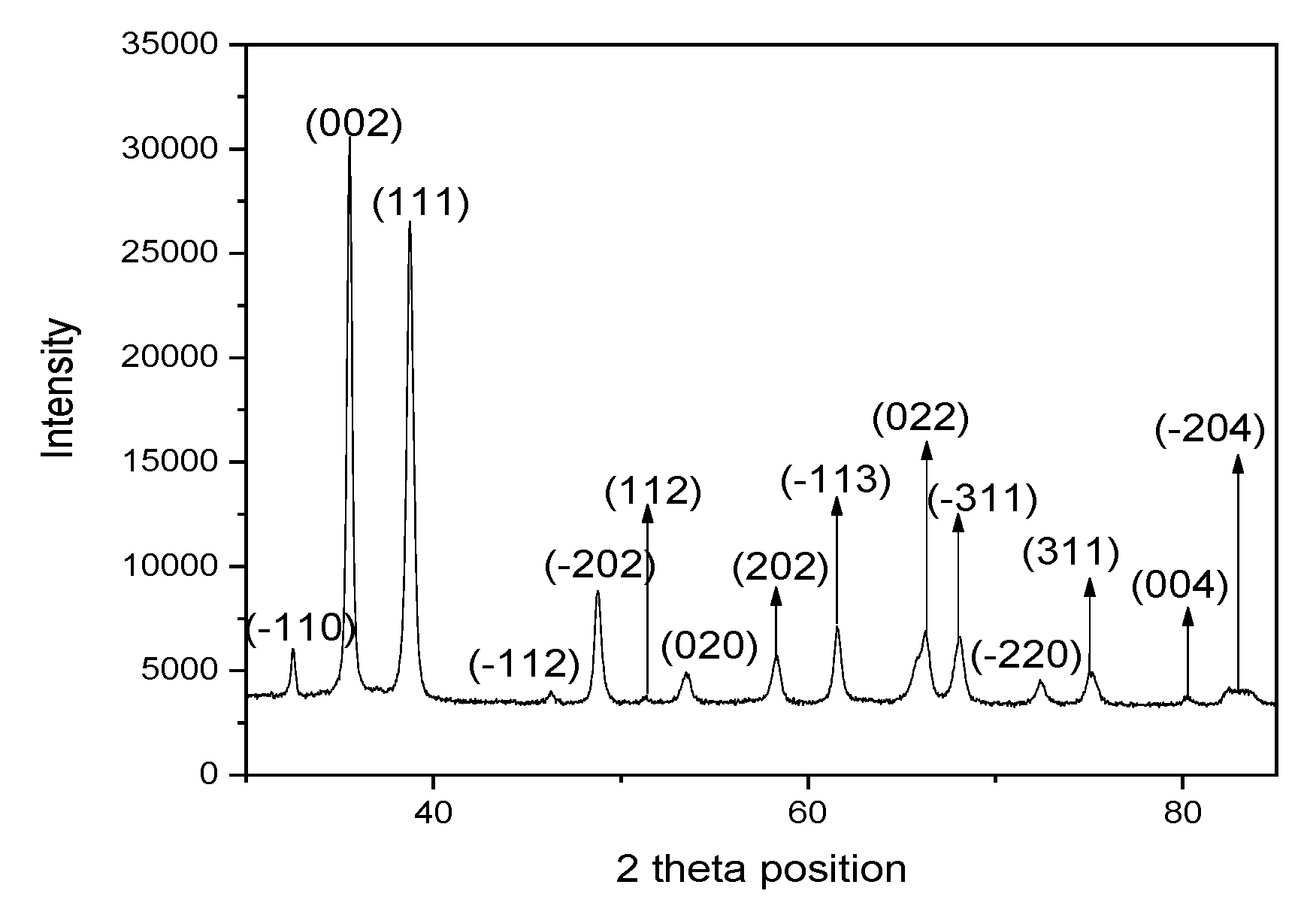
3.1. XRD Analysis
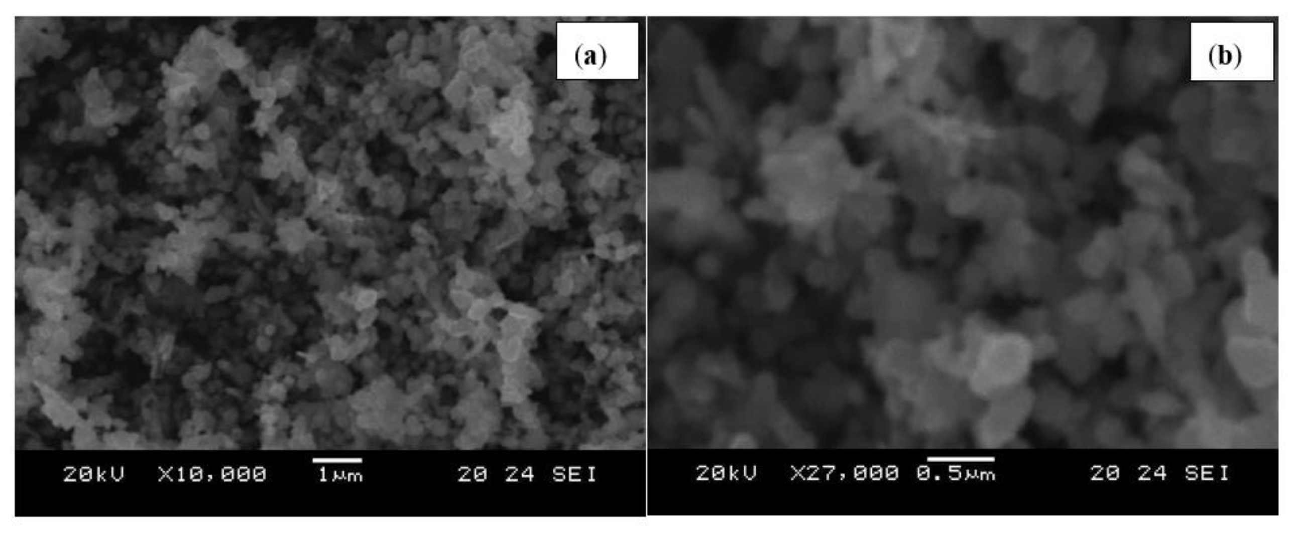
3.2. SEM Analysis
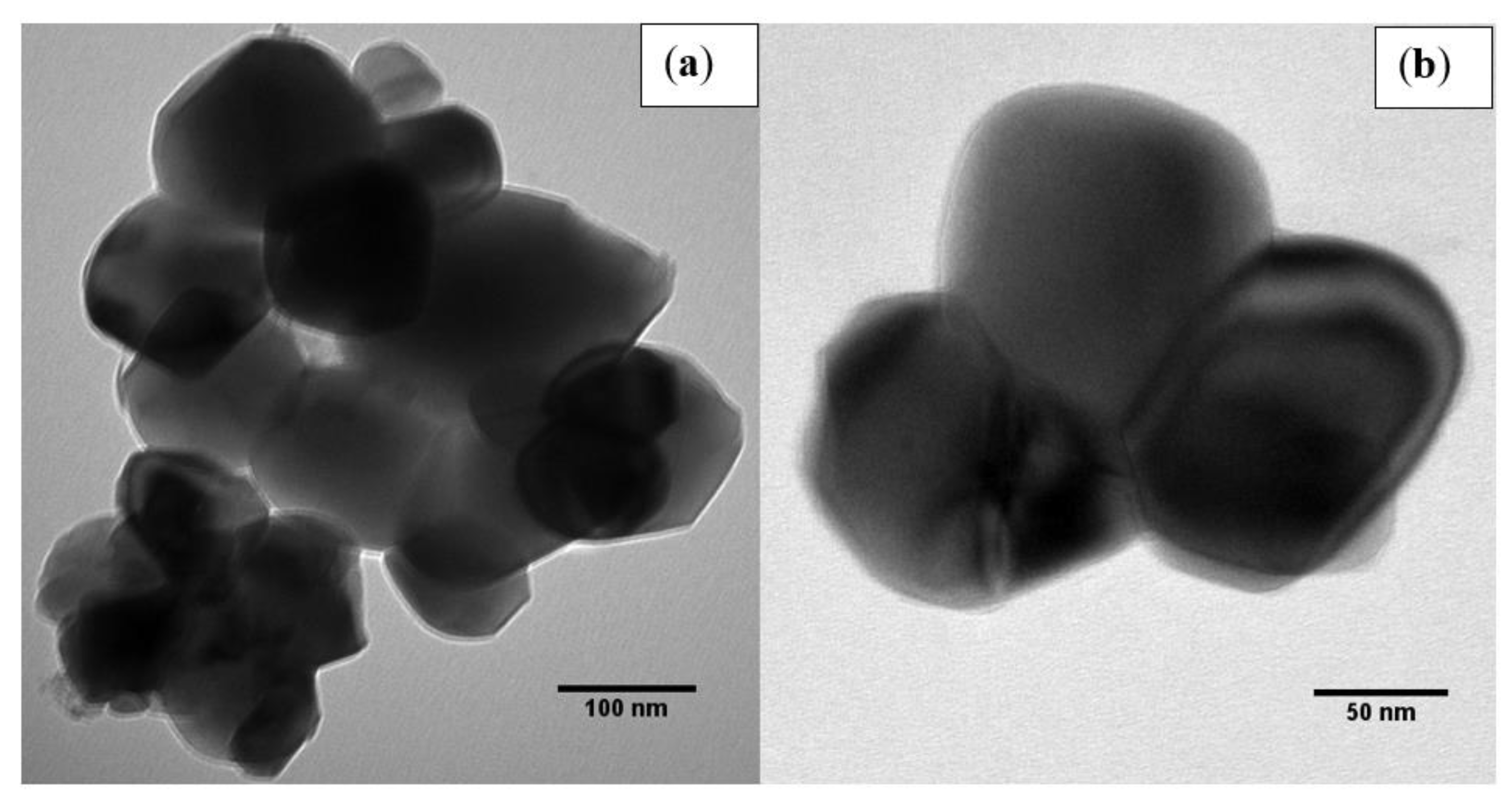
3.3. TEM Analysis
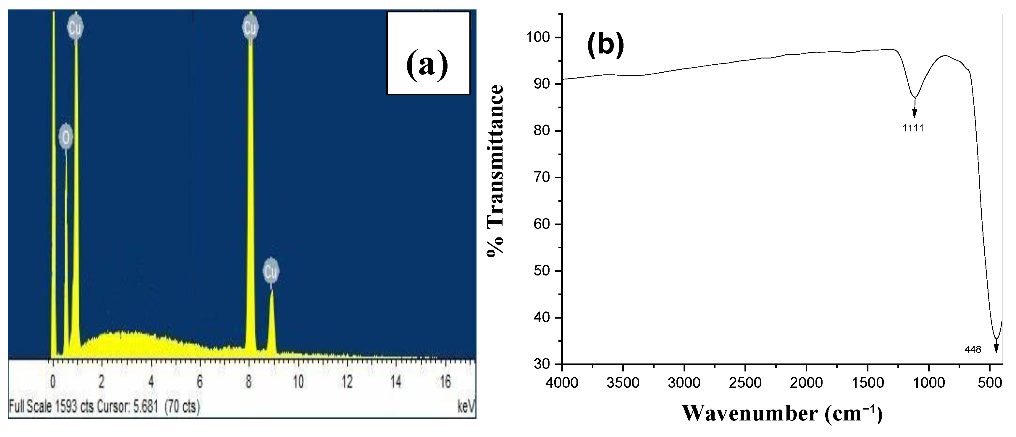
3.4. EDX and FTIR Analysis
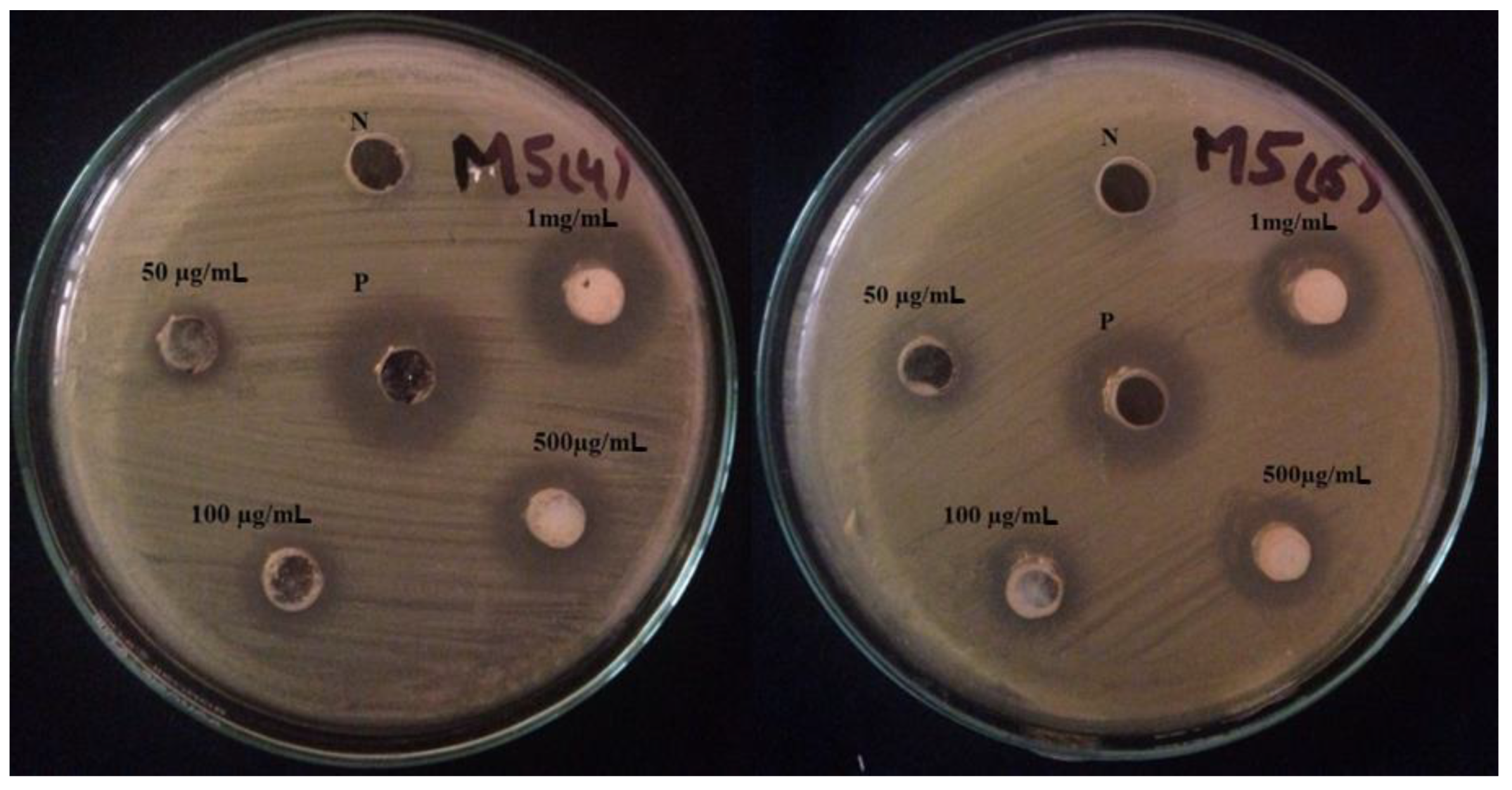
3.5. Antibacterial Activity
3.6. Antioxidant Activity
4. Conclusions
Author Contributions
Funding
Institutional Review Board Statement
Informed Consent Statement
Data Availability Statement
Acknowledgments
Conflicts of Interest
References
- Hajipour, M.J.; Fromm, K.M.; Akbar Ashkarran, A.; de Jimenez Aberasturi, D.; de Larramendi, I.R.; Rojo, T.; Serpooshan, V.; Parak, W.J.; Mahmoudi, M. Antibacterial properties of nanoparticles. Trends Biotechnol. 2012, 30, 499–511. [Google Scholar] [CrossRef] [PubMed] [Green Version]
- Fouda, A.; Hassan, S.E.D.; Abdo, A.M.; El-Gamal, M.S. Antimicrobial, Antioxidant and Larvicidal Activities of Spherical Silver Nanoparticles Synthesized by Endophytic Streptomyces spp. Biol. Trace Elem. Res. 2020, 195, 707–724. [Google Scholar] [CrossRef] [PubMed]
- Kazemi, M.; Akbari, A.; Sabouri, Z.; Soleimanpour, S.; Zarrinfar, H.; Khatami, M.; Darroudi, M. Green synthesis of colloidal selenium nanoparticles in starch solutions and investigation of their photocatalytic, antimicrobial, and cytotoxicity effects. Bioprocess Biosyst. Eng. 2021, 44, 1215–1225. [Google Scholar] [CrossRef] [PubMed]
- Sawai, J. Quantitative evaluation of antibacterial activities of metallic oxide powders (ZnO, MgO and CaO) by conductimetric assay. J. Microbiol. Methods 2003, 54, 177–182. [Google Scholar] [CrossRef] [PubMed]
- Kazemi, M.; Akbari, A.; Zarrinfar, H.; Soleimanpour, S.; Sabouri, Z.; Khatami, M.; Darroudi, M. Evaluation of Antifungal and Photocatalytic Activities of Gelatin-Stabilized Selenium Oxide Nanoparticles. J. Inorg. Organomet. Polym. Mater. 2020, 30, 3036–3044. [Google Scholar] [CrossRef]
- Bafghi, M.H.; Nazari, R.; Darroudi, M.; Zargar, M.; Zarrinfar, H. The effect of biosynthesized selenium nanoparticles on the expression of CYP51A and HSP90 antifungal resistance genes in Aspergillus fumigatus and Aspergillus flavus. Biotechnol. Prog. 2022, 38, e3206. [Google Scholar] [CrossRef]
- Abdelfatah, M.; Salah, H.Y.; EL-Henawey, M.I.; Oraby, A.H.; El-Shaer, A.; Ismail, W. Insight into Co concentrations effect on the structural, optical, and photoelectrochemical properties of ZnO rod arrays for optoelectronic applications. J. Alloy. Compd. 2021, 873, 159875. [Google Scholar] [CrossRef]
- Feng, Y.; Jiao, T.; Yin, J.; Zhang, L.; Zhang, L.; Zhou, J.; Peng, Q. Facile Preparation of Carbon Nanotube-Cu 2 O Nanocomposites as New Catalyst Materials for Reduction of P-Nitrophenol. Adv. Mater. 2019, 14, 78. [Google Scholar]
- Abdelfatah, M.; Ismail, W.; El-Shafai, N.M.; El-Shaer, A. Effect of thickness, bandgap, and carrier concentration on the basic parameters of Cu2O nanostructures photovoltaics: Numerical simulation study. Mater. Technol. 2020, 36, 1–9. [Google Scholar] [CrossRef]
- Chandraker, S.K.; Lal, M.; Ghosh, M.K.; Tiwari, V.; Ghorai, T.K.; Shukla, R. Green synthesis of copper nanoparticles using leaf extract of Ageratum houstonianum Mill. and study of their photocatalytic and antibacterial activities. Nano Express 2020, 1, 010033. [Google Scholar] [CrossRef]
- Kwak, K.; Kim, C. Viscosity of Thermal Conductivity of Copper Oxide Nanofluid Dispersed in Ethylene Glycol Viscosity and thermal conductivity of copper oxide nanofluid dispersed in ethylene glycol. Korea-Aust. Rheol. J. 2005, 17, 35–40. [Google Scholar]
- Rehana, D.; Mahendiran, D.; Kumar, R.S.; Rahiman, A.K. Evaluation of antioxidant and anticancer activity of copper oxide nanoparticles synthesized using medicinally important plant extracts. Biomed. Pharmacother. 2017, 89, 1067–1077. [Google Scholar] [CrossRef]
- Elemike, E.E.; Onwudiwe, D.C.; Singh, M. Eco-friendly synthesis of copper oxide, zinc oxide and copper oxide–zinc oxide nanocomposites, and their anticancer applications. Journal of Inorganic and Organometallic Polymers and Materials. 2020, 30(2), 400–409. [Google Scholar] [CrossRef]
- Derbalah, A.; Abdelsalam, I.; Behiry, S.I.; Abdelkhalek, A.; Abdelfatah, M.; Ismail, S.; Elsharkawy, M.M. Copper oxide nanostructures as a potential method for control of zucchini yellow mosaic virus in squash. Pest Manag. Sci. 2022, 78, 3587–3595. [Google Scholar] [CrossRef]
- Salah, H.Y.; Mahmoud, K.R.; Ismail, W.; El-Shaer, A.; Oraby, A.H.; Abdelfatah, M.; EL-Henawey, M.I. Influence of Nickel Concentration on the Microstructure, Optical, Electrical, and Photoelectrochemical Properties of ZnO Nanorods Synthesized by Hydrothermal Method. J. Electron. Mater. 2022, 51, 910–920. [Google Scholar] [CrossRef]
- Oprea, O.; Andronescu, E.; Ficai, D.; Ficai, A.; Oktar, F.N.; Yetmez, M. ZnO Applications and Challenges. Curr. Org. Chem. 2014, 18, 192–203. [Google Scholar] [CrossRef]
- Unsoy, G.; Gunduz, U.; Oprea, O.; Ficai, D.; Sonmez, M.; Radulescu, M.; Alexie, M.; Ficai, A. Magnetite: From Synthesis to Applications. Curr. Top. Med. Chem. 2015, 15, 1622–1640. [Google Scholar] [CrossRef]
- Sankar, R.; Manikandan, P.; Malarvizhi, V.; Fathima, T.; Shivashangari, K.S.; Ravikumar, V. Green synthesis of colloidal copper oxide nanoparticles using Carica papaya and its application in photocatalytic dye degradation. Spectrochim. Acta—Part A Mol. Biomol. Spectrosc. 2014, 121, 746–750. [Google Scholar] [CrossRef]
- El-Shafai, N.M.; Ibrahim, M.M.; Abdelfatah, M.; Ramadan, M.S.; El-Mehasseb, I.M. Synthesis, characterization, and cytotoxicity of self-assembly of hybrid nanocomposite modified membrane of carboxymethyl cellulose/graphene oxide for photocatalytic antifouling, energy storage, and supercapacitors application. Colloids Surf. A Physicochem. Eng. Asp. 2021, 626, 127035. [Google Scholar] [CrossRef]
- Devi, H.S.; Singh, T.D. Synthesis of Copper Oxide Nanoparticles by a Novel Method and its Application in the Degradation of Methyl Orange. Adv. Electron. Electr. Eng. 2014, 4, 83–88. [Google Scholar]
- Haq, S.; Yasin, K.A.; Rehman, W.; Waseem, M.; Ahmed, M.N.; Shahzad, M.I.; Shahzad, N.; Shah, A.; Rehman, M.U.; Khan, B. Green Synthesis of Silver Oxide Nanostructures and Investigation of Their Synergistic Effect with Moxifloxacin Against Selected Microorganisms. J. Inorg. Organomet. Polym. Mater. 2020, 31, 1134–1142. [Google Scholar] [CrossRef]
- Altikatoglu, M.; Attar, A.; Erci, F.; Cristache, C.M.; Isildak, I. Green synthesis of copper oxide nanoparticles using ocimum basilicum extract and their antibacterial activity. Fresenius Environ. Bull. 2017, 25, 7832–7837. [Google Scholar]
- Sharma, M.; Singh, J.; Hazra, S.; Basu, S. Adsorption of heavy metal ions by mesoporous ZnO and TiO2@ZnO monoliths: Adsorption and kinetic studies. Microchem. J. 2019, 145, 105–112. [Google Scholar] [CrossRef]
- Zhao, X.; Lv, L.; Pan, B.; Zhang, W.; Zhang, S.; Zhang, Q. Polymer-supported nanocomposites for environmental application: A review. Chem. Eng. J. 2011, 170, 381–394. [Google Scholar] [CrossRef]
- Iravani, S. Green synthesis of metal nanoparticles using plants. Green Chem. 2011, 13, 2638–2650. [Google Scholar] [CrossRef]
- Keabadile, O.P.; Aremu, A.O.; Elugoke, S.E.; Fayemi, O.E. Green and traditional synthesis of copper oxide nanoparticles—comparative study. Nanomaterials 2020, 10, 2502. [Google Scholar] [CrossRef]
- Hafeez, M.; Arshad, R.; Khan, J.; Akram, B.; Ahmad, M.N.; Hameed, M.U.; Haq, S. Populus ciliata mediated synthesis of copper oxide nanoparticles for potential biological applications. Mater. Res. Express 2019, 6, 055043. [Google Scholar] [CrossRef]
- Hamid, A.; Haq, S.; Ur Rehman, S.; Akhter, K.; Rehman, W.; Waseem, M.; Ud Din, S.; Zain-ul-Abdin; Hafeez, M.; Khan, A.; et al. Calcination temperature-driven antibacterial and antioxidant activities of fumaria indica mediated copper oxide nanoparticles: Characterization. Chem. Pap. 2021, 75, 4189–4198. [Google Scholar] [CrossRef]
- Mahfooz-Ur-Rehman, M.; Rehman, W.; Waseem, M.; Shah, B.A.; Shakeel, M.; Haq, S.; Zaman, U.; Bibi, I.; Khan, H.D. Fabrication of Titanium-Tin Oxide Nanocomposite with Enhanced Adsorption and Antimicrobial Applications. J. Chem. Eng. Data 2019, 64, 2436–2444. [Google Scholar] [CrossRef]
- Shoukat, S.; Rehman, W.; Haq, S.; Waseem, M.; Shah, A. Synthesis and characterization of zinc stannate nanostructures for the adsorption of chromium (VI) ions and photo-degradation of rhodamine 6G. Mater. Res. Express 2019, 6, 115052. [Google Scholar] [CrossRef]
- Shah, A.; Tauseef, I.; Ali, M.B.; Yameen, M.A.; Mezni, A.; Hedfi, A.; Haleem, S.K.; Haq, S. In-Vitro and In-Vivo Tolerance and Therapeutic Investigations of Phyto-Fabricated Iron Oxide Nanoparticles against Selected Pathogens. Toxics 2021, 9, 105. [Google Scholar] [CrossRef]
- Rehman, F.U.; Mehmood, R.; Ali, B.M.; Hedfi, A.; MOhammed, A.; Mezni, A.; Rehman, W.; Waseem, M. Bergenia ciliate–Mediated Mixed-Phase Synthesis and Characterization of Silver-Copper Oxide Nanocomposite for Environmental and Biological Applications. Materials 2021, 14, 6085. [Google Scholar] [CrossRef]
- Song, H.M.; Kim, S.; Lee, B.; Hong, H.; Lee, G. Chemically ordered FePt 3 nanoparticles synthesized by a bimetallic precursor and their magnetic transitions. J. Mater Chem. 2009, 19, 3677–3681. [Google Scholar] [CrossRef]
- Ahmed, S.; Ahmad, M.; Swami, B.L.; Ikram, S. A review on plants extract mediated synthesis of silver nanoparticles for antimicrobial applications: A green expertise. J. Adv. Res. 2016, 7, 17–28. [Google Scholar] [CrossRef]
- Yadi, M.; Mostafavi, E.; Saleh, B.; Davaran, S.; Aliyeva, I.; Khalilov, R.; Nikzamir, M.; Nikzamir, N.; Akbarzadeh, A.; Panahi, Y.; et al. Current developments in green synthesis of metallic nanoparticles using plant extracts: A review. Artif. Cells Nanomed. Biotechnol. 2018, 46, S336–S343. [Google Scholar] [CrossRef] [Green Version]
- Haq, S.; Dildar, S.; Ali, M.B.; Mezni, A.; Hedfi, A.; Shahzad, M.I.; Shahzad, N.; Shah, A. Antimicrobial and antioxidant properties of biosynthesized of NiO nanoparticles using Raphanus sativus (R. sativus) extract. Mater. Res. Express 2021, 8, 055006. [Google Scholar] [CrossRef]
- Haq, S.; Rehman, W.; Waseem, M.; Shah, A.; Khan, A.R.; Rehman, M.U.; Ahmad, P.; Khan, B.; Ali, G. Green synthesis and characterization of tin dioxide nanoparticles for photocatalytic and antimicrobial studies. Mater. Res. Express 2020, 7, 025012. [Google Scholar] [CrossRef]
- Haq, S.; Ahmad, P.; Khandaker, M.U.; Faruque, M.R.I.; Rehman, W.; Waseem, M.; Din, S.U. Antibacterial, antioxidant and physicochemical investigations of tin dioxide nanoparticles synthesized via microemulsion method. Mater. Res. Express 2021, 8, 035013. [Google Scholar] [CrossRef]
- Magdalane, C.M.; Kaviyarasu, K.; Vijaya, J.J.; Siddhardha, B.; Jeyaraj, B. Photocatalytic activity of binary metal oxide nanocomposites of CeO2/CdO nanospheres: Investigation of optical and antimicrobial activity. J. Photochem. Photobiol. B Biol. 2016, 163, 77–86. [Google Scholar] [CrossRef]
- Haq, S.; Abbasi, F.; Ben Ali, M.; Hedfi, A.; Mezni, A.; Rehman, W.; Waseem, M.; Khan, A.R.; Shaheen, H. Green synthesis of cobalt oxide nanoparticles and the effect of annealing temperature on their physiochemical and biological properties. Mater. Res. Express 2021, 8, 075009. [Google Scholar] [CrossRef]


| Microorganisms | Zones of Inhibition (mm) at Different Concentrations | Variance (S2) | STD Deviation (S) | Correlation b/w Dose and Activity | PC | NC | |||
|---|---|---|---|---|---|---|---|---|---|
| 50 (µg/mL) | 100 (µg/mL) | 500 (µg/mL) | 1 (mg/mL) | ||||||
| E. coli | 5 | 8 | 13 | 19 | 3.00 | 1.74 | 0.08 | 24 | 00 |
| S. aureus | 3 | 5 | 10 | 16 | 3.78 | 1.95 | 0.07 | 21 | 00 |
| Sample | Concentration (µg/mL) | %RSA | IC50 (µg/mL) | Variance (S2) | Std Deviation (S) | Correlation Constant b/w Dose and %RSA |
|---|---|---|---|---|---|---|
| CuO NPs | 5 | 22.62 | 110.50 | |||
| 25 | 30.36 | |||||
| 50 | 44.83 | 2.82 | 1.68 | 0.97 | ||
| 100 | 59.27 | |||||
| 200 | 73.01 | |||||
| 400 | 88.27 | |||||
| Ascorbic acid | 5 | 14.1 | 171.04 | 3.71 | 1.93 | 0.78 |
| 25 | 22.15 | |||||
| 50 | 34.13 | |||||
| 100 | 47.83 | |||||
| 200 | 64.98 | |||||
| 400 | 78.39 |
Publisher’s Note: MDPI stays neutral with regard to jurisdictional claims in published maps and institutional affiliations. |
© 2022 by the authors. Licensee MDPI, Basel, Switzerland. This article is an open access article distributed under the terms and conditions of the Creative Commons Attribution (CC BY) license (https://creativecommons.org/licenses/by/4.0/).
Share and Cite
Rehman, F.U.; Mahmood, R.; Haq, S.; Ahmad, P.; Din, S.U.; Khandaker, M.U.; Idris, A.M.; Zekker, I. Phytogenic Fabrication of Copper Oxide Nanoparticles for Antibacterial and Antioxidant Screening: Physico-Chemical Study. Crystals 2022, 12, 1796. https://doi.org/10.3390/cryst12121796
Rehman FU, Mahmood R, Haq S, Ahmad P, Din SU, Khandaker MU, Idris AM, Zekker I. Phytogenic Fabrication of Copper Oxide Nanoparticles for Antibacterial and Antioxidant Screening: Physico-Chemical Study. Crystals. 2022; 12(12):1796. https://doi.org/10.3390/cryst12121796
Chicago/Turabian StyleRehman, Fazal Ur, Rashid Mahmood, Sirajul Haq, Pervaiz Ahmad, Salah Ud Din, Mayeen Uddin Khandaker, Abubakr M. Idris, and Ivar Zekker. 2022. "Phytogenic Fabrication of Copper Oxide Nanoparticles for Antibacterial and Antioxidant Screening: Physico-Chemical Study" Crystals 12, no. 12: 1796. https://doi.org/10.3390/cryst12121796