Article

Initial Study on Physiochemical Property and Antibacterial Activity against Skin-Infecting Bacteria of Silver Nanoparticles Biologically Produced Using Crude Melanin from Xylaria sp.

1
School of Chemical and Environmental Engineering, International University—Vietnam National University, Block 6, Linh Trung Ward, Thu Duc, Ho Chi Minh City 70000, Vietnam
2
Nanomaterials Engineering Research & Development (NERD) Lab, International University—Vietnam National University, Block 6, Linh Trung Ward, Thu Duc, Ho Chi Minh City 70000, Vietnam
3
Applied Microbiology Laboratory, School of Biotechnology, International University—Vietnam National University, Block 6, Linh Trung Ward, Thu Duc, Ho Chi Minh City 70000, Vietnam
*
Author to whom correspondence should be addressed.
Cosmetics 2023, 10(6), 150; https://doi.org/10.3390/cosmetics10060150
Submission received: 31 July 2023 / Revised: 10 October 2023 / Accepted: 25 October 2023 / Published: 1 November 2023

Abstract

:
Silver nanoparticles (AgNPs) produced by biological methods are safer for biomedical applications. Melanins were initially reported to facilitate AgNPs synthesis. Our research found that the stromata of some Xylaria species contained significant amounts of melanins, which had strong antioxidant and anti-ultraviolet activities without toxicity toward human skin cells. This study reported the characteristics and antibacterial activities against skin-infecting bacteria (Staphylococcus aureus and Cutibacterium acnes) of AgNPs synthesized using crude melanin extracted from stromata of Xylaria sp. AgNPs were successfully synthesized by mixing the crude melanin solution with 0.1 M AgNO3 (25:1, v/v) and incubating for 3 h at 100 °C. The SEM found that the average size of the synthesized AgNPs was 18.85 ± 3.75 nm. The melanin-mediated AgNPs displayed significantly higher antibacterial activities against the tested acne-causing bacteria compared to the positive control (Erythromycin). Specifically, the melanin-mediated AgNPs inhibited 90% of S. aureus and C. acnes at 62.5 (µg/mL) and 15.625 (µg/mL), respectively, whereas it required erythromycin up to 4000 (µg/mL) to achieve the same activities. This research illustrated the feasibility of using crude melanin of Xylaria sp. for the direct synthesis of AgNPs and the potential use of the synthesized AgNPs for treating acne-causing bacteria (with further investigation needed).

1. Introduction

The chronic inflammatory and recurrent skin condition, acne vulgaris, is a disease of the pilosebaceous unit which is the eighth most prevalent disease worldwide, affecting approximately 10% of the world’s population, with adolescents accounting for the majority (67–95%) of those affected [1,2]. Cutibacterium acnes infection is one of the factors leading to acne formation [2]. In most cases, acne is not prescribed as any sort of life-threatening disease; however, if left untreated, it may lead to grave repercussions impacting physical as well as mental health [3]. On the other hand, Staphylococcus aureus is the most prevalent nosocomial infection found on the skin or mucous membranes (particularly in the nasal area) of healthy individuals with mortality rates ranging from 6% to 40% [4]. This bacterium is a more concerning pathogen due to its potential to produce a wide variety of life-threatening infections as well as its ability to rapidly adapt to different environmental settings. Within the infection sites with the presence of oxygen, C. acnes together with S. aureus can increase the biofilm formation and thus enhance the survivability of C. acnes [5]. Penicillins, cephalosporins, erythromycin, clindamycin, and tetracyclines are a few of the commonly used antibiotics for acne therapy [6,7,8], but there has been a substantial increase in antibiotic resistance in those bacteria as a consequence of topical antibiotic formulations and a heavy dependence on antibiotics in long-term treatment [9,10].
Silver nanoparticles (AgNPs) have been widely utilized in various fields, including medical, food, health care, cosmetics, and industrial purposes due to their unique physical and chemical characteristics and powerful biological properties [11,12]. Regarding antimicrobial activity, AgNPs possess remarkable antibacterial activity (against both Gram-positive and Gram-negative bacteria, including multidrug-resistant pathogens) and antifungal activity [13,14,15,16].
AgNPs can be synthesized using two main approaches—top–down and bottom–up. The top–down approach uses physical forces such as mechanical energy, electrical energy, and thermal energy to convert bulk silver to AgNPs [17,18,19]. On the other hand, the bottom–up approach uses chemical synthesis (photochemical, electrochemical, microwave-assisted, and sonochemical) or biological synthesis. The bottom–up approach using the chemical method requires a silver salt precursor and reducing agent [20]. Even though the process is simple and inexpensive with high yields of uniform size and shape of AgNPs, the commonly used reductants and/or stabilizers (e.g., ethylene glycol, sodium borohydride, sodium citrate) are toxic, hazardous, harmful, and may not be appropriate for biochemical applications [11,12,13,14]. The reductants and/or stabilizers could cause negative impacts on health, ranging from minor effects, e.g., nausea and vomiting, diarrhea, and dizziness (caused by sodium citrate) to severe effects, e.g., coma, loss of brain lining tissues, kidney failure (caused by ethylene glycol). In addition, many solvents used in chemical synthesis are also toxic to aquatic ecosystems, e.g., oleyl amine [14]. The biological/green synthesis method can overcome these limitations. In most cases, a reducing biological agent (e.g., biopolymers and polysaccharides) is used to replace the mentioned conventional reducing agents [11,12,13,15,21,22]. This method works similarly to other chemical bottom–up methods via the electron donation of the reducing agents to reduce silver ions (Ag+) to Ag0 [23], but without the presence of toxic chemicals, the synthesized AgNPs are safer for biomedical applications.
Melanins, natural pigments, can be present in all domains of life, namely animals, fungi, plants, microorganisms, and humans [9,10,24]. In terms of structure, melanins are polymers generated from the oxidation of phenolic or indolic substrates [25]. Melanins possess outstanding biological functions, such as anti-radiation and photoprotective [26], antioxidation [27,28], anti-aging [29], antimicrobial [29,30], and anticancer activities [31]. Melanins are considered to effectively chelate metal ions, which have initially been exploited for the synthesis of metal nanoparticles, specifically silver nanoparticles (AgNPs) [23,24]. It is proposed that the conversion of hydroxyl groups to the quinone groups results in the generation of reducing equivalents, which mediates the transformation of silver into AgNPs in the free form. Previous studies found that purified melanin of actinobacterium Nocardiopsis alba MSA10 facilitated the synthesis of AgNPs. The optimized reaction conditions were found to consist of 1 mM AgNO3 with 10 mg melanin in 100 mL 0.1 M KOH at 100 °C for 1 h. The synthesized AgNPs possessed a broad-spectrum antimicrobial activity against food pathogens, including Bacillus subtilis, Bacillus cereus, Staphylococcus aureus, Escherichia coli, Vibrio cholerae, Vibrio parahaemolyticus, Vibrio vulnificus, Pseudomonas fragi and Salmonella typhi [32]. In another study, purified melanin extracted from yeast cells Yarrowia lipolytica was found to mediate AgNPs formation, and the AgNPs displayed excellent antimicrobial inhibition toward Salmonella paratyphi [33]. The research also employed the same incubation conditions as the previously described, except 2.5 mM AgNO3 was mixed with 0.5 mg melanin instead. It is notable that the amounts of melanins used for the AgNPs synthesis are relatively low. However, it was unclear how much of the “unreacted” melanins remained in the reaction mixtures.
Over 300 Xylaria species have been discovered worldwide, and 40 of those are documented in Vietnam [10]. The feasibility of using of melanin extracted from the stromata of Xylaria sp. or fruiting body of any other fungi for AgNPs production has not yet been studied. Our initial studies found that melanins extracted from fungi in general and Xylaria species, in particular, are safe for human skin cells. Thus, the main objectives of this study were to (1) generate AgNPs using crude melanin obtained from Xylaria sp., (2) initially characterize the synthesized AgNPs, and (3) evaluate antibacterial potential against acne-causing bacteria including C. acnes and S. aureus of the melanin-mediated AgNPs.
The research attempted to use crude melanin instead of the pure form for cost and time-effective purposes.

2. Materials and Methods

2.1. Materials

Xylaria sp. stromata/fruiting bodies (Figure S2) were collected from Ma Da (Dong Nai, Vietnam).
Staphylococcus aureus was obtained from International University—VNU HCM (Vietnam). This bacterium was grown in LB, which contained 0.5% yeast extract powder (Himedia, Mumbai, India), 1% Tryptone (Himedia, Mumbai, India) and 1% NaCl.
Cutibacterium acnes was generously given by Dr. Bich from Tay Do University (Can Tho, Vietnam). The bacterium was cultured in brain heart infusion broth supplemented with 1% Dextrose (Merck, NJ, USA).
All chemicals used in this study were of analytical grade. The extraction of melanin involved 1 M KOH and 2 M HCl to dissolve and precipitate the pigment. Moreover, 0.1 M AgNO3 (Shantou, China) was used for the synthesis of all nano-samples in this project.

2.2. Methods

2.2.1. Preparation of Crude Melanin from Xylaria sp. Stromata

The dried powder of Xylaria sp. stromata was incubated with 1 M KOH (1:40 w/v) for 36–48 h at room temperature. The supernatant was collected after the mixture was centrifuged at 5000 rpm for 15 min at 25 °C [34]. The solution then was acidified by 2 M HCl to a pH below 2.5 and incubated for 2 h for melanin precipitation. The sample was then centrifuged again under the same above-described conditions. The precipitated melanin was collected and washed with distilled water until the solution was colorless (at least 3 times). Eventually, the precipitate was vacuum freeze-dried [34].

2.2.2. Silver Nanoparticles Synthesis Using the Crude Melanin

The synthesis was conducted following the method of Kiran with minor modifications [32]. A volume of 100 mL melanin solution (20 μg/mL) was mixed with 4 mL of 0.1 M AgNO3 and vigorously stirred for 5 min. The mixture was incubated at 100 °C for 3 h. After incubation, the synthesized AgNPs were collected by heat drying the mixture at 60 °C in an oven for water vaporization followed by a freeze-drying process using a freeze dryer (Labconco, United States).
To avoid degradation, the freeze-dried samples were then kept in the dark at 4 °C for further use.

2.2.3. Chemical Characterization of Melanin-Mediated Silver Nanoparticles

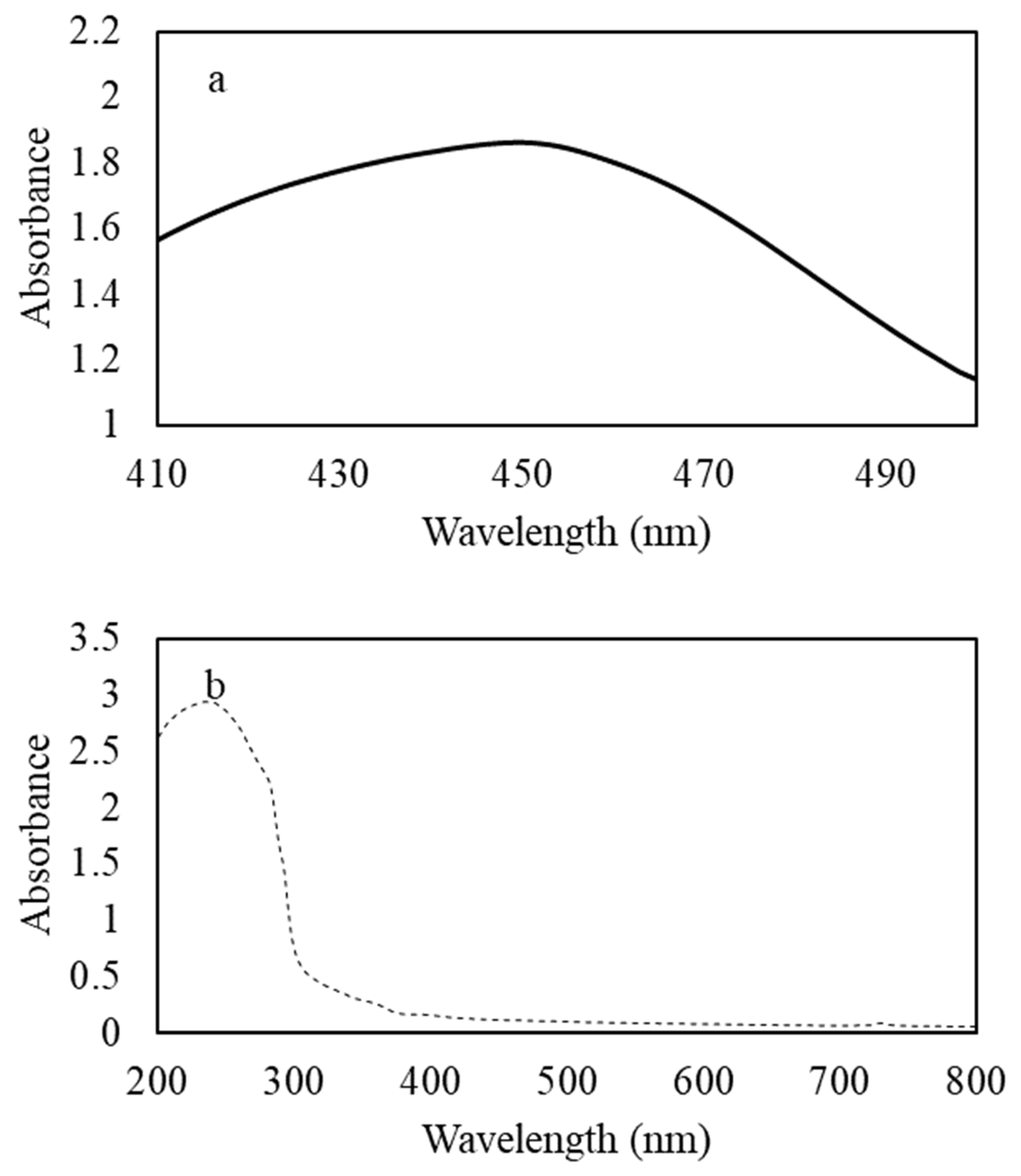
Ultraviolet-Visible Spectroscopy (UV-Vis)

UV-Vis spectroscopy was used to identify the preliminary characterization of the AgNPs. The absorbance spectrum of AgNPs solution was measured with UV-Vis Spectrophotometer (Jasco) over a range of wavelengths from 200 to 800 nm at a resonance of 1 nm [13,35].

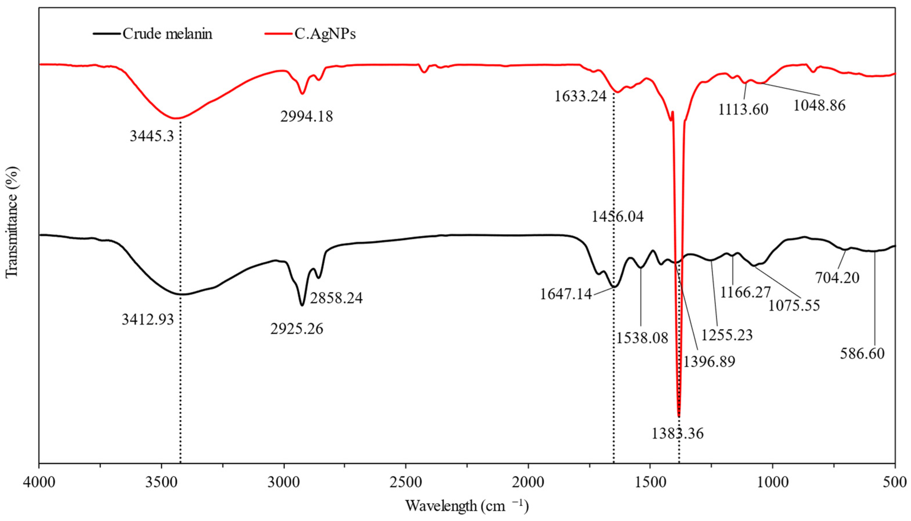
Fourier Transform Infrared (FTIR) and Field Emission Scanning Electron Microscope (FE-SEM)

The sample (freeze-dried AgNPs) was mixed with KBr powder in the ratio of 1:10 and pressed into a pellet, which was then placed on the detector of a Bruker Tensor 27 photometer (Bruker, Ettlingen, Germany) to determine functional groups and their corresponding involvements in nanoparticle synthesis. The FTIR spectra were collected at the resolution of 4 cm−1 in the transmission mode (4000–400 cm−1). In addition, KBr acted as a blank control for the test. FTIR spectra of the crude melanin and the melanin solution were also obtained.
The dried sample was adhered onto a glass slide to conduct FE-SEM analysis for the morphology and size of nanoparticles [33,36].

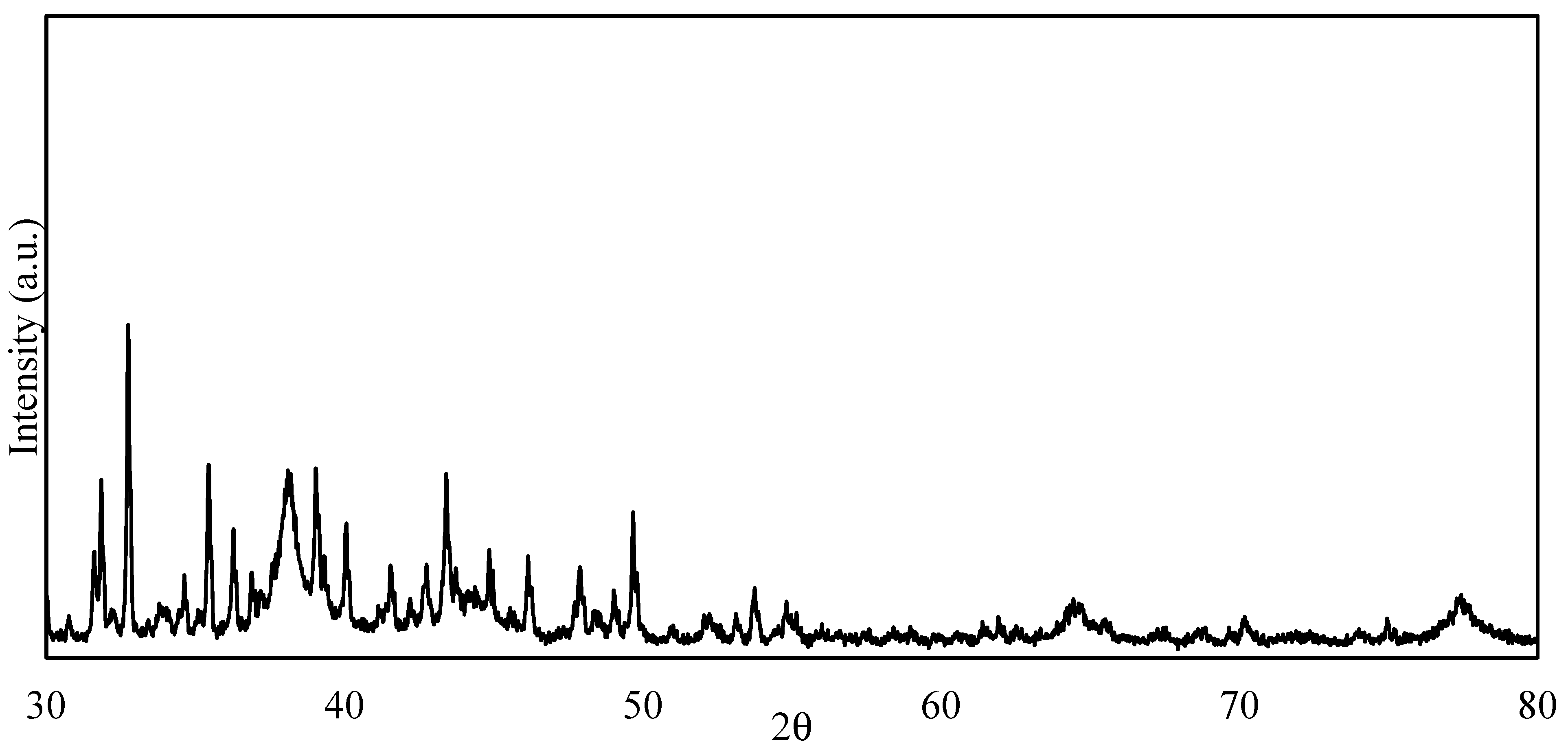
X-ray Diffraction (XRD)

The dried powder of the synthesized nanoparticles was analyzed using an X-ray diffractometer operating with a Cu Kα radiation source (λ = 1.54 Å) at a voltage of 40 kV and a current of 30 mA at a 2θ pattern [33,36].

2.3. Evaluating Antibacterial Activities of the Melanin-Mediated AgNPs

The antibacterial activities of the melanin-mediated AgNPs were evaluated using the microdilution method. The minimum inhibitory concentration (MIC) was assessed using the Clinical and Laboratory Standards Institute (CLSI) guidelines with a few modifications according to a study of Nelson [37]. Briefly, bacterial broth culture was standardized to 5 × 10⁷ CFU/mL (OD590 of 0.05) in sterile BHI broth. The growth inhibition was established in sterile flat-bottom 96-well plates (Biologix, MO, United State). Each well of the sterile 96-well plate was filled with 100 µL of the standardized bacterial suspension (either C. acnes or S. aureus) and 100 µL of a certain concentration of AgNPs suspension, making the final test concentration of AgNP range from 4000 to 7.8125 µg/mL for S. aureus, and 62.5 to 0.98 µg /mL for C. acnes.
The 96-well plates were incubated at 37 °C in 24 h in aerobic conditions for S. aureus and 37 °C for 72 h in anaerobic conditions for C. acnes. Optical density was read by a Synergy HT multimode plate reader (BioTek, Winooski, VT, USA).
Erythromycin was used as the positive control.
The percentage of inhibition was calculated by using the formula below:
% i n h i b i t i o n = 1 O D 600 / t O D 600 / t 0 O D ( ) 600 / t O D ( ) 600 / t 0 × 100
where
OD600/t = optical density (600 nm) of the test well at 24 h or 72 h post-inoculation;
OD600/t0 = optical density (600 nm) of the test well at 0 h post-inoculation;
OD(−)600/t = optical density (600 nm) of the negative control well at 24 h or 72 h post-inoculation;
OD(−)600/t0 = optical density (600 nm) of the negative control well at 0 h post-inoculation.

2.4. Minimum Bactericidal Concentration (MBC)

Minimum bactericidal concentration was determined following the method of Parvekar [38] in which an aliquot of 50 μL (taken from the wells in the microtiter plates where no growth was observed after 24 h or 72 h of incubation) was then inoculated onto the surface of agar plates by the spread plate technique for S. aureus and pour plate technique for C. acnes. The plates were then incubated for another 24 h or 72 h at the desired temperature for optimal growth of the pathogens with MBC being taken to be the lowest concentration of the substance at which no colonies formed under these conditions.

2.5. Data Analysis

IBM SPSS Statistics 22 was used for the data analysis. The analyzed data are reported as means ± standard deviation (p ≤ 0.05 is significant). The FE-SEM images were analyzed with ImageJ software (ver. 1.53) while the FTIR spectrum and XRD patterns were built using OriginPro 8.5.0 SR1. One-way ANOVA was applied to compare the difference in the antibacterial activities of different tested concentrations, and p < 0.05 was considered as there being a significant difference.

3. Results and Discussion

3.1. Chemical and Physical Characteristics of the Synthesized AgNPs

3.1.1. UV-Vis Spectroscopy

The absorbance profiles of AgNPs solution and the crude melanin of Xylaria sp. are presented in Figure 1.
In addition to the formation of a brown color in the reaction mixture, the UV-Vis spectroscopy band observed clearly at 450 nm (Figure 1a) is the indicator of the AgNPs being successfully synthesized in the mixture.
Comparing to Figure 1b, the 450 nm band was not visible. According to the literature, AgNPs UV-VIS bands had a range from 410 to 428 nm [39,40,41]. However, in this study, the AgNPs had a slightly larger band. This phenomenon can be explained by the possibility that the concentration of AgNPs synthesized by the crude melanin would be higher than that of others [42] or the presence of impurities in the synthesized AgNPs solution. However, to determine the specific cause of the shift, further investigation is necessary.

3.1.2. FTIR Analysis

The FTIR spectra of the synthesized AgNPs and the crude melanin are demonstrated in Figure 2.
In general, FTIR bands of crude melanin derived from Xylaria sp. displayed typical bands of melanin, which are in the range of 3600–3000 cm−1, 1650–1600 cm−1, and 1500–1400 cm−1 [24]. These absorption peaks were caused by the stretching vibration of such functional groups normally present in melanin. The intense broad absorption peaks at 3412.93 cm−1 (Figure 2) clearly indicated the existence of –OH and –NH groups belonging to the amine, amide, carboxylic acid, phenolic, and aromatic amino functions present in the indolic and pyrrolic systems. In addition, the FTIR spectra also showed strong peaks at 1647.14 cm−1 associating with the oscillation of aromatic C=C and C=O stretches of carboxylic functions. Furthermore, the bending vibration of N–H and the stretching vibration of C–N present within the indolic-based structure possessed a characteristic band at 1456.04 cm−1. Apart from these typical characteristic bands, melanin has been confirmed by several other absorption ranges. For example, the peaks at 2925.26 cm−1 and 2858.24 cm−1 could be assigned to the stretching oscillation of aliphatic C–H groups. In addition, peaks between 1465 and 1375 cm−1 arose from the oscillation of these functional groups and were detected at 1456.04 cm−1 and 1396.89 cm−1 from the spectrum of crude melanin above. More confirmative information could be retrieved from the FTIR spectrum of Xylaria sp crude melanin, while the presence of absorption bands between 1255 and 1180 cm−1 corresponding to the stretching vibration of phenolic –OH groups that have been discovered in several melanins from different fungal species was revealed at 1255.23 cm−1. Nevertheless, the spectra also exhibited peaks at 1166.27 cm−1 and 1075.55 cm−1, which could be assigned to the stretching vibration of C–O in the phenolic hydroxyl [43,44]. Weak absorption peaks below 1100 cm−1 were attributed to the deformation of the C–H bond, aromatics CH groups, or alkene CH substitution/conjugated systems.
The formation of AgNPs caused some marginal shifts in peak position in the spectra of AgNPs mixture synthesized by crude melanin including 3412.93 to 3445.33 cm−1, 2925.26 to 2924.18 cm−1, and 1647.14 to 1633.24 cm−1 (Figure 2). These confirmed the involvement of corresponding functional groups in facilitating the formation of the nanoparticles. Nevertheless, the disappearance of the peak at 1538.08 cm−1 from the crude melanin exhibited the participation of carboxylic acid in crude melanin in reducing Ag+ to Ag0. Notably, after the synthesis of the AgNPs, a new, sharp peak at 1383.36 cm−1 showed evidence of the impact of carbonyl groups present in melanin on the transformation of silver ions to AgNPs in free forms. Regarding the mechanism, silver possibly interacted with the indole ring in melanin, and the FTIR results were in accordance with those of earlier studies [45].

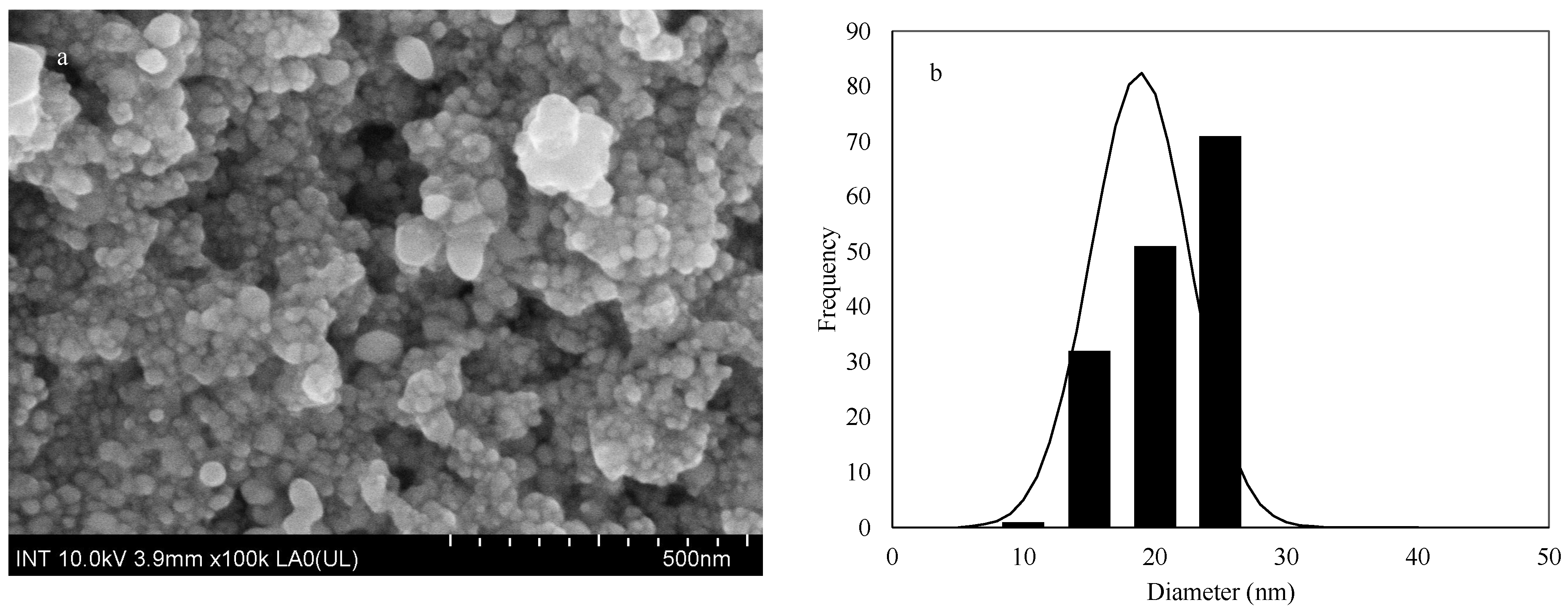
3.1.3. FE-SEM Analysis

The size and the morphology of AgNPs were analyzed using FE-SEM. The representative micrographs along with the size distribution of the sample are shown in Figure 3.
Figure 3 illustrates that the morphology of the generated AgNPs was nearly spherical and agglomerate. The agglomeration of nanoparticles is rather common when the nanoparticle suspension is freeze-dried prior to any test performance [32,33]. The average size of the synthesized AgNPs in this research was 18.85 ± 3.75 nm. In green synthesis, it seems that the size of the synthesized AgNPs significantly depends on the temperature, concentration of AgNO3, type and/or source, and concentration of the biomolecule that facilitates AgNP synthesis [12]. The AgNPs synthesized using various biomolecules can have a size range of 2–100 nm [33,36,40,41,46]. For example, incubating 1 mL (10 μg) L–DOPA-melanin from Y. lipolytica (the melanin was produced by adding L–DOPA as the precursor and used Y. lipolytica cells to convert L–DOPA into melanin) with 2.0 mM AgNO3 solution at 100 °C for 10 min resulted in AgNPs with a size of 7 nm [36]. In another research study, incubating the mixture of 500 μg of melanin naturally produced by Y. lipolytica with 2.5 mM AgNO3 under the same condition led to the production of AgNPs with an average size of 15 nm [33].
It should be noted that the size of the AgNPs can influence their biological activities and smaller sizes are normally preferable, but that also depends on the applications of AgNPs [35].

3.1.4. XRD Analysis

XRD is normally used to determine the structure of nanoparticles. The XRD analysis showed diffraction planes (d-spacings planes) of the spinel cubic structure of the nanoparticles [47], specifically AgNPs. The XRD pattern of the synthesized AgNP is shown in Figure 4.
In Figure 4, the XRD pattern the AgNPs generated from crude melanin of Xylaria sp. displayed four diffraction peaks at (2 θ ) 38.10°, 44.85°, 64.40° and 77.43°, which were indexed to the (111), (200), (220) and (311) crystalline planes, respectively [48]. These suggested that the synthesized AgNPs having a face-centered cubic (fcc) structure.

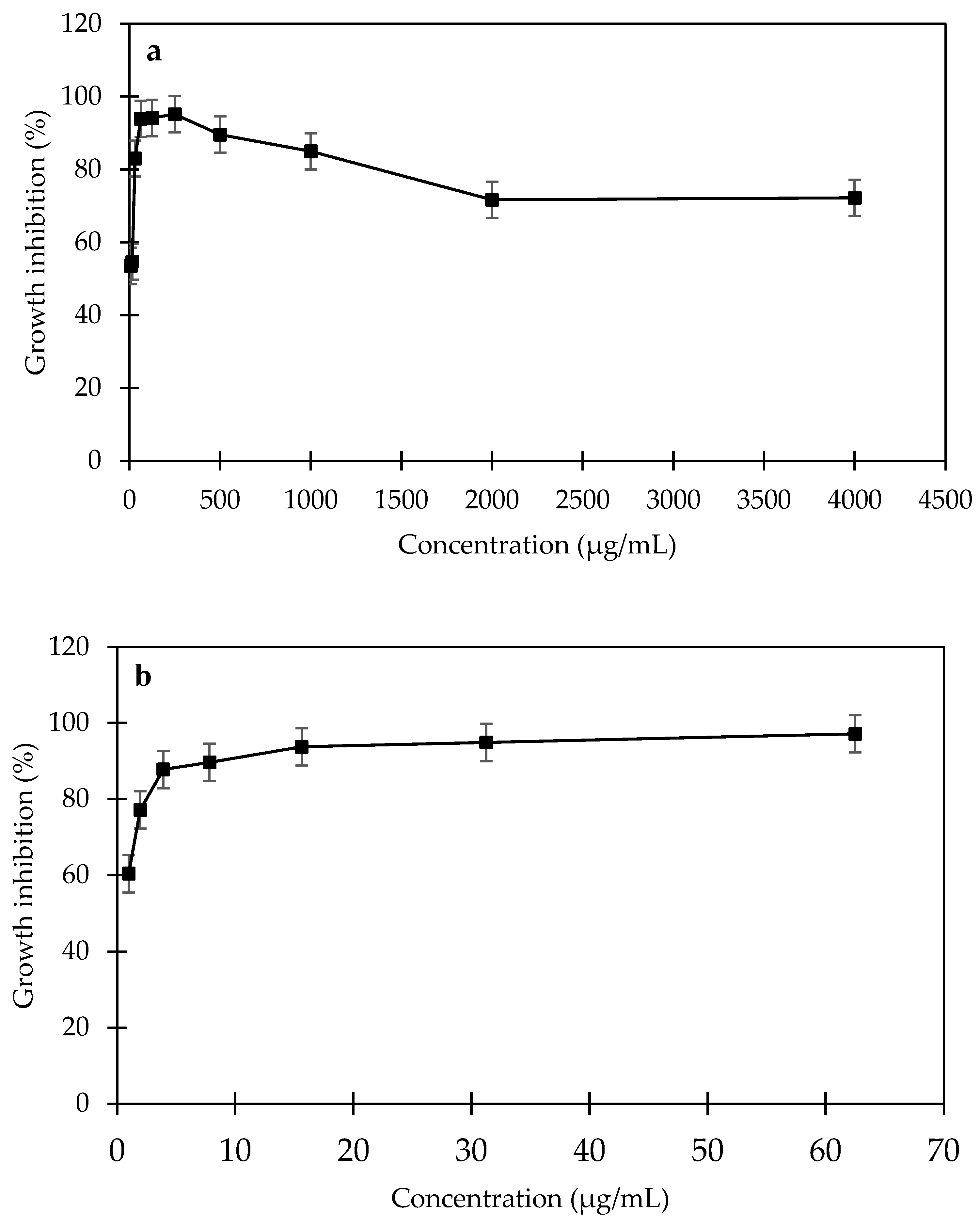
3.2. Antibacterial Activities of the Melanin-Mediated AgNPs against C. acnes and S. aureus

Antibacterial activities of the melanin–mediated AgNPs against S. aureus and C. acnes were evaluated using the serial dilution method. The inhibition activities of various AgNPs concentrations on the tested bacteria are displayed in Figure 5.
Generally, the synthesized AgNPs displayed a dose-dependent inhibitory pattern against both S. aureus and C. acnes (Figure 5). The antibacterial activities of AgNPs toward C. acnes significantly and steadily increased when increasing the concentration of AgNPs from 0.97 to 62.5 μg/mL (Figure 5a). However, that is not the case for S. aureus, as the activities only showed significant increases when the concentration of AgNPs increased from 7.81 to 250 μg/mL. After that the activities remained insignificantly different regardless of the AgNP concentration rising up to 4000 μg/mL.
The minimum inhibitory concentration (MIC) that inhibits 50% (MIC50) and 90% (MIC90) and the minimum bactericidal concentration of (MBC) that kills 99.9% of the bacteria were calculated for the synthesized AgNPs and the positive control (Erythromycin), and the data are shown in Table 1. The values were also confirmed by plate techniques (spread plate for S. aureus and pour plate for C. acnes) (Figure S1).
The MIC and MBC values suggested that the melanin-mediated AgNPs displayed better inhibitory activities against C. acnes than S. aureus (Table 1). In addition, the antibacterial activities of the AgNPs sample against C. acnes and S. aureus are significantly higher than the positive control (erythromycin) (Table 1). There have been only a few studies attempting to evaluate the antibacterial activity of green synthesized AgNPs against C. acnes and S. aureus. The AgNPs synthesized by using Phoenix sylvestris extract had a size range of 40–50 nm, were mostly spherical without forming agglomeration, and had an MIC of 0.687 (mg/mL) against C. acnes [38]. However, the AgNPs synthesized by using Coriandrum sativum leaf extract had a size of 37 nm, and their MIC50 toward C. acnes was only 3.1 (μg/mL) [35]. It could be seen that the MIC50 value of the AgNPs in our research is significantly lower than these reported values, which suggests the synthesized AgNPs by melanin from Xylaria sp. had better antibacterial activity toward C. acnes. In terms of the antibacterial activity of green synthesized AgNPs against S. aureus, AgNPs synthesized by Gardenia thailandica leaf extract possessed MIC50 values ranging from 4 to 64 µg/mL toward different clinical isolates of S. aureus [16]. These values are comparable with what was found in our research (Table 1). We are not aware of any report on the antibacterial activity against C. acnes and S. aureus of AgNPs synthesized by using melanin.
The main antibacterial mechanisms of AgNPs have been reported as membrane and cell wall disruption, interference of DNA replication, interruption of ATP, denaturation of ribosomes, and possible disruption of bacteria signal transduction [49]. Generally, Gram-negative bacteria are more susceptible to AgNPs because they have thinner cell walls that AgNP can penetrate more easily [49]. On the other hand, biofilm-forming bacteria are less susceptible to AgNPs, as the biofilm prevents AgNP from penetrating into the inner parts of the bacterial cells. Both S. aureus and C. acnes are Gram-positive bacteria and can form biofilms, but their susceptibility to the synthesized AgNPs in this research is significantly different. This would be due to the detailed interaction between AgNPs and their cells, and that requires further research to figure out.
It should be noted that the synthesized AgNPs were collected by drying (heat-dry and freeze-dry methods) the entire reaction mixture. Thus, there would be some level of impurities in the synthesized AgNPs sample. The impurities would consist of the unreacted KOH and melanin (although melanin was used with a very small amount, and it was proved to have no toxicity to skin cells). Thus, the exact impurities and their levels in the AgNPs sample and optimization of the reaction to reduce the possible impurities should be reported in follow-up research. In addition, when attempting to use the synthesized AgNPs for antiacne cream production, the removal of the impurities or evaluating the toxicity of different concentrations of AgNP samples including the impurities on skin cells is crucial.

4. Conclusions

Silver nanoparticles were successfully synthesized using crude melanin obtained from the black stromata of Xylaria sp. The synthesized AgNPs had spherical morphology with an average size of 18.85 ± 3.75 nm. In addition, the AgNPs displayed remarkable antibacterial activities against two common acne-causative pathogens including C. acnes and S. aureus. Remarkably, the MBC values toward these bacteria were 125 and 15.6 μg/mL, respectively. These figures are significantly lower than that of the positive control erythromycin (4000 μg/mL), suggesting that the synthesized AgNPs have significantly higher antibacterial activities.
Many species of Xylaria are widely distributed in the tropical regions including Vietnam and normally form black bodies indicating the presence of melanin; thus, they would be good sources of melanin for AgNPs synthesis.
As melanin is safe for human skin, and the synthesized AgNPs with the facilitation of melanin possess powerful antibacterial activities against skin-infecting bacteria, thus, they would have potential for the use as an active ingredient in antiacne cream formulation. However, further investigation into the toxicity (if any) of the synthesized AgNPs toward human skin should be studied before such application is viable.

Supplementary Materials

The following supporting information can be downloaded at https://www.mdpi.com/article/10.3390/cosmetics10060150/s1, Figure S1: Visualization of minimum bactericidal concentration assay on (1) S. aureus and (2) C. acnes with (a) AgNPs (125 µg/mL for S. aureus and 15.625 µg/mL for C. acnes), (b) negative control (sterile water), and (c) erythromycin (4000 µg/mL); Figure S2: Black stromata/fruiting bodies of Xylaria sp.

Author Contributions

L.D. analyzed and interpreted the data. N.K.H.V. performed the experiments. H.T.M.T. contributed most of the manuscript. All authors have read and agreed to the published version of the manuscript.

Funding

This research received no external funding.

Institutional Review Board Statement

Not applicable.

Informed Consent Statement

Not applicable.

Data Availability Statement

All data generated or analyzed during this study are included in this published article [and its Supplementary Information Files].

Acknowledgments

The authors would like to thank Duong Thi Bich (Tay Do University, Can Tho, Vietnam) for the Cutibacterium acnes culture.

Conflicts of Interest

The authors declare no conflict of interest.

References

  1. Cavallo, I.; Sivori, F.; Truglio, M.; De Maio, F.; Lucantoni, F.; Cardinali, G.; Pontone, M.; Bernardi, T.; Sanguinetti, M.; Capitanio, B.; et al. Skin Dysbiosis and Cutibacterium acnes Biofilm in Inflammatory Acne Lesions of Adolescents. Sci. Rep. 2022, 12, 21104. [Google Scholar] [CrossRef] [PubMed]
  2. McLaughlin, J.; Watterson, S.; Layton, A.M.; Bjourson, A.J.; Barnard, E.; McDowell, A. Propionibacterium acnes and Acne Vulgaris: New Insights from the Integration of Population Genetic, Multi-Omic, Biochemical and Host-Microbe Studies. Microorganisms 2019, 7, 128. [Google Scholar] [CrossRef] [PubMed]
  3. Mayslich, C.; Grange, P.A.; Dupin, N. Cutibacterium acnes as an Opportunistic Pathogen: An Update of Its Virulence-Associated Factors. Microorganisms 2021, 9, 303. [Google Scholar] [CrossRef] [PubMed]
  4. Khorvash, F.; Abdi, F.; Kashani, H.H.; Naeini, F.F.; Narimani, T. Staphylococcus aureus in Acne Pathogenesis: A Case-Control Study. N. Am. J. Med. Sci. 2012, 4, 573–576. [Google Scholar] [CrossRef]
  5. Abbott, C.; Grout, E.; Morris, T.; Brown, H.L. Cutibacterium acnes Biofilm Forming Clinical Isolates Modify the Formation and Structure of Staphylococcus aureus Biofilms, Increasing Their Susceptibility to Antibiotics. Anaerobe 2022, 76, 102580. [Google Scholar] [CrossRef]
  6. Dreno, B.; Martin, R.; Moyal, D.; Henley, J.B.; Khammari, A.; Seité, S. Skin Microbiome and Acne Vulgaris: Staphylococcus, a New Actor in Acne. Exp. Dermatol. 2017, 26, 798–803. [Google Scholar] [CrossRef]
  7. Layton, A.M. Top Ten List of Clinical Pearls in the Treatment of Acne Vulgaris. Dermatol. Clin. 2016, 34, 147–157. [Google Scholar] [CrossRef]
  8. Zaenglein, A.L.; Pathy, A.L.; Schlosser, B.J.; Alikhan, A.; Baldwin, H.E.; Berson, D.S.; Bowe, W.P.; Graber, E.M.; Harper, J.C.; Kang, S.; et al. Guidelines of Care for the Management of Acne Vulgaris. J. Am. Acad. Dermatol. 2016, 74, 945–973.e33. [Google Scholar] [CrossRef]
  9. Sardana, K.; Gupta, T.; Garg, V.K.; Ghunawat, S. Antibiotic Resistance to Propionobacterium acnes: Worldwide Scenario, Diagnosis and Management. Expert Rev. Anti-Infect. Ther. 2015, 13, 883–896. [Google Scholar] [CrossRef]
  10. Morris, H.; Smith, K.; Robinson, S.; Göttelmann, M.; Fink, S.; Schwarze, F. The Dark Side of Fungal Competition and Resource Capture in Wood: Zone Line Spalting from Science to Application. Mater. Des. 2021, 201, 109480. [Google Scholar] [CrossRef]
  11. Bruna, T.; Maldonado-Bravo, F.; Jara, P.; Caro, N. Silver Nanoparticles and Their Antibacterial Applications. Int. J. Mol. Sci. 2021, 22, 7202. [Google Scholar] [CrossRef] [PubMed]
  12. Lee, S.H.; Jun, B.H. Silver Nanoparticles: Synthesis and Application for Nanomedicine. Int. J. Mol. Sci. 2019, 20, 865. [Google Scholar] [CrossRef] [PubMed]
  13. Patil, M.P.; Singh, R.D.; Koli, P.B.; Patil, K.T.; Jagdale, B.S.; Tipare, A.R.; Kim, G.-D. Antibacterial Potential of Silver Nanoparticles Synthesized Using Madhuca longifolia Flower Extract as a Green Resource. Microb. Pathog. 2018, 121, 184–189. [Google Scholar] [CrossRef] [PubMed]
  14. Pandit, R. Green Synthesis of Silver Nanoparticles from Seed Extract of Brassica nigra and Its Antibacterial Activity. Nusant. Biosci. 2015, 7, 15–19. [Google Scholar] [CrossRef]
  15. Anandalakshmi, K.; Venugobal, J.; Ramasamy, V. Characterization of Silver Nanoparticles by Green Synthesis Method Using Pedalium murex Leaf Extract and Their Antibacterial Activity. Appl. Nanosci. 2016, 6, 399–408. [Google Scholar] [CrossRef]
  16. Attallah, N.G.M.; Elekhnawy, E.; Negm, W.A.; Hussein, I.A.; Mokhtar, F.A.; Al-Fakhrany, O.M. In Vivo and In Vitro Antimicrobial Activity of Biogenic Silver Nanoparticles against Staphylococcus aureus Clinical Isolates. Pharmaceuticals 2022, 15, 194. [Google Scholar] [CrossRef]
  17. Xu, L.; Wang, Y.-Y.; Huang, J.; Chen, C.-Y.; Wang, Z.-X.; Xie, H. Silver Nanoparticles: Synthesis, Medical Applications and Biosafety. Theranostics 2020, 10, 8996–9031. [Google Scholar] [CrossRef]
  18. Bouafia, A.; Laouini, S.E.; Ahmed, A.S.A.; Soldatov, A.V.; Algarni, H.; Chong, K.F.; Ali, G.A.M. The Recent Progress on Silver Nanoparticles: Synthesis and Electronic Applications. Nanomaterials 2021, 11, 2318. [Google Scholar] [CrossRef]
  19. Alharbi, N.S.; Alsubhi, N.S.; Felimban, A.I. Green Synthesis of Silver Nanoparticles Using Medicinal Plants: Characterization and Application. J. Radiat. Res. Appl. Sci. 2022, 15, 109–124. [Google Scholar] [CrossRef]
  20. Suriati, G.; Mariatti, M.; Azizan, A. Synthesis of Silver Nanoparticles by Chemical Reduction Method: Effect of Reducing Agent and Surfactant Concentration. Int. J. Automot. Mech. Eng. 2014, 10, 1920–1927. [Google Scholar] [CrossRef]
  21. Kim, K.D.; Han, D.N.; Kim, H.T. Optimization of Experimental Conditions Based on the Taguchi Robust Design for the Formation of Nano-Sized Silver Particles by Chemical Reduction Method. Chem. Eng. J. 2004, 104, 55–61. [Google Scholar] [CrossRef]
  22. Mallick, K.; Witcomb, M.J.; Scurrell, M.S. Polymer Stabilized Silver Nanoparticles: A Photochemical Synthesis Route. J. Mater. Sci. 2004, 39, 4459–4463. [Google Scholar] [CrossRef]
  23. Alviar-Agnew, M. Henry Agnew Oxidation and Reduction—Some Definitions. In Introductory Chemistry; Libre Texts, 2022; p. 2. Available online: https://libretexts.org/ (accessed on 10 October 2023).
  24. Pralea, I.E.; Moldovan, R.C.; Petrache, A.M.; Ilieș, M.; Hegheș, S.C.; Ielciu, I.; Nicoară, R.; Moldovan, M.; Ene, M.; Radu, M.; et al. From Extraction to Advanced Analytical Methods: The Challenges of Melanin Analysis. Int. J. Mol. Sci. 2019, 20, 3943. [Google Scholar] [CrossRef] [PubMed]
  25. Singh, S.; Nimse, S.B.; Mathew, D.E.; Dhimmar, A.; Sahastrabudhe, H.; Gajjar, A.; Ghadge, V.A.; Kumar, P.; Shinde, P.B. Microbial Melanin: Recent Advances in Biosynthesis, Extraction, Characterization, and Applications. Biotechnol. Adv. 2021, 53, 107773. [Google Scholar] [CrossRef]
  26. Ye, M.; Guo, G.Y.; Lu, Y.; Song, S.; Wang, H.Y.; Yang, L. Purification, Structure and Anti-Radiation Activity of Melanin from Lachnum YM404. Int. J. Biol. Macromol. 2014, 63, 170–176. [Google Scholar] [CrossRef]
  27. Gessler, N.N.; Aver’yanov, A.A.; Belozerskaya, T.A. Reactive Oxygen Species in Regulation of Fungal Development. Biochemistry 2007, 72, 1091–1109. [Google Scholar] [CrossRef]
  28. Kumar, C.G.; Mongolla, P.; Pombala, S.; Kamle, A.; Joseph, J. Physicochemical Characterization and Antioxidant Activity of Melanin from a Novel Strain of Aspergillus bridgeri ICTF-201. Lett. Appl. Microbiol. 2011, 53, 350–358. [Google Scholar] [CrossRef]
  29. Lu, Y.; Ye, M.; Song, S.; Li, L.; Shaikh, F.; Li, J. Isolation, Purification, and Anti-Aging Activity of Melanin from Lachnum singerianum. Appl. Biochem. Biotechnol. 2014, 174, 762–771. [Google Scholar] [CrossRef]
  30. García-Rivera, J.; Casadevall, A. Melanization of Cryptococcus neoformans Reduces Its Susceptibility to the Antimicrobial Effects of Silver Nitrate. Med. Mycol. 2001, 39, 353–357. [Google Scholar] [CrossRef]
  31. Shi, F.; Li, J.; Ye, Z.; Yang, L.; Chen, T.; Chen, X.; Ye, M. Antitumor Effects of Melanin from Lachnum YM226 and Its Derivative in H22 Tumor-Bearing Mice. MedChemComm 2018, 9, 1059–1068. [Google Scholar] [CrossRef]
  32. Kiran, G.S.; Dhasayan, A.; Lipton, A.N.; Selvin, J.; Arasu, M.V.; Al-Dhabi, N.A. Melanin-Templated Rapid Synthesis of Silver Nanostructures. J. Nanobiotechnol. 2014, 12, 18. [Google Scholar] [CrossRef] [PubMed]
  33. Apte, M.; Sambre, D.; Gaikawad, S.; Joshi, S.; Bankar, A.; Kumar, A.R.; Zinjarde, S. Psychrotrophic Yeast Yarrowia lipolytica NCYC 789 Mediates the Synthesis of Antimicrobial Silver Nanoparticles via Cell-Associated Melanin. AMB Express 2013, 3, 32. [Google Scholar] [CrossRef] [PubMed]
  34. Dadachova, E.; Bryan, R.A.; Huang, X.; Moadel, T.; Schweitzer, A.D.; Aisen, P.; Nosanchuk, J.D.; Casadevall, A. Ionizing Radiation Changes the Electronic Properties of Melanin and Enhances the Growth of Melanized Fungi. PLoS ONE 2007, 2, e457. [Google Scholar] [CrossRef]
  35. Sathishkumar, P.; Preethi, J.; Vijayan, R.; Mohd Yusoff, A.R.; Ameen, F.; Suresh, S.; Balagurunathan, R.; Palvannan, T. Anti-Acne, Anti-Dandruff and Anti-Breast Cancer Efficacy of Green Synthesised Silver Nanoparticles Using Coriandrum sativum Leaf Extract. J. Photochem. Photobiol. B 2016, 163, 69–76. [Google Scholar] [CrossRef] [PubMed]
  36. Apte, M.; Girme, G.; Bankar, A.; Ravikumar, A.; Zinjarde, S. 3,4-Dihydroxy-L-Phenylalanine-Derived Melanin from Yarrowia lipolytica Mediates the Synthesis of Silver and Gold Nanostructures. J. Nanobiotechnol. 2013, 11, 2. [Google Scholar] [CrossRef] [PubMed]
  37. Nelson, K.; Lyles, J.T.; Li, T.; Saitta, A.; Addie-Noye, E.; Tyler, P.; Quave, C.L. Anti-Acne Activity of Italian Medicinal Plants Used for Skin Infection. Front. Pharmacol. 2016, 7, 425. [Google Scholar] [CrossRef]
  38. Parvekar, P.; Palaskar, J.; Metgud, S.; Maria, R.; Dutta, S. The Minimum Inhibitory Concentration (MIC) and Minimum Bactericidal Concentration (MBC) of Silver Nanoparticles against Staphylococcus aureus. Biomater. Investig. Dent. 2020, 7, 105–109. [Google Scholar] [CrossRef] [PubMed]
  39. Venkatesham, M.; Ayodhya, D.; Madhusudhan, A.; Santoshi Kumari, A.; Veerabhadram, G.; Girija Mangatayaru, K. A Novel Green Synthesis of Silver Nanoparticles Using Gum Karaya: Characterization, Antimicrobial and Catalytic Activity Studies. J. Clust. Sci. 2014, 25, 409–422. [Google Scholar] [CrossRef]
  40. Yoksan, R.; Chirachanchai, S. Silver Nanoparticles Dispersing in Chitosan Solution: Preparation by γ-Ray Irradiation and Their Antimicrobial Activities. Mater. Chem. Phys. 2009, 115, 296–302. [Google Scholar] [CrossRef]
  41. Zielińska, A.; Skwarek, E.; Zaleska, A.; Gazda, M.; Hupka, J. Preparation of Silver Nanoparticles with Controlled Particle Size. Procedia Chem. 2009, 1, 1560–1566. [Google Scholar] [CrossRef]
  42. Aldabib, J.; Edbeib, M. The Effects of Concentration Based on the Absorbance Form the Ultraviolet–Visible (UV-VIS) Spectroscopy Analysis. Int. J. Sci. Lett. 2020, 2, 1–11. [Google Scholar] [CrossRef]
  43. Sun, S.; Zhang, X.; Chen, W.; Zhang, L.; Zhu, H. Production of Natural Edible Melanin by Auricularia auricula and Its Physicochemical Properties. Food Chem. 2016, 196, 486–492. [Google Scholar] [CrossRef]
  44. Zong, S.; Li, L.; Li, J.; Shaikh, F.; Yang, L.; Ye, M. Structure Characterization and Lead Detoxification Effect of Carboxymethylated Melanin Derived from Lachnum sp. Appl. Biochem. Biotechnol. 2017, 182, 669–686. [Google Scholar] [CrossRef]
  45. Malaikozhundan, B.; Parthasarathy, A.; Saravanakumar, K.; Wang, M.-H.; Vaseeharan, B. Nano Biomedical Potential of Biopolymer Chitosan-Capped Silver Nanoparticles with Special Reference to Antibacterial, Antibiofilm, Anticoagulant and Wound Dressing Material. J. Clust. Sci. 2019, 31, 355–366. [Google Scholar] [CrossRef]
  46. Carlson, C.; Hussain, S.M.; Schrand, A.M.; Braydich-Stolle, L.K.; Hess, K.L.; Jones, R.L.; Schlager, J.J. Unique Cellular Interaction of Silver Nanoparticles: Size-Dependent Generation of Reactive Oxygen Species. J. Phys. Chem. B 2008, 112, 13608–13619. [Google Scholar] [CrossRef]
  47. Doan, L.; Nguyen, L.T.; Nguyen, N.T.N. Modifying Superparamagnetic Iron Oxides Nanoparticles for Doxorubicin Delivery Carriers: A Review. J. Nanopart. Res. 2023, 25, 73. [Google Scholar] [CrossRef]
  48. Thirugnanasambandan, T.; Pal, K.; Sidhu, A.; Elkodous, M.A.; Prasath, H.; Kulasekarapandian, K.; Ayeshamariam, A.; Jeevanandam, J. Aggrandize Efficiency of Ultra-Thin Silicon Solar Cell via Topical Clustering of Silver Nanoparticles. Nano-Struct. Nano-Objects 2018, 16, 224–233. [Google Scholar] [CrossRef]
  49. Yin, I.X.; Zhang, J.; Zhao, I.S.; Mei, M.L.; Li, Q.; Chu, C.H. The Antibacterial Mechanism of Silver Nanoparticles and Its Application in Dentistry. Int. J. Nanomed. 2020, 15, 2555–2562. [Google Scholar] [CrossRef]
Figure 1. UV-VIS spectrum of (a) AgNPs generated from Xylaria sp. crude melanin and (b) Xylaria sp. crude melanin.
Cosmetics 10 00150 g001
Figure 2. FTIR spectra of crude melanin of Xylaria sp. and AgNPs synthesized using the crude melanin of Xylaria sp.
Cosmetics 10 00150 g002
Figure 3. FE-SEM image (a) and size distribution (b) of the synthesized AgNPs synthesized by using crude melanin.
Cosmetics 10 00150 g003
Figure 4. XRD pattern of melanin–AgNPs produced from crude melanin.
Cosmetics 10 00150 g004
Figure 5. Growth inhibition of the melanin–mediated AgNPs against (a) S. aureus and (b) C. acnes.
Cosmetics 10 00150 g005
Table 1. MIC and MBC values of the melanin-mediated AgNPs against S. aureus and C. acnes.
MIC50 (μg/mL)MIC90 (μg/mL)MBC (μg/mL)
S. aureusC. acnesS. aureusC. acnesS. aureusC. acnes
AgNPs7.8125<0.97762.5<15.62512515.625
Erythromycin<500<500<4000<4000<4000<4000
Disclaimer/Publisher’s Note: The statements, opinions and data contained in all publications are solely those of the individual author(s) and contributor(s) and not of MDPI and/or the editor(s). MDPI and/or the editor(s) disclaim responsibility for any injury to people or property resulting from any ideas, methods, instructions or products referred to in the content.

