Article

Development of Hybrid Electrodes Based on a Ti/TiO2 Mesoporous/Reduced Graphene Oxide Structure for Enhanced Electrochemical Applications

1
Condensed Matter Department, National Institute for Research and Development in Electrochemistry and Condensed Matter, 1 Plautius Andronescu Street, 300254 Timisoara, Romania
2
Department of Applied Chemistry and Engineering of Inorganic Compounds and Environment, Politehnica University of Timisoara, Blv. Vasile Parvan 6, 300223 Timisoara, Romania
3
Department of Materials and Manufacturing Engineering, Faculty of Mechanical Engineering, Politehnica University of Timisoara, Mihai Viteazu 1, 300222 Timisoara, Romania
*
Author to whom correspondence should be addressed.
Coatings 2023, 13(8), 1359; https://doi.org/10.3390/coatings13081359
Submission received: 29 June 2023 / Revised: 24 July 2023 / Accepted: 31 July 2023 / Published: 3 August 2023
(This article belongs to the Special Issue Thin Films and Heterostructures for Optoelectronics)

Abstract

:
Titanium/TiO2 mesoporous/reduced graphene oxide structure for construction of a hybrid electrode was successfully developed using a facile and effective spin-coating technique. The as-prepared structures were characterized using ultraviolet-visible spectroscopy (UV-Vis), X-ray diffraction (XRD), Brunauer-Emmett-Teller (BET) analysis, RAMAN analysis, scanning electron microscopy (SEM) coupled with elemental analysis (EDX), and atomic force microscopy (AFM). In addition, the electrochemical behavior was assessed by cyclic voltammetry (CV) in a 1M KNO3 supporting electrolyte and in the presence of 4 mM K3Fe(CN)6 to determine the electroactive surface area and apparent diffusion coefficient of the hybrid electrode. The charge transfer resistance was investigated via electrochemical impedance spectroscopy (EIS) in a 0.1 M Na2SO4 supporting electrolyte to confirm the role of reduced graphene oxide on the electrode’s surface. The potential application of as-obtained hybrid electrodes in electroanalysis was tested through cyclic voltammetry in the presence of doxorubicin as the target analyte, in the concentration range between 1 to 7 mg L−1 DOX. By using mesoporous TiO2 with a high specific surface area (~140 m2 g−1) in the synthesis of the composite material based on a Ti/TiO2(Ms)/rGO hybrid structure, was obtained a 2.3-times increase in electroactive surface area than the geometrical surface area of the hybrid electrode. These results provide new insights into the development of high-performance and cost-effective electrochemical sensors based on reduced graphene oxide films on metallic structures for applications in the detection processes of drugs from wastewater.

1. Introduction

Nowadays, the increase in environmental pollution with toxic chemicals has led to the necessity of monitoring various points of industrial processes, and recycling of effluents and wastewater, as well as in industrial, agricultural, and urban areas. In addition, for the continuous monitoring of environmental pollution, it is necessary to obtain sensors with fast response, low cost, robustness, high sensitivity, and long lifetime [1]. Since electrochemical sensors can be easily automated and miniaturized, they represent a reliable alternative to current methods of on-site detection of pharmaceutical residues such as spectrophotometry [2,3], chromatography [4,5], thin-layer chromatography (TLC) [6,7], gas chromatography (GC) [8,9], capillary zone electrophoresis [10], and non-aqueous titration [11] methods. The presence in wastewaters of pharmaceutical pollutants, at various concentrations, can affect water quality and have devastating impacts on drinking water supply, ecosystems, and human health [12]. Doxorubicin (DOX) is one of the most widely used pharmaceutical drugs, a cytostatic with a suitable response for the treatment of cancer, and its presence in water has environmental concern because it can be detected in oncologic hospital wastewater at the concentration range of 0.26–1.35 μg L−1 [13], as well as that in effluents reported by wastewater treatment plants (WWTPs) at 0.02–0.042 μg L−1 concentrations [14]. Different analytical technologies have been developed for the determination of DOX in water, including high-performance liquid chromatography [15,16], electrophoresis [17,18], spectrometry [19], fluorescent probes [20], liquid chromatography/mass spectrometry [21], and advanced electro degradation process [22], etc. Due to the characteristics of low instrumental cost, high analytical speed, and sensitivity, electrochemical measurement techniques have attracted extensive attention [23]. The use of electrochemical sensing is a notable field of research, and its success can only bring more advantages for chemical sensing [24]. Electrochemical methods offer the same sensitivity with less complex operational procedures and fast on-site detection. Lately, metal oxide nanostructured materials have been extensively used in electrochemical applications due to their physical and chemical properties such as large specific surface area, the ability of strong adsorption, and high catalytic efficiency [25]. Metal-oxide nanostructured materials, such as Fe3+, Ga3+, La3+, Ce3+, TiO2, ZrO2, and Al2O3, with excellent electrochemical properties (e.g., supercapacitors [26], electrochemical sensor [27], electrochemical degradation [28], water splitting [29]) and good conductive nature at room temperature [30] (10−12 to 2.25 104 S m−1) have often been used as some important materials for the construction of electrodes. Among them, TiO2 has been widely used as a promising semiconductor due to its non-toxicity, chemical stability, photocatalytic activity, and low cost [31]. The mesoporous structure of TiO2 can be used as a support for conductive materials because it can serve as a solution buffering reservoir to minimize diffusion distance, and thus porosity facilitates fast mass transport, resulting in improved electrode performance [32]. Recently, many researchers have synthesized different sensors based on mesoporousTiO2, for example, Xie et al. [33] have synthesized a mesoporous TiO2-modified carbon paste electrode (CPE) electrode to determine hypoxanthine in human blood serum samples obtaining a relative standard deviation (RSD) of 5.7% and the limit of detection of 5.0 10−8 mol L−1. In another study, Lin et al. [34] studied the electrochemical performance of nano-TiO2/CPE and meso-TiO2/CPE compared with CPE and obtained remarkably enhanced oxidation signal for meso-TiO2-CPE, the value for the standard heterogeneous rate constant (k°) being 4.60 10−4 cm s−1 for CPE which improved to 8.86 10−4 cm s−1 at the nano-TiO2/CPE and remarkably increases to 2.81 10−3 cm s−1 at the meso-TiO2/CPE. Due to its good adsorptive capacity and biocompatibility, the incorporation of TiO2 nanoparticles with graphene significantly promotes the electrochemical sensing performance for pharmaceutical detection [35].
Fan et al. [36] developed a heterojunction of Co3O4 nanosheets decorated TiO2 nanobelt array supported on titanium plate (Co3O4@TiO2/TP) as electrocatalyst for NO3 to NH3 conversion. The obtained results showed that Co3O4@TiO2/TP acts as an efficient electrocatalyst toward converting NO3 to NH3 at ambient conditions, and achieved a high Faradaic efficiency of 93.1% at −0.7 V and a large NH3 yield of up to 875 μmol/h cm−2 at −0.9 V. The incorporation of the TiO2 nanomaterials into graphene bulk structure provides further enhancements because of the physicochemical properties such as good biocompatibility, strong adsorptive ability, high surface area, thermal stability, non-toxicity, and electrical/electrochemical properties [37]. Recently, graphene has been widely used with excellent results in electrochemical sensors because of its superior electrical conductivity in the range of 104–105 S m−1 [38], high surface volume ratio about of 2640 m2 g−1 [39], and rapid electron transfer rate at various thickness between 0.13 and 2.09 cm s−1 [40]. Incorporating the metal oxide nanoparticles into the structure of graphene exhibits enhanced sensing abilities due to the synergism between these materials compared to their individual counterparts [41,42]. Various methods, such as sol–gel, hydrothermal, and solvothermal [43,44] have been reported to fabricate hybrid composite based on TiO2 nanoparticles, nanospheres, and nanofibers with graphene nanosheets. For liquid deposition, there are some different techniques such as dip-coating, spray-coating, Doctor Blade, spin-coating, etc. [45,46]. Furthermore, most of the applied techniques for the development of nanoparticle-based films have a tendency to form cracks after thermal treatment, which is a main disadvantage for practical applications. In this way, it is of great interest to develop some methodology that is user-friendly, easy to use, and not time-consuming. The spin-coating method is a facile, rapid, and effective technique that uses centrifugal force to form a liquid precursor film, and the liquid viscosity, concentration, volatility of the solvent, and the speed of centrifugation influence the thickness of the film and the structural properties [47]. The highlights of the spin-coating methodology are related to the good homogeneity, facile manipulation, and inexpensiveness of the method which requires a low processing temperature, and is very suitable for large coating areas [48,49,50,51].
Among the reports over the past few years, Nehru et al. [52] demonstrated the benefit and good electrocatalytic activity (storage stability of 91.5%) for the detection of hazardous 4-nonylphenol (4-NP) pollutants by using the graphene oxide (GO)-TiO2 composite. Using the hydrothermal method, Wen et al. [53] prepared nitrogen-doped mesoporous TiO2 nanomaterials and nitrogen-doped mesoporous TiO2/reduced graphene oxide (RGO) composites that exhibited a 100% degradation of Methyl Orange under UV irradiation in 30 min.
Starting with our previous research [54] on the in-situ deposition of reduced graphene oxide on the Ti-TiO2 structure by microwave-assisted hydrothermal reaction, and based on the obtained results regarding the design model of the Ti-TiO2-rGO hybrid electrodes, these structures exhibit good electrochemical and electrical properties that provide new perspectives in obtaining hybrid electrodes used for both electrochemical sensor development and gas detection.
In this study, a crack-free titanium/mesoporous titanium dioxide/reduced graphene oxide (Ti/TiO2(Ms)/rGO) hybrid electrode was developed by spin-coating technique, by deposition of a mixed paste based on mesoporous TiO2 and rGO on a Ti foil (previous chemically treated). Mesoporous TiO2 was used due to the high specific surface area which improves the structural stability of the composite material deposited on the Ti support determining a large reaction site, resulting in a high density of active sites and rapid electron transfer for redox reactions. Based on the literature data, the most widely used model designs of electrochemical sensors are based on the use of commercial electrodes, such as Au [55] and Pt [56] functionalized with sensitive layers based on graphene-composite materials. In our work, the support electrode was made of a conductive Ti foil (purity 99.99%) and by chemical surface etching with 0.5 M HF solution was obtained an electrode surface with a favorable morphology and porosity, adequate for a good deposition of the composite material. Moreover, the obtained results demonstrated that the use of Ti foil (or any metallic foil) as a support for the construction of the electrode, has the advantage that several design experimental models can be made in which the size, surface porosity, and metal oxide layer of the electrode of the electrode can be predetermined. In addition, the obtaining of a metal oxide layer on the surface of the electrode by a thermal treatment process eliminates an important step that can be difficult and time, energy, and materials consuming, and depending on the previous chemical etching the metal-oxide layer from the foil surface have different morphologies. The Ti/TiO2(Ms)/rGO hybrid electrode was characterized morpho structurally and electrochemically and tested in the detection processes of different concentrations of the DOX drug.

2. Materials and Methods

2.1. Chemicals

Synthesis reagents as follows, graphene oxide (GO) 4 mg mL−1 dispersed in H2O, titanium foil (99.99% purity), hydrofluoric acid (HF) solution (0.5 M), titanium isopropoxide (TTIP, 98%), hexadecyl anime (HDA), acetone, ethyl alcohol, alpha-terpinol, ethylcellulose, and doxorubicin hydrochloride were all purchased from Sigma-Aldrich Company (St. Louis, MO, USA) and used without any pre-treatment.

2.2. Development of the Ti/TiO2(Ms)/rGO Hybrid Electrode

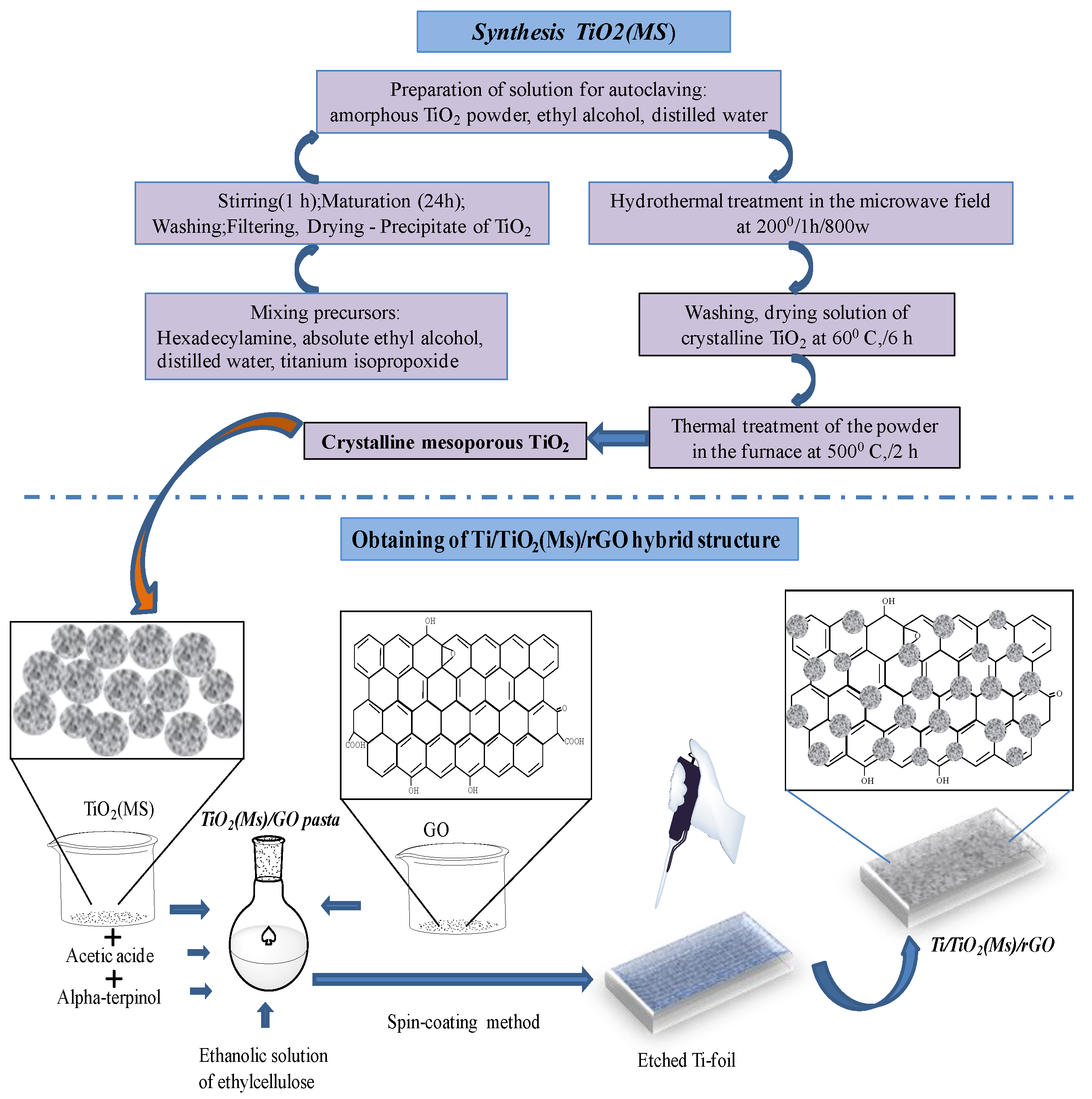
2.2.1. Synthesis of Mesoporous TiO2

The synthesis of TiO2(Ms)was carried out by microwave-assisted hydrothermal method, the schematic representation is presented in Figure 1. Briefly, an aqueous solution consisting of 1.3 g of HDA (surfactant), absolute ethyl alcohol, and 10 mL of TTIP was stirred for 1 h at room temperature. The homogeneous solution was matured (the interaction between TTIP and surfactant molecules) at room temperature for 24 h, and then the precipitate was washed intensively by centrifugation with distilled water and dried in an oven at a temperature of 60 °C, for 6 h. Due to the gel maturation by slow evaporation at room temperature, micellar self-assembly and the formation of ordered mesostructured composite microspheres were formed. Finally, a mixed solution containing 1 g of amorphous TiO2 powder, previously obtained, 15 mL of ethyl alcohol, and 25 mL of distilled water were introduced into a quartz autoclave with 50% fullness degree in a microwave-assisted hydrothermal reaction, at a temperature of 200 °C for 1 h (Anton PaarMultiwave 3000 Microwave Digestion Oven, USA). After the process was completed, the as-obtained powder was washed extensively with distilled water and dried in an oven at a temperature of 60 °C for 6 h; to create a porous structure the mesoporous TiO2 powder was thermally treated in the calcination furnace, at a temperature of 500 °C for 2 h. Microwave-assisted hydrothermal treatment facilitated the formation of TiO2 nanocrystals with an anatase crystalline phase, but the mesoporous TiO2 was obtained by calcination of mesostructured microspheres in air.

2.2.2. Synthesis of TiO2(Ms)/GO Paste

The TiO2(Ms)/GO stock solution paste was obtained by mixing a solution of 2 mL of GO, acetic acid, and 0.1 g of TiO2(Ms) at room temperature for 1 h, over which an ethanolic solution of ethylcellulose was added (previously prepared by heating to 40 °C and stirring for 30 min); finally, 2 mL of alpha-terpinol was added dropwise and continued stirring for 1 h at room temperature. The TiO2(Ms)/GO stock solution paste was used for spin-coating deposition.

2.2.3. Construction of Ti/TiO2(Ms)/rGO Hybrid Structure

The Ti/TiO2(Ms)/rGO hybrid structure was achieved as illustrated in Figure 1. The Ti foil (effective area 1 × 1.5 cm2) was etched in a 0.5 M HF solution for 3 h at room temperature, then washed and dried in an oven at 60 °C for 2 h [57]. Before deposition, the Ti supports were degreased in an ultrasonic bath in acetone and ethyl alcohol solutions, and the residues were removed by UV Ozone Cleaner (Ossila Producer, Sheffield, UK) for 15 min. To obtain a Ti/TiO2(Ms)/rGO hybrid structure, a facile and effective spin-coating (WS-400-6NPPB Spin Coater-Laurell Technology Corporation, Lansdale, PA, USA) methodology was conducted at 1500 rot for 10 s, which was then repeated 6 times on both sides of Ti support. Only 100 µL of TiO2(Ms)/GO stock solution paste for each layer was used by spin-coating deposition. After each deposited layer, the structure was dried in an oven at a temperature of 60 °C for 15 min. Finally, the hybrid structure was heated at a temperature of 350 °C (ramping rate of 2 °C min−1) for 2 h using GSL1100X Tube Furnace Vacuum System (MTI Corporation, Richmond, CA, USA) with constant nitrogen flow (100 mL min−1).

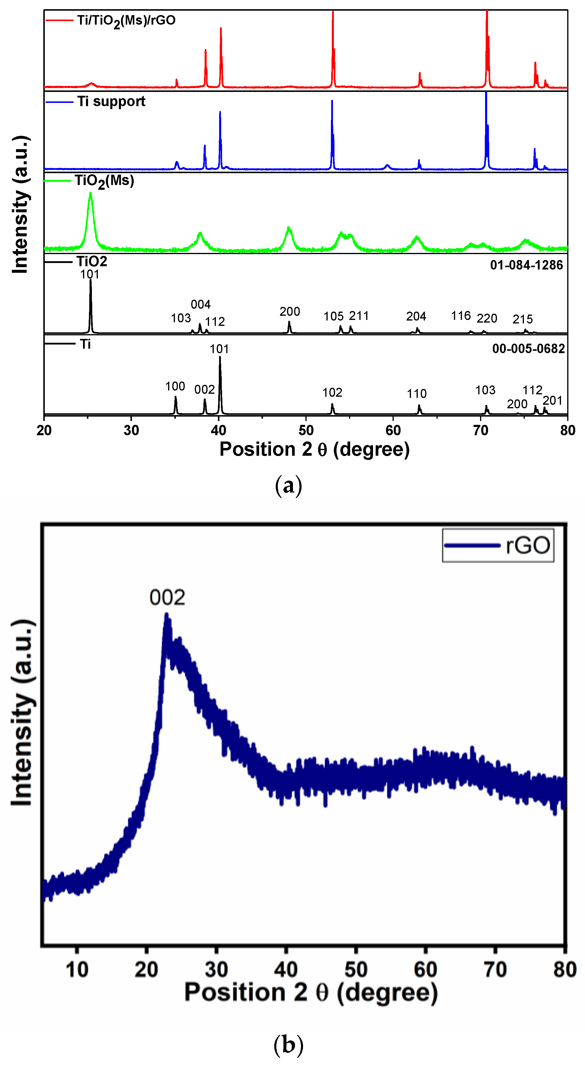
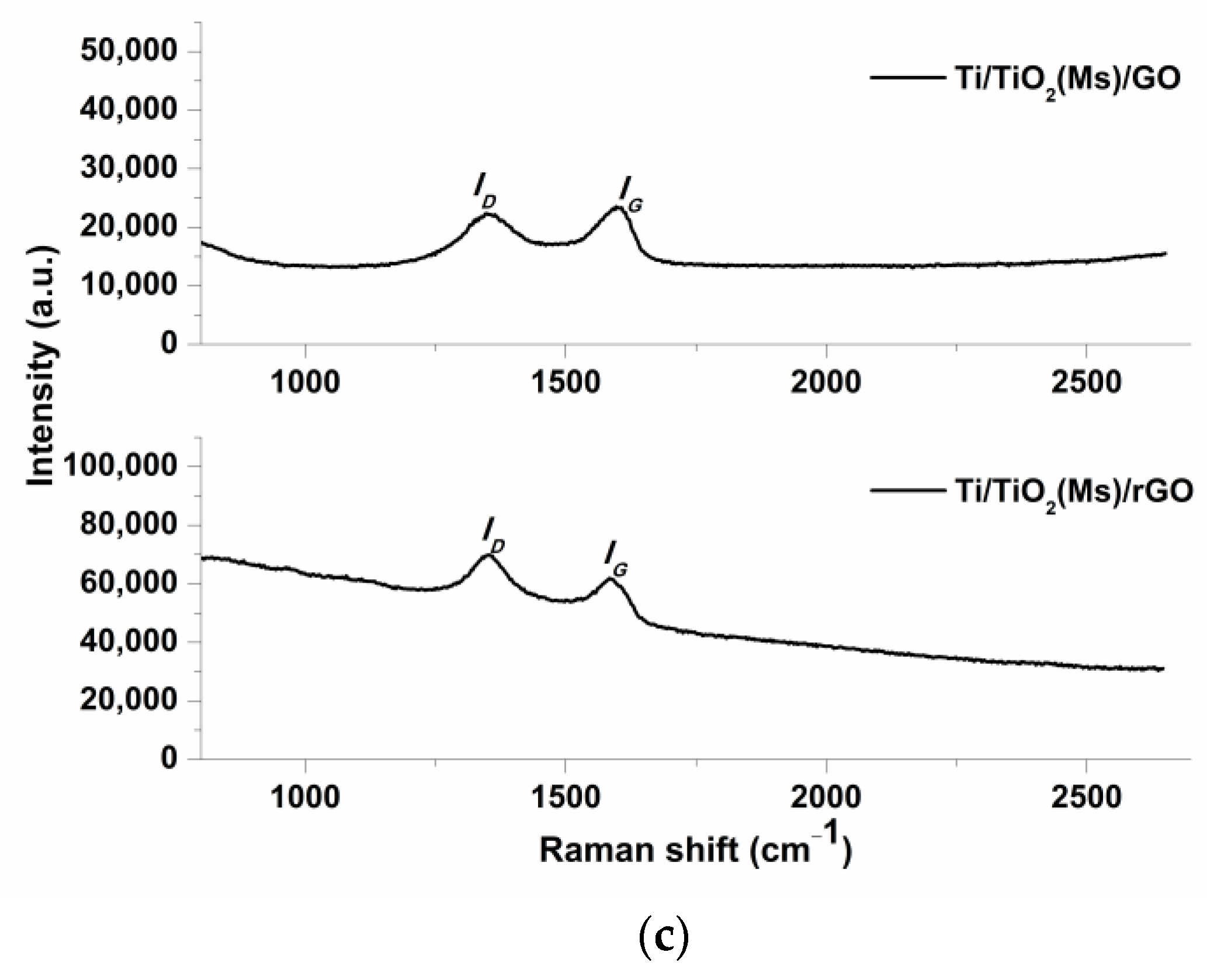
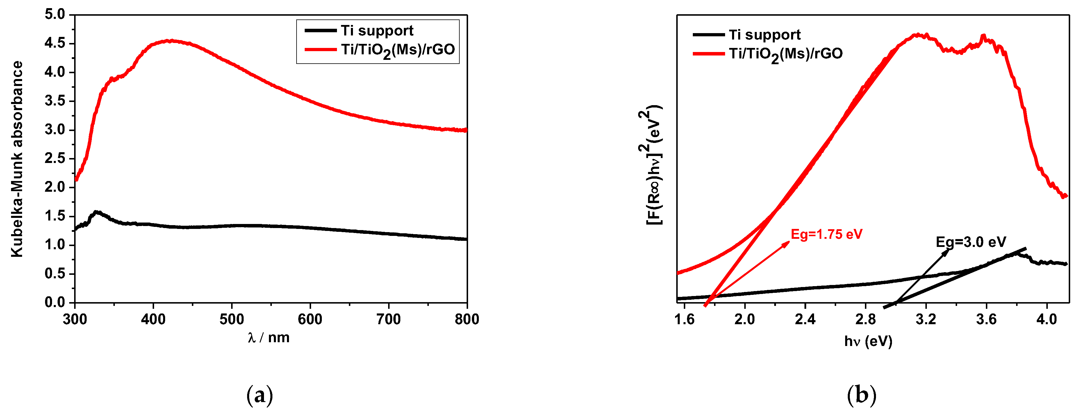
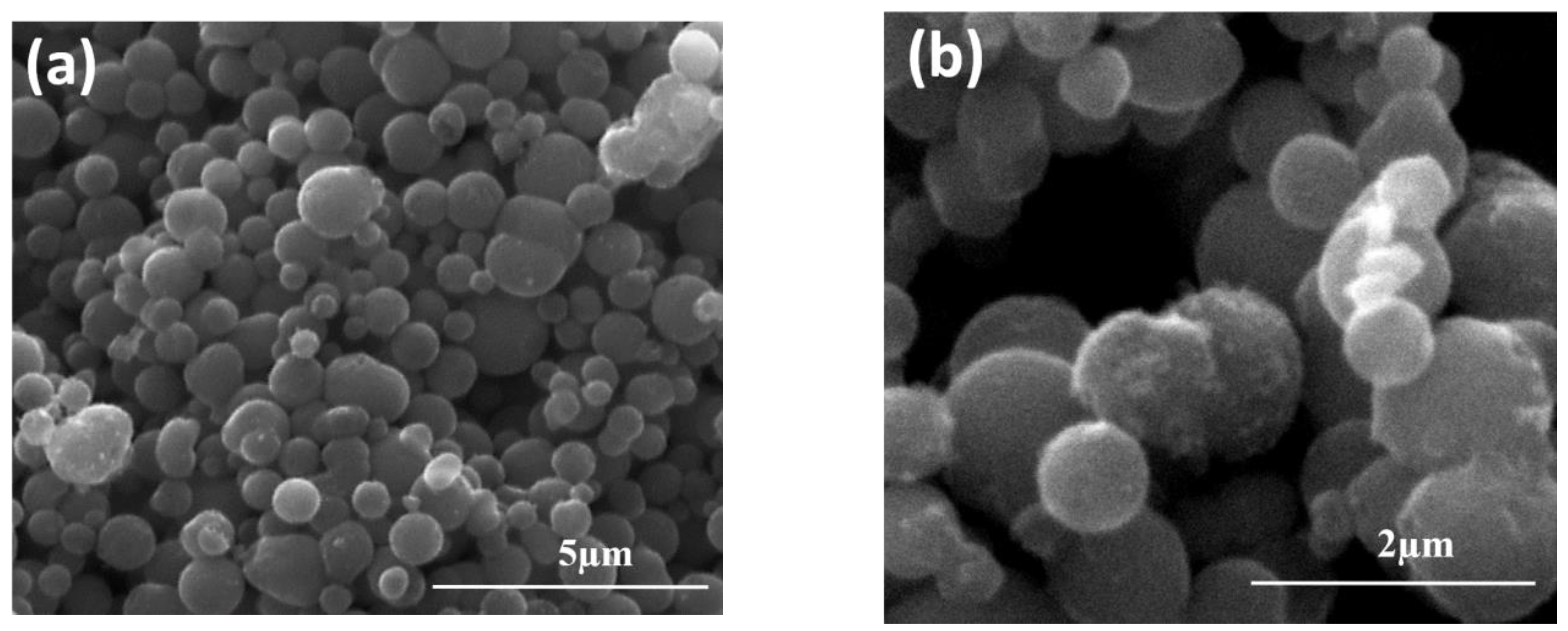
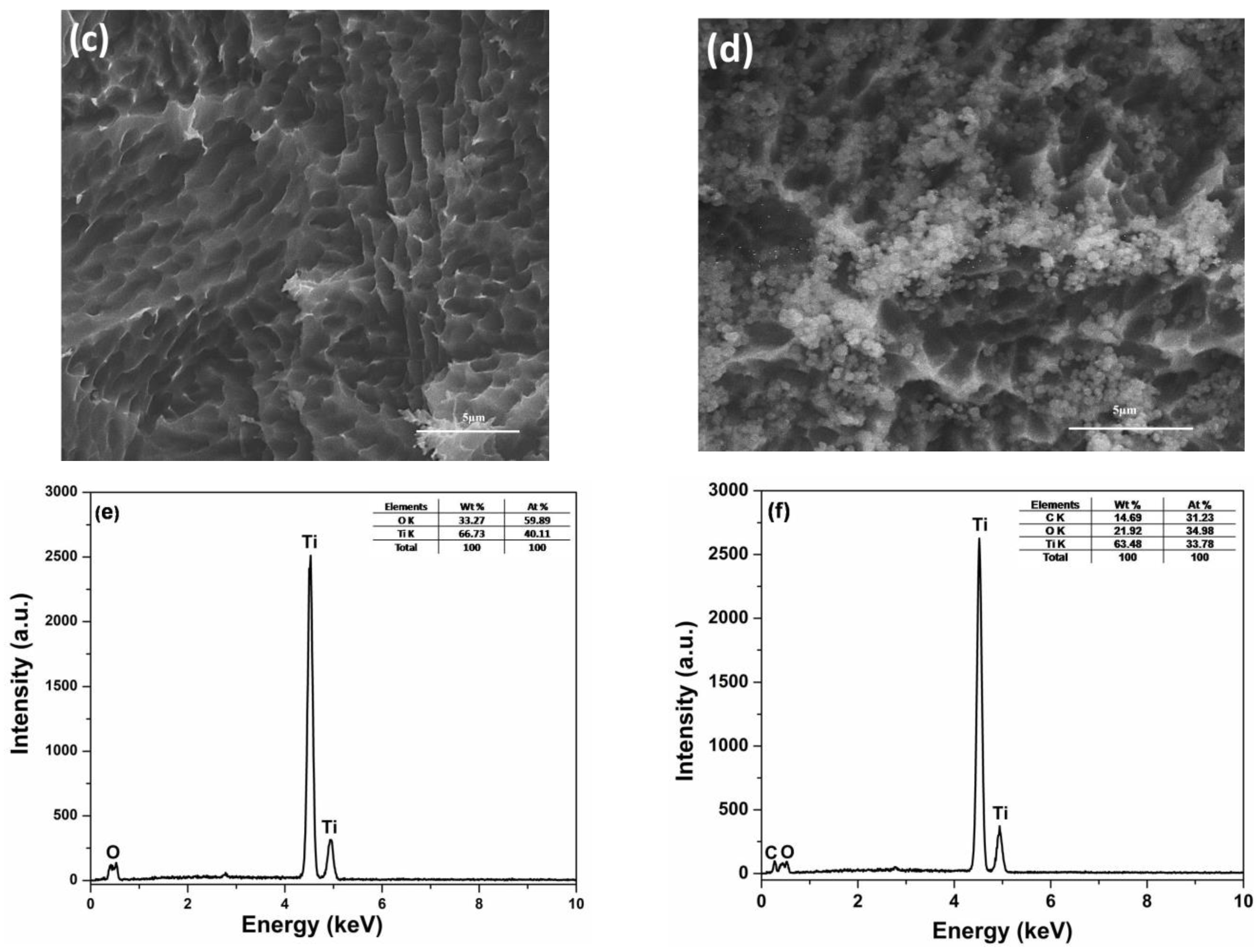
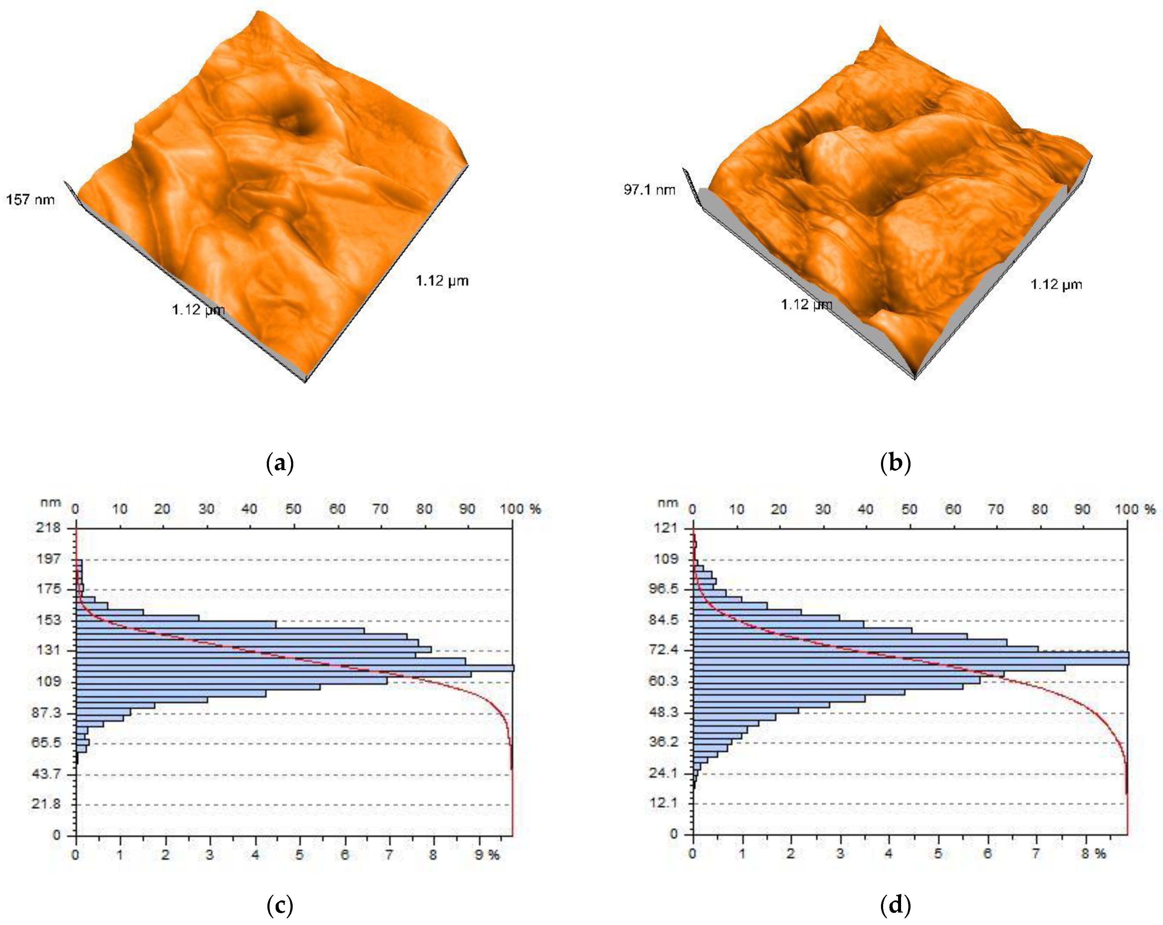
2.3. Hybrid Structure Characterization

The crystalline structure of the hybrid electrode was investigated by X-ray diffraction analysis (XRD, PANalyticalX’Pert PRO MPD Diffractometer, Almelo, The Netherlands) with Cu-Kα radiation in the range 2θ = 20–80°. The morphology of the structures was examined using atomic force microscopy (AFM, Model Nanosurf® EasyScan 2 Advanced Research, Liestal, Switzerland), with non-contact mode (scan size of 1 × 1 μm,) and scanning electron microscopy (SEM, FEI Inspect S model, Eindhoven, The Netherlands) coupled with the energy dispersive X-ray analysis detector (EDX) for elemental analysis. UV-visible spectroscopy (PerkinElmer Lambda 950 UV/Vis spectrophotometer, Shelton, CT, USA) with an integrating sphere in the range of 300–800 nm was used to investigate the optical characteristics of the hybrid structures. The microstructural characteristics of mesoporous TiO2 were investigated by Brunauer-Emmett-Teller (BET) analysis surface area (ASAP 2020 Model Instrument, Micromeritics Instrument Corporation, Norcross, GA, USA). RAMAN analysis was used with a Nanonics Imaging (Israel)—MultiProbe Imaging—MultiView 1000™Platform (SPM) equipped with a 532 nm laser for vibrational states identification. An Autolab potentiostat/galvanostat (PGSTAT 302 Metrohm Autolab B.V., Utrecht, The Netherlands) controlled with GPES 4.9 software using a three-electrode cell system in 1M KNO3 solution was used for electrochemical measurements, where Ti/TiO2(Ms)/rGO (an effective area of 1.5 cm2) acted as working electrodes, Ag/AgCl served as the reference electrode and platinum plate acted as the counter electrode.

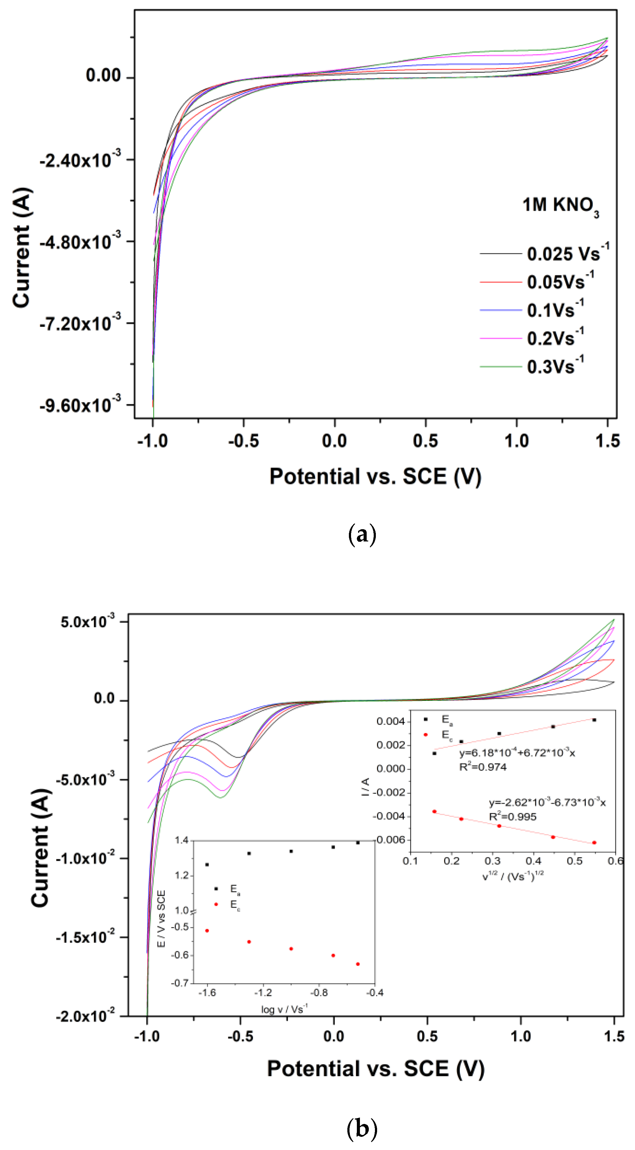
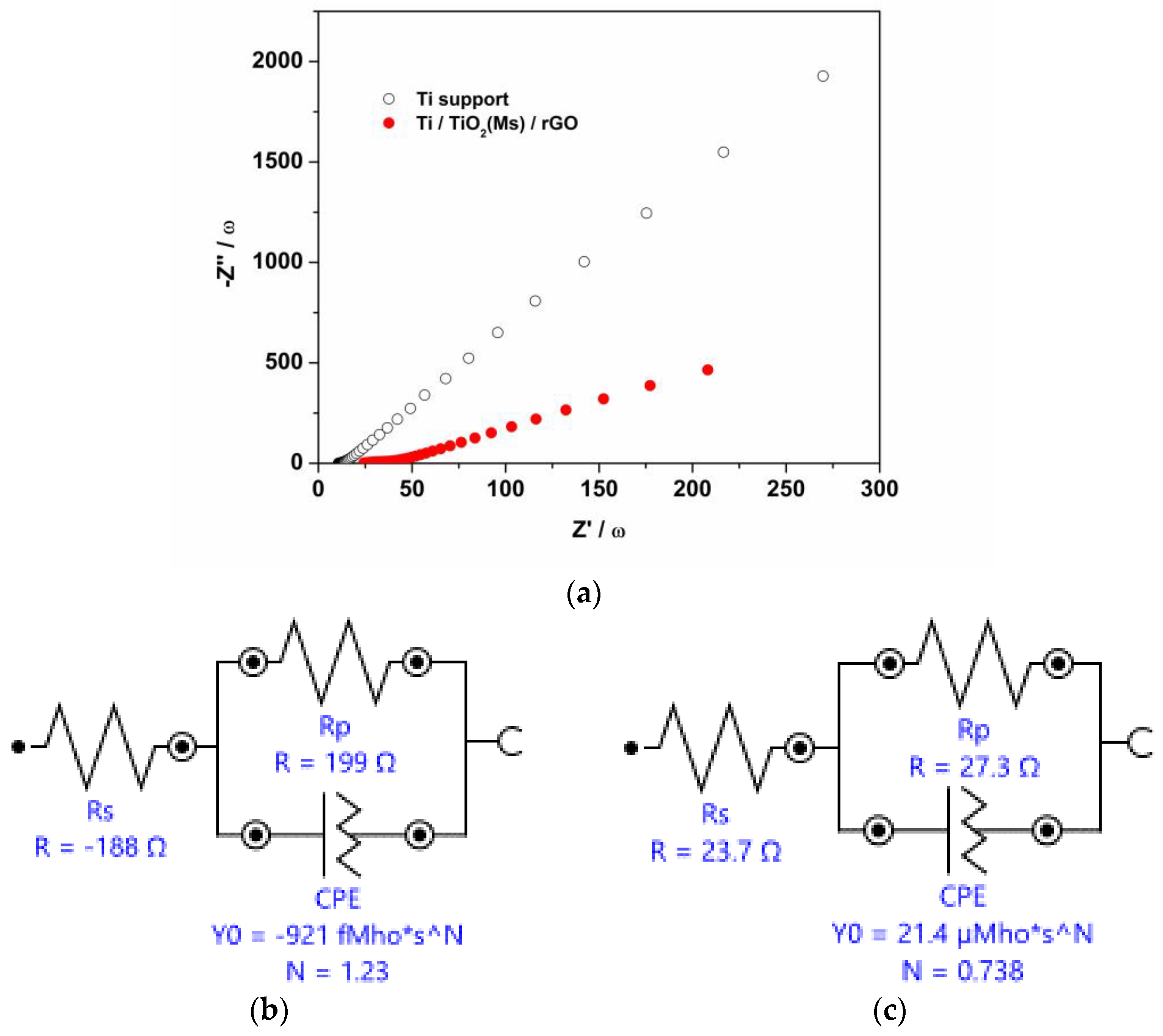
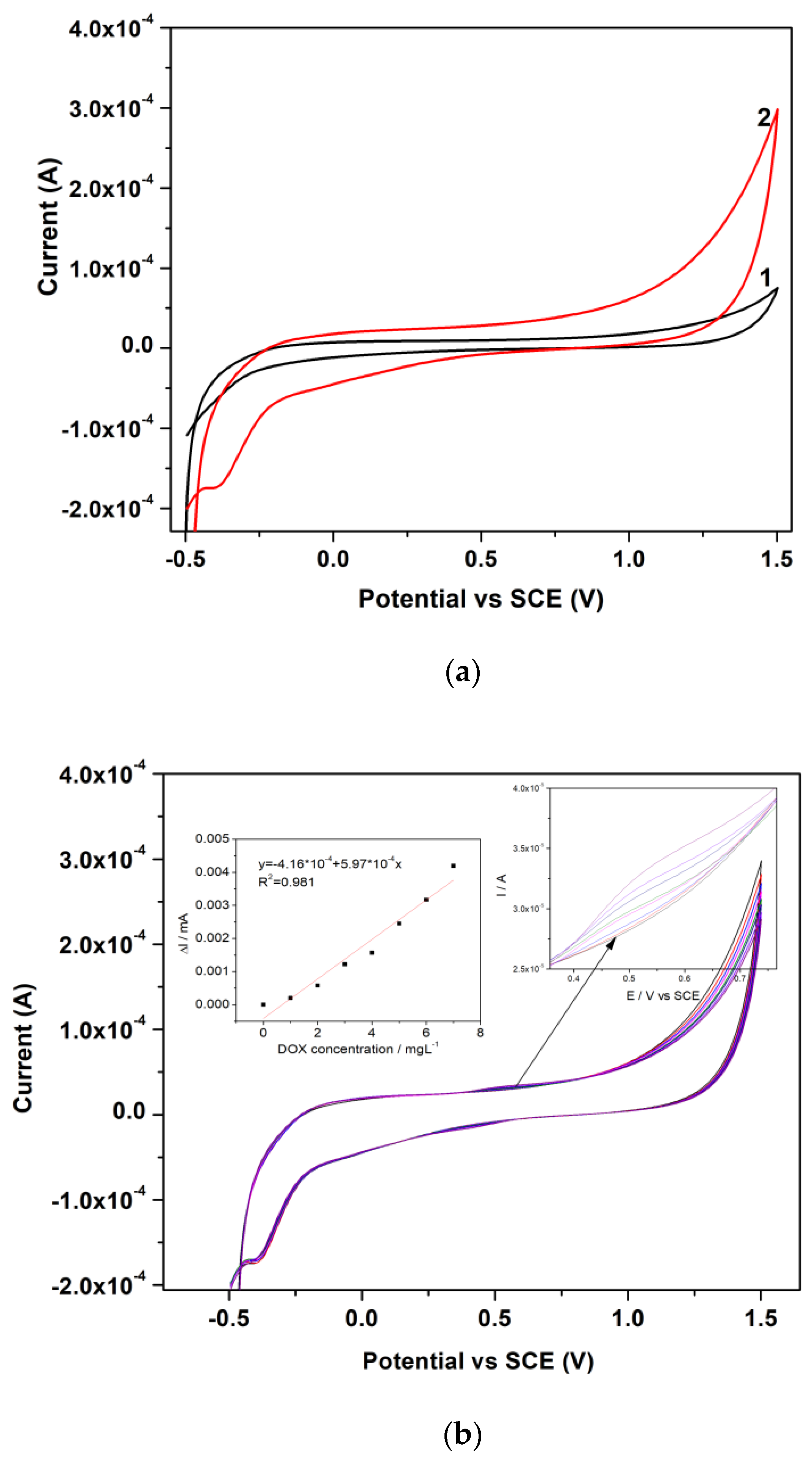
3. Results and Discussion

X-ray patterns for the Ti support, TiO2(Ms), and Ti/TiO2(Ms)/rGO hybrid structures are presented in Figure 2a. It can be seen the presence of strong peaks at 2θ values of 25.36°, 37.75°, 48.01°, 54.03°, 62.78°, 68.90°, 70.38°, and 75.29°, specifically for the anatase phase of mesoporous TiO2 (JCPDS No. 01-084-1286). Additionally, the main peaks for Ti support were identified at 2θ: 35.20°, 38.39°, 40.15°, 53.00°, 62.95°, 70.74°, and 76.35° (JCPDS No. 00-005-0682). The specific peaks of rGO are difficult to be identified in the Ti/TiO2(Ms)/rGO hybrid structure due to the high intensity of the diffraction peaks from the Ti support, instead, it was observed that the diffraction peak for the anatase phase of TiO2(Ms) at 2theta of 25.32°. Based on X-ray analysis from Figure 2a, the average crystallite size for mesoporous TiO2 was calculated using the Debye–Scherrer formula [58] and was estimated to be about 8.43 nm. Figure 2b shows the XRD pattern for pure rGO, in the range of 2θ = 10–30°, presenting a broad intensity peak on the range at 2θ of 20–30° corresponding to 002 diffraction plane of rGO which reflects the nanocrystalline state and the thermal reduction from GO to rGO layer [59]. RAMAN analysis was performed on Ti/TiO2 (Ms)/GO and Ti/TiO2 (Ms)/rGO hybrid structures to evaluate the graphitized structures, i.e., from 800 to 2700 cm−1, as presented in Figure 1c. The first peak, known as the D band (structural defect), and the second peak, the G band (graphitized structure) were both observed in as-synthesized samples. The ID/IG ratio of Ti/TiO2 (Ms)/GO (0.84) was lower than that of Ti/TiO2 (Ms)/rGO (1.13), indicating the reduction of graphene oxide.
The specific surface area is a significant microstructural parameter of the mesoporous materials, which depends on the geometrical shape and porosity. The pore size distribution, pore volume, and porosity were also determined using the BET analysis for mesoporous TiO2. According to IUPAC [60], the adsorption-desorption isotherm for mesoporous TiO2 shows a type IV isotherm with a hysteresis of type H2, specific for mesoporous materials. It can be observed that the actual N2 adsorption isotherm presented in Supplementary Material in Figure S1a,b, starts from a relative pressure (P/P0) of approximately 0.55, a fact that can be correlated with the existing plateau in the pore distribution graph in the range of 1.5–2.77 nm, signifying the fact that the presence of micropores is not detected on the surface of the material. Starting with the relative pressure (P/P0) in the range of 0.55–0.97, this area is characteristic of the mesoporous domain [61] with pore sizes in the range of 3.8–11.67 nm, a fact that can be correlated with the SEM analysis of the TiO2(Ms) material, which shows a high specific surface area of 140.4 m2 g−1 and a pore surface area of 151.7 m2 g−1, which means a high porosity throughout the surface. The diameter of the desorption pores is 7.795 nm with a pore volume of 0.3051 cm3 g−1.
Figure 3 shows UV-Vis absorption spectra of the Ti support and the Ti/TiO2(Ms)/rGO hybrid structure in the range of 300–800 nm. In both samples, the strong absorption in the UV region is attributed to the presence of TiO2. Figure 3a shows the corresponding wavelength for the Ti support and Ti/TiO2 (Ms)/rGO hybrid structure was about 400 nm, and it is observed that the presence of TiO2 (Ms)/rGO film influences the optical properties of light absorption. The increased background in the UV region is caused by the incorporation of mesoporous TiO2 into the rGO structure, suggesting the interaction between the TiO2 and rGO upon the generation of charge carriers [62,63]. The bandgaps of the Ti support and Ti/TiO2 (Ms)/rGO hybrid structure were evaluated using a Tauc method, the Eg optical bandgap energy is derived from the intersection of the tangent line of each plot with the -axis of the Tauc plot [64]. As presented in Figure 3b, the estimated bandgap of the Ti support and Ti/TiO2 (Ms)/rGO hybrid structure has been reduced from a value of 3.0 eV to value of 1.75 eV, respectively. The enhanced light-harvesting intensity of the hybrid structure compared with that of Ti support could be possibly explained by the formation of chemical bonds between TiO2 and rGO, where Ti-O-C facilitates charge transfer upon light excitation [63].
The morphologies of the as-synthesized samples are presented in Figure 4. Figure 4a,b shows that the mesoporous TiO2 has a well-defined spherical shape and it can be observed that numerous nanoparticles are present on the rough surface of the microspheres. Using ImageJ software (Version 1.53 t) the mesoporous TiO2 particle size was measured, and the values were in the range of 0.36 to 1.66 µm. The results revealed in Figure 4c demonstrate that for Ti support, the etching treatment of titanium foil causes the complete removal of the passivation layer from the surface. Therefore, the geometry of the surface (a very porous appearance with small craters) facilitates good adhesion and stability of the TiO2(Ms)/rGO film on the titanium support (Figure 4d). Analyzing the surface morphology of the deposited Ti/TiO2(Ms)/rGO film (Figure 4d) by the spin-coating method on the etched Ti support, the scanning electron micrographs highlights that the film is homogeneous, uniform, and cracks-free. It can also be seen that the paste based on TiO2(Ms) and rGO was deposited in a thin layer and covered the entire surface of the Ti support, following the geometry of the surface. EDX elemental analysis confirms the purity of the as-synthesized structures with specific elements, such as Ti and O for the Ti support (Figure 4e), and Ti, O, and C for the Ti/TiO2(Ms)/rGO hybrid structure, respectively (Figure 4f).
The 3D atomic force micrographs and particle distribution for the Ti support and the TiO2(Ms)/rGO film are presented in Figure 5, which are in accordance with the results obtained by SEM analysis (Figure 4c,d). In our work, the Nanosurf EasyScan 2 software (version 3.10.0) was used to estimate the particle size from the surface of the as-synthesized structures, and the obtained results are displayed in Figure 5c,d. Additionally, the surface roughness (measured surface area of about 1.329 pm2), which is represented by the average roughness Sa and the mean square root roughness Sq, and the layer thickness were determined from the AFM analysis data. The topographical parameters Sp, which refers to the maximum peak height deviation of the roughness, and Sv, which refers to the maximum valley depth deviation of the roughness that was evaluated on the calculated 3D surface (Figure 5a,c), were used for the calculation of the layers’ thicknesses [65], and the obtained values are presented in Table 1. Therefore, as expected the surface roughness of the Ti/TiO2(Ms)/rGO film is smaller than that of the Ti support, which confirms the uniform deposition of the layer on the Ti support.
The electrochemical behavior of the Ti/TiO2(Ms)/rGO hybrid electrode was assessed using the cyclic voltammetry method in the presence of the ferro/ferricyanide redox couple, as an electrochemical model of a reversible redox system. The electroactive surface area of the Ti/TiO2(Ms)/rGO hybrid electrode and the apparent diffusion coefficient were calculated based on the Randles-Sevcik Equation (1) [67]:
I p = 2.69 × 10 5 A D 1 / 2 n 3 / 2 v 1 / 2 C
where: Ip—peak current (0.00245 A); A—geometric surface area of the sensor (2.42 cm2), n—the number of participating electrons in the reaction, equal to 1, D—the diffusion coefficient of the molecules in solution, C—the molar concentration of K3Fe(CN)6 in the solution set up at 4 mM, and v—the scanning speed (0.025 Vs−1), the apparent diffusion coefficient of K3Fe(CN)6 was determined to be 3.55 10−5 cm2 s−1for the Ti/TiO2(Ms)/rGO hybrid electrode. Compared to the theoretical diffusion coefficient value of 6.7 10−6 cm2 s−1 reported for twin electrode thin-layer electrochemical cell by Kenopka et al. [68], we found that our as-developed Ti/TiO2(Ms)/rGO hybrid electrode exhibited a higher diffusion coefficient of 3.55 10−5 cm2 s−1. Due to the presence of TiO2(Ms)/rGO film at the electrode surface, the recorded electroactive surface area of 5.57 cm2 was 2.3 times higher than the geometrical surface area of 2.42 cm2. Figure 6 shows the cyclic voltammograms of 1 M KNO3 supporting electrolyte in the presence of 4 mM K3Fe(CN)6 recorded for the tested Ti/TiO2(Ms)/rGO hybrid electrode at increasing potential scan rate from 0.025 to 0.3 Vs−1.
The Randles–Sevick equation predicts a linear dependence of the anodic and cathodic peak currents with respect to the square root of the scan rate for the reversible process. The linear trends of Ip vs. log (ν½) presented in Figure 6b (right) indicate a diffusive electrochemical process (i.e., a process controlled by the mass transport velocity of the electroactive species moving toward the electrode surface due to a concentration gradient) [69]. However, the anodic-to-cathodic peak ratio was smaller than 1 for all applied scan rates, so the Ti/TiO2(Ms)/rGO hybrid electrode shows a quasi-reversible behavior.
Electrochemical impedance spectroscopy (EIS) experiments were performed for the Ti support and Ti/TiO2(Ms)/rGO hybrid electrode in order to obtain additional information for electrical, electrochemical, and physical processes that take place in the electrochemical system. The supporting electrolyte used for the EIS tests was 0.1 M Na2SO4, which was further used in the electrochemical detection application for the DOX target analyte. The impedance diagrams presented in Figure 7a, recorded in a 0.1 M Na2SO4 supporting electrolyte and a frequency range between 100 kHz and 0.1 Hz for the Ti support and Ti/TiO2(Ms)/rGO hybrid electrode were fit, considering the electrical equivalent circuits given in Figure 7b,c. Both equivalent circuits include the electronic elements: Rs—the electrolyte resistance between the working and reference electrodes, and Rp—the charge transfer resistance and a CPE (constant phase element) that simulates the nonideal behavior of the capacitor. The charge transfer resistance for the Ti/TiO2(Ms)/rGO electrode decreased compared with Ti support, while the capacitance value increased in the Ti/TiO2(Ms)/rGO equivalent circuit.
In order to characterize the voltametric behavior of Ti support and Ti/TiO2(Ms)/rGO hybrid structure in an aqueous solution, the cyclic voltammograms for 0.1 M Na2SO4 supporting electrolyte were conducted at both electrodes. The presence of the TiO2(Ms)/rGO film on the electrode surface is highlighted by the higher background current and oxygen evolution behavior (Figure 8a). The potential applicability of the Ti/TiO2(Ms)/rGO hybrid electrode in electroanalysis was tested for DOX detection by the cyclic voltammetry technique. DOX oxidation on the electrode surface occurred at the potential of +0.52 V/SCE, in the concentration range between 1 to 7 mg L−1 DOX (Figure 8b).
The choice of the design model and the synthesis method to obtain the Ti/TiO2(Ms)/rGO hybrid electrode were validated by electrochemical experiments for DOX detection on its surface. Furthermore, during the electrochemical tests, the TiO2(Ms)/rGO film deposited on the Ti support showed good adhesion and stability, and no exfoliation of the electrode was observed.

4. Conclusions

A hybrid electrode based on a Ti/TiO2(Ms)/rGO structure was successfully achieved by a facile and effective spin-coating methodology. Using mesoporous TiO2 with a high specific surface area (~140 m2 g−1) in the synthesis of the composite material based on a Ti/TiO2(Ms)/rGO, we have achieved a 2.3 times increase in electroactive surface area than the geometrical surface area of the hybrid electrode and determined a good adherence and stability of rGO. The as-developed electrode was structurally, morphologically, and electrochemically characterized by specific methods, and tested for detection at different concentrations of DOX drug. The SEM and AFM micrographs revealed that the Ti/TiO2(Ms)/rGO films are uniformly coated and crack-free on the Ti support, and the EDX analysis confirms the purity of the samples. The development of the Ti/TiO2(Ms)/rGO hybrid electrode provides a novel and sensitive method for DOX detection by exploiting the remarkable properties of reduced graphene oxide on the electrode surface, and the synergistic relationship between these two materials led to an improvement of the surface adsorption characteristics. Moreover, the EIS tests confirmed the important role of the rGO deposited onto the electrode surface, showing a higher charge transfer resistance for the Ti/TiO2(Ms)/rGO hybrid structure in comparison with Ti support. The electrochemical characterization and detection tests revealed an electrode with enhanced electroactive surface area and electrocatalytic properties for DOX detection, opening perspectives for further developments in enhancing the electroanalytical performance of the Ti/TiO2(Ms)/rGO hybrid electrode for the detection of other interest target analytes.

Supplementary Materials

The following supporting information can be downloaded at: https://www.mdpi.com/article/10.3390/coatings13081359/s1, Figure S1: N2 adsorption-desorption isotherm curve (a) and pore size distribution curve (b) of the TiO2 (Ms).

Author Contributions

C.B. was involved in conceptualization, methodology, investigation, writing—original draft; M.I.P. was involved in methodology, investigation; C.O. was involved in conceptualization, methodology, investigation, writing—original draft; M.N. was involved in conceptualization, methodology, investigation; A.P. was involved in methodology, investigation, writing—original draft; C.L. was involved in methodology, investigation, writing—original draft, supervision. All authors have read and agreed to the published version of the manuscript.

Funding

This research was funded by a grant of the Ministry of Research, Innovation and Digitization, CNCS-UEFISCDI, project number PN-III-P1-1.1-TE-2021-0963, within PNCDI III, with contract number TE13/2022 (DD-CyT) and project code PN 23 27 01 02 INOMAT, 23-27 29N/2023.

Institutional Review Board Statement

Not applicable.

Informed Consent Statement

Not applicable.

Data Availability Statement

Not applicable.

Conflicts of Interest

The authors declare no conflict of interest.

References

  1. Li, T.; Shang, D.; Gao, S.; Wang, B.; Kong, H.; Yang, G.; Shu, W.; Xu, P.; Wei, G. Two-dimensional material-based electrochemical sensors/biosensors for food safety and biomolecular detection. Biosensors 2022, 12, 314. [Google Scholar] [CrossRef] [PubMed]
  2. Collado, M.S.; Mantovani, V.E.; Goicoechea, H.C.; Olivieri, A.C. Simultaneous spectrophotometric-multivariate calibration determination of several components of ophthalmic solutions: Phenylephrine, chloramphenicol, antipyrine, methylparaben and thimerosal. Talanta 2000, 52, 909–920. [Google Scholar] [CrossRef] [PubMed]
  3. Ribone, M.É.; Pagani, A.P.; Olivieri, A.C. Simultaneous multivariate spectrophotometric analysis of ear drops containing a ternary mixture of antipyrine, sulfathiazole, and rivanol. Anal. Lett. 2001, 34, 2077–2088. [Google Scholar] [CrossRef]
  4. Atsriku, C.; Watson, D.; Tettey, J.N.; Grant, M.; Skellern, G. Determination of diminazene aceturate in pharmaceutical formulations by HPLC and identification of related substances by LC/MS. J. Pharm. Biomed. Anal. 2002, 30, 979–986. [Google Scholar] [CrossRef] [PubMed]
  5. Zuo, M.; Duan, G.L.; Ge, Z.G. Simultaneous determination of ropivacaine and antipyrine by high performance liquid chromatography and its application to the in vitro transplacental study. Biomed. Chromatogr. 2004, 18, 752–755. [Google Scholar] [CrossRef] [PubMed]
  6. Abdel-Hafez, G.A.; Aboraia, A.S.; Mohammad, A.-M.I.; Youssef, A.F. Development and validation of a high-performance thin-layer chromatography densitometric method for the simultaneous determination of novel 1-acridinyl-1,2,3-triazole derivatives. JPC J. Planar Chromatogr. Mod. TLC 2022, 35, 349–362. [Google Scholar] [CrossRef]
  7. Tiwari, S.; Talreja, S. Thin Layer Chromatography (TLC) VS. Paper Chromatography: A Review. Acta Sci. Pharm. Sci. 2022, 6, 05–09. [Google Scholar] [CrossRef]
  8. Woźniak, M.K.; Wiergowski, M.; Aszyk, J.; Kubica, P.; Namieśnik, J.; Biziuk, M. Application of gas chromatography–tandem mass spectrometry for the determination of amphetamine-type stimulants in blood and urine. J. Pharmaceut. Biomed. Anal. 2018, 148, 58–64. [Google Scholar] [CrossRef]
  9. Moeder, M.; Schrader, S.; Winkler, M.; Popp, P. Solid-phase microextraction–gas chromatography–mass spectrometry of biologically active substances in water samples. J. Chromatogr. A. Mar. 2000, 873, 95–106. [Google Scholar] [CrossRef]
  10. Malak, M.; Ebrahim, H.; Sonbol, H.; Ali, A.; Aboulella, Y.; Hadad, G.; Emara, S. Highly Sensitive In-Capillary Derivatization and Field Amplified Sample Stacking to Analyze Narcotic Drugs in Human Serum by Capillary Zone Electrophoresis. Separations 2023, 10, 58. [Google Scholar] [CrossRef]
  11. Nagib, Q.; Ezzouhra, E.-M. Spectrophotometric/Titrimetric Drug Analysis. In Drug Formulation Design; Rahul, S., Aleksey, K., Akbar, A., Eds.; IntechOpen: Rijeka, Croatia, 2023; p. 10. [Google Scholar]
  12. Rivera-Utrilla, J.; Sánchez-Polo, M.; Ferro-García, M.A.; Prados-Joya, G.; Ocampo-Pérez, R. Pharmaceuticals as emerging, contaminants and their removal from water. A review. Chemosphere 2013, 93, 1268–1287. [Google Scholar] [CrossRef] [PubMed]
  13. Mahnik, S.N.; Lenz, K.; Weissenbacher, N.; Mader, R.M.; Fuerhacker, M. Fate of 5-fluorouracil, doxorubicin, epirubicin, and daunorubicin in hospital wastewater and their elimination by activated sludge and treatment in a membrane-bio-reactor system. Chemosphere 2007, 66, 30–37. [Google Scholar] [CrossRef]
  14. Martín, J.; Camacho-Muñoz, D.; Santos, J.L.; Aparicio, I.; Alonso, E. Occurrence and ecotoxicological risk assessment of 14 cytostatic drugs in wastewater. Water Air Soil Pollut. 2014, 225, 1896–1906. [Google Scholar] [CrossRef]
  15. Sylvester, B.; Tefas, L.; Vlase, L.; Tomuta, I.; Porfire, A. A Quality by Design (QbD) approach to the development of a gradient high-performance liquid chromatography for the simultaneous assay of curcuminoids and doxorubicin from long-circulating liposomes. J. Pharmaceut. Biomed. Anal. 2018, 158, 395–404. [Google Scholar] [CrossRef]
  16. Lucas, A.T.; O’Neal, S.K.; Santos, C.M.; White, T.F.; Zamboni, W.C. A sensitive high performance liquid chromatography assay for the quantification of doxorubicin associated with DNA in tumor and tissues. J. Pharmaceut. Biomed. Anal. 2016, 119, 122–129. [Google Scholar] [CrossRef] [Green Version]
  17. Ansar, S.M.; Mudalige, T. Direct and simultaneous determination of intraliposomal and external sulfate in liposomal doxorubicin formulations by capillary electrophoresis/inductively coupled plasma-tandem mass spectrometry (CE/ICP-MS/MS). Int. J. Pharm. 2019, 561, 283–288. [Google Scholar] [CrossRef]
  18. Ansar, S.M.; Jiang, W.; Mudalige, T. Direct quantification of unencapsulated—doxorubicin in liposomal doxorubicin formulations using capillary electrophoresis. Int. J. Pharm. 2018, 549, 109–114. [Google Scholar] [CrossRef]
  19. Zandanel, C.; Legouffe, R.; Trochon-Joseph, V.; Tomezyk, A.; Gaudin, M.; Bonnel, D.; Stauber, J.; Vasseur, B.; Bromet, N. Biodistribution of polycyanoacrylate nanoparticles encapsulating doxorubicin by matrix-assisted laser desorption ionization (MALDI) mass spectrometry imaging (MSI). J. Drug Deliv. Sci. Technol. 2018, 47, 55–61. [Google Scholar] [CrossRef]
  20. Zhu, J.; Chu, H.; Shen, J.; Wang, C.; Wei, Y. Green preparation of carbon dots from plum as a ratiometric fluorescent probe for detection of doxorubicin. Opt. Mater. 2021, 114, 110941. [Google Scholar] [CrossRef]
  21. Semreen, M.H.; Alniss, H.Y.; Mousa, M.K.; El-Awady, R.; Khan, F.; Al-Rub, K.A. Quantitative determination of doxorubicin in the exosomes of A549/MCF-7 cancer cells and human plasma using ultra performance liquid chromatography-tandem mass spectrometry. Saudi Pharmaceut. J. 2018, 26, 1027–1037. [Google Scholar] [CrossRef] [PubMed]
  22. Orha, C.; Bandas, C.; Lazau, C.; Popescu, M.I.; Baciu, A.; Manea, F. Advanced electrodegradation of doxorubicin in water using a 3D Ti/SnO2 anode. Water 2022, 14, 821. [Google Scholar] [CrossRef]
  23. Rong, S.; Zou, L.; Meng, L.; Yang, X.; Dai, J.; Wu, M.; Qiu, R.; Tian, Y.; Feng, X.; Ren, X.; et al. Dual function metal-organic frameworks based ratiometric electrochemical sensor for detection of doxorubicin. Anal. Chim. Acta 2022, 1196, 339545. [Google Scholar] [CrossRef] [PubMed]
  24. Thobakgale, L.; Ombinda-Lemboumba, S.; Mthunzi-Kufa, P. Chemical sensor nanotechnology in pharmaceutical drug research. Nanomater 2022, 12, 2688. [Google Scholar] [CrossRef]
  25. Al-Tamimi, S.A. Biogenic green synthesis of metal oxide nanoparticles using oat biomass for ultrasensitive modified polymeric sensors. Green Chem. Let. Rev. 2021, 14, 166–179. [Google Scholar] [CrossRef]
  26. Lakra, R.; Kumar, R.; Kumar, S.; Thatoi, D.; Soam, A. Synthesis of TiO2 nanoparticles as electrodes for supercapacitor. Mater. Today Proc. 2023, 74, 863–866. [Google Scholar] [CrossRef]
  27. Wahab, R.; Alam, M. Highly efficient and fast electrochemical sensor based on aluminium oxide (Al2O3) nanoparticle for the detection of organosulfur compounds. Sens. Actuators A Phys. 2022, 347, 113967. [Google Scholar] [CrossRef]
  28. Xu, L.; Tang, S.; Li, D.; Ma, X.; Zhu, Y.; Lu, J.; Niu, J. Electrochemical degradation of tris(2-chloroethyl) phosphate by metal-oxide-coated Ti anodes: Kinetics, toxicity and mechanism. Sep. Purif. Technol. 2021, 265, 118489. [Google Scholar] [CrossRef]
  29. Rodrigues, M.P.D.S.; Miguel, V.M.; Germano, L.D.; Córdoba de Torresi, S.I. Metal oxides as electrocatalysts for water splitting: On plasmon-driven enhanced activity. Electrochem. Sci. Adv. 2021, 2, e2100079. [Google Scholar] [CrossRef]
  30. Shooshtari, M.; Salehi, A.; Vollebregt, S. Effect of temperature and humidity on the sensing performance of TiO2 nanowire-based ethanol vapor sensors. Nanotechnology 2021, 32, 325501. [Google Scholar] [CrossRef]
  31. Morozova, M.; Kluson, P.; Krysa, J.; Vesely, M.; Dzik, P.; Solcova, O. Electrochemical properties of TiO2 electrode prepared by various methods. Procedia Eng. 2012, 42, 573–580. [Google Scholar] [CrossRef] [Green Version]
  32. Liang, Y.; Xiong, X.; Xu, Z.; Xia, Q.; Wan, L.; Liu, R.; Chen, G.; Chou, S.L. Ultrathin 2D mesoporous TiO2/rGO heterostructure for high-performance lithium storage. Small 2020, 16, e2000030. [Google Scholar] [CrossRef]
  33. Xie, X.F.; Yang, K.F.; Sun, D. Voltammetric determination of hypoxanthine based on the enhancement effect of mesoporous TiO2-modified electrode. Colloids Surf. B Biointerfaces 2008, 67, 261–264. [Google Scholar] [CrossRef] [PubMed]
  34. Lin, H.G.; Ji, X.B.; Chen, Q.Y.; Zhou, Y.K.; Banks, C.E.; Wu, K.B. Mesoporous-TiO2 nanoparticles-based carbon paste electrodes exhibit enhanced electrochemical sensitivity for phenols. Electrochem. Commun. 2009, 11, 1990–1995. [Google Scholar] [CrossRef]
  35. Fan, Y.; Liu, J.H.; Lu, H.T.; Zhang, Q. Electrochemical behavior and voltametric determination of paracetamol on Nafion/TiO2–graphene modified glassy carbon electrode. Colloids Surf. B Biointerfaces 2011, 85, 289–292. [Google Scholar] [CrossRef]
  36. Fan, X.; Ma, C.; Zhao, D.; Deng, Z.; Zhang, L.; Wang, Y.; Luo, Y.; Zheng, D.; Li, T.; Zhang, J.; et al. Unveiling selective nitrate reduction to ammonia with Co3O4nanosheets/TiO2 nanobelt heterostructure catalyst. J. Colloid Interface Sci. 2023, 630, 714–720. [Google Scholar] [CrossRef]
  37. Mohamed, M.A.; Atty, S.A.; Merey, H.A.; Fattah, T.A.; Foster, C.W.; Banks, C.E. Titanium nanoparticles (TiO2)/graphene oxide nanosheets (GO): An electrochemical sensing platform for the sensitive and simultaneous determination of benzocaine in the presence of antipyrine. Analyst 2017, 142, 3674. [Google Scholar] [CrossRef] [Green Version]
  38. Fang, C.; Zhang, J.; Chen, X.; Weng, G.J. Calculating the electrical conductivity of graphene nanoplatelet polymer composites by a Monte Carlo method. Nanomater 2020, 10, 1129. [Google Scholar] [CrossRef]
  39. Kamedulski, P.; Skorupska, M.; Binkowski, P.; Arendarska, W.; Ilnicka, A.; Lukaszewicz, J.P. High surface area micro-mesoporous graphene for electrochemical applications. Sci. Rep. 2021, 11, 22054. [Google Scholar] [CrossRef]
  40. Velick, M.; Bradley, D.F.; Cooper, A.J.; Hill, E.W.; Kinloch, I.A.; Mishchenko, A.; Novoselov, K.S.; Patten, H.V.; Toth, P.S.; Valota, A.T.; et al. Electron transfer kinetics on mono-and multilayer graphene. ACS Nano 2014, 8, 10089–10100. [Google Scholar] [CrossRef] [PubMed]
  41. Fu, L.; Mao, S.; Chen, F.; Zhao, S.; Su, W.; Lai, G.; Yu, A.; Lin, C.T. Graphene-based electrochemical sensors for antibiotic detection in water, food and soil: A scientometric analysis in Cite Space (2011–2021). Chemosphere 2022, 297, 134127. [Google Scholar] [CrossRef] [PubMed]
  42. Sfragano, P.S.; Laschi, S.; Renai, L.; Fichera, M.; Del Bubba, M.; Palchetti, I. Electrochemical sensors based on sewage sludge–derived biochar for the analysis of anthocyanins in berry fruits. Anal. Bioanal. Chem. 2022, 414, 6295–6307. [Google Scholar] [CrossRef] [PubMed]
  43. Zhang, X.; Suresh Kumar, P.; Aravindan, V.; Liu, H.H.; Sundaramurthy, J.; Mhaisalkar, S.G.; Duong, H.M.; Ramakrishna, S.; Madhavi, S. Electrospun TiO2-graphene composite nanofibers as a highly durable insertion anode for Lithium ion batteries. J. Phys. Chem. C 2012, 116, 14780–14788. [Google Scholar] [CrossRef]
  44. Minella, M.; Versaci, D.; Casino, S.; Di Lupo, F.; Minero, C.; Battiato, A.; Penazzi, N.; Bodoardo, S. Anodic materials for Lithium-ion batteries: TiO2-rGO composites for high power applications. Electrochim. Acta 2017, 230, 132–140. [Google Scholar] [CrossRef]
  45. Lazau, C.; Bandas, C.; Nicolaescu, M.; Orha, C.; Pop, A. A facile dip-coating process graphene-TiO2 on titanium foil for hybrid electrode fabrication. In Proceedings of the 2022 International Semiconductor Conference (CAS), Poiana Brasov, Romania, 12–14 October 2022; pp. 277–280. [Google Scholar] [CrossRef]
  46. Chou, H.T.; Chen, S.T.; Wang, C.H.; Hsu, H.C.; Kao, T.Y.; Lin, Z.H. Bilayer structure of TiO2/TiO2-graphene for optimizing working electrode applied in dye sensitized solar cells. IEEE J. Photovolt. 2021, 11, 1236–1242. [Google Scholar] [CrossRef]
  47. Timoumi, A.; Alamri, S.N.; Alamri, H. The development of TiO2-graphene oxide nano composite thin films for solar cells. Results Phys. 2018, 11, 46–51. [Google Scholar] [CrossRef]
  48. Ali, H.A. Jalaukhan, Optical investigation of TiO2/Graphene Oxide thin film prepared by Spin Coating technique. IOP Conf. Ser. Mater. Sci. Eng. 2020, 871, 012087. [Google Scholar] [CrossRef]
  49. Becerril, H.A.; Mao, J.; Liu, Z.; Stoltenberg, R.M.; Bao, Z.; Chen, Y. Evaluation of solution-processed reduced graphene oxide films as transparent conductors. ACSN 2008, 2, 463–470. [Google Scholar] [CrossRef]
  50. Sui, D.; Huang, Y.; Huang, L.; Liang, J.; Ma, Y.; Chen, Y. Flexible and transparent electrothermal film heaters based on graphene materials. Small 2011, 7, 3186–3192. [Google Scholar] [CrossRef]
  51. Timoumi, A.; Albetran, H.M.; Alamri, H.R.; Alamri, S.N.; Low, I.M. Impact of annealing temperature on structural, morphological and optical properties of GO-TiO2 thin films prepared by spin coating technique. Superlattices Microstruct. 2020, 139, 106423. [Google Scholar] [CrossRef]
  52. Nehru, R.; Gopi, P.K.; Chen, S.M. Enhanced sensing of hazardous 4-nitrophenol by a graphene oxide–TiO2 composite: Environmental pollutant monitoring applications. New J. Chem. 2020, 44, 4590–4603. [Google Scholar] [CrossRef]
  53. Wen, X.; Zhao, S.; Asuha, S. Preparation of nitrogen-doped mesoporous TiO2/RGO composites and its application to visible light-assisted photocatalytic degradation. J. Nanomater. 2019, 2019, 6467107. [Google Scholar] [CrossRef] [Green Version]
  54. Lazau, C.; Nicolaescu, M.; Orha, C.; Pop, A.; Căprărescu, S.; Bandas, C. In situ deposition of reduced graphene oxide on Ti foil by a facile, microwave-assisted hydrothermal method. Coatings 2022, 12, 1805. [Google Scholar] [CrossRef]
  55. Shan, C.; Yang, H.; Han, D.; Zhang, Q.; Ivaska, A.; Niu, L. Graphene/AuNPs/chitosan nanocomposites film for glucose biosensing. Biosens. Bioelectron. 2010, 25, 1070–1074. [Google Scholar] [CrossRef] [PubMed]
  56. Liu, Y.; Yu, D.S.; Zeng, C.; Miao, Z.C.; Dai, L.M. Biocompatible graphene oxide-based glucose biosensors. Langmuir 2010, 26, 6158–6160. [Google Scholar] [CrossRef]
  57. Nicolaescu, M.; Bandas, C.; Orha, C.; Şerban, V.; Lazău, C.; Căprărescu, S. Fabrication of a UV photodetector based on n-TiO2/p-CuMnO2 heterostructures. Coatings 2021, 11, 1380. [Google Scholar] [CrossRef]
  58. Cullity, B.D. Elements of X-ray Diffraction; Addison-Wesley Pub. Co.: Reading, MA, USA, 1978. [Google Scholar]
  59. Mikhaylov, P.A.; Vinogradov, M.I.; Levin, I.S.; Shandryuk, G.A.; Lubenchenko, A.V.; Kulichikhin, V.G. Synthesis and characterization of polyethylene terephthalate-reduced graphene oxide composites. IOP Conf. Ser. Mater. Sci. Eng. 2019, 693, 012036. [Google Scholar] [CrossRef] [Green Version]
  60. Nishi, Y.; Inagaki, M. Chapter 11—Gas Adsorption/Desorption Isotherm for Pore Structure Characterization. In Materials Science and Engineering of Carbon: Characterization; Inagaki, M., Ed.; Elsevier: Amsterdam, The Netherlands, 2016; pp. 227–247. [Google Scholar] [CrossRef]
  61. Shaikh, S.; Mane, R.; Min, B.; Hwang, Y.; Joo, O. D-sorbitol-induced phase control of TiO2 nanoparticles and its application for dye-sensitized solar cells. Sci. Rep. 2016, 6, 20103. [Google Scholar] [CrossRef] [Green Version]
  62. Raja, R.; Govindaraj, M.; Antony, M.D.; Krishnan, K.; Velusamy, E.; Sambandam, A.; Subbaiha, M.; WilliamsRayar, V. Effect of TiO2/reduced graphene oxide composite thin film as a blocking layer on the efficiency of dye-sensitized solar cells. J. Solid State Electrochem. 2017, 21, 891–903. [Google Scholar] [CrossRef]
  63. Sheshmani, S.; Nayebi, M. Modification of TiO2 with graphene oxide and reduced graphene oxide; enhancing photocatalytic activity of TiO2 for removal of Remazol Black B. Polym. Compos. 2019, 40, 210–216. [Google Scholar] [CrossRef] [Green Version]
  64. Makuła, P.; Pacia, M.; Macyk, W. How to correctly determine the band gap energy of modified semiconductor photocatalysts based on UV–Vis spectra. J. Phys. Chem. Lett. 2018, 9, 6814–6817. [Google Scholar] [CrossRef] [Green Version]
  65. Skoneczny, W.; Niedźwiedź, M.; Bara, M. The Effect of production parameters of oxide layers on their nanostructure, nanomorphology, and surface free energy. Appl. Sci. 2018, 8, 2251. [Google Scholar] [CrossRef] [Green Version]
  66. Birdeanu, A.-V.; Birdeanu, M.; Fagadar-Cosma, E. Corrosion protection characteristics of ceramics, porphyrins and hybrid ceramics/porphyrins, deposited as single and sandwich layers, by pulsed laser deposition (PLD). J. Alloys Compd. 2017, 706, 220–226. [Google Scholar] [CrossRef]
  67. González-Meza, O.A.; Larios-Durán, E.R.; Gutiérrez-Becerra, A.; Casillas, N.; Escalante, J.I.; Bárcena-Soto, M. Development of a Randles-Ševčík-like equation to predict the peak current of cyclic voltammetry for solid metal hexacyanoferrates. J. Solid State Electrochem. 2019, 23, 3123–3133. [Google Scholar] [CrossRef]
  68. Kenopka, S.J.; McDuffle, B. Diffusion coefficient of feri-and ferrocyanide ions in aqueous media, using twin electrode thin layer electrochemistry. Anal. Chem. 1970, 42, 1741–1746. [Google Scholar] [CrossRef]
  69. Bertana, V.; Scordo, G.; Parmeggiani, M.; Scaltrito, L.; Ferrero, S.; Gomez, M.G.; Cocuzza, M.; Vurro, D.; D’Angelo, P.; Iannotta, S.; et al. Rapid prototyping of 3D Organic Electrochemical Transistors by composite photocurable resin. Sci. Rep. 2020, 10, 13335. [Google Scholar] [CrossRef]
Figure 1. Schematic representation of the development process for Ti/TiO2(Ms)/rGO hybrid electrode.
Coatings 13 01359 g001
Figure 2. X-ray patterns for as-synthesized structures (a) and pure rGO (b). RAMAN analysis of the obtained structures (c).
Coatings 13 01359 g002aCoatings 13 01359 g002b
Figure 3. Kubelka-Munk absorbance of Ti support and Ti/TiO2(Ms)/rGO hybrid structure (a); Band-gap calculation using the Tauc method (b).
Coatings 13 01359 g003
Figure 4. SEM micrographs of TiO2(Ms) (a,b); Ti support (c) and Ti/TiO2(Ms)/rGO hybrid structure (d). EDX elemental analysis of Ti support (e) and Ti/TiO2(Ms)/rGO hybrid structure (f).
Coatings 13 01359 g004aCoatings 13 01359 g004b
Figure 5. 3D AFM micrographs for Ti support (a) and TiO2(Ms)/rGO film (b); Particle size distribution for Ti support (c) and TiO2(Ms)/rGO film (d).
Coatings 13 01359 g005
Figure 6. Cyclic voltammograms of 1M KNO3 supporting electrolyte (a) and in the presence of 4 mM K3Fe(CN)6 (b) for the Ti/TiO2(Ms)/rGO hybrid electrode. Inset (b): Plots of the anodic and cathodic potential peaks vs. logarithm of scan rates (i.e., 0.025, 0.05, 0.1, 0.2 and 0.3 Vs−1) (left). Calibration plots of the anodic and cathodic current peaks vs. square root of the scan rates (right).
Coatings 13 01359 g006
Figure 7. (a) Nyquist plots for Ti support and Ti/TiO2(Ms)/rGO electrode in 0.1 M Na2SO4 supporting electrolyte. Electrical equivalent circuits for Ti support (b) and Ti/TiO2 (Ms)/rGO electrode (c).
Coatings 13 01359 g007
Figure 8. Cyclic voltammograms recorded in 0.1 M Na2SO4 supporting electrolyte on TiO2 support (curve 1) and Ti/TiO2(Ms)/rGO (curve 2) (a); Cyclic voltammograms recorded in 0.1 M Na2SO4 supporting electrolyte, and in the presence of 1 to 7 mg L−1 DOX concentrations (scan rate of 50 mV s−1) on Ti/TiO2(Ms)/rGO; Insets: (left) the linear relationship between DOX concentration and anodic current peak and (right) the detail of anodic peak (b).
Coatings 13 01359 g008
Table 1. Surface particle size, nano-roughness, and layer thickness of the structures from the AFM micrographs, calculated with Nanosurf EasyScan 2 software.
SampleAverage Particle Size (nm)Sa (nm)Sq (nm)Sp (nm)Sv (nm)Layer Thickness
Sp-Sv (nm) [66]
Ti support12027.3138.95121.18−146.41267.59
Ti/TiO2(Ms)/rGO film7013.2417.5779.58−136.35215.93
Disclaimer/Publisher’s Note: The statements, opinions and data contained in all publications are solely those of the individual author(s) and contributor(s) and not of MDPI and/or the editor(s). MDPI and/or the editor(s) disclaim responsibility for any injury to people or property resulting from any ideas, methods, instructions or products referred to in the content.

