
Comparison and Research Progress of Protein Detection Technology for Cultural Relic Materials

1
Department of Cultural Heritage and Museology, Zhejiang University, Hangzhou 310028, China
2
Department of Chemistry, Zhejiang University, Hangzhou 310027, China
*
Author to whom correspondence should be addressed.
Coatings 2023, 13(8), 1319; https://doi.org/10.3390/coatings13081319
Submission received: 8 June 2023
/
Revised: 16 July 2023
/
Accepted: 20 July 2023
/
Published: 27 July 2023
(This article belongs to the Special Issue Advanced Coating Material for Heritage Preservation)
Abstract
:The analysis of historical materials is an essential component of cultural heritage conservation. Protein was one of the most important and pervasive organic substances in ancient human societies. Through the qualitative and quantitative examination of protein-based materials, it is possible to clarify their source and functions of substances in cultural heritages, investigate the manufacturing technology of cultural heritage, and identify their deterioration mechanism. On the basis of these analyses, corresponding measurements are therefore feasible. Currently, mass spectrometry, chromatography, spectroscopy, nuclear magnetic, proteomics, and immunoassay are used to analyse protein materials. Proteomics techniques and enzyme-linked immunosorbent assay (ELISA) technology are two of the most common methods for detecting ancient proteins. This article discusses the evolution of protein component detection in ancient materials, as well as the implementation of proteomics and ELISA techniques for the analysis of proteins. In addition, the characteristics of these two techniques were contrasted in order to propose the most recent analytical techniques and the direction of future research.
1. Introduction
In the conservation of cultural heritage and archaeological excavations, the materials, and especially the key components, such as the binder of murals and paintings and the organic components of building mortars, are not only closely related to human production life, material life, spiritual life, and all aspects of social life, but they also serve as significant carriers of historical information. In addition, they are the focus of research on traditional building techniques, the prevention and control of the deterioration of cultural relic materials, the elucidation of the physical and chemical causes of material deterioration, and the development of future protective measures based on analysis and testing [1].
Proteins are one of the most significant components of cultural heritage artefacts and are frequently employed as essential additives [2,3,4]. Additionally, proteins can be used to restore and reinforce cultural heritage artefacts [5,6,7]. It is difficult to analyze protein-containing cultural heritage artefacts due to the limited number of available samples or the low protein content of the samples. Moreover, after prolonged exposure to multiple environmental factors, proteins encounter a variety of issues, including ageing, degradation, and contamination [8,9]. The earliest research on protein detection in artefacts of cultural heritage dates back to the 1950s [10]. Archaeologists used mass spectrometry (MS) to detect amino acids, the building blocks of proteins, in archaeological and paleontological artefacts [11]. Since the 1980s, physical chemistry techniques such as mass spectrometry, chromatography, and spectroscopy have been utilised extensively in the field of cultural heritage research and conservation. However, these methods have the disadvantage of requiring a large number of samples, making it challenging to distinguish the complex protein components in cultural heritage objects and lacking species specificity [12]. In recent years, with the development of biotechnology, bioinformatics technology, and MS, proteomics techniques and enzyme-linked immunosorbent assay (ELISA) techniques have been increasingly applied to the study of proteins in cultural relics materials, and these two techniques offer high sensitivity and low detection limits [13].
In addition to discussing the evolution of protein analysis in the field of cultural patrimony, this article will describe two cutting-edge techniques: proteomics and ELISA technology. Additionally, a general overview of the challenges and prospective directions of protein identification in cultural heritage materials will be presented.
2. The Role of Protein Analysis in the Protection of Cultural Heritage
2.1. Types of Cultural Heritage Containing Protein
Based on their composition, cultural relic materials can be classified as organic or inorganic. Both categories of ancestry share a substantial number of protein components (Table 1). For example, gum is required to enhance the writing effect on biological cultural heritage objects because the paper has numerous pores between the fibres, which easily generates a halo in the ink and affects writing and painting. The utilised adhesive is derived from either plants or animals and contains either soy protein or collagen [14,15,16]. The lacquer of lacquerware cultural heritage artefacts contains glycoproteins [17]. Also present in bone cultural heritage artefacts is collagen [18]. A significant number of organic cultural heritage objects are natural macromolecular proteins. Silk protein, for example, is the primary component of silk [19], whereas collagen and keratin are the primary components of leather and fur cultural heritage objects [6,18].
Proteins are frequently utilized as a binder in the production and restoration of inorganic cultural heritage objects, such as in the bonding of ceramic masonry artifacts, the strengthening of building materials, and the fixation of pigments in painted cultural heritage objects [2,12]. These cement-based building materials may be found easily and are commonly employed at historical and archaeological sites around the globe. Chinese architectural cement, murals, ancient architectural paintings, and pottery paintings frequently use animal glue, egg whites, and other materials as binders; these materials were later examined and found to contain collagen, ovalbumin, and other materials [2,3]. When creating dry murals and tempera paintings in the West, egg whites and milk were frequently utilized. Ovalbumin and casein have also been detected [12,20].
2.2. Characteristics of Protein-Containing Cultural Relics Materials
Physical, chemical, and biological elements in the environment can cause proteins in cultural relic materials to age and degrade. The breakdown of hydrogen and peptide bonds and the disruption of internal protein structures, which result in protein fragmentation, are induced by external stimuli or enzymatic action. For further analysis, protein fragmentation presents difficulties. Protein translational modifications (PTMs), which also cause protein fragmentation, modify the structure of proteins and alter their chemical and biological properties. The characteristics of peptides and proteins are impacted by these structural changes in significant chemical and biological ways. As a result, modifications to the side chains of some proteins can also be used as markers for the degradation of proteins as they age. One biomarker of protein degradation and age, for instance, is protein deamidation in cultural relic materials. The spontaneous nonenzymatic deamidation of glutaminyl and asparaginyl residues has been shown to alter the structure of proteins and peptides [21,22]. In addition to causing protein breakdown, these PTMs also make it challenging to distinguish between the various protein species found in cultural relic materials.
For instance, silk protein makes up the majority of silk. It is influenced by elements such as pH, microbes, oxygen, ultraviolet light, temperature, and humidity because it is a naturally occurring macromolecular protein. Silk cultural relic materials age and deteriorate as a result, losing their original form and luster and turning yellowish, brittle, or even carbonized [13,19]. This is caused by the degradation of the proteins’ heavy chain and light chain, P25 chain, crystalline area, and non-crystalline region. Collagen that has been gelatinized and either been tanned or not is typically what makes up leather cultural relic materials. The acids, alkalis, and salts in the water breakdown the protein in the leather when leather cultural relic items are in a humid environment, such as when submerged in groundwater for a prolonged period of time; hence, the state is frequently quite fragile [23,24]. The morphology of leather coils, deterioration worsens, collagen and fat gradually breakdown, and leather volume diminishes, becoming thinner and more brittle over time, according to examinations of the ageing pattern of leather cultural relic materials [24].
A low temperature is ideal for preserving proteins since it slows down their aging and breakdown. In these circumstances, collagen molecules fold into a chain area, preventing protease attack and delaying protein decomposition. It has been demonstrated that collagen solutions hydrolyze at their lowest rates when the pH is between 5–7 [16]. The protein components that exist in low temperatures, have pH levels that are mildly alkaline to neutral, and are in airtight circumstances are therefore easier to preserve and analyze as residues because well-preserved collagen is also found in enclosed bone samples.
2.3. The Significance of Protein Analysis for the Protection of Cultural Relics Materials
First, protein analysis can aid in understanding the function of the protein in materials used to create cultural relics. For instance, egg white mortar, which is frequently used in construction and building decorating, can be used to track how a substance changes over time by looking for protein components in the mortar. According to research, egg whites can help to aerate, bind, sterilize, and waterproof the mortar. The interfacial activity of protein molecules and hydrated products, as well as the regulatory function of biomineralization, are related to the mechanism of these effects [2,3,7].
Second, protein analysis can show how the artifact was made. For instance, protein analysis enables the actual condition of the cultural relic materials to be compared with the records to validate the authenticity of the records. Some relics contain a preserved production formula detailing the components and production process. Vasily Yakovlevich Struve wrote documents by hand from 1793 to 1864. In 2021, Pankin et al. [25] studied the dried film used for the paper connection and discovered that its constituents were gelatine and fish glue. This outcome is in line with the typical ingredients used in dry film making that were listed in a 19th-century encyclopedia.
In addition, protein analysis can also show how proteins age and degrade in materials from cultural relics. Collagen, for instance, is a key ingredient in leather cultural relic materials. Collagen is highly vulnerable to severe distortion, fracture, and interweaving caused by high temperatures, according to an analysis of the proteins in leather cultural relic materials. Chemical forces also break the hydrogen bonds and peptide chains of collagen, making leather cultural relic materials tougher, more fragile, and even fully ruined.
Finally, resource and approach for associated restoration and conservation activities, protein component analysis is useful. In the case of leather cultural relic materials, for instance, the degree of deterioration of the leather is assessed starting from the retention status and distribution law of the polypeptides of collagen degradation products, and a new protection method is proposed: treating degraded leather with a mixture of glutaraldehyde and tannins can strengthen the inter- and intramolecular hydrogen bond and covalently cross-linked collagen molecules to achieve reinforcement [26]. Protein component analysis can also eliminate any remaining protein clumps and offer a solution without harming the cultural heritage artifact [27]. For the first time, Paul Banks removed remaining gelatine from paper in 1969 using collagenase, a highly specialized protease [28]. Since the egg white varnish coating on the 16th-century painting was insoluble in organic solvents and mechanical removal might have irreparably ruined the original work, the enzyme removal procedure was employed while repainting the damaged area [29]. In recent years, biological purification and collagenase protease treatment have been employed successfully to remove the thick gauze bound with animal glue that was used to protect murals during World War II [30]. Different proteases may break down various kinds of proteins, so suitable proteases must be chosen in accordance with the findings of the protein analysis.
2.4. Protein Detection Method in Cultural Relics Materials
The small number of samples available for the analysis of proteins-containing cultural relic materials, the low protein content of those samples, and the difficulties in isolating proteins that are affected by aging and degradation make this approach difficult to use. Moreover, the majority of protein samples are mixtures. As a result, precise detection of protein components in mixtures is also required, in addition to efficient and sensitive approaches that can evaluate small volumes of samples [31]. Although chromatography, spectroscopy, and other technologies have been employed for protein detection, the chromatographic technology’s pre-treatment process is complicated, necessitates a large number of samples, and has a high sample loss rate [32]. Furthermore, the signals produced by the spectrum in samples of cultural relic materials are subject to interference from other substances, which makes it difficult to recognize proteins, and the majority of ancient protein materials lack standard spectra, which makes it challenging to identify species [33].
3. Proteomics Technology
In 1994, Marc Wilkins, a student at Maquaire University in Australia, coined the term proteome. It refers to all proteins that are expressed by the genes of cells or tissues under specific conditions [34]. various protein expressions at various stages, locations, and environments contribute to the complexity of the proteome, making the protein group extremely complex. Proteomics is the study of the functions, structures, and interactions of all proteins expressed by genes using protein separation, mass spectrometry (MS), and biological information technologies [35,36].
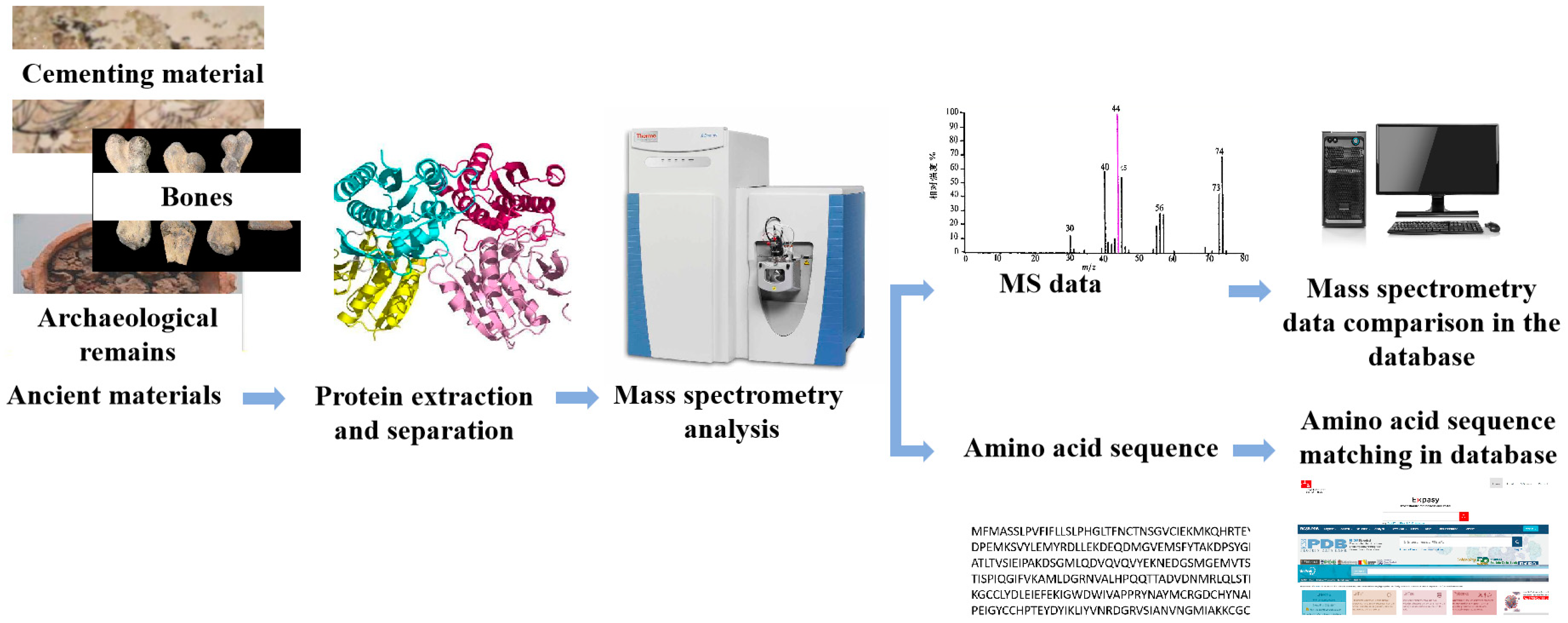
Expression proteomics, which examines the qualitative and quantitative expression of proteins in cells, organs, or organisms, is the most fundamental research in proteomics [37]. Figure 1 depicts the principal processes as protein extraction and separation, MS analysis, and retrieval of biological information databases [38]. Select a suitable method for protein extraction from the sample, and then separate the extracted proteins. In the separation procedure, HPLC can be used for independent separation, or HPLC–MS can be used in series [39]. The specifics will be provided in Section 3.1.
MS analysis is the cornerstone of proteomics technology, and it consists of two distinct techniques. The top-down method analyses multiple complete proteins in a mélange, including large segment proteins, modified proteins, and peptides. The steps are as follows: First, the proteins are separated by reversed-phase liquid chromatography, and then, after ionisation and dissociation, they are analysed. This method is applicable to both sequence analysis and analysis of post-translational modifications. Sequence coverage is substantial, and sample preparation is relatively straightforward. However, there are some physical and technical challenges associated with using this method to analyse cultural relic materials. On the one hand, proteins in cultural relic materials are typically aged and degraded, and it is challenging to extract and purify complete proteins from these materials. On the other hand, mass spectrometry cannot detect aged proteins sensitively [39]. Second, the bottom-up method, also known as the shotgun method, employs a protease such as trypsin to cleave the protein backbone at specific amino acids to generate peptides, which are then analysed by mass spectrometry to derive amino acid sequence information [40]. This technique is suitable for the analysis of complex samples and has a high throughput [8]. Consequently, this technique is presently the most prevalent and mature method for protein identification, particularly in the analysis of ancient proteins. Depending on the testing requirements, various MS options can be selected or used in series due to the rapid development of instruments [38,41]. The ionisation sources of a mass spectrometer are primarily of two types: electric spray ionisation (ESI) and matrix assisted laser desorption ionisation (MALDI). These two soft ionisation techniques enable the analysis of large molecular weight molecules and proteins with post-translational modifications. Both MALDI and ESI have advantages and disadvantages and can be selected based on the sample’s characteristics and the type of information needed. In addition to the ion source, the mass spectrometer consists of a mass analyzer, a detector, and a device for processing data. The mass analyzer is an essential component of the mass spectrometer, which sorts particles according to their mass-to-charge ratios (m/z). In Section 3.2, the functions and selection of the mass spectrometer will be discussed.
The detected original data can then be compared to the database to identify proteins. After digestion, each protein yields a unique set of peptides; the molecular weights of these peptides constitute the peptide fingerprint profile (PMF) of this protein. Using matrix-assisted laser desorption ionization–time-of-flight MS (MALDI-TOF-MS), for instance, we can detect the relative molecular weight of peptide segments, acquire the PMF, and then identify protein species by comparing the PMFS to a standard database. Alternatively, ESI-MS can be used to determine the amino acid sequences of various peptide segments. For protein identification, bioinformatics techniques can be used to compare the amino acid sequence analysis results with the amino acid sequences in the protein database. Using various data processing software and module analysis techniques, it is possible to detect the protein species present in a sample [42,43]. Or, tandem mass spectrometry (MS/MS or MS2), which couples two or more mass analyzers via additional reaction stages, can be utilised. Before accessing the mass spectrometer for examination, proteins must be enzymatically cleaved into peptide segments. After enzyme digestion, the mass of the peptide segment is determined by primary mass spectrometry, and significant high-abundance peptide fragments are assessed by secondary mass spectrometry. After colliding with a high flow rate of inert gas, secondary mass spectrometry dissociates peptide segments, resulting in smaller peptide fragment ions. A detector analyses the fragment ions to determine the amino acid sequence of the peptide segment. MS/MS can obtain accurate and high-quality data from abundant peptide fragment ions, enhancing the reliability of subsequent database retrieval and enabling the detection of trace proteins in mixtures.
With the gradual development of MS technology and the improvement of protein databases, proteomics technology has begun to be applied to the protein analysis of cultural relic materials; for instance, protein analysis was used to identify protein cements in paintings [44] in the early 2000s. The approach has been implemented in the field of archaeology, for example in the identification of animal remains and the analysis of animal and plant products. In proteomics research on cultural relic materials, protein extraction technology determines the efficacy of detection due to the rarity and value of the relics.
3.1. Protein Extraction
How to extract trace quantities of proteins is the first issue that must be resolved for protein identification in materials from cultural relics. Noemi Proietti used SDS-PAGE to separate and analyse the protein components in paper samples from the Camerino Fabriano region [45] in 2020, comparing the protein recovered from the samples to cellulose that had been treated with animal gelatin. The presence of a band with the same molecular weight as the standard sample indicated the presence of animal gelatin. RP-HPLC was then utilised to analyse the separated proteins. Regarding the RP-HPLC outcomes, both analysed samples exhibit chromatographic peaks with UV spectra indicative of protein material. The results indicate that the paintings contain animal gelatin. However, such procedures typically require a large number of samples, and the resulting information regarding species is frequently unclear. In an effort to reduce the weight of the sample, Caroline Tokarski [46] utilised an extraction technique that breaks down proteins from cultural relics into peptide compounds in 2006. Different extraction solutions (HCl, HCOOH, NH3, NaOH) and conditions under ultrasonic immersion, mortar grinding, and resin grinding were investigated, as were the extraction techniques for protein-cemented materials in Renaissance art paintings. The finest extraction results were obtained by grinding the grinding resin in an aqueous solution of 1% trifluoroacetic acid and performing a multi-step ultrasonic bath, according to the authors. The protein extraction efficacy was then evaluated using matrix-assisted laser desorption ionization–time-of-flight mass spectrometry (MALDI–TOF MS). Enzymatic hydrolysis of the extracted protein yields a peptide mixture, which is subsequently analysed and identified using nano-liquid chromatography (nano-LC), nano-electrospray ionisation (nano-ESI), quadrupole time-of-flight mass spectrometry (Q-q-TOF-MS), and mass spectrometry (MS). The results demonstrated that the method only required 10 g of samples to identify the binder in the painting as ovalbumin. This method employs a small sample size and does not hydrolyze the protein into amino acids during extraction, preserving more information about the genus and species.
Similarly, the extraction of proteins from cultural relics should be as minimally invasive as feasible; therefore, a microwave combined protease extraction method for digestion on the surface of cultural relics has been developed. In 2009, Gabriella Leo et al. [47] used microwave-assisted trypsin to extract protein from the pigment layer of a 13th-century Italian cathedral dome fresco, and then used nano-LC-MS/MS and ESI to detect the extracted peptide segments. It was discovered that the binder was milk. Using this method to extract proteins will not harm cultural artefacts and is appropriate for samples with trace quantities, ageing, and degradation.
Recently, a novel hydrogel protein extraction material comprised of trypsin and chymotrypsin has been utilised in the analysis of cultural relic protein materials. Calvano et al. [48] directly coated the surface of an oil painting and analysed the peptides obtained after enzymatic hydrolysis using MALDI–TOF/TOF. They obtained a large number of casein, rabbit collagen, and egg yolk immunoglobulin peptide peaks, which increased protein coverage. It was discovered that bovine adhesive, eggs, and milk were used as binders when studying Italian statues painted in the 16th century using this technology. Using hydrogel to extract proteins prevents injury to the pigment layer; additionally, compared to samples treated with trypsin, more and shorter peptide fragments are obtained, which facilitates the identification of proteins based on the analysis of peptide fragments.
Ethyl-vinyl acetate (EVA) films are used to extract proteins from artefacts by coating the surface of cultural relic materials with moisture for 15–30 min. As follows is how EVA film is prepared: Vinyl acetate, strong anion, and strong cation exchange resin are fused and compressed to form a 150–200 m-thick film. This technology enables for the extraction of precious cultural relic materials in situ without damaging them. In addition, it leaves no residue on the surface of the artefacts. However, the preparation procedure is complex and requires specialised equipment, making it difficult to promote its current use. In 2017, Zilberstein et al. [49] utilised this method to conduct a proteomic analysis of the remnants on the margins of the manuscript of the renowned writer Mikhail Bulgakov. Proteins on the paper may be adsorbed by saliva or perspiration and left at the page’s edge. Following the extraction of the EVA film, the protein is eluted, reduced, alkylated, and digested with trypsin. Proteins like periostein, N-acetyl--glucosaminidase, and norepinephrine were identified using liquid chromatography–tandem mass spectrometry (LC–MS/MS). All of these are biomarkers of kidney disease, proving that the author died from this condition.
3.2. Mass Spectrometry Analysis
Core to proteomics technology is the qualitative and quantitative analysis of extracted proteins by mass spectrometry. Chromatography and spectroscopy are the most comprehensive methods of protein analysis. Chromatographic techniques include gas chromatography (GC) [50], high-performance liquid chromatography (HPLC), and pyrolysis-gas chromatography (Py-GC), among others. Fluorescence spectroscopy [51], Fourier transform infrared spectroscopy (FTIR) [52], and laser Raman spectroscopy [53] are spectroscopic techniques. However, the disadvantages of chromatographic and spectroscopic techniques include their susceptibility to interference from other inorganic substances, their inability to characterise a single protein in mixtures of multiple proteins, and the similar amino acid compositions of proteins derived from different biological sources. Consequently, the chromatographic and spectroscopic techniques listed above are typically incapable of identifying species and are inconvenient for detecting protein mixtures.
With the continuous development of MS technology, proteomics research applications are expanding in both scope and depth. MS can detect amino acid sequences, post-translational modifications, and the molecular weight of peptides and proteins with precision. MS has become the most significant identification technology in proteomics [9,42,54,55] because of its high sensitivity, accuracy, and ease of automation. Triple quadrupoles, time-of-flight (TOF), and Orbitraps are the most prevalent types of mass analyzers. Triple quadrupoles have limited resolution, but their advantages are sensitivity and speed. TOF’s ultra-high resolution and precision are more conducive to the identification of undiscovered species in complex situations. In addition, their selection is of superior quality. Due to the fact that MALDI generates molecules with a high mass-to-charge ratio (m/z), they are frequently employed in tandem with MALDI. Orbitraps have the highest resolution and mass accuracy and are coupled with ESI/nano-ESI due to their ability to resolve the numerous charge states. Each mass analyzer has distinct precision, sensitivity, and resolution, and can be chosen based on the detection needs [56]. MALDI-TOF-MS is able to obtain the relative molecular weights of numerous enzymatic peptide fragments and PMFS. It is suitable for large-scale rapid detection and is resistant to salt, detergents, and high-concentration buffer solutions. This technology has the following limitations: the peak expression of the obtained protein is frequently similar or overlapping; the number of different proteins cannot be determined; it has relatively low reproducibility and separation; it lacks stringing capability, thereby limiting further qualitative capabilities; and it requires precision instruments with high maintenance costs [57,58]. ESI-MS can be utilised to sequence peptides and determine the amino acid sequences of various peptides. Its benefits include broad solvent applicability, high ionisation efficiency, low sample volume requirement, and broad mass range [59]. Several disadvantages include: Capillaries can easily obstruct when detecting higher concentrations of samples or samples containing impurities; only highly volatile buffers (such as ammonium acetate) can be used; separate ESIs will interfere with random peaks when analysing the mixture; and the spectrum is too chaotic. QMS is appropriate for bottom-up methodologies, and its benefits include a simple structure, low cost, simple maintenance, high quantitative capability and sensitivity, and a wider linear range. There is no tandem polarity capability, insufficient qualitative capability, low resolution of fragment ions, and sluggish speed among the disadvantages.
MALDI-TOF-MS is capable of identifying not only individual proteins, but also composites and biomolecules. Due to the frequent presence of protein compounds derived from various biological sources (eggs, animal glue, milk, etc.) in cultural relics, MALDI-TOF-MS is employed for differentiation. For instance, mortar samples collected in 2009 from the Romanesque rotunda of Saint Catherine in Znojmo (Czech Republic) were analysed using MALDI–TOF MS. It can distinguish commonly used proteins (casein, collagen, egg protein) as additives and is used to determine mixtures and biological macromolecules. The results revealed the presence of two proteins in the mortar sample [60]: casein and collagen.
ESI-MS can generate ions with multiple charges, and molecules with a high molecular weight typically possess multiple charges. The distribution of charge states can precisely quantify molecular mass, thereby simultaneously providing accurate molecular mass and structural information. Therefore, it can be used for the analysis of organic macromolecules. Yoko Taniguchi [61] examined the Bamiyan Wall Paintings’ binders in 2022. Using ESI-MS/MS for protein analysis, researchers detected type I collagen in cattle, which is distinct from collagen from swine and horses, as well as type III collagen from cattle. This not only validates the previous species detection results, but also suggests that the material used is bovine skin as opposed to bovine bone because the abundance of this protein in bones and tendons is significantly higher than in skin. Consequently, ESI-MS detection allows for the retrieval of more information regarding the protein materials used in cultural relics, leading to more in-depth findings.
The QMS technique has higher ion and protein fractions and is both rapid and sensitive. Stepanka Kuckova [62] analysed four distinct protein additives (animal glue, blood, egg, and milk) added to aged mortar samples using LC-ESI-Q-TOF MS. LC-ESI-Q-TOF MS was able to identify milk additives that could not be identified by MALDI-TOF MS, as it was able to identify more specific peptides in the peptide mixture.
Several mass spectrometry techniques can be used in series to detect peptides with various physical and chemical properties in order to improve the ability to detect proteins in cultural relic materials. Proteins can be extracted from low-volume samples without the need for protein hydrolysis. Using a combination of MALDI-TOF-MS, LC-MS/MS, and LTQ-Orbitrap, Tripkovic T. et al. [63] identified 19th-century Eastern Orthodox paintings in 2015. Various methods were used to identify distinct peptides of the same protein in this investigation. Using LC-MS/MS with an ESI-LTQ-Orbitrap, less abundant proteins were identified compared to MALDI-TOF MS/MS. Since only a few peptides elute at once, chromatographic separation in front of the ESI source enabled the identification of multiple proteins. MALDI-TOF MS/MS has higher throughput and shorter analysis periods, whereas HPLC-ESI-LTQ-Orbitrap provides more detailed results and detects proteins at lower concentrations. This combination of ionisation techniques allowed for overlap and compensation.
3.3. Biological Information Database Retrieval
The third essential component of proteomics technology is bioinformatics research, the most important phase of which is the creation of a database. Researchers can identify the source species of the protein by comparing the MS data obtained after the aforementioned analyses with the database data. Completeness and breadth of the database are essential for confirming the labelled peptides and identifying species. If ancient samples lack sequence information in the database or if the sequence information is incomplete, modern samples can be used as a reference. In 2007, Kuckova et al. [62] compiled a database of modern samples of commonly used binders (egg yolks, egg whites, casein, milk, curds, whey, gelatine, and various types of animal glue); then, the programme Mass-2.0-alpha4 simulated the decomposition of the aforementioned substances and compared this to the amino acid sequence of a single protein in a database (such as ExPASy). The outcomes matched. The peptide mixture obtained from a few micrograms of pigment layers from Edvard Munch paintings was then analysed using MALDI–TOF MS, which revealed that eggs were the binder. Dallongeville et al. [64] utilised proteomics techniques based on Fourier transform ion cyclotron resonance MS (FTICR-MS), nano-LC, nano-ESI, high-resolution MS (HR/MS), and MS/MS to identify sequence information that was not in the protein database in 2011. Animal gels from several species (bovine, rabbits, and fish) that are commercially available were analysed, and the specific peptides of fifteen species of cattle, three species of rabbit, and three species of fish were identified.
Subsequent analyses revealed that the complex simulation materials consisted of lead white, animal adhesive, and linseed oil; the source species of the animal glue species was also identified. Last but not least, the technology was successfully applied to a 50 g gold-plated sample of a church from the 18th century, demonstrating that the adhesive glue used for plaster brackets and gold foil was bovine glue. Azémard et al. [65] utilised electrospray quadrupole time-of-flight (ESI-Q-TOF) MS in conjunction with ultra-performance liquid chromatography-MS (UHPLC-MS) and LC–MS/MS to identify the species of ancient fibres in Xinjiang in 2019. Due to the arid climate of the Tarim Basin, where the sample is located, the area is ideal for preserving animal fibres. The difference in keratin deamidation rates between ancient and contemporary samples is negligible. By analysing the keratin-specific peptides of ancient samples from this region for species differentiation, the proteomic database of extinct species can be expanded. The results showed that goats, sheep, and cattle were the primary sources of fibre for textiles. In 2021, Dubrovskii et al. [66] proposed a broadband collision-induced dissociation (bbCID) model, which was used to acquire characteristic peptides of ovalbumin and collagen for database construction. After testing the samples with HPLC-MS and nano-LC-MS, the protein was identified by comparing it to the database. This method was used to analyse building samples from the 19th century. Collagen was identified as the primary component using six characteristic peptides. This technology can test samples with multiple layers, and there is no need to re-prepare samples for subsequent analyses.
3.4. Characteristics of Proteomics Technology
Proteomics technology is a high-sensitivity, high-resolution, and high-throughput detection technology that enables the analysis and detection of a dozen complex samples simultaneously; it can detect with only a few micrograms of samples, and numerous sampling methods that do not require the destruction of cultural relic materials have been developed. In addition, the detection limit is low, the tested samples can be repeatedly applied to other detection methods, making it appropriate for samples of valuable cultural relic materials, and the distinction between protein-derived animals can be accurate to the species level. However, the primary disadvantage of this technology is that samples typically require complex pre-treatment procedures, such as separation, purification, and hydrolysis in inorganic solutions, which reduces the amount of information pertaining to biological sources. In addition, the intricate pre-treatment procedure increases the likelihood of sample loss and contamination. Complex instruments impose stringent requirements on experimental platforms and operators. They are only appropriate for professional scientific research institutions and cannot be analysed and examined by various cultural relic material preservation units.
4. ELISA Technology
4.1. The Concept of Enzyme-Linked Immunosorbent Assay
ELISA is a labelled immunoassay technique that combines the specific binding reaction of antigen–antibody with the color-rendering reaction of enzymes and catalytic substrate to enhance sensitivity [3] by amplifying the signal. In a published article promoting the establishment and development of ELISA [67], Swedish researchers Engvall and Perlmann first proposed the use of ELISA for the quantitative detection of antibodies during the 1970s. The substrate is added after the enzyme is labelled on the antibody, the antigen is immunologically bound to the enzyme-labeled antibody, and then the antigen is specifically bound to the enzyme-labeled antibody. The product of the colour reaction between the substrate and the enzyme is coloured. The colour intensity of the product correlates positively with the quantity of antigen or antibody in the test substance. Using an enzyme marker, the absorbance of the product at a particular wavelength can be measured, and the antigen or antibody can be quantitatively analysed. An antigen is a molecule that can stimulate an organism’s immune system, and an antibody is a glycoprotein that can bind to an antigen in a specific manner. Proteins can be used as an antigen to generate antibodies, and for immune experiments to generate specific antibodies, only the protein must be extracted and purified.
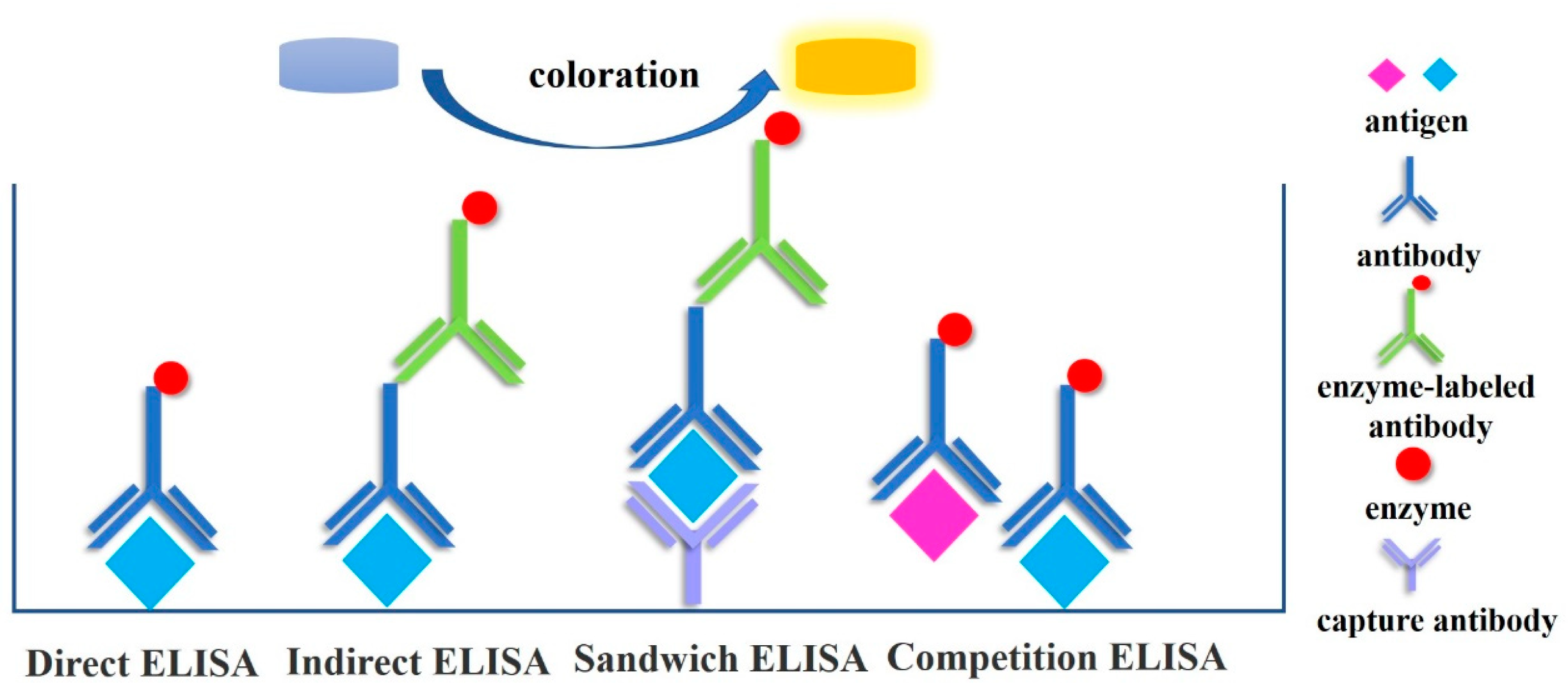
The direct method, double antibody sandwich method, indirect method, and competition method are typical ELISA detection techniques used for protein analysis of cultural relic materials [68] (Figure 2). The direct method involves adsorbing the antigen onto a solid-phase carrier, incubating it at an appropriate temperature and relative humidity, and then washing away any unbound antigens and impurities. To bind other unbound sites on the solid-phase carrier, a high concentration of nonspecific proteins and enzyme-labeled specific antibodies are added. At an optimal temperature and relative humidity, the antigen and antibody react completely, and the sample is then rinsed to remove unreacted antibodies. Finally, the chromogenic substrate is added, and within a certain amount of time, the enzyme-catalyzed colour develops. The experimental operation requirements for the labelling reaction of antibodies are high; each antigen detected by the direct method must be labelled with a specific antibody that interacts with it, resulting in a high cost of detection. Using the sandwich method, a known antibody is affixed to the surface of a solid-phase carrier, followed by the test sample. If the sample contains an antigen, specific binding will occur; then, an enzyme-labeled antibody is added to produce an antibody–antigen–enzyme-labeled antibody “sandwich” structure. After the addition of the substrate, the antigen in the sample is detected and analysed using a color-generating reaction. Double antibody sandwiching is a common technique for detecting macromolecular antigens [69]. However, when using the double antibody sandwich method to detect each antigen, the specific antibody that reflects that antigen must be labelled; when combined with indirect methods, each antigen requires two antibodies, making the double antibody sandwich method more expensive. The indirect technique can be used to detect antigens. The principle is to adsorb the antigen to be tested onto the solid phase, followed by the addition of specific antibodies and enzyme-labeled antibodies. Alternately, after the antigen has been adsorbed in the solid phase, the antibody to be examined and the enzyme-labeled antibody are successively added. In the indirect method, the colour intensity is directly proportional to the concentrations of the coated antigen and the primary antibody. The binding between the first and second antibodies is based on the principle that cross-reactions can occur between distinct subclasses of the same antibody. Therefore, specific antibodies can be applied directly to ELISA without being labelled, preserving the high activity of the specific antibody, reducing the number of experimental steps, and saving time and money. Consequently, indirect methods are the most popular. It is also the most popular method for detecting proteins in cultural relics [70]. The competition method is distinguished by the interaction between the antigen being tested and the enzyme-labeled antigen and the solid-phase antibody. The more the enzyme-labelled antigen binds to the solid-phase antibody, and the darker the colour, the lower the antigen concentration in the test sample. The intensity of the colour of the competitive solution is directly proportional to the antigen concentration in the mixed solution and inversely proportional to the antigen concentration on the solid carrier. This method is primarily used to determine small-molecule hapten [71,72,73].
4.2. Application of Early ELISA in Protein Detection of Cultural Relics Materials
Since the introduction of ELISA technology, it has been widely used for the detection of viruses, antibiotics, heavy metal ions, disease-related proteins, residues, etc., due to its high sensitivity, easy and rapid operation, the small amount of sample required, low cross-reactivity, and high specificity. The development of ELISA technology has also piqued the interest of archaeologists, who have started applying it to the detection of proteins in materials from cultural relics. Earlier studies on the application of ELISA in archaeology were numerous. In 1990, for instance, ELISA was used to detect albumin protein in ancient corpses and animal remains [74]. It was also used to determine that the pigment deposits of the Chumash Indians in the United States consisted of both animal and human blood. Cattabeo [75] utilised ELISA to analyse ancient human bone extracts in 1992. Albumin was discovered in 23 of the 31 skeletons, whereas only one skeleton contained IgG. Therefore, it has been demonstrated that albumin is a significantly superior target molecule for long-term survival in ancient bones. The results demonstrated that there is no cross-reaction between human and animal materials when using ELISA to detect albumin in bones, and that as little as 10 ng of protein is detectable, thereby expanding the possibilities for bone archaeology. In 2002, Schweitzer [76] used ELISA to identify immunologically active polypeptides in 100,000 to 300,000-year-old fossilised skulls. This demonstrates that ELISA is sensitive enough to detect proteins that are extremely aged.
4.3. ELISA Detection of Proteins in Cultural Relics Materials in a Complex Environment
With the ongoing development and enhancement of ELISA, a large number of research results for protein analysis and detection in materials from cultural relics have emerged, and the sensitivity and specificity of detection have improved continuously. In the protein processing method, ELISA employs the trypsin and other prior procedures of aged proteins so that the protein’s antigen determinant is exposed and can react with antibodies. This method detects ageing and denatured proteins in cultural relic materials without the need for a complex protein separation and purification procedure [45]. Protein components that must be detected in cultural relic materials frequently exist in complex environments and are difficult to separate; for instance, protein-cemented materials in mural layers are frequently mixed with inorganic materials, and cations of lead, copper, calcium, and iron are present in pigment layers. When amino acid analysis is performed using mass spectrometry, it will interfere with the derivatization process and alter the results [77]. In the presence of impurities, such as inorganic materials, active cations, and organic polymers, ELISA is superior to other detection methods for identifying proteins in cultural relic materials. In 2010, Palmieri et al. [78] analysed proteins in samples of cultural relics materials containing inorganic matrices using ELISA. After testing samples of 13th-century murals, it was determined that casein could still be detected in common mural substrates, such as carbonated stucco or gypsum, with a detection limit as low as 1 nanogram, indicating that these inorganic matrices will not affect the detection ability and sensitivity of ELISA. Similarly, the use of ELISA technology for determining the age of protein binders in pigments containing distinct metal ions will not affect the test results. In 2015, Lee et al. [77] modified the colour portrayal of Alkaline Phosphatase (AP), a commonly used enzyme in ELISA, to horseradish peroxidase (HRP), which is more sensitive than AP and has a greater magnification range. Animal cements containing pigments such as lead white and ultramarine are mined for proteins. The ELISA signal remains unaffected, and the protein type can still be identified. Similarly, a sample was extracted from a 12th- to 13th-century Peruvian feather robe. The ELISA results revealed that the sample’s adhesive was arabino-galactose gum, not animal glue as was previously believed. Type I collagen was detected in paper samples obtained from 19th-century to early 20th-century watercolour paintings. Ultimately, type I collagen and ovalbumin were discovered in tempera paintings on canvas from the 14th century. These results demonstrate that ELISA is capable of detecting various classes of proteins containing distinct metal ions.
4.4. ELISA Detection of Trace Samples
ELISA can detect and identify proteins in cultural relic materials that are stored in adverse environments, severely damaged, ageing, or even completely denatured, as well as those that are difficult to distinguish visually. It can also differentiate between protein types, determine their biological origin [79] and detect and identify even trace concentrations of proteins. Liu [80] used indirect ELISA and indirect competitive ELISA to detect and identify samples of wool and leather from cultural relics. Cowhide was identified as the variety of leather found in samples from dry regions of ancient Xinjiang. The ELISA indirect detection threshold for keratin was 10 ng/mL. Additionally, the collagen type I in the three ancient leather samples was identified, and the results of various proportions were obtained, thereby completing the species identification. Wu [81] laboured in 2017 to detect lacquer in samples of cultural relic material. Due to the complex composition of the remaining samples, which were contaminated and contained only trace quantities, the FTIR and Py-GC/MS results for lacquer phenol were insufficient. The glycoproteins in the dried lacquer films were analysed using ELISA, and the results were applied to eight samples collected from six remnants or ancient structures in different Chinese cities. This study’s detection limit reached 106 g/mL, and muddy samples were also analysed. In 2021, Weng [82] used ELISA to analyse samples of Chinese building mortars from 4300 years ago (approximately 2300 BC) to determine if they contained traces of commonly used binders, such as glutinous rice, tung oil, sugar, and animal adhesive. The results indicated that the samples contained components of animal adhesive. ELISA was able to detect protein components in old, exceedingly low-content mortars.
5. The Combination of ELISA and Other Technologies
There have been an increasing number of techniques in recent years to enhance ELISA’s detection effectiveness by fusing it with other technologies. In 2016, Liu [83] employed immunofluorescence microscopy (IFM) and ELISA to pinpoint the precise location of protein binders in the mural layer of the Xumi mountain caves and determine their composition. Animal glue and egg whites, two frequent sticky ingredients found in ancient Chinese paints, were found. IFM can characterise the structure distribution information of each layer of binder materials because it is a multi-layer structure that is challenging to mechanically peel off, and ELISA combined with IFM has high detection sensitivity. The protein components in it can still be detected and classified when the cemented material is mixed with the pigment.
Immune-labeled magnetic beads can bind protein more effectively and withstand interference from impurities when used in conjunction with ELISA. The magnetic beads are appropriate for use at archaeological sites and can be mass-produced as well. Zhang Wei [84] created an ELISA assay for the detection of silk protein in 2020 using immune-labeled magnetic beads. Silk protein was used to adsorb immune-labeled magnetic beads, and following adsorption, the silk protein was eluted for indirect ELISA testing. Testing of soil samples from the Qingtai site in Zhengzhou, which had evidence of probable silk breakdown, revealed the presence of silk protein there. Because immune-labeled magnetic beads have good durability, this approach offers higher sensitivity than standard ELISA. Its portability and strong anti-interference ability make it suited for trace protein identification in ancient sites. Table 2 provides a summary of the detection methods and outcomes for samples of historical proteins.
5.1. Characteristics of Enzyme-Linked Immunosorbent Assay
The minimal sample purity requirements, straightforward sample pre-treatment, limited number of experimental procedures, lack of need for complicated instruments, ease of operation, and low cost of ELISA all contribute to its high sensitivity and low sample volume requirement. Therefore, it is appropriate for the initial classification of different cultural relic elements at archaeological sites that contain proteins. It is also appropriate for the quick and simple identification of cultural heritage artefacts with mixed proteins since protein detection is selective and non-target proteins have less of an impact on target proteins. It can be widely employed by cultural and insurance sites with various situations because it has few criteria for experiment instruments and laboratory workers. Because it is impossible to distinguish between distinct species of animals belonging to the same genus, ELISA can only accurately identify the species source of proteins at the genus level. Additionally, because ELISA cannot detect many substances simultaneously, molecules with related structures may react with one another.
5.2. Combined Use of Proteomics and ELISA
Proteomics and ELISA have increasingly been used in research in recent years to analyse proteins. Proteomics approaches have high throughput and accuracy benefits, and the evaluated samples can be reused for ELISA testing. Low sample purity requirements, easy operation without complicated equipment, low cost, and little influence of non-target proteins on the results are all characteristics of ELISA. The composition and source of the sample can be correctly recognised by combining the benefits of the two analysis techniques (Table 3). In order to characterise the organic adhesives employed in painted wood panels from the 13th century, two techniques were applied in 2018: time of flight secondary ion mass spectrometry (ToF-SIMS) and Dot-ELISA [85]. While ELISA has great specificity and sensitivity and can perform a single identification of proteins in trace samples, ToF-SIMS offers high spatial resolution and high surface sensitivity. The outcomes of both techniques demonstrated the existence of a rabbit glue and milk mixture in the paint layer of the sample while ruling out the usage of eggs. Proteins can be qualitatively analysed and their species identified using immunoassay and proteomics techniques.
Sericin and silk fibroin were successfully identified as marker proteins in 2019 by Chen [13] using LC-MS/MS to identify silkworm cocoon protein. The heavy chain of the mulberry silk protein’s distinctive peptides were tested as haptens, while the tussah cocoon’s silk protein was used as a full antigen. Through animal immunology, specific antibodies to various silks were developed, and distinct species’ silk proteins were successfully identified. In 2021, Zheng et al. [86] created a technique for quick enrichment and detection of silk residues based on immune-labeled magnetic beads and ELISA. The detection threshold was at 5.12 ng/mL. After that, they employed LC-MS/MS to further validate the findings. The study demonstrates that the composition and source of silk residues may be precisely determined by combining immunological and proteomic approaches.
5.3. Expectation
Proteomics technology and ELISA have gradually been used to analyse the proteins in cultural relic materials in recent years. This has not only satiated the demands for low destructiveness, high resolution, high throughput, and high sensitivity, but also revealed information about the biological sources of proteins. To increase the effectiveness of protein extraction, proteomics technology must further optimise the extraction process. Additionally, it is impossible to compare many significant spectra with the database. To identify the iconic proteins and create a more comprehensive database of the proteins found in cultural relic materials, it is important to examine the proteins in additional samples of cultural relic materials. The accuracy and specificity of biological source identification will keep rising thanks to the ongoing development of bioinformatics, databases, and analysis software. The future of this field lies on combining conventional ELISA technology with new technologies for extraction and detection. For instance, ELISA with immune-labeled magnetic beads can be used to detect minute quantities of proteins. ELISA and other sensors can also be used to create biosensors, such as electrochemical immunosensors. These techniques enable successful qualitative and quantitative studies, they are simple to use, they can be automatically recognised, they can achieve higher sensitivity, and they can identify proteins in a larger range of materials used to create cultural relics. Additionally, because protein is reversible, has strong biocompatibility, and the non-changing morphology, content, and qualities of cultural relic materials, it has steadily been employed as a bioremediation material in the restoration and reinforcement of such materials. In the future, this could develop into an improved repair and protection strategy for cultural heritage items. Finally, the methods for analysing protein in materials from cultural relics will considerably improve with the advancement of protein analysis technology.
Author Contributions
Writing—original draft preparation, Q.W. writing—review and editing, B.Z. and Y.H. All authors have read and agreed to the published version of the manuscript.
Funding
This research was funded by [National Key R&D Program of China] grant number [No. 2022YFF0903802].
Institutional Review Board Statement
Not applicable.
Informed Consent Statement
Not applicable.
Data Availability Statement
No new data were created or analyzed in this study. Data sharing is not applicable to this article.
Conflicts of Interest
The authors declare no conflict of interest.
References
- Maria, G.G.; Roberto, M.; Enrica, P. Back to the past: Deciphering cultural heritage secrets by protein identification. Appl. Microbiol. Biotechnol. 2018, 102, 5445–5455. [Google Scholar]
- Xu, L. Study on the Comprehensive Analysis Method of Traditional Mortar Materials. Master’s Thesis, Zhejiang University, Hangzhou, China, 2018. [Google Scholar]
- Hu, W.J. Immunoassays Technology of Binding Media in Precious Ancient Polychrome Cultural Relics. Master’s Thesis, Zhejiang University, Hangzhou, China, 2016. [Google Scholar]
- Chen, H.F.; Gong, D.C.; Huang, W.C.; Liu, B. SDS-PAGE Analysis of Aging Characteristics of Liao Dynasty Silk Unearthed in Yemaotai, Faku, Liaoning. Sci. Conserv. Archaeol. 2010, 22, 9–13. [Google Scholar]
- Wang, K.; Hu, D.B. Hydroxyapatite: Collagen Biomimetic Composite Material in Conservation of Tortoise Shell Relics. JNMCH 2013, 3, 141–152. [Google Scholar]
- Zhang, Y.; Chen, Z.; Liu, X.; Shi, J.; Chen, H.; Gong, Y. SEM, FTIR and DSC Investigation of Collagen Hydrolysate Treated Degraded Leather. J. Cult. Herit. 2021, 48, 205–210. [Google Scholar] [CrossRef]
- Pasian, C.; Secco, M.; Piqué, F.; Artioli, G.; Rickerby, S.; Cather, S. Lime-based injection grouts with reduced water content: An assessment of the effects of the water-reducing agents ovalbumin and ethanol on the mineralogical evolution and properties of grouts. J. Cult. Herit. 2018, 30, 70–80. [Google Scholar]
- Wang, Z.Y. Proteomics Analysis and Ultra-Sensitive Detection of Archaeological Hairs. Master’s Thesis, Zhejiang Sci-Tec University, Hangzhou, China, 2021. [Google Scholar]
- Wang, J.; Li, J.; Chao, X.; Chen, Y.; Huang, Y.; Mai, B.; Li, Y.; Cao, J. The influence of the photothermal aging exerting on the GC-MS identification of the common proteinaceous binding media in polychromy artworks. JNWU 2020, 50, 595–605. [Google Scholar]
- Liu, L.Y. Multi-Analytical Approach to the Study of Pigments and Binding Media in Precious Ancient Polychrome Cultural Relics. Master’s Thesis, Zhejiang University, Hangzhou, China, 2017. [Google Scholar]
- Abelson, P.H. Paleobiochemistry: Organic Constituents of Fossils; Yearbook, No. 531954; Carnegie Institution of Washington: Washington, DC, USA; pp. 97–101.
- Li, J.J.; Zhang, B.J. Research status of binders in ancient painted cultural relics—Based on data analysis of research papers from the Web of Science. Sci. Conserv. Archaeol. 2019, 31, 119–129. [Google Scholar]
- Chen, R.R. Characterization of Ancient Silk Fabrics Based on Proteomics and Immunological Techniques. Master’s Thesis, Zhejiang Sci-Tec University, Hangzhou, China, 2020. [Google Scholar]
- He, Q.J. Study on the Role of Sizing Agent in Ancient Calligraphy & Painting and the Development of Neutral Aluminum Salt Sizing Precipitator. Ph.D. Thesis, Northwest University, Xi’an, China, 2019. [Google Scholar]
- Chi, H.H. Artificial Aging and Consolidation of Silk Painting Colored by Mineral Pigment. Ph.D. Thesis, Zhejiang Sci-Tec University, Hangzhou, China, 2020. [Google Scholar]
- He, Q.J.; Wang, L.Q. Study on application of modern instruments analysis in identification of painting relics binding medium. China Adhes. 2007, 19, 19–22. [Google Scholar]
- Deng, Z.M.; Jiang, Z.H.; Qin, D.C. Evaluation on the moisture-proof performance of bamboo coated by lacquer in hot and humid environment. China For. Prod. Ind. 2015, 42, 14–16. [Google Scholar]
- Wang, C.; Qi, L.X.; Yang, J.; Zhou, W.; Dang, X. Status of research on the conservation of bone and antler relics. Sci. Conserv. Archaeol. 2016, 28, 118–125. [Google Scholar]
- Wang, J. Research on the Phenomenon of Decline in the Solubility of Silk Protein in Silk Cultural Relics and Its Solution. Master’s Thesis, Shandong University, Jinan, China, 2021. [Google Scholar]
- Zhang, B.J.; Hu, W.J.; Zhang, K. Research on the Egg White in Ancient Tabia Mortar by Enzyme-Linked Immunosorbent Assay (ELISA). J. Build. Mater. 2015, 18, 716–720. [Google Scholar]
- Wilson, J.; van Doorn, N.L.; Collins, M.J. Assessing the extent of bone degradation using glutamine deamidation in collagen. Anal. Chem. 2012, 84, 9041–9048. [Google Scholar] [CrossRef]
- Solazzo, C.; Wilson, J.; Dyer, J.M.; Clerens, S.; Plowman, J.E.; Von Holstein, I.; Collins, M.J.; Walton Rogers, P.; Peacock, E.E. Modeling deamidation in sheep α-keratin peptides and application to archeological wool textiles. Anal. Chem. 2014, 86, 567–575. [Google Scholar] [CrossRef] [PubMed]
- Xie, J. Thermal aging Mechanism and Soften Technology of Leather. Master’s Thesis, Zhejiang Sci-Tec University, Hangzhou, China, 2016. [Google Scholar]
- Zhang, M. Study on Collagen Extraction Methods from Degraded Leather Cultural Relics. Master’s Thesis, Shandong University, Jinan, China, 2021. [Google Scholar]
- Pankin, D.; Povolotckaia, А.; Borisov, E.; Rongonen, S.; Mikhailova, А.; Tkachenko, T.; Dovedova, N.; Rylkova, L.; Kurochkin, A. Investigation of wafers used as paper binding in the academician von Struve manuscripts. J. Cult. Herit. 2021, 51, 125–131. [Google Scholar] [CrossRef]
- Zhao, L. Tracing the animal origin of organic cultural relics in the Palace Museum through mitochondrial DNA technology. China Cult. Herit. Sci. Res. 2019, 3, 75–82. (In Chinese) [Google Scholar]
- Paolo, C.; Antonella, C. Enzymes as tools for conservation of works of art. J. Cult. Herit. 2021, 50, 73–87. [Google Scholar]
- Banks, P.N. Paper Cleaning. Restaur. Int. J. Preserv. Libr. Arch. Mater. 1969, 1, 52–66. [Google Scholar] [CrossRef]
- Frantisek, M. Enzymatic consolidation of a painting: Seventeenth century landscape from skokloster palace. Stud. Conserv. 1982, 27, 135–138. [Google Scholar]
- Paolo, A.; Giacomo, Z.; Pamela, A. Art-loving bugs: The resurrection of Spinello Aretino from Pisa’s cemetery. Proteomics 2005, 5, 2453–2459. [Google Scholar]
- Zhou, Y. The Discovery of Silk in the Ritual Pit of Sanxingdui and its Significance. Chin. LH 2021, 12, 37–48. [Google Scholar]
- Su, B.M.; Zhang, A.M.; Hu, Z.M.; Li, Z.X. Application of Chromatographic Method in the Analysis of Cementitious Materials in Ancient Paintings. Dunhuang Res. 2000, 1, 82–86. [Google Scholar]
- Chen, D.M.; Si, C.D.; Long, S.J. Research Progress of Spectral Technologies of Binging Media Used in Paintings. Spectrosc. Spect. Anal. 2020, 40, 961–966. [Google Scholar]
- Zhao, Q.; Zhang, L.H.; Zhang, Y.K. Recent Advances in Proteomics. Chin. J. Appl. Chem. 2018, 35, 977–983. [Google Scholar]
- Candiano, G.; Bruschi, M.; Musante, L.; Santucci, L.; Ghiggeri, G.M.; Carnemolla, B.; Orecchia, P.; Zardi, L.; Righetti, G. Blue silver: A very sensitive colloidal Coomassie G-250 staining for proteome analysis. Electrophoresis 2004, 25, 1327–1333. [Google Scholar] [CrossRef]
- Jennifer, A.; Milana, P.; Ei, W.W. Comparison of protein expression levels and proteomically-inferred genotypes using human hair from different body sites. Forensic. Sci. Int.-Gen. 2019, 41, 19–23. [Google Scholar]
- Xin, P.; Kuang, H.-X.; Li, X.-L.; Wang, Y.; Zhang, B.-M.; Bu, H.; Wang, Z.-B.; Meng, Y.-H.; Wang, Y.-H.; Wang, Q.-H. Proteomics and its application to determine mechanism of action of traditional chinese medicine. China J. Chin. Mater. Med. 2018, 43, 904–912. [Google Scholar]
- Yang, Q.; Wang, D.; Chang, L.L.; Sun, Y.; Jin, X.; Yang, X. Progress in Mass Spectrometry and its Application in Proteomics. Chin. Agric. Sci. Bull. 2015, 31, 239–246. [Google Scholar]
- Gong, P.T.; Xu, R.S.; Fang, X.J. Proteogenomics: Progress, Strategy and Problem. Genom. Appl. Biol. 2014, 33, 1169–1180. [Google Scholar]
- Li, N.; Wu, S.-F.; Zhu, Y.-P.; Yang, X.-M. The Progress of Protein Quality Control Methods in Shotgun Proteomics. Prog. Biochem. Biophys. 2009, 36, 668–675. [Google Scholar] [CrossRef]
- Zhao, J.; Wang, H.W.; Tian, E.J. Proteomics Experiment Technologies and Their Applications. Prog. Vet. Med. 2015, 36, 116–120. [Google Scholar]
- Yu, Z.N.; Zhang, Y.; Huang, X.W. Application of proteomics in diagnosis and pathogenesis of genetic metabolic diseases. Chin. J. Lab. Med. 2022, 45, 300–304. [Google Scholar]
- Zhang, W. Progress in Mass Spectrometry Acquisition Approach for Quantitative Proteomics. Chin. J. Anal. Chem. 2014, 42, 1859–1868. [Google Scholar] [CrossRef]
- Radovan, H.; Stepanka, K.; Janka, H. Matrix-assisted laser desorption/ionization time-of-flight mass spectrometry as a tool for fast identification of protein binders in color layers of paintings. RCM 2004, 18, 1896–1900. [Google Scholar]
- Proietti, N.; Roselli, G.; Capitani, D.; Pettinari, C.; Pucciarelli, S.; Basileo, S.; Scognamiglio, F. Characterization of handmade papers (13th–15th century) from Camerino and Fabriano (Marche, Italy). J. Cult. Herit. 2020, 42, 8–18. [Google Scholar] [CrossRef]
- Tokarski, C.; Martin, E.; Rolando, C.; Cren-Olivé, C. Identification of proteins in renaissance paintings by proteomics. Anal. Chem. 2006, 78, 1494–1502. [Google Scholar] [CrossRef]
- Leo, G.; Cartechini, L.; Pucci, P.; Sgamellotti, A.; Marino, G.; Birolo, L. Proteomic strategies for the identification of proteinaceous binders in paintings. Anal. Bioanal. Chem. 2009, 395, 2269–2280. [Google Scholar] [CrossRef] [PubMed] [Green Version]
- Calvano, C.D.; Rigante, E.C.; Cataldi, T.R.; Sabbatini, L. In situ hydrogel extraction with dual-enzyme digestion of proteinaceous binders: The key for reliable mass spectrometry investigations of artworks. Anal. Chem. 2020, 92, 10257–10261. [Google Scholar] [CrossRef] [PubMed]
- Zilberstein, G.; Maor, U.; Baskin, E.; D’Amato, A.; Righetti, P.G. Unearthing Bulgakov’s trace proteome from the Master i Margarita manuscript. J. Proteom. 2017, 152, 102–108. [Google Scholar] [CrossRef]
- Castellani, L.; Ferrantelli, P.; Sinibaldi, M.; Vigliano, G. Identification of proteinaceous adhesives in the wooden backing of Piero della Francesca’s painting Pala of Saint Bernardino: A gas chromatographic study. J. Cult. Herit. 2001, 2, 209–215. [Google Scholar] [CrossRef]
- Chirco, G.; Portale, E.C.; Caponetti, E.; Renda, V.; Martino, D.F.C. Investigation on four centuripe vases (late 3rd-2nd cent. B.C.) by portable X-ray fluorescence and total reflectance-FTIR. J. Cult. Herit. 2021, 48, 326–335. [Google Scholar] [CrossRef]
- Gomaa, A.M.; Mostafa, A.H.; Hany, A. Analytical techniques used for condition assessment of a late period mummy. J. Cult. Herit. 2005, 48, 83–92. [Google Scholar]
- Edwards; Howell, G.M.; Chalmers, J.M. Raman spectroscopy in archaeology and art history. J. Raman Spectrosc. 2004, 35, 607. [Google Scholar]
- Ying, C.F.; Qing, X.; Shu, Z. Characterization and quantitation study of ancient lacquer objects by NIR spectroscopy and THM-Py-GC/MS. J. Cult. Herit. 2020, 46, 95–101. [Google Scholar]
- Lu, R.; Kamiya, Y.; Miyakoshi, T. Applied analysis of lacquer films based on pyrolysis-gas chromatography/mass spectrometry. Talanta 2006, 70, 370–376. [Google Scholar] [CrossRef]
- Ji, M.C.; Fu, B.; Zhang, Y.J. Recent Progress of Analytical Methods of Proteomics Based on Mass Spectrometry. J Chin. Mass Spectrom. Soc. 2021, 42, 862–877. [Google Scholar]
- Yan, X.Y.; Gao, J.Z. Application of MALDI-TOF MS in early diagnosis of non-small cell lung cancer. J. Med. Theory Pract. 2022, 35, 1478–1481. [Google Scholar]
- Wang, Y. Quantitative Analysis of Three Kings of Mycotoxins in Distillers Dried Grains with Soluble by Ultral-High Performance Liquid Chromatography-Tandem Mass Spectrometry. Master’s Thesis, Jilin University, Jilin, China, 2018. [Google Scholar]
- Li, J.; Ren, Q.L.; Pan, Y.J. Mass Spectrometric Studies on the Addition Reaction of HCCA and Sulfhydryl-Containing Living Substance in MALDI-TOF MS Analysis. J Chin. Mass. Spectrom. Soc. 2022, 43, 133–141. [Google Scholar]
- Stepanka, K.; Radovan, H.; Milan, K. Application of peptide mass mapping on proteins in historical mortars. J. Cult. Herit. 2009, 10, 244–247. [Google Scholar]
- Taniguchi, Y.; Kawahara, K.; Takashima, M.; Cotte, M.; Mazurek, J.; Kumazawa, Y.; Taga, Y.; Nakazawa, T. Organic Materials Used for Giant Buddhas and Wall Paintings in Bamiyan, Afghanistan. Appl. Sci. 2022, 12, 9476. [Google Scholar] [CrossRef]
- Kuckova, S.; Hynek, R.; Kodicek, M. Identification of proteinaceous binders used in artworks by MALDI-TOF mass spectrometry. Anal. Bioanal. Chem. 2007, 388, 201–206. [Google Scholar] [CrossRef]
- Tripković, T.; Charvy, C.; Alves, S.; Lolić, A.; Baošić, R.; Nikolić-Mandić, S.; Tabet, J. Electrospray ionization linear trap quadrupole Orbitrap in analysis of old tempera paintings: Application to nineteenth-century Orthodox icons. Eur. J. Mass. Spectrom. 2015, 21, 679–692. [Google Scholar] [CrossRef]
- Sophie, D.; Monika, K.; Nicolas, G. Identification of animal glue species in artworks using proteomics: Application to a 18th century gilt sample. Anal. Chem. 2011, 83, 9431–9437. [Google Scholar]
- Azémard, C.; Zazzo, A.; Marie, A.; Lepetz, S.; Debaine-Francfort, C.; Idriss, A.; Zirah, S. Animal fibre use in the Keriya valley (Xinjiang, China) during the Bronze and Iron Ages: A proteomic approach. J. Archaeol. Sci. 2019, 110, 104996. [Google Scholar] [CrossRef]
- Yaroslav, D.; Timur, K.; Gavrilenko, L.; Solovyev, N. Targeted proteomics for the analysis of cultural heritage: Application of broadband collision-induced dissociation mass spectrometry. Anal. Bioanal. Chem. 2022, 414, 1723–1737. [Google Scholar]
- Eva, E.; Karin, J.; Peter, P. Enzyme-linked immunosorbent assay. II. Quantitative assay of protein antigen, immunoglobulin G, by means of enzyme-labelled antigen and antibody-coated tubes. BBA-Protein Struct. 1971, 251, 427–434. [Google Scholar]
- Pang, B.; Cheng, J.; Chen, Z. Comparision of Three Methods of Enzyme linked Immunosorbent Assay with A Monoclonal Antibody. Chin. J. Biol. Control 1999, 1, 32–35. [Google Scholar]
- Duan, X.; Huang, X.; Huang, L.F.; Wei, H.; Lai, W.H. Detection of Listeria monocytogenes in Food by Sandwich ELISA. Food Sci. 2010, 31, 272–276. [Google Scholar]
- Liu, J.Y.; Li, X.C.; Chen, D.K.; Zhang, Y.X.; Chen, J.; Wu, F.X. Establishment of indirect ELISA for detection of serum antibodies against canine parvovirus disease. China AHS 2006, 3, 27–29. [Google Scholar]
- Liu, S.Z.; Feng, D.H.; Qian, C.F.; Sergei, A.E. The Residue of Carbaryl in Brassica chinensis and Apple Determined by Method of Antibody Immobilized Direct Competitive ELISA. Chin. J. Pestic. Sci. 2001, 3, 69–73. [Google Scholar]
- Doreen, W.L.; Mohamed, S.; Abdel, R. Alterations in Rat Liver Microsomal Enzymes Following Exposure to Carbaryl and Malathion in Combination. Arch. Environ. Contam. Toxicol. 1985, 14, 451–457. [Google Scholar]
- Bu, G.H.; Zheng, Z.; Zheng, H. Amethod for detecting β-lactoglobulin in cowmilk allergens by indirect competitive enzyme-linked immunosorbent assay. J. China Agric. Univ. 2008, 6, 71–76. [Google Scholar]
- Cattaneo, C.; Gelsthorpe, K.; Phillips, P.; Sokol, R.J.; Smillie, D. Identification of ancient blood and tissue—ELISA and DNA analysis. Antiquity 1991, 65, 878–881. [Google Scholar] [CrossRef]
- Cattaneo, C.; Gelsthorpe, K.; Phillips, P.; Sokol, R.J. Detection of blood proteins in ancient human bone using ELISA: A comparative study of the survival of IgG and albumin. Int. J. Osteoarchaeol. 1992, 2, 103–107. [Google Scholar] [CrossRef]
- Schweitzer, M.; Hill, C.L.; Asara, J.M.; Lane, W.S.; Pincus, S.H. Identification of Immunoreactive Material in Mammoth Fossils. J. Mol. Evol. 2002, 55, 696–705. [Google Scholar] [CrossRef] [PubMed] [Green Version]
- Lee, H.Y.; Atlasevich, N.; Granzotto, C.; Schultz, J.; Loike, J.; Arslanoglu, J. Development and application of an ELISA method for the analysis of protein-based binding media of artworks. Anal. Methods 2015, 7, 187–196. [Google Scholar] [CrossRef]
- Palmieri, M.; Vagnini, M.; Pitzurra, L.; Rocchi, P.; Brunetti, B.G.; Sgamellotti, A.; Cartechini, L. Development of an analytical protocol for a fast, sensitive and specific protein recognition in paintings by enzyme-linked immunosorbent assay (ELISA). Anal. Bioanal. Chem. 2011, 399, 3011–3023. [Google Scholar] [CrossRef]
- Hu, W.; Zhang, H.; Zhang, B. Identification of organic binders in ancient Chinese paintings by immunological techniques. Microsc. Microanal. 2015, 21, 1278–1287. [Google Scholar] [CrossRef]
- Liu, Y. Micro-Trace-Identification of Ancient Proteinaceou Relics Based on Enzyme-Linked Immunosorbent Assay. Master’s Thesis, Zhejiang Sci-Tec University, Hangzhou, China, 2017. [Google Scholar]
- Wu, M.; Zhang, B.; Sun, G.; Jiang, L. Determination of lacquer contained in samples of cultural relics by enzyme-linked immunosorbent assay. New J. Chem. 2017, 41, 6226–6231. [Google Scholar] [CrossRef]
- Xin, W.; Ming, Z.M.; Bing, J.Z. Detection of early Chinese organic-inorganic composite lime surface from the Lushanmao site, 4300 years ago. J. Cult. Herit. 2021, 52, 128–133. [Google Scholar]
- Liu, L.; Shen, W.; Zhang, B.J. Determination of Proteinaceous Binders for Polychrome Relics of Xumi Mountain Grottoes by Using Enzyme-Linked Immunosorbent Assay and Immunofluorescence Microscopy. Int. J. Conserv. Sci. 2016, 7, 3–14. [Google Scholar]
- Zhang, W. Development of Immunomagctic Bead Double Antibody Sandwich ELISA for the Detection of Trace Fibroin. Master’s Thesis, China Jiliang University, Hangzhou, China, 2020. [Google Scholar]
- Andrea, A.; Francesca, B.; Mariangela, P. Characterization of organic binders in a 13th century painted wooden panel: Comparison of ToF-SIMS and Dot-ELISA results. Int. J. Mass. Spectrom. 2018, 430, 63–68. [Google Scholar]
- Zheng, H.; Yang, H.; Zhou, Y.; Li, T.; Ma, Q.; Wang, B.; Fang, Q.; Chen, H. Rapid Enrichment and Detection of Silk Residues from Tombs by Double-Antibody Sandwich ELISA Based on Immunomagnetic Beads. Anal. Chem. 2021, 93, 14440–14447. [Google Scholar] [CrossRef] [PubMed]
Figure 1.
Main processes of proteomics.

Figure 2.
Common detection methods of ELISA.


Table 1.
Types of cultural heritage artifacts and proteins contained therein.
| Cultural Heritage Material Composition | Cultural Heritage Category | Protein Containing Part | Protein Type |
|---|---|---|---|
| Organic | Paper | Glue in paper or silk | Collagen; Soybean protein |
| Textile; Fur | Silk products; Wool fabric | Silk protein; Keratin | |
| Lacquerware; Woodware; Bambooware | Lacquer artifacts | Glycoprotein | |
| Leather | Leather products; Parchment | Collagen | |
| Carcass | Mummified corpse; Wax corpse; Wet corpse; | Collagen | |
| Bone; antler | Animal bones, teeth, and horns | Collagen | |
| Inorganic | Ceramics; bricks | Pottery adhesive | Collagen; Ovalbumin; Casein |
| Painted murals | Colored drawing; Mortar | Collagen; Ovalbumin; Casein |
Table 2.
Detection techniques and results of ancient protein samples.
| Sample | Country of Sample | Time | Detection Method | Detected Protein | |
|---|---|---|---|---|---|
| Proteomics | Cementitious Materials in Renaissance Art Painting [46] | Italy | 2006 | MALDI-TOF, nano-LC, nano-ESI, Q-q-TOF-MS, MS | Ovalbumin |
| 17th Century Painings [62] | Norway | 2007 | MALDI-TOF MS | Egg protein | |
| Cathedral dome painting binder [47] | Italy | 2009 | nano-LC-MS/MS; ESI | Casein | |
| Mortar sample for Romanesque rotunda [60] | Czech Republic | 2009 | MALDI-TOF-MS | Casein; Collagen; Egg protein | |
| 18th century church binder [64] | France | 2011 | FTICR-MS, nano-LC, nano-ESI, HR MS, MS/MS | Bovine collagen | |
| Orthodox paintings [63] | Serbia | 2015 | LTQ-Orbitra, MALDI-TOF MS/MS | Yolk protein | |
| The manuscript of the famous writer Mikhail Bulgakov [49] | Russia | 2017 | LC-MS/MS | Periostein; acetyl-β-glucosaminidase; Norepinephrine | |
| Ancient fibers [65] | China | 2019 | ESI-Q-TOF, UHPLC-MS, LC-MS/MS | Goat keratin; Sheep keratin; Sovine keratin | |
| Handmade Papers (13th–15th century) [45] | Italy | 2020 | SDS-PAGE | Collagen | |
| Handmade Papers (13th–15th century) [45] | Italy | 2020 | SDS-PAGE | Collagen | |
| Painted statues [48] | Italy | 2020 | MALDI-TOF/TOF | Casein; Rabbit collagen; Egg yolk immunoglobulin | |
| 19th-century buildings [66] | Russia | 2021 | HPLC–MS, nano-LC–MS | Collagen | |
| Wall Paintings [61] | Afghanistan | 2022 | ESI-MS/MS | Type I collagen Type III collagen | |
| ELISA | 13th Century Murals [78] | Italy | 2010 | ELISA | Casein |
| Peruvian feather robe from the 12th–13th centuries [77] | Peru | 2015 | ELISA | Arabino-galactose gum | |
| Watercolor paintings from the 19th century to the early 20th century [76] | America | 2015 | ELISA | Type I collagen | |
| 14th-century tempera paintings [77] | Italy | 2015 | ELISA | Type I collagen; Ovalbumin | |
| Wool and leather [80] | China | 2016 | ELISA | Type I collagen; Keratin | |
| Murals binders [83] | China | 2016 | ELISA, IFM | Egg protein; Collagen | |
| Dried lacquer films [81] | China | 2017 | ELISA | Glycoproteins | |
| Soil samples [84] | China | 2020 | IMB, ELISA | Silk protein | |
| Building mortars 4300 years ago [82] | China | 2021 | ELISA | Collagen | |
| Combination of the two methods | Painted wood panels from the 13th century [85] | China | 2018 | ToF-SIMS, Dot-ELISA | Casein; Rabbit collagen; Egg protein |
| Silks [13] | China | 2019 | LC–MS/MS, ELISA | Sericin; Silk fibroin | |
| Silk residues [86] | China | 2021 | IMB, ELISA, LC−MS/MS | Silk protein |
Table 3.
Comparison of characteristics of two detection methods.
| Proteomics | ELISA | |
|---|---|---|
| Sample weight | μg, non-destructive sampling | mg |
| Pre-treatment process | complex | simple |
| Number of samples for single test | more | less |
| Instrument | precision | simple |
| Operating procedure | complex | simple |
| Inspection cost | high cost | low cost |
| Sample recovery | recyclable | non-recyclable |
| Limit of detection | 1015 (mol/L) | 109 (mol/L) |
| Species identification precision | species | genus |
| Whether or not false negative | yes | no |
Disclaimer/Publisher’s Note: The statements, opinions and data contained in all publications are solely those of the individual author(s) and contributor(s) and not of MDPI and/or the editor(s). MDPI and/or the editor(s) disclaim responsibility for any injury to people or property resulting from any ideas, methods, instructions or products referred to in the content. |
© 2023 by the authors. Licensee MDPI, Basel, Switzerland. This article is an open access article distributed under the terms and conditions of the Creative Commons Attribution (CC BY) license (https://creativecommons.org/licenses/by/4.0/).
Share and Cite
MDPI and ACS Style
Wu, Q.; Zhang, B.; Hu, Y. Comparison and Research Progress of Protein Detection Technology for Cultural Relic Materials. Coatings 2023, 13, 1319. https://doi.org/10.3390/coatings13081319
AMA Style
Wu Q, Zhang B, Hu Y. Comparison and Research Progress of Protein Detection Technology for Cultural Relic Materials. Coatings. 2023; 13(8):1319. https://doi.org/10.3390/coatings13081319
Chicago/Turabian StyleWu, Qian, Bingjian Zhang, and Yulan Hu. 2023. "Comparison and Research Progress of Protein Detection Technology for Cultural Relic Materials" Coatings 13, no. 8: 1319. https://doi.org/10.3390/coatings13081319
Note that from the first issue of 2016, this journal uses article numbers instead of page numbers. See further details here.