
Microstructure and Properties of MAO-Cu/Cu-(HEA)N Composite Coatings on Titanium Alloy
1
Shaanxi Office of Science, Technology and Industry for National Defense, Xi’an 710061, China
2
Xi’an Saitesimai Titanium Industry Co., Ltd., Xi’an 710201, China
3
School of Materials Science and Chemical Engineering, Xi’an Technological University, Xi’an 710032, China
*
Author to whom correspondence should be addressed.
Coatings 2022, 12(12), 1877; https://doi.org/10.3390/coatings12121877
Submission received: 4 October 2022
/
Revised: 8 November 2022
/
Accepted: 11 November 2022
/
Published: 3 December 2022
(This article belongs to the Special Issue Advances in Thin Film Fabrication by Magnetron Sputtering)
Abstract
:In this paper, MAO-Cu/Cu-(HEA)N composite coatings on TC4 titanium alloy were prepared by combining micro arc oxidation (MAO) with magnetron sputtering (MS) to enhance the wear resistance and antibacterial ability of the substrate in simulated seawater. The number of micropores on the surface of the composite coatings decreased with increasing CuSO4 concentration in the electrolyte, causing the surfaces to be flat and smooth. XPS and EDS analyses revealed that the MAO-Cu/Cu-(HEA)N composite coatings predominately contained TiO2, Cu2O, and (HEA)N. Moreover, the addition of CuSO4 increased the growth rate of the MAO coatings. Comparatively, the MAO-Cu/Cu-(HEA)N composite coating with 5 g/L CuSO4 showed superior wear resistance, reduced friction coefficient (approximately 0.2), and shallow and narrow grinding cracks were observed compared to the other coatings. Antibacterial experiments showed that the MAO-Cu/Cu-(HEA)N composite coatings had better bacterial killing effects than the TC4 substrate, which is of great significance to the antifouling abilities of titanium alloys in marine applications.
1. Introduction
Titanium alloys are widely used in the marine and biomedical field owing to their light weights, high specific strengths, and many other advantages [1,2,3,4,5,6]. Titanium alloys are extensively used as biomedical materials in various artificial joints, bone fixings, dental implants, heart stents, etc., [5,6,7,8]. The use of titanium alloys in the marine industry can effectively reduce the weight of equipment, thus improving their payloads, which ultimately improves their reliability and reduces their maintenance costs. They are therefore ideal materials for marine equipment. However, titanium alloys face extremely harsh service conditions in marine environments, such as abrasion resulting from the erosion and collision of seawater and stones, corrosion of other alloy parts after connection, and adhesion and fouling of marine bacteria and microorganisms. Although titanium alloys exhibit excellent corrosion resistance when used as marine materials, they have poor wear resistance compared with stainless steel and other alloys [6,9,10]. In addition, the loss of titanium alloys from biological fouling is also a problem requiring urgent solutions. Micro arc oxidation (MAO) is a promising surface engineering technology which is used for the surface modification of various metals [11,12,13,14,15,16,17]. Some studies have shown that metallurgically bonded MAO layers on titanium alloy substrates can effectively improve the corrosion resistance, wear resistance, and hardness of the alloy [18,19,20]. It has also been reported that the addition of various soluble chemicals into the electrolyte used for the fabrication of MAO coatings can modify the structure of the coatings, which in turn affects the performance of titanium substrates [9,21,22,23]. The addition of particles into the electrolyte can also affect the discharge channel in the MAO process, change the structures and surface morphologies of the films, and affect the protective performances of titanium alloys [24]. However, the porous oxide coating formed on the surface of titanium alloys provides more possibilities for the adhesion of bacteria and microorganisms due to its usually large surface area. The addition of antibacterial elements into the coating is one of the methods adopted to improve the antibacterial properties of the alloy [25]. Noting that Cu is a common antibacterial element [26,27,28], V. Stranak et al. [29] found that the Ti-Cu films prepared by the dual-HiPIMS technique had good antibacterial ability against Staphylococcus epidermidis and Staphylococcus aureus. Zhang et al. [30] found that Cu exists in the form of Cu2O and CuO for MAO coating doped with Cu. The addition of Cu significantly improved the antifouling performance of the studied metal substrate. Despite having excellent antibacterial properties, Cu has relatively poor corrosion and wear resistance [31,32]. Therefore, using a single Cu film as a protective coating for titanium alloy in marine environments will pose some protective limitations. Recently, high-entropy alloy nitride ((HEA)N) films have gained popularity due to their high hardness, improved oxidation and corrosion resistance, and high temperature stability [33,34], and a comprehensive literature survey proves that there is scant information existing for the addition of Cu to (HEA)N films.
In this paper, the morphology and structure of MAO coatings were adjusted by adding different concentrations of CuSO4 in a phosphate system, followed by the deposition of Cu and high-entropy alloy nitrides (Cu-(HEA)N) film by magnetron sputtering. Cu and (HEA)N films were deposited on the surface of the MAO layer to provide a feasible approach for fabricating anti-wear and antibacterial composite coatings. The MAO-Cu/Cu-(HEA)N composite coating prepared by this method is expected to improve the wider application of titanium alloys in the marine field.
2. Experimental
2.1. Preparation of MAO-Cu/Cu-(HEA)N Composite Coatings
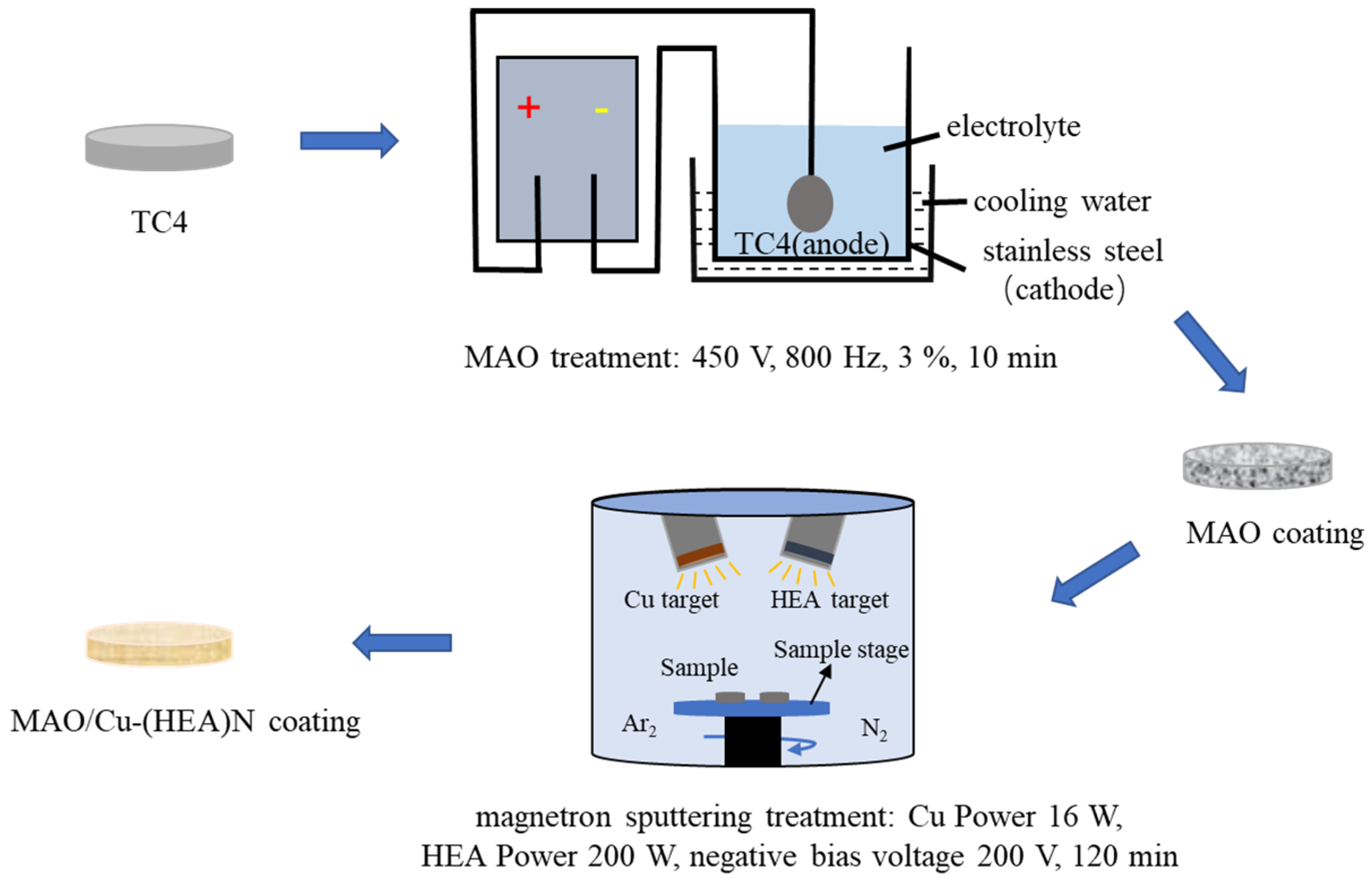
TC4 titanium alloy (Baoji Titanium Industry Co., Ltd, Xi’an, China) circular disk samples with dimension of ϕ 20 mm × 4 mm were used as the substrate material in the experiments. The chemical composition of TC4 is displayed in Table 1. The TC4 substrates were polished by SiC sandpaper from 200# to 2000#, ultrasonically cleaned with alcohol for 10 min to remove surface stains, and dried by a blower. The MAO equipment was independently developed by Xi’an University of technology, with TC4 substrates as anode and stainless-steel barrel as cathode. In the micro-arc oxidation process, DC pulsed constant current mode is used. The basic electrolyte contained sodium hexametaphosphate ((NaP2O5)6, 20 g/L), potassium hydroxide (KOH, 4 g/L), sodium tungstate (Na2WO4·2H2O, 3 g/L) and potassium fluoride (KF·2H2O, 3 g/L). Varying concentrations of CuSO4 solution (5, 10, and 15 g/L) were added to the base electrolyte with a MAO process of 10 min at constant voltage of 450 V, frequency of 800 Hz, and a duty cycle of 3%.
The MAO coatings with different CuSO4 concentrations were ultrasonically cleaned with alcohol for 10 min to remove the electrolyte on the surface of the coatings. Copper alloy target and AlSiTiCrNbV high entropy alloy target prepared by an arc melting furnace were cut into 50 mm × 2 mm disc. Cu-(HEA)N films with thickness of 1 µm were deposited on the surface of the MAO coatings by a double target magnetron sputtering deposition system. Pretreatment of the substrate cavities was carried out before deposition. The surface impurities on the sleeve of the fixed target were removed by a sandblasting machine (Taseken Trading Co., Shanghai, China) and the substrates were ultrasonically cleaned with acetone and alcohol at room temperature for 10 min. All the substrates were wiped clean with dry non-woven cloths, and residual surface impurities or oxides were removed by setting a negative bias of 400 V for 20 min before deposition. During the deposition, the powers of the copper alloy target and AlSiTiCrNbV high entropy alloy target were 20 W and 200 W, respectively. The negative bias voltage was 200 V and deposition time was 120 min. The preparation process of the MAO-Cu/Cu-(HEA)N composite coatings are shown in Figure 1.
2.2. Coating Characterization
The surface morphologies and elemental compositions of the coatings were analyzed by VEGA3-SBH scanning electron microscope (Taseken Trading Co., Shanghai, China) and energy dispersive spectrometer with a working voltage of 10 KV and a working distance of 3 mm (Taseken Trading Co., Shanghai, China). The composition and chemical states of coatings were analyzed by XPS (Shimadzu-Kratos Co., Hadano, Japan). The thicknesses of the MAO coatings were measured with a coating thickness gauge, and the average thickness after fifteen random measurements at different points was taken as the final thickness value. The friction coefficients of the coatings in simulated seawater were obtained using a HT-1000 high temperature friction and wear tester (Zhongke Kaihua Technology Development Co., Lanzhou, China). The chemical composition of the simulated seawater is shown in Table 1. A GCr15 bearing steel ball with a diameter of 6 mm was used as the wear material, and the wear time in the wear resistance test was set at 20 min. The Staphylococcus aureus (S. aureus) used for antimicrobial testing was cultured in LB medium and added into the coatings with simulated seawater by a pipette gun, and then cultured for 3 days at room temperature. The optical density of the bacterial solution was measured by a microplate reader, and bacterial adhesion was observed and measured based on the drying of the coating. Then, the bacteria liquid was coated on the surface of solid medium for 24 h, and colony growth was observed.
3. Results and Discussion
3.1. Analysis of Surface Morphology and Composition
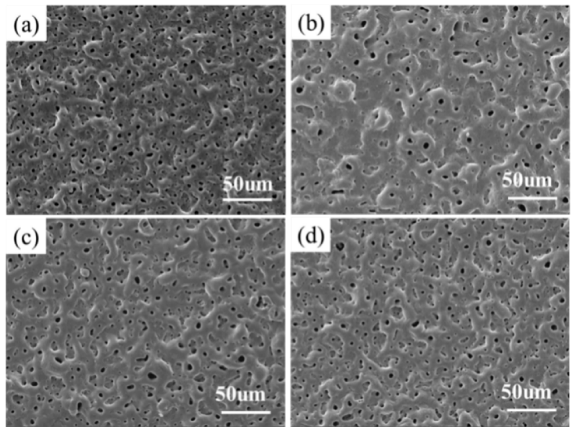
The surface morphologies and compositions of the MAO-Cu/Cu-(HEA)N composite coatings with different concentrations of CuSO4 are shown in Figure 2 and Table 2. It can be seen that the four composite coatings exhibited typical crater-like morphologies with uniform pore distributions. The pores on the surface of the MAO coatings were not completely covered by the Cu-(HEA)N films, maintaining the typical feature of microarc oxidation coatings [35,36,37]. Figure 2 shows that the nanoscale representation of the deposited Cu-(HEA)N films on micron-scale MAO layers had no obvious effect on the surface morphology of the MAO coating. The composite coating prepared from the base solution had a considerable number of protrusions and pores in various sizes, as observed in Figure 2a. However, with the addition of CuSuO4 into electrolyte, the number of pores and protrusions on the coating surface decreased, as shown in Figure 2b. It is noteworthy that small amounts of CuSO4 increased the conductivity of the solution, thereby promoting the growth of the MAO coating. The conductivity of the solution and its reaction rate were continuously enhanced with increasing CuSO4 concentration. More melt was produced at high temperatures and high pressures during the micro arc oxidation [31], which solidified and accumulated on the surface of the TC4 substrate after expulsion through the discharge channel. Therefore, the increase in concentration to 10 g/L and 15 g/L CuSO4 did not reduce the number of micropores and protrusions on the surface of the composite coatings, as displayed in Figure 2c,d. Table 2 shows that the composition of the MAO-Cu/Cu-(HEA)N composite coatings with different CuSO4 concentrations exhibited a certain trend. The coatings were mainly composed of nitrides formed by the reaction of the elements in the (HEA)N target with N2 and oxides of Cu and Ti. It was observed that the Cu content in the coatings increased with increasing CuSO4 concentration in the electrolyte.
3.2. XPS Analysis
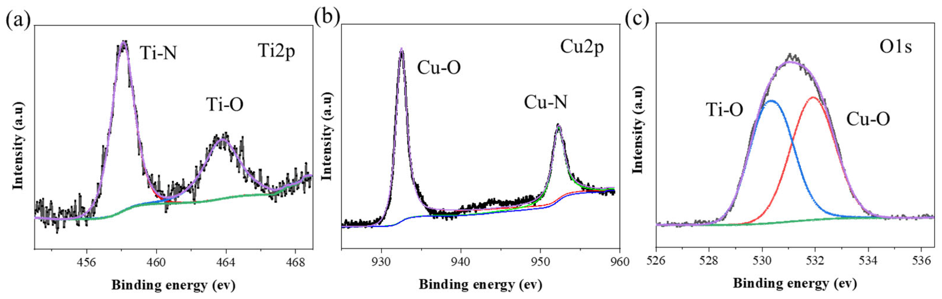
The surface characteristics of the MAO-Cu/Cu-(HEA)N samples were analyzed by XPS and displayed in Figure 3. The XPS fit was carried out using advantage software (version 5), and the peak of C1s was used to calibrate the peak of each element. The results show that the MAO-Cu/Cu-(HEA)N composite coatings surfaces were mainly composed of Ti, Cu, and O elements. The binding energy of Ti 2p was located at 458.17 eV and 464.48 eV, and assigned to Ti 2p3/2 and Ti 2p1/2 of Ti4+, as shown in Figure 3a, indicating that O element mainly existed in the MAO-Cu/Cu-(HEA)N composite coatings in the form of TiN and TiO2. The Cu 2p two main peaks were observed at 932.71 eV and 952.78 eV, and assigned to Cu 2p3/2 and Cu 2p1/2 of Cu+, but no obvious peaks were detected around 944.0–941.5 eV according to Figure 3b, signifying that the Cu+ of Cu2O rather than the Cu2+ of CuO was the existing form of Cu in the MAO-Cu/Cu-(HEA)N composite coating [38,39,40]. The peak value of the O 1s peak was detected at 530.45 eV and 532.09 eV in Figure 3c, confirming the existence of TiO2 and Cu2O [41,42]. In the micro-arc oxidation process, the arc discharge generates instantaneous high temperature and high pressure. CuSO4 decomposes at high temperatures to produce CuO, which in turn decomposes to produce Cu2O.
3.3. Thickness
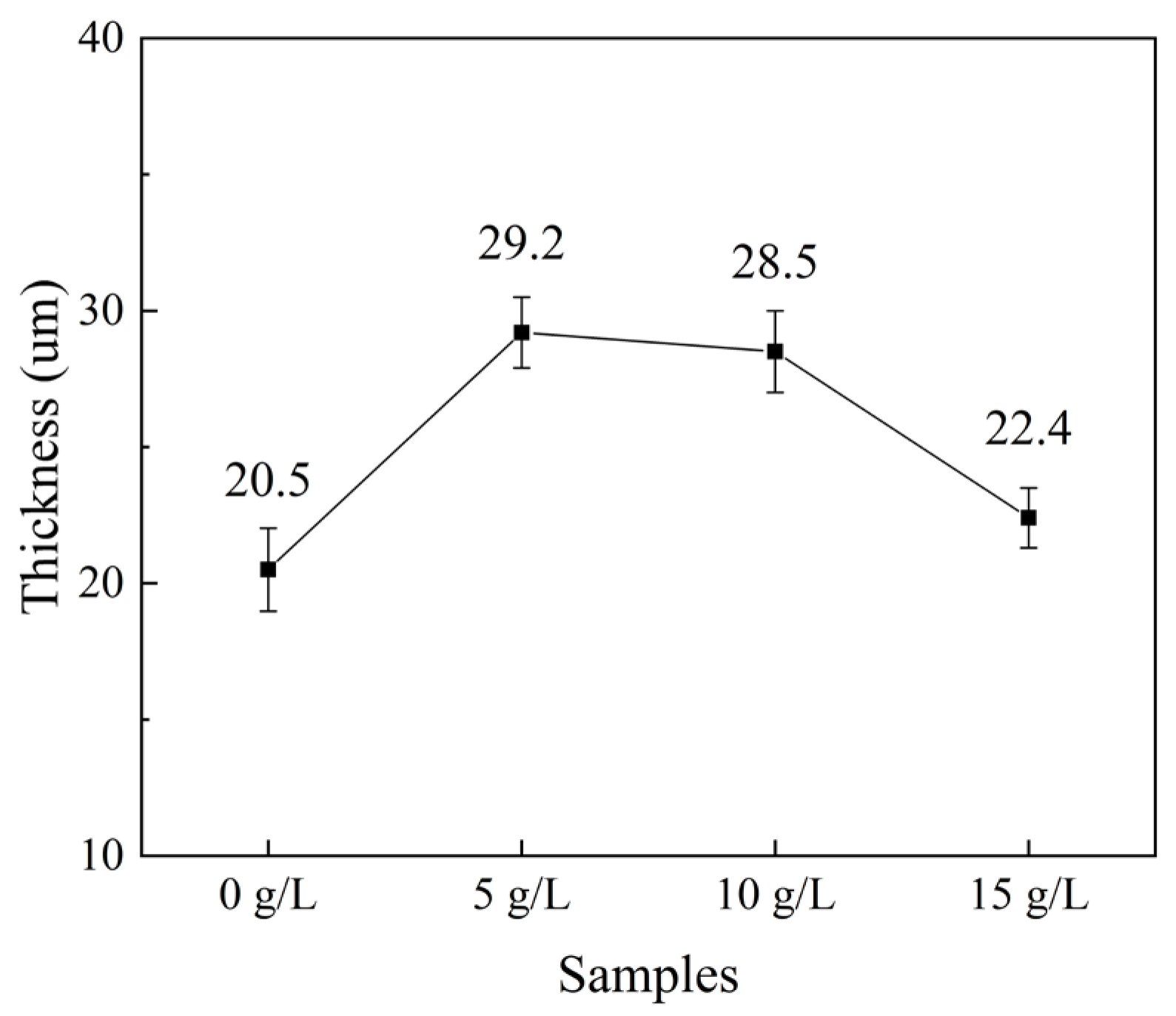
The thicknesses of the MAO coatings are shown in Figure 4. It can be seen from the figure that the thickness values of the four coatings were between 20 and 30 µm. It is possible that the Cu2+ in CuSO4 solution was responsible for the enhancement of the conductivity of the electrolyte and the increase in current which significantly increased the generation and dissolution rate of the surface melt, which improved the growth rate of the MAO coatings. Compared to the coating prepared by the base solution, the thickness of the MAO coating increased by more than 40% with the addition of 5 g/L CuSO4 solution. However, the effect of the MAO treatment was limited at higher CuSO4 concentrations because electric breakdown was more difficult in the thicker coatings [43]. Hence, the thickness of the composite coating did not necessarily increase at CuSO4 concentrations above 5 g/L.
3.4. Wear Resistance
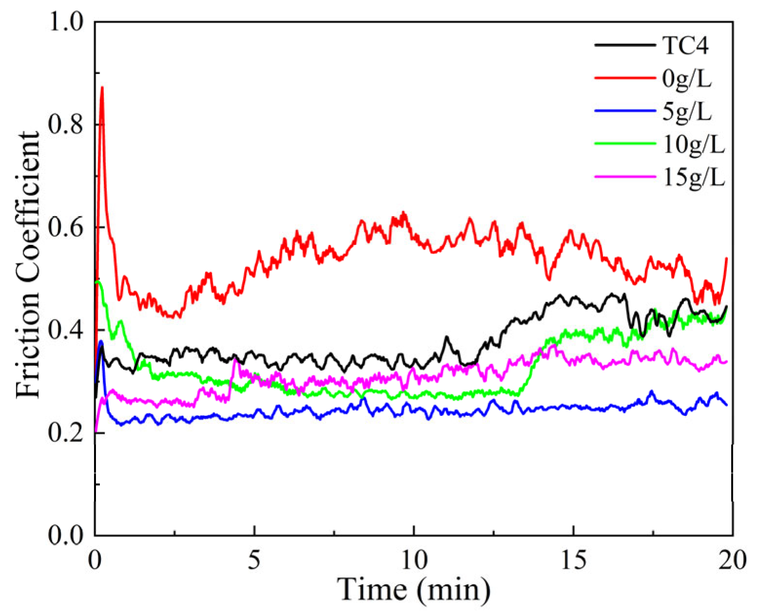
Friction coefficient is an important parameter employed to evaluate the wear resistances of materials [44,45,46]. Figure 5 depicts the friction coefficient (CoF) vs. sliding time for the TC4 material and the four MAO-Cu/Cu-(HEA)N composite coatings at an applied load of 2 N in simulated seawater. The friction coefficient of the TC4 material fluctuated significantly in simulated seawater with values ranging from 0.3 to 0.8. The TC4 material had a relatively low hardness and high viscosity [37]. With continuous friction of the grinding ball and the TC4 surface, the material viscosity and wear contact surface increased together with the increasing frictional force. Therefore, TC4 material had large friction coefficient and obvious fluctuations. The friction coefficient curve of the composite coating with the base solution exhibited a heavy fluctuation, and the friction coefficient gradually increased with increasing wear time. Specifically, the friction coefficient of the MAO-Cu/Cu-(HEA)N composite coating with 5 g/L CuSO4 was about 0.21 with the extension of wear time, and the friction coefficient curve was relatively stable. However, the friction coefficient of the MAO-Cu/Cu-(HEA)N composite coatings with 10 g/L and 15 g/L CuSO4 increased to 0.3 and 0.4, respectively. The addition of CuSO4 to the electrolyte reduced the number of micropores in the MAO layers, making the coatings flat and smooth [47,48,49], and the Cu-(HEA)N films covered parts of the tiny pores in the MAO layers. The low friction of the Cu element in the MAO layers decreased the friction coefficient, thereby improving the wear resistance. In addition, the simulated seawater stored in the MAO layers formed a liquid film during wear, which further decreased the friction coefficient. The weightlessness percentages of the four composite coatings calculated after wear are shown in Table 3. As can be seen from the table, the weightlessness percentages of the four composite coatings were lower than that of the TC4 material. The lowest weightlessness percent of the MAO-Cu/Cu-(HEA)N composite coating was recorded for the coating with 5 g/L CuSO4, indicating that it had the best wear resistance of the four coatings. Overall, the MAO-Cu/Cu-(HEA)N composite coatings improved the wear damage usually associated with TC4 materials.
The surface morphologies by SEM examination and EDS micrographs of the wear scars are shown in Figure 6. Additionally, the widths of the wear scars were marked in the SEM micrographs. After wear, the surface of the TC4 material had the widest wear mark width of 551.45 µm with obvious furrow morphology, indicating a typical abrasive wear characteristic (Figure 6a) [43]. The rough surface of the GCr15 grinding ball played a vital role in ploughing the surface of the TC4 alloy, leaving ploughing grooves on the worn surface. Compared to the TC4 material, the wear marks on the surface of the MAO-Cu/Cu-(HEA)N composite coatings were shallow without obvious grooves, as displayed in Figure 6b–e. The four MAO-Cu/Cu-(HEA)N composite coatings had narrow wear scar widths, which first increased and then decreased with increasing CuSO4 concentrations. The composite coating with 5 g/L CuSO4 exhibited the least wear mark width and friction coefficient. The surface of the composite coating without the addition of CuSO4 has more micropores and larger bumps, with a large degree of wear on the grinding balls. The introduction of CuSO4 improves the structure of the coating, the number of micropores is reduced and the surface of the coating is smooth, so the wear on the grinding balls and the coating is slight. The TiO2 layer of microporous sodium storage seawater formed a liquid film to reduce the degree of wear on the grinding ball and the coating. As can be seen from the EDS diagram, Fe element did not only exist at the wear mark locations, but also on the whole surface of the TC4 material, as shown in Figure 6a. Obviously, the Fe element was largely characterized on the wear marks of the composite coatings. This was due to compaction of the wear debris produced by repeated friction and wear on the grinding balls in the numerous micropores on the surface of the composite coatings (Figure 6b–e). The GCr15 grinding ball only wore out the surface of the four composite coatings but did not damage the TC4 substrate, indicating that the four composite coatings played effective roles in protecting the TC4 substrate.
3.5. Antibacterial Ability
Figure 7 shows the macrophotographs of Staphylococcus aureus (S. aureus) colonies cultured on TC4 and MAO-Cu/Cu-(HEA)N composite coatings with different CuSO4 concentrations attached to the surface of LB solid medium. The bacteria cultured on the surface of TC4 survived well on the surface of LB solid medium and they proliferated and grew in large numbers, as shown in Figure 7a. With increasing CuSO4 concentrations, the MAO-Cu/Cu-(HEA)N composite coatings demonstrated excellent antibacterial abilities. For the coating containing 5 g/L CuSO4, only few S. aureus existed on the surface of the solid medium after culture, indicating that the increasing CuSO4 concentration was beneficial to the improvement of the antibacterial properties of the MAO-Cu/Cu-(HEA)N composite coatings (Figure 7b). It was reported that the presence of Cu2O nanoparticles will completely inactivate bacterial growth through the release of Cu ions which generates reactive oxygen species [50]. In this study, the inactivation of S. aureus on the surface of the solid medium is also attributed to the presence of Cu ions in the MAO-Cu/Cu-(HEA)N composite coatings.
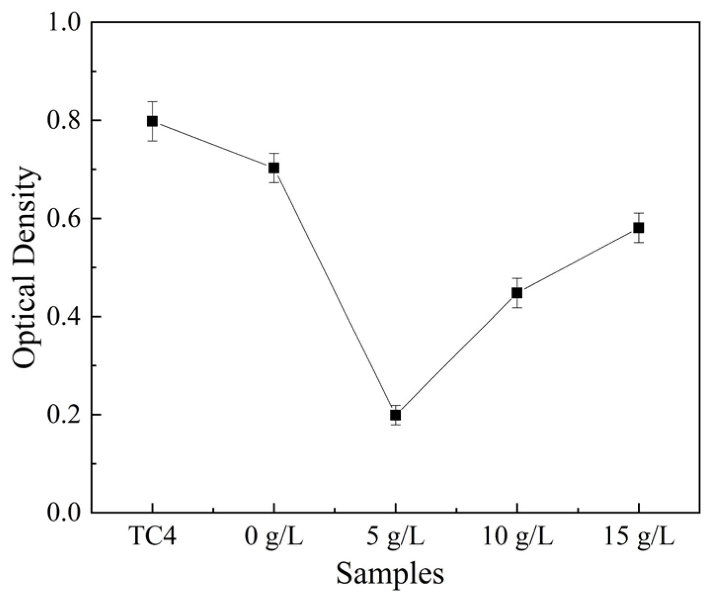
Figure 8 shows the optical density of S. aureus increment in the simulated seawater. Optical density reflects the antibacterial ability of samples to S. aureus [51], and a low optical density represents poor livability of S. aureus and an excellent antibacterial ability. Comparing the optical density of the S. aureus-cultured TC4 with those of the different composite coatings, it was observed that the optical density decreased significantly with increasing CuSO4 concentration. The optical densities of the four S. aureus-cultured composite coatings were lower than that of the S. aureus-cultured TC4. As Cu content in MAO-Cu/Cu-(HEA)N composite coatings increased, the precipitation of Cu ions increased, and the sterilization performance of the MAO-Cu/Cu-(HEA)N composite coating were also enhanced. The optical density of the composite coating containing 5 g/L CuSO4 was 0.19, and this presented the most favorable condition for bacterial deactivation of all the composite coatings.
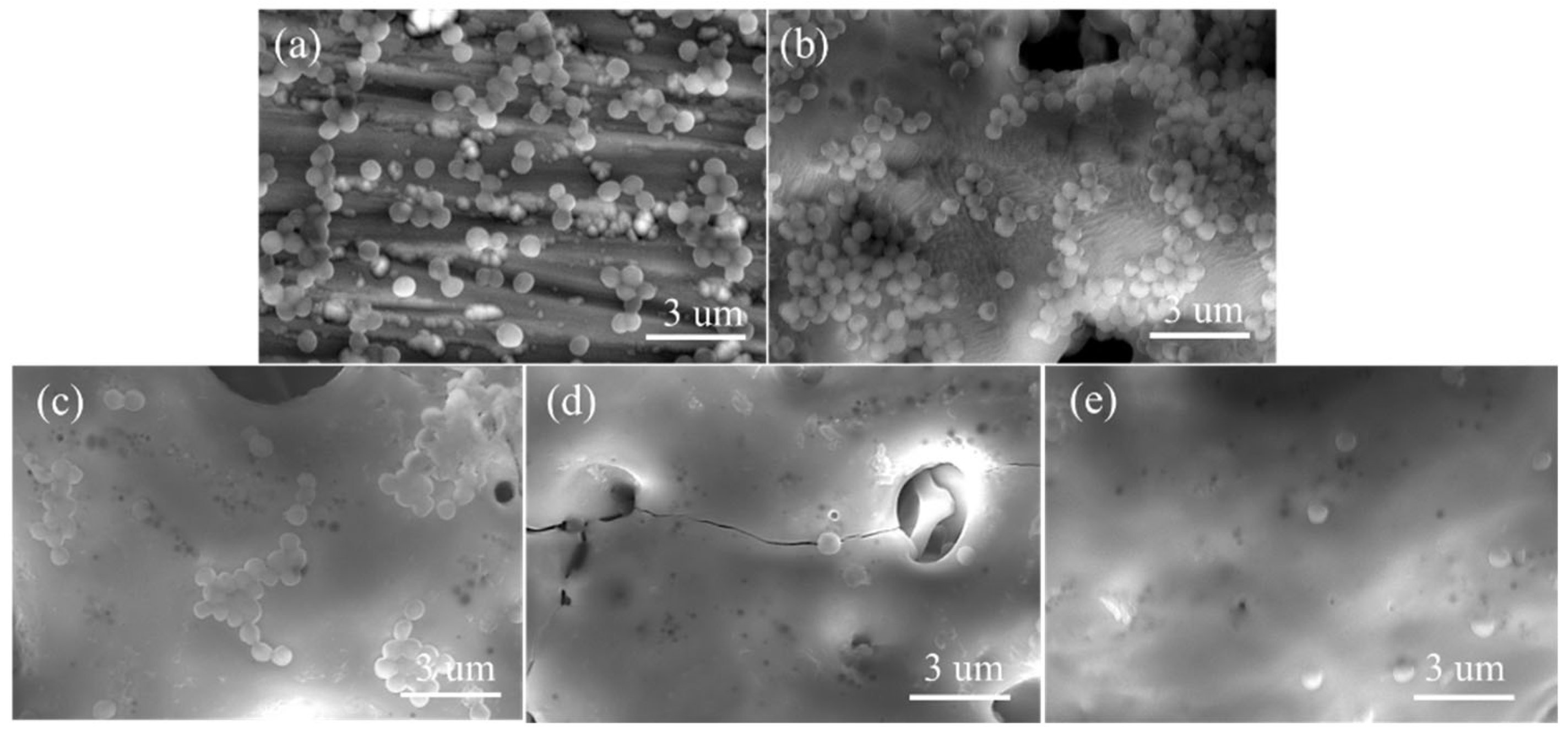
Figure 9 represents the microcosmic surface adhesion of S. aureus on TC4 and the MAO-Cu/Cu-(HEA)N composite coatings. A large number of S. aureus were attached to the surface of the TC4 sample, and the TC4 provided favorable living conditions for the growth and reproduction of the S. aureus, as depicted in Figure 9a. The adhesion rates of the S. aureus-cultured composite coatings containing different concentrations of CuSO4 in basic electrolyte were comparatively lower than that of the S. aureus-cultured TC4. Some of the S. aureus agglomerated to form a grape-like structure which adhered to the surface of the MAO-Cu/Cu-(HEA)N composite coatings, as shown in Figure 9c. In addition, there were almost no agglomerations of S. aureus on the surface of the MAO-Cu/Cu-(HEA)N composite coatings containing 10 g/L and 15 g/L CuSO4 (Figure 9d,e).
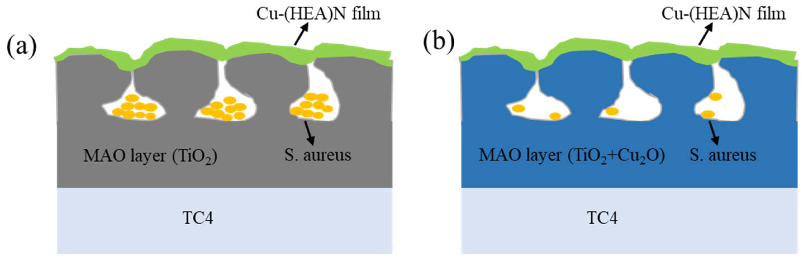
Cu2O has been proven to have good antibacterial effects [50,52,53,54,55,56]. Studies have shown that reactive oxygen species (OH, H2O2 and O2−) produced by Cu2O may interact with bacterial cell membranes and promote bacterial permeation [57]. Disturbance of bacterial cell membranes by Cu2O can cause a number of functional disorders that impede the growth of bacteria and may eventually lead to the growth of bacterial species and possibly eventually their death [57,58]. The small size (nanoscale) of Cu2O makes it easy for Cu2O to penetrate the cell membrane compared to the pore size (microns) of bacteria [59]. The XPS result displayed in Figure 4 shows that after the MAO process and sputtering, TiO2 and Cu2O were the main components on the surface of the coatings. Based on the above observation, it is reasonable to conclude that Cu2O was generated after the addition of CuSO4 to the electrolyte, and it played an important role in the antibacterial behavior of the coatings. As shown in Figure 10a, there was no Cu2O in the pores on the MAO layer without CuSO4 addition. Here, the antibacterial effect was mainly dependent on the Cu2O on the surface of the Cu-(HEA)N films. This means that the incompletely killed bacteria continued to grow and reproduce in the pores. The increasing concentration of CuSO4 changed the structure and Cu2O content of the MAO-Cu/Cu-(HEA)N composite coatings. From Figure 10b, the Cu2O in the pores of the MAO layer interacted with the active bacteria on the surface of the MAO-Cu/Cu-(HEA)N film and continued to kill them, hence bacterial inactivation continued in the pores [60]. Moreover, the rough structure of the coatings increased their surface areas or contact interface areas, thereby improving their antibacterial performances.
4. Conclusions
MAO-Cu/Cu-(HEA)N composite coatings were successfully prepared on titanium alloy by MAO and MS. With the addition of CuSO4 to the electrolyte, the surface of the composite coating became flat and smooth with the existence of dense and uniform pores. Cu mainly existed as Cu2O in the composite coatings, and the introduction of CuSO4 promoted both the growth rates and thicknesses of the coatings. The wear resistance and antibacterial property of the composite coating was improved in simulated seawater with increments in CuSO4 concentration. The friction coefficient of the composite coating with 5 g/L CuSO4 was reduced to about 0.2, and the wear marks were shallow and narrow. Compared with the TC4 material, the optical density of the composite coatings decreased to 0.19, and the adhesion of S. aureus on the surface of the composite coatings were significantly weakened. MAO-Cu/Cu-(HEA)N composite coatings are important for surface modification of titanium alloys, but the wear and antimicrobial mechanisms of the composite coatings need to be investigated in more depth. In addition, the composite coatings research will focus on corrosion and erosion resistance in the marine environment.
Author Contributions
Conceptualization, N.L. and Z.W.; methodology, Z.W.; software, Y.Z.; validation, Y.Z., Z.W. and W.D.; formal analysis, Z.W.; investigation, Y.Z.; resources, W.D.; data curation, Z.W. and Y.Z.; writing—original draft preparation, Z.W.; writing—review and editing, N.L.; visualization, N.L.; supervision, N.L; project administration, Z.W; funding acquisition, Z.W. All authors have read and agreed to the published version of the manuscript.
Funding
The authors gratefully acknowledge financial support of National Natural Science Foundation of China (No. 52071252) and Key research and development plan of Shaanxi province industrial project (2021GY-208, 2022GY-407 and 2021ZDLSF03-11).
Institutional Review Board Statement
Not applicable.
Informed Consent Statement
Not applicable.
Data Availability Statement
Not applicable.
Conflicts of Interest
The authors declare no conflict of interest.
References
- Bai, H.; Zhong, L.; Kang, L.; Liu, J.; Zhuang, W.; Lv, Z.; Xu, Y. A review on wear-resistant coating with high hardness and high toughness on the surface of titanium alloy. J. Alloy. Compd. 2021, 882, 160645. [Google Scholar] [CrossRef]
- Bai, X.; Tu, B. Ellipsoid non-probabilistic reliability analysis of the crack growth fatigue of a new titanium alloy used in deep-sea manned cabin. Theor. Appl. Fract. Mech. 2021, 115, 103041. [Google Scholar] [CrossRef]
- Pan, X.; He, W.; Cai, Z.; Wang, X.; Liu, P.; Luo, S.; Zhou, L. Investigations on femtosecond laser-induced surface modification and periodic micropatterning with anti-friction properties on Ti6Al4V titanium alloy. Chin. J. Aeronaut. 2021, 35, 521–537. [Google Scholar] [CrossRef]
- Dhanda, M.; Haldar, B.; Saha, P. Development and Characterization of Hard and Wear Resistant MMC Coating on Ti-6Al-4V Substrate by Laser Cladding. Procedia Mater. Sci. 2014, 6, 1226–1232. [Google Scholar] [CrossRef] [Green Version]
- Chowdhury, M.A.; Hossain, N.; Shahid, M.A.; Alam, M.J.; Hossain, S.M.; Uddin, M.I.; Rana, M.M. Development of SiC–TiO2-Graphene neem extracted antimicrobial nano membrane for enhancement of multiphysical properties and future prospect in dental implant applications. Heliyon 2022, 8, e10603. [Google Scholar] [CrossRef]
- Kujala, S.; Ryhänen, J.; Danilov, A.; Tuukkanen, J. Effect of porosity on the osteointegration and bone ingrowth of a weight-bearing nickel–titanium bone graft substitute. Biomaterials 2003, 24, 4691–4697. [Google Scholar] [CrossRef]
- Wang, X.; Li, Y.; Xiong, J.; Hodgson, P.D.; Wen, C. Porous TiNbZr alloy scaffolds for biomedical applications. Acta Biomater. 2009, 5, 3616–3624. [Google Scholar] [CrossRef]
- Li, Y.; Ding, Y.; Munir, K.; Lin, J.; Brandt, M.; Atrens, A.; Xiao, Y.; Kanwar, J.R.; Wen, C. Novel β-Ti35Zr28Nb alloy scaffolds manufactured using selective laser melting for bone implant applications. Acta Biomater. 2019, 87, 273–284. [Google Scholar] [CrossRef]
- Muhaffel, F.; Kaba, M.; Cempura, G.; Derin, B.; Kruk, A.; Atar, E.; Cimenoglu, H. Influence of alumina and zirconia incorporations on the structure and wear resistance of titania-based MAO coatings. Surf. Coat. Technol. 2019, 377, 124900. [Google Scholar] [CrossRef]
- Dong, H. Tribological properties of titanium-based alloys. In Surface Engineering of Light Alloys; Dong, H., Ed.; Elsevier: Amsterdam, The Netherlands, 2010; pp. 58–80. [Google Scholar]
- He, R.; Wang, B.; Xiang, J.; Pan, T. Effect of copper additive on microstructure and anti-corrosion performance of black MAO films grown on AZ91 alloy and coloration mechanism. J. Alloy. Compd. 2021, 889, 161501. [Google Scholar] [CrossRef]
- Chen, J.; Xu, J.L.; Huang, J.; Dai, L.; Xue, M.S.; Luo, J.M. Corrosion resistance of T-ZnOw/PDMS-MAO composite coating on the sintered NdFeB magnet. J. Magn. Magn. Mater. 2021, 534, 168049. [Google Scholar] [CrossRef]
- Lan, N.; Yang, W.; Gao, W.; Guo, P.; Zhao, C.; Chen, J. Characterization of ta-C film on micro arc oxidation coated titanium alloy in simulated seawater. Diam. Relat. Mater. 2021, 117, 108483. [Google Scholar] [CrossRef]
- Zuo, Y.; Li, T.; Yu, P.; Zhao, Z.; Chen, X.; Zhang, Y.; Chen, F. Effect of graphene oxide additive on tribocorrosion behavior of MAO coatings prepared on Ti6Al4V alloy. Appl. Surf. Sci. 2019, 480, 26–34. [Google Scholar] [CrossRef]
- Parichehr, R.; Dehghanian, C.; Nikbakht, A. Preparation of PEO/silane composite coating on AZ31 magnesium alloy and in-vestigation of its properties. J. Alloys Compd. 2021, 876, 159995. [Google Scholar] [CrossRef]
- Sopchenski, L.; Robert, J.; Touzin, M.; Tricoteaux, A.; Olivier, M.-G. Improvement of wear and corrosion protection of PEO on AA2024 via sol-gel sealing. Surf. Coat. Technol. 2021, 417, 127195. [Google Scholar] [CrossRef]
- Rizwan, M.; Alias, R.; Zaidi, U.Z.; Mahmoodian, R.; Hamdi, M. Surface modification of valve metals using plasma electrolytic oxidation for antibacterial applications: A review. J. Biomed. Mater. Res. Part A 2017, 106, 590–605. [Google Scholar] [CrossRef]
- Sun, W.; Liu, Y.; Li, T.; Cui, S.; Chen, S.; Yu, Q.; Wang, D. Anti-corrosion of amphoteric metal enhanced by MAO/corrosion inhibitor composite in acid, alkaline and salt solutions. J. Colloid Interface Sci. 2019, 554, 488–499. [Google Scholar] [CrossRef]
- Mashtalyar, D.V.; Sinebryukhov, S.L.; Imshinetskiy, I.M.; Gnedenkov, A.S.; Nadaraia, K.V.; Ustinov, A.Y.; Gnedenkov, S.V. Hard wearproof PEO-coatings formed on Mg alloy using TiN nanoparticles. Appl. Surf. Sci. 2019, 503, 144062. [Google Scholar] [CrossRef]
- Li, X.; Dong, C.; Zhao, Q.; Pang, Y.; Cheng, F.; Wang, S. Characterization of Microstructure and Wear Resistance of PEO Coatings Containing Various Microparticles on Ti6Al4V Alloy. J. Mater. Eng. Perform. 2018, 27, 1642–1653. [Google Scholar] [CrossRef]
- Jiang, B.L.; Wang, Y.M. Plasma electrolytic oxidation treatment of aluminium and titanium alloys. In Surface Engineering of Light Alloys; Elsevier: Amsterdam, The Netherlands, 2010; pp. 110–154. [Google Scholar] [CrossRef]
- Yao, Z.; Xu, Y.; Jiang, Z.; Wang, F. Effects of cathode pulse at low frequency on the structure and composition of plasma elec-trolytic oxidation ceramic coatings. J. Alloys Compd. 2009, 488, 273–278. [Google Scholar] [CrossRef]
- Rafieerad, A.; Ashra, M.; Mahmoodian, R.; Bushroa, A. Surface characterization and corrosion behavior of calcium phosphate-base composite layer on titanium and its alloys via plasma electrolytic oxidation: A review paper. Mater. Sci. Eng. C 2015, 57, 397–413. [Google Scholar] [CrossRef] [PubMed]
- Gowtham, S.; Hariprasad, S.; Arunnellaiappan, T.; Rameshbabu, N. An investigation on ZrO2 nanoparticle incorporation, surface properties and electrochemical corrosion behaviour of PEO coating formed on Cp-Ti. Surf. Coat. Technol. 2017, 313, 263–273. [Google Scholar]
- Cerchier, P.; Pezzato, L.; Brunelli, K.; Dolcet, P.; Bartolozzi, A.; Bertani, R.; Dabalà, M. Antibacterial effect of PEO coating with silver on AA7075. Mater. Sci. Eng. C 2017, 75, 554–564. [Google Scholar] [CrossRef] [PubMed]
- Xia, Z.; Min, J.; Zhou, S.; Ma, H.; Zhang, B.; Tang, X. Photocatalytic performance and antibacterial mechanism of Cu/Ag-molybdate powder material. Ceram. Int. 2021, 47, 12667–12679. [Google Scholar] [CrossRef]
- Lu, M.; Zhang, Z.; Zhang, J.; Wang, X.; Qin, G.; Zhang, E. Enhanced antibacterial activity of Ti-Cu alloy by selective acid etching. Surf. Coat. Technol. 2021, 421, 127478. [Google Scholar] [CrossRef]
- Athinarayanan, J.; Periasamy, V.S.; Krishnamoorthy, R.; Alshatwi, A.A. Evaluation of antibacterial and cytotoxic properties of green synthesized Cu2O/Graphene nanosheets. Mater. Sci. Eng. C 2018, 93, 242–253. [Google Scholar] [CrossRef]
- Stranak, V.; Wulff, H.; Rebl, H.; Zietz, C.; Arndt, K.; Bogdanowicz, R.; Nebe, B.; Bader, R.; Podbielski, A.; Hubicka, Z.; et al. Deposition of thin titanium–copper films with antimicrobial effect by advanced magnetron sputtering methods. Mater. Sci. Eng. C 2011, 31, 1512–1519. [Google Scholar] [CrossRef]
- Zhang, X.; Wu, Y.; Wang, J.; Xia, X.; Lv, Y.; Cai, G.; Liu, H.; Xiao, J.; Liu, B.; Dong, Z. Microstructure, formation mechanism and antifouling property of multi-layered Cu-incorporated Al2O3 coating fabricated through plasma electrolytic oxidation. Ceram. Int. 2019, 46, 2901–2909. [Google Scholar] [CrossRef]
- Li, X.; Guo, P.; Sun, L.; Wang, A.; Ke, P. Ab Initio Investigation on Cu/Cr Codoped Amorphous Carbon Nanocomposite Films with Giant Residual Stress Reduction. ACS Appl. Mater. Interfaces 2015, 7, 27878–27884. [Google Scholar] [CrossRef]
- Wu, Y.; Zhou, S.; Zhao, W.; Ouyang, L. Comparative corrosion resistance properties between (Cu, Ce)-DLC and Ti co-doped (Cu, Ce)/Ti-DLC films prepared via magnetron sputtering method. Chem. Phys. Lett. 2018, 705, 50–58. [Google Scholar] [CrossRef]
- Cui, P.; Li, W.; Liu, P.; Zhang, K.; Ma, F.; Chen, X.; Feng, R.; Liaw, P.K. Effects of nitrogen content on microstructures and mechanical properties of (AlCrTiZrHf)N high-entropy alloy nitride films. J. Alloys Compd. 2020, 834, 155063. [Google Scholar] [CrossRef]
- Xu, Y.; Li, G.; Li, G.; Gao, F.; Xia, Y. Effect of bias voltage on the growth of super-hard (AlCrTiVZr)N high-entropy alloy nitride films synthesized by high power impulse magnetron sputtering. Appl. Surf. Sci. 2021, 564, 150417. [Google Scholar] [CrossRef]
- Yang, W.; Ke, P.; Fang, Y.; Zheng, H.; Wang, A. Microstructure and properties of duplex (Ti:N)-DLC/MAO coating on magnesium alloy. Appl. Surf. Sci. 2013, 270, 519–525. [Google Scholar] [CrossRef]
- Mazinani, A.; Nine, M.J.; Chiesa, R.; Candiani, G.; Tarsini, P.; Tung, T.T.; Losic, D. Graphene oxide (GO) decorated on multi-structured porous titania fabricated by plasma electrolytic oxidation (PEO) for enhanced antibacterial performance. Mater. Des. 2021, 200, 109443. [Google Scholar] [CrossRef]
- Sunilraj, S.; Blessto, B.; Sivaprasad, K.; Muthupandi, V. Microstructural and corrosion behavior of MAO coated 5052 aluminum alloy. Mater. Today Proc. 2020, 41, 1120–1124. [Google Scholar] [CrossRef]
- Poulston, S.; Parlett, P.M.; Stone, P.; Bowker, M. Surface oxidation and reduction of CuO and Cu2O studied using XPS and XAES. Surf. Interface Anal. 1996, 24, 811–820. [Google Scholar] [CrossRef]
- Cheng, Y.; Zhu, Z.; Zhang, Q.; Zhuang, X.; Cheng, Y. Plasma electrolytic oxidation of brass. Surf. Coat. Technol. 2020, 385, 125366. [Google Scholar] [CrossRef]
- Shaikh, J.S.; Pawar, R.C.; Moholkar, A.V.; Kim, J.H.; Patil, P.S. CuO-PAA hybrid films: Chemical synthesis and supercapacitor behavior. Appl. Surf. Sci. 2011, 257, 4389–4397. [Google Scholar] [CrossRef]
- Park, J.; Lam, S.S.; Park, Y.K.; Kim, B.J.; An, K.H.; Jung, S.C. Fabrication of Ni/TiO2 visible light responsive photocatalyst for de-composition of oxytetracycline. Environ. Res. 2023, 216, 114657. [Google Scholar] [CrossRef]
- Tang, Y.; Hu, X.; Liu, Y.; Chen, Y.; Zhao, F.; Zeng, B. An antifouling electrochemiluminescence sensor based on mesoporous CuO2@SiO2/luminol nanocomposite and co-reactant of ionic liquid functionalized boron nitride quantum dots for ultrasensitive NSE detection. Biosens. Bioelectron. 2022, 214, 114492. [Google Scholar] [CrossRef]
- Xie, R.; Lin, N.; Zhou, P.; Zou, J.; Han, P.; Wang, Z.; Tang, B. Surface damage mitigation of TC4 alloy via micro arc oxidation for oil and gas exploitation application: Characterizations of microstructure and evaluations on surface performance. Appl. Surf. Sci. 2018, 436, 467–476. [Google Scholar] [CrossRef]
- Yang, S.; Xiong, J.; Guo, Z.; Wu, B.; You, Q.; Liu, J.; Deng, C.; Fang, D.; Gou, S.; Yu, Z.; et al. Effects of CrMnFeCoNi additions on microstructure, mechanical properties and wear resistance of Ti (C, N)-based cermets. J. Mater. Res. Technol. 2022, 17, 2480–2494. [Google Scholar] [CrossRef]
- Santaella-González, J.B.; Hernández-Torres, J.; Morales-Hernández, J.; Flores-Ramírez, N.; Ferreira-Palma, C.; Rodríguez-Jiménez, R.C.; García-González, L. Effect of the number of bilayers in Ti/TiN coatings on AISI 316L deposited by sputtering on their hardness, adhesion, and wear. Mater. Lett. 2022, 316, 132037. [Google Scholar] [CrossRef]
- Chowdhury, M.A.; Hossain, N.; Al Masum, A.; Islam, M.S.; Shahin, M.; Hossain, M.I.; Nandee, M.R. Surface coatings analysis and their effects on reduction of tribological properties of coated aluminum under motion with ML approach. Mater. Res. Express 2021, 8, 086508. [Google Scholar] [CrossRef]
- Wang, S.Q.; Wang, Y.M.; Zou, Y.C.; Chen, G.L.; Wang, Z.; Ouyang, J.H.; Jia, D.C.; Zhou, Y. Generation, Tailoring and Functional Ap-plications of Micro-Nano Pores in Microarc Oxidation Coating: A Critical Review. Surf. Technol. 2021, 250, 1–22. [Google Scholar]
- Dan, M.; Tong, H.H.; Shen, L.R. The effect of sodium hexametaphosphate on the structure and corrosion resistance of LD7 aluminum alloy micro-arc oxidation ceramic film. Mater. Prot. 2011, 44, 11–13. [Google Scholar]
- Li, J.Z.; Shao, Z.C.; Tian, Y.W. The action of the forms of P element on the process of the micro arc oxidation. J. Chin. Soc. Corros. Prot. 2004, 24, 222–225. [Google Scholar]
- Zhang, W.; Zhang, S.; Liu, H.; Ren, L.; Wang, Q.; Zhang, Y. Effects of surface roughening on antibacterial and osteogenic properties of Ti-Cu alloys with different Cu contents. J. Mater. Sci. Technol. 2021, 88, 158–167. [Google Scholar] [CrossRef]
- Meyers, A.; Furtmann, C.; Jose, J. Direct optical density determination of bacterial cultures in microplates for high-throughput screening applications. Enzym. Microb. Technol. 2018, 118, 1–5. [Google Scholar] [CrossRef]
- Lee, Y.-J.; Kim, S.; Park, S.-H.; Park, H.; Huh, Y.-D. Morphology-dependent antibacterial activities of Cu2O. Mater. Lett. 2011, 65, 818–820. [Google Scholar] [CrossRef]
- Zhang, J.; Liu, J.; Peng, Q.; Wang, A.X.; Li, Y. Nearly Monodisperse Cu2O and CuO Nanospheres: Preparation and Applications for Sensitive Gas Sensors. Chem. Mater. 2006, 18, 867–871. [Google Scholar] [CrossRef]
- Pang, H.; Gao, F.; Lu, Q. Morphology effect on antibacterial activity of cuprous oxide. Chem. Commun. 2009, 9, 1076–1078. [Google Scholar] [CrossRef]
- Ren, J.; Wang, W.; Sun, S.; Zhang, L.; Wang, L.; Chang, J. Crystallography Facet-Dependent Antibacterial Activity: The Case of Cu2O. Ind. Eng. Chem. Res. 2011, 50, 10366–10369. [Google Scholar] [CrossRef]
- Jung, H.Y.; Seo, Y.; Park, H.; Huh, Y.D. Morphology-controlled synthesis of octahedral-to-rhombic dodecahedral Cu2O micro-crystals and shape-dependent antibacterial activities. Bull. Kor. Chem. Soc. 2015, 36, 1828–1833. [Google Scholar] [CrossRef]
- Kabir, M.H.; Ibrahim, H.; Ayon, S.A.; Billah, M.; Neaz, S. Structural, nonlinear optical and antimicrobial properties of sol-gel derived, Fe-doped CuO thin films. Heliyon 2022, 8, e10609. [Google Scholar] [CrossRef]
- Ayon, S.A.; Billah, M.M.; Nishat, S.S.; Kabir, A. Enhanced photocatalytic activity of Ho3þ doped ZnO NPs synthesized by modified sol-gel method: An experimental and theoretical investigation. J. Alloys Compd. 2021, 856, 158217. [Google Scholar] [CrossRef]
- Das, D.; Nath, B.C.; Phukon, P.; Dolui, S.K. Synthesis and evaluation of antioxidant and antibacterial behavior of CuO nano-particles. Colloids Surf. B Biointerfaces 2013, 101, 430–433. [Google Scholar] [CrossRef]
- Liu, S.; Zhang, Z.; Zhang, J.; Qin, G.; Zhang, E. Construction of a TiO2/Cu2O multifunctional coating on Ti-Cu alloy and its influence on the cell compatibility and antibacterial properties. Surf. Coat. Technol. 2021, 421, 127438. [Google Scholar] [CrossRef]
Figure 1.
Preparation process of MAO-Cu/Cu-(HEA)N composite coatings.

Figure 2.
The surface morphologies of MAO-Cu/Cu-(HEA)N composite coatings with different CuSO4 concentrations:(a) 0 g/L; (b) 5 g/L; (c) 10 g/L; and (d) 15 g/L.
Figure 2.
The surface morphologies of MAO-Cu/Cu-(HEA)N composite coatings with different CuSO4 concentrations:(a) 0 g/L; (b) 5 g/L; (c) 10 g/L; and (d) 15 g/L.

Figure 3.
XPS spectra analysis (black line) and XPS fitting (purple line) on the surface of the MAO-Cu/Cu-(HEA)N composite coating: (a) Ti 2p (red line is Ti-N, blue line is Ti-O and green line is base line), (b) Cu 2p (red line is Cu-O, green line is Cu-N and blue line is base line), (c) O 1s (red line is Ti-O, red line is Cu-O and green line is base line).

Figure 4.
The thickness of MAO coatings with different CuSO4 concentrations.

Figure 5.
Friction coefficient curves of TC4 and MAO-Cu/Cu-(HEA)N composite coatings with different CuSO4 concentrations.
Figure 5.
Friction coefficient curves of TC4 and MAO-Cu/Cu-(HEA)N composite coatings with different CuSO4 concentrations.

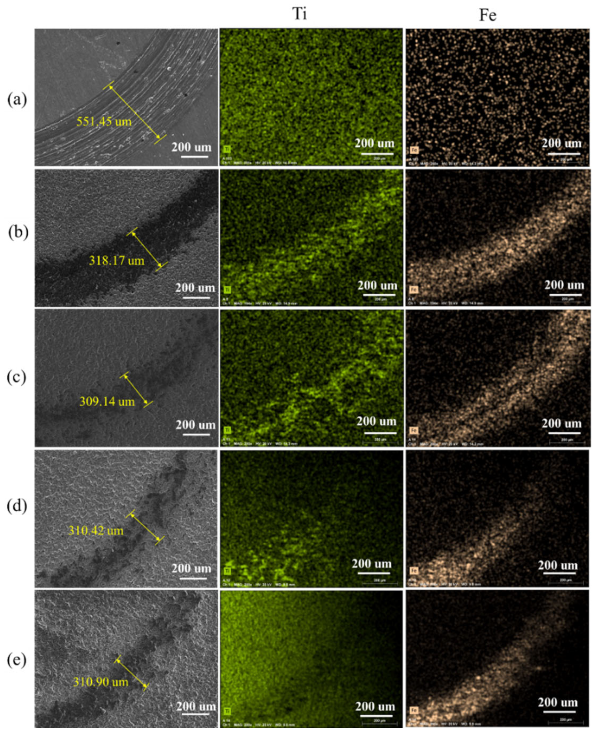
Figure 6.
The surface SEM and EDS of TC4 and MAO-Cu/Cu-(HEA)N composite coatings with different CuSO4 concentrations: (a) TC4; (b) 0 g/L; (c) 5 g/L; (d) 10 g/L; and (e) 15 g/L.
Figure 6.
The surface SEM and EDS of TC4 and MAO-Cu/Cu-(HEA)N composite coatings with different CuSO4 concentrations: (a) TC4; (b) 0 g/L; (c) 5 g/L; (d) 10 g/L; and (e) 15 g/L.

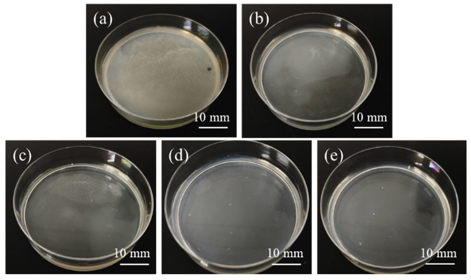
Figure 7.
The macrophotographs of Staphylococcus aureus colonies cultured by TC4 and MAO-Cu/Cu-(HEA)N composite coatings with different CuSO4 concentrations attached to the surface of LB solid medium: (a)TC4; (b) 0 g/L; (c) 5 g/L; (d) 10 g/L; and (e) 15 g/L.
Figure 7.
The macrophotographs of Staphylococcus aureus colonies cultured by TC4 and MAO-Cu/Cu-(HEA)N composite coatings with different CuSO4 concentrations attached to the surface of LB solid medium: (a)TC4; (b) 0 g/L; (c) 5 g/L; (d) 10 g/L; and (e) 15 g/L.

Figure 8.
Optical density of S. aureus cultured by TC4 and MAO-Cu/Cu-(HEA)N composite coatings with different CuSO4 concentrations.
Figure 8.
Optical density of S. aureus cultured by TC4 and MAO-Cu/Cu-(HEA)N composite coatings with different CuSO4 concentrations.

Figure 9.
The microcosmic adhesion of S. aureus on TC4 and MAO-Cu/Cu-(HEA)N composite coatings surface with different CuSO4 concentrations attached: (a) TC4; (b) 0 g/L; (c) 5 g/L; (d) 10 g/L; and (e) 15 g/L.
Figure 9.
The microcosmic adhesion of S. aureus on TC4 and MAO-Cu/Cu-(HEA)N composite coatings surface with different CuSO4 concentrations attached: (a) TC4; (b) 0 g/L; (c) 5 g/L; (d) 10 g/L; and (e) 15 g/L.

Figure 10.
Mechanism of the effect of CuSO4 on the antibacterial ability of MAO-Cu/Cu-(HEA)N composite coating: (a) without CuSO4; and (b) with CuSO4.
Figure 10.
Mechanism of the effect of CuSO4 on the antibacterial ability of MAO-Cu/Cu-(HEA)N composite coating: (a) without CuSO4; and (b) with CuSO4.


Table 1.
The composition of the simulated seawater solution.
| Composition | NaCl | MgCl2 | Na2SO4 | CaCl2 | KCl | NaHCO3 | KBr | HBO3 | SrCl | NaF |
|---|---|---|---|---|---|---|---|---|---|---|
| g/L | 24.53 | 11.11 | 4.09 | 1.16 | 0.685 | 0.201 | 0.101 | 0.027 | 0.028 | 0.003 |
Table 2.
The composition of MAO-Cu/Cu-(HEA)N composite coatings with different CuSO4 concentrations.
Table 2.
The composition of MAO-Cu/Cu-(HEA)N composite coatings with different CuSO4 concentrations.
| Samples | N | O | Al | Si | Ti | Cr | Nb | V | Cu |
|---|---|---|---|---|---|---|---|---|---|
| 0 g/L | 24.0 | 24.0 | 4.5 | 2.8 | 10.1 | 3.7 | 4.1 | 4.1 | 21.6 |
| 5 g/L | 24.5 | 24.5 | 4.3 | 2.7 | 10.3 | 3.6 | 4.2 | 4.1 | 20.6 |
| 10 g/L | 21.7 | 21.7 | 4.5 | 3.0 | 11.8 | 3.8 | 4.6 | 4.6 | 22.7 |
| 15 g/L | 22.9 | 22.9 | 2.3 | 1.5 | 12.6 | 3.8 | 4.6 | 3.4 | 25.0 |
Table 3.
Percent of weight loss of TC4 and MAO-Cu/Cu-(HEA)N composite coatings with different CuSO4 concentrations.
Table 3.
Percent of weight loss of TC4 and MAO-Cu/Cu-(HEA)N composite coatings with different CuSO4 concentrations.
| Samples | Percent of Weightloss (%) |
|---|---|
| TC4 | 1.310 ± 0.004 |
| 0 g/L | 0.016 ± 0.001 |
| 5 g/L | 0.011 ± 0.001 |
| 10 g/L | 0.016 ± 0.001 |
| 15 g/L | 0.014 ± 0.001 |
Publisher’s Note: MDPI stays neutral with regard to jurisdictional claims in published maps and institutional affiliations. |
© 2022 by the authors. Licensee MDPI, Basel, Switzerland. This article is an open access article distributed under the terms and conditions of the Creative Commons Attribution (CC BY) license (https://creativecommons.org/licenses/by/4.0/).
Share and Cite
MDPI and ACS Style
Wang, Z.; Lan, N.; Zhang, Y.; Deng, W. Microstructure and Properties of MAO-Cu/Cu-(HEA)N Composite Coatings on Titanium Alloy. Coatings 2022, 12, 1877. https://doi.org/10.3390/coatings12121877
AMA Style
Wang Z, Lan N, Zhang Y, Deng W. Microstructure and Properties of MAO-Cu/Cu-(HEA)N Composite Coatings on Titanium Alloy. Coatings. 2022; 12(12):1877. https://doi.org/10.3390/coatings12121877
Chicago/Turabian StyleWang, Zhao, Nan Lan, Yong Zhang, and Wanrong Deng. 2022. "Microstructure and Properties of MAO-Cu/Cu-(HEA)N Composite Coatings on Titanium Alloy" Coatings 12, no. 12: 1877. https://doi.org/10.3390/coatings12121877
Note that from the first issue of 2016, this journal uses article numbers instead of page numbers. See further details here.