Review

Antiviral Biodegradable Food Packaging and Edible Coating Materials in the COVID-19 Era: A Mini-Review

1
BioNanocomposite Research Center, Department of Food and Nutrition, Kyung Hee University, Seoul 02447, Korea
2
School of Chemical Engineering, Yeungnam University, 280 Daehak-Ro, Gyeongsan 38541, Korea
3
School of Bioengineering and Food Technology, Shoolini University, Solan 173229, India
4
Department of Food Engineering and Technology, Tezpur University, Tezpur 784028, India
*
Authors to whom correspondence should be addressed.
These authors contributed equally to this work.
Coatings 2022, 12(5), 577; https://doi.org/10.3390/coatings12050577
Submission received: 21 March 2022 / Revised: 15 April 2022 / Accepted: 21 April 2022 / Published: 24 April 2022

Abstract

:
With the onset of the COVID-19 pandemic in late 2019, and the catastrophe faced by the world in 2020, the food industry was one of the most affected industries. On the one hand, the pandemic-induced fear and lockdown in several countries increased the online delivery of food products, resulting in a drastic increase in single-use plastic packaging waste. On the other hand, several reports revealed the spread of the viral infection through food products and packaging. This significantly affected consumer behavior, which directly influenced the market dynamics of the food industry. Still, a complete recovery from this situation seems a while away, and there is a need to focus on a potential solution that can address both of these issues. Several biomaterials that possess antiviral activities, in addition to being natural and biodegradable, are being studied for food packaging applications. However, the research community has been ignorant of this aspect, as the focus has mainly been on antibacterial and antifungal activities for the enhancement of food shelf life. This review aims to cover the different perspectives of antiviral food packaging materials using established technology. It focuses on the basic principles of antiviral activity and its mechanisms. Furthermore, the antiviral activities of several nanomaterials, biopolymers, natural oils and extracts, polyphenolic compounds, etc., are discussed.

1. Introduction

Coronavirus disease, popularly known as COVID-19, is a highly transmissible viral infection caused by the SARS-CoV-2 viral strain. It was initially identified in December 2019, and the initial infections were presumed to be linked to the Huanan Seafood Market in Wuhan city of China [1]. The transmission rate of this virus among humans was so severe that it was ultimately declared a pandemic by the World Health Organization (WHO) before too long in March 2020 [1]. This sudden unforeseen incident caused global turmoil, especially among biotechnologists and virologists, who struggled to decode this puzzle. Zhou et al. carried out the genetic sequencing of the SARS-CoV-2 virus. They compared it with the bat coronavirus and found 96.2% similarity between the genetic organization, suggesting the role of bats in the spread of this pandemic [2]. As the coronavirus research evolved, the United States Center for Disease Control revealed that the first report on the human coronavirus dates back to the 1960s. Several variants have been discovered to date [3,4]. The viruses are capable of causing mild respiratory symptoms in humans and, with genetic evolution over the years, have differentiated into several strains with different properties. SARS-CoV-2, which was the variant responsible for COVID-19, belongs to the same category of coronaviruses to which Severe Acute Respiratory Syndrome (SARS)-CoV and Middle East Respiratory Syndrome (MERS)-CoV belong [4]. Since the primary transmission route of this category of viruses is the spread of droplets during coughing and sneezing, they are capable of easily infecting a large population.
With the advancement of this pandemic, several countries enforced nationwide lockdowns to control viral transmission. People were forced to stay indoors and work from home, which, along with the total or partial closure of food establishments, made them largely dependent on online ordering to meet their hunger needs [5]. This sudden and unusual shift in the consumer behavior of ordering food online resulted in an exponential increase in packaging-based non-biodegradable municipal solid waste, the highest contributor being single-use plastics. This further worsened the already existing massive problem of municipal waste disposal, resulting in negative environmental consequences [5].
In addition to the negative consequences on the environment, the increased use of packaging material led to negative health repercussions. It was reported that viral droplets > 5 μm in size were too heavy to stay airborne and landed on surfaces and objects [6]. These infected surfaces and objects emerged as the indirect and more prevalent source of cross-contamination, as the viruses were reported to stay active on surfaces depending on the material type [5,7]. Studies reported that coronavirus persisted on plastic for 72 h and cardboard surfaces for 24 h [7]. These materials, which play an important role in the packaging of processed and ready-to-eat food, came under suspicion. The long stability of SARS-CoV-2 on the surfaces of packaging materials created substantial risks and worries regarding the global trade of packaged food, as the virus was capable of surviving on the surface for the whole duration from production to consumption. Meanwhile, China reported the presence of coronavirus strains on animal product packages of Brazilian origin, which provided sufficient evidence that food packaging material may carry viruses, leading to cross-contamination hazards [7]. The European Union also highlighted the possibility of viral transmission via food packages [7].
These issues may be addressed by promoting the research on biopolymer composites for food packaging and developing practical applications. In the last decade, much research has been conducted on natural biodegradable polymers for food packaging applications [8,9,10,11]. Moreover, there have been many reports on the natural antibacterial and antifungal additives present in these biopolymer films that could help in extending the shelf life of packaged food products [12,13]. Furthermore, these biopolymer composites have been studied as a standalone packaging material and as surface coatings, either directly on the food surface or as a coated layer on other packaging materials such as paperboard [14]. Nevertheless, to date, the focus has been entirely on the antibacterial and antifungal aspects of these functional biopolymer composite materials. Many of these active components and base biopolymers possess antiviral properties that have long gone unnoticed [15,16,17,18]. Biopolymers (such as chitosan [19] and carrageenan [20]), nanomaterials (such as silver [21]), polyphenolic components (such as lignin [22]), and natural oils and extracts (such as thyme [23], eucalyptus [24], and clove [25]), have been widely reported to possess strong antiviral activities.
This review aims to cover the different perspectives of antiviral food packaging materials using established technology. The prime focus is on the basic principles of antiviral activity and its mechanisms. Furthermore, the antiviral activities of several nanomaterials, biopolymers, natural oils and extracts, polyphenolic compounds, etc., are debated. Finally, the current developments in the research on biodegradable antiviral food packaging materials and coatings are reviewed, and possible future progress in this research area is discussed.

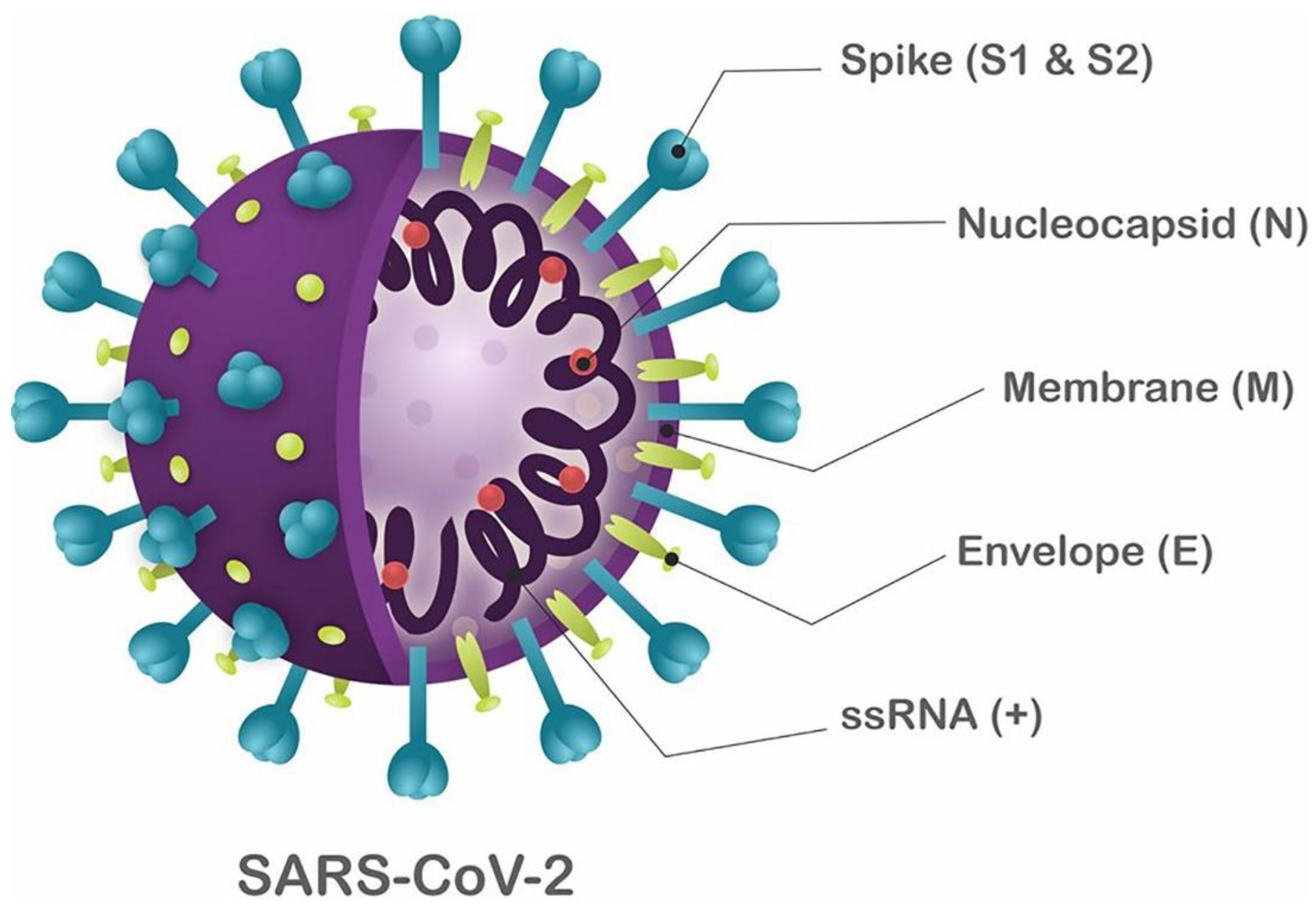
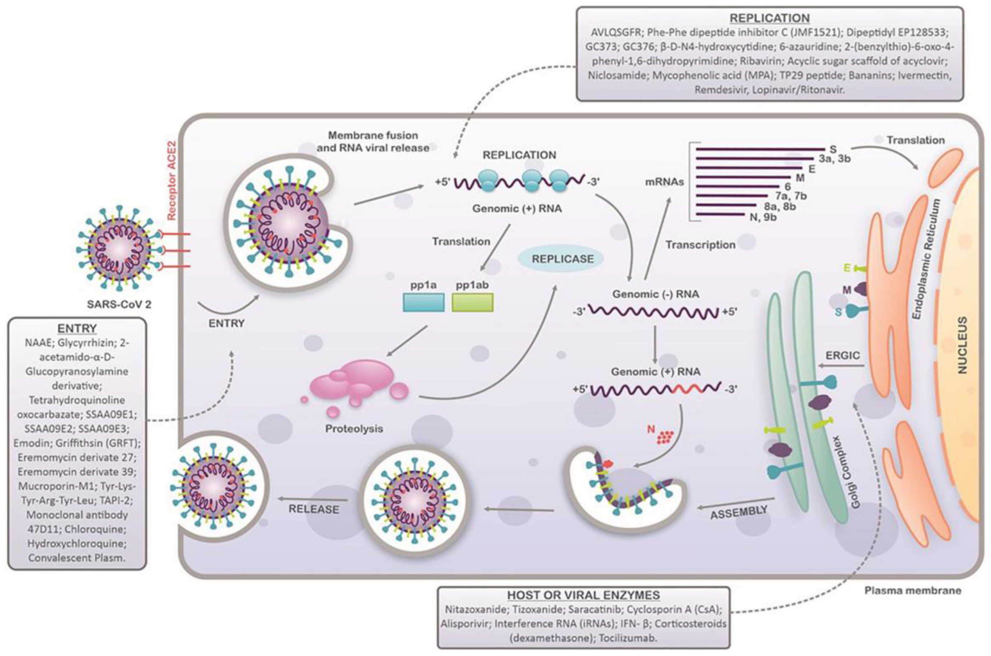
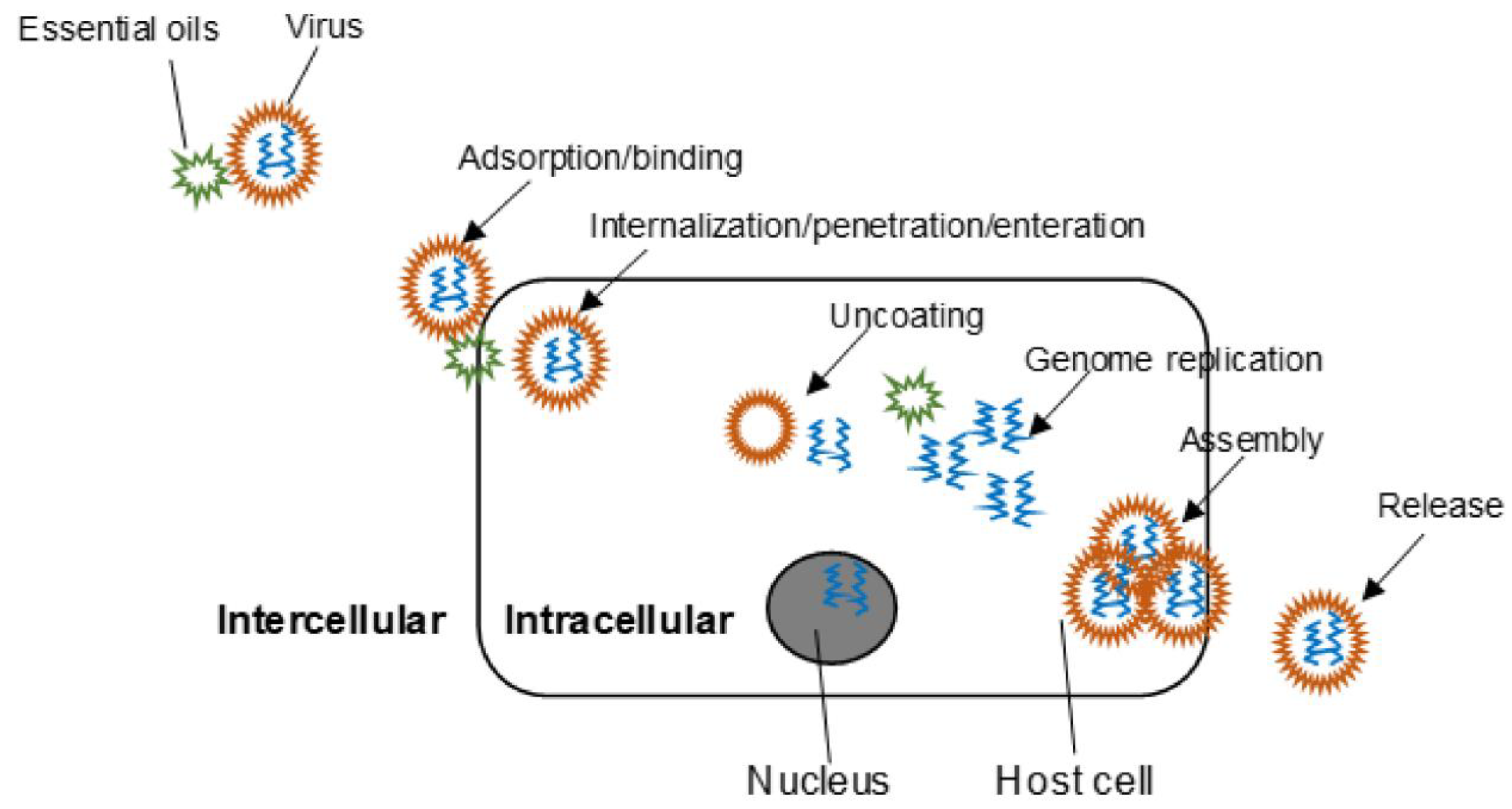
2. Virus Structure and Infection Mechanisms

For the development of antiviral materials, understanding the virus types, their structure, and infection mechanisms is paramount. Viruses are tiny opportunistic intracellular parasites with a structure consisting of an outer protein coat covering nucleic acid (RNA or DNA) in its core. A complete virus particle is called a virion. Viruses require a complex metabolic and biosynthetic machinery of eukaryotic or prokaryotic host cells for propagation and proliferation. Therefore, the virion transports its RNA or DNA genome to host cells for it to be transcribed and translated. This leads to the formation of new virus particles, where a new copy of the genome results from transcription, while their protein capsid is formed due to translation. The viral genome and linked proteins are wrapped in a symmetric protein capsid to form new virions. The nucleic acid-linked protein is called nucleoprotein, and together with the genome, it forms the nucleocapsid. In enveloped viruses, the nucleocapsid is encircled by a lipid bilayer derived from the modified host cell membrane and studded with an outer layer of virus envelope glycoproteins [26].
Viruses are classified based on their nucleic acid content, the shape of their protein capsid, their size, and the surrounding lipoprotein envelope. Their major taxonomic distribution involves two classes based on nucleic acid content: DNA and RNA viruses [27]. The DNA or RNA viruses are further sub-divided based on whether they have double-stranded or single-stranded DNA/RNA. An additional sub-division of the RNA viruses is carried out based on the segmentation of the RNA genome. Single-stranded RNA viruses are further classified into positive-sense viruses (i.e., RNA can be directly translated into proteins) or negative-sense viruses (i.e., RNA requires a polymerase for transcription into mRNA).
Coronaviruses are spherical, enveloped, single-stranded, positive-sense RNA viruses. In clinical practice, the most frequent coronaviruses are OC43, 229E, HKU1, and NL63, which characteristically depict common cold- and flu-related symptoms in immune-competent people. SARS-CoV-2 is the third virus in the coronavirus family that has globally stimulated serious ailments in humans [28] after Severe Acute Respiratory Syndrome (SARS) [29] and Middle East Respiratory Syndrome (MERS) [30].
SARS-CoV-2 has a spherical shape with a diameter ranging from 60 nm to 140 nm and distinctive spikes ranging from 9 nm to 12 nm. This gives SARS-CoV-2 virions a look similar to that of the solar corona (Figure 1) [31]. SARS-CoV-2 is assumed to infect new hosts by changing its spike protein and structure through genetic recombination and variation.
The virus infection cycle commences with the invasion of the host cell by the virion. The virion is adsorbed on the host cell surface and undergoes attachment in this step. After that, it either infiltrates the exterior layer of the host cell to enter the cytoplasm or instills its genetic material into the cell interior while the outer protein capsid and/or envelope relics at the surface of the host cell. A consequent uncoating step occurs inside the host cell when the virion structure infiltrates completely. This step releases genetic material from the virion to the host cell. In both scenarios, the virus’s genetic material cannot initiate protein synthesis until it is released from the virion structure.
In the case of coronaviruses (SARS-CoV and SARS-CoV-2), when viral infection occurs and the virion comes in contact with the host cell, the viral surface glycoprotein attaches to the ACE2 receptor on the host cell surface. As this happens, viral endocytosis is triggered, and endosome formation is initiated. The S glycoprotein comprises two subunits, S1 and S2. As endocytosis commences, the S1 subunit undergoes proteolytic cleavage by cellular proteases, exposing the S2 subunit, a fusion peptide responsible for fusing the viral envelope with the endosome membrane. This process ultimately releases the viral capsid, exposing the viral RNA. Following this, the single-stranded, positive-sense RNA of the virus is translated to produce nonstructural proteins that assemble to form a replicase–transcriptase complex (RTC) responsible for the RNA synthesis, replication, and transcription of nine subgenomic RNAs. These subgenomic RNAs are finally translated to generate S, E, and M structural proteins, which are forwarded to the endoplasmic reticulum (ER). In this cell organelle, the viral genomes are encapsulated by N proteins and assembled with these structural proteins to form new virions, which are finally transported to the cell surface in vesicles and released in a pathway mediated by exocytosis [32]. The basic infection cycle of a SARS-CoV-2 virion is depicted in Figure 2.

3. Antiviral Materials and Mechanism of Action
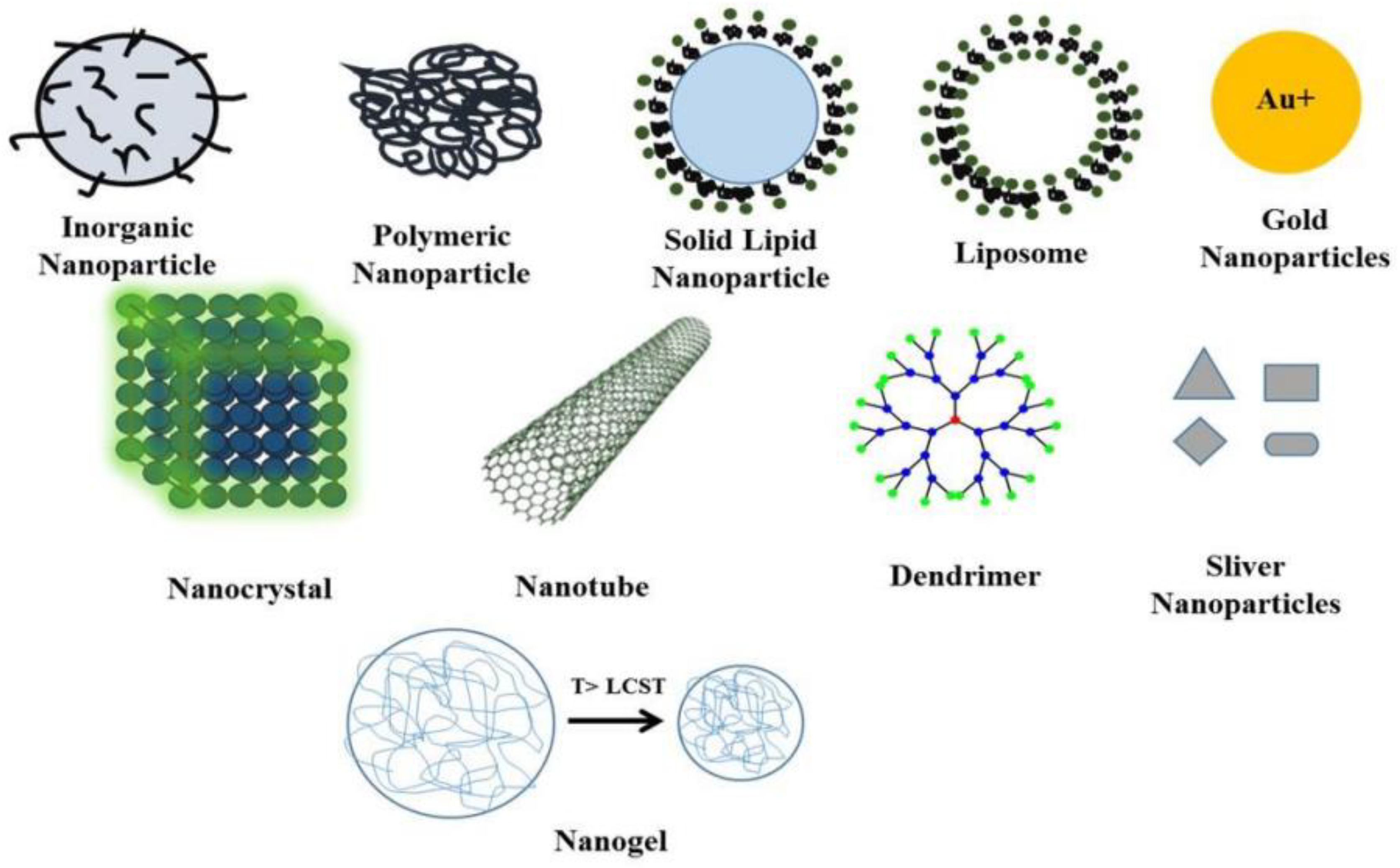
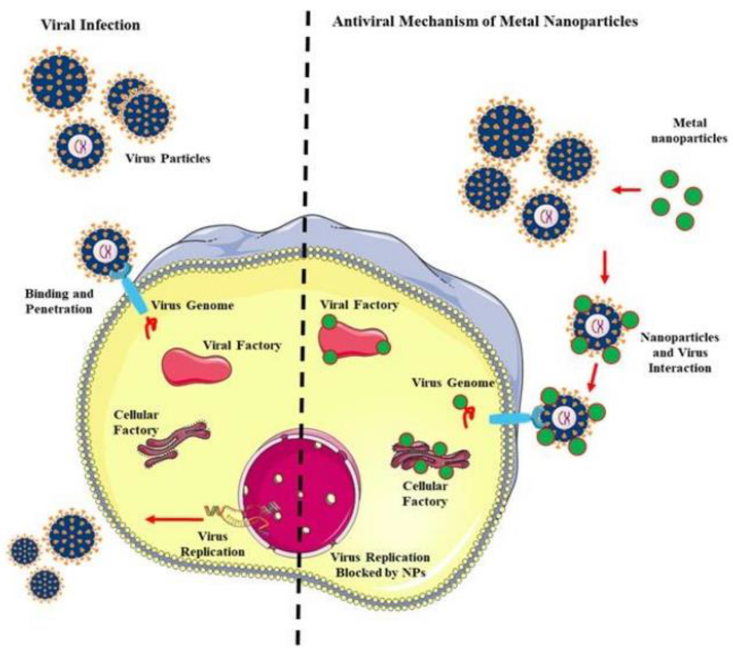
3.1. Nanomaterials

Nanoparticles are the most commonly employed antimicrobial agents, and they are known to be the most effective in inhibiting microbial growth. Specifically, it has been observed that many nanoparticles show excellent antiviral activity [33,34]. In this section, the antiviral properties of various types of nanoparticles, such as metal (silver and gold), metal oxide (zinc oxide and copper oxide), quantum dots, and other nanomaterials (graphene oxide, mesoporous silicon, functionalized nanoparticles, etc.), are briefly overviewed. Various types of nanomaterials known for antiviral applications are shown in Figure 3.
Even though silver nanoparticles have been the most popular antimicrobial agent used since ancient times [12], their antiviral activity has not been clearly elucidated. Studies show that silver nanoparticles effectively reduce the growth of many viruses, such as hepatitis, H1N1, influenza, HIV, HSV1, and HSV2 [21,35,36]. Silver nanoparticles reportedly bind to the viral surface of glycoproteins, blocking their adsorption and interaction with the target host cell. However, it is also known that silver nanoparticles generate reactive oxygen species (ROS) to exert an antiviral effect [34]. Besides silver, gold nanoparticles have also been extensively utilized in biomedical applications. Various researchers have reported that the antiviral activity of gold nanoparticles is effective against HSV1, hepatitis, influenza, HIV, and many more [37,38]. Similar to silver, gold nanoparticles also exert an antiviral effect by preventing the entry of virions into the host cells [34]. It is to be noted that the antiviral action of metallic nanoparticles is highly reliant on the size and shape of the nanoparticles [39]. Moreover, surface modifications play a major role in determining their efficacy [34].
Metal oxide nanoparticles are another most widely studied class of antimicrobial nanoparticles. Zinc oxide nanoparticles have been extensively studied and reported to exhibit antiviral activity [40,41,42].
Surface-modified zinc oxide nanoparticles can block the multiplication of HSV1, HSV2, and H1N1 viruses [42,43]. In addition to blocking the interaction between virions and host cells, zinc oxide nanoparticles are known to exert antiviral effects by releasing Zn2+ ions in host cells and increasing their intracellular concentration, which results in a hindered replication of a variety of RNA viruses [44]. Copper oxide has also been used to control viral infection. Copper oxide-incorporated antiviral materials have been found to effectively control HSV1 [45,46,47].
Apart from metal and metal oxides, other nanoparticles also possess antiviral potential. Graphene oxide in its native form, or after functionalization, is also known to show antiviral activity against RSV, HSV1, and HIV viruses [48,49]. The antiviral action of graphene oxide is mainly presumed to be due to the inactivation of the virus and most likely due to the inhibition of viral replication and hindrance in host–virus attachments [50]. Ag2S is also antiviral against RNA viruses such as diarrhea viruses [51].
Quantum dots are zero-dimensional nanoparticles, usually below 10 nm in size. They are emerging nanomaterials that have been used in the biomedical field, especially for cell imaging, due to their photoluminescence property [52]. Although quantum dots have mostly been used in electronics and related applications, some recent studies focused exclusively on their immense antimicrobial potential [53]. Recently, the antiviral activity of CdTe quantum dots against pseudorabies virus has been reported [54]. The antiviral action is mainly due to the inhibition of the formation of viral antisense RNA. Recently, the antiviral potential of low toxic carbon dots has been established [55,56]. Carbon dots effectively eliminate diarrhea viruses, porcine parvovirus, and adenovirus [57].
Nanomesoporous silicon is another candidate effective against many viruses, such as VEEV and HIV [58]. Silicon-based nanomaterials can also act as drug carriers [59]. Silicon-based antiviral nanomaterials are superior to their metallic counterparts due to their excellent stability and lower toxicity [60].
Moreover, some organic nanoparticles have exhibited decent antiviral efficiency. Polyhexylcyanoacrylate nanoparticles have been reported to inhibit HIV. Poly(lactic-co-glycolic acid) (PLGA) nanoparticles have been found to block the replication of the HCV virus [61,62]. Polymer-based nanomaterials are also known as suitable antiviral agents due to their ability to carry and sustainably release antiviral drugs [63].
As already discussed in previous sections, viral infection comprises multiple stages (Figure 2). Nanoparticles exert antimicrobial activity using several mechanisms as illustrated in Figure 4. The most common is the hindrance caused by nanoparticles in the attachment of virus cells to the host cells’ surface receptor proteins [64]. Engineered nanoparticles can block this primary viral infection step, thus protecting the host from infection [65]. Viral infection can also be inhibited by hindering the entry of the virus inside the target cell. In this context, nanoparticles can effectively modify the host cell membrane and surface protein structures such that the virions are left unable to penetrate the host cells [34]. Another effective strategy to control viral infection is to obstruct viral replication by regulating the enzymatic machinery for viral DNA or RNA replication [64]. The last strategy to prevent viral infection is preventing the budding of the host-infected virus [66]. It is a well-known phenomenon that the progenies of viruses can be more virulent to the host than the parent virion. To this end, engineered functional nanoparticles can be useful to block budding processes. The antiviral activity mechanisms of various kinds of nanoparticles are provided in Table 1.

3.2. Natural Oils and Extracts

Natural compounds, such as essential oils and plant extracts, are also gaining attention as antiviral materials. The main functionality of essential oils and natural extracts originates from their bioactive compounds, especially the polyphenols that are present in them. These compounds are generally recognized as safe (GRAS) and are suitable for consumption without any side effects [18]. There are numerous higher plan species (~17,000) where essential oils are present, and over 3000 types have already been used [67]. At the same time, the antiviral properties of many of these essential oils have already been established [68]. This section briefly overviews the antiviral properties and potential of various plant-based materials.
In the case of the influenza virus, essential oils from sources, such as oregano, artemisia, and salvia, were found to be very effective antiviral agents [18,68]. To inhibit HSV1 and HSV2 infection, essential oils, such as anise, tea tree, eucalyptus, ginger, and artemisia, were used [69,70,71]. Moreover, several essential oils were also found to be very effective against many other viruses, such as dengue, polio, tobacco mosaic virus, herpes, mumps, and HIV [71,72]. Interestingly, many essential oils were found to be highly effective in controlling the novel coronavirus strain SARS-CoV-2, responsible for the COVID 19 pandemic [72,73,74].
Plant-based antiviral compounds commonly hinder viral growth and replication in a dose-dependent manner. In this regard, it has been observed that essential oils exhibit more efficient antiviral activity than commercially available drugs [68]. Even drug-resistant viral strains have been found to be vulnerable to exposure to essential oils. For instance, HSV1 virus strains were reportedly inhibited by Salvia desoleana essential oils [75]. Clove (Syzygium aromaticum) essential oils are effective against different viruses, such as herpes adenovirus, poliovirus, and coxsackievirus [18]. Moreover, it has also been revealed that essential oils can effectively suppress viral infection with drugs. Recently, the combination of oseltamivir and Melissa officinalis essential oils showed synergistic effects against influenza virus H9N2 [76]. Besides essential oils, natural extracts from several medicinal plants also possess strong antiviral effects. Tribulus (Tribulus terrestris) extracts are reported to be composed of several flavonoids, tannin, and phenolic acids, which make them biologically active and antiviral against HIV [18]. Anti-HIV activity is also found in turmeric (Curcuma longa), which has curcumin as the active component. The antiviral activity and other curcumin biological activities were previously verified and reported by several researchers [77]. Cinnamon (Cinnamomum verum and Cinnamomum zeylanicum) and ginger (Zingiber officinale) extracts also possess high anti-influenza virus activities [18].
The mechanism of the antiviral action of essential oils is dependent on various factors. The first and foremost factor is the time that they are added during the viral infection cycle [68]. For further insight into antiviral action, research on the morphological change in viruses must be conducted. Essential oils can destroy or mask the action of a virus, and electron microscopy has been used to study this aspect [78]. Earlier, it was reported that murine norovirus treated with oregano essential oils had slightly modified morphology, while treatment with carvacrol showed capsid disintegration [78]. Another key strategy of antiviral activity is protein inhibition. Generally, viral surface proteins (such as hemagglutinin) help in the attachment and invasion of the virus in the host cell, while some other proteins (Tat protein) help in viral transcription [79]. Recently, it has been reported that cedar leaf essential oils hinder the hemagglutinin protein, while thyme essential oils destabilize the Tat protein [80,81]. Hence, essential oils render the viral metabolic machinery inactive by inhibiting protein functions, resulting in antiviral effects. The target sites of the antiviral action of essential oils are shown in Figure 5.

3.3. Biopolymers

Polymeric materials are extensively used to develop various packaging materials, including active packaging and smart packaging. Packaging systems should improve food safety and enhance the food’s shelf life by protecting and maintaining its quality. Polymeric materials possess characteristic features, such as easy processability, abundance, and surface chemistry, that make them suitable for use in the development of antiviral active packaging materials. Moreover, polymers are capable of easy strategic modifications, including chemical modifications, physical modifications, blending, and incorporation with active materials. Additionally, biopolymeric materials have been considered noteworthy for food packaging due to their biodegradability and excellent sustained-release properties, making them a lucrative option for use as a base material incorporated with various active components [13,82]. Various biopolymers incorporated with nanomaterials, essential oils, and plant extracts have been used to fabricate antiviral packaging film and coatings (Table 2). These studies demonstrate the utilization of biopolymers as carrier materials for active components with antiviral activities [16]. The active components are trapped in polymeric materials and can be released depending on the storage conditions, such as relative humidity, temperature, and pH.
Apart from utilizing biopolymers in the form of carrier matrices to active antiviral components, several biopolymers are known to possess antiviral properties themselves. Although they are widely distributed in several classes of biopolymers, a few important ones are heparin, dextran sulfate, sulfoevernan, cellulose sulfate, carrageenan, chondroitin polysulfate, chitosan, hyaluronic acid, etc. These polymers display decent antiviral activity [83,84].
Polyanionic biopolymers, including sulfated polysaccharides, such as dextran sulfate, heparin, and agar, have been observed to show excellent antiviral activities [84]. The mechanism by which polyanions exert an antiviral effect is adsorption inhibition. They block cell attachment of the virions by adsorbing on the virus surface and preventing their interaction with host cells (Figure 6). However, this virion–biopolymer interaction is also reversible and, hence, not efficient. Another reported mechanism involves the production of interferons by host cells on exposure to anionic polysaccharides [85]. Interferons are signaling proteins released by a virus-infected cell to warn nearby cells so that they can prepare and defend themselves against possible viral infection. It has been clinically confirmed that sulfated polysaccharides, dextran sulfate, and heparin are strong HIV inhibitors [86]. Additionally, most polysaccharides obtained from sea algae, such as carrageenans, fucans, alginates, galactans, naviculans, and sea algae extract, are known to possess strong broad-spectrum antiviral activity [84]. It has also been reported that the antiviral activity of sulfated polysaccharides depends on their degree of sulfation (DS). DS refers to the number of sulfate groups per monosaccharide unit. The λ-carrageenan (DS = 35%) is observed to possess a 10 times higher antiviral potency against HIV than κ-carrageenan (DS = 25%) [86]. Other non-sulfated polyanions such as polyhydroxycarboxylates also possess antiviral activity [84,85]. Non-sulfated mucopolysaccharide hyaluronic acid has also been reported as a potential antiviral compound. It was reported as a wide-spectrum antiviral material inhibiting the growth of coxsackievirus B5, mumps virus, influenza virus, herpes simplex virus-1, etc. [87].
Besides polyanionic biopolymers, biopolymers with a polycationic character, such as chitosan, polylysine, polyarginine, and cationic dextran derivatives, are also reported to be antiviral. The mechanism of antiviral action of polycationic biopolymers involves their electrostatic interaction with the viral capsids and their destabilizations. Moreover, they can inhibit virus–host cell interactions by binding to the negatively charged domains on the virus surface [83,88]. Chitosan has been used for the inhibition of norovirus. However, despite demonstrating an insufficient effect, some methods have been reported to elicit its antiviral effect—higher concentrations, longer incubation times, a higher degree of deacetylation, and higher molecular weight can result in the enhanced antiviral activity of chitosan [83,88].
Therefore, several biopolymers widely studied in food packaging applications possess innate antiviral activity that has not been explored until recently. As discussed in the Introduction, the first signs of packaging-based cross-contamination from the SARS-CoV-2 virus emerged from animal product packaging. There have been several studies where ionic polysaccharides, such as alginate [89], agar [53], carrageenan [90], and chitosan [91], have been suggested for the packaging of animal products. Since it is widely reported that these biodegradable food packaging materials are naturally antiviral, it can be logically presumed that they can protect packaged food from viral infections. Hence, biodegradable polymer materials could be used as antiviral packaging materials, especially those based on ionic polysaccharides.

4. Current Developments in Antiviral Food Packaging

Antiviral coatings and packaging systems are potential candidates for improving food safety and quality against foodborne pathogens [16,92]. As already discussed, the contamination of food products by viral pathogens occurs in seafood, vegetable products, and fruit products, and it is a prime reason for the outbreak of viral diseases. Additionally, the packaging material is suspected to play a critical role in indirectly transmitting viral infection via cross-contamination. There has been a surge in research and development to fabricate antiviral materials for targeted food packaging, such as active coatings, films, and multilayer packaging systems (Table 2). As described in the previous sections, biopolymers, essential oils, active components from plant materials, and antiviral active nanomaterials have been widely used in antiviral food packaging. In this section, the use of multilayer systems and edible films and coatings with antiviral activity for the packaging of food products is discussed.
Multilayered packaging systems are developed to improve packaging properties, such as barrier properties, mechanical properties, and storage stability. However, adding antiviral materials as a layer to the packaging system is a strategic route to develop antiviral packaging systems. Multilayered packaging systems are developed following lamination, coextrusion, coating technologies, etc. Functional carbon dots, metal-nanoparticle-based graphene oxide, and other nanomaterial-based coatings can be used as antiviral materials to develop antiviral food packaging [92,93]. In active packaging, the organic compound cinnamaldehyde with virucidal activity was used to develop biodegradable multilayer systems [94], where the efficacy of cinnamaldehyde against norovirus surrogates, murine norovirus, feline calicivirus, and hepatitis A virus was tested. Some researchers have also reported biodegradable antiviral coatings on non-biodegradable packaging materials. Antiviral materials based on rosemary, raspberry, and pomegranate extracts, prepared by supercritical CO₂ extraction, as active coatings are used to cover low-density polyethylene (LDPE) films in order to develop functional food packaging, where the antiviral activity has been found to decrease the number of Φ6 bacteriophage (a surrogate for airborne viruses) [95,96]. Additionally, ZnO nanoparticles, carvacrol, and geraniol-based antiviral external coatings have been found to be effective against Φ6 bacteriophage and can be used to develop multilayer active polyethylene packaging [97]. An electrospun coating based on silver nanoparticles, silver nitrate, and polyhydroxyalkanoates was tested against norovirus surrogates [98]. In a dose-dependent manner (2.1 and 21 mg/L), the silver ions and nanoparticles could decrease norovirus surrogates (feline calicivirus and murine norovirus).
Moreover, the safety of food products can also be improved by developing antiviral active edible coatings and film materials. Edible films and coatings differing in their method of application on food products are developed from biodegradable materials that possess biodegradability similar to that of the food products [99]. An edible coating is an edible food packaging system where a thin layer of edible materials is applied to food products for improved shelf life and quality [99]. Edible food packaging is fabricated from biomaterials, including polysaccharides, proteins, and lipids. At the same time, incorporating edible active components, including plant extract, essential oils, and other active polymeric materials, into edible coating solutions can provide them with the required antiviral functionality [100]. However, edible films are used as sandwich materials or as a wrapper on food products for improved food quality and properties [101]. Nanomaterials and nanomaterial-based coating with antiviral activity protect from viral transmission [92]. The use of edible films and coatings can significantly reduce the viral contaminations on the surface of the food products without affecting the physicochemical properties of the food products.
In this regard, several antiviral films and coatings have been reported. Edible films and coatings based on chitosan and grape seed extract effectively reduced foodborne viruses such as human norovirus [19]. A solution of chitosan (2% w/w) with added grape seed extract (5%) reduced the virus by 4.00 log PFU/mL after 3 h [19]. Active edible films based on alginate, lipid, grape seed extract, and green tea extract provide antiviral activity tested against murine norovirus and hepatitis A virus [82]. A 2-log reduction for alginate films incorporated with 0.75 g of extract (per g of alginate) was reported. Green tea extract was found to be more efficient than grape seed extract, and it provided more potency against infection. Another study reported edible coatings based on carrageenan and green tea extract and their application to blueberries and raspberries. The addition of green tea extract improved antiviral activity at ambient and refrigerated temperatures [20]. Additionally, the effectiveness of a food-grade edible coating based on alginate-oleic acid incorporated with green tea extract exhibited more antiviral efficacy in strawberries than in raspberries [102]. Compared to control, a reduction of 1.5–2 log of murine norovirus and hepatitis A virus has been observed in fresh strawberries at 10 °C for 4 days’ storage. The development of edible antiviral coatings of Persian gum, gelatin, and allyl isothiocyanate was also effective against murine norovirus. At refrigerated conditions, the addition of allyl isothiocyanate improved the antiviral property of Persian gum [103]. Thus, several antiviral active edible films or coatings on food products have been developed, and they can be useful to reduce viral contamination and improve food safety and quality.
Table 2. Antiviral active packaging system for food products.
ComponentsTypes of PackagingTested AgainstReference
Cinnamaldehyde
Zein
Polyhydroxybutyrate
Biodegradable multilayer systemMurine norovirus
Feline calicivirus
Hepatitis A virus
[94]
Chitosan
Grape seed extract
Edible coatings and edible filmsMurine norovirus
(MNV-1)
[19]
ZnO
Carvacrol
Geraniol
Active external coating Φ6 bacteriophage[97]
Polyethylene film
Rosemary, raspberry, and pomegranate extracts
Films covered with active coatings
(functional food packaging)
Φ6 bacteriophage[95,96]
Alginate
Lipid
Grape seed extract
Green tea extract
Emulsified edible filmsMurine norovirus
Hepatitis A virus
[82]
Silver nanoparticles
Silver nitrate
Polyhydroxyalkanoates
Electrospun coatingNorovirus surrogates[98]
Carrageenan
Green tea extract
Edible coatingMurine norovirus
Hepatitis A virus
[20]
Alginate
Oleic acid
Green tea extract
Edible coatingHuman norovirus
Hepatitis A virus
[102]
Persian gum
Gelatin
Allyl isothiocyanate
Antiviral edible coatingMurine norovirus[103]

5. Conclusions and Future Perspectives

Since the onset of the SARS-CoV-2 virus-related global pandemic in 2019, the food industry has faced many impediments, especially in food packaging. Since the majority of the packaging material used throughout the world involves the use of non-biodegradable plastics, it has caused two major issues: (a) with the global lockdown and upsurge in food delivery services to meet the hunger needs of people, the utilization of plastic-based packaging increased, which, in turn, led to an increase in non-biodegradable municipal solid waste; (b) there had been reports on the transmission of viral infection due to cross-contamination caused by the packaging material while in use or even after disposal, as the coronavirus actively persists on plastic for long periods of 72 h.
Biopolymer-based food packaging materials are possible alternatives to non-biodegradable packaging and can help solve these issues. Since biopolymers are biodegradable, their contribution to municipal solid waste will be markedly reduced. Moreover, several biopolymers, such as alginate, carrageenan, and chitosan, commonly used in the fabrication of biodegradable food packaging films, have been reported to possess antiviral activity. Packaging materials made from antiviral biopolymers will prevent the persistence of viral particles on their surfaces and, hence, will avert cross-contamination. Furthermore, incorporating sustainable additives into these polymer films can enhance the antiviral potential of these films. Antiviral additives, such as nanomaterials, natural oils, and herbal extracts, will help facilitate the packaging material’s physicochemical properties while contributing to its antiviral efficacy. From a sustainability perspective, biopolymer films incorporated with natural oils and plant extracts could be a completely natural, economic, safer, and eco-friendly option for the fabrication of biopolymer-based antiviral packaging.
Biodegradable polymeric materials incorporated with natural oils and plant extracts have long been studied for potential food packaging applications. Although these materials have been widely studied for their several functionalities, such as antibacterial, antifungal, and antioxidant properties, there is a scarcity of reports discussing their antiviral food packaging properties. However, independent reports elaboratively discuss the antiviral properties of ionic biopolymers, plant extracts, and essential oils, which can help researchers reach a logical conclusion that many of the biodegradable packaging materials studied to date tend to possess antiviral functionality. However, concrete quantitative and qualitative research is still needed to prove this hypothesis. To ensure food safety and sustainability, exploring the potential of natural antiviral bioactive components in food packaging is essential. Moreover, it is presumed that the demand for biodegradable antiviral food packaging and coatings will increase further in the post-pandemic period, and efforts are required to analyze the practicality of these natural antiviral materials and their potential to be quickly commercialized.

Author Contributions

Conceptualization, R.P.; investigation, R.P. and S.D.P.; writing—original draft, R.P., S.D.P., S.R. and T.G.; writing—review and editing, J.-W.R., R.P. and S.S.H.; visualization, R.P. and S.D.P.; funding acquisition, J.-W.R.; supervision, J.-W.R. and S.S.H. All authors have read and agreed to the published version of the manuscript.

Funding

This research was supported by the Brain Pool Program funded by the Ministry of Science, ICT and Future Planning through the National Research Foundation of Korea (2019H1D3A1A01070715), and the National Research Foundation of Korea (NRF) grant funded by the Korean government (MSIT) (2022R1A2B5B02001422).

Institutional Review Board Statement

Not applicable.

Informed Consent Statement

Not applicable.

Conflicts of Interest

The authors declare there are no conflicts of interest.

References

  1. Wu, Y.-C.; Chen, C.-S.; Chan, Y.-J. The Outbreak of COVID-19: An Overview. J. Chin. Med. Assoc. 2020, 83, 217–220. [Google Scholar] [CrossRef] [PubMed]
  2. Zhou, P.; Yang, X.L.; Wang, X.G.; Hu, B.; Zhang, L.; Zhang, W.; Si, H.R.; Zhu, Y.; Li, B.; Huang, C.L.; et al. A Pneumonia Outbreak Associated with a New Coronavirus of Probable Bat Origin. Nature 2020, 579, 270–273. [Google Scholar] [CrossRef] [PubMed] [Green Version]
  3. Kahn, J.S.; McIntosh, K. History and Recent Advances in Coronavirus Discovery. Pediatr. Infect. Dis. J. 2005, 24, S223–S227. [Google Scholar] [CrossRef]
  4. Yan, Y.; Shin, W.I.; Pang, Y.X.; Meng, Y.; Lai, J.; You, C.; Zhao, H.; Lester, E.; Wu, T.; Pang, C.H. The First 75 Days of Novel Coronavirus (SARS-CoV-2) Outbreak: Recent Advances, Prevention, and Treatment. Int. J. Environ. Res. Public Health 2020, 17, 2323. [Google Scholar] [CrossRef] [PubMed] [Green Version]
  5. De Oliveira, W.Q.; de Azeredo, H.M.C.; Neri-Numa, I.A.; Pastore, G.M. Food Packaging Wastes amid the COVID-19 Pandemic: Trends and Challenges. Trends Food Sci. Technol. 2021, 116, 1195–1199. [Google Scholar] [CrossRef] [PubMed]
  6. Klompas, M.; Baker, M.A.; Rhee, C. Airborne Transmission of SARS-CoV-2: Theoretical Considerations and Available Evidence. JAMA 2020, 324, 441–442. [Google Scholar] [CrossRef] [PubMed]
  7. Jyoti; Bhattacharya, B. Impact of COVID-19 in Food Industries and Potential Innovations in Food Packaging to Combat the Pandemic—A Review. Sci. Agropecu. 2021, 12, 133–140. [Google Scholar] [CrossRef]
  8. Fabra, M.J.; López-Rubio, A.; Lagaron, J.M. Biopolymers for Food Packaging Applications. In Smart Polymers And Their Applications; Woodhead Publishing: Sawston, UK, 2014; pp. 476–509. [Google Scholar] [CrossRef]
  9. Grujić, R.; Vujadinović, D.; Savanović, D. Biopolymers as Food Packaging Materials. In Advances In Applications of Industrial Biomaterials; Springer: Cham, Switzerland, 2017; pp. 139–160. [Google Scholar] [CrossRef]
  10. Adeyeye, O.A.; Sadiku, E.R.; Babu Reddy, A.; Ndamase, A.S.; Makgatho, G.; Sellamuthu, P.S.; Perumal, A.B.; Nambiar, R.B.; Fasiku, V.O.; Ibrahim, I.D.; et al. The Use of Biopolymers in Food Packaging. In Green Biopolymers and Their Nanocomposites; Springer: Singapore, 2019; pp. 137–158. [Google Scholar] [CrossRef]
  11. Porta, R.; Sabbah, M.; Di Pierro, P. Biopolymers as Food Packaging Materials. Int. J. Mol. Sci. 2020, 21, 4942. [Google Scholar] [CrossRef]
  12. Priyadarshi, R.; Roy, S.; Ghosh, T.; Biswas, D.; Rhim, J.-W. Antimicrobial Nanofillers Reinforced Biopolymer Composite Films for Active Food Packaging Applications—A Review. Sustain. Mater. Technol. 2021, e00353. [Google Scholar] [CrossRef]
  13. Priyadarshi, R.; Rhim, J.-W. Chitosan-Based Biodegradable Functional Films for Food Packaging Applications. Innov. Food Sci. Emerg. Technol. 2020, 62, 102346. [Google Scholar] [CrossRef]
  14. Shankar, S.; Rhim, J.-W. Antimicrobial Wrapping Paper Coated with a Ternary Blend of Carbohydrates (Alginate, Carboxymethyl Cellulose, Carrageenan) and Grapefruit Seed Extract. Carbohydr. Polym. 2018, 196, 92–101. [Google Scholar] [CrossRef] [PubMed]
  15. Balasubramaniam, B.; Prateek; Ranjan, S.; Saraf, M.; Kar, P.; Singh, S.P.; Thakur, V.K.; Singh, A.; Gupta, R.K. Antibacterial and Antiviral Functional Materials: Chemistry and Biological Activity toward Tackling COVID-19-like Pandemics. ACS Pharmacol. Transl. Sci. 2020, 4, 8–54. [Google Scholar] [CrossRef] [PubMed]
  16. Randazzo, W.; Fabra, M.J.; Falcó, I.; López-Rubio, A.; Sánchez, G. Polymers and Biopolymers with Antiviral Activity: Potential Applications for Improving Food Safety. Compr. Rev. Food Sci. Food Saf. 2018, 17, 754–768. [Google Scholar] [CrossRef] [Green Version]
  17. Sha-Tshibey Tshibangu, D.; Liyongo Inkoto, C.; T Tshibangu, D.S.; Matondo, A.; Lengbiye, E.M.; Inkoto, C.L.; Ngoyi, E.M.; Kabengele, C.N.; Bongo, G.N.; Gbolo, B.Z.; et al. Possible Effect of Aromatic Plants and Essential Oils against COVID-19: Review of Their Antiviral Activity. J. Complement. Altern. Med. Res. 2020, 11, 10–22. [Google Scholar] [CrossRef]
  18. Parham, S.; Kharazi, A.Z.; Bakhsheshi-Rad, H.R.; Nur, H.; Ismail, A.F.; Sharif, S.; Ramakrishna, S.; Berto, F. Antioxidant, Antimicrobial and Antiviral Properties of Herbal Materials. Antioxidants 2020, 9, 1309. [Google Scholar] [CrossRef] [PubMed]
  19. Amankwaah, C.; Li, J.; Lee, J.; Pascall, M.A. Development of Antiviral and Bacteriostatic Chitosan-Based Food Packaging Material with Grape Seed Extract for Murine Norovirus, Escherichia coli, and Listeria innocua Control. Food Sci. Nutr. 2020, 8, 6174–6181. [Google Scholar] [CrossRef]
  20. Falcó, I.; Randazzo, W.; Sánchez, G.; López-Rubio, A.; Fabra, M.J. On the Use of Carrageenan Matrices for the Development of Antiviral Edible Coatings of Interest in Berries. Food Hydrocoll. 2019, 92, 74–85. [Google Scholar] [CrossRef]
  21. Dung, T.T.N.; Nam, V.N.; Nhan, T.T.; Ngoc, T.T.B.; Minh, L.Q.; Nga, B.T.T.; Phan Le, V.; Quang, D.V. Silver Nanoparticles as Potential Antiviral Agents against African Swine Fever Virus. Mater. Res. Express 2020, 6, 1250g9. [Google Scholar] [CrossRef]
  22. Shu, F.; Jiang, B.; Yuan, Y.; Li, M.; Wu, W.; Jin, Y.; Xiao, H. Biological Activities and Emerging Roles of Lignin and Lignin-Based Products—A Review. Biomacromolecules 2021, 22, 4905–4918. [Google Scholar] [CrossRef]
  23. Santoyo, S.; Jaime, L.; García-Risco, M.R.; Lopez-Hazas, M.; Reglero, G. Supercritical Fluid Extraction as an Alternative Process to Obtain Antiviral Agents from Thyme Species. Ind. Crops Prod. 2014, 52, 475–480. [Google Scholar] [CrossRef]
  24. Schnitzler, P.; Schön, K.; Reichling, J. Antiviral Activity of Australian Tea Tree Oil and Eucalyptus Oil against Herpes Simplex Virus in Cell Culture. Pharmazie 2001, 56, 343–347. [Google Scholar] [PubMed]
  25. Vicidomini, C.; Roviello, V.; Roviello, G.N. Molecular Basis of the Therapeutical Potential of Clove (Syzygium aromaticum L.) and Clues to Its Anti-COVID-19 Utility. Molecules 2021, 26, 1880. [Google Scholar] [CrossRef] [PubMed]
  26. Brown, T.A. Gene Cloning and DNA Analysis: An Introduction; Brown, T.A., Ed.; Blackwell Science: Oxford, UK, 2001. [Google Scholar]
  27. Gelderblom, H.R. Structure and Classification of Viruses. In Medical Microbiology; Baron, S., Ed.; University of Texas Medical Branch at Galveston: Galveston, TX, USA, 1996; ISBN 0963117211. [Google Scholar]
  28. Zhu, N.; Zhang, D.; Wang, W.; Li, X.; Yang, B.; Song, J.; Zhao, X.; Huang, B.; Shi, W.; Lu, R.; et al. A Novel Coronavirus from Patients with Pneumonia in China, 2019. N. Engl. J. Med. 2020, 382, 727–733. [Google Scholar] [CrossRef] [PubMed]
  29. Zhong, N.S.; Zheng, B.J.; Li, Y.M.; Poon, L.L.M.; Xie, Z.H.; Chan, K.H.; Li, P.H.; Tan, S.Y.; Chang, Q.; Xie, J.P.; et al. Epidemiology and Cause of Severe Acute Respiratory Syndrome (SARS) in Guangdong, People’s Republic of China, in February 2003. Lancet 2003, 362, 1353–1358. [Google Scholar] [CrossRef] [Green Version]
  30. Zaki, A.M.; Van Boheemen, S.; Bestebroer, T.M.; Osterhaus, A.D.M.E.; Fouchier, R.A.M. Isolation of a Novel Coronavirus from a Man with Pneumonia in Saudi Arabia. N. Engl. J. Med. 2012, 367, 1814–1820. [Google Scholar] [CrossRef]
  31. Goldsmith, C.S.; Tatti, K.M.; Ksiazek, T.G.; Rollin, P.E.; Comer, J.A.; Lee, W.W.; Rota, P.A.; Bankamp, B.; Bellini, W.J.; Zaki, S.R. Ultrastructural Characterization of SARS Coronavirus. Emerg. Infect. Dis. 2004, 10, 320. [Google Scholar] [CrossRef]
  32. Santos, I.D.A.; Grosche, V.R.; Bergamini, F.R.G.; Sabino-Silva, R.; Jardim, A.C.G. Antivirals Against Coronaviruses: Candidate Drugs for SARS-CoV-2 Treatment? Front. Microbiol. 2020, 11, 1818. [Google Scholar] [CrossRef]
  33. Jazie, A.A.; Albaaji, A.J.; Abed, S.A. A Review on Recent Trends of Antiviral Nanoparticles and Airborne Filters: Special Insight on COVID-19 Virus. Air Qual. Atmos. Health 2021, 14, 1811–1824. [Google Scholar] [CrossRef]
  34. Gurunathan, S.; Qasim, M.; Choi, Y.; Do, J.T.; Park, C.; Hong, K.; Kim, J.H.; Song, H. Antiviral Potential of Nanoparticles—Can Nanoparticles Fight Against Coronaviruses? Nanomaterials 2020, 10, 1645. [Google Scholar] [CrossRef]
  35. Akbarzadeh, A.; Kafshdooz, L.; Razban, Z.; Dastranj Tbrizi, A.; Rasoulpour, S.; Khalilov, R.; Kavetskyy, T.; Saghfi, S.; Nasibova, A.N.; Kaamyabi, S.; et al. An Overview Application of Silver Nanoparticles in Inhibition of Herpes Simplex Virus. Artif. Cells Nanomed. Biotechnol. 2017, 46, 263–267. [Google Scholar] [CrossRef]
  36. Lin, Z.; Li, Y.; Guo, M.; Xu, T.; Wang, C.; Zhao, M.; Wang, H.; Chen, T.; Zhu, B. The Inhibition of H1N1 Influenza Virus-Induced Apoptosis by Silver Nanoparticles Functionalized with Zanamivir. RSC Adv. 2017, 7, 742–750. [Google Scholar] [CrossRef] [Green Version]
  37. Kim, J.; Yeom, M.; Lee, T.; Kim, H.O.; Na, W.; Kang, A.; Lim, J.W.; Park, G.; Park, C.; Song, D.; et al. Porous Gold Nanoparticles for Attenuating Infectivity of Influenza A Virus. J. Nanobiotechnol. 2020, 18, 1–11. [Google Scholar] [CrossRef] [PubMed] [Green Version]
  38. Lee, M.Y.; Yang, J.A.; Jung, H.S.; Beack, S.; Choi, J.E.; Hur, W.; Koo, H.; Kim, K.; Yoon, S.K.; Hahn, S.K. Hyaluronic Acid-Gold Nanoparticle/Interferon α Complex for Targeted Treatment of Hepatitis C Virus Infection. ACS Nano 2012, 6, 9522–9531. [Google Scholar] [CrossRef] [PubMed]
  39. Bai, Y.; Zhou, Y.; Liu, H.; Fang, L.; Liang, J.; Xiao, S. Glutathione-Stabilized Fluorescent Gold Nanoclusters Vary in Their Influences on the Proliferation of Pseudorabies Virus and Porcine Reproductive and Respiratory Syndrome Virus. ACS Appl. Nano Mater. 2018, 1, 969–976. [Google Scholar] [CrossRef]
  40. Priyadarshi, R.; Negi, Y.S. Effect of Varying Filler Concentration on Zinc Oxide Nanoparticle Embedded Chitosan Films as Potential Food Packaging Material. J. Polym. Environ. 2017, 25, 1087–1098. [Google Scholar] [CrossRef]
  41. Priyadarshi, R.; Kim, S.-M.; Rhim, J.-W. Carboxymethyl Cellulose-Based Multifunctional Film Combined with Zinc Oxide Nanoparticles and Grape Seed Extract for the Preservation of High-Fat Meat Products. Sustain. Mater. Technol. 2021, 29, e00325. [Google Scholar] [CrossRef]
  42. Ghaffari, H.; Tavakoli, A.; Moradi, A.; Tabarraei, A.; Bokharaei-Salim, F.; Zahmatkeshan, M.; Farahmand, M.; Javanmard, D.; Kiani, S.J.; Esghaei, M.; et al. Inhibition of H1N1 Influenza Virus Infection by Zinc Oxide Nanoparticles: Another Emerging Application of Nanomedicine. J. Biomed. Sci. 2019, 26, 1–10. [Google Scholar] [CrossRef]
  43. Tavakoli, A.; Ataei-Pirkooh, A.; Mm Sadeghi, G.; Bokharaei-Salim, F.; Sahrapour, P.; Kiani, S.J.; Moghoofei, M.; Farahmand, M.; Javanmard, D.; Monavari, S.H. Polyethylene Glycol-Coated Zinc Oxide Nanoparticle: An Efficient Nanoweapon to Fight against Herpes Simplex Virus Type 1. Nanomedicine 2018, 13, 2675–2690. [Google Scholar] [CrossRef]
  44. Ishida, T. Review on The Role of Zn2+ Ions in Viral Pathogenesis and the Effect of Zn2+ Ions for Host Cell-Virus Growth Inhibition. Am. J. Biomed. Sci. Res. 2019, 2, 28–37. [Google Scholar] [CrossRef] [Green Version]
  45. Tavakoli, A.; Hashemzadeh, M.S. Inhibition of Herpes Simplex Virus Type 1 by Copper Oxide Nanoparticles. J. Virol. Methods 2020, 275, 113688. [Google Scholar] [CrossRef]
  46. Mazurkow, J.M.; Yüzbasi, N.S.; Domagala, K.W.; Pfeiffer, S.; Kata, D.; Graule, T. Nano-Sized Copper (Oxide) on Alumina Granules for Water Filtration: Effect of Copper Oxidation State on Virus Removal Performance. Environ. Sci. Technol. 2020, 54, 1214–1222. [Google Scholar] [CrossRef] [PubMed]
  47. Merkl, P.; Long, S.; Mcinerney, G.M.; Sotiriou, G.A.; Ahonen, M.; Kogermann, K.; Pestryakov, A.; Mihailescu, I.N.; Papini, E. Antiviral Activity of Silver, Copper Oxide and Zinc Oxide Nanoparticle Coatings against SARS-CoV-2. Nanomaterials 2021, 11, 1312. [Google Scholar] [CrossRef] [PubMed]
  48. Sametband, M.; Kalt, I.; Gedanken, A.; Sarid, R. Herpes Simplex Virus Type-1 Attachment Inhibition by Functionalized Graphene Oxide. ACS Appl. Mater. Interfaces 2014, 6, 1228–1235. [Google Scholar] [CrossRef] [PubMed]
  49. Yang, X.X.; Li, C.M.; Li, Y.F.; Wang, J.; Huang, C.Z. Synergistic Antiviral Effect of Curcumin Functionalized Graphene Oxide against Respiratory Syncytial Virus Infection. Nanoscale 2017, 9, 16086–16092. [Google Scholar] [CrossRef] [PubMed]
  50. Iannazzo, D.; Pistone, A.; Ferro, S.; De Luca, L.; Monforte, A.M.; Romeo, R.; Buemi, M.R.; Pannecouque, C. Graphene Quantum Dots Based Systems As HIV Inhibitors. Bioconjug. Chem. 2018, 29, 3084–3093. [Google Scholar] [CrossRef] [PubMed]
  51. Du, T.; Liang, J.; Dong, N.; Lu, J.; Fu, Y.; Fang, L.; Xiao, S.; Han, H. Glutathione-Capped Ag2S Nanoclusters Inhibit Coronavirus Proliferation through Blockage of Viral RNA Synthesis and Budding. ACS Appl. Mater. Interfaces 2018, 10, 4369–4378. [Google Scholar] [CrossRef]
  52. Pal, A.; Arshad, F.; Sk, M.P. Emergence of Sulfur Quantum Dots: Unfolding Their Synthesis, Properties, and Applications. Adv. Colloid Interface Sci. 2020, 285, 102274. [Google Scholar] [CrossRef]
  53. Priyadarshi, R.; Riahi, Z.; Rhim, J.-W.; Han, S.; Lee, S.-G. Sulfur Quantum Dots as Fillers in Gelatin/Agar-Based Functional Food Packaging Films. ACS Appl. Nano Mater. 2021, 4, 14292–14302. [Google Scholar] [CrossRef]
  54. Du, T.; Cai, K.; Han, H.; Fang, L.; Liang, J.; Xiao, S. Probing the Interactions of CdTe Quantum Dots with Pseudorabies Virus. Sci. Rep. 2015, 5, 1–10. [Google Scholar] [CrossRef]
  55. Łoczechin, A.; Séron, K.; Barras, A.; Giovanelli, E.; Belouzard, S.; Chen, Y.T.; Metzler-Nolte, N.; Boukherroub, R.; Dubuisson, J.; Szunerits, S. Functional Carbon Quantum Dots as Medical Countermeasures to Human Coronavirus. ACS Appl. Mater. Interfaces 2019, 11, 42964–42974. [Google Scholar] [CrossRef]
  56. Lin, C.J.; Chang, L.; Chu, H.W.; Lin, H.J.; Chang, P.C.; Wang, R.Y.; Unnikrishnan, B.; Mao, J.Y.; Chen, S.Y.; Huang, C.C. High Amplification of the Antiviral Activity of Curcumin through Transformation into Carbon Quantum Dots. Small 2019, 15, 1902641. [Google Scholar] [CrossRef] [PubMed]
  57. Huang, S.; Gu, J.; Ye, J.; Fang, B.; Wan, S.; Wang, C.; Ashraf, U.; Li, Q.; Wang, X.; Shao, L.; et al. Benzoxazine Monomer Derived Carbon Dots as a Broad-Spectrum Agent to Block Viral Infectivity. J. Colloid Interface Sci. 2019, 542, 198–206. [Google Scholar] [CrossRef] [PubMed]
  58. De Souza E Silva, J.M.; Hanchuk, T.D.M.; Santos, M.I.; Kobarg, J.; Bajgelman, M.C.; Cardoso, M.B. Viral Inhibition Mechanism Mediated by Surface-Modified Silica Nanoparticles. ACS Appl. Mater. Interfaces 2016, 8, 16564–16572. [Google Scholar] [CrossRef] [PubMed]
  59. LaBauve, A.E.; Rinker, T.E.; Noureddine, A.; Serda, R.E.; Howe, J.Y.; Sherman, M.B.; Rasley, A.; Brinker, C.J.; Sasaki, D.Y.; Negrete, O.A. Lipid-Coated Mesoporous Silica Nanoparticles for the Delivery of the ML336 Antiviral to Inhibit Encephalitic Alphavirus Infection. Sci. Rep. 2018, 8, 13990. [Google Scholar] [CrossRef] [Green Version]
  60. Bimbo, L.M.; Denisova, O.V.; Mäkilä, E.; Kaasalainen, M.; De Brabander, J.K.; Hirvonen, J.; Salonen, J.; Kakkola, L.; Kainov, D.; Santos, H.A. Inhibition of Influenza A Virus Infection in vitro by Saliphenylhalamide-Loaded Porous Silicon Nanoparticles. ACS Nano 2013, 7, 6884–6893. [Google Scholar] [CrossRef]
  61. Jyothi, K.R.; Beloor, J.; Jo, A.; Nguyen, M.N.; Choi, T.G.; Kim, J.H.; Akter, S.; Lee, S.K.; Maeng, C.H.; Baik, H.H.; et al. Liver-Targeted Cyclosporine A-Encapsulated Poly (Lactic-Co-Glycolic) Acid Nanoparticles Inhibit Hepatitis C Virus Replication. Int. J. Nanomed. 2015, 10, 903. [Google Scholar] [CrossRef] [Green Version]
  62. Illescas, B.M.; Rojo, J.; Delgado, R.; Martín, N. Multivalent Glycosylated Nanostructures To Inhibit Ebola Virus Infection. J. Am. Chem. Soc. 2017, 139, 6018–6025. [Google Scholar] [CrossRef] [Green Version]
  63. Kandeel, M.; Al-Taher, A.; Park, B.K.; Kwon, H.J.; Al-Nazawi, M. A Pilot Study of the Antiviral Activity of Anionic and Cationic Polyamidoamine Dendrimers against the Middle East Respiratory Syndrome Coronavirus. J. Med. Virol. 2020, 92, 1665–1670. [Google Scholar] [CrossRef]
  64. Cagno, V.; Andreozzi, P.; D’Alicarnasso, M.; Jacob Silva, P.; Mueller, M.; Galloux, M.; Le Goffic, R.; Jones, S.T.; Vallino, M.; Hodek, J.; et al. Broad-Spectrum Non-Toxic Antiviral Nanoparticles with a Virucidal Inhibition Mechanism. Nat. Mater. 2018, 17, 195–203. [Google Scholar] [CrossRef]
  65. Donskyi, I.; Drüke, M.; Silberreis, K.; Lauster, D.; Ludwig, K.; Kühne, C.; Unger, W.; Böttcher, C.; Herrmann, A.; Dernedde, J.; et al. Interactions of Fullerene-Polyglycerol Sulfates at Viral and Cellular Interfaces. Small 2018, 14, 1800189. [Google Scholar] [CrossRef]
  66. Chen, L.; Liang, J. An Overview of Functional Nanoparticles as Novel Emerging Antiviral Therapeutic Agents. Mater. Sci. Eng. C 2020, 112, 110924. [Google Scholar] [CrossRef] [PubMed]
  67. Wani, A.R.; Yadav, K.; Khursheed, A.; Rather, M.A. An Updated and Comprehensive Review of the Antiviral Potential of Essential Oils and Their Chemical Constituents with Special Focus on Their Mechanism of Action against Various Influenza and Coronaviruses. Microb. Pathog. 2021, 152, 104620. [Google Scholar] [CrossRef] [PubMed]
  68. Ma, L.; Yao, L. Antiviral Effects of Plant-Derived Essential Oils and Their Components: An Updated Review. Molecules 2020, 25, 2627. [Google Scholar] [CrossRef] [PubMed]
  69. Pang, J.; Wang, M.X.; Ang, I.Y.H.; Tan, S.H.X.; Lewis, R.F.; Chen, J.I.P.; Gutierrez, R.A.; Gwee, S.X.W.; Chua, P.E.Y.; Yang, Q.; et al. Potential Rapid Diagnostics, Vaccine and Therapeutics for 2019 Novel Coronavirus (2019-NCoV): A Systematic Review. J. Clin. Med. 2020, 9, 623. [Google Scholar] [CrossRef] [PubMed] [Green Version]
  70. Astani, A.; Reichling, J.; Schnitzler, P. Screening for Antiviral Activities of Isolated Compounds from Essential Oils. Evid. Based Complement. Altern. Med. 2011, 2011, 253643. [Google Scholar] [CrossRef] [PubMed] [Green Version]
  71. Salem, M.A.; Ezzat, S.M. The Use of Aromatic Plants and Their Therapeutic Potential as Antiviral Agents: A Hope for Finding Anti-COVID 19 Essential Oils. J. Essent. Oil Res. 2021, 33, 105–113. [Google Scholar] [CrossRef]
  72. Al-Harrasi, A.; Bhatia, S.; Behl, T.; Kaushik, D. Essential Oils in the Treatment of Respiratory Tract Infections. In Role of Essential Oils in the Management of COVID-19; CRC Press: Boca Raton, FL, USA, 2022; pp. 319–328. [Google Scholar] [CrossRef]
  73. Garzoli, S.; Mohamed, M.E.; Tawfeek, N.; Elbaramawi, S.S.; Fikry, E. Agathis Robusta Bark Essential Oil Effectiveness against COVID-19: Chemical Composition, In Silico and In Vitro Approaches. Plants 2022, 11, 663. [Google Scholar] [CrossRef]
  74. Badraoui, R.; Saoudi, M.; Hamadou, W.S.; Elkahoui, S.; Siddiqui, A.J.; Alam, J.M.; Jamal, A.; Adnan, M.; Suliemen, A.M.E.; Alreshidi, M.M.; et al. Antiviral Effects of Artemisinin and Its Derivatives against SARS-CoV-2 Main Protease: Computational Evidences and Interactions with ACE2 Allelic Variants. Pharmaceuticals 2022, 15, 129. [Google Scholar] [CrossRef]
  75. Cagno, V.; Sgorbini, B.; Sanna, C.; Cagliero, C.; Ballero, M.; Civra, A.; Donalisio, M.; Bicchi, C.; Lembo, D.; Rubiolo, P. In Vitro Anti-Herpes Simplex Virus-2 Activity of Salvia desoleana Atzei & V. Picci Essential Oil. PLoS ONE 2017, 12, e0172322. [Google Scholar] [CrossRef] [Green Version]
  76. Pourghanbari, G.; Nili, H.; Moattari, A.; Mohammadi, A.; Iraji, A. Antiviral Activity of the Oseltamivir and Melissa officinalis L. Essential Oil against Avian Influenza A Virus (H9N2). VirusDisease 2016, 27, 170–178. [Google Scholar] [CrossRef] [Green Version]
  77. Roy, S.; Priyadarshi, R.; Ezati, P.; Rhim, J.W. Curcumin and its uses in active and smart food packaging applications-A comprehensive review. Food Chem. 2021, 375, 131885. [Google Scholar] [CrossRef] [PubMed]
  78. Gilling, D.H.; Kitajima, M.; Torrey, J.R.; Bright, K.R. Antiviral Efficacy and Mechanisms of Action of Oregano Essential Oil and Its Primary Component Carvacrol against Murine Norovirus. J. Appl. Microbiol. 2014, 116, 1149–1163. [Google Scholar] [CrossRef] [PubMed]
  79. Vimalanathan, S.; Hudson, J. Anti-Influenza Virus Activity of Essential Oils and Vapors. Am. J. Essent. Oils Nat. Prod. 2014, 2, 47–53. [Google Scholar]
  80. Selvarani, V.; James, H. The Activity of Cedar Leaf Oil Vapor Against Respiratory Viruses: Practical Applications ARTICLE INFO ABSTRACT. J. Appl. Pharm. Sci. 2013, 3, 11. [Google Scholar] [CrossRef]
  81. Feriotto, G.; Marchetti, N.; Costa, V.; Beninati, S.; Tagliati, F.; Mischiati, C. Chemical Composition of Essential Oils from Thymus vulgaris, Cymbopogon citratus, and Rosmarinus officinalis, and Their Effects on the HIV-1 Tat Protein Function. Chem. Biodivers. 2018, 15, e1700436. [Google Scholar] [CrossRef]
  82. Fabra, M.J.; Falcó, I.; Randazzo, W.; Sánchez, G.; López-Rubio, A. Antiviral and Antioxidant Properties of Active Alginate Edible Films Containing Phenolic Extracts. Food Hydrocoll. 2018, 81, 96–103. [Google Scholar] [CrossRef]
  83. Bianculli, R.H.; Mase, J.D.; Schulz, M.D. Antiviral Polymers: Past Approaches and Future Possibilities. Macromolecules 2020, 53, 9158–9186. [Google Scholar] [CrossRef]
  84. Merigan, T.C.; Finkelstein, M.S. Interferon-Stimulating and in Vivo Antiviral Effects of Various Synthetic Anionic Polymers. Virology 1968, 35, 363–374. [Google Scholar] [CrossRef]
  85. Ito, M.; Baba, M.; Sato, A.; Pauwels, R.; De Clercq, E.; Shigeta, S. Inhibitory Effect of Dextran Sulfate and Heparin on the Replication of Human Immunodeficiency Virus (HIV) in Vitro. Antivir. Res. 1987, 7, 361–367. [Google Scholar] [CrossRef]
  86. Witvrouw, M.; De Clercq, E. Sulfated Polysaccharides Extracted from Sea Algae as Potential Antiviral Drugs. Gen. Pharmacol. Vasc. Syst. 1997, 29, 497–511. [Google Scholar] [CrossRef]
  87. Cermelli, C.; Cuoghi, A.; Scuri, M.; Bettua, C.; Neglia, R.G.; Ardizzoni, A.; Blasi, E.; Iannitti, T.; Palmieri, B. In vitro evaluation of antiviral and virucidal activity of a high molecular weight hyaluronic acid. Virol. J. 2011, 8, 1–8. [Google Scholar] [CrossRef] [Green Version]
  88. Mallakpour, S.; Azadi, E.; Hussain, C.M. Recent Breakthroughs of Antibacterial and Antiviral Protective Polymeric Materials during COVID-19 Pandemic and after Pandemic: Coating, Packaging, and Textile Applications. Curr. Opin. Colloid Interface Sci. 2021, 55, 101480. [Google Scholar] [CrossRef] [PubMed]
  89. Priyadarshi, R.; Kim, H.-J.; Rhim, J.-W. Effect of Sulfur Nanoparticles on Properties of Alginate-Based Films for Active Food Packaging Applications. Food Hydrocoll. 2021, 110, 106155. [Google Scholar] [CrossRef]
  90. Saedi, S.; Shokri, M.; Priyadarshi, R.; Rhim, J.-W. Carrageenan-Based Antimicrobial Films Integrated with Sulfur-Coated Iron Oxide Nanoparticles (Fe3O4@SNP). ACS Appl. Polym. Mater. 2021, 3, 4913–4923. [Google Scholar] [CrossRef]
  91. Roy, S.; Priyadarshi, R.; Rhim, J.-W. Development of Multifunctional Pullulan/Chitosan-Based Composite Films Reinforced with ZnO Nanoparticles and Propolis for Meat Packaging Applications. Foods 2021, 10, 2789. [Google Scholar] [CrossRef]
  92. Basak, S.; Packirisamy, G. Nano-Based Antiviral Coatings to Combat Viral Infections. Nano-Struct. Nano-Objects 2020, 24, 100620. [Google Scholar] [CrossRef]
  93. Delumeau, L.V.; Asgarimoghaddam, H.; Alkie, T.; Jones, A.J.B.; Lum, S.; Mistry, K.; Aucoin, M.G.; Dewitte-Orr, S.; Musselman, K.P. Effectiveness of Antiviral Metal and Metal Oxide Thin-Film Coatings against Human Coronavirus 229E. APL Mater. 2021, 9, 111114. [Google Scholar] [CrossRef]
  94. Fabra, M.J.; Castro-Mayorga, J.L.; Randazzo, W.; Lagarón, J.M.; López-Rubio, A.; Aznar, R.; Sánchez, G. Efficacy of Cinnamaldehyde Against Enteric Viruses and Its Activity After Incorporation Into Biodegradable Multilayer Systems of Interest in Food Packaging. Food Environ. Virol. 2016, 8, 125–132. [Google Scholar] [CrossRef] [Green Version]
  95. Ordon, M.; Zdanowicz, M.; Nawrotek, P.; Stachurska, X.; Mizielińska, M. Polyethylene Films Containing Plant Extracts in the Polymer Matrix as Antibacterial and Antiviral Materials. Int. J. Mol. Sci. 2021, 22, 13438. [Google Scholar] [CrossRef]
  96. Ordon, M.; Nawrotek, P.; Stachurska, X.; Mizielińska, M. Polyethylene Films Coated with Antibacterial and Antiviral Layers Based on CO2 Extracts of Raspberry Seeds, of Pomegranate Seeds, and of Rosemary. Coatings 2021, 11, 1179. [Google Scholar] [CrossRef]
  97. Mizielińska, M.; Nawrotek, P.; Stachurska, X.; Ordon, M.; Bartkowiak, A. Packaging Covered with Antiviral and Antibacterial Coatings Based on ZnO Nanoparticles Supplemented with Geraniol and Carvacrol. Int. J. Mol. Sci. 2021, 22, 1717. [Google Scholar] [CrossRef] [PubMed]
  98. Castro-Mayorga, J.L.; Randazzo, W.; Fabra, M.J.; Lagaron, J.M.; Aznar, R.; Sánchez, G. Antiviral Properties of Silver Nanoparticles against Norovirus Surrogates and Their Efficacy in Coated Polyhydroxyalkanoates Systems. LWT—Food Sci. Technol. 2017, 79, 503–510. [Google Scholar] [CrossRef] [Green Version]
  99. Ghosh, T.; Mondal, K.; Giri, B.S.; Katiyar, V. Silk Nanodisc Based Edible Chitosan Nanocomposite Coating for Fresh Produces: A Candidate with Superior Thermal, Hydrophobic, Optical, Mechanical, and Food Properties. Food Chem. 2021, 360, 130048. [Google Scholar] [CrossRef] [PubMed]
  100. Ghosh, T.; Das, D.; Katiyar, V. Edible Food Packaging: Targeted Biomaterials and Synthesis Strategies. In Nanotechnology in Edible Food Packaging; Springer: Singapore, 2021; pp. 25–63. [Google Scholar]
  101. Ghosh, T.; Katiyar, V. Edible Food Packaging: An Introduction BT—Nanotechnology in Edible Food Packaging: Food Preservation Practices for a Sustainable Future; Katiyar, V., Ghosh, T., Eds.; Springer: Singapore, 2021; pp. 1–23. ISBN 978-981-33-6169-0. [Google Scholar]
  102. Falcó, I.; Flores-Meraz, P.L.; Randazzo, W.; Sánchez, G.; López-Rubio, A.; Fabra, M.J. Antiviral Activity of Alginate-Oleic Acid Based Coatings Incorporating Green Tea Extract on Strawberries and Raspberries. Food Hydrocoll. 2019, 87, 611–618. [Google Scholar] [CrossRef] [Green Version]
  103. Sharif, N.; Falcó, I.; Martínez-Abad, A.; Sánchez, G.; López-Rubio, A.; Fabra, M.J. On the Use of Persian Gum for the Development of Antiviral Edible Coatings against Murine Norovirus of Interest in Blueberries. Polymers 2021, 13, 224. [Google Scholar] [CrossRef] [PubMed]
Figure 1. Schematic structure of SARS-CoV-2. The viral structure is primarily formed by structural proteins, such as spike (S), membrane (M), envelope (E), and nucleocapsid (N) proteins. The S, M, and E proteins are embedded in the viral envelope, a lipid bilayer derived from the host cell membrane. The N protein interacts with the viral RNA in the core of the virion. Adapted with permission from ref. [32], published by Frontiers, 2020.
Coatings 12 00577 g001
Figure 2. Schematic representation of SARS-CoV-2 replication cycle in host cells. SARS-CoV-2 attaches to the host cells by interacting with the ACE2 receptors and spike proteins. After entry, the viral uncoating process releases the viral genome, and the replication stage occurs (translation and transcription). Structural proteins are produced in the intermediate compartment of the endoplasmic reticulum with the Golgi complex and forwarded to assembly, packaging, and virus release. Compounds with antiviral activity against SARS-CoV-2 are indicated in each step of the virus replication cycle. Adapted with permission from ref. [32], published by Frontiers, 2020.
Coatings 12 00577 g002
Figure 3. Various types of nanomaterials that are used in antiviral applications. Adapted with permission from ref. [34], published by MDPI, 2020.
Coatings 12 00577 g003
Figure 4. Antiviral mechanisms of engineered nanoparticles. Adapted with permission from ref. [34], published by MDPI, 2020.
Coatings 12 00577 g004
Figure 5. Potential target sites for essential oils to exhibit antiviral action. Adapted with permission from ref. [68], published by MDPI, 2020.
Coatings 12 00577 g005
Figure 6. Inhibition of influenza by polyvalent SA-containing polymers. SA side chains bind to HA spikes on influenza’s surface, rendering the virus unable to bind to cells. Adapted with permission from ref. [83], published by ACS, 2020.
Coatings 12 00577 g006
Table 1. Antiviral activity mechanisms of various nanoparticles. Adapted with permission from ref. [66], published by Elsevier, 2020.
NanomaterialVirusMechanism
Graphene oxideRespiratory syncytial virusDirectly inactivates the virus and inhibits attachment
NanogelPRRSVShields attachment and penetration
Silver nanoparticleHerpesvirusAffects viral attachment
Graphene oxideHerpesvirusAttachment inhibition
Gold nanoparticlesHerpesvirusPrevent viral attachment and penetration
NanocarbonHerpesvirusInhibits virus entry at the early stage
Silicon nanoparticlesInfluenza AReduce the amount of progeny virus
Ag2S nanoclustersCoronavirusBlock viral RNA synthesis and budding
Gd2O3:Tb3+/Er3+ nanoparticlesZika virusAntigen microcarriers for Zk2 peptide of ZIKV
Copper oxide nanoparticlesHerpes simplex virus type 1Oxidation of viral proteins and degradation of the viral genome
NiO nanostructuresCucumber mosaic virusIncrease the expression of the pod, pr1, and pal1 genes
Zirconia nanoparticlesH5N1 influenza virusPromote the expression of cytokines
Zinc oxide nanoparticlesH1N1 influenza virusInhibit virus only after viral entry
into host cells
Publisher’s Note: MDPI stays neutral with regard to jurisdictional claims in published maps and institutional affiliations.

