
Theoretical and Experimental Investigate for the Magnetic and Optical Properties of Mn-ZnO Nanowire Microspheres

1
School of Physics and Electronic Information, Yan’an University, Yan’an 716000, China
2
Network Information Center, Yan’an University, Yan’an 716000, China
3
School of Information Science and Technology, Northwest University, Xi’an 710127, China
*
Authors to whom correspondence should be addressed.
Coatings 2022, 12(2), 205; https://doi.org/10.3390/coatings12020205
Submission received: 29 December 2021
/
Revised: 22 January 2022
/
Accepted: 28 January 2022
/
Published: 4 February 2022
Abstract
:A Mn-ZnO nanowire microsphere was prepared by using the hydrothermal method. The effects of Mn doping concentration and hydrothermal growth conditions on the crystal structures, morphologies, magnetic and optical properties of ZnO nanowire microsphere were studied. The characterization results showed Mn-ZnO nanowire microsphere with uniform and dense distributions along the [0001] direction with a hexagonal wurtzite structure. No impurity phases were detected in microsphere specimens. The room-temperature ferromagnetism of the Mn-ZnO nanowire microsphere was detected, with the saturation magnetization of 2.4 × 10⁻¹ emu/g and a coercive field of 369 Oe. Furthermore, with the increase of Mn2+ ions doping concentration, the luminescence intensity of the sample decreases in both UV and visible regions, and slight blueshift in the visible light regions was observed. The theoretical results presented obvious spin polarization near the Fermi level, with strong Mn 3d and O 2p hybridization effects. The magnetic moments were mainly generated by Mn 3d and partial contribution of O 2p orbital electrons. Therefore, the Mn-ZnO nanowire microsphere can be used as a potential magneto-optical material.
1. Introduction
Metal oxide semiconductor materials are studied in depth in application aspects of energy storage, photoelectrochemistry and sensors [1,2], due to their good optical, electrical and chemical stability. Diluted magnetic semiconductors (DMSs) are a kind of magnetic semiconductor in which some atoms of the non-magnetic semiconductor crystal lattice are replaced with transition metal (TM) atoms. However, as the DMS has two major characteristics, semiconductor and magnetism, it can break through the quantum mechanical bottleneck faced by traditional electronics and realize the transmission, processing and storage of information. Since the spin property of electrons was first explained in theory by Dirac in 1928, SnO2, ZnO, TiO2 and other materials have successively become the focus and research direction of researchers. Myndrul et al. [3] fabricated 1D ZnO nanomaterials using a combination of two independent methods: electrospinning and atomic layer deposition (ALD) and studied the photoluminescence properties. Later, the room-temperature ferromagnetism of transition metal-doped ZnO became popular in early research work [4]. Many TM ion (Co2+, Mn2+, cmmmCr2+, Nd2+)-doped ZnO materials have been extensively studied [5,6,7,8,9,10,11,12], but the research on Mn-doped ZnO nanowires microsphere is very limited. Therefore, it also stimulates our research interest.
The theoretical calculations of Dietl et al. [4] and Sato et al. [13] have shown that ZnO doping may achieve room temperature ferromagnetism, and the DMS with ZnO semiconductor as the parent material may achieve a higher doping concentration. Therefore, ZnO semiconductors have attracted much attention in the research of DMS. Geburt et al. [14] successfully prepared Co-doped ZnO nanowires by ion injection, and the sample exhibited exotic photoemission properties; Cheng et al. [15] prepared Zn1−xCoxO nanowires of different nano sizes using hydrothermimetry, and magnetic tests showed a ferromagnetic stability at 330 K. Recent progress has also been made in Ni, Cr, V, Fe doping studies. Xu et al. [16] has successfully prepared a V-doped ZnO nano barod material, and measurements showed ferromagnetism at room temperature, and the photoluminescence (PL) spectra also show excellent UV and green light emission properties; Chu et al. [17] has successfully synthesized single-crystal Cr-doped ZnO magnetic nanowire materials; Zhu et al. [18] investigated the application of Fe-doped ZnO nanoarray materials. We have shown that nanowire-ordered arrays can serve as candidates for nanognatators; Cui et al. [19] successfully synthesized Ni- and Co-doped ZnO magnetic nanowire materials at 90 °C and the results showed that the ZnO magnetic nanowires doped by Ni and Co had anisotropic ferromagnetism. Woo et al. [20] synthesized Ni-doped ZnO nanowires using a three-step gas-phase method, and showed that the samples had excellent aerodynamic properties; Gopalakrishnan et al. [21] synthesized Zn0.98Ni0.02O (Z1), Zn0.97Ni0.02Mn0.01O (Z2), and Zn0.96Ni0.02Mn0.02O (Z3) NPs by the co-precipitation method and improved magnetic and antibacterial properties. Singh et al. [22] investigated the electrical, optical, and magnetic characteristics of ZnO materials doped with transition metals. Liu et al. [23] synthesized ZnO nanowires with different Mn doping concentrations, and their studies show that the stable ferromagnetism originated from the Mn O Mn coupling. Bandyopadhyay et al. [24] used the Monte Carlo simulation method within the Ising model framework in two dimensions and presented a study of the magnetic properties of Zn(1−x)MnxO (x = 0.05, 0.1, 0.2). Ma et al. [25] prepared Fe- and Mn-doped ZnO nanowires and studied the magneto-photoluminescence. Li et al. [26] synthesized Mn-doped ZnO thin films with various thickness and different doping concentration by a low cost sol-gel method and investigated its structural and optical properties. However, Mn-doped ZnO nanowires microsphere has not yet achieved the best morphology and obtained ideal optical and magnetic performance, which is still important in the research of ZnO DMS.
In this work, the hydrothermal method was used for synthesizing Mn-ZnO nanowire microsphere arrays. X-ray diffraction (XRD) and scanning electron microscopy (SEM) were employed to analyze the structures and morphologies. PL spectroscopy and energy-dispersive X-ray (EDX) spectroscopy were used to collect the PL and X-ray spectra, respectively. The magneto-optical characteristics were investigated by using magnetic hysteresis curves (M-H). A systematic analysis of the ferromagnetic and photoelectric properties was conducted. The geometrical and electronic structures of Mn-ZnO nanowire microsphere were examined by using density functional theory (DFT) with spin polarization. The experimental and theoretical results of magnetic coupling and magnetic sources of a Mn-ZnO nanowire microsphere in this work could shed some light on preparing superb high-Tc ZnO magnetic nanorods in the future.
2. Experimental Section
2.1. Materials
The chemical reagents were all analytical grade in this work. Zinc acetate (Zn(CH3COO)2) and sodium hydroxide (NaOH) was obtained from Kermel (Tianjin, China). Manganese acetate (Mn(CH3COO)2) was purchased from RHAWN (Shanghai, China).
2.2. Synthesis of Mn-ZnO Nanowire Microsphere
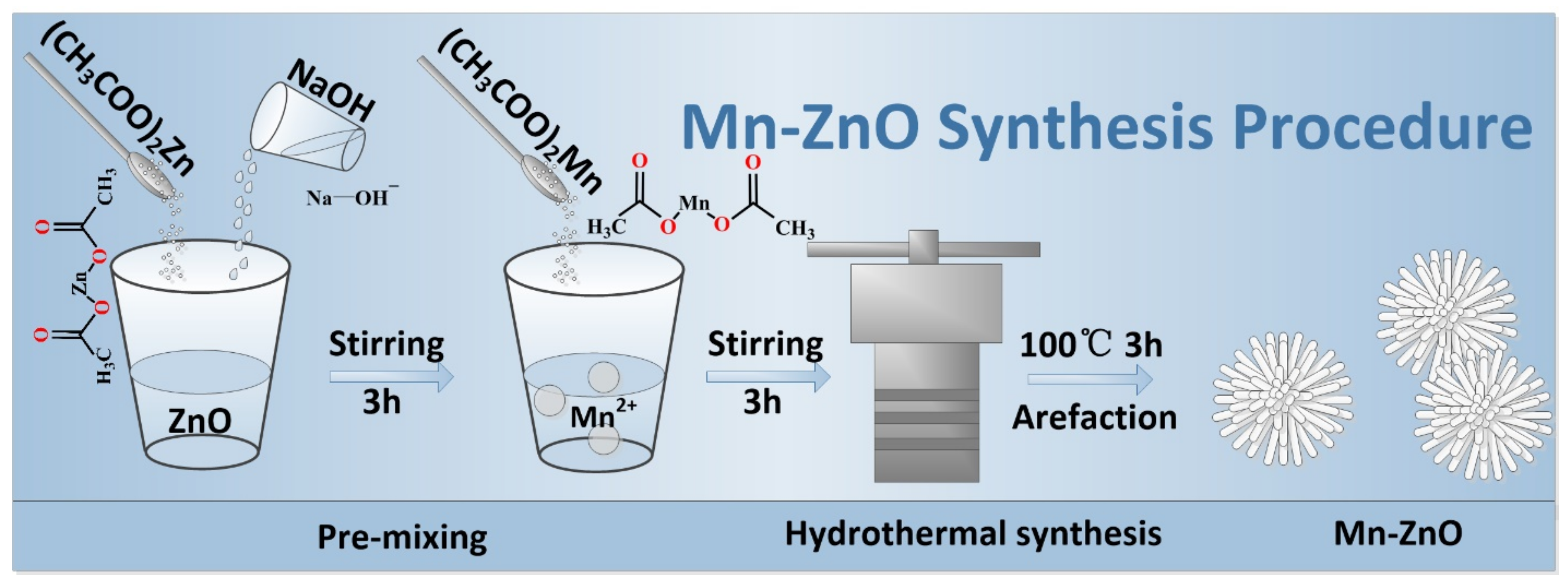
The Mn-ZnO nanowire microsphere was synthesized by using hydrothermal route. Briefly, adding 2.058 g Zn(CH3COO)2 into deionized water, the solution A with concentration of 0.125 mol/L could be obtained. Then, we added the certain concentration of NaOH solution into the solution A, which was magnetically stirred for 3 h. After that, ed added 1%, 2%, 3%, 4% and 5% of Mn2+ into the obtained solution and magnetically stirred it for 3 h as a precursor solution. The mixture solution was transferred into the polytetrafluoroethylene stainless steel reactor for 100 °C for 3 h. After the reaction, the solution was centrifuged in a centrifuge tube. The precipitates after centrifugation were washed three times with anaerobic ethanol and pure water, followed by 12 h baking at 80 °C. Figure 1 shows the specific operating process for making the Mn-doped ZnO nanowire microsphere, and Table 1 lists the specific parameters for the experimental preparation.
2.3. Characterization
A Bruker Advance D8 X-ray diffractometer (XRD, Billerica, MA, USA) with Cu Kα radiation at 40 kV and 30 mA was utilized to investigate the crystal phases of Mn-ZnO nanowire microspheres. The diffraction date was measured from 20° to 80° with a scanning rate of 6°/min and a step size of 0.02°. A Carl Zeiss SIGMA scanning electron microscope (SEM, Jena, Germany) and JEM-3010 high-resolution transmission electron microscope (HRTEM, Tokyo, Japan) were employed to characterize morphologies and structures, operating at 3 kV and 200 kV, respectively. An EDX spectroscope (X-MaxN 80T, Oxfordshire, UK) was utilized to detect the EDX spectra and elemental mappings of samples. With a Horiba Jobin–Yvon Fluoromax-4 spectrophotometer (Kyoto, Japan) using 325 nm excitation wavelength, the PL spectra of Mn-ZnO nanowire microsphere were collected at 325–625 nm. A physical property measurement system (San Diego, CA, USA), which is completely liquid-free, was used for the magnetic property characterization.
3. Results and Discussion
3.1. Morphology and Structure Characterization
Figure 2 provides structural information for Mn-ZnO nanowire microsphere: XRD patterns (Figure 2a), EDX (Figure 2b) and a stick crystal model of Mn-ZnO nanowire microsphere (Figure 2c). Device Studio [27] program provides a number of functions for performing visualization, modeling and simulation. And a stick crystal model of Mn-ZnO simulation using device studio software integrated in Device Studio program. Figure 2a shows that the diffraction peaks of Mn-ZnO at various doping concentrations basically agree with the structure of pristine ZnO (PDF# 36-1451). The diffraction peaks of 2θ = 31.8°, 34.4°, 36.3°, 47.5°, 56.6°, 62.9°, 68.0° match crystal planes (100), (002), (101), (102), (110), (103) and (112), respectively. Figure 2b shows that no impurity atoms appeared. Therefore, the doping of Mn2+ has no change in the ZnO crystal structure, and only Mn2+ replaced the Zn2+ ions in a fixed position (Figure 2c). While Mn2+ concentration increases, the energy density of the ZnO (101) crystal surface is relatively low, and has no effects on the preferred growth along the (101) crystal surface [28]. It is shown that the diffraction peak intensity of Mn-ZnO nanowire microsphere decreases with the doped Mn concentration, but the crystal structure has no significant effect.
Figure 3 shows the microstructure of samples. The SEM characterization of pristine ZnO and Mn-ZnO is presented in Figure 3a,b and Figure 3c,d, respectively. After Mn was introduced, the single size of ZnO nanorods decreases, and uniform nanorod microspheres are formed. Figure 3e–h shows TEM images of individual nanorods in the Mn-ZnO nanowire microsphere. Individual nanorods are about 200 nm in diameter, in line with the SEM results. Figure 3h presents the clear lattice stripes indicating that the Mn-ZnO nanowire microsphere has an excellent crystal structure. The spacing of neighboring (002) crystal planes in the wurtzite ZnO is around 0.26 nm inline with our XRD results in the paper. Regular spot patterns in the SAED pattern (inset of Figure 3h) indicates the existence of a single-crystalline Mn-ZnO nanowire microsphere. The elemental mappings (Figure 3j–m) present uniform distribution of Mn atoms in ZnO nanorods, indicating the presence of the prepared Mn-ZnO nanowire microsphere.
3.2. Optical Performance
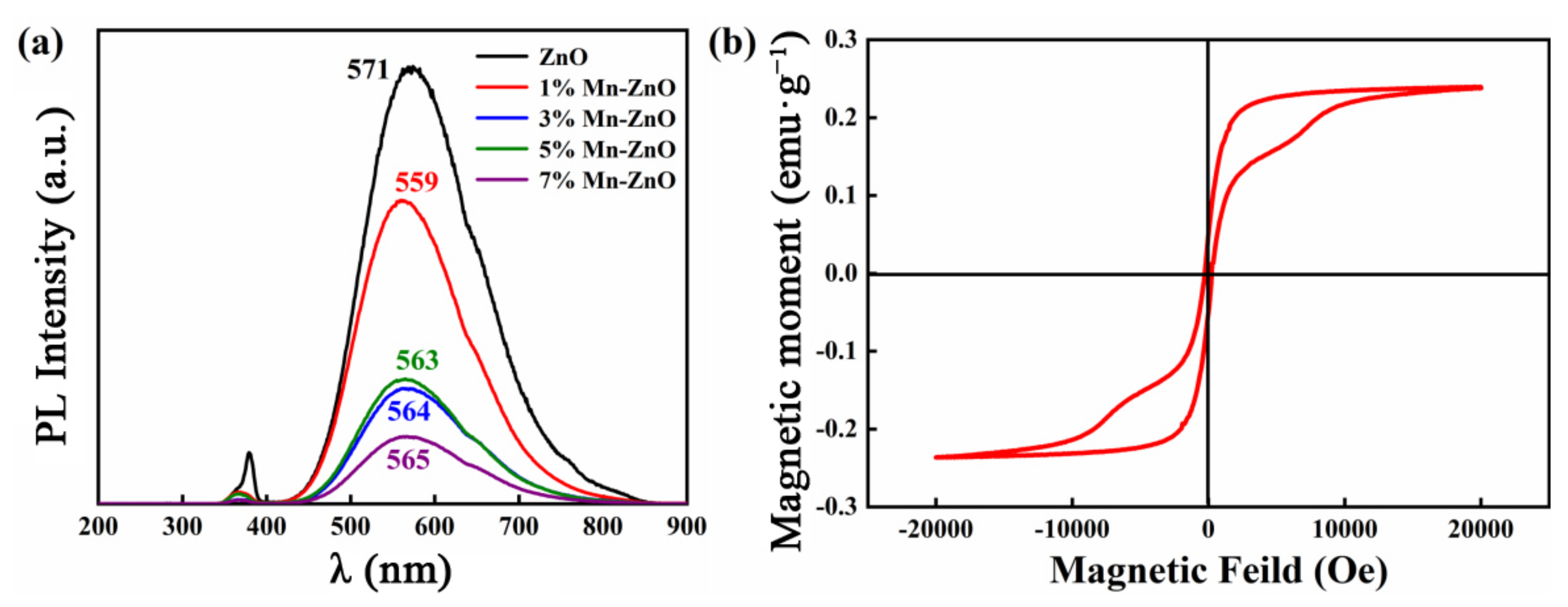
Figure 4a presents the PL spectrum of all samples. There is a small UV emission peak around 380 nm and a wide emission band between 400 and 800 nm. Table 2 shows the optical performance comparison between this research work and other metal-doped ZnO nano materials. The spectrum intensity drops when Mn2+ concentration increases, which is primarily affected by the dopant (Mn) impact on the recombination processes and density of free excitons. The wide-band visible emissions can be attributed to the intrinsic defect concentration in pristine ZnO, since it decreases when Mn in ZnO is substituted. We note that increasing the Mn2+ ion concentration can also possibly weaken the visible emission intensity according to Fabbiyola et al. [29], which is caused by increasing absorption because more distortion centers and surface defects were generated [30]. In addition, with increasing concentration of the dopant ion (Mn2+), non-radiative recombination occurred due to non-radiative decay of excited Mn2+ ions [31]. Mn2+ ion doping affects the luminescence intensity of pristine ZnO, especially the visible light-emitting band. The defective visible emission and PL quenching in ZnO nanorods due to the competitive effect of Fe cations on excitonic recombination was reported in [32]. Similarly, the chemical properties of Mn and Fe are very similar, which will lead to a similar influence on the luminescence intensity of pristine ZnO. The surface defect concentration drops with reducing vacancies of Zn or unpaired/dangling oxygen atom 2p states. Thus, O–Mn bonds can be formed by incorporating unpaired/dangling oxygen atom 2p states at either interstitial Mni defects or VZn [33].
3.3. Magnetic Properties
In Figure 4b, the Mn-ZnO nanowire microsphere displays ferromagnetism under normal temperature with 2.4 × 10⁻¹ emu/g saturation magnetization, 4.9 × 10⁻² emu/g residual magnetization, and 369 Oe coercive field (Hc), which can be explained by the exchange interaction of localized spins associated with unpaired electrons in the oxygen vacancies on the nanoparticle surface [34,35]. Several orders of magnitude enhancement of the saturation magnetization of the pure ZnO by Mn doping (0.0133 → 0.24 emu/g) reflect the significant impact of Mn dopants on the ferromagnetism. Hence, increasing saturation magnetization indicates that doped Mn ions may improve the exchange interaction of localized spins associated with unpaired electrons in pure ZnO, and therefore the defects induced magnetization increases. Therefore, addition of Mn2+ greatly improved the saturation magnetization and coercive field of the ZnO nanowire microsphere, and thus its magnetic storage capacity and demagnetization resistance. These data further demonstrated that the ZnO nanowire microsphere could be used in magnetic memory equipment. Table 2 shows the performance comparison between this research work and other metal-doped ZnO nano materials.
3.4. Theoretical Calculation
The DFT calculation of magnetic properties of Mn-ZnO was conducted with the VASP package [36], with the consideration of HSE hybrid exchange-correlation functions and pin polarization [37,38]. The electronic structural properties were studied with generalized gradient approximation (GGA)-based Perdew–Burke–Ernzerhof (PBE) [39] and projector-augmented wave (PAW) [40] methods, with 500 eV cutoff energy, and fixed 10−5 eV and 0.02 eV/Å convergence criteria. The Brillouin-zone approach was sampled with 1× 1 × 16 k-point grid. The strong correlation effect was described by the Hubbard parameter U (repulsion energy) in the model, by which the band gap can be modified according to experimental results [14]. The electronic structures of valence electrons of atomic Zn, O and Mn were 3p63d104s2, 2s22p4 and 4s23d5, respectively.
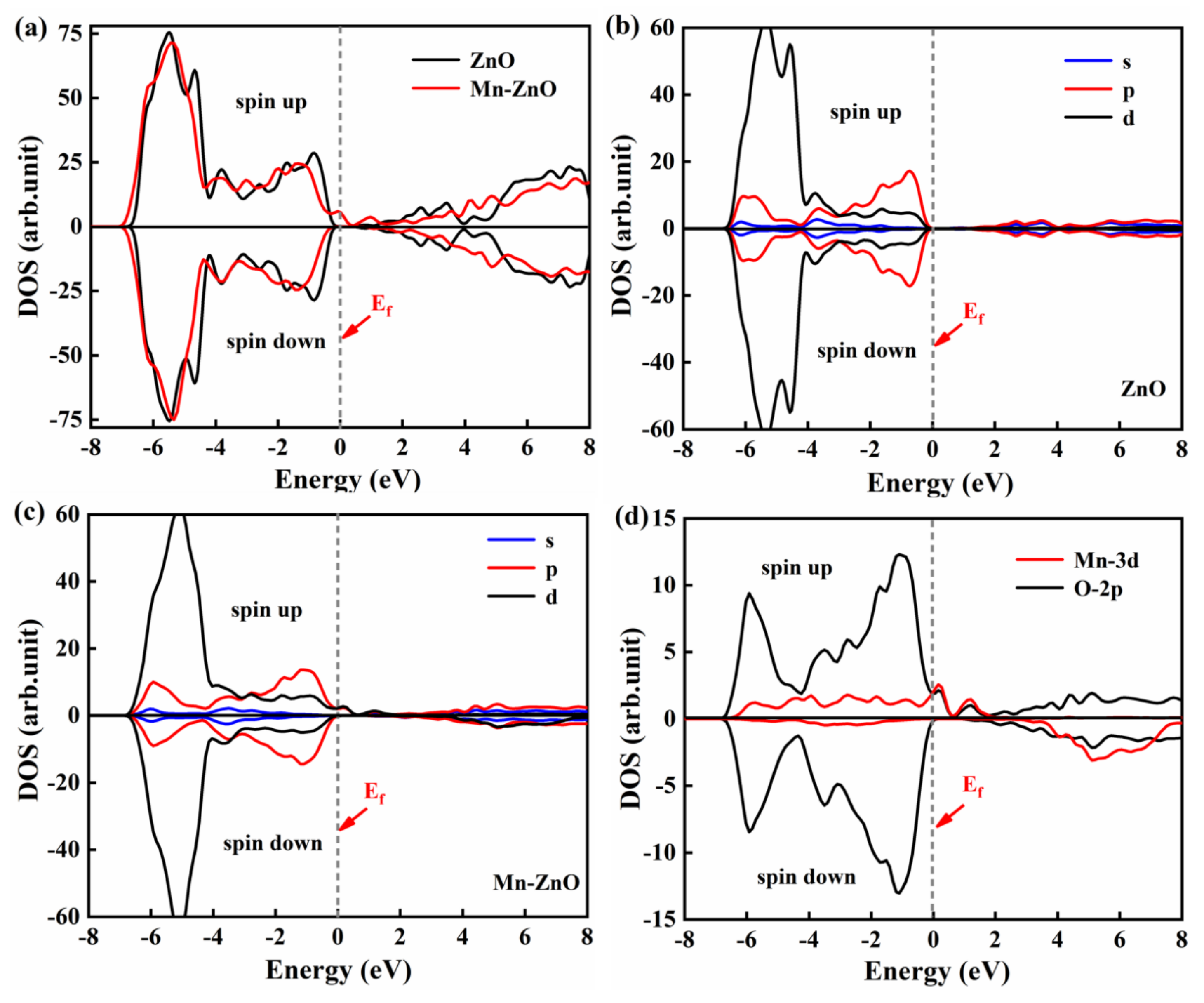
Compared to pure ZnO nanowire microsphere, the total density of states (DOS) of Mn-ZnO nanowire microsphere slightly shifted towards a lower energy region, as shown in Figure 5a. From the partial DOS in Figure 5c, the upper (−4.5–0 eV) and lower valence band (−6.5–3 eV) of Mn-ZnO nanowire microsphere primarily contributed to the Mn 3d state and partially to the O 2p state. Figure 5d shows strong hybridization coupling between Mn 3d and O 2p obits, as non-local characteristics. The spin splitting in the upper and lower spin states was crucial in regulating different magnetic coupling between magnetic atoms. The local characteristics of Mn 3d, O 2p and 2s states primarily determined conduction bands. For a doped system in Figure 5d, the exchange between spin-polarized Mn 3d and O 2p states mainly determined its total magnetic moment due to the 3d electron configuration. Since electron spin configurations (↑↑↑↑↑) were partially occupied by Mn2+ (3d5), some 3d electrons could roam freely and the dominant interaction was double exchange, leading to ferromagnetic-doped DMS, which was confirmed by the experimental and theoretical data.
In order to specifically show the distribution of the charge in the case of the Mn-ZnO system, the bond length of the doping system was analyzed, when the energy of the doping system was calculated (Table 3). In the doping system, after replacing Zn by Mn, excess negative charges are generated, and the mutual exclusion between the charges weakens, making the bond length shorter, weakens the ionic bonds, and enhances the covalent bonds. It is shown that the doping of Mn ions reduces the bond lengths of X, Y, and Z and reduces the corresponding bond angles, which increases the population value, reduced ion bonds and enhanced covalent bonds. Population values are common parameters used to analyze ionic or covalent bonds. The larger the population value, the stronger the covalent bond, the smaller the population value, and the stronger the ion bond. Furthermore, by analyzing the differential charge density distribution map of Mn-doped ZnO, we can more vividly and visually talk about the bonding situation between each atoms in the system of Mn-doped ZnO. The yellow and the blue regions (Figure 6) represent the electron depletion and accumulation in the charge difference distribution calculation, respectively. The electron cloud overlap between the Mn atoms in the Mn-ZnO doping systems and the surrounding O atoms is enhanced, with enhanced covalent bonds, weakened ionic bonds, and stable doping systems [26]. As shown in Figure 6, the electron transfer is mainly between Mn and O ions, which matches the results of the density of state distribution.
4. Conclusions
Mn-ZnO nanowire microsphere was prepared by a hydrothermal method, and the the as-prepared powders were analyzed to reveal the morphological, structural and magneto-optical properties. The luminescence intensity of the sample decreased in both UV and visible regions as the Mn2+ ions doping concentration increased. Mn-ZnO nanowire microsphere showed obvious ferromagnetism. Several orders of magnitude enhancement of the saturation magnetization of the pristine ZnO by Mn2+ doping (0.0133 → 0.24 emu/g) reflected a significant impact of Mn dopants on the ferromagnetism. The incorporation of Mn2+ greatly improved the saturation magnetization and stress field of the zinc oxide nanowire microsphere and enhanced the magnetic storage and magnetoresistance of ZnO. The magnetic source of Mn-ZnO nanorod arrays was further revealed by theoretical calculations. In sum, the Mn-ZnO nanowire microsphere was an excellent magnetic material and has huge potential applications in magnetic storage devices.
Author Contributions
L.Z.: conceptualization, writing—original draft, preparation, contributed to the general discussion. W.W.: validation, methodology, writing—review and editing, contributed to the general discussion. R.D.: formal analysis, contributed to the general discussion. J.N.: data curation, contributed to the general discussion. F.Z. and J.Y.: supervision, funding acquisition, contributed to the general discussion. All authors have read and agreed to the published version of the manuscript.
Funding
This research was funded by National Natural Science Foundation of China, grant number 61664008; Natural Science of Foundation of ShannXi Province, grant number 2021JQ-635; Scientific and Technological Innovation Team, grant number 2017CXTD-01; 2020 Innovation and Entrepreneurship for Undergraduates S202010719016.
Institutional Review Board Statement
Not applicable.
Informed Consent Statement
Not applicable.
Data Availability Statement
Data is contained within the article.
Acknowledgments
We acknowledge Suqin Xue for partially supporting the funding of this manuscript with the postgraduate research opportunities program of HZWTECH (HZWTECH-PROP).
Conflicts of Interest
The authors declare that they have no conflict of interest.
References
- Yang, Y.; Niu, S.; Han, D.; Liu, T.; Wang, G.; Li, Y. Progress in developing metal oxide nanomaterials for photoelectrochemical water splitting. Adv. Energy Mater. 2017, 7, 1700555. [Google Scholar] [CrossRef]
- Cai, L.; Ren, F.; Wang, M.; Cai, G.; Chen, Y.; Liu, Y.; Shen, S.; Guo, L. V ions implanted ZnO nanorod arrays for photoelectrochemical water splitting under visible light. Int. J. Hydrogen Energy 2015, 40, 1394–1401. [Google Scholar] [CrossRef]
- Myndrul, V.; Vysloužilová, L.; Klápšt’ová, A.; Coy, E.; Jancelewicz, M.; Iatsunskyi, I. Formation and Photoluminescence Properties of ZnO Nanoparticles on Electrospun Nanofibers Produced by Atomic Layer Deposition. Coatings 2020, 10, 1199. [Google Scholar] [CrossRef]
- Dietl, T.; Ohno, H.; Matsukura, F.; Cibert, J.; Ferrand, D. Zene rmodel description of ferromagnetism in zinc-blend emagnetic semiconductors. Science 2000, 287, 1019–1022. [Google Scholar] [CrossRef] [PubMed] [Green Version]
- Chang, Y.Q.; Luo, X.H.; Xu, X.Y.; Li, L.; Chen, J.P.; Wang, R.M.; Yu, D.P. Synthesis, Characterization and Magnetic Property Measurements of Zn1−xMnxO Nanoparticles via Vapor Phase Growth. Chin. Phys. Lett. 2003, 20, 2058–2060. [Google Scholar]
- Roy, V.A.L.; Djurii, A.B.; Liu, H.; Zhang, X.X.; Leung, Y.H.; Xie, M.H.; Gao, J.; Lui, H.F.; Surya, C. Magnetic Properties of Mn Doped ZnO Tetrapod Structures. Appl. Phys. Lett. 2004, 84, 756–758. [Google Scholar] [CrossRef] [Green Version]
- Norton, D.P.; Pearton, S.J.; Hebard, A.F.; Theodoropoulou, N.; Boatner, L.A.; Wilson, R.G. Ferromagnetism in Mn-implanted ZnO: Sn Single Crystals. Appl. Phys. Lett. 2003, 82, 239–241. [Google Scholar] [CrossRef] [Green Version]
- Ip, K.; Frazier, R.M.; Heo, Y.W.; Norton, D.P.; Abernathy, C.R.; Pearton, S.J.; Zavada, J.M.; Kelly, J.; Rairigh, R.; Hebard, A.; et al. Ferromagnetism in Mn- and Co-implanted ZnO Nanorods. J. Vac. Sci. Technol. 2003, 21, 1476. [Google Scholar] [CrossRef] [Green Version]
- Jung, S.W.; An, S.J.; Yi, G.C.; Jung, C.U.; Lee, S.I.; Cho, S. Ferromagnetic Properties of Zn1−xMnxO Epitaxial Thin Films. Appl. Phys. Lett. 2002, 80, 4561–4563. [Google Scholar] [CrossRef] [Green Version]
- Chang, Y.Q.; Wang, D.B.; Luo, X.H.; Xu, X.Y.; Chen, X.H.; Li, L.; Chen, C.P.; Wang, R.M.; Xu, J.; Yu, D.P. Synthesis, Optical and Magnetic Properties of Diluted Magnetic SemiconductorZn1−xMnxO Nanowires via Vapor Phase Growth. Appl. Phys. Lett. 2003, 83, 4020. [Google Scholar] [CrossRef]
- Chang, Y.Q.; Xu, X.Y.; Luo, X.H.; Long, Y.; Ye, R.C. Magnetic Properties of Diluted Magnetic Semiconductor Zn1−xMnxO Nanowires. Chin. Phys. Lett. 2005, 22, 991–994. [Google Scholar]
- Wang, D.D.; Chen, Q.; Xing, G.Z.; Yi, J.B.; Bakaul, S.R.; Ding, J.; Wang, J.L.; Wu, T. Robust Room-Temperature Ferromagnetism with Giant Anisotropy in Nd-Doped ZnO Nanowire Arrays. Nano Lett. 2012, 12, 3994–4000. [Google Scholar] [CrossRef]
- Sato, K.; Katayama-Yoshida, H. Stabilization of ferromagnetic states by electron doping in Fe-, Co- or Ni-doped ZnO. Jpn. J. Appl. Phys. 2001, 40, 334. [Google Scholar] [CrossRef]
- Geburt, S.; Röder, R.; Kaiser, U.; Chen, L.M.; Chu, M.H.; Ruiz, J.S.; Criado, G.M.; Heimbrodt, W.; Ronning, C. Intense intra-3d luminescence and waveguide properties of single Co-doped ZnO nanowires. Phys. Status Solidi 2013, 7, 886–889. [Google Scholar] [CrossRef]
- Cheng, C.; Xu, G.Y.; Zhang, H.Q.; Li, Y. Solution synthesis, optical and magnetic properties of Zn1−xCoxO nanowires. Mater. Lett. 2008, 62, 3733–3737. [Google Scholar] [CrossRef]
- Xu, C.K.; Yang, K.K.; Wang, L. Ferromagnetism of aligned Zn1−xVxO nanorods grown by a vapour transport route. J. Phys. D Appl. Phys. 2008, 41, 195005. [Google Scholar] [CrossRef]
- Chu, D.W.; Zeng, Y.P.; Jiang, D.L. Synthesis and growth mechanism of Cr-doped ZnO single crystalline nanowires. Solid State Commun. 2007, 143, 308–312. [Google Scholar] [CrossRef]
- Zhu, D.; Hu, T.X.; Zhao, Y.Y.; Zang, W.L.; Xing, L.L.; Xue, X.Y. High-performance self-powered/active humidity sensing of Fe-doped ZnO nanoarray nanogenerator. Sens. Actuator B-Chem. 2015, 213, 382–389. [Google Scholar] [CrossRef]
- Cui, J.B.; Gibson, U.J. Electrodeposition and room temperature ferromagnetic anisotropy of Co and Ni-doped ZnO nanowire arrays. Appl. Phys. Lett. 2010, 87, 133108. [Google Scholar] [CrossRef]
- Woo, H.S.; Kwak, C.H.; Chung, J.H.; Lee, J.H. Highly selective and sensitive xylene sensors using Ni-doped branched ZnO nanowire networks. Sens. Actuator B-Chem. 2015, 216, 358–366. [Google Scholar] [CrossRef]
- Gopalakrishnan, R.; Kabilan, R.; Ashokkumar, M. Investigations of Mn introduced structural modifications on Ni-doped ZnO diluted magnetic semiconductors and improved magnetic and antibacterial properties. J. Mol. Struct. 2022, 1251, 132060. [Google Scholar] [CrossRef]
- Singh, S.; Rama, N.; Sethupathi, K.; Rao, M.S.R. Correlation between elctrical transport optical and magnetic properties of transition metal ion doped ZnO. J. Appl. Phys. 2008, 103, 07D108. [Google Scholar] [CrossRef]
- Liu, R.B.; Shi, L.J.; Zou, B.S. Magnetic exciton relaxation and spin–spin interaction by the time-delayed photoluminescence spectra of ZnO: Mn nanowires. Appl. Mater. Interfaces 2014, 6, 10353–10366. [Google Scholar] [CrossRef]
- Bandyopadhyay, A.; Gupta, N.; Nath, M.; Chakraborty, S.; Sutradhar, S. Magnetic properties of Mn doped ZnO: A Monte Carlo simulation analysis. Vacuum 2021, 183, 109786. [Google Scholar] [CrossRef]
- Ma, Y.F.; Gao, H.; Huang, R.Q.; Guo, R.B.; Yang, S.J.; Han, Y.B.; Zuo, H.K. Green emission in Fe- and Mn-doped ZnO nanowires studied by magneto-photoluminescence. J. Lumin. 2022, 241, 118521. [Google Scholar] [CrossRef]
- Li, X.; Zhu, X.H.; Jin, K.X.; Yang, D.Y. Study on structural and optical properties of Mn-doped ZnO thin films by sol-gel method. Opt. Mater. 2020, 100, 109657. [Google Scholar] [CrossRef]
- Hongzhiwei Technology, Device Studio, Version 2021A, China. 2021. Available online: https://iresearch.net.cn/cloudSoftware (accessed on 28 December 2021).
- Wang, W.; Zhang, F.C.; Wang, X.Y.; Zhang, S.L.; Yan, J.F.; Zhang, W.B.; Zhang, W.H. Magnetic and optical properties of Co-doped ZnO nanorod arrays. Eur. Phys. J. Plus 2020, 135, 40. [Google Scholar] [CrossRef]
- Fabbiyola, S.; John Kennedy, L.; Dakhel, A.A.; Bououdina, M.; Judith Vijaya, J.; Ratnaji, T. Structural, microstructural, optical and magnetic properties of Mn doped ZnO nanostructures. J. Mol. Struct. 2016, 1109, 89–96. [Google Scholar] [CrossRef]
- Zhang, F.C.; Cui, H.W.; Zhang, W.H. Identifying properties of Co-doped ZnO nanowires from first-principles calculations. Vacuum 2015, 119, 131–135. [Google Scholar] [CrossRef]
- Khalid, M.; Ziese, M.; Setzer, A.; Esquinazi, P.; Lorenz, M.; Hochmuth, H.; Grundmann, M.; Spemann, D.; Butz, T.; Brauer, G.; et al. Defect-induced magnetic order in pure ZnO films. Phys. Rev. B 2009, 80, 035331. [Google Scholar] [CrossRef] [Green Version]
- Dev, P.; Zeng, H.; Zhang, P. Defect-induced magnetism in nitride and oxide nanowires: Surface effects and quantum confinement. Phys. Rev. B 2010, 82, 165319. [Google Scholar] [CrossRef]
- Hu, P.; Han, N.; Zhang, D.; Ho, J.C.; Chen, Y. Highly formaldehyde-sensitive, transition-metal doped ZnO nanorods prepared by plasma-enhanced chemical vapor deposition. Sens. Actuators B 2012, 69, 74–80. [Google Scholar] [CrossRef]
- Lee, J.J.; Xing, G.Z.; Yi, J.B.; Chen, T.; Ionescu, M.; Li, S. Tailoring the coercivity in ferromagnetic ZnO thin films by 3d and 4f elements co-doping. Appl. Phys. Lett. 2014, 104, 012405. [Google Scholar] [CrossRef] [Green Version]
- El-Hilo MDakhel, A.A.; Yacoob, Z.J. Magnetic interactions in Co2+ doped ZnO synthesised by coprecipitation method: Efficient effect of hydrogenation on the long-range ferromagnetic order. J. Magn. Magn. Mater. 2019, 482, 125–134. [Google Scholar] [CrossRef]
- Kresse, G.; Furthmüller, J. Efficiency of ab-initio total energy calculations for metals and semiconductors using a plane-wave basis set. Comp. Mater. Sci. 1996, 6, 15–50. [Google Scholar] [CrossRef]
- Grimme, S. Semiempirical GGA-type density functional constructed with a long-range dispersion correction. Comput. Chem. 2006, 27, 1787–1799. [Google Scholar] [CrossRef] [PubMed]
- Liu, B.; Wu, L.J.; Zhao, Y.Q.; Wang, L.Z.; Cai, M.Q. A first-principles study of magnetic variation via doping vacancy in monolayer VS2. Magn. Magn. Mater. 2016, 420, 218–224. [Google Scholar] [CrossRef]
- Dudarev, S.L.; Botton, G.A.; Savrasov, S.Y.; Humphreys, C.J.; Sutton, A.P. Electron-energy-loss spectra and the structural stability of nickel oxide: An LSDA+U study. Phys. Rev. 1998, 57, 1505. [Google Scholar] [CrossRef]
- Perdew, J.P.; Burke, K.; Ernzerhof, M. Generalized gradient approximation made simple. Phys. Rev. Lett. 1996, 77, 3865. [Google Scholar] [CrossRef] [Green Version]
Figure 1.
Process flow of the Mn-ZnO nanowire microsphere.

Figure 2.
(a) X-ray diffraction (XRD) patterns, (b) energy-dispersive spectroscopy (EDX) and (c) model of Mn-ZnO nanowire microsphere.
Figure 2.
(a) X-ray diffraction (XRD) patterns, (b) energy-dispersive spectroscopy (EDX) and (c) model of Mn-ZnO nanowire microsphere.

Figure 3.
(a–d) Scanning electron microscopy (SEM) images, (e–i) transmission electron microscopy (TEM) images and (j–m) mappings of 5% Mn-ZnO nanowire microsphere.
Figure 3.
(a–d) Scanning electron microscopy (SEM) images, (e–i) transmission electron microscopy (TEM) images and (j–m) mappings of 5% Mn-ZnO nanowire microsphere.

Figure 4.
(a) Photoluminescence (PL) of Mn-ZnO nanowire microsphere. (b) Room-temperature M–H curve of Mn-ZnO nanowire microsphere.
Figure 4.
(a) Photoluminescence (PL) of Mn-ZnO nanowire microsphere. (b) Room-temperature M–H curve of Mn-ZnO nanowire microsphere.

Figure 5.
Total density of states (DOS) and partial DOS. (a) DOS of ZnO and Mn-ZnO nanowire microsphere, (b) partial DOS of ZnO nanowire microsphere, (c) partial DOS of Mn-ZnO, (d) partial DOS of Co 3d and O 2p.
Figure 5.
Total density of states (DOS) and partial DOS. (a) DOS of ZnO and Mn-ZnO nanowire microsphere, (b) partial DOS of ZnO nanowire microsphere, (c) partial DOS of Mn-ZnO, (d) partial DOS of Co 3d and O 2p.

Figure 6.
The calculated charge density difference of Mn-ZnO.


Table 1.
Preparation parameters of Mn-ZnO nanowire microsphere.
| Factors | OH−/Zn2+ | Zn2+ Concentration (mol/L) | Temperature (°C) | Mn-Doped Amount (%) | Time (h) |
|---|---|---|---|---|---|
| Sample 1 | 10 | 0.125 | 100 | 1 | 3 |
| Sample 2 | 10 | 0.125 | 100 | 2 | 3 |
| Sample 3 | 10 | 0.125 | 100 | 3 | 3 |
| Sample 4 | 10 | 0.125 | 100 | 4 | 3 |
| Sample 5 | 10 | 0.125 | 100 | 5 | 3 |
Table 2.
The performance comparison between this research work and other metal-doped ZnO nano materials.
Table 2.
The performance comparison between this research work and other metal-doped ZnO nano materials.
| Samples | Magnetic | Optical | Ref | |
|---|---|---|---|---|
| Ms | Hc | PL | ||
| Zn1−xCoxOnanowires | 0.1 emu/g | 70 Oe | - | [15] |
| Fe- and Mn-doped ZnO | - | - | Green emission | [25] |
| Mn doped ZnO | 2.169 emu/g | 21.6 Oe | UV and visible regions | [29] |
| Ni-doped ZnO | 270.1 emu/g | - | All visible except red | [21] |
| ZnO Nanoparticles | - | - | Broad visible regions | [3] |
| Mn-ZnO nanowire | 0.24 emu/g | 369 Oe | UV and visible regions | This work |
Table 3.
Changes in the bond length and bond angle before and after Mn ions are doped.
| Material | Bond Length | Bond Angle | |||
|---|---|---|---|---|---|
| Z | X | Y | Z-XOY | XOY | |
| ZnO | 2.037 | 2.021 | 2.065 | 110.038 | 108.859 |
| Mn-ZnO | 1.969 | 1.959 | 2.019 | 107.091 | 106.420 |
Publisher’s Note: MDPI stays neutral with regard to jurisdictional claims in published maps and institutional affiliations. |
© 2022 by the authors. Licensee MDPI, Basel, Switzerland. This article is an open access article distributed under the terms and conditions of the Creative Commons Attribution (CC BY) license (https://creativecommons.org/licenses/by/4.0/).
Share and Cite
MDPI and ACS Style
Zhang, L.; Wang, W.; Dai, R.; Ning, J.; Zhang, F.; Yan, J. Theoretical and Experimental Investigate for the Magnetic and Optical Properties of Mn-ZnO Nanowire Microspheres. Coatings 2022, 12, 205. https://doi.org/10.3390/coatings12020205
AMA Style
Zhang L, Wang W, Dai R, Ning J, Zhang F, Yan J. Theoretical and Experimental Investigate for the Magnetic and Optical Properties of Mn-ZnO Nanowire Microspheres. Coatings. 2022; 12(2):205. https://doi.org/10.3390/coatings12020205
Chicago/Turabian StyleZhang, Lei, Wei Wang, Rong Dai, Jing Ning, Fuchun Zhang, and Junfeng Yan. 2022. "Theoretical and Experimental Investigate for the Magnetic and Optical Properties of Mn-ZnO Nanowire Microspheres" Coatings 12, no. 2: 205. https://doi.org/10.3390/coatings12020205
Note that from the first issue of 2016, this journal uses article numbers instead of page numbers. See further details here.