
Photocatalytic and Biological Activity of ZnO Nanoparticles Using Honey
by
 ,
,
M. Sharmila
1,*,
R. Jothi Mani
1,
Abdul Kader
1,
Awais Ahmad
2,* ,
,
Gaber E. Eldesoky
3,*,
Adel E. M. Yahya
3 and
Aboud Ahmed Awadh Bahajjaj
3 1
Department of Physics, Sadakathullah Appa College, Tirunelveli 627011, India
2
Departamento de Quimica Organica, Universidad de Cordoba, Edificio Marie Curie (C-3), Ctra Nnal IV-A, Km 396, 14014 Cordoba, Spain
3
Chemistry Department, College of Science, King Saud University, P.O. Box 2455, Riyadh 11451, Saudi Arabia
*
Authors to whom correspondence should be addressed.
Coatings 2021, 11(9), 1046; https://doi.org/10.3390/coatings11091046
Submission received: 20 June 2021
/
Revised: 4 August 2021
/
Accepted: 16 August 2021
/
Published: 30 August 2021
Abstract
:The innovation and development of water purification methods have been at the center of extensive research for several decades. Many nanoparticles are frequently seen in industrial waste water. In this research, zinc oxide nanoparticles (ZnO) were synthesized following an autocombustion method with and without honey capping. Structural crystallinity and bonding structure were examined via X-ray diffraction (XRD) analysis and Fourier transform infrared (FTIR) spectroscopy. Optical behavior was analyzed using ultraviolet–visible (UV–Vis) spectroscopy and photoluminescence (PL). Size estimation and surface morphology were studied using scanning electron microscopy (SEM), while energy-dispersive spectroscopy (EDS) was performed to analyze the sample purity and elemental composition. The photocatalytic degradation of methylene blue (MB) by ZnO was assessed as it is an efficient water treatment process with high potential. The biological activity of ZnO nanoparticles was also investigated in terms of antibacterial and antifungal activities against different bacterial and fungal species. Surprisingly, the as-synthesized ZnO nanoparticles were found to be substantially bioactive compared to conventional drugs. Honey-mediated nanoparticles displayed 86% dye degradation efficiency, and that of bare ZnO was 60%. Therefore, the involvement of honey in the synthesis of ZnO nanoparticles has great potential due to its dual applicability in both biological and environmental remediation processes.
1. Introduction
Water pollutants are hazardous to life on earth and in waters. Water bodies have been polluted by several sources such as industrial waste, e-waste, medical waste and chemical waste [1]. Methylene blue (MB) is a frequently released effluent from industrial wastes that is used as a dye in the textile industry. Such pollutants can be eradicated by the use of metal oxide nanoparticles as photocatalysts [2,3]. To date, many methods using metal oxide nanoparticles have been developed because of their effective catalytic results. Among various metal oxides, ZnO nanoparticles are frequently used due to their biofunctionality, photocatalytic efficiency, and gas sensing properties with varying chemical components, architectures, and microstructures. The II-VI group of ZnO nanoparticles has a high band gap of ~3.37 eV, along with large excitation binding energy (60 meV) [4,5,6,7,8,9,10]. These intrinsic properties of zinc oxide enable its application in water purification because of its easily movable valence band electrons, which is highly favorable for the photodegradation of organic pollutants in water.
In recent years, green synthesis has attained much more attention than chemical synthesis [11]. Various plant extracts have already been employed to prepare ZnO NPs [12,13,14,15,16,17]. Rathnasamy et al. prepared ZnO nanoparticles by incorporating Carica Papaya leaf extract in a green synthesis method for photocatalytic application [18]. The biosynthesis of ZnO nanoparticles has been used for various applications such as pigments, catalysts, optics and electronics, etc. Compared to several other plant extracts, our precursor (i.e., honey) has significant advantages since it is a herbal product with good antibacterial properties, a high yield and purity, minimal time consumption, and biocompatibility [19,20]. Natural honey is a high-viscosity medium. Honey contains vitamins, minerals, enzymes, carbohydrates, and antioxidants, which are used for surfactants, reducers, and stabilizing agents. Indeed, mono- and disaccharides participate in the reduction of the metal ions, while polysaccharides along with enzymes, minerals, and amino acids provide the encapsulating medium to prevent the agglomeration of metal nanoparticles. The medium also controls the shape, structure, and catalytic activity of the ZnO nanoparticles [21,22,23]. Hoseini, Seyed, Javad et al. synthesized ZnO nanopowders from honey for cytotoxic applications [24], and Ranjithkumar et al. prepared ZnO nanoparticles for antimicrobial activities [25]. The results of these studies confirm the biological activity of honey-mediated ZnO nanoparticles, and they can therefore be employed for other biological processes as well.
Moreover, bacterial and fungal toxicities are the cause of innumerable severe ailments. Such medical problems might be reduced by the development of superefficient, ecofriendly, and cost-effective antibacterial and antifungal agents. Therefore, the biological activities obtained using green nanotechnology approaches for different metal oxide nanoparticles are now seen in antibacterial and antifungal experiments. Additionally, the synthesis of honey-mediated metal oxide nanoparticles, in this era of safe technology, also leads to final products with adequate biological activities [26,27,28]. Honey acts as a capping and reducing agent in the green synthesis of metal nanoparticles.
The present work reports the synthesis of ZnO nanoparticles mediated by honey for photocatalytic MB degradation for the first time. Both Gram-positive and Gram-negative bacteria and three different fungi were studied to analyze the nanoparticles’ antibacterial and antifungal activities, respectively. The photocatalytic degradation efficiency was investigated by monitoring the degradation of methylene blue in water samples via UV–Vis spectroscopy.
2. Materials and Methods
2.1. Chemicals and Reagents
Zinc Nitrate (Zn(NO3)2) of 99% (AR Grade; Himedia, Mumbai, IN, USA) was used as the precursor to obtain ZnO NPs. Honey was purchased from Marthandam, Tamilnadu, India. All the bacteria and fungi strains used were purchased from the American Type Culture Collection (ATCC), Manassas, VA, USA. Solutions were made in freshly prepared deionized water (DI). The chemicals were utilized as received without any further treatment.
2.2. Synthesis of ZnO Nanoparticles
The autocombustion method was adapted for the green synthesis of ZnO nanoparticles. Zn(NO3)2 (0.3 M) was prepared in 50 mL DI water, followed by the addition of 2 mL honey. Honey was employed as a capping agent and as a fuel for autocombustion that preserves the crystal morphology of nanoparticles [25]. The mixed solution was stirred for 1 h at 60 °C. The yellowish solution became a brownish gel. The semisolid mixture was subjected to drying at 100 °C for 1 h, and then annealed in air at 550 °C for 2 h. Dried powder was stored at room temperature and proceeded for further characterization studies.
2.3. Instrumentation
A PANalytical X’Pert PRO X-ray diffractometer (JDX-3532, JEOL, Tokyo, Japan) operating at 40 kV, 30 mA with Bragg Brentano geometry using Cu Kα radiation was used to observe structural parameters. An FTIR spectrometer (IR Prestige 21, Shimadzu, Kyoto, Japan) was used to study the chemical composition. The optical spectrum was obtained using a UV–Vis DRS spectrometer from Shimadzu. The photoluminescence study was carried out by a spectrofluorometric PC1 (ISS, Champaign, IL, USA) xenon lamp exhibiting a wavelength equal to 325 nm at room temperature. The morphology of the sample was evaluated by FESEM (Sigma, Zeiss, Jena, Germany).
2.4. Photocatalysis Setup
Photocatalysis was performed for the cationic dye methylene blue under UV–Vis light irradiation. An amount of 15 mg of synthesized ZnO nanoparticles were dissolved in 100 mL MB solution and stirred for 30 min. At regular time intervals, the decrease in dye concentration was observed via UV–Vis spectral analysis in the range of 200–800 nm by obtaining a 4 mL mixture from the reaction system. Ultraviolet and visible light was delivered from mercury and xenon lamps exhibiting the light intensities of 5.8 × 10⁻² Wcm−2 and 6.0 × 10⁻² Wcm−2, respectively. Photodegradation was observed under ambient temperature and pressure under visible light irradiation. The samples were first ultrasonicated and stirred prior to spectrophotometric analysis. The solution (10 mg ZnO nanoparticles and 100 mg MB) was stirred for 30 min in dark to maintain an adsorption–desorption equilibrium.
3. Results and Discussion
3.1. XRD Analysis
XRD displayed a clear crystal morphology with sharp crystalline peaks for ZnO nanoparticles. The hexagonal wurtzite crystalline nature of the nanoparticles was indicated by the hkl estimated for Figure 1, and the observed peak values are in good accordance with the standard JCPDS file No. 89-0510 [29]. There were no other impurities observed. The average crystallite size of the synthesized ZnO nanoparticles was calculated from XRD data employing the Scherrer formula presented in Equation (1) [30].
where D represents the average crystallite size, k is the constant shape factor (i.e., 0.9), λ indicates the X-ray wavelength, β corresponds to the full width at half maximum (FWHM), and θ is the Bragg angle. The peaks obtained for bare ZnO were of low intensity and comparatively broader as compared to those of honey-mediated zinc oxide. Consequently, honey-mediated ZnO nanoparticles were observed to be more crystalline than bare ZnO nanoparticles. The peaks of both bare ZnO and honey-mediated ZnO were analyzed from JCPDS No. 00-890510 [31]. Herein, the D values estimated for ZnO nanoparticles ZnO were found to be 22.4 nm and 39 nm, respectively, indicating the successful formation of nanoparticles [32].
3.2. FTIR Analysis
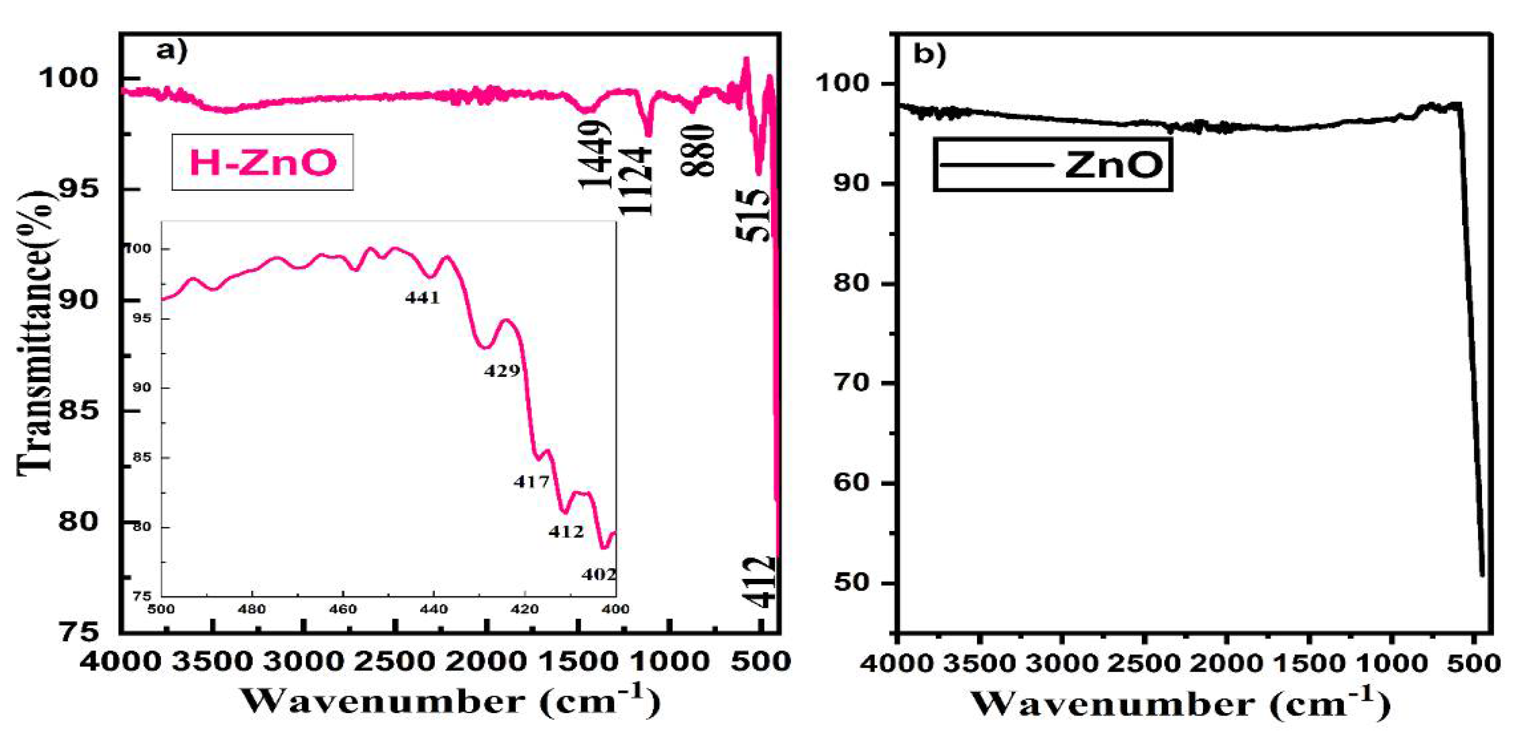
The biomolecules responsible for capping and efficient stabilization in the synthesized sample were observed by FTIR analysis, as displayed in Figure 2. The two absorption peaks at 1449 cm−1 and 1124 cm−1 denote the vibrations of C–O and C–N, respectively, corresponding to the protein conformation of the capped ZnO nanoparticles [33,34]. The bare zinc oxide also expressed zinc and oxygen stretching on the surface of the nanoparticles. The absorption peak at 515 cm−1 indicates the presence of ZnO nanoparticles [35]. The results indicate that the stabilization was formed through the interaction of carboxylate ions with amino acid groups on the ZnO nanoparticles. Other weak peaks are shown as amino acid residues from the binding of ZnO nanoparticles. The results clearly indicate the formation of ZnO nanoparticles by honey components such as fructose, glucose, sucrose, proteins, minerals, and vitamins. The presence of these molecules was confirmed by FTIR results, suggesting that they contribute to the capping of the nanoparticles.
3.3. UV–Visible Spectral Analysis
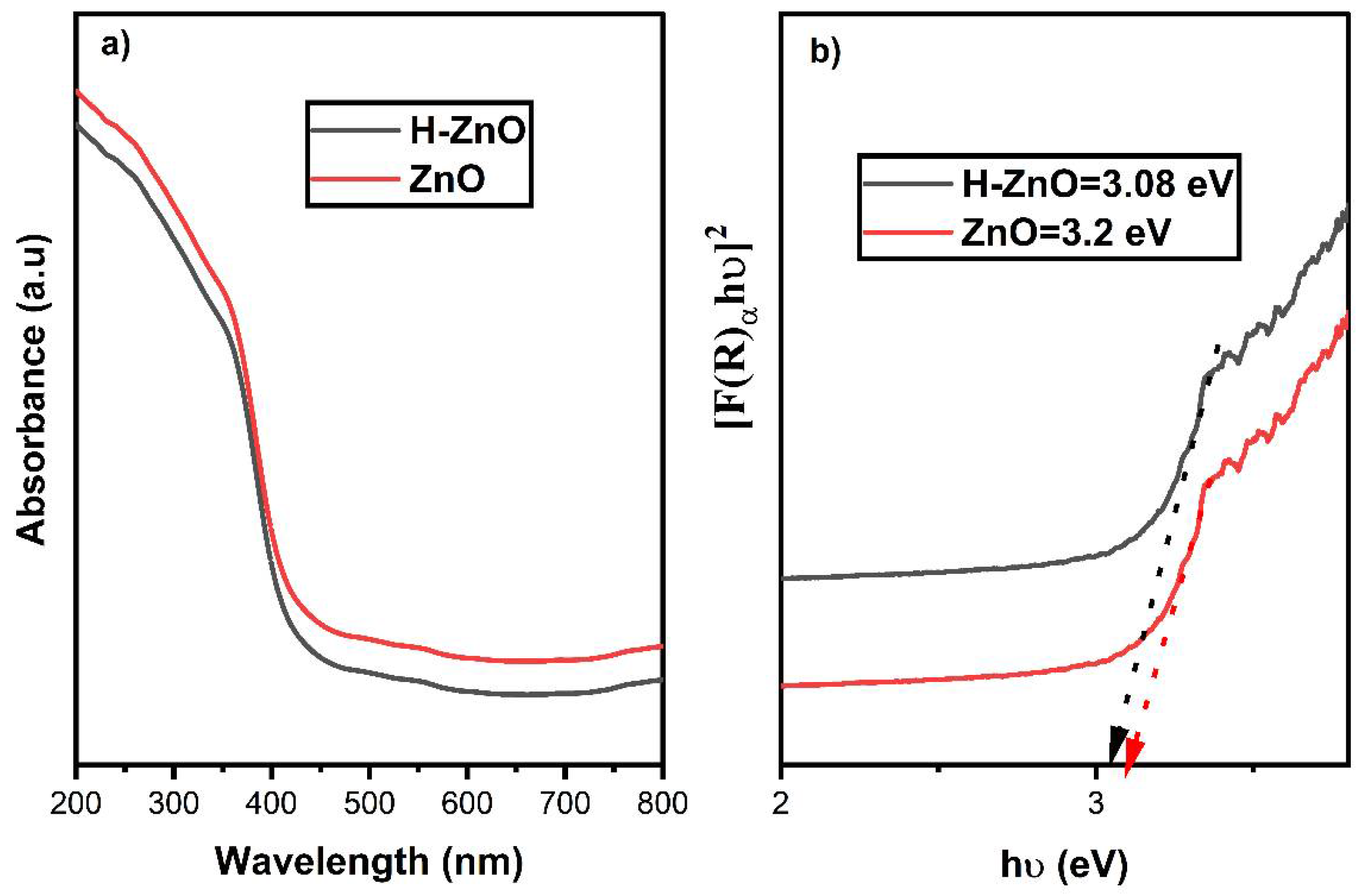
UV–Vis spectral analysis in the range of 200–800 nm was done to confirm the presence of ZnO. The color changes were confirmed by the reduction of Zn2+ ions to ZnO nanoparticles by gaining electrons [36], as the color changed from yellow to brown in the course of synthesis. ZnO nanoparticles were principally formed by dissolution–precipitation processes according to the reaction [37]:
Zn2+ + 3H2O → ZnO + 2H3O+
The absorbance spectrum at around 402 nm (Figure 3) confirms that the excited electron traveled from valance band to conduction band (O2p ≥ Zn3d) [38]. The plot of (F(R)αhv)2) vs. (hv) gives the energy band gap (e.g., 3.08 eV of the H-ZnO sample (Figure 3a)). The bare ZnO produced a low absorption edge compared to honey-mediated zinc oxide. The absorption edge led to a large band gap of the bare zinc oxide materials (i.e., 3.20 eV).
3.4. Photoluminescence Spectroscopy (PL)
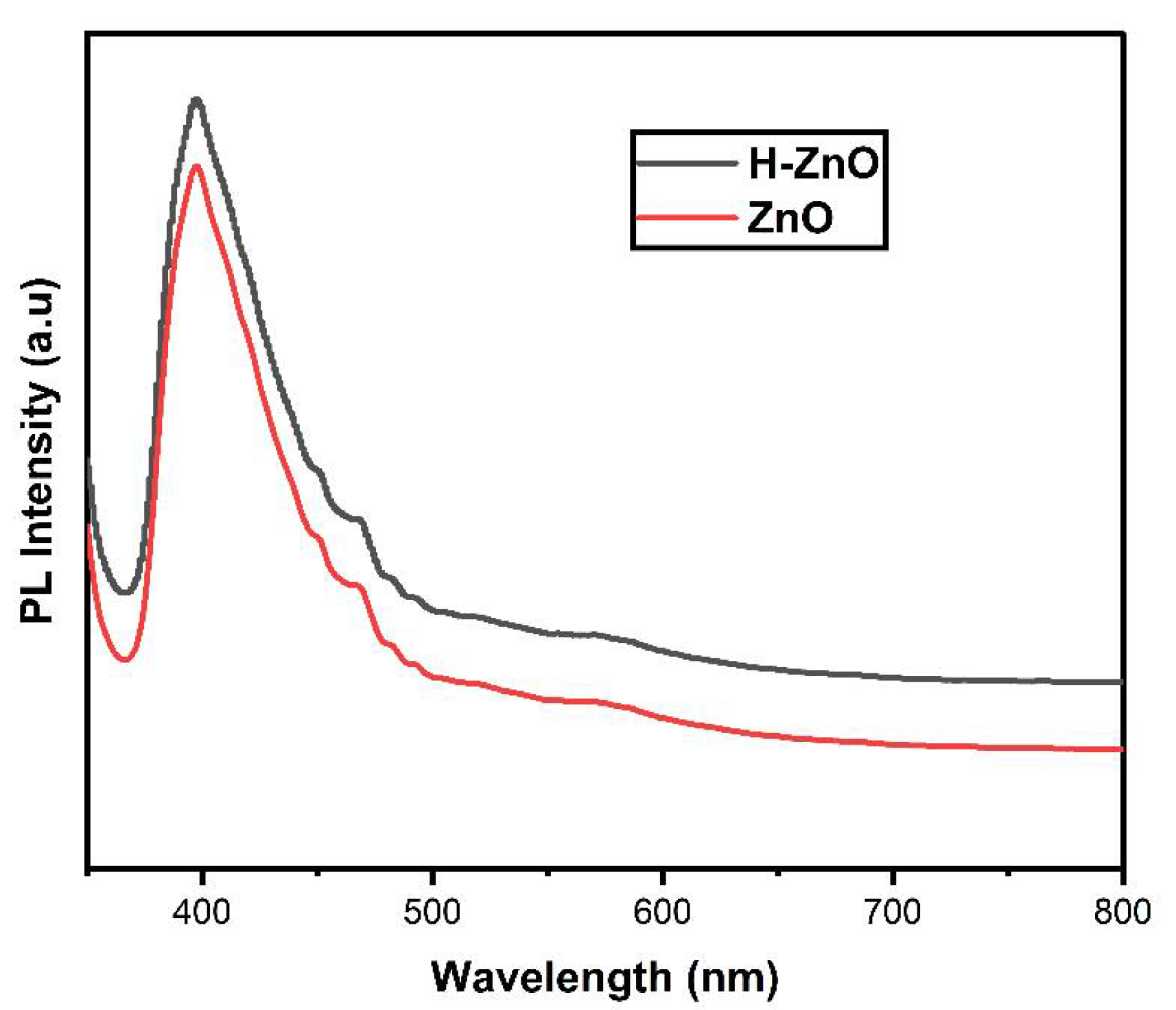
The photoluminescence spectra (Figure 4) taken at room temperature exhibit a peak at 398 nm, assigned to the near-band-edge emission of ZnO nanoparticles [39]. The detailed mechanism is explained by the defect of Zn2+ ions between the oxygen vacancies. These are:
The valence band electron excited at conduction band holes. The electrons were trapped at a high energy level, which led to their being doubly oxidized by the valence band electron.
The photogenerated oxygen was singly oxidized by the Zn2+ ions due to the direct exciton recombination process. The formation of broad peaks in the PL spectra proved the presence of fewer intrinsic defects in the origin for PL emission, which was observed with the transformation of nanoparticles and attributable to quantum size effects. The low PL intensity for bare ZnO indicated the emission of fewer charge carriers than honey-mediated zinc oxide nanoparticles, which displayed higher PL intensity. The energy interval from the shallow donor level to the top of the valence band was 398 nm, which is consistent with the photon energy of the blue emission observed in the present work.
3.5. Scanning Electron Microscopy (SEM)-EDS
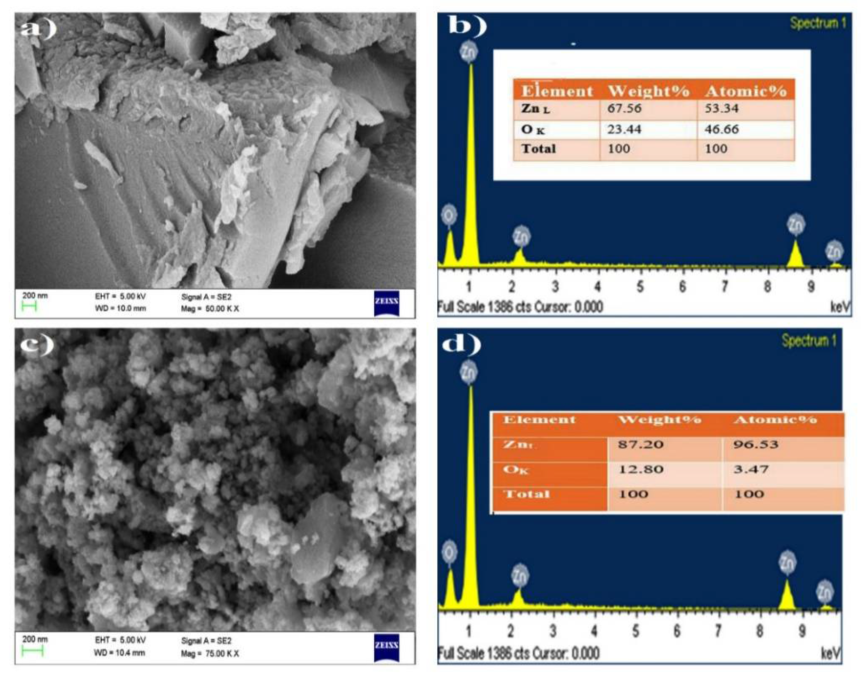
The images in Figure 5 confirm the quasi-spherical shape of ZnO nanoparticles. The bare zinc oxide nanoparticles exhibited an irregularly shaped morphology. The image at 100 nm magnification shows that most of the particles were well shaped compared to images at other magnifications [39]. Honey-capped ZnO nanoparticles consisted of Zn and O, as confirmed by the EDS spectrum. No other elements were present in the ZnO. No carbon species were observed since the honey acted only as a chelating/capping agent and retained its chemical nature through the course of the synthesis reaction. The composition report indicates that the Zn content was high compared to that of O. The obtained results indicate that honey-capped ZnO was more effective than chemically synthesized ZnO nanoparticles. The obtained zinc and oxygen compositions are presented in the EDS tables and spectra, and it was observed that the amount of Zn was greater in the honey-capped ZnO nanoparticles.
3.6. Biological Analysis
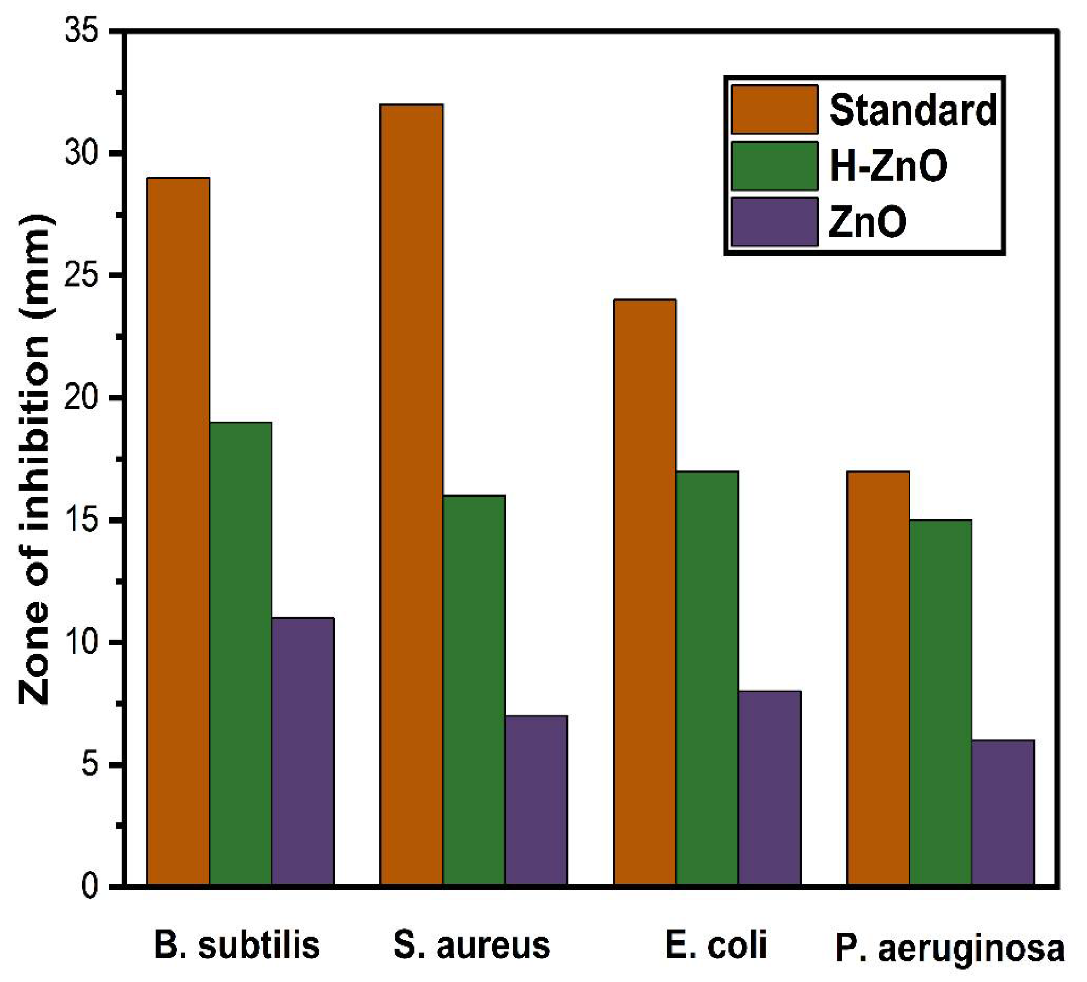
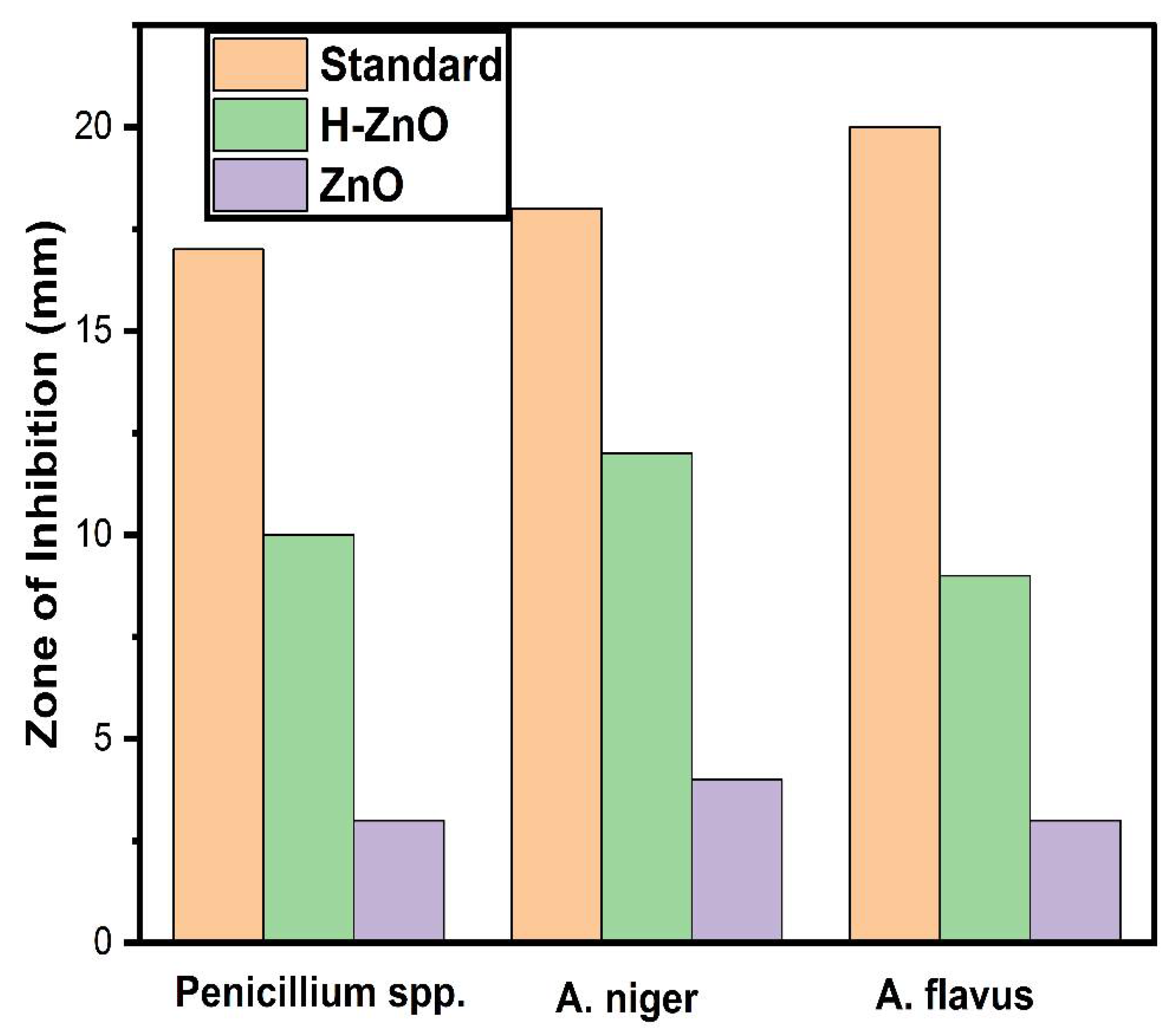
The disc diffusion method was used to observe the antibacterial activity of the synthesized ZnO nanoparticles. The activity of the ZnO nanoparticles was evaluated in both Gram-positive bacteria (Staphylococcus aureus (ATCC 6538) and Bacillus subtilis (ATCC 6633)) and Gram-negative bacteria (Escherichia coli (ATCC 8739) and Pseudomonas aeruginosa (ATCC 27853)). The agar well diffusion method was adopted to evaluate the antifungal activity in three fungi (Penicillium (ATCC 11597), Aspergillus niger (ATCC 16404), and A. flavus (ATCC 9643)). Muller–Hinton agar and PDA medium nearly equal to 20 mL/each were poured into Petri plates and a cork borer was used to bore 5 mm wells into the Petri plates. A volume of 50 μL of ZnO nanoparticles was loaded into the wells with the same concentration (20 μg) in each plate to evaluate the activity. Amikacin antibiotic discs and nystatin were used as controls for antibacterial and antifungal activity, respectively. The inhibition zone was measured after one day at 37 °C. The zone formation indicated the antibacterial and antifungal activity of the ZnO nanoparticles.
3.6.1. Antibacterial Activity
The obtained results are shown in Figure 6 and Table 1. The zone formation was confirmed by the activity of the pathogens. According to the results, ZnO nanoparticles had the highest inhibitory activity against P. aeruginosa, and the inhibition zone was 80% that seen in the control treatment amikacin. The diameter of the inhibition zone of ZnO nanoparticles against E. coli bacteria was 71% that of the control treatment [40,41]. Based on our results, ZnO nanoparticles could be effective against bacterial pathogens (Table 1).
3.6.2. Antifungal Activity
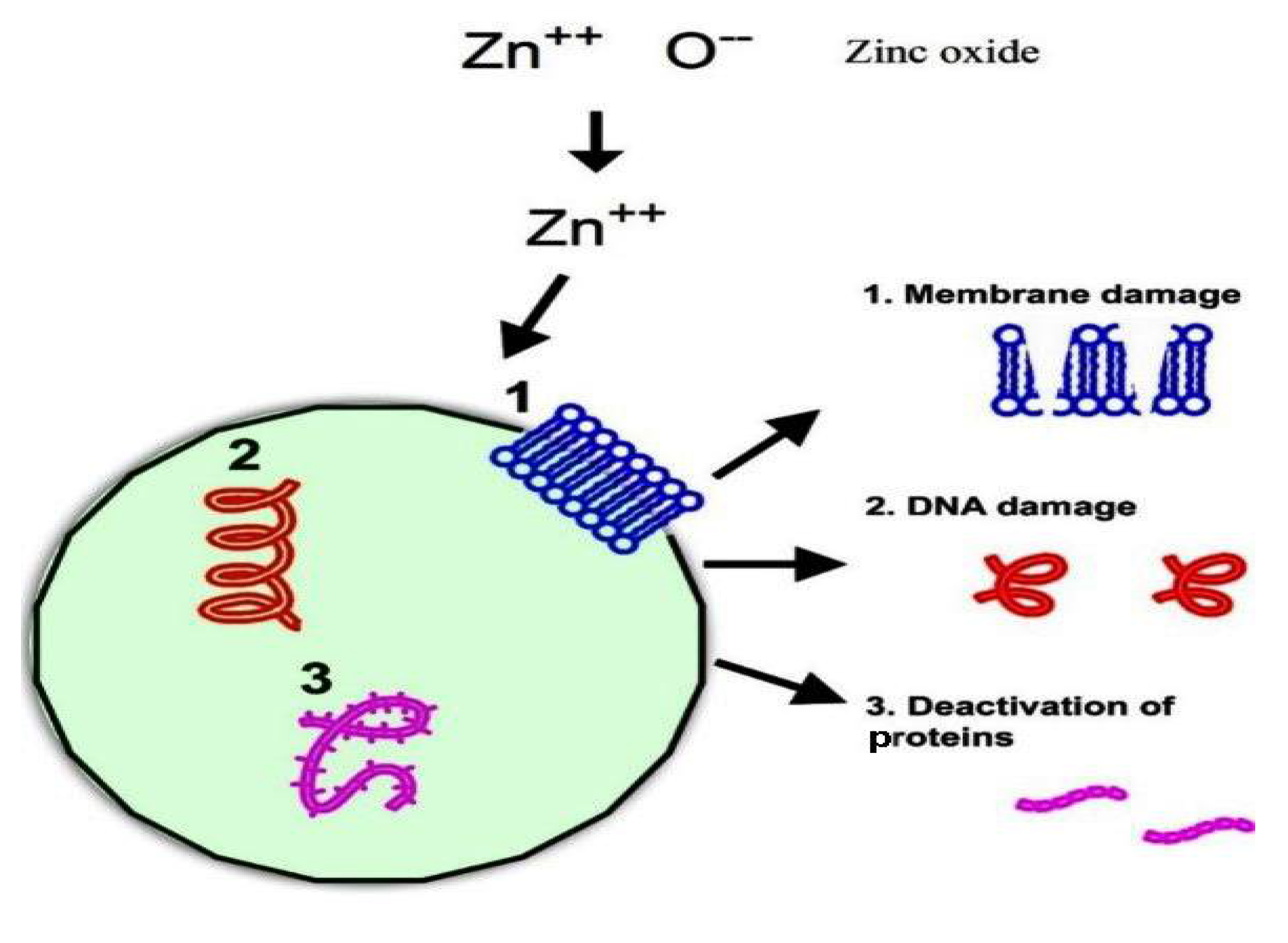
The human immune system can be damaged by fungal infection. We analyzed the antifungal activity of the synthesized nanoparticles against Penicillium, Aspergillus niger, and A. flavus using the agar diffusion method [42]. The obtained results were compared to those obtained using the standard antifungal drug nystatin. The results of the antifungal activity are shown in Table 2. The results clearly indicate that ZnO nanoparticles exhibited good antifungal activity against Penicillium and A. niger, as shown in Figure 7. The mechanism is shown in Figure 8. It seems that the formation of a large number of free ions in the case of honey-mediated ZnO nanoparticles led to better biological activity compared to that of bare ZnO nanoparticles. Free radicals from ZnO nanoparticles result in potential attachment to biomolecules such as DNA and protein, leading to cell death.
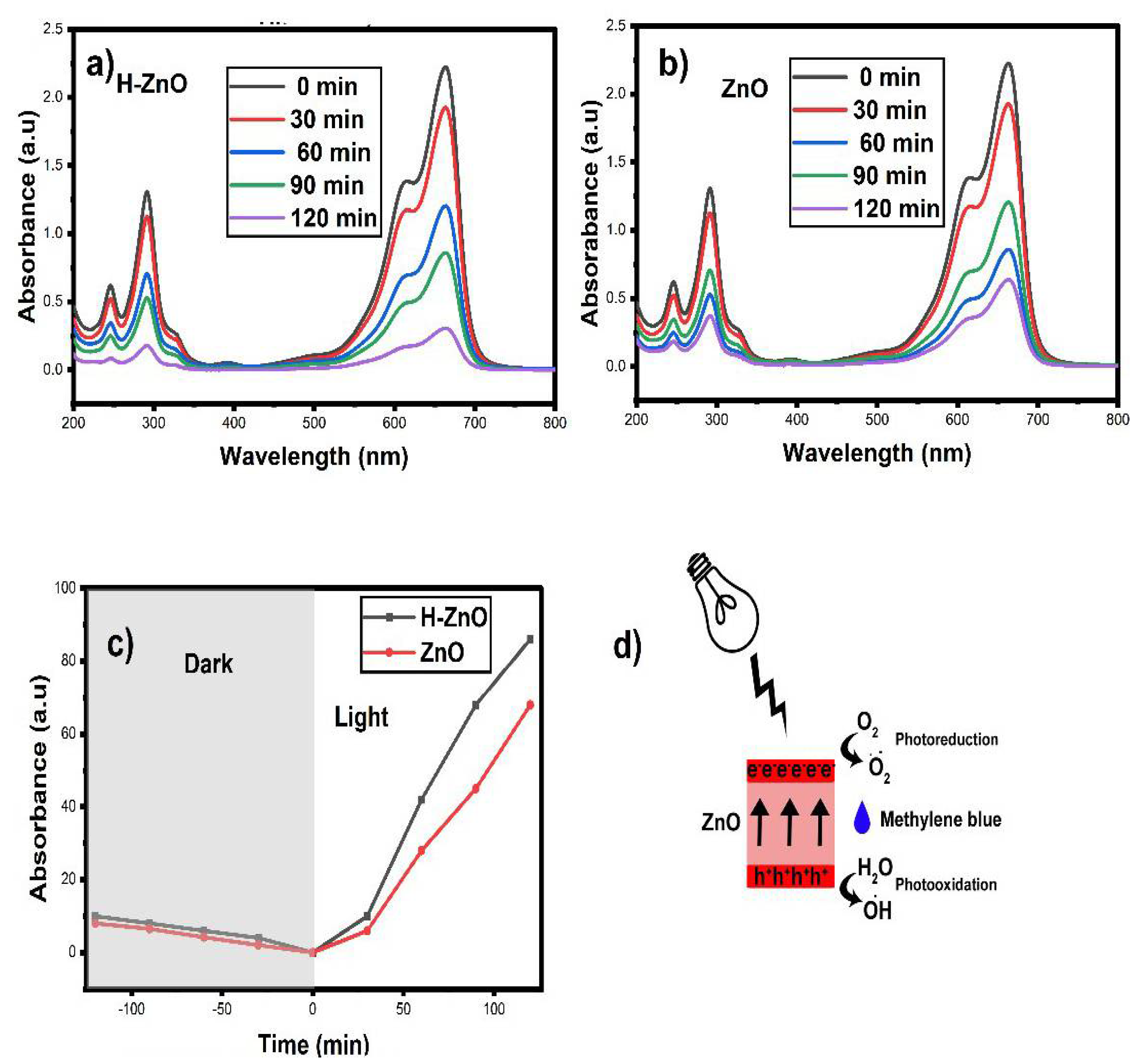
3.7. Photocatalytic Dye Degradation
Figure 9a shows the photocatalytic activity of the sample before and after irradiation. When the sample was exposed to light, absorbance gradually decreased, indicating the degradation of methylene blue. After 120 min, the maximum degradation achieved was 86%, which is a promising result compared with the effectiveness of other plant extracts and chemically synthesized ZnO nanoparticles. The degradation efficiency was estimated from Equation (3) [43]
where η is the removal efficiency, and Co and Ct correspond to the initial concentration and concentration of MB at any time interval (mg/L), respectively.
The corresponding efficiency is comparatively much higher than that of TiO2 (62%) for MB degradation [44]. The obtained dye degradation efficiency is shown in Figure 9b [45]. The detailed photocatalytic mechanism is shown in Figure 9c.
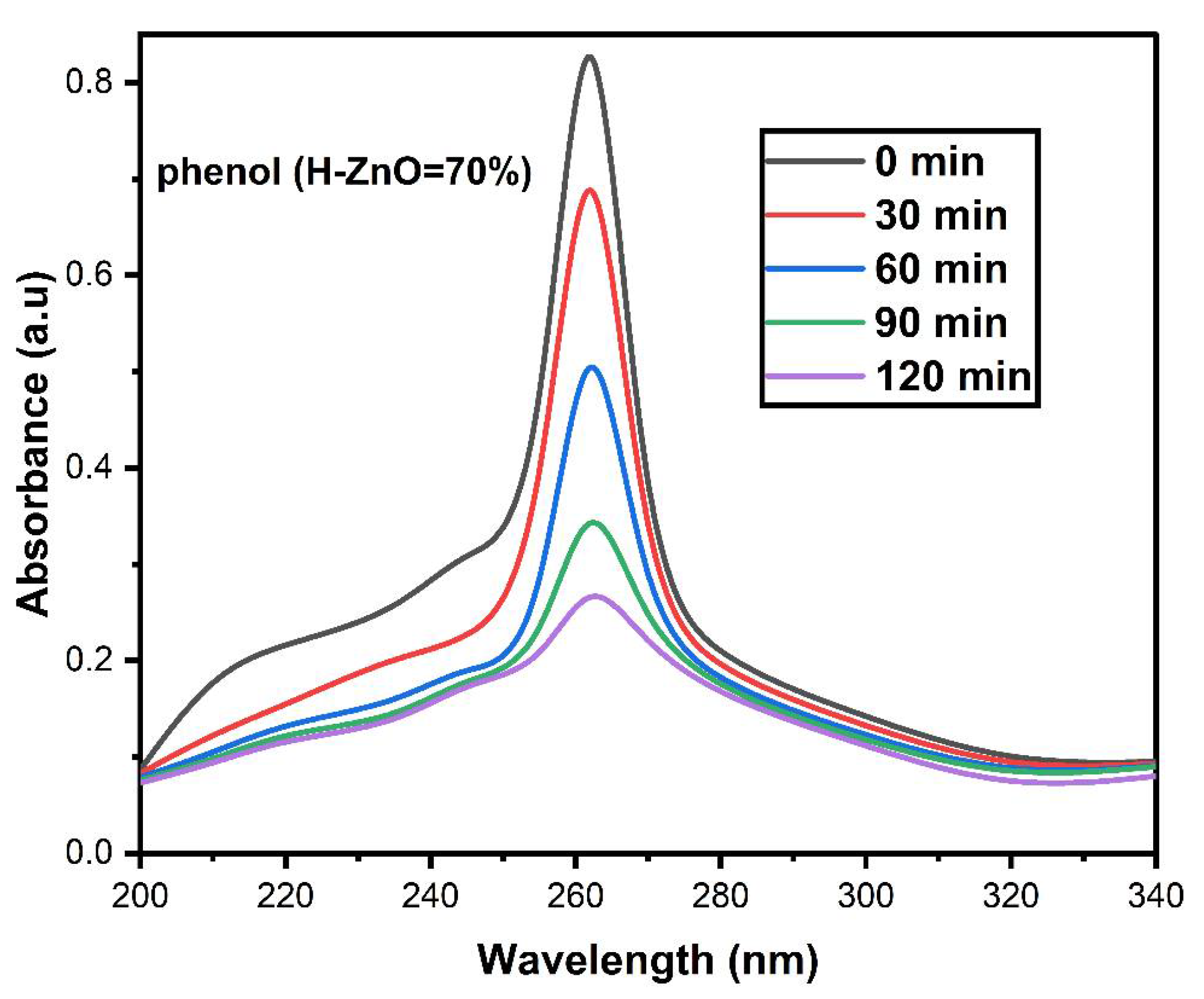
The photodegradation of MB over as-synthesized ZnO nanoparticles was maximal within 120 min. The sample was excited by visible light. This degradation time is comparatively longer than that displayed by other MB degradation catalysts, but this honey-mediated ZnO nanoparticle photocatalyst is exceptionally more economical and ecofriendly than several common photocatalysts. The degradation efficiency obtained for bare ZnO nanoparticles was found to be 60% and 7% under visible light irradiation and in the dark, respectively. Comparatively, honey-mediated zinc oxide nanoparticles acquired their enhanced degradation due to the greater number of active sites on the nanoparticles and the larger surface area able to interact with the target dye molecules. The observed degradation efficiency was much higher compared to many other catalytic systems [46,47,48]. Therefore, these green-synthesized ZnO nanoparticles could be employed in water treatment plants that process industrial waste water.
We also analyzed the degradation of a colorless solution of phenol pollutant by the honey-mediated zinc oxide nanoparticles. In the presence of the phenolic solution, 70% degradation was achieved (Figure 10). The obtained results indicate the endurance of the zinc oxide nanoparticles. The electron–hole pair recombination in the honey-mediated zinc oxide nanoparticles can produce free radicals and promote catalytic activity.
The free radicals increase the dye degradation efficacy of the ZnO nanoparticles. The photocatalytic mechanism is as follows:
ZnO+ hν → ZnO+ + ZnO(e−)
ZnO(e−) + O2 → O−2
O−2 + H+ → HO−2
HO−2 + H+ → OH + OH
ZnO+ + OH → OH
O−2/OH + Dye+ → CO2 + H2O (Byproduct)
4. Conclusions
Honey-mediated ZnO nanoparticles were synthesized using an autocombustion method. Structural crystallinity and phase purity were analyzed via XRD analysis, which also demonstrated that the particle size was in the nanodomain (i.e., 39 nm). FTIR spectroscopy displayed metal oxide bond vibrations in the range 400–500 cm−1. The as-synthesized ZnO nanoparticles were investigated for their biological activity and photocatalytic properties. ZnO nanoparticles were found to be bioactive with respect to both antibacterial and antifungal applications. Moreover, photocatalytic efficiency was investigated by employing the ZnO nanoparticles as photocatalysts for methylene blue degradation. The proposed honey-mediated ZnO catalyst displayed an impressive degradation efficiency of 86% against MB dye, which was significantly higher than that of bare ZnO, which had an efficiency of 60%. In this way, the nanoparticles synthesized from greener synthetic routes by the incorporation of natural capping and chelating ingredients could be more viable than chemical synthesis methods, owing these ingredients’ availability, cost effectiveness, and ecofriendliness. The catalytic, antibacterial, and antifungal activities indicate that the prepared ZnO nanoparticles could be implemented effectively in biomedical and environmental fields.
Author Contributions
Conceptualization, M.S. and R.J.M.; methodology, A.K.; software, G.E.E.; validation, A.E.M.Y. and A.A.A.B.; formal analysis, A.A.; investigation, M.S.; resources, R.J.M.; data curation, M.S.; writing—original draft preparation, A.A.; writing—review and editing, A.A. All authors have read and agreed to the published version of the manuscript.
Funding
The authors extend their appreciation to the Deanship of Scientific Research at King Saud University for funding this work through research group No. RGP-130.
Institutional Review Board Statement
Not applicable.
Informed Consent Statement
Not applicable.
Data Availability Statement
Not applicable.
Conflicts of Interest
The authors declare no conflict of interest.
References
- Zhang, Y.; Chen, Y.; Westerhoff, P.; Hristovski, K.; Crittenden, J.C. Stability of commercial metal oxide nanoparticles in water. Water Res. 2008, 42, 2204–2212. [Google Scholar] [CrossRef]
- Mohammadzadeh, A.; Khoshghadam-Pireyousefan, M.; Shokrianfard-Ravasjan, B.; Azadbeh, M.; Rashedi, H.; Dibazar, M.; Mostafaei, A. Synergetic photocatalytic effect of high purity ZnO pod shaped nanostructures with H2O2 on methylene blue dye degradation. J. Alloy. Compd. 2020, 845, 156333. [Google Scholar] [CrossRef]
- Ahmad, I.; Jamal, M.A.; Iftikhar, M.; Ahmad, A.; Hussain, S.; Asghar, H.; Saeed, M.; Yousaf, A.B.; Karri, R.R.; Al-kadhi, N.S. Lanthanum-zinc binary oxide nanocomposite with promising heterogeneous catalysis performance for the active conversion of 4-nitrophenol into 4-aminophenol. Coatings 2021, 11, 537. [Google Scholar] [CrossRef]
- Selvaraj, S.; Mohan, M.K.; Navaneethan, M.; Ponnusamy, S.; Muthamizhchelvan, C. Synthesis and photocatalytic activity of Gd doped ZnO nanoparticles for enhanced degradation of methylene blue under visible light. Mater. Sci. Semicond. Process. 2019, 103, 104622. [Google Scholar] [CrossRef]
- Ganesan, V.; Hariram, M.; Vivekanandhan, S.; Muthuramkumar, S. Periconium sp. (endophytic fungi) extract mediated sol-gel synthesis of ZnO nanoparticles for antimicrobial and antioxidant applications. Mater. Sci. Semicond. Process. 2020, 105, 104739. [Google Scholar] [CrossRef]
- Shim, Y.J.; Soshnikova, V.; Anandapadmanaban, G.; Mathiyalagan, R.; Perez, Z.E.J.; Markus, J.; Kim, Y.J.; Castro-Aceituno, V.; Yang, D.C. Zinc oxide nanoparticles synthesized by Suaeda japonica Makino and their photocatalytic degradation of methylene blue. Optik 2019, 182, 1015–1020. [Google Scholar] [CrossRef]
- Najafidoust, A.; Allahyari, S.; Rahemi, N.; Tasbihi, M. Uniform coating of TiO2 nanoparticles using biotemplates for photocatalytic wastewater treatment. Ceram. Int. 2020, 46, 4707–4719. [Google Scholar] [CrossRef]
- Dey, S.; Das, S.; Kar, A.K. Role of precursor dependent nanostructures of ZnO on its optical and photocatalytic activity and influence of FRET between ZnO and methylene blue dye on photocatalysis. Mater. Chem. Phys. 2021, 270, 124872. [Google Scholar] [CrossRef]
- Borsagli, F.G.M.; Paiva, A.E. Eco-friendly luminescent ZnO nanoconjugates with thiol group for potential environmental photocatalytic activity. J. Environ. Chem. Eng. 2021, 9, 105491. [Google Scholar] [CrossRef]
- Yulizar, Y.; Apriandanu, D.O.B.; Ashna, R.I. La2CuO4-decorated ZnO nanoparticles with improved photocatalytic activity for malachite green degradation. Chem. Phys. Lett. 2020, 755, 137749. [Google Scholar] [CrossRef]
- Younas, U.; Hassan, S.T.; Ali, F.; Hassan, F.; Saeed, Z.; Pervaiz, M.; Khan, S.; Jannat, F.T.; Bibi, S.; Sadiqa, A. Radical Scavenging and Catalytic Activity of Fe-Cu Bimetallic Nanoparticles Synthesized from Ixora finlaysoniana Extract. Coatings 2021, 11, 813. [Google Scholar] [CrossRef]
- Omri, K.; Najeh, I.; Dhahri, R.; El Ghoul, J.; El Mir, L. Effects of temperature on the optical and electrical properties of ZnO nanoparticles synthesized by sol–gel method. Microelectron. Eng. 2014, 128, 53–58. [Google Scholar] [CrossRef]
- Elumalai, K.; Velmurugan, S.; Ravi, S.; Kathiravan, V.; Ashokkumar, S. Bio-fabrication of zinc oxide nanoparticles using leaf extract of curry leaf (Murraya koenigii) and its antimicrobial activities. Mater. Sci. Semicond. Process. 2015, 34, 365–372. [Google Scholar] [CrossRef]
- Ramesh, P.S.; Kokila, T.; Geetha, D. Plant mediated green synthesis and antibacterial activity of silver nanoparticles using Emblica officinalis fruit extract. Spectrochim. Acta Part A Mol. Biomol. Spectrosc. 2015, 142, 339–343. [Google Scholar] [CrossRef]
- Sathyavathi, R.; Krishna, M.; Rao, D.N. Biosynthesis of silver nanoparticles using Moringa oleifera leaf extract and its application to optical limiting. J. Nanosci. Nanotechnol. 2011, 11, 2031–2035. [Google Scholar] [CrossRef] [PubMed]
- González-Miret, M.L.; Terrab, A.; Hernanz, D.; Fernández-Recamales, M.Á.; Heredia, F.J. Multivariate correlation between color and mineral composition of honeys and by their botanical origin. J. Agric. Food Chem. 2005, 53, 2574–2580. [Google Scholar] [CrossRef] [PubMed]
- Baneto, M.; Enesca, A.; Mihoreanu, C.; Lare, Y.; Jondo, K.; Napo, K.; Duta, A. Effects of the growth temperature on the properties of spray deposited CuInS2 thin films for photovoltaic applications. Ceram. Int. 2015, 41, 4742–4749. [Google Scholar] [CrossRef]
- Rathnasamy, R.; Thangasamy, P.; Thangamuthu, R.; Sampath, S.; Alagan, V. Green synthesis of ZnO nanoparticles using Carica papaya leaf extracts for photocatalytic and photovoltaic applications. J. Mater. Sci. Mater. Electron. 2017, 28, 10374–10381. [Google Scholar] [CrossRef]
- Debanath, M.K.; Karmakar, S. Study of blueshift of optical band gap in zinc oxide (ZnO) nanoparticles prepared by low-temperature wet chemical method. Mater. Lett. 2013, 111, 116–119. [Google Scholar] [CrossRef]
- Groot, A.P.D. Protein and Amino Acid Requirements of the Honeybee (Apis mellifica L.); Food and Agriculture Organization: Sacramento, CA, USA, 1953. [Google Scholar]
- Cooper, R.A.; Halas, E.; Molan, P.C. The efficacy of honey in inhibiting strains of Pseudomonas aeruginosa from infected burns. J. Burn. Care Rehabil. 2002, 23, 366–370. [Google Scholar] [CrossRef] [Green Version]
- Mueller, N.C.; Nowack, B. Exposure modeling of engineered nanoparticles in the environment. in Abstracts of papers of the American Chemical Society. Environ. Sci. Technol. 2008, 42, 4447–4453. [Google Scholar] [CrossRef]
- Porrini, C.; Sabatini, A.G.; Girotti, S.; Ghini, S.; Medrzycki, P.; Grillenzoni, F.; Bortolotti, L.; Gattavecchia, E.; Celli, G. Honey bees and bee products as monitors of the environmental contamination. Apiacta 2003, 38, 63–70. [Google Scholar]
- Hoseini, S.J.; Darroudi, M.; Oskuee, R.K.; Gholami, L.; Zak, A.K. Honey-based synthesis of ZnO nanopowders and their cytotoxicity effects. Adv. Powder Technol. 2015, 26, 991–996. [Google Scholar] [CrossRef]
- Ranjithkumar, B.; Ramalingam, H.B.; Kumar, E.R.; Srinivas, C.; Magesh, G.; Rahale, C.S.; El-Metwaly, N.M.; Shekar, B.C. Natural fuels (Honey and Cow urine) assisted combustion synthesis of zinc oxide nanoparticles for antimicrobial activities. Ceram. Int. 2021, 47, 14475–14481. [Google Scholar] [CrossRef]
- Neupane, B.P.; Chaudhary, D.; Paudel, S.; Timsina, S.; Chapagain, B.; Jamarkattel, N.; Tiwari, B.R. Himalayan honey loaded iron oxide nanoparticles: Synthesis, characterization and study of antioxidant and antimicrobial activities. Int. J. Nanomed. 2019, 14, 3533. [Google Scholar] [CrossRef] [Green Version]
- Balasooriya, E.R.; Jayasinghe, C.D.; Jayawardena, U.A.; Ruwanthika, R.W.D.; Mendis de Silva, R.; Udagama, P.V. Honey mediated green synthesis of nanoparticles: New era of safe nanotechnology. J. Nanomater. 2017, 2017, 5919836. [Google Scholar] [CrossRef]
- Ismail, N.A.; Shameli, K.; Wong, M.M.T.; Teow, S.Y.; Chew, J.; Sukri, S.N.A.M. Antibacterial and cytotoxic effect of honey mediated copper nanoparticles synthesized using ultrasonic assistance. Mater. Sci. Eng. C 2019, 104, 109899. [Google Scholar] [CrossRef]
- Saleem, M.; Fang, L.; Ruan, H.B.; Wu, F.; Huang, Q.L.; Xu, C.L.; Kong, C.Y. Effect of zinc acetate concentration on the structural and optical properties of ZnO thin films deposited by Sol.-Gel method. Int. J. Phys. Sci. 2012, 7, 2971–2979. [Google Scholar] [CrossRef]
- Khan, M.; Janjua, N.K.; Khan, S.; Qazi, I.; Ali, S.; Saad Algarni, T. Electro-oxidation of ammonia at novel Ag2O−PrO2/γ-Al2O3 catalysts. Coatings 2021, 11, 257. [Google Scholar] [CrossRef]
- Mallem, S.P.R.; Koduru, M.; Chandrasekhar, K.; Prabhakar Vattikuti, S.V.; Manne, R.; Reddy, V.R.; Lee, J.H. Potato Chip-Like 0D Interconnected ZnCo2O4 Nanoparticles for High-Performance Supercapacitors. Crystals 2021, 11, 469. [Google Scholar] [CrossRef]
- Khan, S.; Shah, S.S.; Anjum, M.A.R.; Khan, M.R.; Janjua, N.K. Electro-oxidation of ammonia over copper oxide impregnated γ-Al2O3 nanocatalysts. Coatings 2021, 11, 313. [Google Scholar] [CrossRef]
- Philip, D. Honey mediated green synthesis of gold nanoparticles. Spectrochim. Acta Part A Mol. Biomol. Spectrosc. 2009, 73, 650–653. [Google Scholar] [CrossRef]
- Balaji, D.S.; Basavaraja, S.; Deshpande, R.; Mahesh, D.B.; Prabhakar, B.K.; Venkataraman, A. Extracellular biosynthesis of functionalized silver nanoparticles by strains of Cladosporium cladosporioides fungus. Colloids Surf. B Biointerfaces 2009, 68, 88–92. [Google Scholar] [CrossRef] [PubMed]
- Kasthuri, J.; Veerapandian, S.; Rajendiran, N. Biological synthesis of silver and gold nanoparticles using apiin as reducing agent. Colloids Surf. B Biointerfaces 2009, 68, 55–60. [Google Scholar] [CrossRef]
- Devi, R.S.; Gayathri, R. Green synthesis of zinc oxide nanoparticles by using Hibiscus rosa-sinensis. Int. J. Curr. Eng. Technol. 2014, 4, 2444–2446. [Google Scholar]
- Yang, Z.; Xie, C. Zn2+ release from zinc and zinc oxide particles in simulated uterine solution. Colloids Surf. B Biointerfaces 2006, 47, 140–145. [Google Scholar] [CrossRef]
- Senthilkumar, N.; Nandhakumar, E.; Priya, P.; Soni, D.; Vimalan, M.; Potheher, I.V. Synthesis of ZnO nanoparticles using leaf extract of Tectona grandis (L.) and their anti-bacterial, anti-arthritic, anti-oxidant and in vitro cytotoxicity activities. New J. Chem. 2017, 41, 10347–10356. [Google Scholar] [CrossRef]
- Fu, L.; Fu, Z. Plectranthus amboinicus leaf extract–assisted biosynthesis of ZnO nanoparticles and their photocatalytic activity. Ceram. Int. 2015, 41, 2492–2496. [Google Scholar] [CrossRef]
- Arvanag, F.M.; Bayrami, A.; Habibi-Yangjeh, A.; Pouran, S.R. A comprehensive study on antidiabetic and antibacterial activities of ZnO nanoparticles biosynthesized using Silybum marianum L. seed extract. Mater. Sci. Eng. C 2019, 97, 397–405. [Google Scholar] [CrossRef]
- Vickers, N.J. Animal communication: When i’m calling you, will you answer too? Curr. Biol. 2017, 27, R713–R715. [Google Scholar] [PubMed]
- Pasquet, J.; Chevalier, Y.; Pelletier, J.; Couval, E.; Bouvier, D.; Bolzinger, M.A. The contribution of zinc ions to the antimicrobial activity of zinc oxide. Colloids Surf. A Physicochem. Eng. Asp. 2014, 457, 263–274. [Google Scholar] [CrossRef]
- Ahmad, A.; Jini, D.; Aravind, M.; Parvathiraja, C.; Ali, R.; Kiyani, M.Z.; Alothman, A. A novel study on synthesis of egg shell based activated carbon for degradation of methylene blue via photocatalysis. Arab. J. Chem. 2020, 13, 8717–8722. [Google Scholar] [CrossRef]
- Bibi, S.; Ahmad, A.; Anjum, M.A.R.; Haleem, A.; Siddiq, M.; Shah, S.S.; Al Kahtani, A. Photocatalytic degradation of malachite green and methylene blue over reduced graphene oxide (rGO) based metal oxides (rGO-Fe3O4/TiO2) nanocomposite under UV-visible light irradiation. J. Environ. Chem. Eng. 2021, 9, 105580. [Google Scholar] [CrossRef]
- Arciniegas-Grijalba, P.A.; Patiño-Portela, M.C.; Mosquera-Sánchez, L.P.; Guerrero-Vargas, J.A.; Rodríguez-Páez, J.E. ZnO nanoparticles (ZnO-NPs) and their antifungal activity against coffee fungus Erythricium salmonicolor. Appl. Nanosci. 2017, 7, 225–241. [Google Scholar] [CrossRef] [Green Version]
- Li, K.; Liu, J.; Li, J.; Wan, Z. Effects of N mono-and N/P dual-doping on H2O2, OH generation, and MB electrochemical degradation efficiency of activated carbon fiber electrodes. Chemosphere 2018, 193, 800–810. [Google Scholar] [CrossRef]
- Chen, Q.; Yan, Z.; Zhang, H.; Zhang, L.C.; Ma, H.; Wang, W.; Wang, W. High. MB solution degradation efficiency of FeSiBZr amorphous ribbon with surface tunnels. Materials 2020, 13, 3694. [Google Scholar] [CrossRef] [PubMed]
- Yao, W.; Zhang, B.; Huang, C.; Ma, C.; Song, X.; Xu, Q. Synthesis and characterization of high efficiency and stable Ag3PO4/TiO2 visible light photocatalyst for the degradation of methylene blue and rhodamine B solutions. J. Mater. Chem. 2012, 22, 4050–4055. [Google Scholar] [CrossRef]
- Aravind, M.; Ahmad, A.; Ahmad, I.; Amalanathan, M.; Naseem, K.; Mary, S.M.M.; Parvathiraja, C.; Hussain, S.; Algarni, T.S.; Pervaiz, M.; et al. Critical green routing synthesis of silver NPs using jasmine flower extract for biological activities and photocatalytical degradation of methylene blue. J. Environ. Chem. Eng. 2021, 9, 104877. [Google Scholar] [CrossRef]
- Zhang, J.; Zhang, X.; Dong, S.; Zhou, X.; Dong, S. N-doped carbon quantum dots/TiO2 hybrid composites with enhanced visible light driven photocatalytic activity toward dye wastewater degradation and mechanism insight. J. Photochem. Photobiol. A Chem. 2016, 325, 104–110. [Google Scholar] [CrossRef]
- Senthilraja, A.; Subash, B.; Krishnakumar, B.; Rajamanickam, D.; Swaminathan, M.; Shanthi, M. Synthesis, characterization and catalytic activity of co-doped Ag–Au–ZnO for MB dye degradation under UV-A light. Mater. Sci. Semicond. Process. 2014, 22, 83–91. [Google Scholar] [CrossRef]
Figure 1.
XRD patterns of bare ZnO and honey-capped ZnO nanoparticles.

Figure 2.
FTIR spectra of bare ZnO (a) and H-ZnO (b) nanoparticles.

Figure 3.
(a) UV–Vis DRS spectra and (b) estimated band gaps of ZnO and H-ZnO.

Figure 4.
PL spectra of ZnO nanoparticles.

Figure 5.
(a,b) SEM-EDS spectra of ZnO nanoparticles and (c,d) SEM-EDS spectra of H-ZnO nanoparticles.
Figure 5.
(a,b) SEM-EDS spectra of ZnO nanoparticles and (c,d) SEM-EDS spectra of H-ZnO nanoparticles.

Figure 6.
Antibacterial activity of ZnO and H-ZnO nanoparticles compared to the standard antibiotic amikacin.
Figure 6.
Antibacterial activity of ZnO and H-ZnO nanoparticles compared to the standard antibiotic amikacin.

Figure 7.
Antifungal activity of bare ZnO and H-ZnO nanoparticles compared to the standard antifungal agent nystatin.
Figure 7.
Antifungal activity of bare ZnO and H-ZnO nanoparticles compared to the standard antifungal agent nystatin.

Figure 8.
Biological activity mechanism of ZnO nanoparticles.

Figure 9.
(a) Photocatalytic activity of H-ZnO; (b) of ZnO; (c) degradation efficiency and (d) mechanism of ZnO nanoparticles.
Figure 9.
(a) Photocatalytic activity of H-ZnO; (b) of ZnO; (c) degradation efficiency and (d) mechanism of ZnO nanoparticles.

Figure 10.
Photocatalytical activity of Honey-mediated ZnO nanoparticles against phenol.


Table 1.
Effect of H-ZnO nanoparticles on antibacterial activity.
| Zone of Inhibition Diameter (mm) | ||
|---|---|---|
| Bacterial Species | Standard Drug (mm) | H-ZnO Nanoparticles (mm) |
| B. subtilis | 29 | 19 |
| S. aureus | 32 | 16 |
| E. coli | 24 | 17 |
| P. aeruginosa | 17 | 15 |
Table 2.
Antifungal activity of H-ZnO nanoparticles on fungal species reported as zone of inhibition diameter (mm) sample−1.
Table 2.
Antifungal activity of H-ZnO nanoparticles on fungal species reported as zone of inhibition diameter (mm) sample−1.
| Fungi | Nystatin (mm) | H-ZnO Nanoparticles (mm) |
|---|---|---|
| Penicillium | 17 | 10 |
| A. niger | 18 | 12 |
| A. flavus | 20 | 9 |
Publisher’s Note: MDPI stays neutral with regard to jurisdictional claims in published maps and institutional affiliations. |
© 2021 by the authors. Licensee MDPI, Basel, Switzerland. This article is an open access article distributed under the terms and conditions of the Creative Commons Attribution (CC BY) license (https://creativecommons.org/licenses/by/4.0/).
Share and Cite
MDPI and ACS Style
Sharmila, M.; Jothi Mani, R.; Kader, A.; Ahmad, A.; Eldesoky, G.E.; Yahya, A.E.M.; Bahajjaj, A.A.A. Photocatalytic and Biological Activity of ZnO Nanoparticles Using Honey. Coatings 2021, 11, 1046. https://doi.org/10.3390/coatings11091046
AMA Style
Sharmila M, Jothi Mani R, Kader A, Ahmad A, Eldesoky GE, Yahya AEM, Bahajjaj AAA. Photocatalytic and Biological Activity of ZnO Nanoparticles Using Honey. Coatings. 2021; 11(9):1046. https://doi.org/10.3390/coatings11091046
Chicago/Turabian StyleSharmila, M., R. Jothi Mani, Abdul Kader, Awais Ahmad, Gaber E. Eldesoky, Adel E. M. Yahya, and Aboud Ahmed Awadh Bahajjaj. 2021. "Photocatalytic and Biological Activity of ZnO Nanoparticles Using Honey" Coatings 11, no. 9: 1046. https://doi.org/10.3390/coatings11091046
Note that from the first issue of 2016, this journal uses article numbers instead of page numbers. See further details here.