3. Results
A summary of patients’ baseline characteristics is shown in
Table 1 and
Table 2. There was a total of 137 patients on RTX. More than half (54%;
n = 74) of the patients were female, with a mean age of 47.69 ± 18.86 years and an average BMI of 28.57 ± 6.55 kg/m
2. The majority (56.9%;
n = 78) of the patients were younger than 50 years old, and only around a third (16.1%;
n = 22) were older than 70 years old. Almost two-quarters (41.6%;
n = 57) of the patients were obese. The most common blood type was AB (36.5%;
n = 50), followed by A (20.4%;
n = 28), B (13.1%;
n = 18), and O (4.4%;
n = 6).
As summarized in
Table 2, the most frequent indications for which RTX was used were hematological malignancies, autoimmune CTD, and benign/non-malignant hematological diseases, accounting for 42.3%, 27%, and 17.5%, respectively. More than half (56.2%;
n = 77) of the patients received the 375 mg/m
2 dose, with a median number of five doses/cycles (Q1, Q3: 2, 7). RTX’s mean cumulative dose was 3216 ± 2282 mg, with a median of 2625 (
Table 3). Besides RTX, almost half (48.9%;
n = 67) of the patients were on steroids, and less than a third (10.2%;
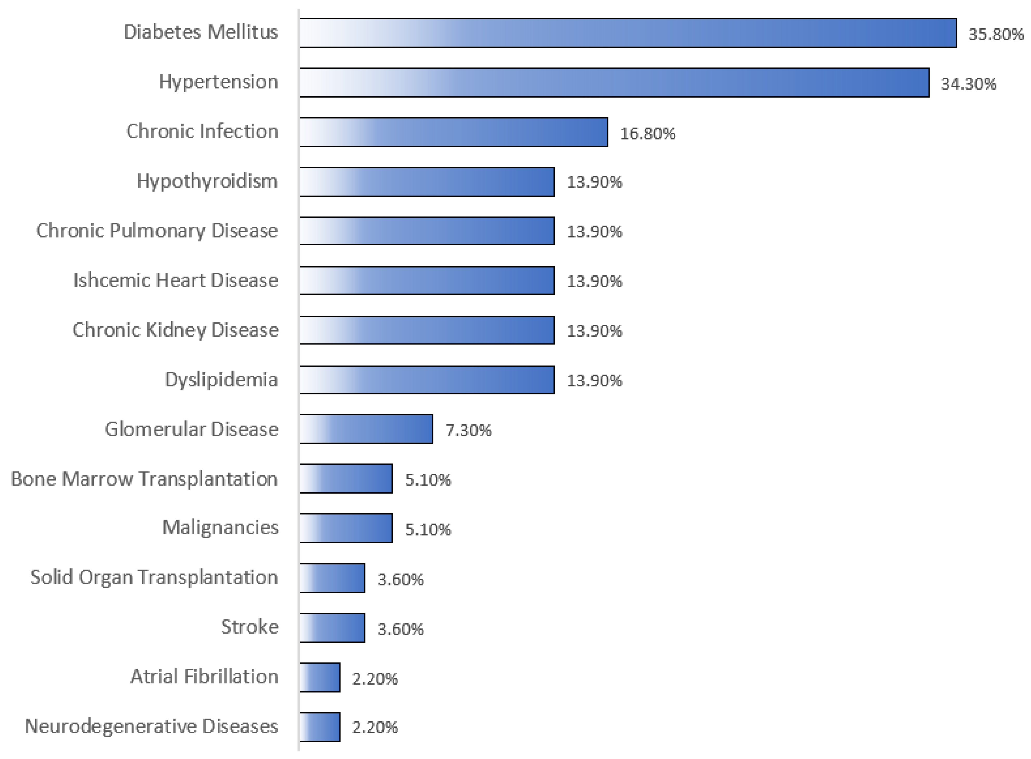
n = 14) were on methotrexate. As shown in
Figure 1, the most notable associated comorbidities were DM, HTN, chronic infection, dyslipidemia, and CKD, accounting for 35.8%, 34.3%, 16.8%, 13.9%, and 13.9%, respectively. Only a few patients had a history of bone marrow (5.1%;
n = 7) or solid organ (3.6%;
n = 5) transplantation. The overall mortality rate was 22.6% (
n = 31), with sepsis/septic shock (45.2%;
n = 14) and COVID-19 infection (16.1%;
n = 5) being the most common underlying causes of mortality (
Table 4).
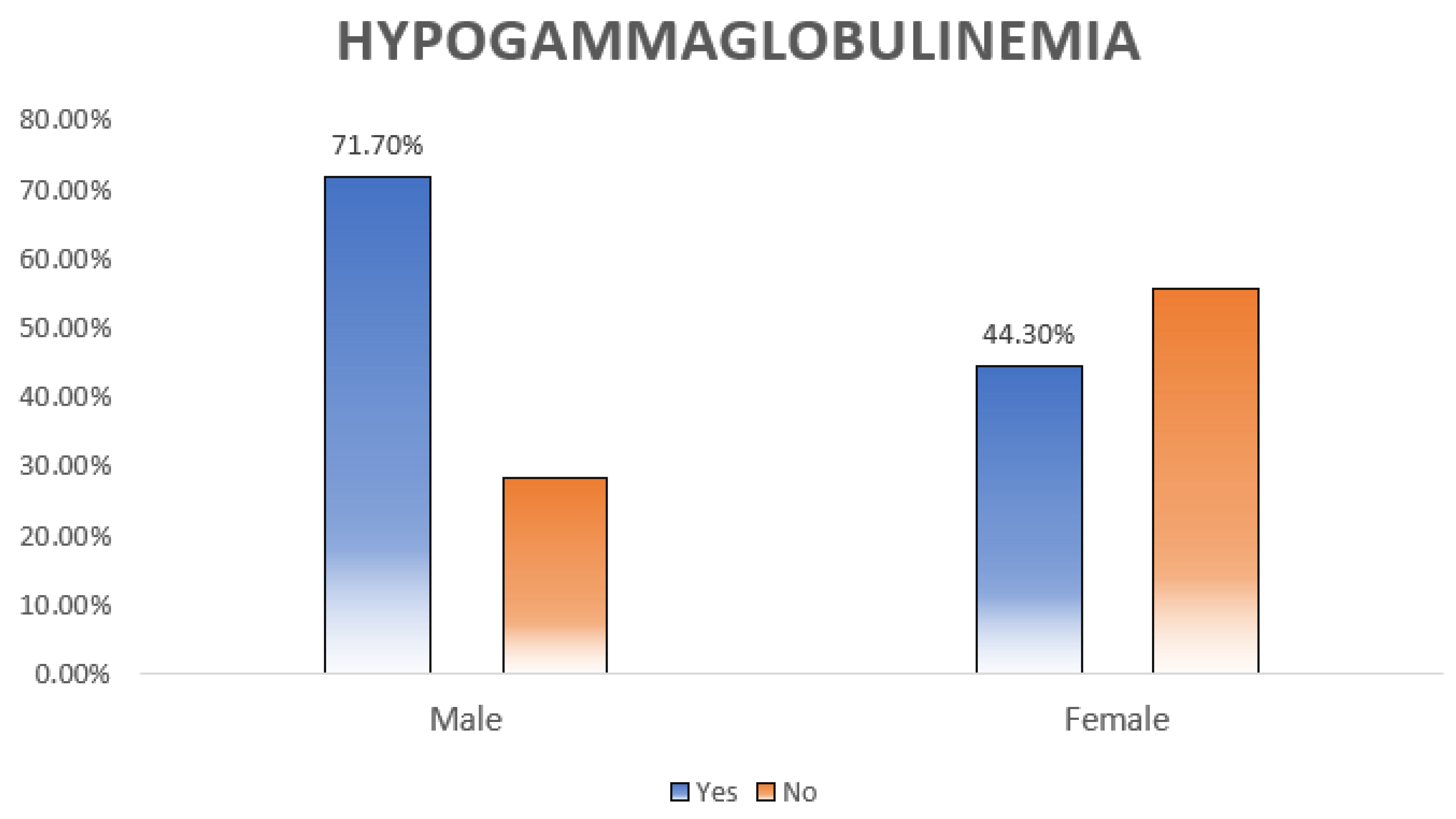
Hypogammaglobulinemia, defined as low IgG, IgM, or IgA, was diagnosed in 43.8% (
n = 60) of the patients. As shown in
Table 5 and
Table 6, hypogammaglobulinemia was significantly more prevalent among males (
p = 0.005) (
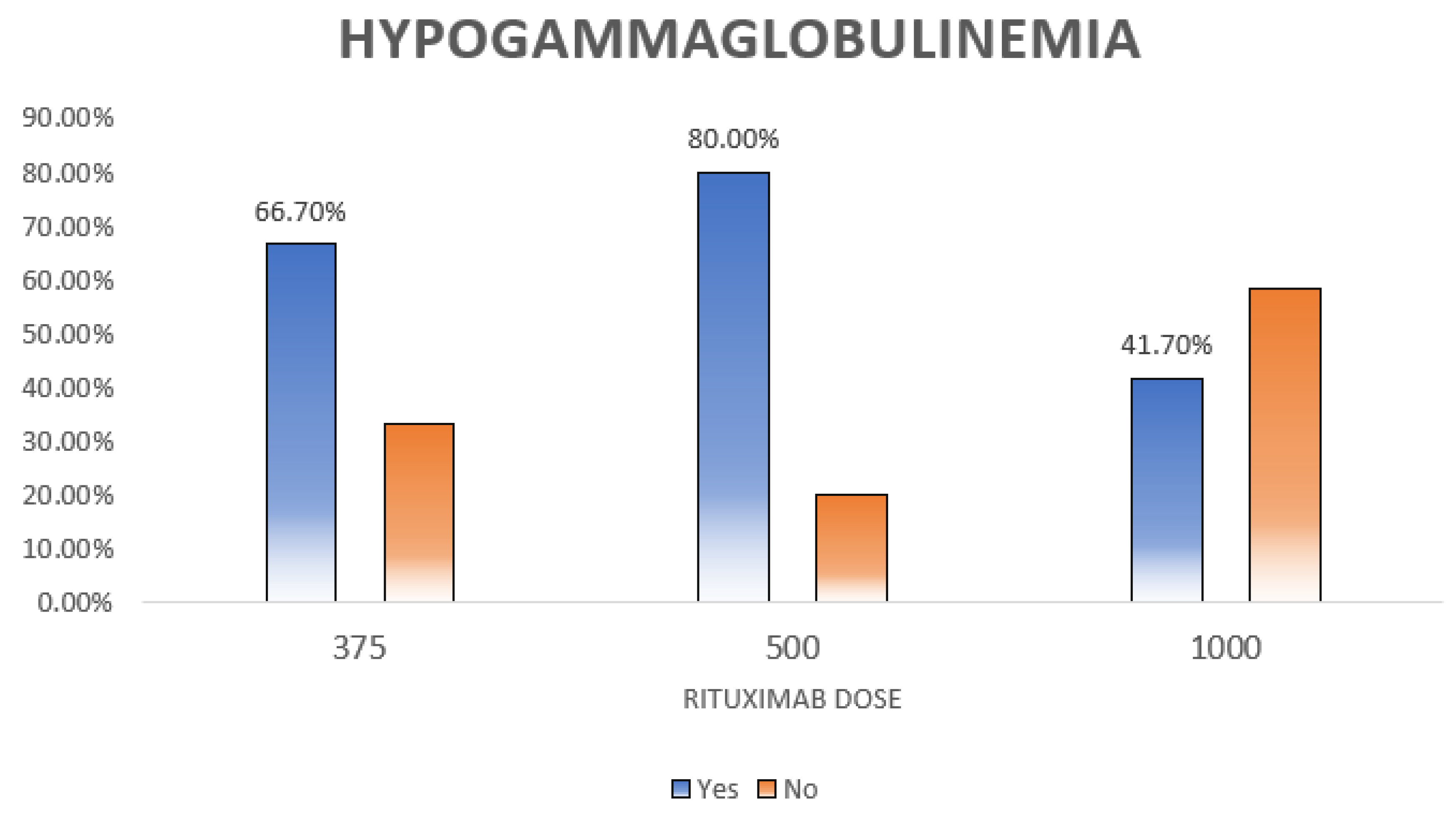
Figure 2). Moreover, patients with different doses of RTX had different percentages of hypogammaglobulinemia (
p = 0.012) (
Figure 3).
In multivariable analysis, patients with hematological malignancies had significantly higher odds of developing a severe infection and being admitted to the ICU than those with no hematological malignancies (OR = 17.77, 95% CI: 1.29 to 245.62,
p = 0.032). On the other hand, patients who received the 1000 mg dose of RTX had significantly lower odds of having severe infection and being admitted to the ICU (OR = 0.04, 95% CI: 0 to 0.8,
p = 0.036) compared to those who received the 375 mg/m
2 dose, as summarized in
Table 7.
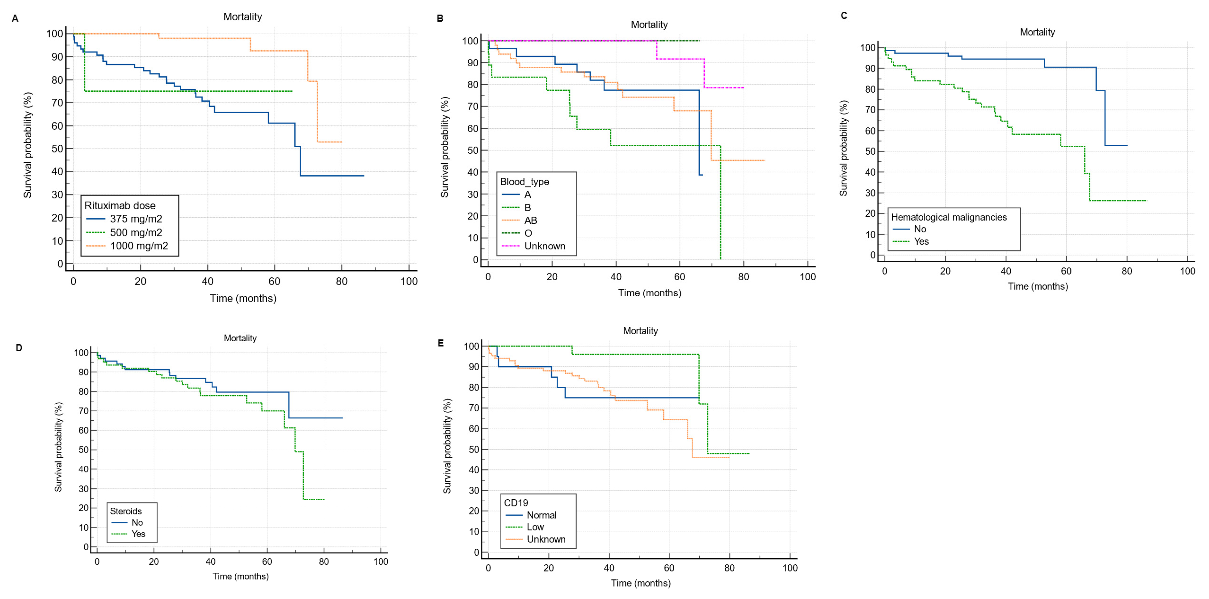
As shown in
Table 8, based on univariate Cox regression analysis, patients with hematological malignancies had a significantly higher risk of mortality than those with no hematological malignancies (HR = 6.18, 95% CI: 2.63 to 14.55,
p < 0.001). However, patients who received RTX in the 1000 mg dose had a significantly lower risk of mortality (HR = 0.15, 95% CI: 0.05 to 0.44,
p = 0.001) compared to those who received the 375 mg/m
2 dose (
Figure 4A). In multivariable Cox regression analysis, patients with blood type B (HR = 6.82, 95% CI: 2 to 23.22,
p = 0.002) and AB (HR = 1.58, 95% CI: 0.5 to 4.98,
p < 0.001) had a significantly higher risk of mortality compared to those with blood type A (
Figure 4B). Additionally, patients with hematological malignancies had a significantly higher probability of mortality than those with no hematological malignancies (HR = 11.74, 95% CI: 2.18 to 63.14,
p = 0.004) (
Figure 4C). Moreover, patients on steroids had a significantly higher risk of mortality than those who were not on steroids (HR = 3.12, 95% CI: 1.24 to 7.83,
p = 0.015) (
Figure 4D). In comparison to patients with normal CD19, those with low CD19 had a significantly lower risk of mortality (HR = 0.08, 95% CI: 0.01 to 0.74,
p = 0.026) (
Figure 4E).
4. Discussion
B cells are derived from hematopoietic stem cells in the bone marrow [
17]. Through a sophisticated mechanism, these cells recognize pathogens and differentiate into antibody-producing plasma cells [
18]. RTX is a human monoclonal antibody targeted against the B-cell surface marker CD20 [
2]. In this study, we evaluated infection risk, mortality rate and predictors, and hypogammaglobulinemia prevalence and associated factors among 137 patients treated with RTX for a variety of clinical indications in one of the largest medical centers in Saudi Arabia.
In this study, the patients’ mean age was 47.69 ± 18.86 years, which is younger than what has been reported in the literature [
13,
19]. This might be explained by the fact that our patients had heterogeneous diagnoses. To clarify, the most common indication for RTX use were hematological malignancies, followed by autoimmune CTD, non-malignant hematological conditions, glomerular diseases, and neurological conditions such as MS and MG. However, most of the available literature addressing RTX and the risk of infection and mortality include a specific disease such as rheumatoid arthritis, or a group of diseases such as autoimmune diseases [
14,
19,
20,
21]. This may also explain the equal sex distribution in our study.
In the current study, infection was observed in over a third (32.8%;
n = 45) of the patients, but only a third (33%;
n = 14) of those who were infected had a severe infection necessitating ICU admission. It is well known that immunosuppressant-induced severe infection is associated with old age, kidney impairment, chronic cardiopulmonary diseases, organ or hematopoietic stem cell recipients, and DM [
22,
23,
24,
25]. In the present study, DM (35.8%;
n = 49) and HTN (34.3%;
n = 47) were the most frequent comorbid conditions, with only a few having a chronic infection, mainly hepatitis B or C, dyslipidemia, CKD, or chronic cardiopulmonary diseases. Additionally, a small proportion of the patients had bone marrow (5.1%;
n = 7) or solid organ transplants (3.6%;
n = 5). Regardless, it is hard to compare our results with the literature owing to the difference in defining a severe infection. Several studies defined an infection as severe once it required hospitalization or IV antibiotics [
6,
13,
14,
19,
20,
21,
22]. In our study, however, it was defined as any infection requiring ICU admission, which happened in only 10.21% (
n = 14) of the patients. Based on a study that evaluated infection risk in 4479 patients treated with RTX for various indications, almost a third (28.2%) had severe infections, mainly in the first 6 months of RTX initiation [
13]. A trial that evaluated the long-term safety and efficacy of RTX in combination with belimumab in 15 patients with systemic lupus erythematosus reported three major infections (20%), requiring hospitalization, and eight minor infections (53.3%) [
26]. Another study evaluated the risk of infection among 1681 rheumatoid arthritis patients treated with RTX and reported that only 5% developed a severe infection requiring hospitalization, IV antibiotics, or resulting in death [
19]. Additionally, a study that assessed infection rates among 147 patients with ANCA-positive vasculitis treated with RTX found a total of 88 (59.9%) infection events, almost a third (29.5%;
n = 26) of which were identified as severe requiring IV antibiotics or hospitalization [
22]. The discrepancy in the definition of a severe infection, the different indications for RTX use, and the various immunosuppressants given with RTX make it challenging to determine the infection risk among RTX receivers.
Since its inception, RTX has reconstituted the treatment and redirected the survival of B-cell malignancies, including diffuse large B-cell lymphoma, follicular lymphoma, and mantle cell lymphoma [
27]. In the present study, the most common primary diagnosis for which RTX was used was hematological malignancy, and it was significantly associated with higher infection (
p = 0.032) and mortality (
p = 0.004) rates. We believe that these worrisome findings are probably attributed to the fact that RTX is usually given with other immunosuppressants such as methotrexate, azathioprine, cyclophosphamide, and mycophenolate or as a part of treatment regimens such as R-CHOP, which includes cyclophosphamide, doxorubicin, vincristine, and prednisone, and R-EPOCH, which includes etoposide, prednisone, vincristine, cyclophosphamide, doxorubicin, in addition to RTX. Additionally, around half (48.9%;
n = 67) of our patients were on corticosteroids, and their use was significantly associated with a higher mortality rate (
p = 0.015). The studied population has an impaired humoral immunity due to RTX use, and adding another immunosuppressant such as azathioprine, mycophenolate, or cyclophosphamide would impair cell-mediated immunity [
28]. Furthermore, steroids are powerful anti-inflammatory agents that disrupt innate immunity as well [
29]. Impairment of both innate and adaptive immunity makes these patients vulnerable to severe and fulminant infections and increases their mortality.
We also found a statistically significant association between the 375 mg/m
2 RTX dose and both infection and mortality. The former can be explained with the same aforementioned explanation, as the 375 mg/m
2 dose weekly for 4 weeks or every 3–4 weeks is the one used in lymphoma treatment. Moreover, in addition to therapy-related factors, the pathogenesis of the primary disease plays a crucial role in susceptibility to infection and subsequent mortality in these patients [
30]. Also, we found that patients who received the 1000 mg RTX dose had a significantly lower risk of mortality compared to those who received the 375 mg/m
2 dose. The reason might be related to the primary diagnosis for which RTX was used, frequency of RTX administration, and the concurrent use of other immunosuppressants. To clarify, the 1000 mg dose is commonly used in the treatment of neurological diseases such as MG and MS. Those patients do not typically require multiple immunosuppressants and are usually young and relatively healthy with no or only mild comorbidities compared to those with hematological malignancies. Additionally, most of those patients were not on steroids as they are often only used for acute relapses and crises. This could be another possible explanation for the observed favorable outcomes in patients receiving the 1000 mg dose compared to the 375 mg/m
2 dose.
Our results revealed a mortality rate of 22.6% (
n = 31), with infection (61.3%;
n = 19) being the most frequent cause of mortality, consistent with the literature [
31]. Since its emergence, an important cause of mortality, especially in immunocompromised individuals such as the studied population, has been COVID-19 infection, which was the underlying cause of mortality in 16.1% (
n = 5) of our patients [
32,
33]. Compared to the general population, B-cell-depleted individuals need a longer time to clear the virus, and because of that, COVID-19 infection is more likely to persist and last for months [
34]. Furthermore, total hospitalization time and COVID-19 complications, including respiratory failure and ICU admission, occur at higher rates among these patients [
34,
35,
36]. Another dilemma is the fact that RTX affects memory B cells in addition to effector B cells, possibly blunting the response to the COVID-19 vaccine [
34]. Since the most frequently observed indications for RTX use in our study were hematological malignancies (42.3%;
n = 58) and autoimmune CTDs (27%;
n = 37), we will discuss the effect of COVID-19 infection on the outcomes of these two groups of diseases. To begin with, based on a systematic review and meta-analysis that investigated COVID-19 morbidity and mortality in cancer patients, the risk of severe COVID-19 infection and mortality increases by 2.84- and 2.60-fold, respectively, among cancer patients [
37]. More specifically, several studies have also found that hematological malignancies compromise the outcomes of COVID-19 infection and the response to the COVID-19 vaccine, and receiving RTX within a year of the vaccine significantly reduces antibody production and, therefore, attenuates the efficacy of the vaccine [
38,
39]. Unfortunately, this is not limited to the COVID-19 vaccine; it has also been observed with the pneumococcal and influenza vaccines [
16]. Similarly, a study that evaluated the outcomes of COVID-19 in 122 patients with a heterogeneous group of inflammatory CTDs found that RTX use was associated with significantly longer hospitalization and higher mortality [
35].
Hypogammaglobulinemia is defined as low serum immunoglobulin levels [
40]. Although it can be primary, hypogammaglobulinemia is frequently diagnosed secondary to medical conditions, such as nephrotic syndrome and infections, and medications, such as corticosteroids and immunomodulators like RTX used for various hematological malignancies, autoimmune CTDs, glomerular conditions, and neurological diseases [
41,
42]. In the present study, hypogammaglobulinemia was defined as a deficiency in either IgG, IgM, and/or IgA, and it was observed in 43.8% (with 95% CI: from 33.4 to 56.4) of our patients. This is in accordance with the literature, as several studies have reported a hypogammaglobulinemia rate ranging from 42 to 47.5% among RTX receivers [
13,
14,
43,
44,
45,
46]. This dose range not apply to all the studies, though; there are published studies with lower [
6,
47,
48] or higher [
21,
49] percentages, but most of the literature reported a percentage within this range. Generally, patients with malignancies are more prone to developing hypogammaglobulinemia during or after treatment, compared to patients with non-malignant conditions. This might be accredited to the nature of cancer itself, as it is already a well-known cause of hypogammaglobulinemia, and to the aggressive treatment regimens used in cancer. To clarify, patients with B-cell lymphoma, for example, might need more than one course of RTX, and this has been linked to a higher risk of hypogammaglobulinemia [
50]. We found a statistically significant relation between the development of hypogammaglobulinemia and the male sex (71.7%) as compared to females (44.3%) (
p = 0.005). We believe that sex may not be directly related to hypogammaglobulinemia. Rather, male predominance observed in most hematological malignancies and the aggressive nature of autoimmune CTDs in males requiring RTX use might be the underlying cause for this finding [
51,
52,
53]. Based on a large study of 4479 patients treated with RTX for various clinical conditions, the male sex was significantly associated with higher mortality particularly in hematological malignancy and CTD groups, supporting the previously mentioned explanation [
13]. Likewise, a study that examined hypogammaglobulinemia and infection risk among 29 granulomatosis with polyangiitis patients treated with RTX found an association between the male sex and hypogammaglobulinemia [
43].
We found that different doses of RTX had significantly (
p = 0.012) different rates of hypogammaglobulinemia (
Table 4); however, we could not find an association between cumulative RTX dose and hypogammaglobulinemia (
p = 0.307). Comparably, a study of 243 patients treated with RTX for several multi-system autoimmune diseases find no association between cumulative RTX dose and hypogammaglobulinemia [
21]. Opposite to our findings, a study of 103 patients with complicated nephrotic syndrome who received at least a single dose of RTX found an association between repeated RTX cycles and hypogammaglobulinemia [
49]. Next, a study of 169 patients with neuromyelitis optica who were treated with RTX reported that the mean annual RTX dose was significantly associated with hypogammaglobulinemia [
44]. We believe that this discrepancy in the results is largely attributed to several factors, including the primary disease and patients’ characteristics. It is also important to mention that some of the studies have significantly linked RTX dose and infection, but not RTX dose and hypogammaglobulinemia [
14,
20,
44,
50]. We could not establish an association between the number of RTX doses and hypogammaglobulinemia (
p = 0.153). Additionally, we failed to prove an association between other immunosuppressants, such as methotrexate, azathioprine, and mycophenolate, or comorbidities and hypogammaglobulinemia. Some studies have linked some immunosuppressants, such as cyclophosphamide, corticosteroids, and mitoxantrone, with the development of hypogammaglobulinemia [
16,
50]. It is also worth mentioning that many studies have found a significant association between particular comorbidities, such as chronic pulmonary disease, heart failure, DM, and cancer, and severe infection, but not hypogammaglobulinemia, suggesting a highly complicated interplay of multiple factors related to the patients, their pathologies, and their treatment regimens [
14,
20,
44,
50].
In this study, the majority (86.13%;
n = 118) of patients underwent B cell immunophenotyping, and over a quarter (24.57%;
n = 29) of them had low CD19. Like immunoglobulin levels, which were not measured in over a quarter of our patients, CD19 was unchecked in almost a third (16.9%;
n = 20) of the patients. Although the percentage of patients who did not undergo immunophenotyping was relatively low, we believe that the status quo can be improved. We also found that low CD19 was significantly associated with a lower risk of mortality (HR = 0.08, 95% CI: 0.01 to 0.74,
p = 0.026). As expected, patients who receive appropriate doses of RTX should have low CD19, which is a surrogate for CD20. And, at least in theory, patients with incomplete B cell depletion, measured by CD19 level, should receive an extra dose of RTX [
54]. An observational study investigating the efficacy of RTX in 71 patients with systemic lupus erythematosus found that patients with B cells in renal biopsy had poor outcomes [
55]. Another study assessed the role of CD19 level in 42 patients with nephrotic syndrome and found a positive correlation between CD19 B cell percentage and risk of relapse [
56]. Also, a study of 44 patients treated with RTX-based desensitization for ABO-incompatible kidney transplantation found that high CD19 significantly increased the risk of acute antibody-mediated rejection [
57]. Although not exactly similar to our findings, we believe these findings explain the same concept, as low CD19 indicates that the patient received an appropriate dose of RTX, which controlled the primary disease and might explain the low mortality.
Unfortunately, despite the current recommendations, more than a quarter (21.9%; n = 30) of our patients did not have their immunoglobulin levels checked, and more than a third (16.9%; n = 20) of our patients did not have their CD19 level checked, reflecting a lack of awareness about the importance of immunological monitoring during the treatment period. We advocate for immunoglobulin monitoring and B cell immunophenotyping throughout the treatment period, especially in male patients with hematological malignancies and those using corticosteroids, to detect hypogammaglobulinemia early, identify immunoglobulin replacement candidates, and possibly mitigate the risk of infection and mortality.
The current study has several limitations. First, a sample size of 137 is considered small, especially among patients with heterogeneous diagnoses and different dosing regimens, which might be another limitation. Second, the retrospective nature of the study makes it challenging to establish a causative relation between the use of RTX and infection and/or mortality as both can be influenced by the underlying primary disease, comorbidities, and the concurrent use of other immunosuppressants. Moreover, we did not gather data about the time of infection following RTX initiation. It would be beneficial to know the median time of infection after RTX initiation. Next, hypogammaglobulinemia was not classified as mild, moderate, or severe. Instead, we collected immunoglobulin levels (IgG, IgM, and IgA) as a numerical variable and then coded the levels as either normal or low. It might have been more beneficial to know the fraction of patients with severe hypogammaglobulinemia and the associated factors. Also, we did not look at immunoglobulin levels before RTX initiation. This may have given us an idea of the awareness among different specialties about the importance of measuring immunoglobulin levels before RTX initiation. In addition, our definition of severe infection, which was any infection requiring ICU admission, might have underestimated the real percentage of infection among the studied population. Finally, some of the findings might not be clinically relevant or could not be well explained by the authors or the literature, such as the statistical significance between specific blood groups (B and AB) and mortality.

 ,
, 


