
A Glance at the Molecules That Regulate Oligodendrocyte Myelination


1
Institute of Life Science & School of Life Sciences, Nanchang University, Nanchang 330031, China
2
School of Basic Medical Sciences, Nanchang University, Nanchang 330031, China
*
Author to whom correspondence should be addressed.
Curr. Issues Mol. Biol. 2022, 44(5), 2194-2216; https://doi.org/10.3390/cimb44050149
Submission received: 30 March 2022
/
Revised: 10 May 2022
/
Accepted: 13 May 2022
/
Published: 15 May 2022
(This article belongs to the Special Issue Multipotent and Multisystem Biomolecules—Associated Interlinking Networks and Crossing Organ Differentiation)
Abstract
:Oligodendrocyte (OL) myelination is a critical process for the neuronal axon function in the central nervous system. After demyelination occurs because of pathophysiology, remyelination makes repairs similar to myelination. Proliferation and differentiation are the two main stages in OL myelination, and most factors commonly play converse roles in these two stages, except for a few factors and signaling pathways, such as OLIG2 (Oligodendrocyte transcription factor 2). Moreover, some OL maturation gene mutations induce hypomyelination or hypermyelination without an obvious function in proliferation and differentiation. Herein, three types of factors regulating myelination are reviewed in sequence.
1. Introduction
OL myelination is critical to the vertebrate central nervous system (CNS) function. It supports not only the myelinating cell in the CNS but also provides metabolic and trophic support to the myelinated axon. The myelin sheath is essential insulation surrounding axons for conduction in the nervous system. Hypermyelination or hypomyelination interferes with saltatory nerve conduction, causing neurological disabilities [1,2,3].
OL progenitor cells (OPCs) come originally from neuroepithelial precursor cells and then proliferate and differentiate into premyelinating OLs, subsequently differentiating into myelinating OLs in the CNS (Figure 1), where an individual OL can myelinate up to 40 axonal segments [4,5,6,7]. Sequential generation of OPCs from specific germinal regions is reviewed by S. Mitew [8].
OPC proliferation and OL differentiation are the two main critical processes in myelination and remyelination. Despite some molecules having opposing functions in OPC proliferation and OL differentiation, the physiological processes they control are not opposed. Transitory expression of signaling molecules that drive OPC proliferation and block OL differentiation or maturation is required for early OPC content control within the CNS. Sequential transitory expression or modification of signaling molecules that drive OL differentiation or maturation and block OPC proliferation are required for OL lineage progression and proper myelination or remyelination. The molecules and the classic signaling pathway controlling the delicate and complicated balance among OPC proliferation, OL differentiation and maturation will be reviewed separately and sequentially.
2. OPC Proliferation
Myelination is a multi-step process from OPCs to OLs. Although OPC proliferation opposes OPC differentiation into OLs, OPC proliferation contributes to myelinating OLs. Therefore, in some respects, OPC proliferation is the first essential step for myelination and remyelination. Many factors have been found to stimulate this process, including classic signaling pathways and transcription factors such as WNT (Wingless-type MMTV integration site family), GPR56 (G protein-coupled receptor 56), GPR17, NG2 (chondroitin sulfate proteoglycan neuron-glia antigen 2), SOX2 (SRY (sex-determining region Y)-box transcription factor 2) and NOTCH1 (Notch receptor 1). Blocking the inhibitory factors is an attractive therapeutic approach to reforming myelin repair.
OPCs specifically express PDGFRα (platelet-derived growth factor receptor α-subunit) and NG2, downregulated during OL differentiation [9,10]. NG2 is regarded as a factor maintaining OPC proliferation and preventing OL differentiation. Cortical NG2-positive cells are highly dynamic, surveying the microenvironment with filopodia, extending growth cones, and continuously migrating. The NG2-positive cell maintains the unique territory via self-avoidance [11].
OPCs also express high HES5 (Hes family bHLH transcription factor 5), ID2 (inhibitor of DNA binding 2), ID4, SOX5, and SOX6, restricting differentiation and myelination. For example, ID2 and ID4 are downstream of BMP (bone morphogenetic protein) [12] and GPR17 [13]. Id2 overexpression limits OPC differentiation, whereas Id2 knockout accelerates OL differentiation [14]. The potential mechanism of ID2 and ID4 hindering differentiation may be the interaction with OLIG1 and OLIG2 to obstruct the latter’s activity [12,14].
2.1. Wnt Signaling Pathway
The Wnt signaling pathway (Wnt-β-Catenin-TCF7L2) promotes OL myelination with β-Catenin binding TCF7L2 (transcription factor 7-like 2); however, the TCF7L2 activation enhances OL differentiation when HDAC (histone deacetylase) replaces β-Catenin (Figure 2).
Canonical Wnt-β-Catenin signaling pathway (also termed Wnt-β-Catenin-TCF-LEF cascade) constrains OL differentiation to an immature state and strongly hampers differentiation [15,16,17,18]. Activating the canonical Wnt signaling pathway, the intranuclear factor TCF7L2 (also known as TCF4) serves as an essential negative regulator of OL differentiation during myelination and remyelination [17]. Under WNT3a treatment, differentiation of OL is strongly delayed or blocked [18], recruiting TCF7L2 to β-Catenin target genes to foster proliferation [17,19]. ID2 and ID4 are the potential targets of the transcriptional complex formed by β-Catenin and TCF7L2 in OL development. Moreover, a Wnt antagonist (rmFz-8/Fc) increases the number of immature OLs [18]. Strikingly, Hao Huang et al. found that Id2 single mutation and Id2/Id4 double mutations display a mild and transient precocity of OL differentiation [20]. Id4 disruption has little effect on OL differentiation and maturation. Although Id2, but not Id4, is weakly expressed in OPCs, overexpression of Id2 and Id4 in embryonic chicken spinal cords strongly inhibits OL differentiation, suggesting Id2/4 may not be the major repressors in OL differentiation [20].
Not surprisingly, β-Catenin disruption leads to enhancing the premyelinating OL. However, OL differentiation is not enhanced but reduced in Tcf7l2 knockout mice [16,17], and differentiation is hampered in β-Catenin-inactivated mice [21], indicating the complexity of β-Catenin-TCF7L2. The potential mechanism is TCF7L2 interacting with HDAC [16]. Four classes of HDACs are recruited to form multiprotein transcriptional complexes and lead to impeding target gene expression. HDAC1 and HDAC2 are essential to OL differentiation. Hdac1/2 double knockout in OLs leads to β-Catenin stabilization and translocation to nuclear, negatively mediating OL development by arresting Olig2 expression [16]. TCF7L2 is verified as a bipartite co-effector in β-Catenin, constraining OL differentiation. TCF7L2 dominant repressive form expression reinforces OL specification [16,19]. Hence, HDAC1/2 may compete with β-Catenin to interact with TCF7L2 controlling OL differentiation, and HDAC binding switches TCF7L2 from an inhibitor to an activator of OL differentiation [16,19]. Moreover, administration of 5-FU (5-Fluorouracil) results in fast demyelination in the CNS by disassociating the interaction between TCF7L2 and HDAC1/2 [22]. TCF7L2 acts like a molecular switch, inhibiting or promoting OL differentiation by associating with the different binding partners [8].
2.2. FGF2/FGFR1
In the early stages, FGF2 (fibroblast growth factor 2) is known as a mitotic and neuroprotective factor, and its receptor is FGFR1 (fibroblast growth factor receptor 1) [23] (Figure 3). Maintaining the FGF2 level by administration stimulates OPC proliferation, hindering OL differentiation and maturation [23,24]. However, transient exposure to FGF2 benefits maturation [24,25]. In addition, FGF2 and FGFR1 are augmented in demyelinated lesions [26,27], and in Fgf2 null mice, OL differentiation is elevated during remyelination [28]. Furthermore, Fgf2-deficient or Fgfr1 knockdown accelerates oligodendrogenesis and remyelination, improving recovery after experimental demyelination [27,28,29]. The FGF signaling pathway contributes to the failure of remyelination.
FGF signaling is initially identified as an OPC proliferative signal retarding differentiation. However, in Fgfr1/Fgfr2 double knockout mice, OPC proliferation and OL differentiation are not affected, and the onset of myelination is on time [30]. Rapid myelin growth in the CNS is firmly restrained, and the latent mechanism of hypomyelination in the double knockout mice is ERK1/2-MAPK (mitogen-activated protein kinase) activity being weakened [30]. The potential mechanism of FGF2 was recently found to hinder myelination, and it regulates activation of Wnt signaling via FGFR2 [31]. Inhibition of Wnt signaling is sufficient to abrogate the negative function of FGF2 in remyelination [31]. Additionally, fibrinogen hampers OL differentiation into myelinating OL via activating the BMP signaling pathway. Fibrinogen deficiency reforms the CNS remyelination [32].
2.3. GPR56
In the CNS, the GPR family plays different roles, such as GPR56 modulating OPC proliferation (Figure 3) [33,34], GPR17 impeding OPC early differentiation [13] (Figure 4), and GPR37 restraining OL differentiation and maturation (Figure 3) [35]. GPR56, also known as ADGRG1 (adhesion G protein-coupled receptor G1), regulates many physiological processes via cell–cell and matrix communications. Loss function mutations of GPR56 lead to human brain malformation bilateral frontoparietal polymicrogyria (BFPP), including CNS hypomyelination [36,37]. GPR56 is required in OPC proliferation during developmental myelination in Zebrafish [34,38] and mice models [39] for enhancing RhoA (Ras homolog family member A) activity. GPR56 activates RhoA via coupling to Gα12/13 and maintains OPCs in an immature, proliferative state by preventing terminal differentiation [39]. Interestingly, Gpr56 deficiency causes corrupted OPC proliferation and decreased myelinated axons. Further research has indicated that Gpr56 conditioned knockout in microglia, astrocytes, or neurons does not impair developmental myelination [33]. Microglia-derived TG2 (transglutaminase-2) signals to GPR56 on OPCs depend on laminin, which was proposed to stimulate OPC proliferation [40]. Tg2 conditioned knockout in microglia reduces OL numbers and myelination [40].
2.4. GPR17
During OL differentiation to premyelinating/myelinating OLs, positive and negative regulators coordinate the timing of OL maturation with complicated roles. GPR17 expression peak occurs in OPCs, and then its expression is gradually silenced in mature OLs [41,42,43]. GPR17 can be activated by either uracil nucleotides (UDP, UDP-glucose and UDP-galactose) or cysteinyl-leukotrienes (LTC4, LTD4, and LTE4) [41,44,45,46,47,48,49], leading to potent inhibition of intracellular cyclic adenosine monophosphate (cAMP) formation (Figure 4) [13,41,42,50] in OPCs. Inflammation-elicited factors may activate GPR17, preventing OPCs from differentiation and proliferating [51]. Therefore, GPR17 in OPCs is a cell-intrinsic timer of myelination [13,42,43,50,52]. Gpr17 promotes OPC proliferation and impedes OL differentiation and myelination via inducing ID2/4 [13]. OLIG1/2, a primary helix-loop-helix transcription factor, benefits OL maturation and remyelination. Although OLIG1/2 regulates Gpr17 expression, GPR17 acts as a negative feedback factor to OLIG1/2 [13]. Gpr17 overexpression bridled OL differentiation and maturation in vitro and in vivo; conversely, OL myelination started early in Gpr17-deficient mice [13].
Although GPR17+/NG2+ glia prefer to remain undifferentiated from GPR17−/NG2+ cells, the double-positive cell effectively reacts to the damage and matures [53]. Chromatin immunoprecipitation (ChIP) sequencing indicated that OLIG2 and GPR17 are critical to regulating OL survival [54]. Following OL injury upregulating Olig2 expression, OLIG2 transcriptionally activates Gpr17 expression by targeting the Gpr17 locus. Then, activated GPR17 stalls OL survival via enhancing Xaf1 (XIAP-associated factor 1, A pro-apoptotic gene) expression and downregulating the PKA-cAMP-CREB signaling pathway [54,55]. Consistently, loss or block of GPR17 results in a rapid onset of remyelination by elevating ERK1/2 activity (phosphor-ERK1/2) [54,56].
2.5. SOX2
Maintained Sox2 expression restrains myelination and remyelination in mice [57] (Figure 3). Although Sox2 expression is low in OPCs [58,59], early research proposed that SOX2 sustains OPC proliferation and restrains OL differentiation [59,60]. Sox2 expression transiently boosts OL differentiation and regeneration during remyelination [61]. Strikingly, recent research demonstrated that SOX2 is essential for OPC proliferation and OL differentiation [61,62]. Unquestionably, Sox2-deficient mice display severe ataxia and tremors, a classical phenotype of hypomyelination [61].
2.6. NOTCH1
Notch signal constrains OPC differentiation because it maintains OPC in an undifferentiation state [63,64] (Figure 3), and Notch1 haploinsufficiency leads to precocious myelination [65]. Notch limits OPC differentiation via transcription factor HES5, which competes with SOX10 to obstruct MBP (myelin basic protein) transcription [66]. The Notch pathway also is the potential target of bisphenol-A (BPA) and curcumin, and several genes are engaged in it, such as Notch1, Hes1, and Mib1(MIB E3 ubiquitin protein ligase 1). BPA impairs the myelination process and neurogenesis, resulting in cognitive dysfunctions. In contrast, curcumin reversed the impeding of BPA myelination by enhancing the Notch signaling pathway [67]. Thus, Notch signaling alteration may disrupt the proliferation of OPCs [67].
2.7. SHH
SHH (Sonic hedgehog) has been identified as a critical factor for OL lineage specification, and it controls both OPC proliferation and OL differentiation. Shh misexpression can induce ectopic OLs, and conversely, ablation of SHH signals leads to OL loss [18,68,69]. The SHH signaling pathway contributes to oligodendrogenesis during corpus callosum myelination in young mice or remyelination in adult mice [70,71,72,73]. SHH signaling inactivation leads to a dose-dependent MBP and MAG (myelin-associated glycoprotein) decrease in OLs [73]. Shh, Smo (smoothened, frizzled class receptor), and Gli1 (GLI family zinc finger 1) are the essential components of the SHH signaling pathway, and BOC (BOC cell adhesion associated, oncogene regulated) is an SHH receptor. Mutant mice exhibit impeded myelination because of a decrease in OL density [69].
3. OL Differentiation
OLs generated from OPCs are fundamental to myelin formation, and all myelination processes are composed of OPC proliferation, OL differentiation, and maturation. Although a supra-threshold axon diameter (>0.3 μm) is necessary for axonal myelination during optic nerve development, OL differentiation is independent of dynamic signals from the myelinated neuron [74]. Supra-threshold axon diameter is only an insufficient necessary factor.
Notably, the first territories to be occupied by OPCs are not necessarily the first regions to be myelinated [75], suggesting a potential regulation of other environmental factors. However, dynamic neuronal signals such as transcriptional changes or neuronal activity may also mediate physiological processes, such as OL differentiation and maturation [76]. The optic nerves are almost myelinated, but most brain regions are partially myelinated, even though the axon caliber is far more than the supra-threshold. Consequently, potential inhibitory factors or repulsive signals likely prevent the onset of myelination [77]. Conversely, it is also possible that attractive factors or signals exist to initiate the onset of myelination [74].
3.1. AKT-mTOR
AKT (AKT serine/threonine kinase 1), as an essential effector of PI3K (phosphatidylinositol-4,5-bisphosphate 3-kinase), promotes myelination in the CNS [78,79] through the mTOR (mechanistic target of rapamycin) pathway (Figure 3). Akt stimulates axonal wrapping and raises myelin thickness by the mTOR pathway. Moreover, maintaining AKT activation induces hypermyelination or demyelination [80]. Accordingly, sustaining the PI3K signaling pathway in OPCs results in gradual hypermyelination, leading to leukodystrophy [81]. Conversely, PTEN (phosphatase and tensin homolog) [82], as an inhibitor of the PI3K-Akt-mTOR signaling pathway, negatively regulates myelination, and DLG1 (discs large MAGUK scaffold protein 1, an interactor of PTEN) has a similar effect during myelination [81,83]. CNP (2,3-cyclic nucleotide 3-phosphodiesterase) controlling constitutive AKT activation boosts MBP staining and increases the corpus callosum size, with pronounced hypermyelination in small-caliber axons [80].
mTOR, as the downstream of the PI3K-AKT signaling pathway, is the core element of two complexes (mTORC1 and mTORC2) with different functions in the complex [82,84]. Ablation of RAPTOR (regulatory-associated protein of MTOR complex 1), as an element of the mTORC1, leads to hypomyelination. Conversely, RICTOR (RPTOR independent companion of MTOR complex 2) deficiency does not affect myelination as an essential element of the mTORC2 [82].
Tuberous sclerosis protein 1 (TSC1) and TSC2 negatively regulate mTORC1 signaling [82,85,86], and downregulation of mTORC1 is essential for OPC differentiation and the subsequent myelination initiation. TSC2, as a member of GAP (GTPase-activating protein), constrains mTORC1 by stimulating the GTPase activity of RHEB (Ras homolog, mTORC1 binding), while RHEB binding to GTP causes the activation of mTORC1 [86]. Although TSC1 is not a GAP (RAS p21 protein activator 1), it stabilizes TSCs to maintain GTPase activity [82]. Thus, TSC1 or TSC2 ablation is supposed to strengthen RHEB-GTP stability, and consequently, abnormal activation of mTORC1 results in arrested differentiation in the early stage [84,87]. In some situations, mTORC1 hyperactivity causes distinctly delayed myelination and remyelination after injury [87]. Surprisingly, TSC1 deletion is detrimental to OL myelination [88,89].
mTORC1 activity also is downregulated by RAB35 (RAB35, member RAS oncogene family), a Ras-related GTPase controlling myelin growth through MTMR2 (myotubularin-related protein 2) and MTMR13 [90]. Accordingly, disruption of Rab35 causes hypermyelination via elevation of PI3P signaling and mTORC1 hyperactivation [90].
Some factors can switch the PI3K pathway to the MAPK pathway, such as Integrin α6 in OLs, a receptor for laminins in the neuronal axon. When the two types of molecules contact each other, myelin-forming OLs activate the switch in survival signaling dependence [91], which reverses neuregulin’s inhibition in OL differentiation, subsequently promoting the myelination process [91,92].
3.2. ERK1/2
The ERK1/2 advocates myelin wrapping during myelination and remyelination, and maintained OL ERK1/2 activation causes hypermyelination in the CNS [1] (Figure 3). ERK1/2 contributes to remyelination after the demyelination induced by lysophosphatidylcholine (LPC) injection into the corpus callosum [93]. MEK1 (MAPK kinase 1) is the upstream activator of ERK1/2 and displays declined expression in experimental autoimmune encephalomyelitis (EAE) induction [1]. Accordingly, maintained ERK1/2 activation upgrades myelin thickness after the LPC injection [94,95]. MEK inhibitors, such as PD0325901, AZD6244, AZD8330, CI-1040, and U0126 [96,97,98], significantly rectify OPC differentiation in a time- and dose-dependent manner, suggesting that regulation of the MAPK–ERK signaling pathway is sufficient to accelerate OL generation, facilitating myelin sheath formation [96].
3.3. GPR37
GPR37 is highly expressed in OLs and significantly intensified during OL differentiation and myelination (Figure 3). GPR37, also known as PAELR (Parkin-associated endothelin B-like receptor), has been identified as a substrate of parkin (an E3 ubiquitin ligase) [99]. Although the OPC number was not affected in Gpr37 null mice, GPR37 is regarded as an inhibitor of OL differentiation and myelination. Yang HJ et al. found that GPR37 restricts OL differentiation and hypermyelination by suppressing the cAMP-dependent Raf-MAPK-ERK1/2 cascade [35]. Gpr37 knockout leads to dramatically diminished MAG expression in the mouse brain. The mutant mice exhibit strikingly enlarged myelin loss during the cuprizone demyelination model without impacting the number of OPCs and OLs [100]. GPR37 suppresses activation of the cAMP-dependent Raf-MAPK-ERK1/2 cascade via inhibiting AC (adenylate cyclase) [35].
3.4. SOX10-MYRF
MYRF (myelin regulatory factor) is a membrane-bound transcriptional factor on the endoplasmic reticulum (ER). It forms homo-trimers in the ER [101] and then undergoes self-cleavage via the intrinsic peptidase [102,103,104]. TMEM98 (transmembrane protein 98) can block MYRF self-cleavage in vitro and in vivo [105,106]. Following the self-proteolysis, the homo-trimer of MYRF N-terminal fragments is translocated to the nucleus and binds the motif to activate the transcription of myelin genes [102,103,107,108,109,110,111,112].
MYRF is required to initiate and maintain myelination [8,113] (Figure 3). Myrf knockout mice sustain the premyelinating stage, leading to myelination failure and postnatal death [10,114], a similar phenotype to that in Olig1 null mice [115] and Sox10 mutant Zebrafish [116]. Loss of Myrf in OPCs does not alter OPC proliferation and recruitment after demyelination but impairs remyelination because of diminishing OL differentiation [107,117]. In addition, OLs deriving from OPCs with Myrf deletion produce few myelin proteins in response to demyelination [107]. Not surprisingly, conditioned knockout Myrf in mature OLs leads to a dramatic downregulation of myelin gene expression and impairment of myelin sheaths [113,117].
Myrf expression is enormously intensified during the initiation of OL differentiation, and SOX10 acts as a Myrf gene enhancer [118]. SOX10 binds the first intron of Myrf, which is also the region regulated by OLIG2 [119]. Once induced by SOX10, MYRF redirects SOX10 to myelin gene expression [112]. Therefore, the feedback and forward regulatory loops compose an essential molecular circuit for OL differentiation and maturation. Surprisingly, despite the importance of MYRF in OL differentiation and maturation, most of the genetic mutations of Myrf do not cause apparent myelin-related human diseases [120,121,122,123,124].
3.5. NRG1
NRG1 (Neuregulin-1) promotes OPC migration, proliferation, and differentiation into OLs [125,126,127], and NRG1/ErbB (Erb-b2 receptor tyrosine kinase) signaling (Figure 3) is required for OL survival and maturation [128,129,130].
NRG1 has more than 30 splice isoforms, sharing an EGF-like function domain to activate the receptors, ErbB2/ErbB3 heterodimer, or ErbB4 homodimer [131,132,133]. Immature NRG1 is a transmembrane protein that releases soluble N-terminal moieties containing the EGF-like domain after proteolytic processing [133]. Soluble NRG1 is a mitogen for OLs, provides an axonal signal for OL survival and increases myelination [134]. Inhibiting its receptors reversed the positive effects of the administration of soluble NRG1 [125]. BACE1 (Beta-site APP-cleaving enzyme 1) cleaves NRG1 type Ⅰ and type Ⅲ. Bace1 deficiency leads to hypomyelination and impairs remyelination [129].
NRG1, acting through ErbB, is an important regulators in OPC proliferation and OL differentiation during development [135]. NRG1-ErbB modulates myelin-related gene expression depending on the PI3K-AKT-mTOR pathway [132,136,137]. Recently, Ding Z et al. found that NRG1 can convert astrocytes into OL lineage cells via the PI3K-AKT-mTOR signaling activation and eventually improves remyelination [138]. Increasing AKT activity of the OLs in Bace1 null mice is sufficient to normalize myelination without inducing hypermyelination [139].
Nrg1 type Ⅲ null mice died at birth owing to the deficiency of functional neuromuscular junction, which also hampers the analysis of NRG1 in the CNS [140,141]. Although OLs normally differentiate when cocultured with Nrg1 type Ⅲ null dorsal root ganglia, the myelination of dorsal root ganglia is impaired in the Nrg1 type Ⅲ deficient mice [142]. Intriguingly, heterozygous Nrg1 type Ⅲ mutant mice exhibit hypomyelination in the brain, but myelination in the spinal cord and the optic nerve is normal [142]. However, others have not found CNS hypomyelination in heterozygous Nrg1 type Ⅲ mutant or Nrg1 null mice [143]. Surprisingly, mice overexpressing Nrg1 type Ⅲ have hypomyelinated axons in the CNS [143]. Nrg1 type Ⅲ overexpression in the spinal cord improves motor function and increases motor neuron survival in mice with amyotrophic lateral sclerosis [144].
3.6. BDNF
CNS myelinating cells develop from slowly dividing adult progenitor cell OPCs through a premyelinating OL stage before maturing into myelinating OLs. Bdnf (brain-derived neurotrophic factor) heterozygous mice display hindered myelination in the CNS [145] (Figure 3). BDNF functions via two different classes of transmembrane receptors: TRKB (tropomyosin-related kinase receptor B) and p75NTR (p75 neurotrophin receptor). Maintaining BDNF enhances myelination, but it cannot be attenuated in the p75Ntr knockout mice [145]. Therefore, p75NTR is not necessary for promoting myelination. Conversely, BDNF cannot rescue the impeding myelination induced by the impairment of TrkB signaling [145].
3.7. GlcNAc
Recently, Michael et al. [146] found that GlcNAc (N-acetylglucosamine) is essential in triggering OL differentiation. N-glycan branching and GlcNAc support OPC differentiation into OLs via suppressing PDGF-α. Furthermore, primary myelination in newborn pups is strengthened when the lactating mice are supplemented with oral GlcNAc. Conversely, by blocking N-glycan branching, primary myelination is impeded. Intriguingly, oral GlcNAc protects neuronal axons from damage in the cuprizone-induced demyelination mouse model via enhancing myelin reparation [146].
3.8. OLIG2
OLIG2 is regarded as a marker of OL family cells, although it is also expressed during development in motoneurons, subgroups of astrocyte precursors, and Purkinje cell precursors [147,148,149,150,151]. OLIG2 acts as a binding upstream enhancer to induce the expression of target genes such as Nkx2.2 (NK2 homeobox 2) and Sox10. Conditional knockout of Olig2 in embryonic neural stem cells confines OL differentiation without the alteration of OL specification, resulting in hypomyelination [152].
OLIG2 is critical to OPC specification and OL differentiation. When cortical OPCs are conditional knockout Olig2, the OPCs are transformed into astrocytes [153]. OPCs are completely absent from most regions of the CNS in Olig2 knockout mice [154,155,156,157]. However, OPCs are still present in dramatically reduced numbers in the forebrain and hindbrain of Olig2 knockout mice [154,158]. Conditioned knockout of Olig2 in OPCs leads to hypomyelination because of the limitation of OL differentiation, while conditional knockout of Olig2 in immature OLs accelerates OL myelination by facilitating maturation [158].
Though Olig2 is closely related to Olig1 [159], Olig1 provides little compensation for Olig2 loss [147]. OLIG2 forms a homodimer or a heterodimer to induce OPC specification [148]. Moreover, OLIG2 exhibits versatile functions in OPCs and OLs via post-translational modification, which was reviewed thoroughly by H Li and WD Richardson [151].
3.9. PDE
PDEs (phosphodiesterases) have been implicated in OL maturation and myelination in the CNS (Figure 3). Inhibitors of PDEs, such as PDE1 inhibitor vinpocetine [160] and PDE5 inhibitor sildenafil [161], not only spoil inflammation but also exert a negative impact on the CNS OL differentiation processes, including diminishing myelin gene expression and enhancing myelination negative transcriptional regulators (such as ID2 and ID4) [161].
4. OL Maturation
OPC proliferation and OL differentiation are the two main stages in the myelination process, and factors commonly have converse roles in these two stages. However, some genes mainly regulate OL maturation, and their mutation induces hypomyelination or hypermyelination without an obvious function in the proliferation and differentiation of OLs.
4.1. DDIT4
DDIT4 (DNA damage inducible transcript 4, also known as REDD1/Dig2/RTP801) is a negative regulator of myelination [83]. DDIT4 maximum expression is in line with the peak activity of AKT and DLG1. Moreover, Ddit4 deficiency provokes hypermyelination by enhancing mTOR activation and enlarged myelin thickness. Intriguingly, Ddit4-deficient mice do not present myelin out-folding (depending on PIP3) or macula (depending on AKT) [83].
4.2. JAM2
Neuronal JAM2 (junction adhesion molecule 2) is sufficient and necessary in the somatodendritic membrane to inhibit OL myelination in the neuronal cell body [77,162], while Galectin-4 is an inhibitor in the axons [163]. Galectin-4 is specifically sorted into segmental domains along the axon membrane, and OLs do not deposit myelin on Galectin-4 covered surfaces, leading to long unmyelinated axon segments [163]. After the JAM2 extracellular portion is fused to the immunoglobulin Fc region, the formed JAM2-Fc is added into cultured OLs, with higher JAM2-Fc binding to MBP+ myelinating OLs than to OPCs, suggesting that the JAM2 receptor is upregulated on the surface during OL differentiation [77,162]. Furthermore, soluble JAM2-Fc arrests myelin formation in cultured OLs from wild-type mice [77,162]. Intriguingly, recent studies have found that JAM2 is also associated with primary familial brain calcification, an uncommon degenerative neurological disease due to abnormal calcium phosphate deposits in the brain [164,165]. In Jam2 null mice, the neuronal soma is sheathed, and contactin-associated protein, which typically localizes only with the paranodal structures of the Nodes of Ranvier, clusters on the neuronal somatic member [77].
4.3. PKD1
PKD1 (protein kinase D1), a serine/threonine kinase belonging to the calcium/calmodulin-dependent kinase family, is implicated in OL maturation, except for its role in tumor progression [166,167]. Pkd1 homozygous deficiency is lethal to the mutant mice, and Pkd1 heterozygous mutant mice exhibit quickly inducible epilepsy and hypermyelination, supporting the finding of the epilepsy patient [168]. In addition, PKD1 can elevate functional synapse formation by enhancing N-cadherin’s stability in an activity-dependent manner [169].
4.4. TTR
TTR (Transthyretin) binds and transfers thyroid hormones in cerebrospinal fluid and blood. Ttr mutation is related to familial amyloid polyneuropathy, a neurodegenerative disorder with TTR deposition in the peripheral nervous system [170,171]. However, Ttr null mice exhibit hypermyelination, magnified OL density in the corpus callosum, and anterior commissure during postnatal development [172]. Moreover, Ttr deficiency magnifies OPC migration and proliferation with debased apoptosis [172]. Intriguingly, TTR is expressed in OPCs, and boosting the pAKT level in OLs may be the mechanism of hypermyelination [172]. During remyelination in the adult mouse corpus callosum, Ttr null mice exhibit an expedited remyelination rate, preferentially remyelinating small axons [173]. Moreover, Ttr null mice display thicker myelin than wild-type mice [173].
4.5. LINGO-1
LINGO-1 (leucine-rich repeat and Ig-like domain-containing Nogo receptor interacting protein 1), a transmembrane protein, is expressed explicitly in OLs and neurons, serving as a potent negative modulation of axonal myelination and regeneration in the CNS [6,174]. Downregulating LINGO-1 functions [6], such as Lingo-1 RNAi [175], sh-RNA [174,176], anti-LINGO-1 antibody [177,178,179,180,181], dominant-negative LINGO-1, or soluble LINGO-1-Fc [182,183], improves OL differentiation and myelination, accompanied by prolonged process length and augment branching, and downregulated RhoA [181,184] activity is the potential mechanism [6]. Conversely, Lingo-1 overexpression results in RhoA activation, negatively regulating OL differentiation and myelination [6]. Neuronal LINGO-1 is a critical component of the Nogo receptor complex, restricting axonal growth via RhoA. The Nogo receptor is absent in OLs, and consequently, LINGO-1 prevents OL myelination through intercellular interactions with self-association in the trans [183] or cytoplasmic gelsolin signaling pathway [179].
4.6. N-WASP
N-WASP (Neural Wiskott–Aldrich syndrome protein) is essential for myelin wrapping in Schwan cell and OL myelination [185,186,187,188]. Conditional knockout N-Wasp continues to ensheathe the onset myelinating axons but fails to extend circumferentially to elaborate myelin, and the affected mice demonstrate apparent motor deficits without progress [187]. In N-Wasp-deficient nerves, most cells arrest at the premyelinating stage and subsequently fail to myelinate, with occasional misfolding myelin forming unusually short internodes and thin myelin sheaths [186]. Strikingly, N-Wasp deficiency leads to hypomyelination and induces remarkably focal hypermyelination, representing long myelin out-folds enclosing neuronal cell bodies and unmyelinated axons [185].
4.7. PRMT5
PRMT5 (protein arginine methyltransferase 5) is a histone arginine methyltransferase catalyzing histone H4R3 methylation. Although suppression or knockout of Prmt5 does not affect OPC proliferation, it attenuates OPC survival and differentiation leading to hypomyelination [189]. Its potential mechanism is heightened nuclear acetylation of H4K5 following histone H4R3 methylation reduction, which can be rescued via bridling histone acetyltransferases [189].
4.8. ZEB2
ZEB2 (zinc finger E-box binding homeobox 2, also known as Zfhx1b and Sip1), a transcription factor, contributes to many essential neurodevelopmental processes [190,191,192,193]. ZEB2 heterozygous mutation in humans leads to Mowat–Wilson syndrome [190,193]. Zeb2 knockout mice have severe impairment of myelination, failing to express myelin genes, and ZEB2 may be one of the direct transcriptional targets of Olig2 [194,195]. Moreover, ZEB2 controls the onset of Schwann cell differentiation by recruiting HDAC1/2 and nucleosome remodeling and deacetylase complex co-repressor complexes in mice [192].
4.9. PAD2
Citrullination, a modification converting peptidyl-arginine residues to peptidyl-citrulline, has been associated with the etiology of several diseases, including inflammation in the CNS [196]. Citrullination by PAD2 (peptidyl-arginine deiminase 2) [197] contributes to OL differentiation and myelination by modifying myelin and chromatin-related proteins [198]. In Pad2 transgenic mice, homozygous mice exhibit thinner myelin and more severe focal demyelination than heterozygous mice [199]. Overexpression of Pad2 increases levels of TNF-α (tumor necrosis factor-α), TNF-α induces predominantly cytosolic PAD4 translocation into the nucleus, and high citrullination of histones by PAD4 causes irreversible changes to OLs, which may contribute to apoptosis [199,200]. Moreover, citrullination of MBP by PAD2 leads to reduced interaction of the arginine residues with negatively charged lipids, forming incompact sheaths lacking an attraction between the MBP and lipids, and consequently, the myelin becomes unstable or remains immature [201].
4.10. NPC1
CNS hypomyelination is one of the pathological characteristics of Niemann–Pick Type C disease (NPC), a rare childhood-onset neurodegenerative disorder due to mutations of NPC1 (NPC intracellular cholesterol transporter 1) or NPC2 [202]. Npc1-deficient mice display hypomyelination and delayed myelination caused by hampered OL maturation [202,203]. NPC patients suffer abnormally swollen axons and intracellular lipid accumulation [204]. A deficiency of NPC1, a transmembrane protein essential for mobilizing cholesterol from late endosomes and lysosomes, in neurons alone does not affect the density of OPCs but results in an arrest of OL maturation [204]. Deletion of Npc1 in OLs leads to a delay rather than a block of myelination [204]. Npc1 is also required for CNS myelin maintenance because OL-conditioned knockout Npc1 in aged mice causes late-stage myelin loss, followed by secondary Purkinje neuron degeneration [204].
5. Conclusions
The generation of myelin by OLs is crucial to the central nervous system of vertebrates. OPCs originally arise from neuroepithelial precursor cells and then proliferate and differentiate into premyelinating OLs, subsequently differentiating into myelinating OLs. OPC proliferation, OL differentiation, and maturation are the critical processes of OL myelination in the CNS. The extracellular signals, intracellular signaling pathways, and transcription factors regulating myelination are increasingly well-established, which shed light on human demyelinating diseases. The regulation of myelination is a very delicate process. Too much or too little, too early or too late—both extremes affect the function of the entire organism. The myelination processes closely connect with each other, and factors involved in myelination or remyelination may execute multiple functions, even exhibiting contradicting roles in a similar process, depending on the research methods. In order to give a clear description, the factors discussed here are summarized in Table 1.
The next challenge is identifying the common critical intracellular and transcriptional factors for OPC proliferation, OL differentiation, and maturation pathways that can be targeted. The precise regulation of related signaling pathways will be challenging, and associated treatment plans need to be considered. Achieving a thorough understanding of the factors regulating CNS myelination will improve our knowledge about human demyelinating diseases, and the heightened understanding will enable more effective treatments.
Author Contributions
S.W. and Y.W. wrote the manuscript. S.W. and S.Z. designed the project and revised the final manuscript. All authors contributed to the article and approved the submitted version. All authors have read and agreed to the published version of the manuscript.
Funding
This work was supported partly by grants from the National Natural Science Foundation of China (31960176 and 31460260) and the Natural Science Foundation of Jiangxi Province (20181BAB205031).
Institutional Review Board Statement
Not applicable.
Informed Consent Statement
Not applicable.
Data Availability Statement
Not applicable.
Acknowledgments
We thank Baoming Li (Institute of Psychological Sciences, Hangzhou Normal University) and Bingxing Pan (Institute of Life Science, Nanchang University) for assisting. Thanks to Erkang Fei, Xinsheng Lai, and Chaolin Ma (Institute of Life Science, Nanchang University) for suggestions in the manuscript writing.
Conflicts of Interest
The authors have no conflict of interest to declare.
Abbreviations
| 5-FU | 5-Fluorouracil |
| AC | Adenylate cyclase |
| ADGRG1 | Adhesion G protein-coupled receptor G1 |
| AKT | AKT serine/threonine kinase 1 |
| BACE1 | Beta-site APP-cleaving enzyme 1 |
| Bdnf | Brain-derived neurotrophic factor |
| BFPP | Bilateral frontoparietal polymicrogyria |
| BMP | Bone morphogenetic protein |
| BOC | BOC cell adhesion associated, oncogene regulated |
| BPA | Bisphenol-A |
| cAMP | Cyclic adenosine monophosphate |
| CNP | 2,3-cyclic nucleotide 3-phosphodiesterase |
| CNS | Central nervous system |
| CREB | cAMP responsive element binding protein 1 |
| DDIT4 | DNA damage inducible transcript 4 |
| DLG1 | Discs large MAGUK scaffold protein 1 |
| ER | Endoplasmic reticulum |
| ErbB | Erb-b2 receptor tyrosine kinase |
| FGF2 | Fibroblast growth factor 2 |
| GAP | RAS p21 protein activator 1 |
| GlcNAc | N-acetylglucosamine |
| Gli1 | GLI family zinc finger 1 |
| GPR56 | G protein-coupled receptor 56 |
| HDAC | Histone deacetylase |
| HES5 | Hes family bHLH transcription factor 5 |
| ID2 | Inhibitor of DNA binding 2 |
| JAM2 | Junction adhesion molecule 2 |
| LEF | Lymphoid enhancer binding factor |
| LINGO-1 | Leucine-rich repeat and Ig-like domain-containing Nogo receptor interacting protein 1 |
| LPC | Lysophosphatidylcholine |
| MAG | Myelin-associated glycoprotein |
| MAPK | Mitogen-activated protein kinase |
| MBP | Myelin basic protein |
| MEK1 | MAPK kinase 1 |
| Mib1 | MIB E3 ubiquitin protein ligase 1 |
| MTMR2 | Myotubularin-related protein 2 |
| mTOR | Mechanistic target of rapamycin |
| MYRF | Myelin regulatory factor |
| NG2 | Chondroitin sulfate proteoglycan neuron-glia antigen 2 |
| Nkx2.2 | NK2 homeobox 2 |
| NOTCH1 | Notch receptor 1 |
| NPC | Niemann–Pick Type C disease |
| NPC1 | NPC intracellular cholesterol transporter 1 |
| NRG1 | Neuregulin-1 |
| N-WASP | Neural Wiskott–Aldrich syndrome protein |
| p75NTR | p75 neurotrophin receptor |
| OL | Oligodendrocyte |
| OLIG2 | Oligodendrocyte transcription factor 2 |
| OPC | Oligodendrocyte progenitor cell |
| PAD2 | Peptidyl-arginine deiminase 2 |
| PAELR | Parkin-associated endothelin B-like receptor |
| PDEs | Phosphodiesterases |
| PDGFRα | Platelet-derived growth factor receptor α-subunit |
| PI3K | Phosphatidylinositol-4,5-bisphosphate 3-kinase |
| PKA | Protein kinase A |
| PKD1 | Protein kinase D1 |
| PPH | Primary palmar hyperhidrosis |
| PTEN | Phosphatase and tensin homolog |
| RAB35 | RAB35, member RAS oncogene family |
| RAPTOR | Regulatory-associated protein of MTOR complex 1 |
| RHEB | Ras homolog, mTORC1 binding |
| RhoA | Ras homolog family member A |
| RICTOR | RPTOR independent companion of MTOR complex 2 |
| SHH | Sonic hedgehog |
| Smo | Smoothened, frizzled class receptor |
| SOX2 | SRY (sex-determining region Y)-box transcription factor 2 |
| TCF7L2 | Transcription factor 7-like 2 |
| TG2 | Transglutaminase-2 |
| TMEM98 | Transmembrane protein 98 |
| TNF-α | Tumor necrosis factor-α |
| TRKB | Tropomyosin-related kinase receptor B |
| TSC1 | Tuberous sclerosis protein 1 |
| TTR | Transthyretin |
| WNT | Wingless-type MMTV integration site family |
| Xaf1 | XIAP-associated factor 1 |
| ZEB2 | Zinc finger E-box binding homeobox 2 |
References
- Jeffries, M.A.; Obr, A.E.; Urbanek, K.; Fyffe-Maricich, S.L.; Wood, T.L. Cnp Promoter-Driven Sustained ERK1/2 Activation Increases B-Cell Activation and Suppresses Experimental Autoimmune Encephalomyelitis. ASN Neuro 2020, 12, 1–18. [Google Scholar] [CrossRef] [PubMed]
- Meyer, N.; Richter, N.; Fan, Z.; Siemonsmeier, G.; Pivneva, T.; Jordan, P.; Steinhauser, C.; Semtner, M.; Nolte, C.; Kettenmann, H. Oligodendrocytes in the Mouse Corpus Callosum Maintain Axonal Function by Delivery of Glucose. Cell Rep. 2018, 22, 2383–2394. [Google Scholar] [CrossRef] [PubMed] [Green Version]
- Simons, M.; Nave, K.A. Oligodendrocytes: Myelination and Axonal Support. Cold Spring Harb. Perspect. Biol. 2015, 8, a020479. [Google Scholar] [CrossRef] [PubMed]
- van Tilborg, E.; de Theije, C.G.M.; van Hal, M.; Wagenaar, N.; de Vries, L.S.; Benders, M.J.; Rowitch, D.H.; Nijboer, C.H. Origin and dynamics of oligodendrocytes in the developing brain: Implications for perinatal white matter injury. Glia 2018, 66, 221–238. [Google Scholar] [CrossRef]
- Emery, B. Regulation of oligodendrocyte differentiation and myelination. Science 2010, 330, 779–782. [Google Scholar] [CrossRef] [Green Version]
- Mi, S.; Miller, R.H.; Lee, X.; Scott, M.L.; Shulag-Morskaya, S.; Shao, Z.; Chang, J.; Thill, G.; Levesque, M.; Zhang, M.; et al. LINGO-1 negatively regulates myelination by oligodendrocytes. Nat. Neurosci. 2005, 8, 745–751. [Google Scholar] [CrossRef]
- Tian, C.; Zou, S.; Hu, B. Extraocular Source of Oligodendrocytes Contribute to RetinalMyelination and Optokinetic Responses in Zebrafish. Investig. Ophthalmol. Vis. Sci. 2016, 57, 2129–2138. [Google Scholar] [CrossRef] [Green Version]
- Mitew, S.; Hay, C.M.; Peckham, H.; Xiao, J.; Koenning, M.; Emery, B. Mechanisms regulating the development of oligodendrocytes and central nervous system myelin. Neuroscience 2014, 276, 29–47. [Google Scholar] [CrossRef]
- Goldman, S.A.; Kuypers, N.J. How to make an oligodendrocyte. Development 2015, 142, 3983–3995. [Google Scholar] [CrossRef] [Green Version]
- Emery, B.; Lu, Q.R. Transcriptional and Epigenetic Regulation of Oligodendrocyte Development and Myelination in the Central Nervous System. Cold Spring Harb. Perspect. Biol. 2015, 7, e020461. [Google Scholar] [CrossRef] [Green Version]
- Hughes, E.G.; Kang, S.H.; Fukaya, M.; Bergles, D.E. Oligodendrocyte progenitors balance growth with self-repulsion to achieve homeostasis in the adult brain. Nat. Neurosci. 2013, 16, 668–676. [Google Scholar] [CrossRef] [PubMed] [Green Version]
- Samanta, J.; Kessler, J.A. Interactions between ID and OLIG proteins mediate the inhibitory effects of BMP4 on oligodendroglial differentiation. Development 2004, 131, 4131–4142. [Google Scholar] [CrossRef] [PubMed] [Green Version]
- Chen, Y.; Wu, H.; Wang, S.; Koito, H.; Li, J.; Ye, F.; Hoang, J.; Escobar, S.S.; Gow, A.; Arnett, H.A.; et al. The oligodendrocyte-specific G protein-coupled receptor GPR17 is a cell-intrinsic timer of myelination. Nat. Neurosci. 2009, 12, 1398–1406. [Google Scholar] [CrossRef] [PubMed] [Green Version]
- Wang, S.; Sdrulla, A.; Johnson, J.E.; Yokota, Y.; Barres, B.A. A Role for the Helix-Loop-Helix Protein Id2 in the Control of Oligodendrocyte Development. Neuron 2001, 29, 603–614. [Google Scholar] [CrossRef] [Green Version]
- Guo, F.; Lang, J.; Sohn, J.; Hammond, E.; Chang, M.; Pleasure, D. Canonical Wnt signaling in the oligodendroglial lineage--puzzles remain. Glia 2015, 63, 1671–1693. [Google Scholar] [CrossRef] [Green Version]
- Ye, F.; Chen, Y.; Hoang, T.; Montgomery, R.L.; Zhao, X.H.; Bu, H.; Hu, T.; Taketo, M.M.; van Es, J.H.; Clevers, H.; et al. HDAC1 and HDAC2 regulate oligodendrocyte differentiation by disrupting the beta-catenin-TCF interaction. Nat. Neurosci. 2009, 12, 829–838. [Google Scholar] [CrossRef] [Green Version]
- Fancy, S.P.; Baranzini, S.E.; Zhao, C.; Yuk, D.I.; Irvine, K.A.; Kaing, S.; Sanai, N.; Franklin, R.J.; Rowitch, D.H. Dysregulation of the Wnt pathway inhibits timely myelination and remyelination in the mammalian CNS. Genes Dev. 2009, 23, 1571–1585. [Google Scholar] [CrossRef] [Green Version]
- Shimizu, T.; Kagawa, T.; Wada, T.; Muroyama, Y.; Takada, S.; Ikenaka, K. Wnt signaling controls the timing of oligodendrocyte development in the spinal cord. Dev. Biol. 2005, 282, 397–410. [Google Scholar] [CrossRef] [Green Version]
- Fu, H.; Kesari, S.; Cai, J. Tcf7l2 is tightly controlled during myelin formation. Cell. Mol. Neurobiol. 2012, 32, 345–352. [Google Scholar] [CrossRef] [Green Version]
- Huang, H.; Wu, H.; He, W.; Zhou, F.; Yu, X.; Yi, M.; Du, J.; Xie, B.; Qiu, M. Id2 and Id4 are not the major negative regulators of oligodendrocyte differentiation during early central nervous system development. Glia 2022, 70, 590–601. [Google Scholar] [CrossRef]
- Dai, J.; Bercury, K.K.; Macklin, W.B. Interaction of mTOR and Erk1/2 signaling to regulate oligodendrocyte differentiation. Glia 2014, 62, 2096–2109. [Google Scholar] [CrossRef] [PubMed]
- Weng, Q.; Tan, B.; Wang, J.; Wang, J.; Zhou, H.; Shi, J.; He, Q.; Yang, B. 5-Fluorouracil causes severe CNS demyelination by disruption of TCF7L2/HDAC1/HDAC2 complex in adolescent mice. Toxicology 2014, 325, 144–150. [Google Scholar] [CrossRef] [PubMed]
- Zhou, Y.X.; Flint, N.C.; Murtie, J.C.; Le, T.Q.; Armstrong, R.C. Retroviral lineage analysis of fibroblast growth factor receptor signaling in FGF2 inhibition of oligodendrocyte progenitor differentiation. Glia 2006, 54, 578–590. [Google Scholar] [CrossRef] [PubMed] [Green Version]
- Magy, L.; Keita, M.; Richard, L.; Piaser, M.; Vallat, J.-M. Transient exposure to FGF2 enhances myelination in embryonic brain cell cocultures. Exp. Neurol. 2003, 181, 17–24. [Google Scholar] [CrossRef]
- Magy, L.; Mertens, C.; Avellana-Adalid, V.; Keita, M.; Lachapelle, F.; Nait-Oumesmar, B.; Fontaine, B.; Baron-Van Evercooren, A. Inducible expression of FGF2 by a rat oligodendrocyte precursor cell line promotes CNS myelination in vitro. Exp. Neurol. 2003, 184, 912–922. [Google Scholar] [CrossRef]
- Zhou, Y.-X.; Armstrong, R.C. Interaction of Fibroblast Growth Factor 2 (FGF2) and Notch Signaling Components in Inhibition of Oligodendrocyte Progenitor (OP) Differentiation. Neurosci. Lett. 2007, 421, 27–32. [Google Scholar] [CrossRef] [Green Version]
- Zhao, L.; Cao, X.; Li, L.; Wang, X.; Wang, Q.; Jiang, S.; Tang, C.; Zhou, S.; Xu, N.; Cui, Y.; et al. Acute Kidney Injury Sensitizes the Brain Vasculature to Ang II (Angiotensin II) Constriction via FGFBP1 (Fibroblast Growth Factor Binding Protein 1). Hypertension 2020, 76, 1924–1934. [Google Scholar] [CrossRef]
- Murtie, J.C.; Zhou, Y.X.; Le, T.Q.; Armstrong, R.C. In vivo analysis of oligodendrocyte lineage development in postnatal FGF2 null mice. Glia 2005, 49, 542–554. [Google Scholar] [CrossRef]
- Mierzwa, A.J.; Zhou, Y.X.; Hibbits, N.; Vana, A.C.; Armstrong, R.C. FGF2 and FGFR1 signaling regulate functional recovery following cuprizone demyelination. Neurosci. Lett. 2013, 548, 280–285. [Google Scholar] [CrossRef]
- Furusho, M.; Dupree, J.L.; Nave, K.A.; Bansal, R. Fibroblast growth factor receptor signaling in oligodendrocytes regulates myelin sheath thickness. J. Neurosci. Off. J. Soc. Neurosci. 2012, 32, 6631–6641. [Google Scholar] [CrossRef] [Green Version]
- Thummler, K.; Rom, E.; Zeis, T.; Lindner, M.; Brunner, S.; Cole, J.J.; Arseni, D.; Mucklisch, S.; Edgar, J.M.; Schaeren-Wiemers, N.; et al. Polarizing receptor activation dissociates fibroblast growth factor 2 mediated inhibition of myelination from its neuroprotective potential. Acta Neuropathol. Commun. 2019, 7, 212. [Google Scholar] [CrossRef] [PubMed] [Green Version]
- Petersen, M.A.; Ryu, J.K.; Chang, K.J.; Etxeberria, A.; Bardehle, S.; Mendiola, A.S.; Kamau-Devers, W.; Fancy, S.P.J.; Thor, A.; Bushong, E.A.; et al. Fibrinogen Activates BMP Signaling in Oligodendrocyte Progenitor Cells and Inhibits Remyelination after Vascular Damage. Neuron 2017, 96, 1003–1012.e1007. [Google Scholar] [CrossRef] [PubMed] [Green Version]
- Chiou, B.; Gao, C.; Giera, S.; Folts, C.J.; Kishore, P.; Yu, D.; Oak, H.C.; Jiang, R.; Piao, X. Cell type-specific evaluation of ADGRG1/GPR56 function in developmental central nervous system myelination. Glia 2021, 69, 413–423. [Google Scholar] [CrossRef] [PubMed]
- Salzman, G.S.; Ackerman, S.D.; Ding, C.; Koide, A.; Leon, K.; Luo, R.; Stoveken, H.M.; Fernandez, C.G.; Tall, G.G.; Piao, X.; et al. Structural Basis for Regulation of GPR56/ADGRG1 by Its Alternatively Spliced Extracellular Domains. Neuron 2016, 91, 1292–1304. [Google Scholar] [CrossRef] [PubMed] [Green Version]
- Yang, H.J.; Vainshtein, A.; Maik-Rachline, G.; Peles, E. G protein-coupled receptor 37 is a negative regulator of oligodendrocyte differentiation and myelination. Nat. Commun. 2016, 7, 10884. [Google Scholar] [CrossRef] [Green Version]
- Piao, X.; Hill, R.S.; Bodell, A.; Chang, B.S.; Basel-Vanagaite, L.; Straussberg, R.; Dobyns, W.B.; Qasrawi, B.; Winter, R.M.; Innes, A.M.; et al. G protein-coupled receptor-dependent development of human frontal cortex. Science 2004, 303, 2033–2036. [Google Scholar] [CrossRef] [Green Version]
- Mehta, P.; Piao, X. Adhesion G-Protein Coupled Receptors and Extracellular Matrix Proteins: Roles in Myelination and Glial Cell Development. Dev. Dyn. 2017, 246, 275–384. [Google Scholar] [CrossRef] [Green Version]
- Ackerman, S.D.; Garcia, C.; Piao, X.; Gutmann, D.H.; Monk, K.R. The adhesion GPCR Gpr56 regulates oligodendrocyte development via interactions with Galpha12/13 and RhoA. Nat. Commun. 2015, 6, 6122. [Google Scholar] [CrossRef] [Green Version]
- Giera, S.; Deng, Y.; Luo, R.; Ackerman, S.D.; Mogha, A.; Monk, K.R.; Ying, Y.; Jeong, S.J.; Makinodan, M.; Bialas, A.R.; et al. The adhesion G protein-coupled receptor GPR56 is a cell-autonomous regulator of oligodendrocyte development. Nat. Commun. 2015, 6, 6121. [Google Scholar] [CrossRef] [Green Version]
- Giera, S.; Luo, R.; Ying, Y.; Ackerman, S.D.; Jeong, S.-J.; Stoveken, H.M.; Folts, C.J.; Welsh, C.A.; Tall, G.G.; Stevens, B.; et al. Microglial transglutaminase-2 drives myelination and myelin repair via GPR56/ADGRG1 in oligodendrocyte precursor cells. eLife 2018, 29, e33385. [Google Scholar] [CrossRef]
- Lecca, D.; Raffaele, S.; Abbracchio, M.P.; Fumagalli, M. Regulation and signaling of the GPR17 receptor in oligodendroglial cells. Glia 2020, 68, 1957–1967. [Google Scholar] [CrossRef] [PubMed]
- Fumagalli, M.; Daniele, S.; Lecca, D.; Lee, P.R.; Parravicini, C.; Fields, R.D.; Rosa, P.; Antonucci, F.; Verderio, C.; Trincavelli, M.L.; et al. Phenotypic changes, signaling pathway, and functional correlates of GPR17-expressing neural precursor cells during oligodendrocyte differentiation. J. Biol. Chem. 2011, 286, 10593–10604. [Google Scholar] [CrossRef] [PubMed] [Green Version]
- Bonfanti, E.; Bonifacino, T.; Raffaele, S.; Milanese, M.; Morgante, E.; Bonanno, G.; Abbracchio, M.P.; Fumagalli, M. Abnormal Upregulation of GPR17 Receptor Contributes to Oligodendrocyte Dysfunction in SOD1 G93A Mice. Int. J. Mol. Sci. 2020, 21, 2395. [Google Scholar] [CrossRef] [PubMed] [Green Version]
- Ciana, P.; Fumagalli, M.; Trincavelli, M.L.; Verderio, C.; Rosa, P.; Lecca, D.; Ferrario, S.; Parravicini, C.; Capra, V.r.; Gelosa, P.; et al. The orphan receptor GPR17 identified as a new dual uracil nucleotides/cysteinyl-leukotrienes receptor. EMBO J. 2006, 25, 4615–4627. [Google Scholar] [CrossRef] [PubMed]
- Benned-Jensen, T.; Rosenkilde, M.M. Distinct expression and ligand-binding profiles of two constitutively active GPR17 splice variants. Br. J. Pharmacol. 2010, 159, 1092–1105. [Google Scholar] [CrossRef] [PubMed] [Green Version]
- Hennen, S.; Wang, H.; Peters, L.; Merten, N.; Simon, K.; Spinrath, A.; Blattermann, S.; Akkari, R.; Schrage, R.; Schroder, R.; et al. Decoding signaling and function of the orphan G protein-coupled receptor GPR17 with a small-molecule agonist. Sci. Signal. 2013, 6, ra93. [Google Scholar] [CrossRef] [PubMed] [Green Version]
- Daniele, S.; Trincavelli, M.L.; Fumagalli, M.; Zappelli, E.; Lecca, D.; Bonfanti, E.; Campiglia, P.; Abbracchio, M.P.; Martini, C. Does GRK-beta arrestin machinery work as a “switch on” for GPR17-mediated activation of intracellular signaling pathways? Cell. Signal. 2014, 26, 1310–1325. [Google Scholar] [CrossRef]
- Agier, J.; Różalska, S.; Wódz, K.; Brzezińska-Błaszczyk, E. Leukotriene receptor expression in mast cells is affected by their agonists. Cell. Immunol. 2017, 317, 37–47. [Google Scholar] [CrossRef]
- Lecca, D.; Trincavelli, M.L.; Gelosa, P.; Sironi, L.; Ciana, P.; Fumagalli, M.; Villa, G.; Verderio, C.; Grumelli, C.; Guerrini, U.; et al. The recently identified P2Y-like receptor GPR17 is a sensor of brain damage and a new target for brain repair. PLoS ONE 2008, 3, e3579. [Google Scholar] [CrossRef] [Green Version]
- Fumagalli, M.; Bonfanti, E.; Daniele, S.; Zappelli, E.; Lecca, D.; Martini, C.; Trincavelli, M.L.; Abbracchio, M.P. The ubiquitin ligase Mdm2 controls oligodendrocyte maturation by intertwining mTOR with G protein-coupled receptor kinase 2 in the regulation of GPR17 receptor desensitization. Glia 2015, 63, 2327–2339. [Google Scholar] [CrossRef]
- Wang, J.; He, X.; Meng, H.; Li, Y.; Dmitriev, P.; Tian, F.; Page, J.C.; Lu, Q.R.; He, Z. Robust Myelination of Regenerated Axons Induced by Combined Manipulations of GPR17 and Microglia. Neuron 2020, 108, 876–886.e874. [Google Scholar] [CrossRef] [PubMed]
- Coppolino, G.T.; Marangon, D.; Negri, C.; Menichetti, G.; Fumagalli, M.; Gelosa, P.; Dimou, L.; Furlan, R.; Lecca, D.; Abbracchio, M.P. Differential local tissue permissiveness influences the final fate of GPR17-expressing oligodendrocyte precursors in two distinct models of demyelination. Glia 2018, 66, 1118–1130. [Google Scholar] [CrossRef] [PubMed]
- Vigano, F.; Schneider, S.; Cimino, M.; Bonfanti, E.; Gelosa, P.; Sironi, L.; Abbracchio, M.P.; Dimou, L. GPR17 expressing NG2-Glia: Oligodendrocyte progenitors serving as a reserve pool after injury. Glia 2016, 64, 287–299. [Google Scholar] [CrossRef] [PubMed]
- Ou, Z.; Sun, Y.; Lin, L.; You, N.; Liu, X.; Li, H.; Ma, Y.; Cao, L.; Han, Y.; Liu, M.; et al. Olig2-Targeted G-Protein-Coupled Receptor Gpr17 Regulates Oligodendrocyte Survival in Response to Lysolecithin-Induced Demyelination. J. Neurosci. Off. J. Soc. Neurosci. 2016, 36, 10560–10573. [Google Scholar] [CrossRef] [PubMed] [Green Version]
- Simon, K.; Hennen, S.; Merten, N.; Blattermann, S.; Gillard, M.; Kostenis, E.; Gomeza, J. The Orphan G Protein-coupled Receptor GPR17 Negatively Regulates Oligodendrocyte Differentiation via Galphai/o and Its Downstream Effector Molecules. J. Biol. Chem. 2016, 291, 705–718. [Google Scholar] [CrossRef] [PubMed] [Green Version]
- Lu, C.; Dong, L.; Zhou, H.; Li, Q.; Huang, G.; Bai, S.J.; Liao, L. G-Protein-Coupled Receptor Gpr17 Regulates Oligodendrocyte Differentiation in Response to Lysolecithin-Induced Demyelination. Sci. Rep. 2018, 8, 4502. [Google Scholar] [CrossRef] [Green Version]
- Roberts, S.L.; Dun, X.P.; Doddrell, R.D.S.; Mindos, T.; Drake, L.K.; Onaitis, M.W.; Florio, F.; Quattrini, A.; Lloyd, A.C.; D’Antonio, M.; et al. Sox2 expression in Schwann cells inhibits myelination in vivo and induces influx of macrophages to the nerve. Development 2017, 144, 3114–3125. [Google Scholar]
- Dai, J.; Bercury, K.K.; Ahrendsen, J.T.; Macklin, W.B. Olig1 function is required for oligodendrocyte differentiation in the mouse brain. J. Neurosci. Off. J. Soc. Neurosci. 2015, 35, 4386–4402. [Google Scholar] [CrossRef]
- Shen, S.; Sandoval, J.; Swiss, V.A.; Li, J.; Dupree, J.; Franklin, R.J.; Casaccia-Bonnefil, P. Age-dependent epigenetic control of differentiation inhibitors is critical for remyelination efficiency. Nat. Neurosci. 2008, 11, 1024–1034. [Google Scholar] [CrossRef] [Green Version]
- Pedre, X.; Mastronardi, F.; Bruck, W.; Lopez-Rodas, G.; Kuhlmann, T.; Casaccia, P. Changed histone acetylation patterns in normal-appearing white matter and early multiple sclerosis lesions. J. Neurosci. Off. J. Soc. Neurosci. 2011, 31, 3435–3445. [Google Scholar] [CrossRef] [Green Version]
- Zhang, S.; Zhu, X.; Gui, X.; Croteau, C.; Song, L.; Xu, J.; Wang, A.; Bannerman, P.; Guo, F. Sox2 Is Essential for Oligodendroglial Proliferation and Differentiation during Postnatal Brain Myelination and CNS Remyelination. J. Neurosci. Off. J. Soc. Neurosci. 2018, 38, 1802–1820. [Google Scholar] [CrossRef] [PubMed] [Green Version]
- Hoffmann, S.A.; Hos, D.; Kuspert, M.; Lang, R.A.; Lovell-Badge, R.; Wegner, M.; Reiprich, S. Stem cell factor Sox2 and its close relative Sox3 have differentiation functions in oligodendrocytes. Development 2014, 141, 39–50. [Google Scholar] [CrossRef] [PubMed] [Green Version]
- Stidworthy, M.F. Notch1 and Jagged1 are expressed after CNS demyelination, but are not a major rate-determining factor during remyelination. Brain A J. Neurol. 2004, 127, 1928–1941. [Google Scholar] [CrossRef]
- Popko, B. Notch signaling: A rheostat regulating oligodendrocyte differentiation? Dev. Cell 2003, 5, 668–669. [Google Scholar] [CrossRef] [Green Version]
- Givogri, M.I.; Costa, R.M.; Schonmann, V.; Silva, A.J.; Campagnoni, A.T.; Bongarzone, E.R. Central nervous system myelination in mice with deficient expression of Notch1 receptor. J. Neurosci. Res. 2002, 67, 309–320. [Google Scholar] [CrossRef]
- Liu, D.; Liang, X.C.; Sun, Y.; Wu, Y.N.; Zhang, H. Combination of Quercetin, Hirudin and Cinnamaldehyde Promotes Schwann Cell Differentiation and Myelination against High Glucose by Inhibiting ERK Signaling Pathway. Chin. J. Integr. Med. 2020, 26, 591–598. [Google Scholar] [CrossRef]
- Tandon, A.; Singh, S.J.; Gupta, M.; Singh, N.; Shankar, J.; Arjaria, N.; Goyal, S.; Chaturvedi, R.K. Notch pathway up-regulation via curcumin mitigates bisphenol-A (BPA) induced alterations in hippocampal oligodendrogenesis. J. Hazard. Mater. 2020, 392, 122052. [Google Scholar] [CrossRef]
- Fang, M.; Yu, Q.; Ou, B.; Huang, H.; Yi, M.; Xie, B.; Yang, A.; Qiu, M.; Xu, X. Genetic Evidence that Dorsal Spinal Oligodendrocyte Progenitor Cells are Capable of Myelinating Ventral Axons Effectively in Mice. Neurosci. Bull. 2020, 36, 1474–1483. [Google Scholar] [CrossRef]
- Zakaria, M.; Ferent, J.; Hristovska, I.; Laouarem, Y.; Zahaf, A.; Kassoussi, A.; Mayeur, M.E.; Pascual, O.; Charron, F.; Traiffort, E. The Shh receptor Boc is important for myelin formation and repair. Development 2019, 146, 172502. [Google Scholar] [CrossRef] [Green Version]
- Sanchez, M.A.; Armstrong, R.C. Postnatal Sonic hedgehog (Shh) responsive cells give rise to oligodendrocyte lineage cells during myelination and in adulthood contribute to remyelination. Exp. Neurol. 2018, 299, 122–136. [Google Scholar] [CrossRef]
- Sanchez, M.A.; Sullivan, G.M.; Armstrong, R.C. Genetic detection of Sonic hedgehog (Shh) expression and cellular response in the progression of acute through chronic demyelination and remyelination. Neurobiol. Dis. 2018, 115, 145–156. [Google Scholar] [CrossRef] [PubMed]
- Laouarem, Y.; Traiffort, E. Developmental and Repairing Production of Myelin: The Role of Hedgehog Signaling. Front. Cell. Neurosci. 2018, 12, 305. [Google Scholar] [CrossRef]
- Wang, L.C.; Almazan, G. Role of Sonic Hedgehog Signaling in Oligodendrocyte Differentiation. Neurochem. Res. 2016, 41, 3289–3299. [Google Scholar] [CrossRef] [PubMed]
- Mayoral, S.R.; Etxeberria, A.; Shen, Y.-A.A.; Chan, J.R. Initiation of CNS Myelination in the Optic Nerve Is Dependent on Axon Caliber. Cell Rep. 2018, 25, 544–550.e543. [Google Scholar] [CrossRef] [PubMed] [Green Version]
- Sabatelli, M.; Mignogna, T.; Lippi, G.; Servidei, S.; Manfredi, G.; Ricci, E.; Bertini, E.; Monaco, M.L.; Tonali, P. Autosomal recessive hypermyelinating neuropathy. Acta Neuropathol. 1995, 87, 337–342. [Google Scholar] [CrossRef] [PubMed]
- Mitew, S.; Gobius, I.; Fenlon, L.R.; McDougall, S.J.; Hawkes, D.; Xing, Y.L.L.; Bujalka, H.; Gundlach, A.L.; Richards, L.J.; Kilpatrick, T.J.; et al. Pharmacogenetic stimulation of neuronal activity increases myelination in an axon-specific manner. Nat. Commun. 2018, 22, 306. [Google Scholar] [CrossRef] [PubMed]
- Redmond, S.A.; Mei, F.; Eshed-Eisenbach, Y.; Osso, L.A.; Leshkowitz, D.; Shen, Y.A.; Kay, J.N.; Aurrand-Lions, M.; Lyons, D.A.; Peles, E.; et al. Somatodendritic Expression of JAM2 Inhibits Oligodendrocyte Myelination. Neuron 2016, 91, 824–836. [Google Scholar] [CrossRef] [Green Version]
- Narayanan, S.P.; Flores, A.I.; Wang, F.; Macklin, W.B. Akt signals through the mammalian target of rapamycin pathway to regulate CNS myelination. J. Neurosci. Off. J. Soc. Neurosci. 2009, 29, 6860–6870. [Google Scholar] [CrossRef]
- Flores, A.I.; Narayanan, S.P.; Morse, E.N.; Shick, H.E.; Yin, X.; Kidd, G.; Avila, R.L.; Kirschner, D.A.; Macklin, W.B. Constitutively active Akt induces enhanced myelination in the CNS. J. Neurosci. Off. J. Soc. Neurosci. 2008, 28, 7174–7183. [Google Scholar] [CrossRef] [Green Version]
- Domenech-Estevez, E.; Baloui, H.; Meng, X.; Zhang, Y.; Deinhardt, K.; Dupree, J.L.; Einheber, S.; Chrast, R.; Salzer, J.L. Akt Regulates Axon Wrapping and Myelin Sheath Thickness in the PNS. J. Neurosci. Off. J. Soc. Neurosci. 2016, 36, 4506–4521. [Google Scholar] [CrossRef] [Green Version]
- Maire, C.L.; Ramkissoon, S.; Hayashi, M.; Haidar, S.; Ramkissoon, L.; DiTomaso, E.; Ligon, K.L. Pten loss in Olig2 expressing neural progenitor cells and oligodendrocytes leads to interneuron dysplasia and leukodystrophy. Stem Cells 2014, 32, 313–326. [Google Scholar] [CrossRef] [PubMed] [Green Version]
- Beirowski, B.; Wong, K.M.; Babetto, E.; Milbrandt, J. mTORC1 promotes proliferation of immature Schwann cells and myelin growth of differentiated Schwann cells. Proc. Natl. Acad. Sci. USA 2017, 114, E4261–E4270. [Google Scholar] [CrossRef] [PubMed] [Green Version]
- Noseda, R.; Belin, S.; Piguet, F.; Vaccari, I.; Scarlino, S.; Brambilla, P.; Martinelli Boneschi, F.; Feltri, M.L.; Wrabetz, L.; Quattrini, A.; et al. DDIT4/REDD1/RTP801 is a novel negative regulator of Schwann cell myelination. J. Neurosci. Off. J. Soc. Neurosci. 2013, 33, 15295–15305. [Google Scholar] [CrossRef] [PubMed] [Green Version]
- Figlia, G.; Norrmen, C.; Pereira, J.A.; Gerber, D.; Suter, U. Dual function of the PI3K-Akt-mTORC1 axis in myelination of the peripheral nervous system. eLife 2017, 6, e29241. [Google Scholar] [CrossRef]
- Kennedy, B.K.; Lamming, D.W. The Mechanistic Target of Rapamycin: The Grand ConducTOR of Metabolism and Aging. Cell Metab. 2016, 23, 990–1003. [Google Scholar] [CrossRef] [Green Version]
- Laplante, M.; Sabatini, D.M. mTOR signaling in growth control and disease. Cell 2012, 149, 274–293. [Google Scholar] [CrossRef] [Green Version]
- Figlia, G.; Gerber, D.; Suter, U. Myelination and mTOR. Glia 2018, 66, 693–707. [Google Scholar] [CrossRef] [Green Version]
- Jiang, M.; Liu, L.; He, X.; Wang, H.; Lin, W.; Wang, H.; Yoon, S.O.; Wood, T.L.; Lu, Q.R. Regulation of PERK-eIF2alpha signalling by tuberous sclerosis complex-1 controls homoeostasis and survival of myelinating oligodendrocytes. Nat. Commun. 2016, 7, 12185. [Google Scholar] [CrossRef] [Green Version]
- Lebrun-Julien, F.; Bachmann, L.; Norrmen, C.; Trotzmuller, M.; Kofeler, H.; Ruegg, M.A.; Hall, M.N.; Suter, U. Balanced mTORC1 activity in oligodendrocytes is required for accurate CNS myelination. J. Neurosci. Off. J. Soc. Neurosci. 2014, 34, 8432–8448. [Google Scholar] [CrossRef] [Green Version]
- Sawade, L.; Grandi, F.; Mignanelli, M.; Patino-Lopez, G.; Klinkert, K.; Langa-Vives, F.; Di Guardo, R.; Echard, A.; Bolino, A.; Haucke, V. Rab35-regulated lipid turnover by myotubularins represses mTORC1 activity and controls myelin growth. Nat. Commun. 2020, 11, 2835. [Google Scholar] [CrossRef]
- Colognato, H.; Nishiyama, A. Introduction to the Special Issue on The oligodendrocyte niche in development and repair. Neurosci. Lett. 2020, 730, 134957. [Google Scholar] [CrossRef] [PubMed]
- Baron, W.; Colognato, H.; ffrench-Constant, C. Integrin-growth factor interactions as regulators of oligodendroglial development and function. Glia 2005, 49, 467–479. [Google Scholar] [CrossRef] [PubMed]
- Michel, K.; Zhao, T.; Karl, M.; Lewis, K.; Fyffe-Maricich, S.L. Translational control of myelin basic protein expression by ERK2 MAP kinase regulates timely remyelination in the adult brain. J. Neurosci. Off. J. Soc. Neurosci. 2015, 35, 7850–7865. [Google Scholar] [CrossRef] [PubMed]
- Ishii, A.; Furusho, M.; Bansal, R. Sustained activation of ERK1/2 MAPK in oligodendrocytes and schwann cells enhances myelin growth and stimulates oligodendrocyte progenitor expansion. J. Neurosci. Off. J. Soc. Neurosci. 2013, 33, 175–186. [Google Scholar] [CrossRef] [Green Version]
- Gaesser, J.M.; Fyffe-Maricich, S.L. Intracellular signaling pathway regulation of myelination and remyelination in the CNS. Exp. Neurol. 2016, 283, 501–511. [Google Scholar] [CrossRef] [Green Version]
- Suo, N.; Guo, Y.E.; He, B.; Gu, H.; Xie, X. Inhibition of MAPK/ERK pathway promotes oligodendrocytes generation and recovery of demyelinating diseases. Glia 2019, 67, 1320–1332. [Google Scholar] [CrossRef] [Green Version]
- Furqan, M.; Akinleye, A.; Mukhi, N.; Mittal, V.; Chen, Y.; Liu, D. STAT inhibitors for cancer therapy. J. Hematol. Oncol. 2013, 6, 90. [Google Scholar] [CrossRef] [Green Version]
- Planz, O. Development of cellular signaling pathway inhibitors as new antivirals against influenza. Antivir. Res. 2013, 98, 457–468. [Google Scholar] [CrossRef]
- Imai, Y.; Soda, M.; Inoue, H.; Hattori, N.; Mizuno, Y.; Takahashi, R. An Unfolded Putative Transmembrane Polypeptide, which Can Lead to Endoplasmic Reticulum Stress, Is a Substrate of Parkin. Cell 2001, 105, 891–902. [Google Scholar] [CrossRef] [Green Version]
- Smith, B.M.; Giddens, M.M.; Neil, J.; Owino, S.; Nguyen, T.T.; Duong, D.; Li, F.; Hall, R.A. Mice lacking Gpr37 exhibit decreased expression of the myelin-associated glycoprotein MAG and increased susceptibility to demyelination. Neuroscience 2017, 358, 49–57. [Google Scholar] [CrossRef]
- Kim, D.; Choi, J.O.; Fan, C.; Shearer, R.S.; Sharif, M.; Busch, P.; Park, Y. Homo-trimerization is essential for the transcription factor function of Myrf for oligodendrocyte differentiation. Nucleic Acids Res. 2017, 45, 5112–5125. [Google Scholar] [CrossRef] [PubMed] [Green Version]
- ffrench-Constant, C.; Bujalka, H.; Koenning, M.; Jackson, S.; Perreau, V.M.; Pope, B.; Hay, C.M.; Mitew, S.; Hill, A.F.; Lu, Q.R.; et al. MYRF Is a Membrane-Associated Transcription Factor That Autoproteolytically Cleaves to Directly Activate Myelin Genes. PLoS Biol. 2013, 11, e1001625. [Google Scholar]
- Li, Z.; Park, Y.; Marcotte, E.M. A Bacteriophage tailspike domain promotes self-cleavage of a human membrane-bound transcription factor, the myelin regulatory factor MYRF. PLoS Biol. 2013, 11, e1001624. [Google Scholar] [CrossRef] [PubMed] [Green Version]
- Wu, P.; Zhen, X.; Li, B.; Yu, Q.; Huang, X.; Shi, N. Crystal structure of the MyRF ICA domain with its upstream beta-helical stalk reveals the molecular mechanisms underlying its trimerization and self-cleavage. Int. J. Biol. Sci. 2021, 17, 2931–2943. [Google Scholar] [CrossRef] [PubMed]
- Huang, H.; Teng, P.; Du, J.; Meng, J.; Hu, X.; Tang, T.; Zhang, Z.; Qi, Y.B.; Qiu, M. Interactive Repression of MYRF Self-Cleavage and Activity in Oligodendrocyte Differentiation by TMEM98 Protein. J. Neurosci. 2018, 38, 9829–9839. [Google Scholar] [CrossRef] [PubMed]
- Cross, S.H.; McKie, L.; Hurd, T.W.; Riley, S.; Wills, J.; Barnard, A.R.; Young, F.; MacLaren, R.E.; Jackson, I.J. The nanophthalmos protein TMEM98 inhibits MYRF self-cleavage and is required for eye size specification. PLoS Genet. 2020, 16, e1008583. [Google Scholar] [CrossRef] [PubMed] [Green Version]
- Duncan, G.J.; Plemel, J.R.; Assinck, P.; Manesh, S.B.; Muir, F.G.W.; Hirata, R.; Berson, M.; Liu, J.; Wegner, M.; Emery, B.; et al. Myelin regulatory factor drives remyelination in multiple sclerosis. Acta Neuropathol. 2017, 134, 403–422. [Google Scholar] [CrossRef]
- Zhen, X.; Li, B.; Hu, F.; Yan, S.; Meloni, G.; Li, H.; Shi, N. Crystal structure of the DNA-binding domain of Myelin-gene Regulatory Factor. Sci. Rep. 2017, 7, 3696. [Google Scholar] [CrossRef] [Green Version]
- Chen, B.; Zhu, Y.; Ye, S.; Zhang, R. Structure of the DNA-binding domain of human myelin-gene regulatory factor reveals its potential protein-DNA recognition mode. J. Struct. Biol. 2018, 203, 170–178. [Google Scholar] [CrossRef]
- Fan, C.; An, H.; Sharif, M.; Kim, D.; Park, Y. Functional mechanisms of MYRF DNA-binding domain mutations implicated in birth defects. J. Biol. Chem. 2021, 296, 100612. [Google Scholar] [CrossRef]
- Choi, J.O.; Fan, C.; Kim, D.; Sharif, M.; An, H.; Park, Y. Elucidating the transactivation domain of the pleiotropic transcription factor Myrf. Sci. Rep. 2018, 8, 13075. [Google Scholar] [CrossRef] [PubMed]
- Aprato, J.; Sock, E.; Weider, M.; Elsesser, O.; Frob, F.; Wegner, M. Myrf guides target gene selection of transcription factor Sox10 during oligodendroglial development. Nucleic Acids Res. 2020, 48, 1254–1270. [Google Scholar] [CrossRef] [PubMed]
- Koenning, M.; Jackson, S.; Hay, C.M.; Faux, C.; Kilpatrick, T.J.; Willingham, M.; Emery, B. Myelin gene regulatory factor is required for maintenance of myelin and mature oligodendrocyte identity in the adult CNS. J. Neurosci. Off. J. Soc. Neurosci. 2012, 32, 12528–12542. [Google Scholar] [CrossRef] [PubMed]
- Emery, B.; Agalliu, D.; Cahoy, J.D.; Watkins, T.A.; Dugas, J.C.; Mulinyawe, S.B.; Ibrahim, A.; Ligon, K.L.; Rowitch, D.H.; Barres, B.A. Myelin gene regulatory factor is a critical transcriptional regulator required for CNS myelination. Cell 2009, 138, 172–185. [Google Scholar] [CrossRef] [Green Version]
- Xin, M.; Yue, T.; Ma, Z.; Wu, F.F.; Gow, A.; Lu, Q.R. Myelinogenesis and axonal recognition by oligodendrocytes in brain are uncoupled in Olig1-null mice. J. Neurosci. Off. J. Soc. Neurosci. 2005, 25, 1354–1365. [Google Scholar] [CrossRef] [Green Version]
- Takada, N.; Kucenas, S.; Appel, B. Sox10 is necessary for oligodendrocyte survival following axon wrapping. Glia 2010, 58, 996–1006. [Google Scholar] [CrossRef] [Green Version]
- McKenzie, I.A.; Ohayon, D.; Li, H.; de Faria, J.P.; Emery, B.; Tohyama, K.; Richardson, W.D. Motor skill learning requires active central myelination. Science 2014, 346, 318–322. [Google Scholar] [CrossRef]
- Hornig, J.; Frob, F.; Vogl, M.R.; Hermans-Borgmeyer, I.; Tamm, E.R.; Wegner, M. The transcription factors Sox10 and Myrf define an essential regulatory network module in differentiating oligodendrocytes. PLoS Genet. 2013, 9, e1003907. [Google Scholar] [CrossRef] [Green Version]
- Yu, Y.; Chen, Y.; Kim, B.; Wang, H.; Zhao, C.; He, X.; Liu, L.; Liu, W.; Wu, L.M.; Mao, M.; et al. Olig2 targets chromatin remodelers to enhancers to initiate oligodendrocyte differentiation. Cell 2013, 152, 248–261. [Google Scholar] [CrossRef] [Green Version]
- Kurahashi, H.; Azuma, Y.; Masuda, A.; Okuno, T.; Nakahara, E.; Imamura, T.; Saitoh, M.; Mizuguchi, M.; Shimizu, T.; Ohno, K.; et al. MYRF is associated with encephalopathy with reversible myelin vacuolization. Ann. Neurol. 2018, 83, 98–106. [Google Scholar] [CrossRef]
- Rossetti, L.Z.; Glinton, K.; Yuan, B.; Liu, P.; Pillai, N.; Mizerik, E.; Magoulas, P.; Rosenfeld, J.A.; Karaviti, L.; Sutton, V.R.; et al. Review of the phenotypic spectrum associated with haploinsufficiency of MYRF. Am. J. Med. Genetics. Part A 2019, 179, 1376–1382. [Google Scholar] [CrossRef] [PubMed]
- Garnai, S.J.; Brinkmeier, M.L.; Emery, B.; Aleman, T.S.; Pyle, L.C.; Veleva-Rotse, B.; Sisk, R.A.; Rozsa, F.W.; Ozel, A.B.; Li, J.Z.; et al. Variants in myelin regulatory factor (MYRF) cause autosomal dominant and syndromic nanophthalmos in humans and retinal degeneration in mice. PLoS Genet. 2019, 15, e1008130. [Google Scholar] [CrossRef] [PubMed] [Green Version]
- Yu, X.; Sun, N.; Yang, X.; Zhao, Z.; Zhang, J.; Zhang, M.; Zhang, D.; Ge, J.; Fan, Z. Myelin regulatory factor deficiency is associated with the retinal photoreceptor defects in mice. Vis. Neurosci. 2021, 38, E005. [Google Scholar] [CrossRef] [PubMed]
- Huang, H.; Zhou, F.; Zhou, S.; Qiu, M. MYRF: A Mysterious Membrane-Bound Transcription Factor Involved in Myelin Development and Human Diseases. Neurosci. Bull. 2021, 37, 881–884. [Google Scholar] [CrossRef] [PubMed]
- Gauthier, M.K.; Kosciuczyk, K.; Tapley, L.; Karimi-Abdolrezaee, S. Dysregulation of the neuregulin-1-ErbB network modulates endogenous oligodendrocyte differentiation and preservation after spinal cord injury. Eur. J. Neurosci. 2013, 38, 2693–2715. [Google Scholar] [CrossRef]
- Canoll, P.D.; Musacchio, J.M.; Hardy, R.; Reynolds, R.; Marchionni, M.A.; Salzer, J.L. GGF/Neuregulin Is a Neuronal Signal That Promotes the Proliferation and Survival and Inhibits the Differentiation of Oligodendrocyte Progenitors. Neuron 1996, 17, 229–243. [Google Scholar] [CrossRef] [Green Version]
- Ortega, M.C.; Bribian, A.; Peregrin, S.; Gil, M.T.; Marin, O.; de Castro, F. Neuregulin-1/ErbB4 signaling controls the migration of oligodendrocyte precursor cells during development. Exp. Neurol. 2012, 235, 610–620. [Google Scholar] [CrossRef] [Green Version]
- Kataria, H.; Alizadeh, A.; Shahriary, G.M.; Saboktakin Rizi, S.; Henrie, R.; Santhosh, K.T.; Thliveris, J.A.; Karimi-Abdolrezaee, S. Neuregulin-1 promotes remyelination and fosters a pro-regenerative inflammatory response in focal demyelinating lesions of the spinal cord. Glia 2018, 66, 538–561. [Google Scholar] [CrossRef]
- Hu, X.; Hicks, C.W.; He, W.; Wong, P.; Macklin, W.B.; Trapp, B.D.; Yan, R. Bace1 modulates myelination in the central and peripheral nervous system. Nat. Neurosci. 2006, 9, 1520–1525. [Google Scholar] [CrossRef]
- Makinodan, M.; Rosen, K.M.; Ito, S.; Corfas, G. A critical period for social experience-dependent oligodendrocyte maturation and myelination. Science 2012, 337, 1357–1360. [Google Scholar] [CrossRef] [Green Version]
- Mei, L.; Xiong, W.-C. Neuregulin 1 in neural development, synaptic plasticity and schizophrenia. Nat. Rev. Neurosci. 2008, 9, 437–452. [Google Scholar] [CrossRef] [PubMed]
- Luo, X.; Prior, M.; He, W.; Hu, X.; Tang, X.; Shen, W.; Yadav, S.; Kiryu-Seo, S.; Miller, R.; Trapp, B.D.; et al. Cleavage of neuregulin-1 by BACE1 or ADAM10 protein produces differential effects on myelination. J. Biol. Chem. 2011, 286, 23967–23974. [Google Scholar] [CrossRef] [PubMed] [Green Version]
- Mei, L.; Nave, K.A. Neuregulin-ERBB signaling in the nervous system and neuropsychiatric diseases. Neuron 2014, 83, 27–49. [Google Scholar] [CrossRef] [Green Version]
- Wang, Z.; Colognato, H.; Ffrench-Constant, C. Contrasting effects of mitogenic growth factors on myelination in neuron-oligodendrocyte co-cultures. Glia 2007, 55, 537–545. [Google Scholar] [CrossRef] [PubMed]
- Kataria, H.; Karimi-Abdolrezaee, S. Neuregulin-1: A novel regulator of glial response in spinal cord injury. Neural Regen. Res. 2017, 12, 1616–1617. [Google Scholar] [PubMed]
- Goebbels, S.; Oltrogge, J.H.; Kemper, R.; Heilmann, I.; Bormuth, I.; Wolfer, S.; Wichert, S.P.; Mobius, W.; Liu, X.; Lappe-Siefke, C.; et al. Elevated phosphatidylinositol 3,4,5-trisphosphate in glia triggers cell-autonomous membrane wrapping and myelination. J. Neurosci. Off. J. Soc. Neurosci. 2010, 30, 8953–8964. [Google Scholar] [CrossRef]
- Nave, K.A. Myelination and support of axonal integrity by glia. Nature 2010, 468, 244–252. [Google Scholar] [CrossRef]
- Ding, Z.; Dai, C.; Zhong, L.; Liu, R.; Gao, W.; Zhang, H.; Yin, Z. Neuregulin-1 converts reactive astrocytes toward oligodendrocyte lineage cells via upregulating the PI3K-AKT-mTOR pathway to repair spinal cord injury. Biomed. Pharmacother. Biomed. Pharmacother. 2021, 134, 111168. [Google Scholar] [CrossRef]
- Hu, X.; Schlanger, R.; He, W.; Macklin, W.B.; Yan, R. Reversing hypomyelination in BACE1-null mice with Akt-DD overexpression. FASEB J. Off. Publ. Fed. Am. Soc. Exp. Biol. 2013, 27, 1868–1873. [Google Scholar] [CrossRef] [Green Version]
- Wolpowitz, D.; Mason, T.B.A.; Dietrich, P.; Mendelsohn, M.; Talmage, D.A.; Role, L.W. Cysteine-Rich Domain Isoforms of the Neuregulin-1 Gene Are Required for Maintenance of Peripheral Synapses. Neuron 2000, 25, 79–91. [Google Scholar] [CrossRef] [Green Version]
- Adlkofer, K.; Lai, C. Role of Neuregulins in Glial Cell Development. Glia 2000, 29, 104–111. [Google Scholar] [CrossRef]
- Taveggia, C.; Thaker, P.; Petrylak, A.; Caporaso, G.L.; Toews, A.; Falls, D.L.; Einheber, S.; Salzer, J.L. Type III neuregulin-1 promotes oligodendrocyte myelination. Glia 2008, 56, 284–293. [Google Scholar] [CrossRef] [PubMed]
- Brinkmann, B.G.; Agarwal, A.; Sereda, M.W.; Garratt, A.N.; Muller, T.; Wende, H.; Stassart, R.M.; Nawaz, S.; Humml, C.; Velanac, V.; et al. Neuregulin-1/ErbB signaling serves distinct functions in myelination of the peripheral and central nervous system. Neuron 2008, 59, 581–595. [Google Scholar] [CrossRef] [PubMed] [Green Version]
- Modol-Caballero, G.; Herrando-Grabulosa, M.; Verdes, S.; Garcia-Lareu, B.; Hernandez, N.; Francos-Quijorna, I.; Lopez-Vales, R.; Bosch, A.; Navarro, X. Gene Therapy Overexpressing Neuregulin 1 Type I in Combination With Neuregulin 1 Type III Promotes Functional Improvement in the SOD1(G93A) ALS Mice. Front. Neurol. 2021, 12, 693309. [Google Scholar] [CrossRef]
- Xiao, J.; Wong, A.W.; Willingham, M.M.; van den Buuse, M.; Kilpatrick, T.J.; Murray, S.S. Brain-derived neurotrophic factor promotes central nervous system myelination via a direct effect upon oligodendrocytes. Neurosignals 2010, 18, 186–202. [Google Scholar] [CrossRef]
- Sy, M.; Brandt, A.U.; Lee, S.-U.; Newton, B.L.; Pawling, J.; Golzar, A.; Rahman, A.M.A.; Yu, Z.; Cooper, G.; Scheel, M.; et al. N-acetylglucosamine drives myelination by triggering oligodendrocyte precursor cell differentiation. J. Biol. Chem. 2020, 295, 17413–17424. [Google Scholar] [CrossRef]
- Sock, E.; Wegner, M. Using the lineage determinants Olig2 and Sox10 to explore transcriptional regulation of oligodendrocyte development. Dev. Neurobiol. 2021, 81, 892–901. [Google Scholar] [CrossRef]
- Kageyama, R.; Shimojo, H.; Ohtsuka, T. Dynamic control of neural stem cells by bHLH factors. Neurosci. Res. 2019, 138, 12–18. [Google Scholar] [CrossRef]
- Cai, J.; Chen, Y.; Cai, W.H.; Hurlock, E.C.; Wu, H.; Kernie, S.G.; Parada, L.F.; Lu, Q.R. A crucial role for Olig2 in white matter astrocyte development. Development 2007, 134, 1887–1899. [Google Scholar] [CrossRef] [Green Version]
- Ju, J.; Liu, Q.; Zhang, Y.; Liu, Y.; Jiang, M.; Zhang, L.; He, X.; Peng, C.; Zheng, T.; Lu, Q.R.; et al. Olig2 regulates Purkinje cell generation in the early developing mouse cerebellum. Sci. Rep. 2016, 6, 30711. [Google Scholar] [CrossRef]
- Li, H.; Richardson, W.D. Evolution of the CNS myelin gene regulatory program. Brain Res. 2016, 1641, 111–121. [Google Scholar] [CrossRef] [PubMed]
- Yue, T.; Xian, K.; Hurlock, E.; Xin, M.; Kernie, S.G.; Parada, L.F.; Lu, Q.R. A critical role for dorsal progenitors in cortical myelination. J. Neurosci. Off. J. Soc. Neurosci. 2006, 26, 1275–1280. [Google Scholar] [CrossRef] [PubMed]
- Zhu, X.; Zuo, H.; Maher, B.J.; Serwanski, D.R.; LoTurco, J.J.; Lu, Q.R.; Nishiyama, A. Olig2-dependent developmental fate switch of NG2 cells. Development 2012, 139, 2299–2307. [Google Scholar] [CrossRef] [PubMed] [Green Version]
- Lu, Q.R.; Sun, T.; Zhu, Z.; Ma, N.; Garcia, M.; Stiles, C.D.; Rowitch, D.H. Common Developmental Requirement for Olig Function Indicates a Motor Neuron/Oligodendrocyte Connection. Cell 2002, 109, 75–86. [Google Scholar] [CrossRef] [Green Version]
- Takebayashi, H.; Nabeshima, Y.; Yoshida, S.; Chisaka, O.; Ikenaka, K.; Nabeshima, Y. The Basic Helix-Loop-Helix Factor Olig2 Is Essential for the Development of Motoneuron and Oligodendrocyte Lineages. Curr. Biol. 2002, 12, 1157–1163. [Google Scholar] [CrossRef] [Green Version]
- Zhou, Q.; Anderson, D.J. The bHLH Transcription Factors OLIG2 and OLIG1 Couple Neuronal and Glial Subtype Specification. Cell 2002, 109, 61–73. [Google Scholar] [CrossRef] [Green Version]
- Ligon, K.L.; Kesari, S.; Kitada, M.; Sun, T.; Arnett, H.A.; Alberta, J.A.; Anderson, D.J.; Stiles, C.D.; Rowitch, D.H. Development of NG2 neural progenitor cells requires Olig gene function. Proc. Natl. Acad. Sci. USA 2006, 103, 7853–7858. [Google Scholar] [CrossRef] [Green Version]
- Mei, F.; Wang, H.; Liu, S.; Niu, J.; Wang, L.; He, Y.; Etxeberria, A.; Chan, J.R.; Xiao, L. Stage-specific deletion of Olig2 conveys opposing functions on differentiation and maturation of oligodendrocytes. J. Neurosci. Off. J. Soc. Neurosci. 2013, 33, 8454–8462. [Google Scholar] [CrossRef] [Green Version]
- Li, C.; Xie, Z.; Xing, Z.; Zhu, H.; Zhou, W.; Xie, S.; Zhang, Z.; Li, M.H. The Notch Signaling Pathway Regulates Differentiation of NG2 Cells into Oligodendrocytes in Demyelinating Diseases. Cell. Mol. Neurobiol. 2021. [CrossRef]
- Tapson, V.F.; Torres, F.; Kermeen, F.; Keogh, A.M.; Allen, R.P.; Frantz, R.P.; Badesch, D.B.; Frost, A.E.; Shapiro, S.M.; Laliberte, K.; et al. Oral treprostinil for the treatment of pulmonary arterial hypertension in patients on background endothelin receptor antagonist and/or phosphodiesterase type 5 inhibitor therapy (the FREEDOM-C study): A randomized controlled trial. Chest 2012, 142, 1383–1390. [Google Scholar] [CrossRef]
- Munoz-Esquivel, J.; Gottle, P.; Aguirre-Cruz, L.; Flores-Rivera, J.; Corona, T.; Reyes-Teran, G.; Kury, P.; Torres, K.J. Sildenafil Inhibits Myelin Expression and Myelination of Oligodendroglial Precursor Cells. ASN Neuro 2019, 11, 1–10. [Google Scholar] [CrossRef] [PubMed] [Green Version]
- Follis, R.M.; Carter, B.D. Myelin Avoids the JAM. Neuron 2016, 91, 713–716. [Google Scholar] [CrossRef] [PubMed] [Green Version]
- Diez-Revuelta, N.; Higuero, A.M.; Velasco, S.; Penas-de-la-Iglesia, M.; Gabius, H.J.; Abad-Rodriguez, J. Neurons define non-myelinated axon segments by the regulation of galectin-4-containing axon membrane domains. Sci. Rep. 2017, 7, 12246. [Google Scholar] [CrossRef] [PubMed] [Green Version]
- Schottlaender, L.V.; Abeti, R.; Jaunmuktane, Z.; Macmillan, C.; Chelban, V.; O’Callaghan, B.; McKinley, J.; Maroofian, R.; Efthymiou, S.; Athanasiou-Fragkouli, A.; et al. Bi-allelic JAM2 Variants Lead to Early-Onset Recessive Primary Familial Brain Calcification. Am. J. Hum. Genet. 2020, 106, 412–421. [Google Scholar] [CrossRef] [Green Version]
- Marinho, W.; de Oliveira, J.R.M. JAM2: A New Culprit at the Pathophysiology of Primary Familial Brain Calcification. J. Mol. Neurosci. MN 2021, 71, 1723–1724. [Google Scholar] [CrossRef]
- Jaggi, M.; Du, C.; Zhang, W.; Balaji, K.C. Protein kinase D1: A protein of emerging translational interest. Front. Biosci. 2007, 12, 3757–3767. [Google Scholar] [CrossRef] [Green Version]
- Youssef, I.; Ricort, J.M. Deciphering the Role of Protein Kinase D1 (PKD1) in Cellular Proliferation. Mol. Cancer Res. MCR 2019, 17, 1961–1974. [Google Scholar] [CrossRef]
- Omer, S.; Jin, S.C.; Koumangoye, R.; Robert, S.M.; Duran, D.; Nelson-Williams, C.; Huttner, A.; DiLuna, M.; Kahle, K.T.; Delpire, E. Protein kinase D1 variant associated with human epilepsy and peripheral nerve hypermyelination. Clin. Genet. 2021, 100, 176–186. [Google Scholar] [CrossRef]
- Cen, C.; Luo, L.D.; Li, W.Q.; Li, G.; Tian, N.X.; Zheng, G.; Yin, D.M.; Zou, Y.; Wang, Y. PKD1 Promotes Functional Synapse Formation Coordinated with N-Cadherin in Hippocampus. J. Neurosci. Off. J. Soc. Neurosci. 2018, 38, 183–199. [Google Scholar] [CrossRef]
- Fleming, C.E.; Saraiva, M.J.; Sousa, M.M. Transthyretin enhances nerve regeneration. J. Neurochem. 2007, 103, 831–839. [Google Scholar] [CrossRef]
- Finsterer, J.; Wanschitz, J.; Quasthoff, S.; Iglseder, S.; Loscher, W.; Grisold, W. Causally treatable, hereditary neuropathies in Fabry’s disease, transthyretin-related familial amyloidosis, and Pompe’s disease. Acta Neurol. Scand. 2017, 136, 558–569. [Google Scholar] [CrossRef] [PubMed]
- Alshehri, B.; Pagnin, M.; Lee, J.Y.; Petratos, S.; Richardson, S.J. The Role of Transthyretin in Oligodendrocyte Development. Sci. Rep. 2020, 10, 4189. [Google Scholar] [CrossRef] [PubMed]
- Pagnin, M.; Dekiwadia, C.; Petratos, S.; Richardson, S.J. Enhanced re-myelination in transthyretin null mice following cuprizone mediated demyelination. Neurosci. Lett. 2022, 766, 136287. [Google Scholar] [CrossRef] [PubMed]
- Ding, L.; Zhu, Z.; Wang, Y.; Zeng, L.; Wang, T.; Luo, J.; Zou, T.B.; Li, R.; Sun, X.; Zhou, G.; et al. LINGO-1 shRNA Loaded by Pluronic F-127 Promotes Functional Recovery After Ventral Root Avulsion. Tissue Engineerin. Part A 2019, 25, 1381–1395. [Google Scholar] [CrossRef] [PubMed]
- Youssef, A.E.H.; Dief, A.E.; El Azhary, N.M.; Abdelmonsif, D.A.; El-Fetiany, O.S. LINGO-1 siRNA nanoparticles promote central remyelination in ethidium bromide-induced demyelination in rats. J. Physiol. Biochem. 2019, 75, 89–99. [Google Scholar] [CrossRef]
- Wang, J.; Ye, Z.; Zheng, S.; Chen, L.; Wan, Y.; Deng, Y.; Yang, R. Lingo-1 shRNA and Notch signaling inhibitor DAPT promote differentiation of neural stem/progenitor cells into neurons. Brain Res. 2016, 1634, 34–44. [Google Scholar] [CrossRef]
- Mi, S.; Hu, B.; Hahm, K.; Luo, Y.; Kam Hui, E.S.; Yuan, Q.; Wong, W.M.; Wang, L.; Su, H.; Chu, T.H.; et al. LINGO-1 antagonist promotes spinal cord remyelination and axonal integrity in MOG-induced experimental autoimmune encephalomyelitis. Nat. Med. 2007, 13, 1228–1233. [Google Scholar] [CrossRef]
- Sun, J.J.; Ren, Q.G.; Xu, L.; Zhang, Z.J. LINGO-1 antibody ameliorates myelin impairment and spatial memory deficits in experimental autoimmune encephalomyelitis mice. Sci. Rep. 2015, 5, 14235. [Google Scholar] [CrossRef]
- Shao, Z.; Lee, X.; Huang, G.; Sheng, G.; Henderson, C.E.; Louvard, D.; Sohn, J.; Pepinsky, B.; Mi, S. LINGO-1 Regulates Oligodendrocyte Differentiation through the Cytoplasmic Gelsolin Signaling Pathway. J. Neurosci. Off. J. Soc. Neurosci. 2017, 37, 3127–3137. [Google Scholar] [CrossRef] [Green Version]
- Hanf, K.J.M.; Arndt, J.W.; Liu, Y.; Gong, B.J.; Rushe, M.; Sopko, R.; Massol, R.; Smith, B.; Gao, Y.; Dalkilic-Liddle, I.; et al. Functional activity of anti-LINGO-1 antibody opicinumab requires target engagement at a secondary binding site. mAbs 2020, 12, 1713648. [Google Scholar] [CrossRef] [Green Version]
- Yang, C.; Tang, J.; Liang, X.; Qi, Y.; Luo, Y.; Xie, Y.; Wang, J.; Jiang, L.; Zhou, C.; Huang, C.; et al. Anti-LINGO-1 antibody treatment improves chronic stress-induced spatial memory impairments and oligodendrocyte loss in the hippocampus. Behav. Brain Res. 2020, 393, 112765. [Google Scholar] [CrossRef] [PubMed]
- Ji, B.; Li, M.; Wu, W.T.; Yick, L.W.; Lee, X.; Shao, Z.; Wang, J.; So, K.F.; McCoy, J.M.; Pepinsky, R.B.; et al. LINGO-1 antagonist promotes functional recovery and axonal sprouting after spinal cord injury. Mol. Cell. Neurosci. 2006, 33, 311–320. [Google Scholar] [CrossRef] [PubMed]
- Jepson, S.; Vought, B.; Gross, C.H.; Gan, L.; Austen, D.; Frantz, J.D.; Zwahlen, J.; Lowe, D.; Markland, W.; Krauss, R. LINGO-1, a transmembrane signaling protein, inhibits oligodendrocyte differentiation and myelination through intercellular self-interactions. J. Biol. Chem. 2012, 287, 22184–22195. [Google Scholar] [CrossRef] [PubMed] [Green Version]
- Mi, S.; Pepinsky, R.B.; Cadavid, D. Blocking LINGO-1 as a therapy to promote CNS repair: From concept to the clinic. CNS Drugs 2013, 27, 493–503. [Google Scholar] [CrossRef] [PubMed]
- Katanov, C.; Novak, N.; Vainshtein, A.; Golani, O.; Dupree, J.L.; Peles, E. N-Wasp Regulates Oligodendrocyte Myelination. J. Neurosci. Off. J. Soc. Neurosci. 2020, 40, 6103–6111. [Google Scholar] [CrossRef]
- Novak, N.; Bar, V.; Sabanay, H.; Frechter, S.; Jaegle, M.; Snapper, S.B.; Meijer, D.; Peles, E. N-WASP is required for membrane wrapping and myelination by Schwann cells. J. Cell Biol. 2011, 192, 243–250. [Google Scholar] [CrossRef]
- Jin, F.; Dong, B.; Georgiou, J.; Jiang, Q.; Zhang, J.; Bharioke, A.; Qiu, F.; Lommel, S.; Feltri, M.L.; Wrabetz, L.; et al. N-WASp is required for Schwann cell cytoskeletal dynamics, normal myelin gene expression and peripheral nerve myelination. Development 2011, 138, 1329–1337. [Google Scholar] [CrossRef] [Green Version]
- Bacon, C.; Lakics, V.; Machesky, L.; Rumsby, M. N-WASP regulates extension of filopodia and processes by oligodendrocyte progenitors, oligodendrocytes, and Schwann cells-implications for axon ensheathment at myelination. Glia 2007, 55, 844–858. [Google Scholar] [CrossRef]
- Scaglione, A.; Patzig, J.; Liang, J.; Frawley, R.; Bok, J.; Mela, A.; Yattah, C.; Zhang, J.; Teo, S.X.; Zhou, T.; et al. PRMT5-mediated regulation of developmental myelination. Nat. Commun. 2018, 9, 2840. [Google Scholar] [CrossRef]
- He, L.; Yu, K.; Lu, F.; Wang, J.; Wu, L.N.; Zhao, C.; Li, Q.; Zhou, X.; Liu, H.; Mu, D.; et al. Transcriptional Regulator ZEB2 Is Essential for Bergmann Glia Development. J. Neurosci. Off. J. Soc. Neurosci. 2018, 38, 1575–1587. [Google Scholar] [CrossRef]
- Quintes, S.; Brinkmann, B.G. Transcriptional inhibition in Schwann cell development and nerve regeneration. Neural Regen. Res. 2017, 12, 1241–1246. [Google Scholar] [PubMed]
- Wu, L.M.; Wang, J.; Conidi, A.; Zhao, C.; Wang, H.; Ford, Z.; Zhang, L.; Zweier, C.; Ayee, B.G.; Maurel, P.; et al. Zeb2 recruits HDAC-NuRD to inhibit Notch and controls Schwann cell differentiation and remyelination. Nat. Neurosci. 2016, 19, 1060–1072. [Google Scholar] [CrossRef] [PubMed]
- Hegarty, S.V.; Sullivan, A.M.; O’Keeffe, G.W. Zeb2: A multifunctional regulator of nervous system development. Prog. Neurobiol. 2015, 132, 81–95. [Google Scholar] [CrossRef] [PubMed]
- Weng, Q.; Chen, Y.; Wang, H.; Xu, X.; Yang, B.; He, Q.; Shou, W.; Chen, Y.; Higashi, Y.; van den Berghe, V.; et al. Dual-mode modulation of Smad signaling by Smad-interacting protein Sip1 is required for myelination in the central nervous system. Neuron 2012, 73, 713–728. [Google Scholar] [CrossRef] [PubMed] [Green Version]
- Quintes, S.; Brinkmann, B.G.; Ebert, M.; Frob, F.; Kungl, T.; Arlt, F.A.; Tarabykin, V.; Huylebroeck, D.; Meijer, D.; Suter, U.; et al. Zeb2 is essential for Schwann cell differentiation, myelination and nerve repair. Nat. Neurosci. 2016, 19, 1050–1059. [Google Scholar] [CrossRef] [PubMed] [Green Version]
- Falcao, A.M.; Meijer, M.; Scaglione, A.; Rinwa, P.; Agirre, E.; Liang, J.; Larsen, S.C.; Heskol, A.; Frawley, R.; Klingener, M.; et al. PAD2-Mediated Citrullination Contributes to Efficient Oligodendrocyte Differentiation and Myelination. Cell Rep. 2019, 27, 1090–1102.e1010. [Google Scholar] [CrossRef] [Green Version]
- Dreyton, C.J.; Knuckley, B.; Jones, J.E.; Lewallen, D.M.; Thompson, P.R. Mechanistic studies of protein arginine deiminase 2: Evidence for a substrate-assisted mechanism. Biochemistry 2014, 53, 4426–4433. [Google Scholar] [CrossRef]
- Liu, Y.; Lightfoot, Y.L.; Seto, N.; Carmona-Rivera, C.; Moore, E.; Goel, R.; O’Neil, L.; Mistry, P.; Hoffmann, V.; Mondal, S.; et al. Peptidylarginine deiminases 2 and 4 modulate innate and adaptive immune responses in TLR-7-dependent lupus. JCI Insight 2018, 3, e124729. [Google Scholar] [CrossRef] [Green Version]
- Musse, A.A.; Li, Z.; Ackerley, C.A.; Bienzle, D.; Lei, H.; Poma, R.; Harauz, G.; Moscarello, M.A.; Mastronardi, F.G. Peptidylarginine deiminase 2 (PAD2) overexpression in transgenic mice leads to myelin loss in the central nervous system. Dis. Models Mech. 2008, 1, 229–240. [Google Scholar] [CrossRef]
- Mastronardi, F.G.; Wood, D.D.; Mei, J.; Raijmakers, R.; Tseveleki, V.; Dosch, H.M.; Probert, L.; Casaccia-Bonnefil, P.; Moscarello, M.A. Increased citrullination of histone H3 in multiple sclerosis brain and animal models of demyelination: A role for tumor necrosis factor-induced peptidylarginine deiminase 4 translocation. J. Neurosci. Off. J. Soc. Neurosci. 2006, 26, 11387–11396. [Google Scholar] [CrossRef]
- Beniac, D.R.; Wood, D.D.; Palaniyar, N.; Ottensmeyer, F.P.; Moscarello, M.A.; Harauz, G. Cryoelectron microscopy of protein-lipid complexes of human myelin basic protein charge isomers differing in degree of citrullination. J. Struct. Biol. 2000, 129, 80–95. [Google Scholar] [CrossRef] [PubMed]
- Yang, F.; Guan, Y.; Feng, X.; Rolfs, A.; Schluter, H.; Luo, J. Proteomics of the corpus callosum to identify novel factors involved in hypomyelinated Niemann-Pick Type C disease mice. Mol. Brain 2019, 12, 17. [Google Scholar] [CrossRef] [PubMed]
- Yang, F.; Feng, X.; Rolfs, A.; Luo, J. Lovastatin promotes myelin formation in NPC1 mutant oligodendrocytes. J. Neurol. Sci. 2018, 386, 56–63. [Google Scholar] [CrossRef] [PubMed]
- Yu, T.; Lieberman, A.P. Npc1 acting in neurons and glia is essential for the formation and maintenance of CNS myelin. PLoS Genet. 2013, 9, e1003462. [Google Scholar] [CrossRef] [Green Version]
Figure 1.
Major markers at the different stages of oligodendrocyte development. OL myelination is mainly divided into proliferation, differentiation, and maturation. The genes associated with the different stages are listed below them. Moreover, genes in blue are the positive markers in OPC proliferation, OL differentiation, or maturation. Specifically, genes in red are inhibitors in OL maturation.

Figure 2.
The role of the Wnt signaling pathway in OPC proliferation and OL differentiation. β-Catenin is the crucial effector in the canonical Wnt signaling pathway. When the WNT is absent, β-Catenin is phosphorated by GSK3β in the complex, and the phosphorylation is the signal for ubiquitination, leading to the digestion of β-Catenin. After WNT is activated, β-Catenin in the free state enters the nucleus and promotes OPC proliferation via binding with TCF7L2. HDAC may compete with β-Catenin to interact with TCF7L2 and switches TCF7L2 from an inhibitor to an activator in OL differentiation.

Figure 3.
The molecules and the potential signaling pathway regulate the OPC proliferation and OL differentiation gene expression. Most of the molecules are exhibited, and the majority of the upstream molecules originate from the cytoplasmic membrane. The sign of blue arrows represents promoting, and the sign of red lines represents inhibiting. AC: Adenylate cyclase.

Figure 4.
GPR17 promotes OPC proliferation and OLIG1/2 mainly induces OL differentiation. OLIG2 modulates Gpr17 expression, and GPR17 acts as a negative feedback factor to Olig1/2 expression. GPR17 mainly promotes OPC proliferation with inhibition in OL differentiation via ERK1/2. OLIG1/2 are the markers for OL family cells and have multiple roles in OL myelination via different downstream factors. Genes in the ellipse with different colors: the red color stands for the genes involved in OPC proliferation, and the green color refers to those correcting with OL differentiation and maturation. Different ratios of red and green in the ellipse imply the genes’ potential ratio in multiple processes. The sign of blue arrows represents promoting, and the sign of red lines represents inhibit.


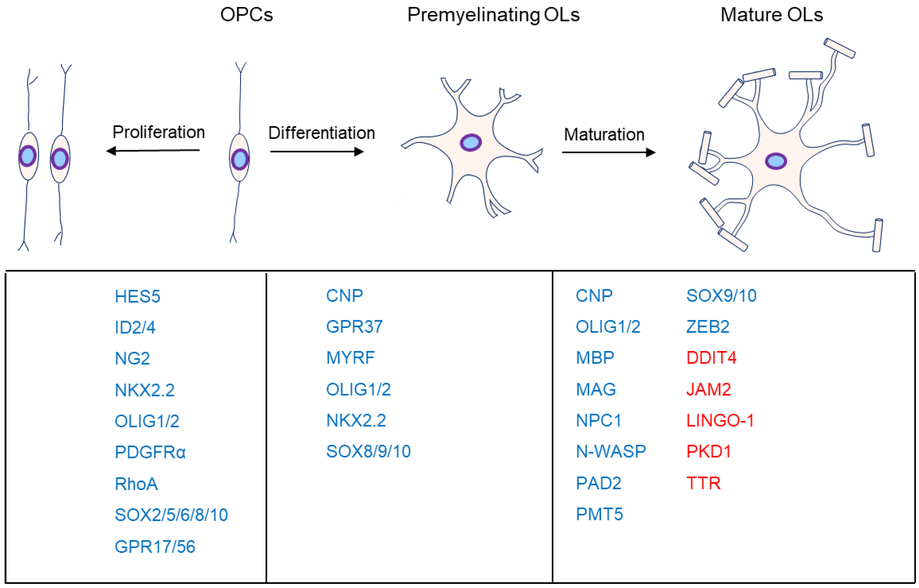
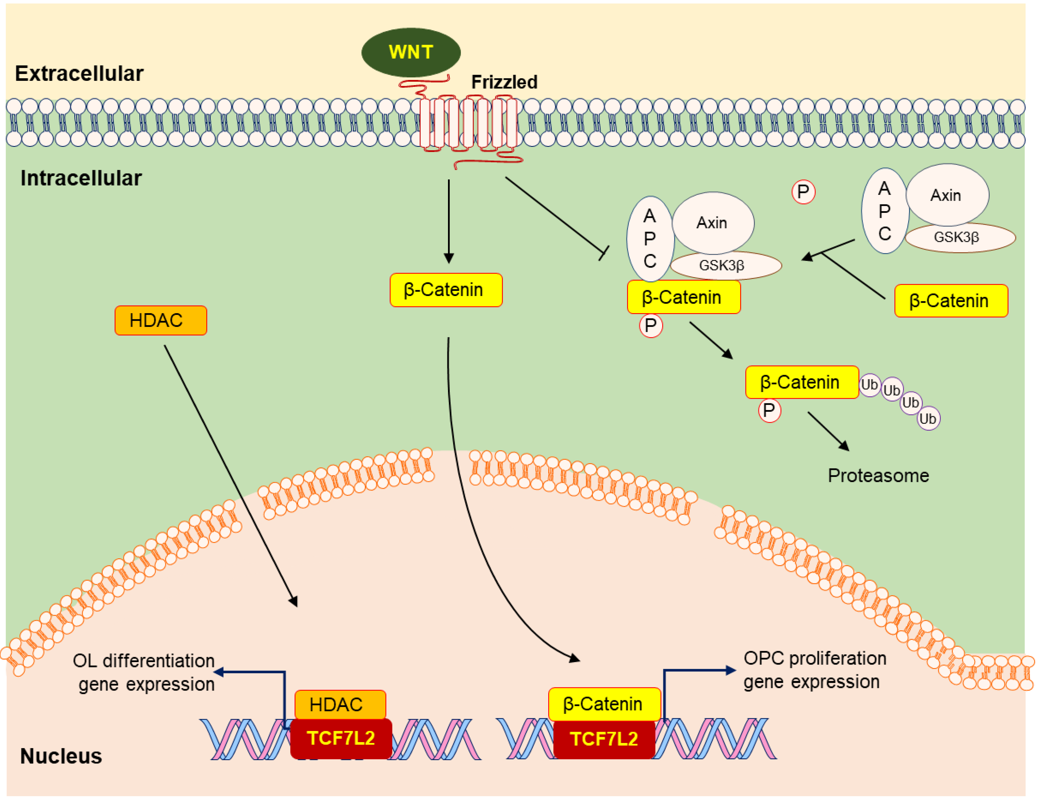
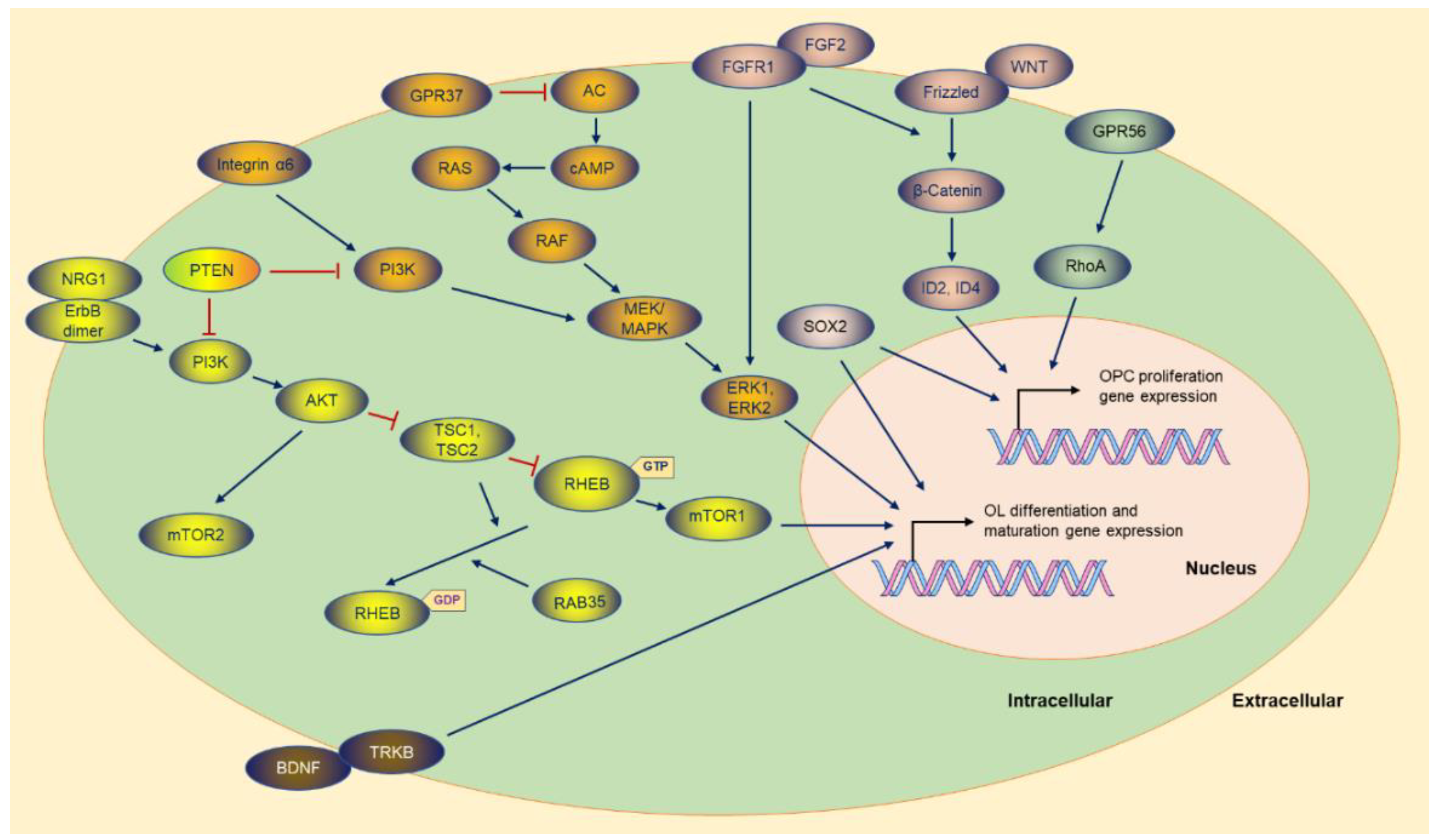
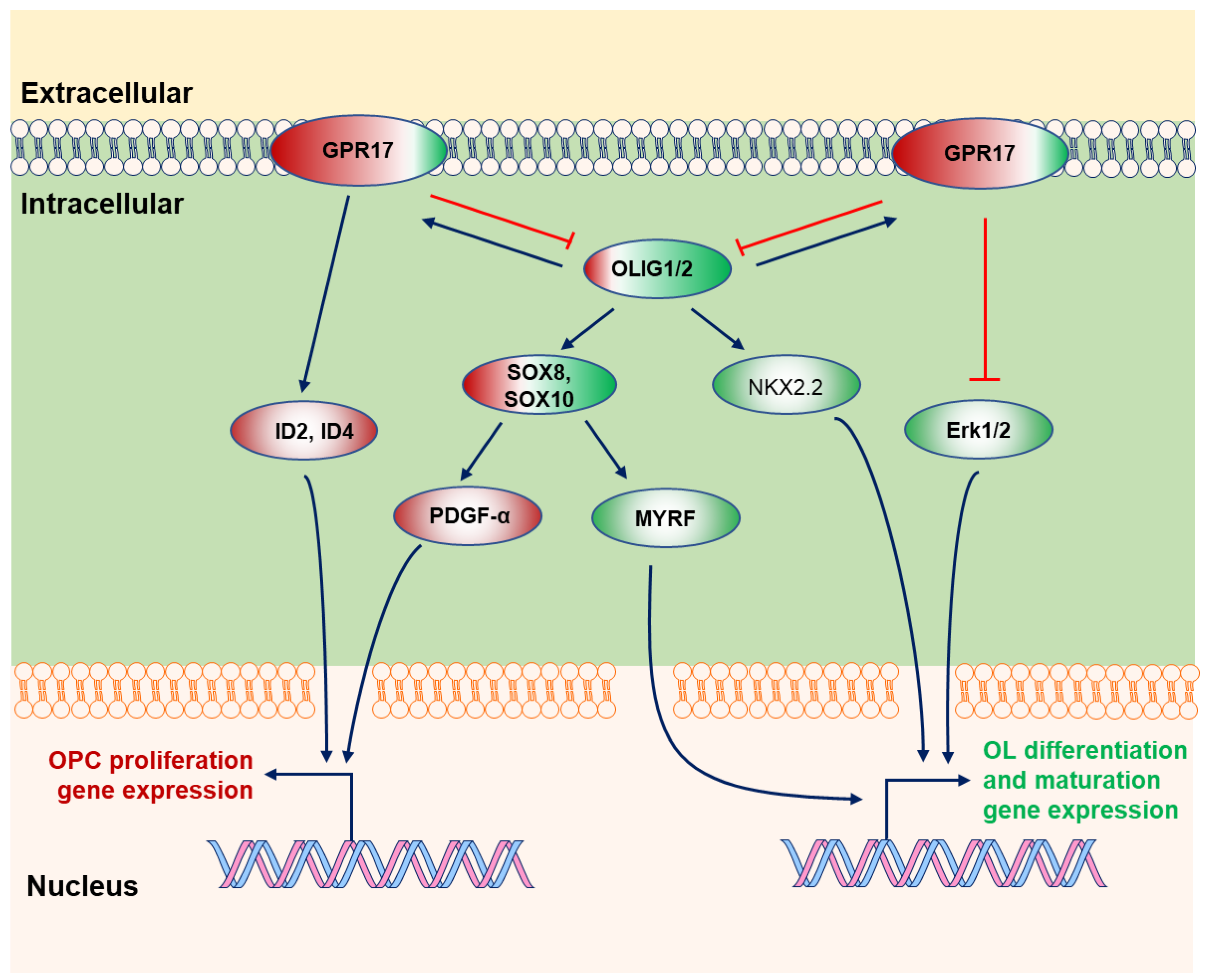
Table 1.
Functions of the factors in the specific stages of OL development.
| Factors | Potential Pathway | OPC Proliferation | OL Differentiation | OL Maturation | References |
|---|---|---|---|---|---|
| HDAC | HDAC-TCF7L2 | + | [16,19,22] | ||
| WNT | WNT-β-Catenin-TCF7L2 | + | − | [17,18,19] | |
| GPR17 | GPR17-ID2/ID4; GPR17-OLIG2; GPR17-ERK1/2 | +/− | − | − | [13,42,43,50,51,52,54,55,56] |
| GPR56 | GPR56/Gα12/13-RhoA | + | − | − | [33,34,36,37,38,39] |
| LINGO-1 | LINGO-1-RhoA | − | − | [6,174,175,176,177,178,179,180,181,182,183] | |
| TG2 | TG2-Laminin-GPR56 | + | [40] | ||
| ERK1/2 | MEK-ERK1/2 | + | + | [93,94,95,96,97,98] | |
| GPR37 | GPR37-cAMP-Raf-MAPK-ERK1/2 | − | +/− | [35,100] | |
| OLIG2 | OLIG2-NKX2.2; OLIG2-SOX8/10 | + | + | +/− | [152,158] |
| ZEB2 | OLIG2-ZEB2 | + | [194,195] | ||
| SOX10-MYRF | SOX10-MYRF | + | + | [102,103,107,109,110,111,112,113,114,117,118] | |
| AKT-mTOR | PI3K-AKT-mTOR | + | + | [78,79,80,81,90] | |
| DDIT4 | DDIT4-mTOR | − | [83] | ||
| NRG1 | NRG1/ErbB- PI3K-AKT-mTOR | + | + | +/− | [125,126,127,128,129,130,134,135,136,137,138,142,143] |
| TTR | TTR-pAKT | − | [172,173] | ||
| BDNF | BDNF/TrkB | + | + | [145] | |
| FGF2 | FGF2/FGFR1 | + | − | +/− | [23,24,25,28,29,31] |
| GlcNAc | GlcNAc-PDGF-α | + | + | [146] | |
| JAM2 | \ | − | [77,162] | ||
| NG2 | \ | + | − | [9,10,11] | |
| NOTCH1 | NOTCH1-HES5 | − | − | − | [63,64,65,66,67] |
| NPC1 | \ | + | [202,203,204] | ||
| N-WASP | \ | + | [185,186,187] | ||
| PAD2 | PAD2-TNF-α; PAD2-MBP | − | − | [196,198,199,200,201] | |
| PDE | \ | + | + | [160,161] | |
| PKD1 | \ | − | [168] | ||
| PRMT5 | \ | + | + | [189] | |
| SOX2 | \ | + | +/− | [59,60,61,62] | |
| SHH | SHH-BOC | + | + | [69,70,71,72,73] |
(Notes: Pathways sharing one or more of the same factors are shown with the same background color. +, activation; −, inhibition; +/−, activation or inhibition, depending on the different research studies; \, unclear).
Publisher’s Note: MDPI stays neutral with regard to jurisdictional claims in published maps and institutional affiliations. |
© 2022 by the authors. Licensee MDPI, Basel, Switzerland. This article is an open access article distributed under the terms and conditions of the Creative Commons Attribution (CC BY) license (https://creativecommons.org/licenses/by/4.0/).
Share and Cite
MDPI and ACS Style
Wang, S.; Wang, Y.; Zou, S. A Glance at the Molecules That Regulate Oligodendrocyte Myelination. Curr. Issues Mol. Biol. 2022, 44, 2194-2216. https://doi.org/10.3390/cimb44050149
AMA Style
Wang S, Wang Y, Zou S. A Glance at the Molecules That Regulate Oligodendrocyte Myelination. Current Issues in Molecular Biology. 2022; 44(5):2194-2216. https://doi.org/10.3390/cimb44050149
Chicago/Turabian StyleWang, Shunqi, Yingxing Wang, and Suqi Zou. 2022. "A Glance at the Molecules That Regulate Oligodendrocyte Myelination" Current Issues in Molecular Biology 44, no. 5: 2194-2216. https://doi.org/10.3390/cimb44050149