Article

Effects of Rapid Palatal Expansion on the Upper Airway Space in Children with Obstructive Sleep Apnea (OSA): A Case-Control Study

1
Dentistry Unit, Bambino Gesù Children’s Hospital, IRCCS, 00165 Rome, Italy
2
Department of Life, Health and Environmental Sciences, University of L’Aquila, 67100 L’Aquila, Italy
3
Unit of Clinical Epidemiology, Medical Direction, Bambino Gesù Children’s Research Hospital, IRCCS, 00165 Rome, Italy
4
Otorhinolaryngology Unit, Department of Specialist Surgeries, Bambino Gesù Children’s Hospital, IRCCS, 00165 Rome, Italy
*
Author to whom correspondence should be addressed.
Children 2023, 10(2), 244; https://doi.org/10.3390/children10020244
Submission received: 13 December 2022 / Revised: 17 January 2023 / Accepted: 27 January 2023 / Published: 30 January 2023
(This article belongs to the Special Issue Advances in Sleep Respiratory Disorders in Children and Adolescents)

Abstract

:
Obstructive Sleep Apnea (OSA) in children needs a multidisciplinary approach. Even if the first-line treatment of pediatric OSA is adenotonsillectomy, nowadays rapid palatal expansion (RPE) is considered a valid additional treatment. The aim of this study is to evaluate cephalometric changes in upper airways dimensions after rapid palatal expansion (RPE) in children suffering from Obstructive Sleep Apnea (OSA). A total of 37 children (range age 4–10 years) with diagnosis of OSA referred to Dentistry Unit of Bambino Gesù Children’s Research Hospital IRCCS (Rome, Italy) were included in this pre-post study and underwent lateral radiographs at the start (T0) and at the end (T1) of a RPE treatment. Inclusion criteria were: diagnosis of OSA confirmed by cardiorespiratory polygraphy (AHI > 1) or pulse oximetry (McGill = >2), skeletal maxillary contraction evaluated by presence of posterior crossbite. A control group of 39 untreated patients (range age 4–11 years), in good general health, was set up. A paired T-test was used to investigate the statistical differences between T0 and T1 values in both groups. The results showed a statistically significant increase of nasopharyngeal width in the treated group after RPE treatment. Moreover, the angle that identifies mandibular divergence compared to palatal plane (PP-MP°) was significantly reduced. In the control group, no statistically significant differences were observed. The present study showed that RPE treatment determines a significant sagittal space increase in the upper airways space and a counterclockwise mandibular growth in children with OSA compared to a control group. These results suggest that a widening of the nasal cavities induced by RPE may support a return to physiological nasal breathing and promote a counterclockwise mandibular growth in children. This evidence confirms the crucial role of the orthodontist in the management of OSA in pediatric patients.

1. Introduction

Obstructive Sleep Apnea (OSA) in children is a common breathing disorder characterized by recurrent episodes of partial or complete upper airway obstruction that disrupt the normal sleep pattern [1].
Other sleep breathing disorders include primary snoring and increased upper airway resistance syndrome (UARS) [2].
Paglia et al., in 2019 [3], reported that primary snoring affects about 27% of children, whereas 1–3% are suffering from OSA, with a peak age between 2 and 5 years, which is the age of greatest increase of adenotonsillar lymphoid tissue [4].
Brunetti et al. [5] estimated that in Italy there is a prevalence of pediatric OSA of 1.8%, especially in pre-school and school years, compared with 4.9% of habitual snoring.
The main risk factors for OSA involve adenotonsillar hypertrophy, neuromuscular disorders, obesity, and craniofacial anomalies such as deficiency in the transversal dimension of the upper jaw, macroglossia, retrognathia, hyperdivergent mandibular pattern, midface hypoplasia and anterior open bite [6,7]. Moreover, the presence of craniofacial anomalies also has a genetic component [8] that may influence the anatomical development of the upper airways [9].
Commonly, children with systemic diseases and specific craniofacial features suffer from OSA [10]. Hill et al. [11], in particular, reported that children with Down syndrome have a high incidence of OSAS, moderate to severe grade, due to the common adenotonsillar hypertrophy and reduced airway dimensions, in relation to common midface hypoplasia, small jaw and hypotonia.
A significant association was evidenced between malocclusion and OSA, in particular the prevalence of posterior crossbite, deviations in overjet and overbite was higher in OSA children [12].
Associated symptoms are snoring, daytime sleepiness or hyperactivity, headache, nocturnal enuresis, and restless sleep [13].
An early diagnosis is very important to prevent serious complications, such as neurocognitive impairment, behavioral, metabolic, and cardiovascular problems [1].
The diagnostic pathway requires above all anamnestic investigation to collect signs and symptoms with the use of parent-specific questionnaires [14,15].
Clinical examination includes pediatric, otorhinolaryngological and myofunctional evaluation [13].
Sleep examination is indispensable for the diagnosis of OSAS. The Gold Standard is nocturnal polysomnography which allows a complete examination of the patient’s sleep (PSG) [16].
Unfortunately, it is not always feasible to perform this examination in pediatric patients as it must be conducted in dedicated sleep laboratories. Thus, alternative methods to study the presence of episodes of apnea and/or oxygen desaturation during sleep have been considered such as cardiorespiratory polygraphy or pulse oximetry [17]. Adenotonsillectomy represents the first-line treatment in the presence of adenotonsillar hypertrophy but this procedure is not always effective [18,19]. After adenotonsillectomy, about 25% of patients retain residual obstructive apnea events during sleep [18,19]. Risk factors for residual OSA after adenotonsillectomy include: more than seven years of age, obesity, severity of sleep disorder and presence of asthma [20]. For this reason, several authors have focused attention on the therapeutic role of an orthodontic approach to obstructive sleep apnea in children [21].
The presence of palatal contraction has been demonstrated to be associated with the presence of OSA in childhood [6] so rapid palatal expansion (RPE) is commonly used in young patients to expand the upper maxilla and it is also recognized as a valid additional treatment in children with OSA [22,23].
Several studies have demonstrated the effectiveness of maxillary expansion to induce widening of the anterior part of the oropharyngeal and nasal space, and the reduction of nasal resistance to the passage of air flow [4,24].
Previous studies evidenced an increase in airway space after the use of orthodontic appliances in OSA children [4,25,26] but few studies have analyzed the upper airway changes after early palatal expansion in a large sample of OSA children.
The aim of this study, therefore, is to evaluate the cephalometric variations of the upper airways dimension after rapid maxillary expansion in children suffering from OSA.

2. Materials and Methods

The study was approved by the Ethics Committee of Bambino Gesù Children’s Research Hospital, IRCCS (Rome, Italy) (protocol number: 1187_2016), 15 September 2016.

2.1. Subjects

A group of 139 children from a previous study [12] with diagnosis of OSA at the Otorhinolaryngology Unit of Bambino Gesù Children’s Research Hospital, IRCCS (Rome, Italy) was considered for this study. Exclusion criteria included: diagnosis of genetic syndromes, missing teeth, previous orthodontic treatment, previous adenoidectomy or adenotonsillectomy and any surgery of the upper airways. The patients underwent dental examination at the Dentistry Unit of the Bambino Gesù Children’s Research Hospital, IRCCS (Rome, Italy) by specialist in Orthodontics. Inclusion criteria were: diagnosis of OSA confirmed by cardiorespiratory polygraphy (AHI > 1) as described in a previous study [7] or pulse oximetry (McGill = >2), with at home overnight sleep studies [25,27]; skeletal maxillary contraction evaluated by the presence of posterior crossbite.
Polygraphy was conducted with the “Somtè/Siesta” portable system (Compumedics, Australia). Parameters such as nasal pressure, tracheal sound, heart rate and pulse oximetry were recorded [7]. Pulse oximetry was conducted as reported by Brouillette et al. [25]. Pulse oximeters set for a mean time of 2 s were used for oxygen saturation (SpO2) (RAD 5, Masimo, Irvine, CA, USA). Data were processed with Profox Oximetry Software, Version Masimo 0706.05D.
After the inclusion and exclusion criteria application, 37 children (range age 4–10 years) were selected for a rapid palatal expansion (RPE) treatment and were enrolled for this pre-post study. The parents accepted to participate the study and signed informed consent.
A control group of 39 subjects (range age 4–11 years), were collected from patients examined in the Dentistry Unit. The children were in good general health, had no systemic disease and no missing teeth. Two lateral cephalograms were performed pre and post an observation period. No orthodontic treatment and adenoidectomy or adenotonsillectomy were performed during this period.

2.2. Methods

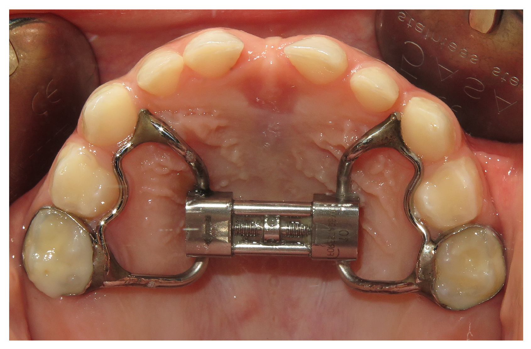
2.2.1. Rapid Palatal Expansion

The patients were treated with rapid palatal expander (RPE) cemented with glass ionomer cement (Fuji Ortho, GC, Cary, NC, USA). Two bands type anchored at the second deciduous maxillary molars or permanent upper first molars (Figure 1), or bite block type (Mc Namara expander) rapid palatal expander were used. The RPE is built with a central screw that is required to be activated to open the midpalatal suture. The expansion screw is placed as close as possible to the palatal vault to deliver force to the center of resistance of the maxillary molars and produce orthopedic effects on the upper jaw. The expansion was performed one turn a day (0.25 mm/turn) until the palatal cusps of the upper molars were contacting the buccal cusps of the lower molars. At the end of the expansion period the RPE was manteined in place as a retainer for 9 months. Mean duration and standard deviation of the therapy was 13.2 ± 3.6 months.

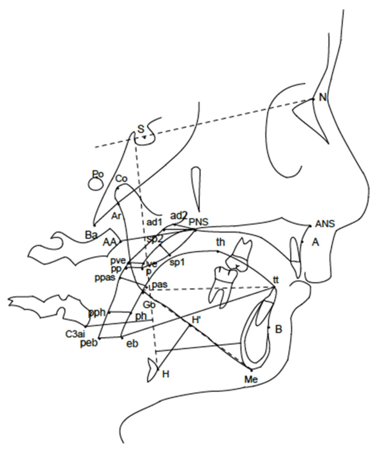
2.2.2. Cephalometric Analysis

74 lateral cephalometric radiographs, 37 at the start (T0) and 37 at the end (T1) of RPE treatment, were taken with the same cephalix cephalostat (OP 3D Pro; Kavo, Milan, Italy) at the Diagnostic Imaging Unit of the Bambino Gesù Children’s Hospital IRCCS (Rome, Italy) for the treated group. Radiographs were taken with the subjects standing, the head fixed in the cephalostat with ear and forehead supports, the teeth in their maximum intercuspal position and the head in the natural position.
39 subjects included in the control group took two lateral cephalometric radiographs at T0 and T1 with a time interval of 9.7 months to 18.6 months (mean 14.2 ± 2.8 months) by means of the same cephalix cephalostat (OP 3D Pro; Kavo, Italy) at the Diagnostic Imaging Unit of the Bambino Gesù Children’s Hospital IRCCS (Rome, Italy). Radiographs were taken with the subjects standing, the head fixed in the cephalostat with ear and forehead supports, the teeth in their maximum intercuspal position and the head in the natural position.
All lateral cephalograms were analyzed and the measurements of the distances and angles were recorded by the same operator, using computerized software “Orisceph Rx CE, 1370” (Elite Computer, Milan, Italy).
The Kirjavainen analysis [28] was used as cephalometric method to evaluate changes of vertical, sagittal and pharyngeal parameters between T0 and T1 for both the study and control groups (Figure 2).
The technical errors of measurement were calculated from 14 randomly selected participants. One examiner revalued all measurements after a period of at least 8 weeks. The method error for the 27 measurements was calculated using Dahlberg’s formula [29]. Systematic differences between duplicated measurements were tested using a paired Student’s t-test with the type I error set at <0.1 [30].

2.3. Sample Size Calculation

The sample size was calculated considering Ad1-PNS distance: the distance of Ad1 point to posterior nasal spine (PNS). Ad1 is the intersection point of the posterior pharyngeal wall and the line from PNS to Basion (the most inferior-posterior point on the anterior margin of foramen magnum). A sample size of 37 patients treated and 39 subjects not treated in the control group achieves 90% of power to detect a difference of 3 mm between the means of the two groups with estimated group standard deviations of 6 mm and with a significance level (alpha) of 0.05 (two-sided).

2.4. Statistical Methods

The Shapiro–Wilk test was used to evaluate the normality of the data. Continuous variables were summarized by mean and standard deviation (SD) and 95% confidence interval of the mean (95% CI).
A paired t-test was used to determine statistical differences between the scores before RPE treatment (T0) and after RPE treatment (T1) and between the scores at T0 and T1 in the control group without treatment.
Further analysis was performed between the cases group and control group considering gender and age. The Pearson chi-square test was used for gender comparison. Two sample t-test with equal variances was used for age comparison. The statistical significance was set at p < 0.05.
Statistical analyses were performed using STATA, Statistical Software Release 13 (StataCorp LP, College Station, TX, USA).

3. Results

37 children (20 males (54.05%) and 17 females (45.95%); mean age ± SD = 6.26 ± 1.26 years) were included in the study and treated with RPE. The control group included 20 males e 19 females (mean age ± SD = 7.36 ± 0.90 years). Cephalometric measurements and their definitions are summarized in Table 1.
All the cephalometric measurements of the treated group, at T0 and T1, and their differences are presented in Table 2.
A significant variation of the nasopharyngeal distance was found after RPE treatment: ad1-PNS (mm) increased by 3.48 mm (p < 0.001) and ad2-PNS (mm) increased by 1.67 (p = 0.003) (Table 2).
A statistically significant reduction of the PP-MP angle of 1.14° (p = 0.043) was evidenced after RPE treatment (Table 2).
No statistically significant changes were shown in maxillary and mandible sagittal positions (SNA angle, SNB angle, ANB angle) or for the other variables (Table 2).
The cephalometric measurements of the control group, at T0 and T1, and their differences are summarized in Table 3.
The analysis performed between case group and control group showed that the two groups are not different by gender (p = 0.809); a statistically significant difference in age of one year (p < 0.001) was found.

4. Discussion

The aim of this study is to evaluate the cephalometric variations of the upper airways dimensions after rapid maxillary expansion in children suffering from OSA.
Rapid palatal expansion (RPE) is considered an effective method to enlarge the upper maxilla and, due to the anatomical contiguity, to wide the floor of the nasal cavity [31], but a very debated and controversial issue is the effectiveness of RPE increasing the width of the upper airways and improving the nasal airflow [32,33].
A 2021 review with 18 articles selected [30] showed an enlargement of upper dental arch, maxillary bone and improvement of nasal breathing in the short term, after RPE treatment.
Several studies have focused on the effects of RPE on the size of airways space, using two-dimensional (2D) and three-dimensional (3D) methods [4,34,35,36,37].
A recent systematic review with meta-analysis [34] showed that the results of the studies are still very conflicting: some authors evidenced a significant association between RPE and increase in the width of the upper airways [34,35,38,39], while other authors showed no significant findings [40]. All of the studies mentioned in this review [34] were performed on children with reduced transverse diameters of the upper jaw who needed palate expansion therapy, but none of these patients were diagnosed with OSA or any other type of sleep breathing disorder.
The present study investigates the sagittal variations of upper airways dimensions after rapid maxillary expansion in children suffering from OSA. We suppose that children with OSA, who have narrower upper airways than normal subjects [41], might respond differently to palatal expansion treatment.
In the present study a significant increase of cephalometric measurements of the nasopharyngeal distance (ad1-PNS = p < 0.001; ad2-PNS = p < 0.003) was found after RPE treatment in OSA children; the increase was 3.48 mm for ad1-PNS and 1.67 for ad2-PNS. These results are consistent with recent evidence reported in a three dimensional- study [42] where the authors performed Cone Beam computed tomography (CBCT) scans of 19 OSA children (mean age of 10.5 years) before (T0) and after (T1) RPE treatment. Pirelli et al. [42] showed that the volume of total upper airways increased significantly after maxillary expansion; in detail, average increases of 9.4 mm3 for total upper airways volume, 7.2 mm3 of nasal volume and of 2.0 mm3 nasopharinx volume were reported. In contrast with our results, these authors reported also an increase of three dimensional oropharyngeal space [42].
As a novelty compared to this previous study, our investigation presents a larger sample of children with OSA and the analysis of sagittal airway values also in a control group. The study of the cephalometric values of the control group did not show a significant difference of the airway space comparing T0 to T1. Therefore, healthy and untreated children did not have a significant change in airway space after the observation period, while the statistical significance of the changes in the group of patients with OSA is high (p < 0.001). These results suggest a clear role of RPE treatment in increasing the sagittal upper airways dimensions in OSA children. Furthermore, considering that the evaluation of this study has the limit of being two-dimensional, it is conceivable that the effects are even more interesting in volumetric terms. Further three-dimensional evaluations are needed to better analyze these aspects.
The comparison between the cases group and control group showed no difference in gender. There was a statistically significant difference in age of about one year when comparing the control group (mean age ± SD = 7.36 ± 0.90 years) with the case group (mean age ± SD = 6.26 ± 1.26 years) and it can be considered in the limits of our study.
It was not possible to perform radiographs in non-treated children for ethical reasons. We therefore used as a control group children who had undergone lateral cephalograms available in our database and who met the inclusion criteria. For this reason, there is also a slight difference in the time interval T1–T0 between cases and controls.
However, we suggest that this statistically significant difference in age of one year between cases and controls does not influence the results of the study: in fact, in clinical terms, the respiratory symptoms related to growth become evident after nine years of age when there is a physiological reduction of the adenoid and tonsillar tissue [43,44].
Another statistically significant value in our analysis is the modification of the PP-MP angle after treatment. This reduction of 1.14 degrees may not have a great impact from a clinical point of view, but it indicates an interesting modification of growth pattern in OSA children who presented always a typical hyperdivergent mandibular growth. Several authors [6] have correlated hyperdivergent growth pattern with a reduction in nasopharyngeal space in children with OSA. It was suggested that the mouth breathing and the lower position of the tongue promote posterior mandible rotation and increased vertical growth [45].
Moreover, the reduction of the angle between the palatal plane and the mandibular plane is an interesting piece of evidence in the orthodontic field. An increase in vertical mandibular growth pattern was previously reported as a consequence of orthodontic treatment [46]; the effect of RPE on craniofacial vertical growth has long been debated and there are still conflicting opinions [46,47].
Therefore, the results of our study suggest that rapid palatal expansion induces skeletal and functional changes in OSA children: a widening of the nasal cavities induced by RPE increase the upper airway space in the adenoidal region; this supports a return to physiological nasal breathing and promotes a counterclockwise mandibular growth in OSA children. On the other hand, the modification of the shape of the palate also allows a more correct repositioning of the tongue which contributes to physiological mandibular growth.
The limitations of our study are:
  • The use of a two-dimensional examination, but it was not possible to use a three-dimensional analysis which exposed patients to a massive radiation dose before and after treatment. For this reason, we evaluated the changes in sagittal and vertical direction, but the modifications were not studied in a transverse direction. Nevertheless, we assume that the increase of upper airways in millimeters might be much more substantial with 3D evaluation which analyzes the entire volume, as suggested by other studies [40,41].
  • No home sleep study was performed after RPE treatment; therefore, this study does not provide data on the efficacy of orthodontic therapy.
  • This study did not present long-term data.
The multidisciplinary diagnostic path is essential in pediatric OSA to attribute the right role to each of the risk factors involved in the etiology of pathology and define the better therapeutic approach.
Orthodontic treatment alone may not be sufficient to solve the sleep breathing disorder. Some studies have shown that the apnea index (AHI) decreases after RPE [48,49], while other studies have reported results that are still unclear [22]. An interesting study evidenced that the combined treatment of adenotonsillectomy and RPE were required to solve OSA in a pediatric sample [50].
Thus, the contribution of different specialists involved in the management of sleep disorder in children is important. The orthodontist plays a crucial role because the craniofacial skeletal changes and functional modification are key aspects in solving sleep respiratory breathing problems in children.

5. Conclusions

In conclusion, our study showed that RPE treatment has significant effects on nasopharyngeal dimensions and mandibular growth in pediatric patients with OSA compared to a control group of untreated children.
Therefore, our results suggest that a widening of the nasal cavities induced by RPE may support a return to physiological nasal breathing and promote a counterclockwise mandibular growth in children. This evidence confirms the crucial role of the orthodontist in the management of OSA in pediatric patients.

Author Contributions

A.G. and P.F. contributed to the study conception, design, data acquisition, analysis and interpretation, and drafted and critically revised the manuscript. R.G. and S.C. contributed to data interpretation and critically revised the manuscript. S.P. contributed to statistical analysis. V.V. contributed to data acquisition and analysis. W.M. contributed data acquisition and drafted the manuscript. G.B. and E.S. critically revised the manuscript. F.A. contributed to checking the integrity of the work. G.M. contributed to final approval of the version to be published. All authors have read and agreed to the published version of the manuscript.

Funding

This research received no external funding.

Institutional Review Board Statement

The study was conducted in accordance with the Declaration of Helsinki, and approved by the Ethics Committee of Bambino Gesù Children’s Research Hospital, IRCCS, (Rome, Italy) (protocol number: 1187_2016, date of approval: 15 September 2016).

Informed Consent Statement

Written informed consent has been obtained from the patient(s) to publish this paper.

Data Availability Statement

The data supporting the results of this study are available from the corresponding author upon reasonable request from any qualified investigator.

Acknowledgments

The authors acknowledge dental technician Patrizio Evangelista (Orthonet, Rome, Italy) for realization of oral devices and Ear Nose and Throat doctors of Bambino Gesù Children’s Hospital for their contribution in the multidisciplinary management of OSA children.

Conflicts of Interest

The authors declare that they have no conflict of interest.

References

  1. American Thoracic Society. Standards and indications for cardiopulmonary sleep studies in children. Am. J. Respir. Crit. Care Med. 1996, 153, 866–878. [Google Scholar] [CrossRef] [PubMed]
  2. Gulotta, G.; Iannella, G.; Vicini, C.; Polimeni, A.; Greco, A.; de Vincentiis, M.; Visconti, I.C.; Meccariello, G.; Cammaroto, G.; De Vito, A.; et al. Risk Factors for Obstructive Sleep Apnea Syndrome in Children: State of the Art. Int. J. Environ. Res. Public Health 2019, 16, 3235. [Google Scholar] [CrossRef] [PubMed] [Green Version]
  3. Paglia, L. Respiratory sleep disorders in children and role of the paediatric dentist. Eur. J. Pediatr. Dent. 2019, 20, 5. [Google Scholar]
  4. Marino, A.; Ranieri, R.; Chiarotti, F.; Villa, M.P.; Malagola, C. Rapid maxillary expansion in children with Obstructive Sleep Apnoea Syndrome (OSAS). Eur. J. Paediatr. Dent. 2012, 13, 57–63. [Google Scholar]
  5. Brunetti, L.; Rana, S.; Lospalluti, M.L.; Pietrafesa, A.; Francavilla, R.; Fanelli, M.; Armenio, L. Prevalence of obstructive sleep apnea syndrome in a cohort of 1207 children of southern Italy. Chest 2001, 120, 1930–1935. [Google Scholar] [CrossRef] [Green Version]
  6. Katyal, V.; Pamula, Y.; Martin, A.J.; Daynes, C.N.; Kennedy, J.D.; Sampson, W.J. Craniofacial and upper airway morphology in pediatric sleep-disordered breathing: Systematic review and meta-analysis. Am. J. Orthod. Dentofacial. Orthop. 2013, 143, 20–30. [Google Scholar] [CrossRef]
  7. Galeotti, A.; Festa, P.; Viarani, V.; Pavone, M.; Sitzia, E.; Piga, S.; Cutrera, R.; De Vincentis, G.C.; D’Antò, V. Correlation between cephalometric variables and obstructive sleep apnoea severity in children. Eur. J. Paediatr. Dent. 2019, 20, 43–47. [Google Scholar]
  8. Paradowska-Stolarz, A. MSX1 gene in the etiology orofacial deformities. Postepy Hig. Med. Dosw. 2015, 69, 1499–1504. [Google Scholar]
  9. Dastan, F.; Ghaffari, H.; Shishvan, H.H.; Zareiyan, M.; Akhlaghian, M.; Shahab, S. Correlation between the upper airway volume and the hyoid bone position, palatal depth, nasal septum deviation, and concha bullosa in different types of malocclusion: A retrospective cone-beam computed tomography study. Dent. Med. Probl. 2021, 58, 509–514. [Google Scholar] [CrossRef]
  10. Brennan, L.C.; Kirkham, F.J.; Gavlak, J.C. Sleep-disordered breathing and comorbidities: Role of the upper airway and craniofacial skeleton. Nat. Sci. Sleep 2020, 12, 907–936. [Google Scholar] [CrossRef]
  11. Hill, C.M.; Evans, H.J.; Elphick, H.; Farquhar, M.; Pickering, R.M.; Kingshott, R.; Martin, J.; Reynolds, J.; Joyce, A.; Rush, C.; et al. Prevalence and predictors of obstructive sleep apnoea in young children with Down syndrome. Sleep Med. 2016, 27–28, 99–106. [Google Scholar] [CrossRef] [PubMed]
  12. Galeotti, A.; Festa, P.; Viarani, V.; D’Antò, V.; Sitzia, E.; Piga, S.; Pavone, M. Prevalence of malocclusion in children with obstructive sleep apnoea. Orthod. Craniofac. Res. 2018, 21, 242–247. [Google Scholar] [CrossRef]
  13. Bitners, A.C.; Arens, R. Evaluation and Management of Children with Obstructive Sleep Apnea Syndrome. Lung 2020, 198, 257–270. [Google Scholar] [CrossRef] [PubMed]
  14. Franco, R.A.; Rosenfeld, R.M.; Rao, M. First place—Resident clinical science award 1999 Quality of life for children with obstructive sleep apnea. Otolaryngol. Head Neck Surg. 2000, 123, 9–16. [Google Scholar] [CrossRef] [PubMed]
  15. Chervin, R.D.; Hedger, K.; Dillon, J.E.; Pituch, K.J. Pediatric sleep questionnaire (PSQ): Validity and reliability of scales for sleep-disordered breathing, snoring, sleepiness, and behavioral problems. Sleep Med. 2000, 1, 21–32. [Google Scholar] [CrossRef]
  16. American Academy of Pediatrics. Clinical practice guideline: Diagnosis and managment of childhood obstructive sleep apnea syndrome. Pediatrics 2002, 109, 704–712. [Google Scholar] [CrossRef] [Green Version]
  17. Villa, M.P.; Pietropaoli, N.; Supino, M.C.; Vitelli, O.; Rabasco, J.; Evangelisti, M.; Del Pozzo, M.; Kaditis, A.G. Diagnosis of Pediatric Obstructive Sleep Apnea Syndrome in Settings With Limited Resources. JAMA Otolaryngol. Head Neck Surg. 2015, 141, 990–996. [Google Scholar] [CrossRef] [PubMed]
  18. Lee, C.H.; Kang, K.T.; Weng, W.C.; Lee, P.L.; Hsu, W.C. Quality of life after adenotonsillectomy in children with obstructive sleep apnea: Short-term and long-term results. Int. J. Pediatr. Otorhinolaryngol. 2015, 79, 210–215. [Google Scholar] [CrossRef]
  19. Katyal, V.; Kennedy, D.; Martin Dreyer, C.; Sampson, W. Paediatric sleep-disorder breathing due to upper airway obstruction in the orthodontic setting: A review. Aust. Orthod. J. 2013, 29, 184–192. [Google Scholar]
  20. Brouillette, R.T.; Horwood, L.; Constantin, E.; Brown, K.; Ross, N.A. Childhood sleep apnea and neighborhood disadavantage. J. Pediatr. 2011, 158, 789–795. [Google Scholar] [CrossRef]
  21. Ngiam, J.; Cistulli, P.A. Dental treatment for paediatric obstructive sleep apnea. Paediatr. Respir. Rev. 2015, 16, 174–181. [Google Scholar] [CrossRef]
  22. Pereira, J.D.S.; Jacob, H.B.; Locks, A.; Brunetto, M.; Ribeiro, G.L.U. Evaluation of the rapid and slow maxillary expansion using cone-beam computed tomography: A randomized clinical trial. Dent. Press J. Orthod. 2017, 22, 61–68. [Google Scholar] [CrossRef] [PubMed] [Green Version]
  23. Buccheri, A.; Chinè, F.; Fratto, G.; Manzon, L. Rapid Maxillary Expansion in Obstructive Sleep Apnea in Young Patients: Cardio-Respiratory Monitoring. J. Clin. Pediatr. Dent. 2017, 41, 312–316. [Google Scholar] [CrossRef] [PubMed]
  24. Baratieri, C.; Alves, M., Jr.; de Souza, M.G.; de Souza Araùjo, M.T.; Cople Maia, L. Does rapid maxillary expansion have long-term effects on airway dimensions and breathing? Am. J. Orthod. Dentofacial. Orthop. 2011, 140, 146–156. [Google Scholar] [CrossRef]
  25. Galeotti, A.; Festa, P.; Pavone, M.; De Vincentis, G.C. Effects of simultaneous palatal expansion and mandibular advancement in a child suffering from OSA. Acta Otorhinolaryngol. Ital. 2016, 36, 328–332. [Google Scholar] [CrossRef] [PubMed]
  26. Pirelli, P.; Fanucci, E.; Giancotti, A.; Di Girolamo, M.; Guilleminault, C. Skeletal changes after rapid maxillary expansion in children with obstructive sleep apnea evaluated by low-dose multi-slice computed tomography. Sleep Med. 2019, 60, 75–80. [Google Scholar] [CrossRef]
  27. Brouillette, R.T.; Morielli, A.; Leimanis, A.; Waters, K.A.; Luciano, R.; Ducharme, F.M. Nocturnal pulse oximetry as an abbreviated testing modality for pediatric obstructive sleep apnea. Pediatrics 2000, 105, 405–412. [Google Scholar] [CrossRef]
  28. Kirjavainen, M.; Kirjavainen, T. Upper airway dimensions in class II malocclusion. Effects of headgear treatment. Angle Orthod. 2007, 77, 1046–1053. [Google Scholar] [CrossRef]
  29. Dahlberg, G. Statistical Methods for Medical and Biological Students; George Allen & Unwin: London, UK, 1940; 232p. [Google Scholar]
  30. Houston, W.J. The analysis of errors in orthodontic measurements. Am. J. Orthod. 1983, 83, 382–390. [Google Scholar] [CrossRef]
  31. Sakai, R.H.; de Assumpção, M.S.; Ribeiro, J.D.; Sakano, E. Impact of rapid maxillary expansion on mouth-breathing children and adolescents: A systematic review. J. Clin. Exp. Dent. 2021, 13, 1258–1270. [Google Scholar] [CrossRef]
  32. Bucci, R.; Montanaro, D.; Rongo, R.; Valletta, R.; Michelotti, A.; D’Antò, V. Effects of maxillary expansion on the upper airways. Evidence from systematic reviews and meta-analyses. J. Oral Rehabil. 2019, 46, 377–387. [Google Scholar] [CrossRef] [PubMed]
  33. Calvo-Henriquez, C.; Capasso, R.; Chiesa-Estomba, C.; Yung Liu, S.; Martins-Neves, S.; Castedo, E.; O’Connor-Reina, C.; Ruano-Ravina, A.; Kahn, S. The role of pediatric maxillary expansion on nasal breathing. A systematic review and metanalysis. Int. J. Pediatr. Otorhinolaryngol. 2020, 135, 110139. [Google Scholar] [CrossRef] [PubMed]
  34. Niu, X.; Di Carlo, G.; Cornelis, M.A.; Cattaneo, P.M. Three-dimensional analyses of short- and long-term effects of rapid maxillary expansion on nasal cavity and upper airway: A systematic review and meta-analysis. Orthod. Craniofac. Res. 2020, 23, 250–276. [Google Scholar] [PubMed]
  35. Radescu, O.D.; Colosi, H.A.; Albu, S. Effects of rapid palatal expansion (RPE) and twin block mandibular advancement device (MAD) on pharyngeal structures in Class II pediatric patients from Cluj-Napoca, Romania. Cranio 2020, 38, 22–29. [Google Scholar] [CrossRef]
  36. Aljawad, H.; Lee, K.M.; Lim, H.J. Three-dimensional evaluation of upper airway changes following rapid maxillary expansion: A retrospective comparison with propensity score matched controls. PLoS ONE 2021, 16, e0261579. [Google Scholar] [CrossRef] [PubMed]
  37. Lee, W.C.; Tu, Y.K.; Chen, R.; Fu, M.W.; Fu, E. Pharyngeal airway changes following maxillary expansion or protraction: A meta-analysis. Orthod. Craniofac. Res. 2018, 21, 4–11. [Google Scholar] [CrossRef] [PubMed]
  38. Aloufi, F.; Preston, C.B.; Zawawi, K.H. Changes in the upper and lower pharyngeal airway spaces associated with rapid maxillary expansion. ISRN Dent. 2012, 2012, 290964. [Google Scholar] [CrossRef] [Green Version]
  39. Iwasaki, T.; Saitoh, I.; Takemoto, Y.; Inada, E.; Kakuno, E.; Kanomi, R.; Hayasaki, H.; Yamasaki, Y. Tongue posture improvement and pharyngeal airway enlargement as secondary effects of rapid maxillary expansion: A cone-beam computed tomography study. Am. J. Orthod. Dentofacial. Orthop. 2013, 143, 235–245. [Google Scholar] [CrossRef]
  40. Ribeiro, A.N.; de Paiva, J.B.; Rino-Neto, J.; Illipronti-Filho, E.; Trivino, T.; Fantini, S.M. Upper airway expansion after rapid maxillary expansion evaluated with cone beam computed tomography. Angle Orthod. 2012, 82, 458–463. [Google Scholar] [CrossRef] [Green Version]
  41. Marcus, C.L.; Brooks, L.J.; Draper, K.A.; Gozal, D.; Halbower, A.C.; Jones, J.; Schechter, M.S.; Ward, S.D.; Sheldon, S.H.; Shiffman, R.N.; et al. American Academy of Pediatrics. Diagnosis and management of childhood obstructive sleep apnea syndrome. Pediatrics 2012, 130, 714–755. [Google Scholar] [CrossRef] [Green Version]
  42. Pirelli, P.; Fiaschetti, V.; Fanucci, E.; Giancotti, A.; Saccomanno, S.; Mampieri, G. Cone beam CT evaluation of skeletal and nasomaxillary complex volume changes after rapid maxillary expansion in OSA children. Sleep Med. 2021, 86, 81–89. [Google Scholar] [CrossRef] [PubMed]
  43. Vogler, R.C.; Ii, F.J.; Pilgram, T.K. Age-specific size of the normal adenoid pad on magnetic resonance imaging. Clin. Otolaryngol. Allied Sci. 2000, 25, 392–395. [Google Scholar] [CrossRef] [PubMed]
  44. Arens, R.; Marcus, C.L. Patophysiology of upper airway obstruction: A developmental perspective. Sleep 2004, 27, 997–1019. [Google Scholar] [CrossRef] [PubMed] [Green Version]
  45. Sousa, J.B.; Anselmo-Lima, W.T.; Valera, F.C.; Gallego, A.J.; Matsumoto, M.A. Cephalometric assessment of the mandibular growth pattern in mouth-breathing children. J. Pediatr. Otorhinolaryngol. 2005, 69, 311–317. [Google Scholar] [CrossRef]
  46. Cozza, P.; Giancotti, A.; Petrosino, A. Rapid palatal expansion in mixed dentition using a modified expander: A cephalometric investigation. J. Orthod. 2001, 28, 129–134. [Google Scholar] [CrossRef] [PubMed]
  47. Lineberger, M.W.; McNamara, J.A.; Baccetti, T.; Herberger, T.; Franchi, L. Effects of rapid maxillary expansion in hyperdivergent patients. Am. J. Orthod. Dentofacial. Orthop. 2012, 142, 60–69. [Google Scholar] [CrossRef]
  48. Machado-Júnior, A.J.; Zancanella, E.; Crespo, A.N. Rapid maxillary expansion and obstructive sleep apnea: A review and meta-analysis. Med. Oral Patol. Oral Cir. Bucal 2016, 21, 465–469. [Google Scholar] [CrossRef] [PubMed]
  49. Vale, F.; Albergaria, M.; Carrilho, E.; Francisco, I.; Guimarães, A.; Caramelo, F.; Maló, L. Efficacy of Rapid Maxillary Expansion in the Treatment of Obstructive Sleep Apnea Syndrome: A Systematic Review With Meta-analysis. J. Evid. Based Dent. Pract. 2017, 17, 159–168. [Google Scholar] [CrossRef]
  50. Guilleminault, C.; Quo, S.; Huynh, N.T.; Li, K. Orthodontic expansion treatment and adenotonsillectomy in the treatment of obstructive sleep apnea in prepubertal children. Sleep 2008, 31, 953–957. [Google Scholar]
Figure 1. Customized rapid palatal expander (RPE).
Children 10 00244 g001
Figure 2. Cephalometric landmarks used for the analysis [7].
Children 10 00244 g002
Table 1. Definitions of cephalometric measurements [7].
Nasopharynx
ad1-PNSThe distance of ad1 point to PNS (posterior nasal spine). Ad1 is the intersection point of posterior pharyngeal wall and the line from PNS to Basion (Ba)
ad2-PNSThe distance of ad2 point to PNS. Ad2 is the intersection point of posterior pharyngeal wall and the line from the midpoint of the line from sella (S) to basion (Ba) to PNS
Oropharynx
p-ppThe distance of the tip of soft palate (p) to horizontal counterpoint on posterior pharyngeal wall (pp)
paThe distance of the intersection points on anterior and posterior pharyngeal wall of the line from supramentale (B) to gonion (Go)
Hyoid bone
H-H′The distance from the most anterior and superior point of hyoid bone (H) perpendicular to mandibular plane (MP).
Maxilla
SNAThe angle sella (S) to nasion (N) to subspinale (A)
Mandible
SNBThe angle sella (S) to nasion (N) to supramentale (B)
ANBThe angle subspinale (A) to nasion (N) to supramentale (B)
Facial heights
S-GoThe distance from sella (S) to gonion (Go)
N-MeThe distance from nasion (N) to menton (Me)
P-A face height (S-Go/N-Me)%The ratio between S-Go/N-Me
Typology
SN-MPThe angle nasion (N) to sella (S) to mandibular plane (MP).
PP-MPThe angle PP to MP. PP is the line from anterior nasal spine (ANS) to PNS
Cranial base
SNThe distance from sella (S) to nasion (N)
Table 2. Cephalometric variables in treated group at time T0 and T1, expressed as mean and standard deviation (SD) and their 95% confidence interval (95% CI).
Cephalometric VariablesTimeMean Difference by Timep-Value
T0T1T1–T0
MeanSD95% CIMeanSD95% CI
SNA (°)80.94.4379.4–82.480.814.7679.2–82.4−0.070.866
SNB (°)75.63.9774.3–76.975.83.974.5–77.20.260.492
ANB (°)5.293.14.2–6.34.962.34.2–5.7−0.320.381
SN (mm)58.35.856.4–60.359.964.358.5–61.41.630.136
SN/MP (°)38.35.836.4–40.337.96.035.9–39.9−0.460.320
PP/MP (°)32.15.630.2–33.930.95.329.2–32.7−1.140.043 *
P-A face height
(S-Go/N-Me) (%)
61.55.259.8–63.261.95.360.2–63.70.470.299
ad1-PNS (mm)AD5.69.2–12.914.65.612.7–16.53.48<0.001 *
ad2-PNS (mm)7.53.66.3–8.69.13.97.8–10.51.670.003 *
p-pp (mm)13.34.211.9–14.713.04.311.6–14.5−0.250.795
pas-ppas (mm)14.33.613.1–15.514.13.512.9–15.2−0.210.760
eb-peb (mm)15.25.013.6–16.914.55.412.7–16.3−0.70.539
H-H′ (GnGo-H) (mm)13.94.912.2–15.514.05.312.2–15.80.140.873
p-value < 0.05 = Significance level.
Table 3. Cephalometric variables in control group at time T0 and T1, expressed as mean and standard deviation (SD) and their 95% confidence interval (95%CI).
Cephalometric VariablesTimeMean Difference by Timep-Value
T0T1T1–T0
MeanSD95% CIMeanSD95% CI
SNA (°)81.24.180.5–83.181.05.079.9–83.20.20.639
SNB (°)78.44.377.0–79.778.34.777.6–79.80.10.809
ANB (°)3.42.42.7–4.23.32.22.6–4.00.10.663
SN (mm)61.35.159.6–62.963.27.060.9–65.4−1.90.038
SN/MP (°)34.55.532.7–36.335.47.233.1–37.7−0.90.375
PP/MP (°)28.45.426.6–30.127.74.826.1–29.30.60.260
P-A face height
(S-Go/N-Me) (%)
63.84.962.2–65.464.04.362.6–65.4−0.20.746
ad1-pns (mm)15.66.513.5–17.716.75.015.1–18.3−1.10.108
ad2-pns (mm)8.64.07.3–9.99.23.28.1–10.2−0.50.267
p-pp (mm)11.34.69.9–12.810.74.69.2–12.10.70.281
pas-ppas (mm)12.53.811.3–13.712.14.010.8–13.40.40.396
eb-peb (mm)16.54.715.0–18.017.04.915.4–18.6−0.40.624
H-H’ (GnGo-H) (mm)11.05.19.4–12.710.35.58.5–12.10.70.299
No statistically significant differences were found in the control group.
Disclaimer/Publisher’s Note: The statements, opinions and data contained in all publications are solely those of the individual author(s) and contributor(s) and not of MDPI and/or the editor(s). MDPI and/or the editor(s) disclaim responsibility for any injury to people or property resulting from any ideas, methods, instructions or products referred to in the content.

