
Antioxidant Determining Using Electrochemical Method
by
 , and
, and
Rani Melati Sukma
1, 
Dyah Iswantini
1,2,*, 
Novik Nurhidayat
3,
Mohamad Rafi
1,2 and
and
Dita Ariyanti
4 1
Department of Chemistry, Faculty of Mathematics and Natural Sciences, IPB University, Bogor 16680, Indonesia
2
Tropical Biopharmaca Research Center, IPB University, Bogor 16128, Indonesia
3
Research Centre for Applied Microbiology, National Research and Innovation Agency, Cibinong 16911, Indonesia
4
Department of Chemistry, Faculty of Military Mathematics and Natural Sciences, Indonesian Defense University, Bogor 16810, Indonesia
*
Author to whom correspondence should be addressed.
Chemistry 2023, 5(3), 1921-1941; https://doi.org/10.3390/chemistry5030131
Submission received: 20 July 2023
/
Revised: 26 August 2023
/
Accepted: 28 August 2023
/
Published: 1 September 2023
(This article belongs to the Section Electrochemistry and Photoredox Processes)
Abstract
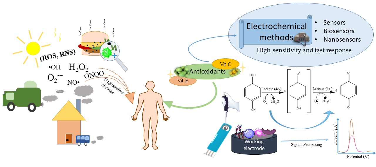
:Antioxidants are very beneficial for health as they protect the body from the effects of free radicals on various degenerative diseases caused by food contamination, air pollution, sunlight, etc. In general, methods for measuring the capacity of antioxidants generally use accurate methods such as spectrophotometry and chromatography. Still, this takes time, accurate sample preparation, and must be performed in a laboratory with particular expertise. Therefore, a new, more practical method needs to be developed for determining antioxidants, namely the electrochemical method. The electrochemical method is a promising method to develop because it comes with several advantages, including high sensitivity and fast response. The electrochemical method discussed in this article reviews sensors, biosensors, and nanosensors. This paper comprehensively analyzes contemporary developments in electrochemical biosensor techniques and antioxidant evaluation methodologies. The discussion centers on utilizing multiple biosensors. Electrochemical biosensors have been determined to be prevalent in analyzing food quality, assessing active factor functionality, and screening practical components. The present study outlines the difficulties linked with electrochemical bio-sensor technology and provides insights into the potential avenues for future research in this domain.

1. Introduction
Increasing body immunity and maintaining health through antioxidant preparations has become a new concern after the COVID-19 pandemic. Antioxidants are chemical substances that interrupt the cascade of free radical reactions within the human body. Free radicals are generated as byproducts of metabolic and physiological processes and are an essential part of the immune system of aerobic organisms, including humans. According to sources [1,2], radicals have one or more unpaired electrons, rendering them unstable and able to damage other atoms by losing electrons in order to become stable. These substances are called reactive oxygen/nitrogen species (ROS/RNS), and include hydroxyl, hydrogen peroxide, superoxide, nitric oxide, and peroxynitrite [3,4]. There are two types of antioxidants: endogenous antioxidants, produced by our bodies [5], and exogenous antioxidants, which are provided by food or nutritional supplements; one example of the latter are polyphenolic compounds [6,7]. The high content of phenolic components in the samples results in high levels of antioxidant activity [8].
In some diets, spices, herbs, fruits, and vegetables can provide additional natural antioxidants to support antioxidant defenses [9]. Antioxidants sourced from food, beverages, and herbal medicines must be controlled for their quality so that the antioxidants consumed can optimally counteract free radicals in the body in order to help prevent disease. Several research studies have indicated that medical conditions, including inflammation, osteoporosis, hepatopathy, diabetes, cancer, and neuro-degenerative diseases, are frequently linked with elevated levels of oxidative stress [10,11,12,13,14]. There are significant differences between RNS and ROS and the ways in which antioxidants protect the body against them. Enhancing cellular defenses through antioxidants can effectively neutralize oxidative stress [15,16].
Spices and herbs are known to have high antioxidant activity and beneficial effects on human health in certain spices. Antioxidants derived from spices include bioactive compounds consisting of flavonoids, phenolic compounds, and compounds containing sulfur, tannins, alkaloids, diterpenes, and vitamins [15]. The compounds exhibit variations in their antioxidant efficacy. For instance, flavonoids can eliminate free radicals and establish associations with catalytic metal ions, rendering them reactive. Numerous academic studies have shown that spices and herbs, including, but not limited to, rosemary, sage, and oregano, possess high levels of phenolic compounds and antioxidants. Antioxidants can safeguard oils against oxidative degradation. When incorporated into food, antioxidants can impede the formation of harmful oxidation byproducts, preserve the nutritional tributes, and prolong the duration of product storage. Spices contain inherent antioxidants that aid in the mitigation of oxidative stress. Oxidative stress is a biological state that results from heightened concentrations of unpaired electrons, known as free radicals, within cellular and tissue environments. Various detrimental factors, including gamma radiation, UV and X-rays, psychological stress, contaminated food, unfavorable environmental circumstances, strenuous physical exertion, tobacco use, and alcohol addiction, can trigger this condition [17,18].
According to previous studies, the process of inhibiting free radicals by antioxidants can be achieved by donating an electron to oxidant compounds, thereby impeding their activity. The efficacy of antioxidants in mitigating the effects of free radicals can be classified into two distinct groups. The initial classification, namely primary, pertains to antioxidants that undergo chain termination to impede the generation of free radicals. In this process, antioxidants donate hydrogen from their active hydroxyl groups to produce more radicals [19]. The second category pertains to the deactivation of free radicals by transferring single electrons to form more stable substances, accomplished by antioxidants [20]. Antioxidants are compounds that have many benefits, including use in beauty and cosmetic products [21], health care [22], food ingredients and preservatives [23], the production of silver nanoparticles [24], and others.
Antioxidant detection can be achieved using spectrophotometry [25], colorimetry [26], chromatography [27], spectroscopy, and electrochemical methods [28]. The utilization of the chromatographic technique comes with a higher cost due to the expense of the equipment involved. While the method can differentiate between distinct antioxidant constituents in various food items, it only furnishes data regarding their concentration. The spectroscopic technique relies on the spectral characteristics of a reference material because of its determinant principle, which leads to unavoidable errors in the measurement results, including determining the actual color of the sample, such as orange juice, etc. For this reason, it is necessary to pay attention to and develop simple, sensitive, and fast methods for analysis, such as the electrochemical method. In general, the conventional method is sensitive and efficient. Still, the work is usually carried out in a centralized laboratory, requires resources and experts in the field, and comes at a high costs and takes a long time. The electrochemical approach offers numerous benefits, including rapid detection time, minimal sample volume requirement, exceptional precision, and heightened sensitivity. By circumventing the need for laborious pre-treatment of samples, interference from colored samples can be minimized [29,30,31]. One method utilized to evaluate antioxidant capacity is the electrochemical approach, which is preferred for its accuracy, affordability, simplicity, rapid response, and high sensitivity [32]. A multitude of electrochemical methodologies, including square wave voltammetry (SWV), cyclic voltammetry (CV), and differential pulse voltammetry (DPV), have been extensively utilized in various research endeavors to explore redox systems and produce results [33,34].
The information literature review was collected from scientific journals, Wiley Online Library, Scopus, Google Scholar, and Science Direct. The keyword is biosensor antioxidant and electrochemical method. The articles obtained are filtered by title, abstract, and full text.
2. Antioxidants
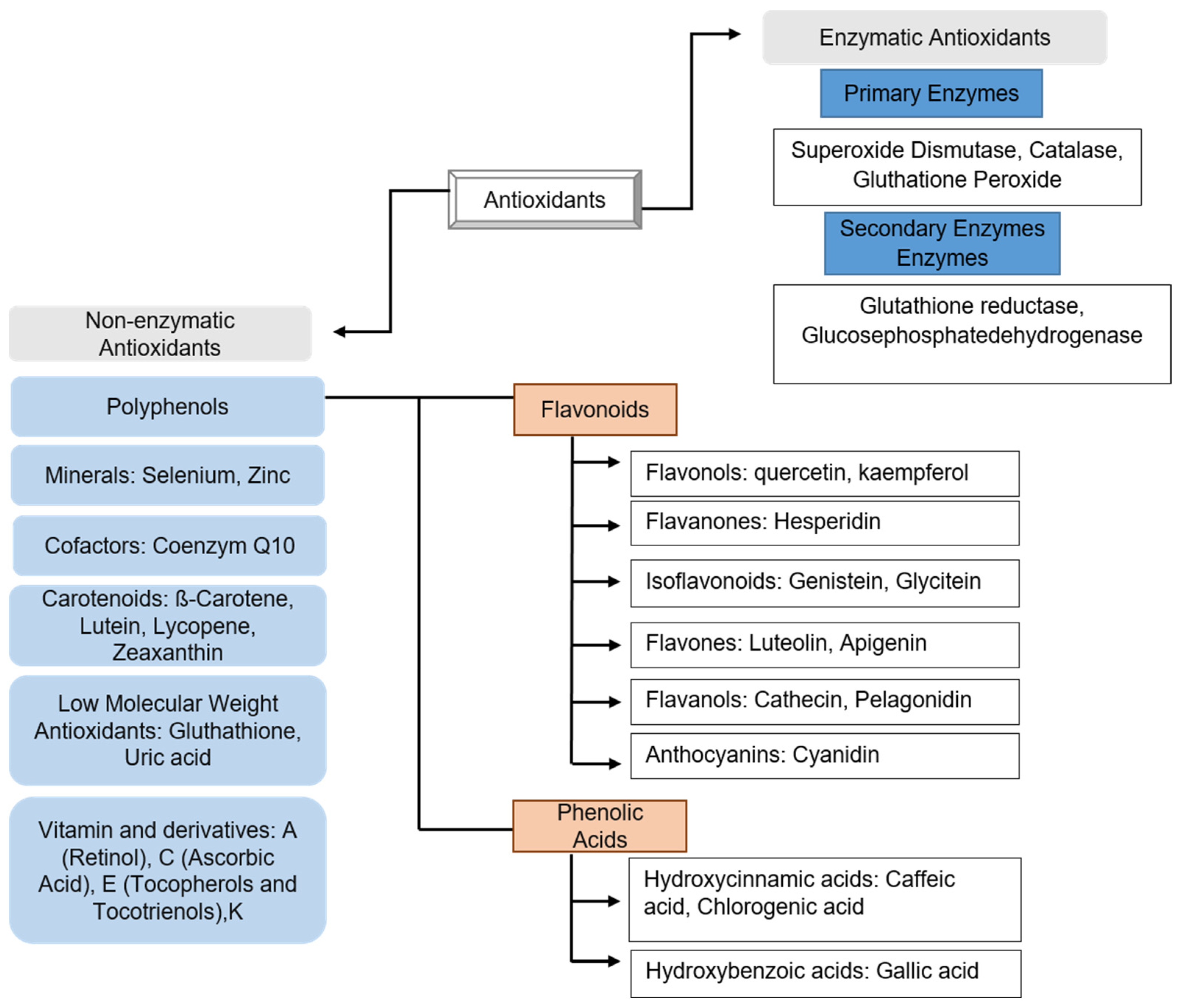
Based on how they react with free radicals, antioxidants are classified as either enzymatic or non-enzymatic. Enzymatic antioxidants remove free radicals by donating electrons to free radical species produced by the body. Meanwhile, non-enzymatic antioxidants remove free radicals by interfering with the chain reaction of free radicals, and many non-enzymatic antioxidants come from food [35]. Enzymatic antioxidants can be classified into two discrete classifications: primary and secondary. The enzyme antioxidant system in the body consists of glutathione peroxidase (GPX), catalase compounds (CAT), and superoxide dismutase (SOD) [36]. Non-enzymatic antioxidants commonly found in crops are polyphenols, including, for example, flavonoids. Non-enzyme antioxidants function in balancing the health system of organisms. Vitamin E (tocopherol) and ascorbic acid (vitamin C) are non-enzyme antioxidants (Figure 1) [37].
Enzymatic antioxidants work by breaking down and removing free radicals. Generally, antioxidant enzymes remove harmful oxygen from the product and then convert it into H2O2 and then H2O2. The process requires several metal cofactors (copper, zinc, manganese, and iron) [38]. Polyphenols are classified into two groups based on their work against free radicals. First, the polyphenol group fights free radicals by breaking down the main chain to reduce or delay free radical production, which will trap the free radicals produced. Free radical scavengers in secondary antioxidants are carried out on the substrate. In contradistinction, primary antioxidants neutralize free radicals through three mechanisms, hydrogen atom transfer (HAT), sequential electron transfer proton transfer (SETPT), and sequential proton loss electron transfer (SPLET), by donating hydrogen atoms to peroxyl radicals [39].
Flavonoids can stop free radicals by binding free electrons to conjugated hydroxyl species [40]. The chelating properties of flavonoids also encourage inhibiting free radicals in the body [41]. Flavonoids can act as anti-allergens, anti-diabetics, anti-inflammatories, and antioxidants [42]. Phenolic acid is an antioxidant with conjugated aromatic rings and substituted hydroxyl groups. The functional groups’ composition and quantity significantly affect the phenolic acid’s activity, as reported in reference [43]. There are several mechanisms of free radical scavenging by phenolic acid, carried out by donating H atoms, donating electrons, and binding of oxidative free radicals [44]. Apart from having many functions for the body, antioxidants also extend the shelf life of food because antioxidants can slow down reactive species, such as ROS and RNS, that can increase the food decay rate [16]. Another function of antioxidants is changing metal peroxide to become more stable and inhibiting lipoxygenase in prooxidative enzymes [45]. The position of the functional group of the active compound is also important, such as OH or NH2, with the ortho position being the most active compared to the para and meta positions.
3. Electrochemical Sensors
The electrochemical sensor transforms the signal resulting from the chemical interaction between the analyte and the identifying component to obtain quantitative or qualitative information. The sensor signal is then transformed into an analytical signal. The resulting signal can change the voltage, conductance, and current. Electrochemical sensors have many advantages over mass, thermal, and optical sensors, and electrochemical sensors have been shown to possess superior sensitivity, ease of use, cost-effectiveness, and versatility in analyzing analytes in various states, including liquid, solid, and gas [46]. Electrochemical sensors for determining antioxidants are developed using various electrodes, receptors, and transducers. Several times, nanomaterials are used to enhance sensor performance, sensitivity, stability, and selectivity. Antioxidants tend to have significant redox properties, so they can be detected directly [47].
The integration of nanomaterials into electrochemical sensors has yielded significant improvements. Nanomaterials exhibit distinct thermal, mechanical, optical, electrical, and magnetic characteristics contingent upon their size and can be readily tailored by manipulating their shape and dimensions [48]. The characteristics of the added nanomaterials have a significant impact on improving the sensor’s electrochemical performance [49]. Measurement of the redox potential of antioxidants using the electrochemical sensor method in reporting the results must be included with the reference electrode utilized because it cannot be quantified in absolute terms. Determining antioxidants through electrochemical sensors is significantly impacted by the solvent employed, as the solvent substantially impacts the reactivity of antioxidants. Therefore, it is necessary to consider the redox reactions occurring in several different solvent types to measure the solvents’ impact on the antioxidant measurement [50]. Measurement of antioxidants with other methods pays little attention to the influence of solvents, electrolytes, radical intermediates, reactant concentrations, and pH. At the same time, the electrochemical sensor method can estimate this well [51]. Several literature studies on sensors in detecting antioxidants using electrochemical methods can be seen in Table 1.
This research concentrated on developing electrochemical sensors for detecting uric acid and gallic acid in green tea and fruit juice samples using carbon paste electrodes modified by ZrO2 nanocomposites, chlorine chloride, and gold nanoparticles using the DPV electrochemical method. It showed promising results, obtaining a LOD value of 2.5 × 10⁻⁸ M and a linear range of 0.22–55 μM for gallic acid samples, while for uric acid samples, the LOD is 1.5 × 10⁻⁸ M and a concentration range of 0.12–55 μM [52]. Numerous electrochemical sensor techniques have been devised for the quantification of antioxidant activity in food specimens, such as determining the type of antioxidant in the form of phloretin (Ph) in spice samples using the cyclic voltammetry method and glassy carbon electrodes to produce a LOD of 4.1 × 10⁻⁶ with a concentration range of 9.9 × 10⁻⁶–1.07 × 10⁻⁴ M [53]. A glassy carbon electrode modified with a hydroxide film based on Zn Al-NO3 on antioxidant measurements of gallic acid and caffeic acid using the pulse differential voltammetry detection method produces a LOD of 1.6−2.6 μM [44].
The development of electrochemical sensors using differential pulse and cyclic voltammetry methods with glass carbon working electrodes with in situ activity for the determination of tertiary antioxidant butyl hydroquinone resulted in a detection limit of 67 nM and a linear range of 1.0−1.1 μm [54]. Electrochemical detection using the differential pulse polarography method with mercury-dropping electrodes in determining gallic acid as an antioxidant resulted in a detection limit of 0.3 μm with a linear range of 1.0−50 μm. The total polyphenols measured in red wine samples averaged 1987 ppm and 238.1 ppm in white wine samples [55]. Development of electrochemical sensors with different measurement methods, namely using an electrochemical detection method in the form of square wave voltammetry with several working electrodes and different samples, first using a glassy carbon working electrode immobilized by purine biosensors in measuring beverage samples to determine total antioxidant capacity, second, using the SPCE working electrode on an antioxidant in the form of ascorbic acid, produced a detection limit of 0.09 nM. Melatonin produced a detection limit of 0.04 nM and N-acetylcysteine produced a detection limit of 0.07 nM [56].
The measurement of chlorogenic acid in nutraceuticals involves using a voltammetry sensor based on an SPCE modified with graphene and gold nanoparticles. This sensor utilizes a glassy carbon electrode modified with 4-methylpyridium iodine to detect the presence of caffeine acid, an antioxidant. The electrochemical sensor comprises three distinct screen-printed electrodes (SPEs): a carbon-based electrode, a graphene-based electrode, and a graphene-modified electrode that incorporates gold nanoparticles dissolved in various solutions. The lowest LOD and LOQ were obtained by SPE based on graphene (GPH) and gold nanoparticles (GNP) with LOD = 0.62 × 10⁻⁷ M and LOQ = 1.97 × 10⁻⁷ M. The present study suggests that utilizing GPH-GNP-SPE enhances sensor response in terms of sensitivity and reversibility for the accurate determination of real-world samples. This conclusion is supported by the validation of FTIR results, which indicates no statistically significant differences [57].

Table 1.
Some literature on antioxidant detection uses electrochemical sensors.
| Electrochemical Detection Method | Electrode | Sample | Antioxidant | Detection Limit | Range Linear | Ref |
|---|---|---|---|---|---|---|
| Cyclic Voltammetry | Glassy carbon | Spices | Curcumin | 4.1 × 10⁻⁶ M | 9.9 × 10⁻⁶–1.07 × 10⁻⁴ M | [53] |
| Amperometry | AgNP/Delph/GCE | Apple juice, lemon juice, peach juice, orange juice, green tea | Gallic acid | 0.28 µmol/L | 6 × 10⁻⁷–8.68 × 10⁻⁶ M | [58] |
| Differential Pulse Voltammetry | GCE | Human serum blood | Total antioxidant capacity | - | - | [59] |
| Differential Pulse Voltammetry | GCE/ZnAl-NO3 layered double hydroxide film | - | Gallic Acid | 1.6 µM | 4–600 µM 7.0–180 µM | [44] |
| Caffeic Acid | 2.6 µM | |||||
| Differential Pulse Voltammetry, Cyclic voltammetry | GCE | - | Tertiary butyl hydroquinone | 67 nM | 1.0 µM–1.1 mM | [54] |
| Differential pulse polarography | Dropping mercury | - | Gallic acid | 0.3 µM | 1.0–50 µM | [60] |
| Differential pulse polarography | Ti3Al0.5Cu0.5C2/GCE | Kiwi | Rutin | 0.015 μmol L−1 | 0.02–50.00 μmol/L | [61] |
| Square wave voltammetry | SPCE | • Ascorbic acid | • 0.09 mmol/I | - | [56] | |
| Square wave voltammetry | SPCE | - | • N-acetylcysteine | • 0.04 mmol/I | - | [56] |
| Square wave voltammetry | SPCE | - | • Melatonin | • 0.07 mmol/I | - | [56] |
| Square wave voltammetry | 4-[(4-decyloxyphenyl)-ethynyl]-1- methylpyridinium iodide modified glassy carbon | Mate herb extracts | Caffeic acid standard | 9.0 × 10⁻⁷ M 8.7 × 10⁻⁶ M | 9.9 × 10⁻⁷ M–3.8 × 10⁻⁵ M 4.7 × 10⁻⁵ M–9.9 × 10⁻⁵ M | [62] |
| Differential Pulse Voltammetry | (ZrO2/Co3O4/rG) | Tea, juice and urine | Gallic acid and uric acid | 2.5 × 10⁻⁸ M | 2.2 × 10⁻⁷–5.5 × 10⁵ M | [52] |
| Differential Pulse Voltammetry | Am-ZrO2-CPE | Wine | Gallic acid | 1.24 × 10⁻⁷ M | 1 × 10⁻⁶–1 × 10⁻³ M | [46] |
| Differential Pulse Voltammetry | Nano-GO-SiO2- nanoparticles-GCE | Red wine | Gallic acid | 6.25 × 10⁻⁶ M | 1 × 10⁻⁶–1 × 10⁻³ M | [63] |
Note: -: Not available.
4. Antioxidants-Based Biosensors
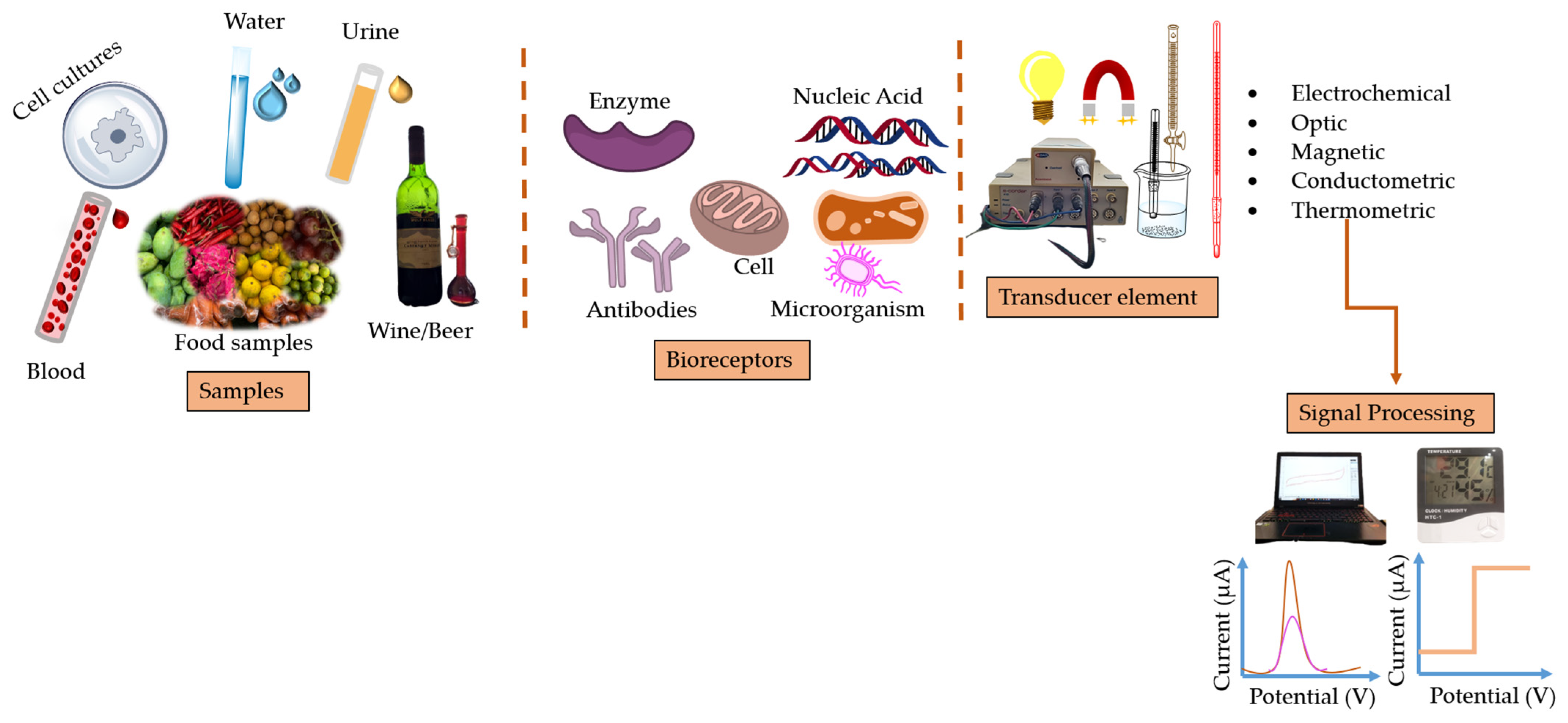
Biosensors are a promising alternative method for several analytical processes in various fields. These techniques have been effectively utilized to detect various ailments, toxins, pathogens, and heightened levels of blood constituents, among other applications [64,65,66]. The biosensor comprises two primary components: the bioreceptor and transducer elements. The bioreceptor component of the biosensor interacts with the target analyte to ensure the sensor’s selectivity. This happens because the identifier element in the biosensor takes advantage of specific biological interactions in compounds to increase the specific binding affinity for the desired molecule. Though transducers convert biological responses resulting from interactions with target analytes into measurable signals, it determines biosensor sensitivity [67]. The variety of transducers employed substantially affects the biosensor’s sensitivity and selectivity.
The bioreceptors used in biosensors include enzymes [68], nucleic acids [69], antibodies [70], cells [31], and receptors. Sensitivity, selectivity, detection limit, stability, and linearity are used to evaluate the biosensor’s performance. In comparison, the transducers used in biosensors are piezoelectric/gravimetric [71], optical [72], thermometric [73], and conductometric [74] (Figure 2).
The biosensor used in this article uses an electrochemical biosensor taken from several literature studies. Electrochemical biosensors are starting to become a focus for development in measuring antioxidant activity because they can analyze biological samples, which can then be converted through biological processes and produce electrochemical signals [16]. Biosensor indicators have high selectivity. The principle of the electrochemical biosensor is that when an analyte is at a different potential, it will cause oxidation to occur on the working electrode and then measure the signal from the movement of electrons. This signal can be measured with an electrochemical detector [75].
4.1. Enzyme-Based Biosensor
Developing enzyme-based biosensors as biocatalysts to accelerate chemical reactions for testing antioxidant activity requires attention to be paid to several aspects, such as immobilization and enzyme stability. It is necessary to formulate the most efficient way of immobilizing the enzyme on the electrode because the immobilization of the enzyme affects the electron transfer rate. Therefore, enzyme immobilization must be integrated with nanomaterials or polymer membranes to produce good efficiency and stability and achieve the desired results. Enzyme-based electrochemical biosensors employ enzymes as bioreceptor elements, and the analysis of samples is predicated on inhibiting enzymatic activity [76]. An enzymatic biosensor is a detection tool with the working principle of changing the measured substance concentration into a digital signal through a transducer. The components used to identify the biomolecules have suitable sensitive components, including an enzyme [77].
4.1.1. Tyrosinase
The enzyme tyrosinase is involved in the process of melanin biosynthesis, which contains copper and serves to catalyze the hydroxylation of mono-phenols to o-diphenols through the monophenolase reaction, as well as the oxidization of o-diphenols to o-quinone through the diphenolase reaction. l-tyrosine is a substrate that plays a role in monophenolase and diphenolase reactions. The enzymatic mechanism described involves a measurement approach involving the reversible electrochemical reduction of o-quinone, produced from phenol during enzymatic reactions. Each signal obtained from this measurement corresponds to the concentration of polyphenols in the solution. Enzyme-based biosensors are commonly utilized due to their remarkable sensitivity and selectivity. Enzymes involved in oxidation-reduction reactions have gained considerable interest in enzyme-based biosensors due to their remarkable ability to catalyze reactions dependent on electron transfer. The tyrosinase’s enzymatic mechanism entails redox activity enabled by the reversible transfer of electrons between copper ions in their +1 and +2 oxidation states (Cu+ ↔ Cu2+).
Electrochemical sensor have been developed with the addition of nanoparticle-based materials. The goal of these additions is to increase sensor performance. Biosensors based on tyrosinase/laccase enzymes with graphite-epoxy modified copper nanoparticles, whose performance is reviewed in the determination of polyphenols, catechols, caffeine acids, and catechins with cyclic voltammetry methods, produce a linear range > 200 µM [78].
Other studies are based on the tyrosinase enzyme with catechol as a substrate, where an excellent enzyme immobilization system produces reticulated BSA with chitosan for tyrosinase. The stability obtained by this tyrosinase-based biosensor lasts one to two days. Biosensor analysis using cyclic voltammetry (CV) and chronoamperometric (CA) has also been performed. The detection limit of each type of sensor is 0.5 μM for tyrosinase, with a linear range of 1−340 μM [79].
An amperometric biosensor with the tyrosinase enzyme immobilized with glutaraldehyde on the SPCE evaluated catechins in black and green tea using cyclic voltammetry. This biosensor has a high sensitivity of 217 nA/μM, a LOD of 0.03 μM, and 85% and 70% biosensor stability after 34 and 53 days, respectively. The biosensor approach was compared with HPLC, with recovery calculations of 90% and 96%, proving that tyrosinase as an enzyme-based biosensor can substitute for testing catechin derivatives in tea [80]. Furthermore, biosensors based on carbon paste electrodes were immobilized on Nafion films to measure hydroquinone and polyphenol antioxidants. The detection limit obtained was 1.6 µmol/L, with a repeatability of 1.2% RSD. Biosensors can determine the phenolic antioxidant capacity in plant and food samples without higher ascorbic acid concentrations [81].
The determination of chlorogenic acid is facilitated by a biosensor that employs the tyrosinase enzyme and screen-printed graphene-based electrodes modified with manganese phthalocyanine. The findings indicated that manganese phthalocyanine could enhance the activity of the tyrosinase enzyme and facilitate electron transfer. The linearity of the GPH-MnPc Tyr/SPE calibration curve against CGA, as determined through cyclic voltammetry and SWV, falls within the concentration range of 0.1–10.48 µM. The LOD and LOQ values are low and comparable to those of other tyrosinase-based biosensors in detecting phenolic compounds. The obtained biosensor results were subjected to comparison with the spectrophotometric method. The two methods yielded comparable validation outcomes, indicating that the biosensor approach for analyzing chlorogenic acid in nutraceutical products is characterized by high selectivity, rapid response, and holds the potential for monitoring phenolic compounds [82].
4.1.2. Laccase
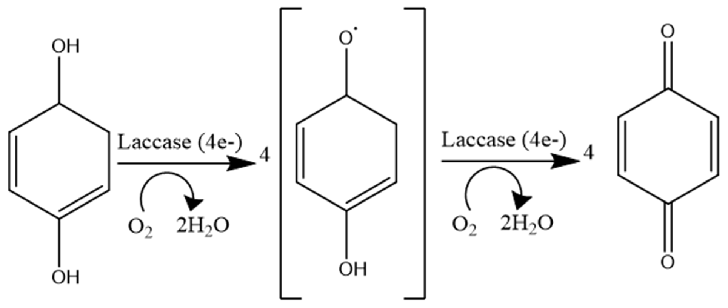
Laccase, an enzyme categorized as benzenediol oxidoreductase and E.C. 1.10.3.2, is a multi-copper oxidase. Due to its ability to facilitate the four-electron reduction of dioxygen to water while simultaneously catalyzing the one-electron oxidation of four molecular sub-substrates, the catalyst in issue is considered green [83]. Laccase can aid in the oxidation of various organic and inorganic compounds, including n-methoxy-substituted phenols, polyphenols, ketones, ascorbates, phosphates, and diamines [84]. The enzyme has undergone thorough investigation due to its capacity to catalyze the oxidation of diverse organic substrates and reduce molecular oxygen to water. Laccase is a novel enzyme that can catalyze the oxidation of diverse substrates without H2O2 within the reaction milieu. The electrode can facilitate the introduction of numerous analytes by utilizing two or more enzymes. The combination of laccase-tyrosinase enzymes is highly efficient at detecting phenolic compounds [85]. Laccases demonstrate responsiveness to ortho- and para-diphenol moieties, which include mono-, di-, and polyphenols, aminophenols, methoxy phenols, aromatic amines, and ascorbate, through reducing the four-electron oxygen to water (Figure 3).
Laccase is a thermostable catalyst, so the biosensor performance increases more rapidly. The image above shows the sensor using three different screen-printed electrodes (SPE based on carbon nanotubes (CNT), gold nanoparticles (GNPs), and nanocarbon tubes and gold nanoparticles (CNT-GNP), which are then modified using laccase enzyme with the addition of glutaraldehyde as a crosslinking agent between the amino group in laccase and the aldehyde group of the reticulating agent. Catechins were analyzed via CV and DPV techniques to explore their electrooxidation characteristics. The results showed that a detection limit established for catechins was 5.6 × 10⁻⁸ M on CNT-Lac/SPE, 1.3 × 10⁻⁷ M on GNP-Lac/SPE, and 4.9 × 10⁻⁸ M on CNT-GNP. The biosensor was exposed to the nutritional composition containing green tea to be analyzed for its catechin content using CNTGNP-Lac/SPE by DPV. The relationship between CNT and GNP significantly increases the sensitivity and selectivity of the biosensor. Catechin content was assessed by paired t-test. Furthermore, the correlation between the response of CNT-GNP-Lac/SPE and the particular efficacy and antioxidant activity of nutrients, ascertained through traditional spectrophotometric techniques (DPPH, galvanoxyl, and ABTS), is examined in relation to the swift development of biosensors for the assessment of comparative antioxidant activity [86].
Yang et al. [87] detected catechins using a laccase-based electrochemical sensor immobilized in a nanocomposite of 4-MBA gold nanoparticles and a polymer from CTS-g-N-CSIDZ. Biomaterials were employed to amplify the reactivity of electrochemical biosensors, which exhibited high specificity, rapid reactivity, economic viability, and uncomplicated configuration, removing the necessity for arduous sample preparation or analytical instrumentation. The performance of laccase-based sensors as catechin sensors in industrial waste was evaluated through conventional methods, namely HPLC. The results obtained by the concentration of catechins in three industrial waste samples confirmed no significant disparity in the concentrations of the samples between the two methods. It shows that the electrochemical sensor can detect catechins. In conclusion, this sensor shows benefits including rapid analysis duration, economic cost, and mobility.
In addition, measurements of electrochemical and electroanalytical antioxidants can be determined simultaneously. Laccase-based electronic detection and biosensors to measure the total phenolic content of honey samples from different countries showed a good correlation between antioxidant strength as measured by the electrochemical index and the FRAP and DPPH tests, as well as between TPC results from an approved biosensor and the Folin–Ciocalteau (FC) test [68].
4.1.3. Peroxidase-Based Biosensors
Peroxidase enzymes catalyze oxidation-reduction reactions through mechanisms involving free radicals [88]. Peroxidases are enzymes that facilitate the conversion of substrates into oxidized products. The HRP enzyme, or horseradish peroxidase, is frequently used in biochemistry and biotechnology. It is a reporting enzyme in affinity-based assays and biosensor recognition elements [89]. However, only some studies on the use peroxidase-based electrochemical biosensors for determining antioxidants exist. Peroxidase-based electrochemical biosensors are starting to develop because they can transfer electrons at low working potentials. Consequently, there is a notable enhancement in the biosensor’s selectivity [90]. The advantages possessed by other peroxide-based biosensors are their good stability in wet test media and their high sensitivity. HRP-based electrochemical biosensors have been extensively developed by immobilization on classical working electrodes, such as carbon or precious metals. Enzyme-based biosensors have advanced electrode immobilization and have many interface functions on the electrode surface [58].
A peroxidase-based electrochemical sensor method has been developed. It shows good results based on the detection limit value and also the resulting linear range, including H2O2 measurements using the electrochemical detection method in the form of cyclic voltammetry using nanoparticle working electrodes encapsulated with HRP yielding LOD values above 0.01 μM and linear regression of 0.01–100 μM [91]. Measurement of l-lactate in wine and must samples using the electrochemical detection method, in the form of amperometric voltammetry using a graphite rod working electrode combined with peroxidase-mimetic nanozymes, produced LOD values above 2 μM and linear regression of 5 μM–14 nm [90]. The peroxidase-based biosensor detects butylated hydroxyanisole (BHA) and propyl gallate (PG) in the food matrix. The HRP enzyme is immobilized by the spiny Au-Pt nanotube (SAP NTs) electrode, which has a wide surface area and is ideal for electron transfer. The SAP NTs structure was synthesized, and the intrinsic peroxidase was proved as an enzymatic biosensor. Linear sweep voltammetry (LSW) showed oxidation maxima for BHA and PG at 624 mV and 655 mV, respectively, with linearity ranges of 0.3–50 mg/L and 0.1–100 mg/L. BHA and PG were detectable at 0.046 and 0.024 mg/L (3 s/slope), respectively. RSD experiments and recoveries were comparable to HPLC, with good sensitivity, stability, and reducibility [92].
4.1.4. Oxidase-Based Biosensor
A biosensor utilizing oxidase enzymes is capable of effectively neutralizing hydrogen peroxide (H2O2) or superoxide radicals (O2•−) produced through oxidative catalysis. The assessment of antioxidant capacity was carried out by Becker et al. [93] through an examination of multiple fruits using a xanthine oxidase-based biosensor and amperometric techniques. The results revealed distinct antioxidant capacities for each fruit, with the Murici fruit exhibiting the highest capacity. Additionally, the study measured the concentration of gallic acid, finding a positive correlation between higher gallic acid concentrations and increased antioxidant capacity. Furthermore, the assessment of antioxidant capacity was conducted by employing identical enzymes through the utilization of polymeric PVA-AWE films that were immobilized on the carbon paste electrode surface. The chamomile sample exhibited the highest antioxidant capacity, which was approximately 70% [94]. The present study focuses on the utilization of an ascorbate oxidase-based sensor-biosensor system (SB) for the purpose of detecting ascorbic acid as well as several types of orange juice, specifically Hamlin, Sanguinello, and Moro. The detection method employed in this study utilizes cyclic voltammetry and amperometric methods. The obtained results pertaining to the antioxidant capacity of this particular method were compared to those of the conventional methods, namely DPPH and ABTS. It was observed that there was no significant difference between the antioxidant capacities obtained from the two methods. Notably, the Hamlin variety exhibited the highest antioxidant capacity [95].
4.2. Cell/Microorganism-Based Biosensors
Cell-based biosensors have gained significant popularity in various domains, such as drug screening, clinical and health, environmental testing, biomedicine, and national security [96,97,98]. Living cells are used as biometric components in electrochemical biosensors to convert biological impulses into electrical signals that can be quickly detected and measured [99]. Cell-based electrochemical biosensors are considered to be highly resilient biosensors that are capable of detecting biochemical effects through living cells. These biosensors possess several distinct advantages, including their non-invasiveness, absence of labeling requirements, their rapid response time, high efficiency, and versatile fabrication.
The hydrogen peroxide (H2O2) discharged from the cell reacts with a catalytic agent on the electrode’s surface. This reaction facilitates reduction and oxidation reactions, subsequently leading to electrochemical signal alterations. The expeditious and precise discharge of H2O2 within cells is crucial in utilizing cell-based electrochemical biosensors for evaluating activity and screening antioxidant components. The study’s methodology involves immobilizing cells onto a working electrode modified with manganese dioxide nanowires (MnO2NWs) and gold nanoparticles (AuNP). It is achieved using a photosensitive hydrogel treated with carbon nanofibers (CNF) as a scaffolding material for a three-dimensional cell culture. This study uses cells as receptor elements to investigate H2O2 release and assess the antioxidant potential of anthocyanins. The A549 cell-based electrochemical biosensor was utilized in the preparation process to detect extracellular H2O2 and to evaluate the efficacy of the active constituents in terms of their antioxidant activity. The findings of this investigation indicate a strong association between the oxidation peak (Ip) and the concentration of H2O2, as evidenced by the linear equation Ip (μA) = 58.199CH2O2 + 5.825 (where CH2O2 represents the concentration of H2O2. Moreover, the limit of detection for H2O2 was determined to be 0.02 μM, indicating a high level of sensitivity, reproducibility, and stability, as reported in the literature [31].
Ge et al. [99] also examined the antioxidant activity of a Chinese dried ham Lactobacillus plantarum extract. An RAW264.7 cell-based electrochemical technique assessed this potential. RAW264 powers the cellular biosensor. Seven cells were placed in a hydrogel of sodium alginate and graphene oxide on a manganese dioxide surface changed with a gold electrode. To construct a 3D hydrogel scaffold, cells/NaAlg/GO/a-MnO2/GE were immersed in calcium. After PMA, RAW264.7 cells produced H2O2. RAW264 cells released H2O2. The MnO2/GE absorbs seven cells and catalyzes them at its active site. MnO2 reduces and electro-oxidizes on the electrode surface, increasing the oxidation-reduction probe and electron transfer rate. The biosensor approach was used to detect H2O2 from RAW246.7 macrophage cells quickly. The study has a detection limit of 0.02 μM and a linear response from 0.05 to 0.85. At 1010 CFU/mL, L. plantarum had the highest relative antioxidant capacity (RAC) of 88.94%. Thus, this study uses RAW264.7 cells to assess L. plantarum’s antioxidant potential quickly, sensitively, and quantitatively via electrochemical biosensing. This approach quickly screens specimens’ antioxidant properties.
Microorganisms producing superoxide dismutase enzymes (D. radiodurans) as baroreceptors were immobilized in zeolite nanocomposites using electrochemical measurements, namely cyclic voltammetry. Xanthine oxidase (XO) catalyzes the enzymatic process of xanthine as a substrate, generating superoxide radicals. The presence of zeolite has the potential to augment the response, concomitantly elevating the value of Km. This is because the immobilization of SOD in zeolite for 24 h does not necessarily imply a lower concentration of SOD involved in the reaction with superoxide radicals when compared to prior research [100]. The detection of uric acid as a non-enzyme antioxidant using Lactobacillus plantarum immobilized using zeolite reported the results of electrode stability until day 18 [101]. Furthermore, the detection of antioxidants using superoxide dismutase-producing microorganisms from D. radiodurans bacteria as receptors immobilized on the surface of SPCE using electrochemical measurements, namely cyclic voltammetry, was carried out. The results showed that the stable D. radiodurans biofilm on the SPCE surface remained stable for 35 days. Optimum SPCE biofilms made have good stability with less than 5% RSD. Repeatability measurements indicated that the optimum SPCE biofilm had fairly good repeatability. The voltammogram produced with three different SPCE biofilms is identical and has an oxidation peak of 0.750 V [102].
4.3. DNA-Based-Biosensors
DNA-based biosensors have advantages, including superior biocompatibility, good thermal stability, alternative functionalization, and detection of specific targets [103,104,105,106,107]. Antioxidants in food samples can be detected using DNA as a bioreceptor in a biosensor. The DNA emulates the mechanisms of interaction with analytical agents that take place within the human body by immobilizing DNA on the transducer surface. This can be achieved through the utilization of genetic material as a biological recipient [108]. DNA has been identified as a highly promising bioreceptor for biosensing applications owing to its extended biological activity, exceptional addressability, and tunable stiffness. DNA-based biosensors have been widely developed and show promising results. These include aptamer, which has better thermal stability [109], adaptable biological affinity [110,111,112], and improved enzyme attack resistance [113]. DNA can also be used to construct supermolecular structures that can be programmed as templates to realize precise positioning and control spatial modifications, significantly improving biosensor performance [114]. DNA-based biosensors encompass various types, such as functional DNA, DNA hybridization, and DNA template-based biosensors.
Typically, electrochemical biosensors that rely on DNA as the bioreceptor element utilize immobilized DNA molecules on the working electrode surface to facilitate the detection of DNA-analyte interactions. Moreover, these interactions elicit modifications in the structure of DNA and its electrochemical characteristics, converting a stimulus into an electrochemical signal [115]. DNA-based biosensors can be used to determine the antioxidant capacity of samples on a routine basis because the changes in the base oxidation peaks are highly distinguishable before and after interactions with analytes. As such, there is no need for labeling or amplification strategies that can reduce analysis time and complexity [16].
Immobilization of purine bases (adenine and guanine) onto the surface of carbon glass electrodes (GCE) utilizes hydroxyl radicals for purine base degradation. Ascorbic acid, caffeic acid, coumaric acid, and resveratrol are compounds with antioxidant properties that can eliminate hydroxyl radicals and safeguard adenine and guanine located on the surface of GCE. The study assessed the interactions between immobilized purine bases and free radicals, both in the presence and absence of antioxidants, by analyzing alterations in anodic peak currents utilizing square wave voltammetry (SWV). The outcomes derived from the five antioxidants exhibited varied efficiencies, from 47% to 79%, in their capacity as hydroxyl radical scavengers. The antioxidant standard with the highest sensitivity, ascorbic acid, yielded the greatest TAC value. The utilization of purine-base-based biosensors can enhance a beverage’s overall antioxidant capacity. The expeditious and uncomplicated nature of the analysis is augmented by its compatibility with portable instrumentation, as noted in reference [116].
Peng et al. [117] used guanine immobilized on the electrode and modified by adding MoS2 nanosheet. This results in an electrochemical biosensor with a large specific surface area and high electrocatalytic capability. The development of a guanine-based biosensor modified by nanosheets used in measuring the antioxidant capacity of three types of flavonoids, namely quercetin, fisetin, and catechins, compared to ascorbic acid shows many advantages, including an extensive linear range, low detection limit, and good stretching. Quercetin, fisetin, and catechin were measured and found to be 45.82%, 34.39%, and 16.99%, respectively. Ascorbic acid is a comparator antioxidant; its capacity value was determined to be 51.84% [118].
The determination of chlorogenic acid (CGA) in coffee samples using the cyclic voltammetry method and SPCEs electrodes modified with single-walled carbon nanotubes (SWCNT-COOH) has been reported. dsDNA interacts with OH free radicals to measure antioxidant activity in a DNA-based biosensor [119]. The current study used square wave voltammetry and deoxy-adenylic acid oligonucleotide (dA20) immobilized on carbon paste electrodes to identify polyphenols in green tea, black tea, peppermint, and senna samples. According to reference [29], the procedure yielded a linear range of 1.0 to 8.0 mg L−1 and a LOD of 0.059 mg L−1. Glutathione and ascorbic acid concentrations were measured using pulse differential voltammetry. This study used pencil graphite electrodes (PGE) modified with MWCNTs and chitosan (CHIT)/ds-DNA. Reference [120] describes this procedure.
5. Nanosensors
Electrochemical techniques are becoming faster, more accurate, and less expensive in the measurement of antioxidant capacity. Traditional spectrophotometric methods have problems, such as long analysis and sample preparation times, reagents that could be better for the environment, expensive reagents, and unknown reaction times [13].
Electroanalytical methods use transfer studies to exclude it. Electrochemical principles explain electron transfer and antioxidant oxidation reactions, although visual approaches are important. Electron transfer methods use a simple oxidant-redox process to generate signals. Electrochemical methods are appropriate for studying electron transfer. Due to their great sensitivity, voltammetric methods are suitable for qualitative analysis and quantifying trace phenolic compounds and antioxidants. Due to their electron-donating capacity, voltammetric methods can detect antioxidants such as polyphenols at low potentials [121]. Several literature studies on nanosensors using electrochemical methods can be seen in Table 2.
Electrochemical sensing is the most prevalent analytical method for identifying analytes due to its speed, sensitivity, accuracy, and affordability. Electrochemical sensors are useful for directly assessing antioxidants and detecting dietary antioxidant capacity as a result of these qualities. Active electrode materials have been used to produce antioxidant biosensors and electrochemical sensors [13]
Manoranjitham et al. used a BHA electrochemical nanosensor to detect synthetic antioxidants. Electro-polymerization created a POC/MWCNTs electrode sensor employing o-cresol phthalein complex-one (OC). The sensor’s detection limit was 0.11 mM [122]. In another study, an electrochemical sensor detected BHA in food and other items. Modifying multi-layer graphite paper with AuNPs and NiO nanoparticles created the sensor. The AuNPs’ conductivity complemented NiO nanoparticles’ electrocatalytic activity. EGP’s broad surface area boosts linear range, lowering LOD and increasing sensitivity. The NiO/AuNPs/EGP combination identified samples well [123].
Ziyatdinova devised an exact and discerning sensor capable of detecting TBHQ and BHA concurrently. The sensitive layer was formed by grouping MWNT with electropolymerized carminic acid. The augmentation of the electrode surface area and electron transfer rate resulted in a notable enhancement in the response of TBHQ and BHA. In turn, it facilitated the observation of their oxidation peaks when they coexisted. The utilization of polycarminic acid as a surface modifier for electrodes is being reported for the first time. The sensor that was obtained exhibited a high level of sensitivity and could identify template molecules. The sensor’s response exhibits linearity within the 0.50–75 µM range for TBHQ and 0.25–75 µM range for BHA, and the respective limits of detection are 0.36 µM and 0.23 µM [124].
Motia et al. developed an electrochemical sensor utilizing molecularly imprinted polymer (MIP) and benzenehexacarboxylic acid (BHA). The electrochemical sensor that underwent chitosan modification and was coated with gold nanoparticles and molecularly imprinted polymers demonstrated high efficacy in detecting BHA. The findings indicate that the electrochemical sensor exhibits remarkable selectivity and robust sensitivity. The created sensor exhibited a limit of detection (LOD) of 0.001 µg/mL. In a recent study, three commercially available food items, namely chewing gum, mayonnaise, and potato chips, were utilized as the original testing medium for evaluating the performance of the molecularly imprinted polymer (MIP) sensor. The findings of the study indicated favorable outcomes [125]. Balram et al. synthesized a novel nanocomposite of CuO nanofibers and NH2-functionalized carbon nanotubes that exhibited superior electrocatalytic activity for detecting cytotoxic TBHQ. The electrocatalytic performance of CuO nanofibers (NFs) supported on amino-functionalized carbon nanotubes (NH2-CNTs) on SPCE was investigated using cyclic voltammetry (CV) and differential pulse voltammetry (DPV) techniques. The sensor exhibited a high level of sensitivity, measuring at 37.7 µA/M/cm2, a substantial detection limit of 3 nM, and a broad linear range, as reported in reference [126]. Yan et al. proposed an alternative approach for the detection of TBHQ utilizing an electrochemical sensor. VMSF/ErGO has modified the recognition layer of GCE film for utilization. The sensitivity of the VMSF/ErGO/GCE sensor towards TBHQ can be attributed to the preconcentration effect of hydrogen bonds in VMSF and the elevated electroactivity of ErGO, as reported in reference [127].
Table 2.
Tabulated some studies about electrochemical nanosensor research for analyzing endogenous antioxidants.
Table 2.
Tabulated some studies about electrochemical nanosensor research for analyzing endogenous antioxidants.
| Method | Electrode | Medium | Antioxidants | Matrix | LOD | Linear Range | Ref. |
|---|---|---|---|---|---|---|---|
| DPV | MIM-PACO/GCE | 0.25 M ABS (pH 6.5) | Curcumin | Turmeric extract | 5.0 nM | 10 nM–2.0 µM | [128] |
| DPV | SNO NRs/GCE | 0.1 M PBS (pH 5.0) | Quercetin | Apple and grape juice | 1.98 nM | 0.01–68.53 µM | [129] |
| DPV | MMIP | 0.1 M PBS (pH 1.0) | Rosmarinic acid | Salvia officinalis, Zataria multiflor, Mentha longifolia, and Rosmarinus officinalis | 0.085 µM | 0.1–100 µM | [130] |
| 100–500 µM | |||||||
| DPV | EGDMA-MIP/IL-GR/GCE | 0.04 M BRBS (pH 2.0) | Rutin | Tablet | 0.12 µM | 0.3–1 µM | [131] |
| DPV | GCE/rGO/ZIF-8/MIP | Rutin | Tablet and orange juice | 0.0001 µM | 0.05–100 µM | [132] | |
| 0.0005–0.05µM | |||||||
| DPV | MIP/AuNPs/EGP | 0.1 M PBS (pH 5.0) | TBHQ | Edible oil | 0.07 µM | 0.08–100 µM | [123] |
| DPV | MIP/CHIT + AuNPs/SPCE | PBS (pH 7.0) | BHA | Chewing gum, mayonnaise and potato chips | 0.001 µg/mL | 0.01–20 µg/mL | [125] |
| DPV | CuO.NFs/NH2-CNTs/SPCE | 0.05 M PBS (pH 7.0) | TBHQ | Coconut oil, sesame oil, soybean oil | 3 nM | 0.013.9 µM | [126] |
| 3.9–147.6 µM | |||||||
| DPV | VMSF/ErGO/GCE | 0.1 M PBS (pH 4.0) | TBHQ | Edible oil, Toning lotion | 0.23 nM | 0.001–0.5 µM | [127] |
| 0.5–120 µM | |||||||
| AMP | poly O-cresolphthalein/MWCNT electrode | 0.1 M PBS (pH 7.0) | BHA | Potato chips | 0.11 µM | 0.33–110 µM | [122] |
| AMP | poly(carminic acid)/MWNT/GCE | BRBS (pH 2.0) | BHA | Linseed oil | 0.23 µM | 0.25–75 µM | [124] |
| TBHQ | 0.36 µM | 0.50–75 µM | |||||
| AMP | 4-aminobenzoic acid/Toray carbon fiber electrode | 0.1 M PBS (pH 7.0) | Bilirubin | Serum | 15 µM | 150–890 µM | [133] |
| AMP | AuNPs/RGO/SPCE | 100 mM PBS (pH 3.5) | Vitamin C | Commercial, pasteurized, and skimmed cow’s milk | 0.088 µg/mL | 50–500 µM | [134] |
| AMP | Mesoporous CuCo2O4/GCE | 0.15 M NaOH solution | Vitamin C | Vitamin C tablets Effervescent tablets | 0.21 µM | 1–100 µM | [135] |
| CV | H-BDDP-printed electrode | 1/15 M PBS (pH 7.0) | l-Cysteine | Bovine plasma | 0.620 µM | 1–194 µM | [136] |
| CV | GOCuNP/CPE | 1.0 M KCl (pH 7.0 | N-acetylcysteine | - | 2.97 × 10⁻⁵ M | 3.0 × 10⁻⁴–6.0× 10⁻³ M | [137] |
Note: -: Not available.
6. Biosensors Trends and Perspective
In recent years, there has been a significant expansion in the field of biosensors. However, more biosensors need to be developed specifically to address issues related to sustainable agriculture. Recent publications have primarily focused on utilizing established sensor principles for detecting food products and other related applications. From a positive perspective, contemporary situations could be encompassed by commercially available sensor technology, thereby emphasizing the importance of advancing integrated application development. The research group focused on leadership status has disclosed the creation of an electrical stimulation machine that has the potential to enhance agricultural productivity and provide autonomous, weather-resistant sensing services. The biosensors have encountered challenges with anti-interference measures, self-calibration techniques, and prolonged monitoring periods, necessitating further attention.
The most important improvements in enzyme-based biosensors involve how the biological materials on the electrode surface are held in place and how the interface works. Even though this technology works well and is important for both applied and basic science, four important things need to be thought about before enzyme biosensors can be used commercially to track active compounds and their antioxidant power:
- Stability and immobilization
In making biosensors, it is hard to immobilize enzymes in a way that works well and speeds up the rate at which electrons are transferred. Nanomaterials and polymers have the potential to serve as carriers or hosts for immobilized enzymes, thereby facilitating electron transfer, enhancing the stability of biosensors, and prolonging their lifespan.
- Several kinds of enzymes
Due to the high specificity of enzyme reactions, it is not feasible for a single type of enzyme to effectively locate all antioxidants or assess the antioxidant properties of all active substances. The enzyme Laccase is incapable of degrading monophenols such as 3-amino phenol due to the positioning of the amino group in the meta position. Consequently, the biosensor necessitates greater specificity, challenging certain substrates such as monophenols. Developing diverse enzyme-based biosensors capable of detecting specific antioxidants through enzymatic mechanisms would be an effective avenue for exploration.
- Interference with the matrix
The issue of matrix interference poses a significant challenge for numerous research methodologies, including biosensor techniques. It is due to the complex nature of real samples. For the matrix interference to be kept to a minimum, not only do new ways of pre-treating samples need to be found, but the specificity and selectivity of the biosensor also need to be improved.
- Sensitivity and usage
Enzymes need to be attached to the electrode surface for biosensors’ effectiveness through bioconjugation. High-specificity sensing is necessary to engineer biocompatible materials that satisfy the criteria for achieving heightened sensitivity. The significance of engaging with the matrix lies in its ability to reduce the effective concentration of enzymes. The forthcoming advancement of enzymatic biosensors will prioritize enhancing the immobilization technology and modifying the biological enzyme to augment the biosensor’s utility.
Author Contributions
For review articles with several authors, all authors were involved in conceptualization, writing review and editing, supervision, and funding acquisition. All authors have read and agreed to the published version of the manuscript.
Funding
This research was funded by the Directorate General Higher Education, Research, and Technology, The Ministry Education, Culture, Research, and Technology, Republic of Indonesia for the research funding in scheme Regular Fundamental Research fiscal year 2023 with grant number 18797/IT3.D10/PT.01.03/P/B/2023.
Institutional Review Board Statement
Not applicable.
Informed Consent Statement
Not applicable.
Data Availability Statement
Data available in a publicly accessible repository and cited in accordance with journal guidelines.
Acknowledgments
The authors would like to thank to the Directorate General Higher Education, Research, and Technology, The Ministry Education, Culture, Research, and Technology, Republic of Indonesia for the research funding in scheme Regular Fundamental Research fiscal year 2023 with grant number 18797/IT3.D10/PT.01.03/P/B/2023.
Conflicts of Interest
The authors declare no conflict of interest.
References
- Li, Q.; Guan, X.; Wu, P.; Wang, X.; Zhou, L.; Tong, Y. Early Transmission Dynamics in Wuhan, China, of Novel Coronavirus–Infected Pneumonia. N. Engl. J. Med. 2020, 382, 1199–1207. [Google Scholar] [CrossRef] [PubMed]
- Lushchak, V.I. Free radicals, reactive oxygen species, oxidative stress and its classification. Chem. Biol. Interact. 2014, 5, 164–175. [Google Scholar] [CrossRef] [PubMed]
- Senoner, T.; Dichtl, W. Oxidative Stress in cardiovascular diseases: Still a therapeutic target. Nutrients 2019, 11, 2090. [Google Scholar] [CrossRef] [PubMed]
- Prasad, M.; Sedlarova, A.; Balukova, M.; Rac, P. Pospisil. Reactive Oxygen Species as a Response to Wounding: In Vivo Imaging in Arabidopsis thaliana. Front. Plant Sci. 2020, 10, 1660. [Google Scholar] [CrossRef] [PubMed]
- Zhang, L.; Liu, Y. Potential interventions for novel coronavirus in China: A systematic review. J. Med. Virol. 2020, 92, 479–490. [Google Scholar] [CrossRef] [PubMed]
- Alkadi, H. A review on free radicals and antioxidants. Infect. Disord. Drug Targets 2020, 20, 16–26. [Google Scholar] [CrossRef]
- Marrocco, I.; Altieri, F.; Peluso, I. Measurement and Clinical Significance of Biomarkers of Oxidative Stress in Humans. Oxid. Med. Cell Longev. 2017, 2017, 6501046. [Google Scholar] [CrossRef]
- Pisoschi, A.M.; Pop, A. The role of antioxidants in the chemistry of oxidative: A review. Eur. J. Med. Chem. 2015, 97, 55–74. [Google Scholar] [CrossRef]
- Sandrasari, D.A.; Andarwulan, N.; Faridah, D.N.; Dewi, F.N.A. Indonesian indigenous plants as a source of antioxidants to treat gastrointestinal disorders. Food Res. 2021, 5, 195–204. [Google Scholar] [CrossRef]
- Costa, C.; Tsatsakis, A.; Mamoulakis, C.; Teodoro, M.; Briguglio, G.; Caruso, E.; Tsoukalas, D.; Margina, D.; Dardiotis, E.; Kouretas, D.J.F.R. Current evidence on the effect of dietary polyphenols intake on chronic diseases. Food Chem. Toxicol. 2017, 110, 286–299. [Google Scholar] [CrossRef]
- Sharma, R.; Padwad, Y.J. Perspectives of the potential implications of polyphenols in influencing the interrelationship between oxi-inflammatory stress, cellular senescence and immunosenescence during aging. Trends Food Sci. Technol. 2020, 98, 41–52. [Google Scholar] [CrossRef]
- Iswantini, D.; Yulian, M.; Mulijani, S.; Trivadila. Inhibition kinetics of Sida rhombifolia L. extract toward xanthine oxidase electrochemical method. Indones. J. Chem. 2014, 14, 71–77. [Google Scholar] [CrossRef]
- Pisoschi, A.M.; Pop, A.; Iordache, F.; Stanca, L.; Predoi, G.; Serban, A.M. Oxidative stress mitigation by antioxidants an overview on their chemistry and influences on health status. Eur. J. Med. Chem. 2021, 116, 112891. [Google Scholar] [CrossRef] [PubMed]
- Ruskovska, T.; Budić-Leto, I.; Corral-Jara, K.F.; Ajdžanović, V.; Arola-Arnal, A.; Bravo, F.I.; Deligiannidou, G.E.; Havlik, J.; Janeva, M.; Kistanova, E.; et al. Systematic bioinformatic analyses of nutrigenomic modifications by polyphenols associated with cardiometabolic health in humans—Evidence from targeted nutrigenomic studies. Nutrients 2021, 13, 2326. [Google Scholar] [CrossRef]
- Butterfield, D.A.; Halliwell, B. Oxidative stress, dysfunctional glucose metabolism and Alzheimer disease. Nat. Rev. Neurosci. 2019, 20, 148–160. [Google Scholar] [CrossRef]
- Ye, Y.; Ji, J.; Sun, Z.; Shen, P.; Sun, X. Recent advances in electrochemical biosensors for antioxidant analysis in foodstuff. Trends Anal. Chem. 2020, 122, 115718. [Google Scholar] [CrossRef]
- Srinivasan, K. Antioxidant Potential of Spices and Their Active Constituents. Crit. Rev. Food Sci. Nutr. 2014, 54, 352–372. [Google Scholar] [CrossRef]
- Yashin, A.; Yashin, Y.; Xia, X.; Nemzer, B. Antioxidant activity of spices and their impact on human health: A review. Antioxidants 2017, 6, 70. [Google Scholar] [CrossRef]
- Nimse, S.; Pal, D. Free radicals, natural antioxidants, and their reaction mechanisms. RSC Adv. 2015, 5, 27986–28006. [Google Scholar] [CrossRef]
- Ojha, K.; Dubey, S.; Chandrakar, J.; Minj, R.A.; Dehariya, R.; Dixit, A.K. A review on different methods of determination of antioxidant activity assay of herbal plant. J. Sci. Bio Pharm. Chem. Sci. 2018, 4, 707–732. [Google Scholar]
- Chen, J.; Liu, Y.; Zhao, Z.; Qiu, J. Oxidative stress in the skin: Impact and related protection. Int. J. Cosmet. Sci. 2021, 43, 495–509. [Google Scholar] [CrossRef] [PubMed]
- Działo, M.; Mierziak, J.; Korzun, U.; Preisner, M.; Szopa, J.; Kulma, A. The potential of plant phenolics in prevention and therapy of skin disorders. Int. J. Mol. Sci. 2016, 17, 160. [Google Scholar] [CrossRef] [PubMed]
- Kobus-Cisowska, J.; Flaczyk, E.; Rudzińska, M.; Kmiecik, D. Antioxidant properties of extracts from Ginkgo biloba leaves in meatballs. Meat Sci. 2014, 97, 174–180. [Google Scholar] [CrossRef] [PubMed]
- Rolim, W.R.; Pelegrino, M.T.; de Araújo Lima, B.; Ferraz, L.S.; Costa, F.N.; Bernardes, J.S.; Rodigues, T.; Brocchi, M.; Seabra, A.B. Green tea extract mediated biogenic synthesis of silver nanoparticles: Characterization, cytotoxicity evaluation and antibacterial activity. Appl. Surf. Sci. 2019, 463, 66–74. [Google Scholar] [CrossRef]
- Vinci, G.; D’ascenzo, F.; Maddaloni, L.; Prencipe, S.A.; Tiradritti, M. The Influence of Green and Black Tea Infusion Parameters on Total Polyphenol Content and Antioxidant Activity by ABTS and DPPH Assays. Beverages 2022, 8, 18. [Google Scholar] [CrossRef]
- Thongsuk, P.; Sameenoi, Y. Colorimetric determination of radical scavenging activity of antioxidants using Fe3O4 magnetic nanoparticles. Arab. J. Chem. 2022, 15, 103475. [Google Scholar] [CrossRef]
- Wang, X.; Luo, Y.; Huang, K.; Cheng, N. Advanced Agrochem Biosensor for agriculture and food safety: Recent advances and future perspectives. Adv. Agrochem. 2022, 1, 3–6. [Google Scholar] [CrossRef]
- Munteanu, I.G.; Apetrei, C. A Review on Electrochemical Sensors and Biosensors Used in Chlorogenic Acid Electroanalysis. Int. J. Mol. Sci. 2021, 22, 13138. [Google Scholar] [CrossRef]
- Barroso, M.F.; Ramalhosa, M.J.; Alves, R.C.; Dias, A.; Soares, C.M.D.; Oliva-Teles, M.T.; Delerue-Matos, C. Total antioxidant capacity of plant infusions: Assessment using electrochemical DNA-based biosensor and spectrophotometric methods. Food Control. 2016, 68, 153–161. [Google Scholar] [CrossRef]
- Karimi-Maleh, H.; Orooji, Y.; Karimi, F.; Alizadeh, M.; Baghayeri, M.; Rouhi, J.; Tajik, S.; Beitollahi, H.; Agarwal, S.; Gupta, V.K.; et al. A critical review on the use of potentiometric based biosensors for biomarkers detection. Biosens. Bioelectron. 2021, 184, 113252. [Google Scholar] [CrossRef]
- Ye, Y.; Sun, X.; Zhang, Y.; Han, X.; Sun, X. A novel cell-based electrochemical biosensor based on MnO2 catalysis for antioxidant activity evaluation of anthocyanins. Biosens. Bioelectron. 2021, 202, 113990. [Google Scholar] [CrossRef] [PubMed]
- Romero, K.J.; Galliher, M.S.; Raycroft, M.A.R.; Chauvin, J.P.R.; Bosque, I.; Pratt, D.A.; Stephenson, C.R.J. Electrochemical Dimerization of Phenylpropenoids and the Surprising Antioxidant Activity of the Resultant Quinone Methide Dimers. Angew. Chem.—Int. Ed. 2018, 57, 17125–17129. [Google Scholar] [CrossRef] [PubMed]
- Ricci, A.; Parpinello, G.P.; Teslić, N.; Kilmartin, P.A.; Versari, A. Suitability of the cyclic voltammetry measurements and DPPH• spectrophotometric assay to determine the antioxidant capacity of food-grade oenological tannins. Molecules 2019, 24, 2925. [Google Scholar] [CrossRef]
- Petković, B.B.; Stanković, D.; Milčić, M.; Sovilj, S.P.; Manojlović, D. Dinuclear copper(II) octaazamacrocyclic complex in a PVC coated GCE and graphite as a voltammetric sensor for determination of gallic acid and antioxidant capacity of wine samples. Talanta 2015, 132, 513–519. [Google Scholar] [CrossRef] [PubMed]
- Rajput, V.D.; Harish; Singh, R.K.; Verma, K.K.; Sharma, L.; Quiroz-Figueroa, F.R.; Meena, M.; Gour, V.S.; Minkina, T.; Sushkova, S.; et al. Recent developments in enzymatic antioxidant defence mechanism in plants with special reference to abiotic stress. Biology 2021, 10, 267. [Google Scholar] [CrossRef]
- Losada-Barreiro, S.; Bravo-Díaz, C. Free radicals and polyphenols: The redox chemistry of neurodegenerative diseases. Eur. J. Med. Chem. 2017, 133, 379–402. [Google Scholar] [CrossRef]
- Vo, T.T.T.; Chu, P.M.; Tuan, V.P.; Te, J.S.L.; Lee, I.T. The promising role of antioxidant phytochemicals in the prevention and treatment of periodontal disease via the inhibition of oxidative stress pathways: Updated insights. Antioxidants 2020, 9, 1211. [Google Scholar] [CrossRef]
- Gupta, D. Methods for determination of antioxidant capacity: A review. Int. J. Pharm. Sci. Res. 2015, 6, 546–566. [Google Scholar] [CrossRef]
- Martínez, A.; Galano, A.; Vargas, R. Free radical scavenger properties of α-mangostin: Thermodynamics and kinetics of HAT and RAF mechanisms. J. Phys. Chem. B 2011, 115, 12591–12598. [Google Scholar] [CrossRef]
- Martinez-Perez, C.; Ward, C.; Cook, G.; Mullen, P.; McPhail, D.; Harrison, D.J.; Langdon, S.P. Novel flavonoids as anti-cancer agents: Mechanisms of action and promise for their potential application in breast cancer. Biochem. Soc. Trans. 2014, 42, 1017–1023. [Google Scholar] [CrossRef]
- Awouafack, M.D.; Tane, P.; Morita, H. Isolation and Structure Characterization of Flavonoids. In Flavonoids—From Biosynthesis to Human Health; InTech: London, UK, 2017. [Google Scholar] [CrossRef]
- Tiwari, S.C.; Husain, N. Biological Activities and Role of Flavonoids in Human Health—A Review. Indian J. Sci. Res. 2017, 12, 193–196. [Google Scholar]
- Badhani, B.; Sharma, N.; Kakkar, R. Gallic acid: A versatile antioxidant with promising therapeutic and industrial applications. RSC Adv. 2015, 5, 27540–27557. [Google Scholar] [CrossRef]
- Kahl, M.; Golden, T.D. Electrochemical determination of phenolic acids at a Zn/Al layered double hydroxide film modified glassy carbon electrode. Electroanalysis 2014, 26, 1664–1670. [Google Scholar] [CrossRef]
- Carocho, M.; Ferreira, I.C.F.R. A review on antioxidants, prooxidants and related controversy: Natural and synthetic compounds, screening and analysis methodologies and future perspectives. Food Chem. Toxicol. 2013, 51, 15–25. [Google Scholar] [CrossRef]
- Chikere, C.O.; Faisal, N.H.; Kong-Thoo-Lin, P.; Fernandez, C. Interaction between amorphous zirconia nanoparticles and graphite: Electrochemical applications for gallic acid sensing using carbon paste electrodes in wine. Nanomaterials 2020, 10, 537. [Google Scholar] [CrossRef] [PubMed]
- Zheng, Y.; Karimi-maleh, H.; Fu, L. Evaluation of Antioxidants Using Electrochemical Sensors: A Bibliometric Analysis. Sensors 2022, 22, 3238. [Google Scholar] [CrossRef] [PubMed]
- Nejad, F.G.; Tajik, S.; Beitollahi, H.; Sheikhshoaie, I. Magnetic nanomaterials based electrochemical (bio)sensors for food analysis. Talanta 2021, 228, 122075. [Google Scholar] [CrossRef] [PubMed]
- Ziyatdinova, G.; Guss, E.; Yakupova, E. Electrochemical sensors based on the electropolymerized natural phenolic antioxidants and their analytical application. Sensors 2021, 21, 8385. [Google Scholar] [CrossRef]
- Gajdár, J.; Goněc, T.; Jampílek, J.; Brázdová, M.; Bábková, Z.; Fojta, M.; Barek, J.; Fischer, J. Voltammetry of a Novel Antimycobacterial Agent 1-Hydroxy-N-(4-nitrophenyl)naphthalene-2-carboxamide in a Single Drop of a Solution. Electroanalysis 2018, 30, 38–47. [Google Scholar] [CrossRef]
- Haque, M.A.; Morozova, K.; Ferrentino, G.; Scampicchio, M. Electrochemical Methods to Evaluate the Antioxidant Activity and Capacity of Foods: A Review. Electroanalysis 2021, 33, 1419–1435. [Google Scholar] [CrossRef]
- Shahamirifard, S.A.; Ghaedi, M.; Razmi, Z.; Hajati, S. A simple ultrasensitive electrochemical sensor for simultaneous determination of gallic acid and uric acid in human urine and fruit juices based on zirconia-choline chloride-gold nanoparticles-modified carbon paste electrode. Biosens. Bioelectron. 2018, 114, 30–36. [Google Scholar] [CrossRef] [PubMed]
- Ziyatdinova, G.K.; Nizamova, A.M.; Budnikov, H.C. Voltammetric determination of curcumin in spices. J. Anal. Chem. 2012, 67, 591–594. [Google Scholar] [CrossRef]
- Wang, X.; Jiao, C.; Yu, Z. Electrochemical biosensor for assessment of the total antioxidant capacity of orange juice beverage based on the immobilizing DNA on a poly l-glutamic acid doped silver hybridized membrane. Sens. Actuators B Chem. 2014, 192, 628–633. [Google Scholar] [CrossRef]
- Souza, L.P.; Calegari, F.; Zarbin, A.J.G.; Marcolino-Júnior, L.H.; Bergamini, M.F. Voltammetric determination of the antioxidant capacity in wine samples using a carbon nanotube modified electrode. J. Agric. Food Chem. 2011, 59, 7620–7625. [Google Scholar] [CrossRef] [PubMed]
- Kračmarová, A.; Pohanka, M. Elektrochemické stanovení nízkomolekulárních antioxidantů v séru. Chem. List. 2014, 108, 64–69. [Google Scholar]
- Munteanu, I.G.; Apetrei, C. Electrochemical determination of chlorogenic acid in nutraceuticals using voltammetric sensors based on screen-printed carbon electrode modified with graphene and gold nanoparticles. Int. J. Mol. Sci. 2021, 22, 8897. [Google Scholar] [CrossRef] [PubMed]
- Ghaani, M.; Nasirizadeh, N.; Ardakani, S.A.Y.; Mehrjardi, F.Z.; Scampicchio, M.; Farris, S. Development of an electrochemical nanosensor for the determination of gallic acid in food. Anal. Methods 2016, 8, 1103–1110. [Google Scholar] [CrossRef]
- Korotkova, E.I.; Freinbichler, W.; Linert, W.; Dorozhko, E.V.; Bukkel, M.V.; Plotnikov, E.V.; Voronova, O.A. Study of total antioxidant activity of human serum blood in the pathology of alcoholism. Molecules 2013, 18, 1811–1818. [Google Scholar] [CrossRef]
- Yilmaz, Ü.T.; Kekillioglu, A.; Mert, R. Determination of Gallic acid by differential pulse polarography: Application to fruit juices. J. Anal. Chem. 2013, 68, 1064–1069. [Google Scholar] [CrossRef]
- Senocak, A.; Sanko, V.; Tumay, S.O.; Orooji, Y.; Demirbas, E.; Yoon, Y.; Khataee, A. Ultrasensitive electrochemical sensor for detection of rutin antioxidant by layered Ti3Al0.5Cu0.5C2 MAX phase. Food Chem. Toxicol. 2022, 164, 113016. [Google Scholar] [CrossRef]
- Silva, T.R.; Westphal, E.; Gallardo, H.; Vieira, I.C. Ionic organic film sensor for determination of phenolic compounds. Electroanalysis 2014, 26, 1801–1809. [Google Scholar] [CrossRef]
- Chikere, C.; Faisal, N.H.; Lin, P.K.T.; Fernandez, C. Zinc oxide nanoparticles modified-carbon paste electrode used for the electrochemical determination of Gallic acid. J. Phys. Conf. Ser. 2019, 1310, 012008. [Google Scholar] [CrossRef]
- Wardak, C.; Paczosa-Bator, B.; Malinowski, S. Application of cold plasma corona discharge in preparation of laccase-based biosensors for dopamine determination. Mater. Sci. Eng. C 2020, 116, 111199. [Google Scholar] [CrossRef] [PubMed]
- Azad, M.S. Experiences of Domestic Abuse within the South Asian Community. J. Glob. Faultlines 2021, 8, 50–68. [Google Scholar] [CrossRef]
- Gil, D.M.A.; Rebelo, M.J.F. Evaluating the antioxidant capacity of wines: A laccase-based biosensor approach. Eur. Food Res. Technol. 2010, 231, 303–308. [Google Scholar] [CrossRef]
- Gahlaut, A.; Gothwal, A.; Chhillar, K.; Hooda, V. Electrochemical Biosensors for Determination of Organophosphorus Compounds: Review. Sci. Res. J. 2012, 1, 1–8. [Google Scholar] [CrossRef]
- de Oliveira Neto, J.R.; Rezende, S.G.; Lobón, G.S.; Garcia, T.A.; Macedo, I.Y.L.; Garcia, L.F.; Alves, V.F.; Torres, I.M.S.; Santiago, M.F.; Schmidt, F.; et al. Electroanalysis and laccase-based biosensor on the determination of phenolic content and antioxidant power of honey samples. Food Chem. 2017, 237, 1118–1123. [Google Scholar] [CrossRef]
- Sun, B.; Yang, Y.; Yu, S.; Bao, L.; Shi, H.; Dang, Q.; Liu, Y.; Yang, L.; Ma, Q.; Shi, X. Electrochemical biosensor based on ds-DNA/N-G@CS/GCE for highly sensitive and rapid measurement of antioxidant activity. ECS Adv. 2023, 2, 1–11. [Google Scholar] [CrossRef]
- Sharma, S.; Byrne, H.; O’Kennedy, R.J. Antibodies and antibody-derived analytical biosensors. Essays Biochem. 2016, 60, 9–18. [Google Scholar] [CrossRef]
- Narita, F.; Wang, Z.; Kurita, H.; Li, Z.; Shi, Y.; Jia, Y.; Soutis, C. A Review of Piezoelectric and Magnetostrictive Biosensor Materials for Detection of COVID-19 and Other Viruses. Adv. Mater. 2021, 33, 2005448. [Google Scholar] [CrossRef]
- Damborský, P.; Švitel, J.; Katrlík, J. Optical Biosensors. Essays Biochem. 2016, 60, 91–100. [Google Scholar] [PubMed]
- Hackney, A.C. Exercise, Sport, and Bioanalytical Chemistry; Elsevier: Amsterdam, The Netherlands, 2016. [Google Scholar]
- Dzyadevych, S.; Jaffrezic-Renault, N. Conductometric Biosensors. In Biological Identification; Elsevier: Amsterdam, The Netherlands, 2014; pp. 153–193. [Google Scholar]
- Fu, Y.; You, Z.; Xiao, A.; Liu, L.; Zhou, W. Electrochemical evaluation of the antioxidant capacity of natural compounds on glassy carbon electrode modified with guanine-, polythionine-, and nitrogen-doped graphene. Open Chem. 2020, 18, 1054–1063. [Google Scholar] [CrossRef]
- Cesewski, E.; Johnson, B.N. Electrochemical biosensors for pathogen detection. Biosens. Bioelectron. 2020, 159, 112214. [Google Scholar] [CrossRef] [PubMed]
- Jin, X.; Liu, C.; Xu, T.; Su, L.; Zhang, X. Artificial intelligence biosensors: Challenges and prospects. Biosens. Bioelectron. 2020, 165, 112412. [Google Scholar] [CrossRef]
- Cetó, X.; Céspedes, F.; Pividori, M.I.; Gutiérrez, J.M.; Del Valle, M. Resolution of phenolic antioxidant mixtures employing a voltammetric bio-electronic tongue. Analyst 2012, 137, 349–356. [Google Scholar] [CrossRef]
- Vlamidis, Y.; Gualandi, I.; Tonelli, D. Amperometric biosensors based on reduced GO and MWCNTs composite for polyphenols detection in fruit juices. J. Electroanal. Chem. 2017, 799, 285–292. [Google Scholar] [CrossRef]
- Nadifiyine, S.; Calas-Blanchard, C.; Amine, A.; Marty, J.L. Tyrosinase Biosensor Used for the Determination of Catechin Derivatives in Tea: Correlation with HPLC/DAD Method. Food Nutr. Sci. 2013, 4, 108–118. [Google Scholar] [CrossRef]
- Sýs, M.; Pekec, B.; Kalcher, K.; Vytřas, K. Amperometric enzyme carbon paste-based biosensor for quantification of hydroquinone and polyphenolic antioxidant capacity. Int. J. Electrochem. Sci. 2013, 8, 9030–9040. [Google Scholar] [CrossRef]
- Munteanu, I.G.; Apetrei, C. Tyrosinase-Based Biosensor—A New Tool for Chlorogenic Acid Detection in Nutraceutical Formulations. Materials 2022, 15, 3221. [Google Scholar] [CrossRef]
- Bounegru, A.V.; Apetrei, C. Laccase and tyrosinase biosensors used in the determination of hydroxycinnamic acids. Int. J. Mol. Sci. 2021, 22, 4881. [Google Scholar] [CrossRef]
- Kadam, A.A.; Saratale, G.D.; Ghodake, G.S.; Saratale, R.G.; Shahzad, A.; Magotra, V.K.; Kumar, M.; Palem, R.R.; Sung, J.S. Recent Advances in the Development of Laccase-Based Biosensors via Nano-Immobilization Techniques. Chemosensors 2022, 10, 58. [Google Scholar] [CrossRef]
- Rodríguez-Delgado, M.M.; Alemán-Nava, G.S.; Rodríguez-Delgado, J.M.; Dieck-Assad, G.; Martínez-Chapa, S.O.; Barceló, D.; Parra, R. Laccase-based biosensors for detection of phenolic compounds. TrAC—Trends Anal. Chem. 2015, 74, 21–45. [Google Scholar] [CrossRef]
- Munteanu, I.G.; Apetrei, C. Assessment of the Antioxidant Activity of Catechin in Nutraceuticals: Comparison between a Newly Developed Electrochemical Method and Spectrophotometric Methods. Int. J. Mol. Sci. 2022, 23, 8110. [Google Scholar] [CrossRef]
- Yang, Y.; Zeng, H.; Zhang, Q.; Bai, X.; Liu, C.; Zhang, Y.H. Direct electron transfer and sensing performance for catechin of nano-gold particles-polymer nano-composite with immobilized Laccase. Chem. Phys. Lett. 2016, 658, 259–269. [Google Scholar] [CrossRef]
- Steevensz, A.; Villegas, L.G.C.; Feng, W.; Taylor, K.E.; Bewtra, J.K.; Biswas, N. Soybean peroxidase for industrial wastewater treatment: A mini review. J. Environ. Eng. Sci. 2014, 9, 181–186. [Google Scholar] [CrossRef]
- Neumann, B.; Wollenberger, U. Electrochemical Biosensors Employing Natural and Artificial Heme Peroxidases on Semiconductors. Sensors 2020, 20, 3692. [Google Scholar] [CrossRef]
- Smutok, O.; Kavetskyy, T.; Prokopiv, T.; Serkiz, R.; Šauša, O.; Novák, I.; Švajdlenková, H.; Maťko, I.; Gonchar, M.; Katz, E. Biosensor Based on Peroxidase-Mimetic Nanozyme and Lactate Oxidase for Accurate L-Lactate Analysis in Beverages. Biosensors 2022, 12, 1042. [Google Scholar] [CrossRef]
- Shin, J.H.; Lee, M.J.; Choi, J.H.; Song, J.; Kim, T.H.; Oh, B.K. Electrochemical H2O2 biosensor based on horseradish peroxidase encapsulated protein nanoparticles with reduced graphene oxide-modified gold electrode. Nano Converg. 2020, 7, 1–8. [Google Scholar] [CrossRef]
- Wu, L.; Yin, W.; Tang, K.; Li, D.; Shao, K.; Zuo, Y.; Ma, J.; Liu, J.; Han, H. Enzymatic biosensor of horseradish peroxidase immobilized on Au-Pt nanotube/Au-graphene for the simultaneous determination of antioxidants. Anal. Chim. Acta 2016, 933, 89–96. [Google Scholar] [CrossRef]
- Becker, M.M.; Ribeiro, E.B.; de Oliveira Marquez, P.R.B.; Marty, J.; Nunes, G.S.; Catanante, G. Development of a highly sensitive xanthine oxidase-based biosensor for the determination of antioxidant capacity in Amazonian fruit samples. Talanta 2019, 204, 626–632. [Google Scholar] [CrossRef]
- Ribeiro, D.B.; Silva, G.S.; Dos Santos, D.; Costa, A.R.C.; Ribeiro, E.B.; Badea, M.; Nunes, G.S. Determination of the antioxidant activity of samples of tea and commercial sources of vitamin c, using an enzymatic biosensor. Antioxidants 2021, 10, 324. [Google Scholar] [CrossRef] [PubMed]
- Barberis, A.; Spissu, Y.; Bazzu, G.; Fadda, A.; Azara, E.; Sanna, D.; Schirra, M.; Serra, P.A. Development and characterization of an ascorbate oxidase-based sensor-biosensor system for telemetric detection of AA and antioxidant capacity in fresh orange juice. Anal. Chem. 2014, 86, 8727–8734. [Google Scholar] [CrossRef] [PubMed]
- Lindeman, N.I.; Cagle, P.T.; Aisner, D.L.; Arcila, M.E.; Beasley, M.B.; Bernicker, E.H.; Colasacco, C.; Dacic, S.; Hirsch, F.R.; Kerr, K.; et al. Updated molecular testing guideline for the selection of lung cancer patients for treatment with targeted tyrosine kinase inhibitors guideline from the college of American pathologists, the international association for the study of lung cancer, and the Association for Molecular Pathology. Arch Pathol. Lab. Med. 2018, 142, 321–346. [Google Scholar] [CrossRef]
- Gupta, N.; Renugopalakrishnan, V.; Liepmann, D.; Paulmurugan, R.; Malhotra, B.D. Cell-based biosensors: Recent trends, challenges and future perspectives. Biosens. Bioelectron. 2019, 141, 111435. [Google Scholar] [CrossRef] [PubMed]
- Inda, M.E.; Mimee, M.; Lu, T.K. Cell-based biosensors for immunology, inflammation, and allergy. J. Allergy Clin. Immunol. 2019, 144, 645–647. [Google Scholar] [CrossRef]
- Ge, Q.; Ge, P.; Jiang, D.; Du, N.; Chen, J.; Yuan, L.; Yu, H.; Xu, X.; Wu, M.; Zhang, W.; et al. A novel and simple cell-based electrochemical biosensor for evaluating the antioxidant capacity of Lactobacillus plantarum strains isolated from Chinese dry-cured ham. Biosens. Bioelectron. 2018, 99, 555–563. [Google Scholar] [CrossRef]
- Iswantini, D.; Weniarti; Nurhidayat, N.; Abidin, Z.; Trivadila. Antioxidant biosensor based on superoxide dismutase from Indonesian microbes immobilized in Indonesian natural zeolite. J. Appl. Pharm. Sci. 2019, 9, 104–109. [Google Scholar] [CrossRef]
- Iswantini, D.; Nurhidayat, N.; Trivadila; Widiyatmoko, O. Activity and stability of uricase from Lactobacillus plantarum immobilizated on natural zeolite for uric acid biosensor. Pak. J. Biol. Sci. 2014, 17, 277–281. [Google Scholar] [CrossRef]
- Febryanti, A.; Mulijani, S.; Iswantini, D. Antioxidant biosensor based on Deinococcus radiodurans biofilm immobilized on screen printed carbon electrode (SPCE) surface. Int. J. Res. Appl. Sci. Biotechnol. 2018, 5, 1–7. [Google Scholar]
- Reder-Christ, K.; Bendas, G. Biosensor applications in the field of antibiotic research-a review of recent developments. Sensors 2011, 11, 9450–9466. [Google Scholar] [CrossRef]
- Liang, G.; Man, Y.; Li, A.; Jin, X.; Liu, X.; Pan, L. DNAzyme-based biosensor for detection of lead ion: A review. Microchem. J. 2017, 131, 145–153. [Google Scholar] [CrossRef]
- Saidur, M.R.; Aziz, A.R.A.; Basirun, W.J. Recent advances in DNA-based electrochemical biosensors for heavy metal ion detection: A review. Biosens. Bioelectron. 2017, 90, 125–139. [Google Scholar] [CrossRef] [PubMed]
- Dehghani, S.; Nosrati, R.; Yousefi, M.; Nezami, A.; Soltani, F.; Taghdisi, S.M.; Abnous, K.; Alibolandi, M.; Ramezani, M. Aptamer-based biosensors and nanosensors for the detection of vascular endothelial growth factor (VEGF): A review. Biosens. Bioelectron. 2018, 110, 23–37. [Google Scholar] [CrossRef] [PubMed]
- Wu, Y.; Gao, H.; Wang, Y.; Peng, Z.; Guo, Z.; Ma, Y.; Zhang, R.; Zhang, M.; Wu, Q.; Xiao, J.; et al. Effects of different extraction methods on contents, profiles, and antioxidant abilities of free and bound phenolics of Sargassum polycystum from the South China Sea. J. Food Sci. 2022, 87, 968–981. [Google Scholar] [CrossRef]
- Thapa, K.; Liu, W.; Wang, R. Nucleic acid-based electrochemical biosensor: Recent advances in probe immobilization and signal amplification strategies. Wiley Interdiscip. Rev. Nanomed. Nanobiotechnol. 2022, 14, e1765. [Google Scholar] [CrossRef]
- Mehlhorn, A.; Rahimi, P.; Joseph, Y. Aptamer-based biosensors for antibiotic detection: A review. Biosensors 2018, 8, 54. [Google Scholar] [CrossRef]
- Li, N.; Du, M.; Liu, Y.; Ji, X.; He, Z. Multipedal DNA Walker Biosensors Based on Catalyzed Hairpin Assembly and Isothermal Strand-Displacement Polymerase Reaction for the Chemiluminescent Detection of Proteins. ACS Sens. 2018, 3, 1283–1290. [Google Scholar] [CrossRef]
- Minagawa, H.; Kataoka, Y.; Kuwahara, M.; Horii, K.; Shiratori, I.; Waga, I. A high affinity modified DNA aptamer containing base-appended bases for human β-defensin. Anal. Biochem. 2020, 594, 113627. [Google Scholar] [CrossRef]
- Yang, H.; Zhou, Y.; Liu, J. G-quadruplex DNA for construction of biosensors. TrAC—Trends Anal. Chem. 2020, 132, 116060. [Google Scholar] [CrossRef]
- Xue, C.; Zhang, S.; Yu, X.; Hu, S.; Lu, Y.; Wu, Z.S. Periodically Ordered, Nuclease-Resistant DNA Nanowires Decorated with Cell-Specific Aptamers as Selective Theranostic Agents. Angew. Chem.—Int. Ed. 2020, 59, 17540–17547. [Google Scholar] [CrossRef]
- Liu, J. DNA-stabilized, fluorescent, metal nanoclusters for biosensor development. TrAC—Trends Anal. Chem. 2014, 58, 99–111. [Google Scholar] [CrossRef]
- Diculescu, V.C.; Chiorcea-Paquim, A.M.; Oliveira-Brett, A.M. Applications of a DNA-electrochemical biosensor. TrAC—Trends Anal. Chem. 2016, 79, 23–36. [Google Scholar] [CrossRef]
- Barroso, M.F.; Delerue-Matos, C.; Oliveira, M.B.P.P. Electrochemical evaluation of total antioxidant capacity of beverages using a purine-biosensor. Food Chem. 2012, 132, 1055–1062. [Google Scholar] [CrossRef]
- Peng, H.; Wang, D.; Li, M.; Zhang, L.; Liu, M.; Fu, S. N-P-Zn-containing 2D supermolecular networks grown on MoS2 nanosheets for mechanical and flame-retardant reinforcements of polyacrylonitrile fiber. Chem. Eng. J. 2019, 372, 873–885. [Google Scholar] [CrossRef]
- Tang, P.; Tang, X.; Mei, S.; Xie, Y.; Liu, L.; Ren, L. Electrochemical antioxidant screening and evaluation based on guanine and chitosan immobilized MoS2 nanosheet modified glassy carbon electrode (guanine/CS/MoS2/GCE). Open Chem. 2020, 18, 1–9. [Google Scholar] [CrossRef]
- Tomac, I.; Šeruga, M.; Labuda, J. Evaluation of antioxidant activity of chlorogenic acids and coffee extracts by an electrochemical DNA-based biosensor. Food Chem. 2020, 325, 126787. [Google Scholar] [CrossRef] [PubMed]
- Ensafi, A.A.; Kazemnadi, N.; Amini, M.; Rezaei, B. Impedimetric DNA-biosensor for the study of dopamine induces DNA damage and investigation of inhibitory and repair effects of some antioxidants. Bioelectrochemistry 2015, 104, 71–78. [Google Scholar] [CrossRef] [PubMed]
- Chiorcea-Paquim, A.M.; Enache, T.A.; De Souza Gil, E.; Oliveira-Brett, A.M. Natural phenolic antioxidants electrochemistry: Towards a new food science methodology. Compr. Rev. Food Sci. Food Saf. 2020, 19, 1680–1726. [Google Scholar] [CrossRef]
- Manoranjitham, J.J.; Narayanan, S.S. Electrochemical sensor for determination of butylated hydroxyanisole (BHA) in food products using poly O-cresolphthalein complexone coated multiwalled carbon nanotubes electrode. Food Chem. 2021, 342, 128246. [Google Scholar] [CrossRef]
- Fan, L.; Kan, X. Sensitive detection of butylated hydroxyanisole based on free-standing paper decorated with gold and NiO nanoparticles. Microchem. J. 2020, 159, 105511. [Google Scholar] [CrossRef]
- Ziyatdinova, G.; Guss, E.; Budnikov, H. Amperometric sensor based on MWNT and electropolymerized carminic acid for the simultaneous quantification of TBHQ and BHA. J. Electroanal. Chem. 2020, 859, 113885. [Google Scholar] [CrossRef]
- Motia, S.; Bouchikhi, B.; El Bari, N. An electrochemical molecularly imprinted sensor based on chitosan capped with gold nanoparticles and its application for highly sensitive butylated hydroxyanisole analysis in foodstuff products. Talanta 2020, 223, 121689. [Google Scholar] [CrossRef] [PubMed]
- Balram, D.; Lian, K.Y.; Sebastian, N.; Rasana, N. Ultrasensitive detection of cytotoxic food preservative tert-butylhydroquinone using 3D cupric oxide nanoflowers embedded functionalized carbon nanotubes. J. Hazard. Mater. 2021, 406, 124792. [Google Scholar] [CrossRef]
- Yan, F.; Wang, M.; Jin, Q.; Zhou, H.; Xie, L.; Tang, H.; Liu, J. Vertically-ordered mesoporous silica films on graphene for anti-fouling electrochemical detection of tert-butylhydroquinone in cosmetics and edible oils. J. Electroanal. Chem. 2021, 881, 114969. [Google Scholar] [CrossRef]
- Gao, Q.; Zang, Y.; Zhang, Y.; Xie, J.; Li, J.; Gao, J.; Xue, H. Composite polymerized molecular imprinting membrane-based electrochemical sensor for sensitive determination of curcumin by using 4-pentenoyl-aminoacyl-chitosan oligosaccharide as functional monomer oligomer. J. Electroanal. Chem. 2020, 879, 114793. [Google Scholar] [CrossRef]
- Vinothkumar, V.; Sangili, A.; Chen, S.M.; Veerakumar, P.; Lin, K.C. Sr-Doped NiO3 nanorods synthesized by a simple sonochemical method as excellent materials for voltammetric determination of quercetin. New J. Chem. 2020, 44, 2821–2832. [Google Scholar] [CrossRef]
- Alipour, S.; Azar, P.A.; Husain, S.W.; Rajabi, H.R. Determination of Rosmarinic acid in plant extracts using a modified sensor based on magnetic imprinted polymeric nanostructures. Sens. Actuators B Chem. 2020, 323, 128668. [Google Scholar] [CrossRef]
- Lu, Y.; Hu, J.; Zeng, Y.; Zhu, Y.; Wang, H.; Lei, X.; Huang, S.; Guo, L.; Li, L. Electrochemical determination of rutin based on molecularly imprinted poly (ionic liquid) with ionic liquid-graphene as a sensitive element. Sens. Actuators B Chem. 2020, 311, 127911. [Google Scholar] [CrossRef]
- El Jaouhari, A.; Yan, L.; Zhu, J.; Zhao, D.; Khan, M.Z.H.; Liu, X. Enhanced molecular imprinted electrochemical sensor based on zeolitic imidazolate framework/reduced graphene oxide for highly recognition of rutin. Anal. Chim. Acta 2020, 1106, 103–114. [Google Scholar] [CrossRef]
- Thangavel, B.; Lingagouder, J.; Berchmans, S.; Ganesh, V. Electrochemical grafting of 4-aminobenzoic acid onto toray carbon—Interfacial investigation and fabrication of non-enzymatic bilirubin sensor. Sens. Actuators B Chem. 2021, 344, 130292. [Google Scholar] [CrossRef]
- Bettazzi, F.; Ingrosso, C.; Sfragano, P.S.; Pifferi, V.; Falciola, L.; Curri, M.L.; Palchetti, I. Gold nanoparticles modified graphene platforms for highly sensitive electrochemical detection of vitamin C in infant food and formulae. Food Chem. 2021, 344, 128692. [Google Scholar] [CrossRef] [PubMed]
- Xiao, X.; Zhang, Z.; Nan, F.; Zhao, Y.; Wang, P.; He, F.; Wang, Y. Mesoporous CuCo2O4 rods modified glassy carbon electrode as a novel non-enzymatic amperometric electrochemical sensors with high-sensitive ascorbic acid recognition. J. Alloys Compd. 2021, 852, 157045. [Google Scholar] [CrossRef]
- Matsunaga, T.; Kondo, T.; Shitanda, I.; Hoshi, Y.; Itagaki, M.; Tojo, T.; Yuasa, M. Sensitive electrochemical detection of L-Cysteine at a screen-printed diamond electrode. Carbon. N. Y. 2021, 173, 395–402. [Google Scholar] [CrossRef]
- Maraldi, V.A.; Colmenares, Y.N.; Barbosa, P.P.F.; Mastelaro, V.; do Carmo, D.R. Graphene Oxide as a Platform for Copper Pentacyanonitrosylferrate Nanoparticles and their Behavior in the Electro-oxidation of N-Acetylcysteine. Electroanalysis 2020, 32, 1408–1416. [Google Scholar] [CrossRef]
Figure 1.
Classification of Antioxidants.

Figure 2.
Schematic of the general biosensor.

Figure 3.
Schematic of the oxidation reaction by laccase enzyme.

Disclaimer/Publisher’s Note: The statements, opinions and data contained in all publications are solely those of the individual author(s) and contributor(s) and not of MDPI and/or the editor(s). MDPI and/or the editor(s) disclaim responsibility for any injury to people or property resulting from any ideas, methods, instructions or products referred to in the content. |
© 2023 by the authors. Licensee MDPI, Basel, Switzerland. This article is an open access article distributed under the terms and conditions of the Creative Commons Attribution (CC BY) license (https://creativecommons.org/licenses/by/4.0/).
Share and Cite
MDPI and ACS Style
Sukma, R.M.; Iswantini, D.; Nurhidayat, N.; Rafi, M.; Ariyanti, D. Antioxidant Determining Using Electrochemical Method. Chemistry 2023, 5, 1921-1941. https://doi.org/10.3390/chemistry5030131
AMA Style
Sukma RM, Iswantini D, Nurhidayat N, Rafi M, Ariyanti D. Antioxidant Determining Using Electrochemical Method. Chemistry. 2023; 5(3):1921-1941. https://doi.org/10.3390/chemistry5030131
Chicago/Turabian StyleSukma, Rani Melati, Dyah Iswantini, Novik Nurhidayat, Mohamad Rafi, and Dita Ariyanti. 2023. "Antioxidant Determining Using Electrochemical Method" Chemistry 5, no. 3: 1921-1941. https://doi.org/10.3390/chemistry5030131