Review

Phosphorylations and Acetylations of Cytochrome c Control Mitochondrial Respiration, Mitochondrial Membrane Potential, Energy, ROS, and Apoptosis

by
Paul T. Morse
1,
Tasnim Arroum
1,
Junmei Wan
1,
Lucynda Pham
1,
Asmita Vaishnav
2,
Jamie Bell
1,3,
Lauren Pavelich
1,2,
Moh H. Malek
4,
Thomas H. Sanderson
5,
Brian F.P. Edwards
2 and
Maik Hüttemann
1,2,*
1
Center for Molecular Medicine and Genetics, Wayne State University, Detroit, MI 48201, USA
2
Department of Biochemistry, Microbiology, and Immunology, Wayne State University, Detroit, MI 48201, USA
3
Division of Pediatric Critical Care, Children’s Hospital of Michigan, Central Michigan University, Detroit, MI 48201, USA
4
Department of Health Care Sciences, Eugene Applebaum College of Pharmacy & Health Sciences, Wayne State University, Detroit, MI 48201, USA
5
Department of Emergency Medicine, University of Michigan Medical School, Ann Arbor, MI 48109, USA
*
Author to whom correspondence should be addressed.
Cells 2024, 13(6), 493; https://doi.org/10.3390/cells13060493
Submission received: 27 January 2024 / Revised: 7 March 2024 / Accepted: 9 March 2024 / Published: 12 March 2024

Abstract

:
Cytochrome c (Cytc) has both life-sustaining and cellular death-related functions, depending on subcellular localization. Within mitochondria, Cytc acts as a single electron carrier as part of the electron transport chain (ETC). When released into the cytosol after cellular insult, Cytc triggers the assembly of the apoptosome, committing the cell to intrinsic apoptosis. Due to these dual natures, Cytc requires strong regulation by the cell, including post-translational modifications, such as phosphorylation and acetylation. Six phosphorylation sites and three acetylation sites have been detected on Cytc in vivo. Phosphorylations at T28, S47, Y48, T49, T58, and Y97 tend to be present under basal conditions in a tissue-specific manner. In contrast, the acetylations at K8, K39, and K53 tend to be present in specific pathophysiological conditions. All of the phosphorylation sites and two of the three acetylation sites partially inhibit respiration, which we propose serves to maintain an optimal, intermediate mitochondrial membrane potential (ΔΨm) to minimize reactive oxygen species (ROS) production. Cytc phosphorylations are lost during ischemia, which drives ETC hyperactivity and ΔΨm hyperpolarization, resulting in exponential ROS production thus causing reperfusion injury following ischemia. One of the acetylation sites, K39, shows a unique behavior in that it is gained during ischemia, stimulating respiration while blocking apoptosis, demonstrating that skeletal muscle, which is particularly resilient to ischemia-reperfusion injury compared to other organs, possesses a different metabolic strategy to handle ischemic stress. The regulation of Cytc by these post-translational modifications underscores the importance of Cytc for the ETC, ΔΨm, ROS production, apoptosis, and the cell as a whole.

Graphical Abstract

1. Introduction

In healthy mitochondria, the electron transport chain (ETC) together with ATP synthase produces the majority of cellular ATP via the oxidative phosphorylation (OxPhos) process [1]. Four complexes and two electron carriers form the ETC. Electrons enter through either complex I, via NADH, or complex II, via FADH2, which couples the citric acid cycle to the ETC. These electrons are transferred to the ubiquinone pool, generating ubiquinol, which reduces complex III. The electrons then reach cytochrome c (Cytc), which passes them to complex IV (cytochrome c oxidase, COX). The movement of Cytc between complex III and COX has been proposed to occur through 2D diffusion or restricted diffusion by Cytc sliding between the complexes, making it more efficient compared to the free diffusion model [2,3]. Finally, COX catalyzes the reduction of oxygen, the terminal electron acceptor, to water. Complexes I, III, and IV pump protons from the matrix into the intermembrane space (IMS). This generates the proton-motive force, which is composed of both a pH gradient and the mitochondrial membrane potential (ΔΨm) [4] allowing ATP synthase (complex V) to harnesses this electrochemical gradient to produce ATP.
Naturally, this process is tightly regulated to ensure that ATP production is equivalent to energy demand. In addition to the complexes being regulated at the transcriptional and translational level, other mechanisms, such as supercomplex formation, allosteric binding of ADP and ATP, and post-translational modifications, allow for direct control over complex activity [2,5,6,7]. There is proposed to be crosstalk between these regulatory mechanisms, as post-translational modifications of Cytc may optimize supercomplex functioning [3]. However, these processes can go awry, with the resulting mitochondrial dysfunction contributing to a variety of pathologies, including neurodegeneration, ischemia-reperfusion injury, and cancer [8]. In many disease conditions, mitochondrial dysfunction triggers increased reactive oxygen species (ROS) production, which then initiates the release of Cytc into the cytosol [9]. Cytc release from the mitochondria takes place via a two-step process. First, Cytc, which is typically associated with cardiolipin within the cristae, dissociates from cardiolipin following cardiolipin peroxidation [10,11]. Cristae remodeling and swelling via tBID allows for further Cytc clearance out of the cristae [12]. Second, Cytc must cross the outer mitochondrial membrane, which occurs via calcium-dependent mechanisms such as the permeability transition pore or calcium-independent mechanisms via the Bcl-2 family proteins [13,14,15]. Once released from the mitochondria into the cytosol, Cytc is rapidly reduced in otherwise healthy cells [16]. However, in apoptotic cells, it is the oxidized form that binds to apoptosis protease-activating factor-1 (Apaf-1), leading to formation of the apoptosome and activation of the caspase cascade, because Cytc is rapidly oxidized by COX to which it has access due to outer mitochondrial membrane permeabilization [17,18]. Pharmacological interventions, such as treatment with anthocyanins that reduce cytosolic Cytc, hold promise as tissue protective treatments by inhibiting apoptosome formation [19,20]. In cancer, it is worth noting that sublethal release of Cytc into the cytosol can desensitize the cancer cell to apoptosis [21]. In effect, the subcellular localization and regulation of Cytc controls the balance between cellular life and death. This review discusses how tissue-specific post-translational modifications of Cytc affect the functions of the protein as well as its impact on various disease pathologies. While reviews of post-translational modifications of Cytc exist [22,23], new discoveries since then demand updating the literature.

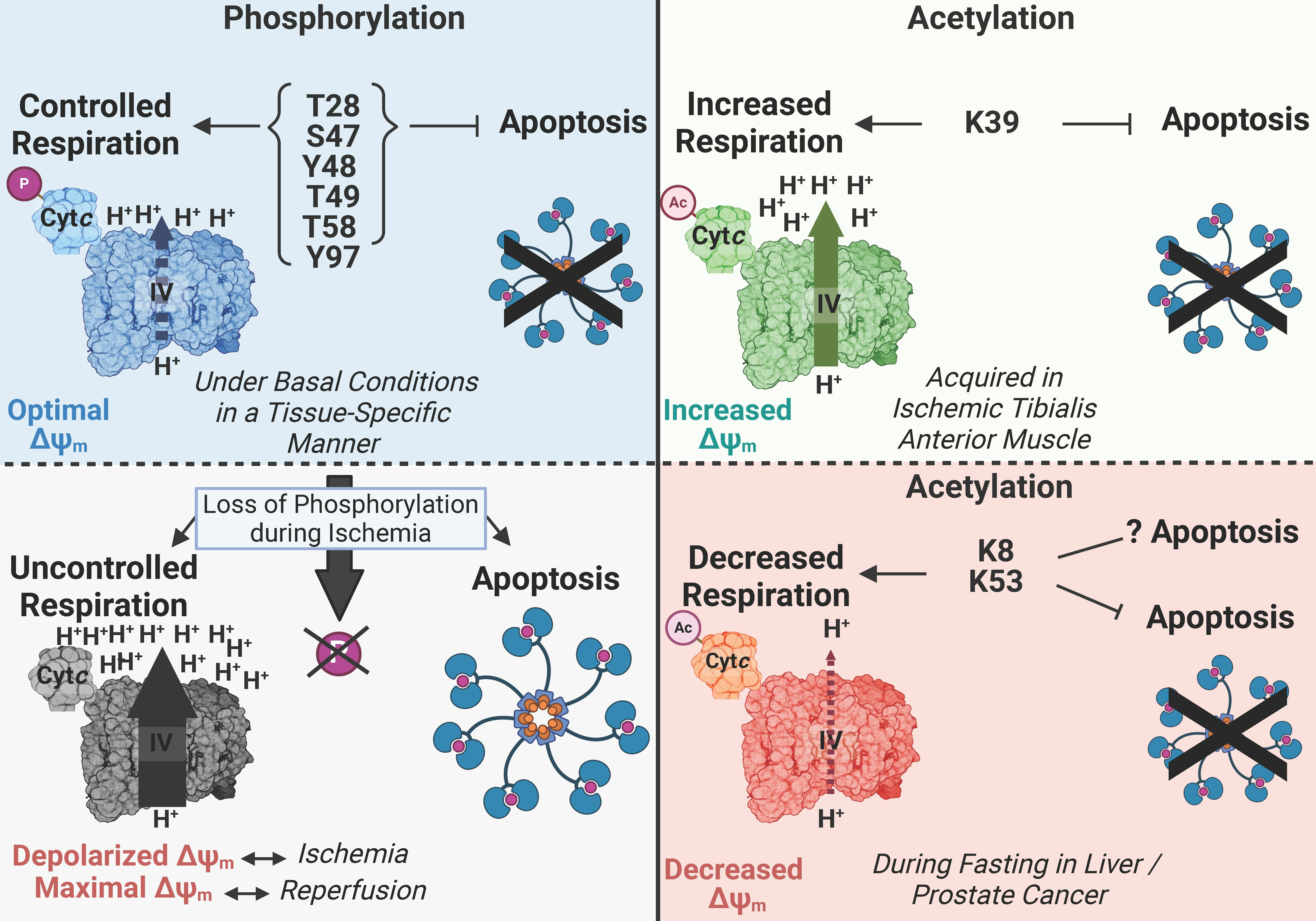
2. Various Functions of Cytc

Cytc is a small, globular, 104 amino acid, evolutionarily optimized protein that carries out a variety of distinct functions. Cytc possesses a covalently attached heme group, which is crucial for carrying out its redox-based functions. The two major functions of Cytc were already introduced above: it operates a single electron carrier from complex III to COX of the mitochondrial ETC and initiates intrinsic apoptosis via binding to Apaf-1 when released into the cytosol. Cytc also supports other crucial, life-sustaining processes (Figure 1). For example, Cytc scavenges and detoxifies ROS including superoxide and hydrogen peroxide [24,25,26] and acts as an electron acceptor of the redox-coupled import pathway of proteins to the IMS via the Erv1-Mia40 pathway [27]. Additionally, a new area of investigation is the role of Cytc in nuclear-mitochondrial crosstalk. In response to DNA damage, Cytc has been shown to translocate to the nucleus, where it promotes both Protein Phosphatase 2A activity via interaction with histone chaperone ANP32A and the release of alternative reading frame protein from nucleophosmin, facilitating DNA damage repair [28,29,30]. However, it still has to be determined if this function of Cytc can rescue cells from apoptosis or only delay it, because binding to Apaf-1 in the cytosol competes with its nuclear function.
Moreover, Cytc is involved in a variety of pro-apoptotic processes beyond its interaction with Apaf-1, and dysfunction in programmed cell death is relevant to numerous disease processes. Cytc is able to produce ROS via p66Shc [31,32], which can damage the cell and trigger cell death when excessive ROS are generated. As an early step during apoptosis, Cytc catalyzes the formation of cardiolipin peroxide [11,33,34,35]. Cardiolipin remodeling is an on-going research topic, which is also related to supercomplex formation, and defects in this process have been implicated in different disease pathologies [36,37,38]. Interestingly, the cardiolipin peroxidase activity of Cytc may be driven by ROS produced during ischemia-reperfusion injury [39].
Overall, the functional diversity of Cytc underscores the need for tight regulatory control, particularly when cellular fate hangs in the balance. Various post-translational modifications of Cytc have been discovered, some of which have been functionally characterized (Figure 2). Some of these modifications, or the loss thereof, result in dysregulation of Cytc and have been associated with specific pathologies. Although other post-translational modifications, such as methylation, nitration, nitrosylation, and homocysteinylation, have been reported, this review focuses on the effects of phosphorylations and acetylations of Cytc that have been identified in vivo, along with any associated pathologies.

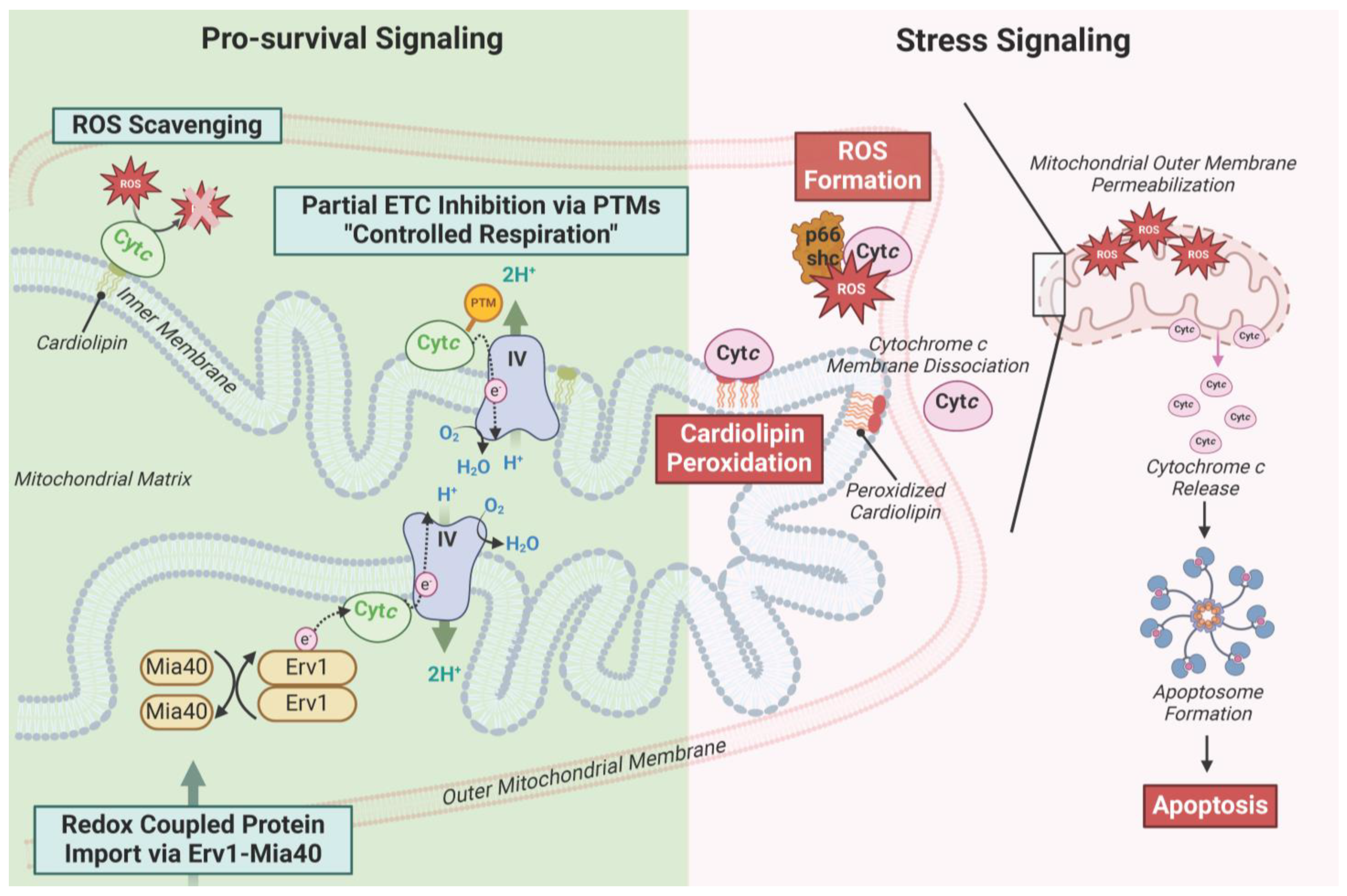
3. Characterized Phosphorylation Sites of Cytc

Phosphorylation of Cytc (Table 1) has been identified on threonine 28 (T28), serine 47 (S47), tyrosine 48 (Y48), threonine 49 (T49), threonine 58 (T58), and tyrosine 97 (Y97). These residues, except for T58, which is replaced with isoleucine in humans, are conserved in mammals. Cytc was not known to be regulated via cell-signaling mechanisms until the discovery of Y97 phosphorylation in 2006, which was mapped in bovine heart [40]. This discovery was made using protein purification protocols that preserve the in vivo phosphorylation state of mitochondrial proteins. Despite this advance, studying post-translational modifications of Cytc remains challenging. Protein purified from animal tissue exists as a mixture of modified and unmodified forms, and depending on the tissue, yields can be low. Therefore, these phosphorylation sites were also functionally characterized using phosphomimetic replacement strategies, most commonly by glutamate replacement, which mimics the negative charge of the phosphate group. Following Y97 phosphorylation, Y48 phosphorylation was identified in bovine liver [41], followed by T28 [42] and T58 [43] in bovine and rat kidney, respectively, S47 phosphorylation in rat and porcine brain [44], and, most recently, T58 in rat heart [45].

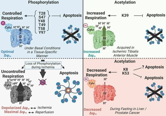
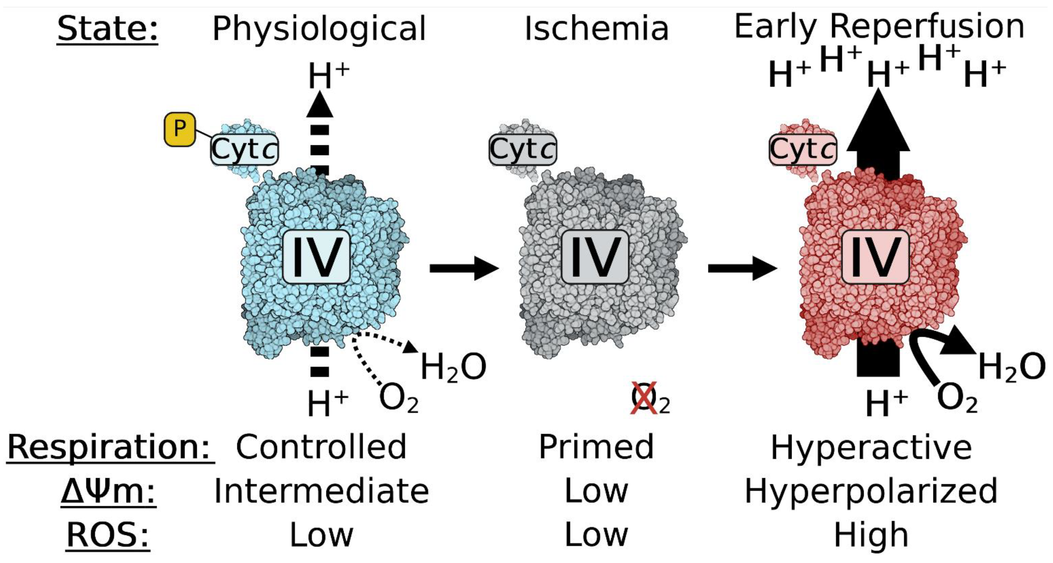
4. Most Phosphorylations of Cytc Are Protective through Partial Inhibition of ETC Flux and Decreased Apoptotic Activity

A general theme for phosphorylation sites identified on Cytc is slowed electron transfer kinetics to COX and, although not universally true, decreased ability to activate downstream caspases. The binding of Cytc, which is a highly basic protein with a pI of 9.6, with both COX and Apaf-1 is largely predicted to be driven by electrostatic interactions, with the positively charged Cytc binding to negatively charged binding pockets on both proteins [2,17,52,53,54,55,56,57,58]. Therefore, it can be expected that phosphorylation, which introduces a negative charge, generally reduces the ability of Cytc to interact with these two key partners.
Reducing ETC flux through phosphorylation of Cytc may at first sight seem disadvantageous. However, similar to batteries that work best at an intermediate charge, the same argument can be made for mitochondria. This is because of the relationship between ETC activity, ΔΨm, and mitochondrial ROS production [22,59,60]; mitochondria with optimal, intermediate ΔΨm levels in the range of about 100–130 mV generate ATP by ATP synthase at close to maximal capacity [61], whereas ROS production is minimal. At membrane potentials exceeding 140 mV, ROS production increases exponentially [62,63,64], which damages the cell and can result in cell death. Therefore, we propose that one major role of Cytc phosphorylation is maintenance of optimal intermediate ΔΨm values, given that these phosphorylation sites tend to partially inhibit ETC activity, thus limiting mitochondrial ROS production to low basal levels. During acute conditions of stress, including ischemia as seen in ischemic stroke and myocardial infarction, these protective phosphorylations are lost in an attempt by the cell to boost ATP production and because kinases cannot operate due to ATP depletion while phosphatases are still operational or even activated due to mitochondrial calcium influx. Since there is no oxygen, the ETC is idling but primed for hyperactivity. During this time, succinate accumulates due to a reversal of complex II [65]. When blood flow is reinstated by mechanical or enzymatic removal of the clot, oxygen reenters the affected tissue. ETC activity resumes at pathological, maximal speed, leading to ΔΨm hyperpolarization and excessive ROS production at complexes I and III, which is further amplified by succinate-driven reverse electron transport to complex I (Figure 3) [65,66,67]. We propose that this sequence mechanistically explains reperfusion injury, which is a significant contributor to cell death on top of that caused initially by ischemia. Therefore, understanding the effect of post-translational modifications of Cytc will provide a mechanistic understanding of pathophysiological processes that occur in a highly tissue-specific manner. Below we discuss in detail the functional effects of identified phosphorylations and acetylations of Cytc.

4.1. Phosphorylation of Threonine 28 (T28)

T28 phosphorylation was mapped in Cytc purified from bovine kidney [42]. For a post-translational modification to be biologically relevant, an appreciable portion of the protein pool must possess the modification. High resolution gel electrophoresis found that up to 83% of the protein pool purified from the entire kidney was phosphorylated. The Vmax of COX in reaction with phosphorylated T28 Cytc was 50% reduced compared to the maximal activity of COX in reaction with non-phosphorylated wild-type (WT) Cytc. Additionally, the Km decreased from 6.3 μM to 4.5 μM. Similar results were obtained using phosphomimetic T28E Cytc, which showed a 73% reduction in Vmax and a decrease in Km from 7.8 μM to 4.7 μM in the reaction with COX [42]. These profound results may be explained by the strategic position of T28, which is part of the negative classical γ turn of Cytc, which stabilizes the protein and is part of the positive epitope that facilitates Cytc binding to its negatively charged binding site on COX [68]. Additionally, during the interaction between Cytc and COX, T28 of Cytc comes into close proximity with D50 and D51 of COX subunit I, which are known to be flexible residues, with D51 proposed to be involved in proton pumping [69,70]. Altogether, this suggests that phosphorylation of T28 perturbs the interaction between Cytc and COX. Another study looked at T28D and found increased electron transfer to COX [46]. However, aspartate is not evolutionarily allowed at the site, while glutamate is allowed in certain species of plants, implying that T28E is the better phosphomimetic since it recapitulates the effects of phosphorylated Cytc [71]. The opposing effects of T28E and T28D on COX activity may be explained based on structural considerations of the Cytc-COX complex in which T28 localizes near K47 of COX subunit II [72]. Based on the charge–charge interaction model, both the T28E and T28D mutations should strengthen the interaction between Cytc and COX, slowing down dissociation and thus the overall reaction. However, considering salt bridge formation, it is possible that electron transfer depends on the length and flexibility of the residue, which would be larger with T28E compared to T28D. With T28D, the complex could be restrained in a conformation allowing faster electron transfer, thereby explaining the opposite effects of the two mutations, despite showing the same charge change. Similar considerations may also apply to S47E and S47D (Figure 4).
The ability to activate downstream caspase-3 activity was unchanged comparing the phosphomimetic T28E to WT. The redox potential of T28E was decreased from 246 mV to 217 mV, which is near the typical reported literature range of 220 mV to 270 mV [73]. Compared to WT, T28E Cytc showed an increased rate of reduction by ascorbate, sometimes considered a stand-in for Cytc superoxide scavenging capability, and no change in rate of oxidation by H2O2. Interestingly, T28E Cytc resisted degradation from high concentrations of H2O2 better than WT, tracked spectrophotometrically via decrease in heme absorbance. T28E also showed slightly reduced cardiolipin peroxidase activity [42].
The effects of T28 phosphorylation were also studied using Cytc double knockout mouse lung fibroblasts, where both the somatic and testes isoforms of Cytc are knocked out. Double knockout is necessary because knockout of just the somatic isoform induces expression of the testes isoform [74]. A construct for T28E was transfected into the double knockout cells. Clones expressing equal levels of WT or phosphomimetic T28E Cytc were selected for further experimentation [42]. Cells expressing phosphomimetic T28E Cytc showed no change in proliferation compared to cells expressing WT after trypan blue staining. It is known that mitochondrial respiration, ΔΨm, and mitochondrial ROS production are linked [62]. This relationship was observed as the cells expressing phosphomimetic T28E showed 60% reduced intact cellular respiration, reduced ΔΨm measured via JC-1 fluorescence, 40% reduced mitochondrial ROS production measured via MitoSOX fluorescence, and 28% reduced ATP levels. Importantly, this demonstrates that modification of a single residue on Cytc by phosphorylation or mutation can control electron flux through the entire ETC. Further supporting the protective role of this modification, cells expressing phosphomimetic T28E Cytc underwent lower levels of cell death after treatment with low levels of H2O2 [42].
The signaling pathway for T28 phosphorylation was also elucidated. In vitro experiments with commercial AMPK and Cytc revealed that AMPK selectively phosphorylated T28. AMPK and Cytc were shown to interact via co-immunoprecipitation and co-localized together in the IMS via submitochondrial fractionation. Chemical activation of AMPK resulted in increased threonine phosphorylation of Cytc, while pharmacological inhibition resulted in loss of threonine phosphorylation of Cytc. Similarly, incubation of kidney tissue homogenates with pharmacological activators or inhibitors of AMPK resulted in increased or decreased respiration, respectively [42]. The kidney is an energetically demanding organ [75], and in contrast to other tissues, the kidney has high basal activity of AMPK and its dysregulation is known to result in kidney disease [76]. Furthermore, pharmacological activation of AMPK has been shown to be an effective treatment in a rat model of kidney ischemia-reperfusion injury [77].
Altogether, these experiments show that T28 phosphorylation is essential for kidney bioenergetic regulation. By partially inhibiting mitochondrial activity under basal conditions, T28 phosphorylation maintains an optimal intermediate ΔΨm and prevents the generation of excessive ROS. When this mechanism is lost, it would facilitate maximal respiration rates and result in increased ROS production [78].

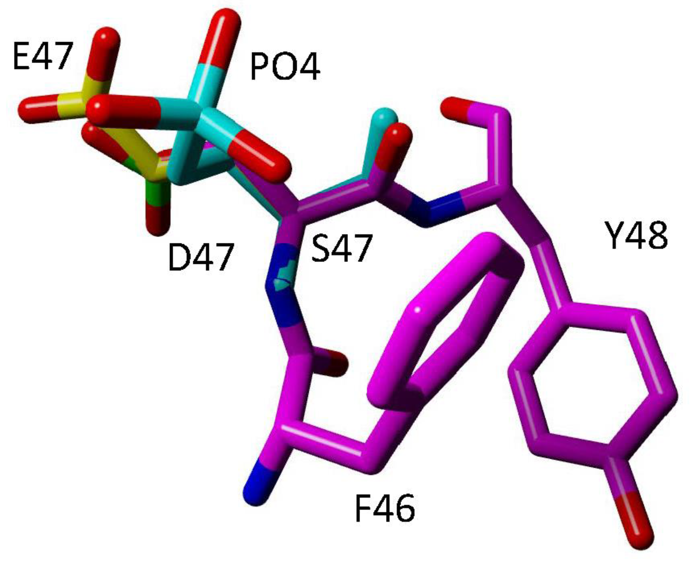
4.2. Phosphorylation of Serine 47 (S47)

S47 phosphorylation was characterized in Cytc purified from both pig and rat brain tissue and characterized [44]. S47 is the only serine residue present in porcine and rat somatic Cytc; however, human Cytc has a second serine residue at S15 [71]. Interestingly, S47 phosphorylation was found to be fully lost in ischemic brain tissue. Phos-tag gel electrophoresis determined that 35% of the pig Cytc pool was phosphorylated, which probably is an underestimation because it takes time to open the skull and remove the brain, a period of time during which the brain is ischemic and likely starts losing the modification. Altered kinetics in reaction with COX were also observed when using S47 phosphorylated protein purified from control brain tissue. The activity of the Cytc-COX reaction was reduced by 48% compared to the dephosphorylated protein purified from ischemic brain tissue. Docking simulations of the Cytc-COX interaction predict that S47 of Cytc would interact with K58 of COX subunit VIIa [52], highlighting the importance of this residue. Looking at the ability to initiate intrinsic apoptosis, S47 phosphorylated Cytc showed 62% decreased caspase-3 activity compared to the dephosphorylated protein [44].
Using glutamate as a phosphomimetic replacement, activity of the Cytc-COX reaction was 54% reduced and downstream caspase-3 activity was 65% reduced when S47E Cytc was used compared to WT. These results are similar to those obtained using the in vivo phosphorylated protein purified from porcine brain, indicating that S47E reproduces functions of in vivo phosphorylation. Other findings using the S47E protein were that it was 33% more resistant to degradation at high concentrations of H2O2 and that the cardiolipin peroxidase activity was decreased [44].
Another study looked at phosphorylation via replacement with aspartate at this site [46]. Using this strategy, caspase-3 and cardiolipin peroxidase activities were decreased whereas Cytc-COX activity was increased, which is opposite to the behavior of in vivo phosphorylated Cytc. In addition, molecular dynamics simulations using the S47E crystal structure (6N10.pdb) demonstrate better spatial similarities between S47E and in silico phosphorylated S47 (Figure 4), implying that glutamate replacement is a better phosphomimetic replacement than aspartate [44].
The Cytc knockout cell culture system was also employed to characterize the phosphomimetic S47E [47]. The cell line expressing phosphomimetic S47E Cytc showed a 52% decrease in basal respiration with a concurrent increased extracellular acidification rate, a stand-in for glycolytic activity. As a result of the decreased ETC activity, cells expressing phosphomimetic S47E Cytc also showed a decreased ΔΨm, a 30% decrease in MitoSOX fluorescence indicating reduced mitochondrial ROS production, and a slight decrease in ATP levels. These experiments again highlight that a single modification or point mutation on Cytc affects electron flux through the entire ETC and is able to induce global changes in overall mitochondrial activity. Using a model of oxygen-glucose deprivation followed by reoxygenation (OGD/R), which simulates ischemia-reperfusion injury in cell culture, the cells expressing WT showed a doubling in mitochondrial ROS production following OGD/R compared to control cells not subjected to OGD/R. In contrast, cells expressing phosphomimetic S47E Cytc were unresponsive to OGD/R and did not show any differences in mitochondrial ROS production. Lastly, in agreement with data suggesting that phosphomimetic S47E replacement reduces the pro-apoptotic abilities of Cytc, the cells expressing phosphomimetic S47E showed less cell death after exposure to H2O2 or OGD/R compared to WT [47].
Akt was identified as the putative kinase mediating S47 phosphorylation. In vitro kinase assays revealed that all three isoforms of Akt were able to phosphorylate Cytc on S47 [47]. Akt, specifically Akt2, is known to translocate to the mitochondrial IMS [79,80,81,82]. Treatment of pig brain homogenates with activators or inhibitors of Akt induced or suppressed the phosphorylation of S47, respectively [47]. Altogether, these data show that S47 phosphorylation acts to maintain a healthy mitochondrial activity level and serves as a protective mechanism in brain tissue. Because this modification is lost during ischemia, and because the loss facilitates high rates of respiration during reperfusion, this causes ΔΨm hyperpolarization and thus mitochondrial ROS production [59], providing a molecular mechanism explaining, at least in part, reperfusion injury seen in brain tissue following stroke treatment [67].

4.3. Phosphorylation of Tyrosine 48 (Y48)

Cytc purified from bovine liver was shown to be phosphorylated on Y48 [41]. The kinetics in reaction with COX were measured using the Y48 phosphorylated protein purified from bovine liver compared to protein dephosphorylated in vitro by shrimp alkaline phosphatase. The Vmax of the Cytc-COX reaction was 55% decreased compared to the dephosphorylated protein, and there was also a slight reduction in the Km from 3.8 μM for dephosphorylated Cytc to 3.0 μM for Y48 phosphorylated Cytc [41]. Using a phosphomimetic glutamate replacement, Y48E Cytc showed a similar change in Vmax, which was 30% decreased compared to WT [48]. Interestingly, phosphomimetic Y48E Cytc showed an increase in Km from 1.1 μM to 3.7 μM. The midpoint redox potential of the phosphomimetic Y48E Cytc was greatly reduced to 192 mV in multiple publications [48,49], which is below the 220 mV to 270 mV range reported in the literature for Cytc [73]. These results suggest significant perturbations to the heme crevice, altering the ability of the protein to carry out efficient electron transfer [49]. Furthermore, this redox potential value is below that of complex III, suggesting that electron transfer from complex III to Cytc may also be impaired [83]. In addition, perturbing Y48 can be expected to disrupt this interaction because Y48 was predicted to be a key residue mediating the interaction between Cytc and complex III [84].
Regarding pro-apoptotic functions, the phosphomimetic Y48E Cytc was unable to activate downstream caspase-9 or caspase-3 activities in multiple studies [48,49,50]. Additionally, the cardiolipin peroxidase activity of phosphomimetic Y48E Cytc was reduced, which can be explained by reduced binding to cardiolipin [48].
Y48 phosphorylation has also been studied using p-carboxymethyl-L-phenylalanine (pCMF) replacement, incorporated via the evolved tRNA synthetase method [85]. Work with the novel phosphomimetic Y48pCMF yielded some different results [50]. While there was agreement regarding decreased downstream caspase-3 activity, Y48pCMF showed increased activity with isolated COX and increased cardiolipin peroxidase activity. Interestingly, although Cytc-COX activity was increased, the overall electron transfer rate through the ETC was decreased, possibly due to the perturbation between the Cytc-complex III interaction.
Y48H is a known disease-causing mutation of Cytc in humans, resulting in mild thrombocytopenia [86]. Y48H resulted in reduced respiration rate and increased apoptotic activity. Another study found that the mutation also caused the heme group to change to a pentacoordinated form, which increased cardiolipin peroxidase activity [87]. Given the crucial functions of Cytc and its high evolutionary conservation, it is surprising that one of the few known disease-causing mutations in humans only causes mild thrombocytopenia. Perhaps only mild mutations of Cytc are compatible with life.

4.4. Phosphorylation of Threonine 49 (T49)

T49 phosphorylation (numbering based on the mature protein, which lacks the start methionine) was reported to increase in aged mouse heart after detection via high-throughput phosphoproteomics [45]. It was characterized using the AC16 cell line, a human cardiomyocyte cell line, which was transfected using lentiviral constructs containing the sequence for phosphomimetic T49E Cytc. Using this system, the authors reported lower levels of cell death and lower levels of caspase-9 and caspase-3 activities. However, the authors did not report knocking out endogenous Cytc in their cell lines prior to transfection with the phosphomimetic T49E Cytc, nor did they use the double Cytc knockout mouse lung fibroblast cell line generated by the Moraes lab [74]. Therefore, the transfected cells likely express an unknown combination of both endogenous Cytc in addition to phosphomimetic T49E Cytc and data interpretation is thus rather limited.

4.5. Phosphorylation of Threonine 58 (T58)

T58 phosphorylation was mapped in Cytc purified from rat kidney [43]. Unlike T28 phosphorylation, which was detected in all five kidney preparations tested [42], T58 phosphorylation was only detected in two of five kidney preparations [43]. T58 is known to be replaced with isoleucine in the testes isoform of Cytc as well as in the single human Cytc gene [71]. Using phosphomimetic T58E Cytc, the kinetics of the Cytc–COX reaction were interrogated. The Vmax was 45% decreased compared to WT while the Km was unchanged. This inhibition of Cytc-COX activity is consistent with that seen with the other Cytc phosphorylations and supports the model that Cytc phosphorylation partially inhibits respiration to maintain optimal mitochondrial functioning [22,59].
Downstream caspase-3 activity of T58E Cytc was 70% decreased compared to WT. The redox potential of T58E was reduced to 209 mV and the ROS scavenging ability of Cytc was also altered; the T58E Cytc demonstrated a 50% reduced rate of oxidation by H2O2 and a 50% increased rate of reduction by ascorbate. The heme group of the T58E Cytc was slightly more resistant to degradation by high H2O2 concentrations compared to WT. Furthermore, the cardiolipin peroxidase activity of T58E Cytc was reduced at high concentrations of cardiolipin. T58 has been reported to be part of the cardiolipin binding site on Cytc, which suggests that the phosphorylation may partially disrupt this interaction [88].
Using the double Cytc knockout system, cells transfected with T58E Cytc showed a similar phenomenon to the other phosphorylations when expressed in cells: 68% decreased respiration rates in intact cells, reduced ΔΨm levels and mitochondrial ROS production, and 66% decreased ATP levels [43]. The cells expressing T58E Cytc also underwent less cell death. Overall, these data establish the role of T58 phosphorylation as an anti-apoptotic modification, supporting the model that Cytc phosphorylation sites maintain optimal mitochondrial functioning to limit ROS generation [22,59]. It still has to be shown in what renal cell type this modification is found and what kinase/phosphatase enzymes regulate this modification.

4.6. Phosphorylation of Tyrosine 97 (Y97)

Y97 was the first phosphorylation site of Cytc to be discovered and characterized. It was initially mapped on Cytc purified from bovine heart [40]. Using the in vivo phosphorylated purified protein, the Km of the Cytc-COX reaction was found to be increased from 2.5 μM of WT to 5.5 μM for the phosphorylated protein whereas the Vmax was unchanged. This alteration of the Cytc-COX kinetics is likely due to a perturbation of the heme group, which was seen on the UV–Vis spectra as the classic 695 nm peak, which assesses heme status via the M80-heme iron coordination, was shifted to 687 nm. Interestingly, another publication using phosphomimetic Y97E found no change of the redox potential and a significant decrease in melting temperature, implying that protein stability may be affected by alteration of this residue [49].
A third publication examined phosphorylation using phosphomimetic Y97pCMF replacement [51]. In contrast with the results obtained using the in vivo Y97 phosphorylated Cytc, Y97pCMF demonstrated increased Cytc-COX activity. The Y97pCMF did demonstrate some reduced pro-apoptotic functionality, with a decrease in downstream caspase-3 activity, although there was no change in cardiolipin binding or cardiolipin peroxidase activity using this phosphomimetic. Recently, a O-sulfotyrosine has become available as a novel mimetic, which should be tested as a model for studying tyrosine phosphorylation [89].
In a different study, Y97 phosphorylation was found to be rapidly induced by neuroprotective insulin treatment after induced ischemia-reperfusion in porcine and rat brains [90]. Induction of Y97 phosphorylation protected the CA-1 hippocampal neurons from cell death following the ischemia-reperfusion injury, supporting the anti-apoptotic role of Y97 phosphorylation seen using the phosphomimetic. Additionally, less Cytc was released after 24 h post-ischemia when Y97 phosphorylation was induced by the insulin treatment, suggesting that phosphorylation may be able to prevent Cytc release into the cytosol.

5. Characterized Acetylation Sites of Cytc

In vitro lysine acetylation of Cytc has been long studied. However, detection of lysine acetylation of Cytc in vivo has only occurred recently and is a new area of research (Table 2). The first acetylation site discovered was lysine 8 (K8) in fasted mouse liver mitochondria via high-throughput acetyl-omics [91]. Later, lysine 53 (K53) was identified in prostate cancer [92]. This was followed by lysine 39 (K39) in ischemic skeletal muscle [93].
Lysine acetylation is the most abundant post-translational modification in mitochondria [94,95]. Lysine acetylation is believed to occur largely non-enzymatically in the mitochondrial matrix, due to the basic pH and high concentration of acetyl-CoA [96]. However, lysine acetylation of Cytc, which resides in the IMS, was found to occur selectively on specific residues tied to a particular tissue or pathology. This suggests that specific acetyltransferase enzymes mediate these modifications. Unlike phosphorylations of Cytc, which are present under basal conditions and are generally lost during ischemia, these acetylations tend to be associated with a specific pathophysiological state. Lysine acetylations suffer from the same methodological problems as phosphorylations: yields are typically low and the modification itself is transient. Therefore, it is common to study acetylation using glutamine as an acetylmimetic. In contrast to the positively charged side chain of lysine, acetyl-lysine and glutamine both have uncharged, polar side chains that contain amide groups [97]. Lysine acetylation of Cytc, which leads to the loss of the positive charge of the ε-amine group, is generally predicted to interfere with binding to COX and Apaf-1, due to making Cytc less positively charged. In this manner, acetylation, which removes a positive charge from a lysine residue, is similar to phosphorylation, which introduces a negative charge on a neutral S/T/Y residue, because both of them result in Cytc becoming more negatively charged.
Table 2. Summary of functionally characterized acetylation sites of Cytc.
ResidueTissue of OriginExperimental ModelsFindings
Lysine 8Fasted Mouse LiverRecombinant acetylmimetic K8Q CytcDecreased Cytc-COX activity, decreased KD to cytochrome c1 [98]
Lysine 39Ischemic Porcine Tibialis Anterior MuscleIn vivo acetylated Cytc purified from ischemic porcine muscleIncreased Cytc-COX Vmax, decreased caspase-3 activity [93]
Recombinant acetylmimetic K39Q CytcIncreased Cytc-COX Vmax, decreased caspase-3 activity, increased rate of oxidation, decreased rate of reduction, decreased cardiolipin peroxidase activity [93]
Cytc double knockout mouse lung fibroblasts expressing K39Q CytcIncreased respiration, increased ΔΨm, increased mitochondrial ROS production, increased ATP levels, decreased cell death, reduced responsiveness to oxygen-glucose deprivation followed by reoxygenation [93]
Lysine 53Human Prostate CancerRecombinant acetylmimetic K53Q CytcDecreased Cytc-COX Vmax, decreased caspase-3 activity, increased rate of oxidation, increased rate of reduction, increased heme degradation, decreased cardiolipin peroxidase activity [92,98]

5.1. Acetylation of Lysine 8 (K8)

K8 acetylation was discovered in a high-throughput study of fasted mouse liver [91]. This residue is known to interact with cytochrome c1 of Complex III [99,100], and it was found that acetylmimetic K8Q Cytc had a perturbed dissociation constant from cytochrome c1 [98]. K8 of Cytc is also known to interact with D139 of COX subunit II [72] as part of the proximal Cytc-COX binding site, explaining the observed decrease in electron transfer to COX using acetylmimetic K8Q protein [98]. While this residue seems crucial for the electron transfer activity of Cytc, not much is known regarding the effects of this modification on the other functions of Cytc.

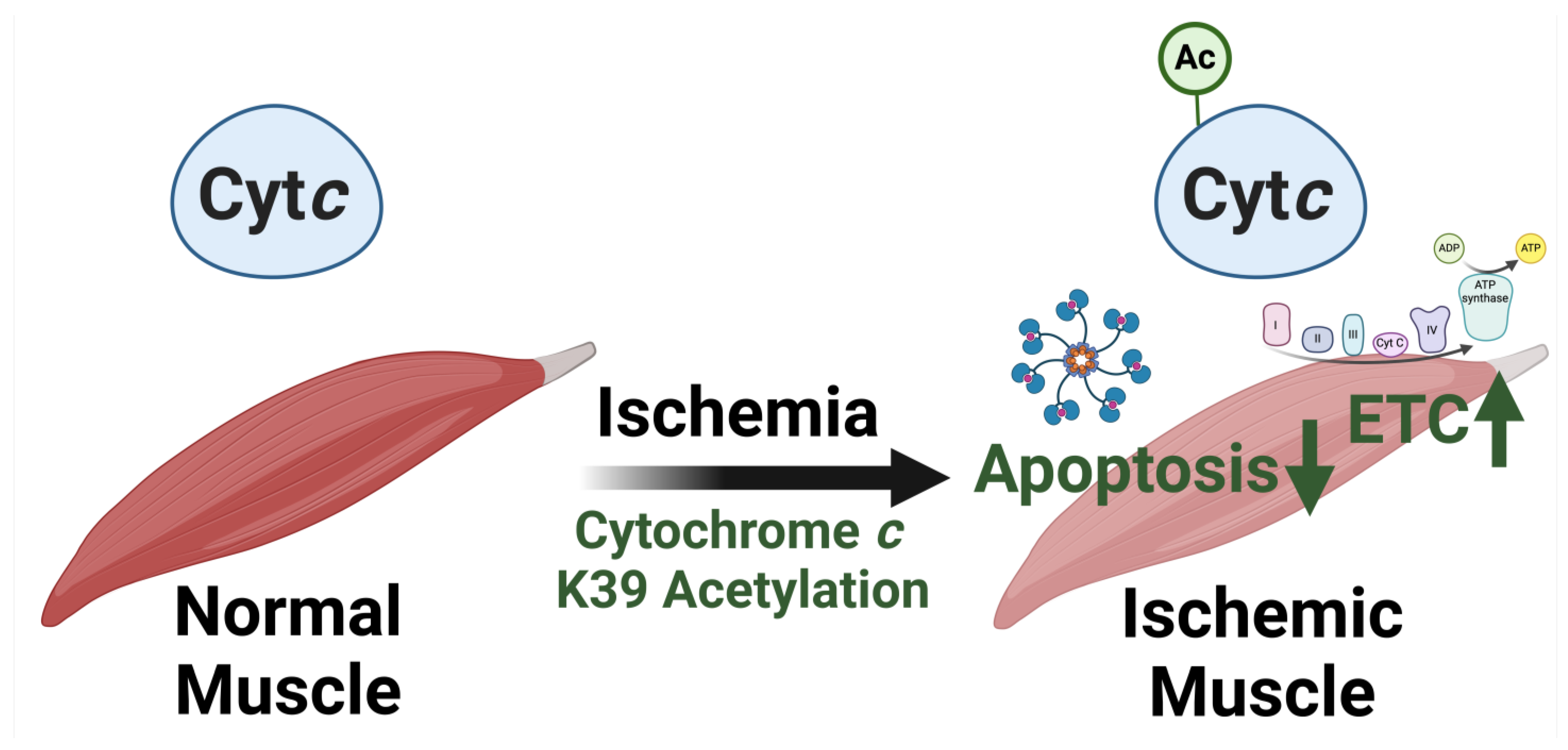
5.2. Acetylation of Lysine 39 (K39)

K39 acetylation was mapped in Cytc purified from ischemic porcine tibialis anterior muscle after 45 min of ischemia [93]. The modification was not found in the muscle under basal conditions, making it the first reported gain of a post-translational modification during ischemia, suggesting that the response to ischemia may be muscle-specific. It is known that muscles tend to be more resilient to ischemia than other tissues such as brain, heart, kidneys, or liver [101,102]. K39 acetylation may provide a unique mechanism to potentially explain this resilience. Whereas other phosphorylations and acetylations of Cytc tend to be present under basal conditions, lost during ischemia, and inhibitory to the Cytc-COX reaction, K39 acetylation of tibialis anterior muscle follows a unique metabolic and apoptotic strategy by stimulating respiration to meet the cell’s energy demand and at the same time inhibiting apoptosis (Figure 5). K39 acetylation was found to be removed by sirtuin5, which is known to co-localize in the IMS with Cytc [103,104]. Sirtuins are known to be downregulated in response to hypoxia, suggesting that preventing the removal of this acetylation during ischemia facilitates the protective effects [105]. The in vivo acetylated Cytc purified from ischemic tibialis anterior muscle showed a 58% increase in Cytc-COX Vmax, compared to the unacetylated Cytc purified from non-ischemic tibialis anterior muscle [93]. Acetylmimetic K39Q Cytc produced a similar increase of 38% compared to WT. Nuclear magnetic resonance studies demonstrated perturbation of residues interacting with or near the heme group such as the M80-containing loop and the K55-to-W59 stretch, possibly explaining the increased electron transfer rate by facilitating more efficient electron transfer. Interestingly, a separate publication studying K39L did not observe any changes in Cytc-COX kinetics [106].
Additionally, this acetylation, which was triggered as a response to ischemia, blocks the pro-apoptotic functions of Cytc, thereby acting as a protective mechanism during ischemia-reperfusion. Downstream caspase-3 activity using the in vivo acetylated protein purified from ischemic tibialis anterior muscle was 45% decreased, and the K39Q Cytc showed a reduction of 90% in the same assay [93]. K39 of Cytc interacts with the carbonyl carbon of F1063 of Apaf-1 [17,56], and the acetylation likely perturbs this interaction, decreasing downstream caspase-3 activity. Furthermore, cardiolipin peroxidase activity, another proapoptotic action, was decreased with the acetylmimetic K39Q Cytc.
Interestingly, acetylmimetic K39Q Cytc had a decreased rate of reduction by superoxide and an increased rate of oxidation by H2O2 compared to WT [93]. This may be because superoxide is a known signaling molecule produced during muscle contraction [107,108,109]. Therefore, reduced scavenging of superoxide could be an adaptative response to short periods of hypoxia that occur during normal muscle exercise, which becomes mal-adaptive in the context of ischemia-reperfusion injury. The crystal structure of the in vivo K39 acetylated Cytc purified from ischemic tibialis anterior was solved at 1.5 Å and had an occupancy of 0.75 for the acetyl group, showing that a large proportion of the protein pool was acetylated following ischemia [93].
Similar to the results for the Cytc-COX kinetics using purified proteins, cells expressing the acetylmimetic K39Q Cytc showed an increase in respiration with a 70% increase in basal oxygen consumption rate. Accordingly, the basal ATP levels were increased 51% over WT. ΔΨm and mitochondrial ROS production also experienced increases compared to cells expressing WT. These experiments demonstrate that single point mutations on Cytc can both inhibit and stimulate overall mitochondrial activity, supporting the concept that the Cytc-COX interaction is the rate-limiting step of the ETC. After exposure to OGD/R, cells expressing K39Q Cytc did not show a statistically significant increase in mitochondrial ROS production, unlike WT. These data show that K39Q Cytc increases respiration, ΔΨm, and ROS under baseline conditions compared to WT, but does not lead to a further increase following OGD/R, potentially protecting the cells from additional oxidative stress upon reperfusion. After exposure to H2O2, OGD/R, and thapsigargin treatment, cells expressing K39Q Cytc experienced reduced levels of cell death. Using cells expressing K39Q Cytc, reduced levels of Cytc were co-immunoprecipitated with Apaf-1, suggesting impaired interaction of the two proteins. Furthermore, this translated into reduced cleavage of procaspase-9 and procaspase-3 after induction of apoptosis in the presence of K39Q Cytc. Overall, skeletal muscle is a unique tissue type with drastic changes in perfusion depending on usage [110,111]. Cytc K39 acetylation may be a skeletal muscles-specific adaptation during exercise or other short-term energy deficiencies, meant to allow the muscle to meet the high energy demand while protecting from cell death (Figure 5) [93].

5.3. Acetylation of Lysine 53 (K53)

Lastly, we will discuss K53 acetylation, which was mapped in Cytc purified from human prostate cancer xenografts and prostate cancer specimens but was not found in normal prostate tissue [92]. Overall, this modification was found to be advantageous to cancer cells by driving two hallmarks of cancer: a metabolic switch from OxPhos to glycolysis and apoptosis evasion. It is known that acetyltransferase enzymes are upregulated in prostate cancer, which may help explain increased K53 acetylation [112,113]. Acetylmimetic K53Q Cytc showed 35% decreased Vmax for the Cytc-COX reaction. K53 of Cytc is within 5 Å of K58 residue of COX subunit VIIa [52], suggesting that K53 is within the Cytc-COX binding domain and acetylation may spatially interfere with the interaction. This inhibition of respiration would assist the prostate cancer cells in metabolic reprogramming toward the glycolytic pathway.
Additionally, cancer cells often manage to evade apoptosis, which K53 acetylation facilitates. Downstream caspase-3 activity was 80% decreased using acetylmimetic K53Q Cytc and may be explained because K53 is a known regulatory epitope for Apaf-1 binding [17]. The ability of acetylmimetic K53Q Cytc to scavenge ROS was also significantly increased, with large increases in rate of oxidation by H2O2 and rate of reduction by superoxide, compared to WT. These may further facilitate prostate cancer cell survival by detoxifying ROS, blocking a strong pro-apoptotic signal. The pro-apoptotic cardiolipin peroxidase activity was also greatly reduced with acetylmimetic K53Q Cytc. K53 is conserved in all mammals, further pointing to an important regulatory role of K53 for the interactions with both COX and Apaf-1 [71]. Future work should expand these studies to other cancers to see if this modification is more broadly involved in cancer pathology and potentially in resistance to cancer therapies relying on apoptosis induction.

6. Conclusions

The tissue and disease specific regulation of Cytc has interesting implications. It has been long debated which step of the ETC is rate-limiting. We have previously proposed that the interaction between Cytc and COX is rate-limiting [22]. Cytc and COX are both highly regulated via allosteric ATP binding, post-translational modifications, and tissue-specific isoforms, and they are the only known mammalian components of the OxPhos that are regulated by all three mechanisms. Cytc acts at the intersection point between pro-survival respiration and pro-death apoptosis, which depends on the subcellular localization of the protein, and, therefore, lends itself as an ideal regulatory target. Several reports employing traditional metabolic flux analysis experiments have identified COX as the rate-limiting step of the ETC [114,115,116,117,118]. For these experiments, an inhibitor of a specific complex is added in increasing amounts, and total flux through the ETC is measured, usually via oxygen consumption of COX as the terminal step of the ETC. The less inhibitors necessary to affect electron flux globally through the entire ETC, the more rate-limiting a particular step is. However, the possibility that Cytc and its post-translational modifications are rate-limiting was overlooked in the past, perhaps due to the lack of a specific inhibitor of Cytc.
Cytc, which is crucial for physiological mitochondria functioning, has been shown to possess tissue-specific post-translational modifications that react to ischemia-reperfusion injury. Patterns within these post-translational modifications of Cytc help explain different tissue sensitivities to ischemia-reperfusion injury. Specifically, various phosphorylations of Cytc, which are present under basal conditions and lost during ischemia, sensitize the tissue to ischemia-reperfusion injury, while K39 acetylation of Cytc in skeletal muscle, which is not present under basal conditions and is gained during ischemia, protects skeletal muscle and contributes to the resilience of this tissue type to ischemia-reperfusion injury. In this review, we have discussed that single post-translational modifications and point mutations of Cytc are able to affect flux through the entire electron transport chain. When cells expressed T28E, S47E, T58E, or K39Q Cytc, oxygen consumption rate by the cells was altered compared to cells expressing WT. While the phosphorylations of Cytc and most acetylations are inhibitory to Cytc-COX kinetics, K39 acetylation of Cytc is stimulatory. This highlights that a change in Cytc-COX activity, whether inhibitory or stimulatory, can decrease or increase total ETC flux, respectively.
This review has focused primarily on phosphorylations and acetylations of Cytc, specifically those which have been detected in vivo in mammals and for which clear evidence exists that they are functionally important. Other post-translational modifications of Cytc exist (reviewed in [23]). However, they were largely detected through high-throughput studies or are only found in vitro, therefore their biological significance is unknown. In most but not all cases, the eight modifications discussed here serve to support healthy cell functioning by blocking pro-apoptotic functions of Cytc and by lowering mitochondrial activity to minimize ROS production. Furthermore, it has long been observed that different organs and even different tissue types within an organ have different sensitivities to ischemia and reperfusion injury [119]. For the phosphorylations, which are present in their respective tissues under basal conditions, their loss tends to drive ischemia-reperfusion injury by stimulating respiration, increasing ROS production, and sensitizing the cell to apoptosis. Overall, the phosphorylations occur in ischemia sensitive tissues, such as brain, heart, kidney, and liver, which may explain the susceptibility of these tissues to ischemia-reperfusion injury. K39 acetylation follows a different pattern, where acetylation is a response to ischemia, stimulating respiration and blocking apoptosis to enforce maximum muscle activity output. The presence of this acetylation in skeletal muscle, which is notoriously less sensitive to ischemic stress, results in skeletal muscle protection and resilience to ischemia-reperfusion injury. Altogether, the diversity of Cytc regulation by post-translational modifications underscores the crucial nature of the protein for both the ETC and the cell as a whole.

Author Contributions

Conceptualization, P.T.M. and M.H.; writing, review, and editing, P.T.M. wrote the manuscript with support of all authors; visualization, T.A. and P.T.M.; resources, funding acquisition, and supervision, M.H. All authors reviewed and edited the manuscript. All authors have read and agreed to the published version of the manuscript.

Funding

This work and some of the work discussed in this article was supported by the Office of the Assistant Secretary of Defense for Health Affairs through the Prostate Cancer Research Program under Award No. HT9425-24-1-0073, NIH grant R01 NS120322, NIH grant U44 NS125160, the Michigan Prostate SPORE Developmental Research Program, NSF grant MCB-2329629, and the Michigan Translational Research and Commercialization (MTRAC) Award. Opinions, interpretations, conclusions, and recommendations are those of the authors and are not necessarily endorsed by the funding agencies including the Department of Defense, the National Institutes of Health, or the National Science Foundation.

Institutional Review Board Statement

Not applicable.

Informed Consent Statement

Not applicable.

Data Availability Statement

Not applicable.

Acknowledgments

We thank the four reviewers for their valuable comments.

Conflicts of Interest

The authors declare no conflicts of interest.

Abbreviations

Apaf-1apoptosis protease-activating factor-1
Cytccytochrome c
COXcytochrome c oxidase (complex IV)
ETCelectron transport chain
IMSintermembrane space
ΔΨmmitochondrial membrane potential
K8lysine 8 (of Cytc)
K39lysine 39 (of Cytc)
K53lysine 53 (of Cytc)
OGD/Roxygen-glucose deprivation followed by reoxygenation
OxPhosoxidative phosphorylation
pCMFp-carboxymethyl-L-phenylalanine
ROSreactive oxygen species
S47serine 47 (of Cytc)
TMPDtetramethyl-p-phenylenediamine
T28threonine 28 (of Cytc)
T49threonine 49 (of Cytc)
T58threonine 58 (of Cytc)
WTwild-type (Cytc)
Y48tyrosine 48 (of Cytc)
Y97tyrosine 97 (of Cytc)

References

  1. Rich, P. Chemiosmotic coupling: The cost of living. Nature 2003, 421, 583. [Google Scholar] [CrossRef]
  2. Moe, A.; Di Trani, J.; Rubinstein, J.L.; Brzezinski, P. Cryo-EM structure and kinetics reveal electron transfer by 2D diffusion of cytochrome c in the yeast III-IV respiratory supercomplex. Proc. Natl. Acad. Sci. USA 2021, 118, e2021157118. [Google Scholar] [CrossRef]
  3. Perez-Mejias, G.; Guerra-Castellano, A.; Diaz-Quintana, A.; De la Rosa, M.A.; Diaz-Moreno, I. Cytochrome c: Surfing Off of the Mitochondrial Membrane on the Tops of Complexes III and IV. Comput. Struct. Biotechnol. J. 2019, 17, 654–660. [Google Scholar] [CrossRef]
  4. Zorova, L.D.; Popkov, V.A.; Plotnikov, E.Y.; Silachev, D.N.; Pevzner, I.B.; Jankauskas, S.S.; Babenko, V.A.; Zorov, S.D.; Balakireva, A.V.; Juhaszova, M.; et al. Mitochondrial membrane potential. Anal. Biochem. 2018, 552, 50–59. [Google Scholar] [CrossRef] [PubMed]
  5. Benard, G.; Bellance, N.; Jose, C.; Melser, S.; Nouette-Gaulain, K.; Rossignol, R. Multi-site control and regulation of mitochondrial energy production. Biochim. Biophys. Acta 2010, 1797, 698–709. [Google Scholar] [CrossRef]
  6. Cogliati, S.; Lorenzi, I.; Rigoni, G.; Caicci, F.; Soriano, M.E. Regulation of Mitochondrial Electron Transport Chain Assembly. J. Mol. Biol. 2018, 430, 4849–4873. [Google Scholar] [CrossRef]
  7. Hüttemann, M.; Lee, I.; Samavati, L.; Yu, H.; Doan, J.W. Regulation of mitochondrial oxidative phosphorylation through cell signaling. Biochim. Biophys. Acta (BBA)—Mol. Cell Res. 2007, 1773, 1701–1720. [Google Scholar] [CrossRef] [PubMed]
  8. Pieczenik, S.R.; Neustadt, J. Mitochondrial dysfunction and molecular pathways of disease. Exp. Mol. Pathol. 2007, 83, 84–92. [Google Scholar] [CrossRef] [PubMed]
  9. Hüttemann, M.; Lee, I.; Pecinova, A.; Pecina, P.; Przyklenk, K.; Doan, J.W. Regulation of oxidative phosphorylation, the mitochondrial membrane potential, and their role in human disease. J. Bioenerg. Biomembr. 2008, 40, 445–456. [Google Scholar] [CrossRef]
  10. Ott, M.; Robertson, J.D.; Gogvadze, V.; Zhivotovsky, B.; Orrenius, S. Cytochrome c release from mitochondria proceeds by a two-step process. Proc. Natl. Acad. Sci. USA 2002, 99, 1259–1263. [Google Scholar] [CrossRef]
  11. Kagan, V.E.; Bayir, H.A.; Belikova, N.A.; Kapralov, O.; Tyurina, Y.Y.; Tyurin, V.A.; Jiang, J.; Stoyanovsky, D.A.; Wipf, P.; Kochanek, P.M.; et al. Cytochrome c/cardiolipin relations in mitochondria: A kiss of death. Free Radic. Biol. Med. 2009, 46, 1439–1453. [Google Scholar] [CrossRef] [PubMed]
  12. Scorrano, L.; Ashiya, M.; Buttle, K.; Weiler, S.; Oakes, S.A.; Mannella, C.A.; Korsmeyer, S.J. A distinct pathway remodels mitochondrial cristae and mobilizes cytochrome c during apoptosis. Dev. Cell 2002, 2, 55–67. [Google Scholar] [CrossRef]
  13. Petronilli, V.; Nicolli, A.; Costantini, P.; Colonna, R.; Bernardi, P. Regulation of the permeability transition pore, a voltage-dependent mitochondrial channel inhibited by cyclosporin A. Biochim. Biophys. Acta 1994, 1187, 255–259. [Google Scholar] [CrossRef] [PubMed]
  14. Petronilli, V.; Penzo, D.; Scorrano, L.; Bernardi, P.; Di Lisa, F. The mitochondrial permeability transition, release of cytochrome c and cell death. Correlation with the duration of pore openings in situ. J. Biol. Chem. 2001, 276, 12030–12034. [Google Scholar] [CrossRef]
  15. Bernardi, P.; Azzone, G.F. Cytochrome c as an electron shuttle between the outer and inner mitochondrial membranes. J. Biol. Chem. 1981, 256, 7187–7192. [Google Scholar] [CrossRef] [PubMed]
  16. Ripple, M.O.; Abajian, M.; Springett, R. Cytochrome c is rapidly reduced in the cytosol after mitochondrial outer membrane permeabilization. Apoptosis 2010, 15, 563–573. [Google Scholar] [CrossRef]
  17. Cheng, T.C.; Hong, C.; Akey, I.V.; Yuan, S.; Akey, C.W. A near atomic structure of the active human apoptosome. Elife 2016, 5, e17755. [Google Scholar] [CrossRef]
  18. Brown, G.C.; Borutaite, V. Regulation of apoptosis by the redox state of cytochrome c. Biochim. Biophys. Acta 2008, 1777, 877–881. [Google Scholar] [CrossRef]
  19. Skemiene, K.; Rakauskaite, G.; Trumbeckaite, S.; Liobikas, J.; Brown, G.C.; Borutaite, V. Anthocyanins block ischemia-induced apoptosis in the perfused heart and support mitochondrial respiration potentially by reducing cytosolic cytochrome c. Int. J. Biochem. Cell Biol. 2013, 45, 23–29. [Google Scholar] [CrossRef]
  20. Liobikas, J.; Skemiene, K.; Trumbeckaite, S.; Borutaite, V. Anthocyanins in cardioprotection: A path through mitochondria. Pharmacol. Res. 2016, 113, 808–815. [Google Scholar] [CrossRef]
  21. Kalkavan, H.; Chen, M.J.; Crawford, J.C.; Quarato, G.; Fitzgerald, P.; Tait, S.W.G.; Goding, C.R.; Green, D.R. Sublethal cytochrome c release generates drug-tolerant persister cells. Cell 2022, 185, 3356–3374.e22. [Google Scholar] [CrossRef]
  22. Kalpage, H.A.; Wan, J.; Morse, P.T.; Zurek, M.P.; Turner, A.A.; Khobeir, A.; Yazdi, N.; Hakim, L.; Liu, J.; Vaishnav, A.; et al. Cytochrome c phosphorylation: Control of mitochondrial electron transport chain flux and apoptosis. Int. J. Biochem. Cell Biol. 2020, 121, 105704. [Google Scholar] [CrossRef]
  23. Guerra-Castellano, A.; Marquez, I.; Perez-Mejias, G.; Diaz-Quintana, A.; De la Rosa, M.A.; Diaz-Moreno, I. Post-Translational Modifications of Cytochrome c in Cell Life and Disease. Int. J. Mol. Sci. 2020, 21, 8483. [Google Scholar] [CrossRef] [PubMed]
  24. Pasdois, P.; Parker, J.E.; Griffiths, E.J.; Halestrap, A.P. The role of oxidized cytochrome c in regulating mitochondrial reactive oxygen species production and its perturbation in ischaemia. Biochem. J. 2011, 436, 493–505. [Google Scholar] [CrossRef] [PubMed]
  25. Wang, Z.B.; Li, M.; Zhao, Y.; Xu, J.X. Cytochrome c is a hydrogen peroxide scavenger in mitochondria. Protein Pept. Lett. 2003, 10, 247–253. [Google Scholar] [CrossRef] [PubMed]
  26. Pereverzev, M.O.; Vygodina, T.V.; Konstantinov, A.A.; Skulachev, V.P. Cytochrome c, an ideal antioxidant. Biochem. Soc. Trans. 2003, 31, 1312–1315. [Google Scholar] [CrossRef]
  27. Allen, S.; Balabanidou, V.; Sideris, D.P.; Lisowsky, T.; Tokatlidis, K. Erv1 mediates the Mia40-dependent protein import pathway and provides a functional link to the respiratory chain by shuttling electrons to cytochrome c. J. Mol. Biol. 2005, 353, 937–944. [Google Scholar] [CrossRef]
  28. Gonzalez-Arzola, K.; Diaz-Quintana, A.; Bernardo-Garcia, N.; Martinez-Fabregas, J.; Rivero-Rodriguez, F.; Casado-Combreras, M.A.; Elena-Real, C.A.; Velazquez-Cruz, A.; Gil-Caballero, S.; Velazquez-Campoy, A.; et al. Nucleus-translocated mitochondrial cytochrome c liberates nucleophosmin-sequestered ARF tumor suppressor by changing nucleolar liquid-liquid phase separation. Nat. Struct. Mol. Biol. 2022, 29, 1024–1036. [Google Scholar] [CrossRef]
  29. Rivero-Rodriguez, F.; Diaz-Quintana, A.; Velazquez-Cruz, A.; Gonzalez-Arzola, K.; Gavilan, M.P.; Velazquez-Campoy, A.; Rios, R.M.; De la Rosa, M.A.; Diaz-Moreno, I. Inhibition of the PP2A activity by the histone chaperone ANP32B is long-range allosterically regulated by respiratory cytochrome c. Redox Biol. 2021, 43, 101967. [Google Scholar] [CrossRef]
  30. Gonzalez-Arzola, K.; Diaz-Moreno, I.; Cano-Gonzalez, A.; Diaz-Quintana, A.; Velazquez-Campoy, A.; Moreno-Beltran, B.; Lopez-Rivas, A.; De la Rosa, M.A. Structural basis for inhibition of the histone chaperone activity of SET/TAF-Ibeta by cytochrome c. Proc. Natl. Acad. Sci. USA 2015, 112, 9908–9913. [Google Scholar] [CrossRef]
  31. Giorgio, M.; Migliaccio, E.; Orsini, F.; Paolucci, D.; Moroni, M.; Contursi, C.; Pelliccia, G.; Luzi, L.; Minucci, S.; Marcaccio, M.; et al. Electron transfer between cytochrome c and p66Shc generates reactive oxygen species that trigger mitochondrial apoptosis. Cell 2005, 122, 221–233. [Google Scholar] [CrossRef]
  32. Di Lisa, F.; Giorgio, M.; Ferdinandy, P.; Schulz, R. New aspects of p66Shc in ischaemia reperfusion injury and other cardiovascular diseases. Br. J. Pharmacol. 2017, 174, 1690–1703. [Google Scholar] [CrossRef] [PubMed]
  33. Steele, H.B.B.; Elmer-Dixon, M.M.; Rogan, J.T.; Ross, J.B.A.; Bowler, B.E. The Human Cytochrome c Domain-Swapped Dimer Facilitates Tight Regulation of Intrinsic Apoptosis. Biochemistry 2020, 59, 2055–2068. [Google Scholar] [CrossRef]
  34. Diaz-Quintana, A.; Perez-Mejias, G.; Guerra-Castellano, A.; De la Rosa, M.A.; Diaz-Moreno, I. Wheel and Deal in the Mitochondrial Inner Membranes: The Tale of Cytochrome c and Cardiolipin. Oxid. Med. Cell Longev. 2020, 2020, 6813405. [Google Scholar] [CrossRef]
  35. Kagan, V.E.; Tyurin, V.A.; Jiang, J.; Tyurina, Y.Y.; Ritov, V.B.; Amoscato, A.A.; Osipov, A.N.; Belikova, N.A.; Kapralov, A.A.; Kini, V.; et al. Cytochrome c acts as a cardiolipin oxygenase required for release of proapoptotic factors. Nat. Chem. Biol. 2005, 1, 223–232. [Google Scholar] [CrossRef]
  36. Xu, Y.; Anjaneyulu, M.; Donelian, A.; Yu, W.; Greenberg, M.L.; Ren, M.; Owusu-Ansah, E.; Schlame, M. Assembly of the complexes of oxidative phosphorylation triggers the remodeling of cardiolipin. Proc. Natl. Acad. Sci. USA 2019, 116, 11235–11240. [Google Scholar] [CrossRef]
  37. Montero, J.; Mari, M.; Colell, A.; Morales, A.; Basanez, G.; Garcia-Ruiz, C.; Fernandez-Checa, J.C. Cholesterol and peroxidized cardiolipin in mitochondrial membrane properties, permeabilization and cell death. Biochim. Biophys. Acta 2010, 1797, 1217–1224. [Google Scholar] [CrossRef]
  38. Paradies, G.; Petrosillo, G.; Paradies, V.; Ruggiero, F.M. Oxidative stress, mitochondrial bioenergetics, and cardiolipin in aging. Free Radic. Biol. Med. 2010, 48, 1286–1295. [Google Scholar] [CrossRef] [PubMed]
  39. Aluri, H.S.; Simpson, D.C.; Allegood, J.C.; Hu, Y.; Szczepanek, K.; Gronert, S.; Chen, Q.; Lesnefsky, E.J. Electron flow into cytochrome c coupled with reactive oxygen species from the electron transport chain converts cytochrome c to a cardiolipin peroxidase: Role during ischemia-reperfusion. Biochim. Biophys. Acta 2014, 1840, 3199–3207. [Google Scholar] [CrossRef]
  40. Lee, I.; Salomon, A.R.; Yu, K.; Doan, J.W.; Grossman, L.I.; Hüttemann, M. New prospects for an old enzyme: Mammalian cytochrome c is tyrosine-phosphorylated in vivo. Biochemistry 2006, 45, 9121–9128. [Google Scholar] [CrossRef]
  41. Yu, H.; Lee, I.; Salomon, A.R.; Yu, K.; Hüttemann, M. Mammalian liver cytochrome c is tyrosine-48 phosphorylated in vivo, inhibiting mitochondrial respiration. Biochim. Biophys. Acta 2008, 1777, 1066–1071. [Google Scholar] [CrossRef] [PubMed]
  42. Mahapatra, G.; Varughese, A.; Ji, Q.; Lee, I.; Liu, J.; Vaishnav, A.; Sinkler, C.; Kapralov, A.A.; Moraes, C.T.; Sanderson, T.H.; et al. Phosphorylation of Cytochrome c Threonine 28 Regulates Electron Transport Chain Activity in Kidney: IMPLICATIONS FOR AMP KINASE. J. Biol. Chem. 2017, 292, 64–79. [Google Scholar] [CrossRef]
  43. Wan, J.; Kalpage, H.A.; Vaishnav, A.; Liu, J.; Lee, I.; Mahapatra, G.; Turner, A.A.; Zurek, M.P.; Ji, Q.; Moraes, C.T.; et al. Regulation of Respiration and Apoptosis by Cytochrome c Threonine 58 Phosphorylation. Sci. Rep. 2019, 9, 15815. [Google Scholar] [CrossRef] [PubMed]
  44. Kalpage, H.A.; Vaishnav, A.; Liu, J.; Varughese, A.; Wan, J.; Turner, A.A.; Ji, Q.; Zurek, M.P.; Kapralov, A.A.; Kagan, V.E.; et al. Serine-47 phosphorylation of cytochrome c in the mammalian brain regulates cytochrome c oxidase and caspase-3 activity. FASEB J. 2019, 33, 13503–13514. [Google Scholar] [CrossRef]
  45. Li, F.; Sun, H.; Lin, X.; Li, Q.; Zhao, D.; Cheng, Z.; Liu, J.; Fan, Q. Increased cytochrome C threonine 50 phosphorylation in aging heart as a novel defensive signaling against hypoxia/reoxygenation induced apoptosis. Aging (Albany NY) 2022, 14, 5699–5709. [Google Scholar] [CrossRef]
  46. Guerra-Castellano, A.; Diaz-Moreno, I.; Velazquez-Campoy, A.; De la Rosa, M.A.; Diaz-Quintana, A. Structural and functional characterization of phosphomimetic mutants of cytochrome c at threonine 28 and serine 47. Biochim. Biophys. Acta 2016, 1857, 387–395. [Google Scholar] [CrossRef] [PubMed]
  47. Kalpage, H.A.; Wan, J.; Morse, P.T.; Lee, I.; Hüttemann, M. Brain-Specific Serine-47 Modification of Cytochrome c Regulates Cytochrome c Oxidase Activity Attenuating ROS Production and Cell Death: Implications for Ischemia/Reperfusion Injury and Akt Signaling. Cells 2020, 9, 1843. [Google Scholar] [CrossRef]
  48. Pecina, P.; Borisenko, G.G.; Belikova, N.A.; Tyurina, Y.Y.; Pecinova, A.; Lee, I.; Samhan-Arias, A.K.; Przyklenk, K.; Kagan, V.E.; Hüttemann, M. Phosphomimetic substitution of cytochrome c tyrosine 48 decreases respiration and binding to cardiolipin and abolishes ability to trigger downstream caspase activation. Biochemistry 2010, 49, 6705–6714. [Google Scholar] [CrossRef]
  49. Garcia-Heredia, J.M.; Diaz-Quintana, A.; Salzano, M.; Orzaez, M.; Perez-Paya, E.; Teixeira, M.; De la Rosa, M.A.; Diaz-Moreno, I. Tyrosine phosphorylation turns alkaline transition into a biologically relevant process and makes human cytochrome c behave as an anti-apoptotic switch. J. Biol. Inorg. Chem. 2011, 16, 1155–1168. [Google Scholar] [CrossRef]
  50. Moreno-Beltran, B.; Guerra-Castellano, A.; Diaz-Quintana, A.; Del Conte, R.; Garcia-Maurino, S.M.; Diaz-Moreno, S.; Gonzalez-Arzola, K.; Santos-Ocana, C.; Velazquez-Campoy, A.; De la Rosa, M.A.; et al. Structural basis of mitochondrial dysfunction in response to cytochrome c phosphorylation at tyrosine 48. Proc. Natl. Acad. Sci. USA 2017, 114, E3041–E3050. [Google Scholar] [CrossRef]
  51. Guerra-Castellano, A.; Diaz-Quintana, A.; Perez-Mejias, G.; Elena-Real, C.A.; Gonzalez-Arzola, K.; Garcia-Maurino, S.M.; De la Rosa, M.A.; Diaz-Moreno, I. Oxidative stress is tightly regulated by cytochrome c phosphorylation and respirasome factors in mitochondria. Proc. Natl. Acad. Sci. USA 2018, 115, 7955–7960. [Google Scholar] [CrossRef]
  52. Roberts, V.A.; Pique, M.E. Definition of the interaction domain for cytochrome c on cytochrome c oxidase. III. Prediction of the docked complex by a complete, systematic search. J. Biol. Chem. 1999, 274, 38051–38060. [Google Scholar] [CrossRef]
  53. Schmidt, T.R.; Wildman, D.E.; Uddin, M.; Opazo, J.C.; Goodman, M.; Grossman, L.I. Rapid electrostatic evolution at the binding site for cytochrome c on cytochrome c oxidase in anthropoid primates. Proc. Natl. Acad. Sci. USA 2005, 102, 6379–6384. [Google Scholar] [CrossRef]
  54. Purring-Koch, C.; McLendon, G. Cytochrome c binding to Apaf-1: The effects of dATP and ionic strength. Proc. Natl. Acad. Sci. USA 2000, 97, 11928–11931. [Google Scholar] [CrossRef]
  55. Yu, T.; Wang, X.; Purring-Koch, C.; Wei, Y.; McLendon, G.L. A mutational epitope for cytochrome C binding to the apoptosis protease activation factor-1. J. Biol. Chem. 2001, 276, 13034–13038. [Google Scholar] [CrossRef]
  56. Zhou, M.; Li, Y.; Hu, Q.; Bai, X.C.; Huang, W.; Yan, C.; Scheres, S.H.; Shi, Y. Atomic structure of the apoptosome: Mechanism of cytochrome c- and dATP-mediated activation of Apaf-1. Genes. Dev. 2015, 29, 2349–2361. [Google Scholar] [CrossRef]
  57. Dorstyn, L.; Akey, C.W.; Kumar, S. New insights into apoptosome structure and function. Cell Death Differ. 2018, 25, 1194–1208. [Google Scholar] [CrossRef] [PubMed]
  58. Elena-Real, C.A.; Diaz-Quintana, A.; Gonzalez-Arzola, K.; Velazquez-Campoy, A.; Orzaez, M.; Lopez-Rivas, A.; Gil-Caballero, S.; De la Rosa, M.A.; Diaz-Moreno, I. Cytochrome c speeds up caspase cascade activation by blocking 14-3-3epsilon-dependent Apaf-1 inhibition. Cell Death Dis. 2018, 9, 365. [Google Scholar] [CrossRef]
  59. Sanderson, T.H.; Reynolds, C.A.; Kumar, R.; Przyklenk, K.; Hüttemann, M. Molecular mechanisms of ischemia-reperfusion injury in brain: Pivotal role of the mitochondrial membrane potential in reactive oxygen species generation. Mol. Neurobiol. 2013, 47, 9–23. [Google Scholar] [CrossRef]
  60. Kalpage, H.A.; Bazylianska, V.; Recanati, M.A.; Fite, A.; Liu, J.; Wan, J.; Mantena, N.; Malek, M.H.; Podgorski, I.; Heath, E.I.; et al. Tissue-specific regulation of cytochrome c by post-translational modifications: Respiration, the mitochondrial membrane potential, ROS, and apoptosis. FASEB J. 2019, 33, 1540–1553. [Google Scholar] [CrossRef] [PubMed]
  61. Kaim, G.; Dimroth, P. ATP synthesis by F-type ATP synthase is obligatorily dependent on the transmembrane voltage. EMBO J. 1999, 18, 4118–4127. [Google Scholar] [CrossRef]
  62. Korshunov, S.S.; Skulachev, V.P.; Starkov, A.A. High protonic potential actuates a mechanism of production of reactive oxygen species in mitochondria. FEBS Lett. 1997, 416, 15–18. [Google Scholar] [CrossRef]
  63. Liu, S.S. Mitochondrial Q cycle-derived superoxide and chemiosmotic bioenergetics. Ann. N. Y Acad. Sci. 2010, 1201, 84–95. [Google Scholar] [CrossRef] [PubMed]
  64. Starkov, A.A.; Fiskum, G. Regulation of brain mitochondrial H2O2 production by membrane potential and NAD(P)H redox state. J. Neurochem. 2003, 86, 1101–1107. [Google Scholar] [CrossRef] [PubMed]
  65. Chouchani, E.T.; Pell, V.R.; Gaude, E.; Aksentijevic, D.; Sundier, S.Y.; Robb, E.L.; Logan, A.; Nadtochiy, S.M.; Ord, E.N.J.; Smith, A.C.; et al. Ischaemic accumulation of succinate controls reperfusion injury through mitochondrial ROS. Nature 2014, 515, 431–435. [Google Scholar] [CrossRef]
  66. Rottenberg, H.; Covian, R.; Trumpower, B.L. Membrane potential greatly enhances superoxide generation by the cytochrome bc1 complex reconstituted into phospholipid vesicles. J. Biol. Chem. 2009, 284, 19203–19210. [Google Scholar] [CrossRef]
  67. Morse, P.T.; Wan, J.; Bell, J.; Lee, I.; Goebel, D.J.; Malek, M.H.; Sanderson, T.H.; Hüttemann, M. Sometimes less is more: Inhibitory infrared light during early reperfusion calms hyperactive mitochondria and suppresses reperfusion injury. Biochem. Soc. Trans. 2022, 50, 1377–1388. [Google Scholar] [CrossRef]
  68. Sanishvili, R.; Volz, K.W.; Westbrook, E.M.; Margoliash, E. The low ionic strength crystal structure of horse cytochrome c at 2.1 A resolution and comparison with its high ionic strength counterpart. Structure 1995, 3, 707–716. [Google Scholar] [CrossRef]
  69. Sugitani, R.; Stuchebrukhov, A.A. Molecular dynamics simulation of water in cytochrome c oxidase reveals two water exit pathways and the mechanism of transport. Biochim. Biophys. Acta 2009, 1787, 1140–1150. [Google Scholar] [CrossRef]
  70. Tsukihara, T.; Shimokata, K.; Katayama, Y.; Shimada, H.; Muramoto, K.; Aoyama, H.; Mochizuki, M.; Shinzawa-Itoh, K.; Yamashita, E.; Yao, M.; et al. The low-spin heme of cytochrome c oxidase as the driving element of the proton-pumping process. Proc. Natl. Acad. Sci. USA 2003, 100, 15304–15309. [Google Scholar] [CrossRef]
  71. Zaidi, S.; Hassan, M.I.; Islam, A.; Ahmad, F. The role of key residues in structure, function, and stability of cytochrome-c. Cell Mol. Life Sci. 2014, 71, 229–255. [Google Scholar] [CrossRef]
  72. Shimada, S.; Shinzawa-Itoh, K.; Baba, J.; Aoe, S.; Shimada, A.; Yamashita, E.; Kang, J.; Tateno, M.; Yoshikawa, S.; Tsukihara, T. Complex structure of cytochrome c-cytochrome c oxidase reveals a novel protein-protein interaction mode. EMBO J. 2017, 36, 291–300. [Google Scholar] [CrossRef] [PubMed]
  73. Cammack, R. Redox States and Potentials. In Bioenergetics: A Practical Approach; Brown, G.C., Cooper, C.E., Eds.; IRL Press: Oxford, UK, 1995; pp. 85–109. [Google Scholar]
  74. Vempati, U.D.; Diaz, F.; Barrientos, A.; Narisawa, S.; Mian, A.M.; Millan, J.L.; Boise, L.H.; Moraes, C.T. Role of cytochrome c in apoptosis: Increased sensitivity to tumor necrosis factor alpha is associated with respiratory defects but not with lack of cytochrome c release. Mol. Cell Biol. 2007, 27, 1771–1783. [Google Scholar] [CrossRef] [PubMed]
  75. Bhargava, P.; Schnellmann, R.G. Mitochondrial energetics in the kidney. Nat. Rev. Nephrol. 2017, 13, 629–646. [Google Scholar] [CrossRef]
  76. Juszczak, F.; Caron, N.; Mathew, A.V.; Decleves, A.E. Critical Role for AMPK in Metabolic Disease-Induced Chronic Kidney Disease. Int. J. Mol. Sci. 2020, 21, 7994. [Google Scholar] [CrossRef] [PubMed]
  77. Lempiainen, J.; Finckenberg, P.; Levijoki, J.; Mervaala, E. AMPK activator AICAR ameliorates ischaemia reperfusion injury in the rat kidney. Br. J. Pharmacol. 2012, 166, 1905–1915. [Google Scholar] [CrossRef]
  78. Hüttemann, M.; Pecina, P.; Rainbolt, M.; Sanderson, T.H.; Kagan, V.E.; Samavati, L.; Doan, J.W.; Lee, I. The multiple functions of cytochrome c and their regulation in life and death decisions of the mammalian cell: From respiration to apoptosis. Mitochondrion 2011, 11, 369–381. [Google Scholar] [CrossRef]
  79. Bijur, G.N.; Jope, R.S. Rapid accumulation of Akt in mitochondria following phosphatidylinositol 3-kinase activation. J. Neurochem. 2003, 87, 1427–1435. [Google Scholar] [CrossRef]
  80. Marchi, S.; Corricelli, M.; Branchini, A.; Vitto, V.A.M.; Missiroli, S.; Morciano, G.; Perrone, M.; Ferrarese, M.; Giorgi, C.; Pinotti, M.; et al. Akt-mediated phosphorylation of MICU1 regulates mitochondrial Ca2+ levels and tumor growth. EMBO J. 2019, 38, e99435. [Google Scholar] [CrossRef]
  81. Xie, X.; Shu, R.; Yu, C.; Fu, Z.; Li, Z. Mammalian AKT, the Emerging Roles on Mitochondrial Function in Diseases. Aging Dis. 2022, 13, 157–174. [Google Scholar] [CrossRef]
  82. Santi, S.A.; Lee, H. The Akt isoforms are present at distinct subcellular locations. Am. J. Physiol. Cell Physiol. 2010, 298, C580–C591. [Google Scholar] [CrossRef] [PubMed]
  83. Nelson, B.D.; Gellerfors, P. The redox properties of the cytochromes of purified complex 3. Biochim. Biophys. Acta 1974, 357, 358–364. [Google Scholar] [CrossRef] [PubMed]
  84. Kokhan, O.; Wraight, C.A.; Tajkhorshid, E. The binding interface of cytochrome c and cytochrome c(1) in the bc(1) complex: Rationalizing the role of key residues. Biophys. J. 2010, 99, 2647–2656. [Google Scholar] [CrossRef]
  85. Guerra-Castellano, A.; Diaz-Quintana, A.; Moreno-Beltran, B.; Lopez-Prados, J.; Nieto, P.M.; Meister, W.; Staffa, J.; Teixeira, M.; Hildebrandt, P.; De la Rosa, M.A.; et al. Mimicking Tyrosine Phosphorylation in Human Cytochrome c by the Evolved tRNA Synthetase Technique. Chemistry 2015, 21, 15004–15012. [Google Scholar] [CrossRef]
  86. De Rocco, D.; Cerqua, C.; Goffrini, P.; Russo, G.; Pastore, A.; Meloni, F.; Nicchia, E.; Moraes, C.T.; Pecci, A.; Salviati, L.; et al. Mutations of cytochrome c identified in patients with thrombocytopenia THC4 affect both apoptosis and cellular bioenergetics. Biochim. Biophys. Acta 2014, 1842, 269–274. [Google Scholar] [CrossRef]
  87. Deacon, O.M.; Karsisiotis, A.I.; Moreno-Chicano, T.; Hough, M.A.; Macdonald, C.; Blumenschein, T.M.A.; Wilson, M.T.; Moore, G.R.; Worrall, J.A.R. Heightened Dynamics of the Oxidized Y48H Variant of Human Cytochrome c Increases Its Peroxidatic Activity. Biochemistry 2017, 56, 6111–6124. [Google Scholar] [CrossRef] [PubMed]
  88. O’Brien, E.S.; Nucci, N.V.; Fuglestad, B.; Tommos, C.; Wand, A.J. Defining the Apoptotic Trigger: THE INTERACTION OF CYTOCHROME c AND CARDIOLIPIN. J. Biol. Chem. 2015, 290, 30879–30887. [Google Scholar] [CrossRef]
  89. Italia, J.S.; Peeler, J.C.; Hillenbrand, C.M.; Latour, C.; Weerapana, E.; Chatterjee, A. Genetically encoded protein sulfation in mammalian cells. Nat. Chem. Biol. 2020, 16, 379–382. [Google Scholar] [CrossRef] [PubMed]
  90. Sanderson, T.H.; Mahapatra, G.; Pecina, P.; Ji, Q.; Yu, K.; Sinkler, C.; Varughese, A.; Kumar, R.; Bukowski, M.J.; Tousignant, R.N.; et al. Cytochrome C is tyrosine 97 phosphorylated by neuroprotective insulin treatment. PLoS ONE 2013, 8, e78627. [Google Scholar] [CrossRef]
  91. Kim, S.C.; Sprung, R.; Chen, Y.; Xu, Y.; Ball, H.; Pei, J.; Cheng, T.; Kho, Y.; Xiao, H.; Xiao, L.; et al. Substrate and functional diversity of lysine acetylation revealed by a proteomics survey. Mol. Cell 2006, 23, 607–618. [Google Scholar] [CrossRef]
  92. Bazylianska, V.; Kalpage, H.A.; Wan, J.; Vaishnav, A.; Mahapatra, G.; Turner, A.A.; Chowdhury, D.D.; Kim, K.; Morse, P.T.; Lee, I.; et al. Lysine 53 Acetylation of Cytochrome c in Prostate Cancer: Warburg Metabolism and Evasion of Apoptosis. Cells 2021, 10, 802. [Google Scholar] [CrossRef]
  93. Morse, P.T.; Perez-Mejias, G.; Wan, J.; Turner, A.A.; Marquez, I.; Kalpage, H.A.; Vaishnav, A.; Zurek, M.P.; Huettemann, P.P.; Kim, K.; et al. Cytochrome c lysine acetylation regulates cellular respiration and cell death in ischemic skeletal muscle. Nat. Commun. 2023, 14, 4166. [Google Scholar] [CrossRef]
  94. Wagner, G.R.; Payne, R.M. Widespread and enzyme-independent Nepsilon-acetylation and Nepsilon-succinylation of proteins in the chemical conditions of the mitochondrial matrix. J. Biol. Chem. 2013, 288, 29036–29045. [Google Scholar] [CrossRef]
  95. Konig, A.C.; Hartl, M.; Boersema, P.J.; Mann, M.; Finkemeier, I. The mitochondrial lysine acetylome of Arabidopsis. Mitochondrion 2014, 19 Pt B, 252–260. [Google Scholar] [CrossRef]
  96. Hong, S.Y.; Ng, L.T.; Ng, L.F.; Inoue, T.; Tolwinski, N.S.; Hagen, T.; Gruber, J. The Role of Mitochondrial Non-Enzymatic Protein Acylation in Ageing. PLoS ONE 2016, 11, e0168752. [Google Scholar] [CrossRef]
  97. Kamieniarz, K.; Schneider, R. Tools to tackle protein acetylation. Chem. Biol. 2009, 16, 1027–1029. [Google Scholar] [CrossRef]
  98. Marquez, I.; Perez-Mejias, G.; Guerra-Castellano, A.; Olloqui-Sariego, J.L.; Andreu, R.; Calvente, J.J.; De la Rosa, M.A.; Diaz-Moreno, I. Structural and functional insights into lysine acetylation of cytochrome c using mimetic point mutants. FEBS Open Bio 2021, 11, 3304–3323. [Google Scholar] [CrossRef]
  99. Moreno-Beltran, B.; Diaz-Moreno, I.; Gonzalez-Arzola, K.; Guerra-Castellano, A.; Velazquez-Campoy, A.; De la Rosa, M.A.; Diaz-Quintana, A. Respiratory complexes III and IV can each bind two molecules of cytochrome c at low ionic strength. FEBS Lett. 2015, 589, 476–483. [Google Scholar] [CrossRef] [PubMed]
  100. Rieder, R.; Bosshard, H.R. Comparison of the binding sites on cytochrome c for cytochrome c oxidase, cytochrome bc1, and cytochrome c1. Differential acetylation of lysyl residues in free and complexed cytochrome c. J. Biol. Chem. 1980, 255, 4732–4739. [Google Scholar] [CrossRef]
  101. Eckert, P.; Schnackerz, K. Ischemic tolerance of human skeletal muscle. Ann. Plast. Surg. 1991, 26, 77–84. [Google Scholar] [CrossRef] [PubMed]
  102. Gillani, S.; Cao, J.; Suzuki, T.; Hak, D.J. The effect of ischemia reperfusion injury on skeletal muscle. Injury 2012, 43, 670–675. [Google Scholar] [CrossRef]
  103. Nakamura, Y.; Ogura, M.; Tanaka, D.; Inagaki, N. Localization of mouse mitochondrial SIRT proteins: Shift of SIRT3 to nucleus by co-expression with SIRT5. Biochem. Biophys. Res. Commun. 2008, 366, 174–179. [Google Scholar] [CrossRef]
  104. Schlicker, C.; Gertz, M.; Papatheodorou, P.; Kachholz, B.; Becker, C.F.; Steegborn, C. Substrates and regulation mechanisms for the human mitochondrial sirtuins Sirt3 and Sirt5. J. Mol. Biol. 2008, 382, 790–801. [Google Scholar] [CrossRef]
  105. Lim, J.H.; Lee, Y.M.; Chun, Y.S.; Chen, J.; Kim, J.E.; Park, J.W. Sirtuin 1 modulates cellular responses to hypoxia by deacetylating hypoxia-inducible factor 1alpha. Mol. Cell 2010, 38, 864–878. [Google Scholar] [CrossRef] [PubMed]
  106. Sato, W.; Hitaoka, S.; Inoue, K.; Imai, M.; Saio, T.; Uchida, T.; Shinzawa-Itoh, K.; Yoshikawa, S.; Yoshizawa, K.; Ishimori, K. Energetic Mechanism of Cytochrome c-Cytochrome c Oxidase Electron Transfer Complex Formation under Turnover Conditions Revealed by Mutational Effects and Docking Simulation. J. Biol. Chem. 2016, 291, 15320–15331. [Google Scholar] [CrossRef]
  107. Davies, K.J.; Quintanilha, A.T.; Brooks, G.A.; Packer, L. Free radicals and tissue damage produced by exercise. Biochem. Biophys. Res. Commun. 1982, 107, 1198–1205. [Google Scholar] [CrossRef] [PubMed]
  108. O’Neill, C.A.; Stebbins, C.L.; Bonigut, S.; Halliwell, B.; Longhurst, J.C. Production of hydroxyl radicals in contracting skeletal muscle of cats. J. Appl. Physiol. (1985) 1996, 81, 1197–1206. [Google Scholar] [CrossRef]
  109. Pearson, T.; Kabayo, T.; Ng, R.; Chamberlain, J.; McArdle, A.; Jackson, M.J. Skeletal muscle contractions induce acute changes in cytosolic superoxide, but slower responses in mitochondrial superoxide and cellular hydrogen peroxide. PLoS ONE 2014, 9, e96378. [Google Scholar] [CrossRef] [PubMed]
  110. Gonzalez-Alonso, J.; Richardson, R.S.; Saltin, B. Exercising skeletal muscle blood flow in humans responds to reduction in arterial oxyhaemoglobin, but not to altered free oxygen. J. Physiol. 2001, 530, 331–341. [Google Scholar] [CrossRef] [PubMed]
  111. Heinonen, I.H.; Kemppainen, J.; Kaskinoro, K.; Peltonen, J.E.; Borra, R.; Lindroos, M.; Oikonen, V.; Nuutila, P.; Knuuti, J.; Boushel, R.; et al. Regulation of human skeletal muscle perfusion and its heterogeneity during exercise in moderate hypoxia. Am. J. Physiol. Regul. Integr. Comp. Physiol. 2010, 299, R72–R79. [Google Scholar] [CrossRef] [PubMed]
  112. Kuhns, K.J.; Zhang, G.; Wang, Z.; Liu, W. ARD1/NAA10 acetylation in prostate cancer. Exp. Mol. Med. 2018, 50, 1–8. [Google Scholar] [CrossRef]
  113. Xie, X.; Xu, Z.; Wang, C.; Fang, C.; Zhao, J.; Xu, L.; Qian, X.; Dai, J.; Sun, F.; Xu, D.; et al. Tip60 is associated with resistance to X-ray irradiation in prostate cancer. FEBS Open Bio 2018, 8, 271–278. [Google Scholar] [CrossRef]
  114. Villani, G.; Attardi, G. In vivo control of respiration by cytochrome c oxidase in wild-type and mitochondrial DNA mutation-carrying human cells. Proc. Natl. Acad. Sci. USA 1997, 94, 1166–1171. [Google Scholar] [CrossRef]
  115. Villani, G.; Greco, M.; Papa, S.; Attardi, G. Low reserve of cytochrome c oxidase capacity in vivo in the respiratory chain of a variety of human cell types. J. Biol. Chem. 1998, 273, 31829–31836. [Google Scholar] [CrossRef] [PubMed]
  116. Kunz, W.S.; Kudin, A.; Vielhaber, S.; Elger, C.E.; Attardi, G.; Villani, G. Flux control of cytochrome c oxidase in human skeletal muscle. J. Biol. Chem. 2000, 275, 27741–27745. [Google Scholar] [CrossRef] [PubMed]
  117. Piccoli, C.; Scrima, R.; Boffoli, D.; Capitanio, N. Control by cytochrome c oxidase of the cellular oxidative phosphorylation system depends on the mitochondrial energy state. Biochem. J. 2006, 396, 573–583. [Google Scholar] [CrossRef] [PubMed]
  118. Rossignol, R.; Malgat, M.; Mazat, J.P.; Letellier, T. Threshold effect and tissue specificity. Implication for mitochondrial cytopathies. J. Biol. Chem. 1999, 274, 33426–33432. [Google Scholar] [CrossRef] [PubMed]
  119. Kalogeris, T.; Baines, C.P.; Krenz, M.; Korthuis, R.J. Ischemia/Reperfusion. Compr. Physiol. 2016, 7, 113–170. [Google Scholar] [CrossRef]
Figure 1. Functions of Cytc divided into survival (left) and stress signaling functions (right). This figure was created using BioRender.com (accessed on 27 February 2024).
Cells 13 00493 g001
Figure 2. Tissue-specific phosphorylation (green) and acetylation (orange) sites of Cytc mapped in mammalian tissues and their consensus functional effects. Discovery order: Y97 (heart), K8 (fasted liver), Y48 (liver), T28 (kidney), S47 (brain), T58 (kidney), K53 (prostate cancer xenografts), T49 (aged heart), K39 (ischemic tibialis anterior skeletal muscle). The heme group is annotated in red. The protein backbone is in blue except for serine/threonine/tyrosine residues that can be phosphorylated are in green and lysine residues that can be acetylated are in orange. The down arrow () refers to decreased activity while the up arrow () refers to increased activity. This figure of Cytc (5c0z.pdb) was created using UCSF Chimera (v1.16) for protein visualization followed by BioRender.com (accessed on 27 February 2024).
Cells 13 00493 g002
Figure 3. Inhibitory phosphorylations of Cytc are lost during ischemia, promoting reperfusion injury. Inhibitory phosphorylations (yellow box) of Cytc control respiration under physiological conditions (left, blue), which maintain an optimal intermediate ΔΨm and minimize ROS production. These partially inhibitory phosphorylations are lost during ischemia (middle, gray). Loss of phosphorylations primes the electron transport chain for hyperactivity, but lacks the terminal substrate, oxygen. When reperfusion occurs and oxygen is reintroduced into the tissue, the lack of inhibitory phosphorylations results in hyperactive mitochondria (right, red). This results in pathological high ΔΨm levels and bursts of ROS during reperfusion. Similar changes likely occur on COX and other components of the ETC. This figure was created using BioRender.com (accessed on 27 February 2024).
Cells 13 00493 g003
Figure 4. Comparison of phosphorylated S47, S47E, and S47D Cytc. Overlay comparison shows that S47E (yellow) better approximates length of phosphorylated S47 (cyan) compared to the more spatially restricted S47D (green). The F46-S47-Y48 epitope (purple) of Cytc is shown using pdb file 5C0Z. The S47E and S47D mutations were introduced with Yasara. The phosphorylation was introduced with COOT.
Cells 13 00493 g004
Figure 5. Tibialis anterior skeletal muscle Cytc gains a post-translational modification during ischemia, activating electron transport chain flux and inhibiting apoptosis. In contrast to phosphorylations that are present under normal conditions but lost during ischemia in tissues such as brain, liver, kidney, and heart, Cytc from tibialis anterior muscle becomes K39-acetylated during ischemia. This modification may explain resilience of skeletal muscle to ischemia-reperfusion injury because this acetylation inhibits the execution of apoptosis while activating respiration to quickly restore ATP production. This figure was created using BioRender.com (accessed on 27 February 2024).
Cells 13 00493 g005
Table 1. Summary of functionally characterized phosphorylation sites of Cytc in mammals. * Phosphomimetic replacement shows some opposite behavior from the phosphorylated protein.
ResidueTissue of OriginExperimental ModelsFindings
Threonine 28Bovine KidneyIn vivo phosphorylated Cytc purified from bovine kidneyDecreased Cytc-COX Vmax and Km, phosphorylated by AMPK [42]
Recombinant phosphomimetic T28E CytcDecreased Cytc-COX Vmax and Km, decreased redox potential, increased rate of reduction, decreased degradation by H2O2, decreased cardiolipin peroxidase activity [42]
Cytc double knockout mouse lung fibroblasts expressing T28E CytcDecreased respiration, decreased ΔΨm, decreased mitochondrial ROS production, decreased ATP levels, decreased cell death [42]
Recombinant phosphomimetic T28D Cytc* Increased Cytc-COX activity, decreased redox potential, increased cardiolipin peroxidase activity [46]
Serine 47Porcine Brain, Rat BrainIn vivo phosphorylated Cytc purified from porcine brainDecreased Cytc-COX activity, decreased caspase-3 activity, phosphorylated by Akt [44,47]
Recombinant phosphomimetic S47E CytcDecreased Cytc-COX activity, decreased caspase-3 activity, decreased heme degradation, decreased cardiolipin peroxidase activity [44]
Cytc double knockout mouse lung fibroblasts expressing S47E CytcDecreased respiration, decreased ΔΨm, decreased mitochondrial ROS production, decreased cell death, reduced responsiveness to oxygen-glucose deprivation followed by reoxygenation [47]
Recombinant phosphomimetic S47D Cytc* Increased Cytc-COX activity, decreased caspase-3 activity, decreased cardiolipin peroxidase activity [46]
Tyrosine 48Bovine LiverIn vivo phosphorylated Cytc purified from bovine liverDecreased Cytc-COX Vmax and Km [41]
Recombinant phosphomimetic Y48E CytcDecreased Cytc-COX Vmax, increased COX Km, decreased caspase-9 activity, decreased caspase-3 activity, decreased redox potential, decreased cardiolipin peroxidase activity [48,49]
Recombinant phosphomimetic Y48pCMF Cytc* Overall decreased supercomplex activity (increased isolated Cytc-COX activity), decreased caspase-3 activity, increased cardiolipin peroxidase activity [50]
Threonine 49Mouse HeartAC16 cardiomyocytes concurrently expressing WT Cytc and T49E CytcDecreased cell death, decreased caspase-9 activity, decreased caspase-3 activity [45]; methodological limitations are discussed in the text
Threonine 58Rat KidneyRecombinant phosphomimetic T58E CytcDecreased Cytc-COX Vmax, decreased caspase-3 activity, decreased rate of oxidation, increased rate of reduction, decreased heme degradation, decreased cardiolipin peroxidase activity [43]
Cytc double knockout mouse lung fibroblasts expressing T58E CytcDecreased respiration, decreased ΔΨm, decreased mitochondrial ROS production, decreased ATP levels, decreased cell death [43]
Tyrosine 97Bovine HeartIn vivo phosphorylated Cytc purified from bovine heartDecreased Cytc-COX Km, spectral shift of characteristic 695 nm peak to 687 nm (indicates changes to heme group) [40]
Recombinant phosphomimetic Y97E CytcDecreased melting temperature [49]
Recombinant phosphomimetic Y97pCMF Cytc* Increased Cytc-COX activity, decreased caspase-3 activity [51]
Disclaimer/Publisher’s Note: The statements, opinions and data contained in all publications are solely those of the individual author(s) and contributor(s) and not of MDPI and/or the editor(s). MDPI and/or the editor(s) disclaim responsibility for any injury to people or property resulting from any ideas, methods, instructions or products referred to in the content.

