Article

Chronic Mg2+ Deficiency Does Not Impair Insulin Secretion in Mice

1
Walther Straub Institute of Pharmacology and Toxicology, LMU Munich, 80539 Munich, Germany
2
Ophthalmology Department, Wenzhou Medical University, Wenzhou 325015, China
3
German Center for Lung Research, 81377 Munich, Germany
*
Authors to whom correspondence should be addressed.
Cells 2023, 12(13), 1790; https://doi.org/10.3390/cells12131790
Submission received: 2 June 2023 / Revised: 28 June 2023 / Accepted: 3 July 2023 / Published: 5 July 2023
(This article belongs to the Section Cell Signaling)

Abstract

:
Magnesium is an essential mediator of a vast number of critical enzymatic cellular reactions in the human body. Some clinical epidemiological studies suggest that hypomagnesemia accounts for declines in insulin secretion in patients with type 2 diabetes (T2D); however, the results of various experimental studies do not support this notion. To address this discrepancy, we assessed the short- and long-term effects of hypomagnesemia on β-cell function and insulin secretion in primary mouse islets of Langerhans and in a mouse model of hypomagnesemia known as Trpm6Δ17 /fl;Villin1-Cre mice. We found that lowering the extracellular Mg2+ concentration from 1.2 mM to either 0.6 or 0.1 mM remarkably increased glucose-induced insulin secretion (GIIS) in primary islets isolated from C57BL/6 mice. Similarly, both the plasma insulin levels and GIIS rose in isolated islets of Trpm6Δ17 /fl;Villin1-Cre mice. We attribute these rises to augmented increases in intracellular Ca2+ oscillations in pancreatic β-cells. However, the glycemic metabolic profile was not impaired in Trpm6Δ17 /fl;Villin1-Cre mice, suggesting that chronic hypomagnesemia does not lead to insulin resistance. Collectively, the results of this study suggest that neither acute nor chronic Mg2+ deficiency suppresses glucose-induced rises in insulin secretion. Even though hypomagnesemia can be symptomatic of T2D, such deficiency may not account for declines in insulin release in this disease.

Graphical Abstract

1. Introduction

Hypomagnesemia (serum Mg2+ levels < 0.7 mmol/l) has been reported in various cohorts of patients with type 2 diabetes (T2D) [1,2]. Notably, intracellular free Mg2+ levels are also significantly lower in these patients than in nondiabetic cohorts. However, the underlying relationship between Mg2+ deficiency and the pathophysiological processes of diabetes remains unclear. It is uncertain whether Mg2+ deficiency plays a role in triggering the onset of diabetes or if it instead occurs as a consequence of the development of the diabetic condition in patients.
There is substantive evidence suggesting that Mg2+ promotes β-cell function and insulin secretion [3,4]. Increases in blood glucose levels stimulate the influx of glucose into pancreatic β-cells via GLUT2, followed by a glucokinase-mediated conversion into glucose-6-phosphate (G6P). Mg2+ directly increases glucokinase activity by acting as a cofactor for adenine nucleotides [5,6]. G6P is further metabolized through glycolysis and the Krebs cycle, resulting in increased ATP levels. These rises inhibit KATP channel activity, which depolarizes the membrane potential difference and increases intracellular Ca2+ influx through stimulating voltage-dependent calcium channel (VDCC) activity. This final step induces rises in insulin release from β-cells [7]. The activity of KATP is tightly controlled by a delicate balance between the opposing effects of changes in intracellular ATP and MgADP levels, which inhibit and stimulate its activity, respectively [8,9,10]. Despite the established role of Mg2+ in controlling β-cell function, the impact of hypomagnesemia on glucose-induced insulin secretion (GIIS) is unclear.
A recent study demonstrated that short-term exposure to low Mg2+ concentrations stimulates GIIS in murine islets [11]. Although we confirmed this positive effect of acute hypomagnesemia on insulin secretion, it is notable that diabetic patients usually experience chronic hypomagnesemia [12]. To the best of our knowledge, the effect of chronic hypomagnesemia on insulin secretion was only studied in the rodent models fed a Mg2+-deficient diet rather than in any genetic model of this condition. In this current study, we used a mouse model of hypomagnesemia to investigate the effect of chronic Mg2+ deficiency on β-cell function. These studies were undertaken to assess if long-term exposure to hypomagnesemia induces decreases in insulin secretion in diabetic patients.
Over the last decade, different genetic forms of hypomagnesemia have been detected in humans. They include mutations in transient receptor potential melastatin type 6 (TRPM6), Paracellin-1, and cyclin M2 (CNNM2) [13,14,15]. Loss-of-function mutations in TRPM6 cause hypomagnesemia in human infants [14,16,17]. A recent study demonstrated that deletion of Trpm6 in mice intestines results in hypomagnesemia, reduced bone Mg2+ content, and diminished urinary Mg2+ excretion [18]. The Mg2+ levels in the serum of intestine-specific Trpm6 KO mice were only 50% of the control values. In this study, to delineate the effects of chronic hypomagnesemia, we used 24-week-old Trpm6Δ17 /fl;Villin1-Cre mice in which Trpm6 is selectively deleted in the intestine [18]. Furthermore, TRPM6 is expressed in the distal convoluted tubule (DCT) of the kidney and facilitates luminal Mg2+ uptake from the pro-urine [17]. Such expression in the DCT is critical for maintaining whole-body Mg2+ balance [19]. Therefore, we additionally used the Trpm6Δ17 /fl;Ksp-Cre mouse model in which Trpm6 is selectively deleted in the kidney. Our results indicate that both short-term and long-term Mg2+ deficiency have a positive effect on insulin secretion from murine pancreatic islets. We found that hypomagnesemia induced rises in insulin secretion through the enhancement of oscillatory Ca2+ responses in pancreatic β-cells.

2. Materials and Methods

2.1. Islet Isolation and Determination of Insulin Secretion

All experimental procedures were approved and performed in accordance with the EU Animal Welfare Act and were approved by the District Government of Upper Bavaria, Germany. C57BL/6 mice were obtained from The Jackson Laboratory. Trpm6Δ17 /fl;Villin1-Cre KO and Trpm6Δ17 /fl;Ksp-Cre mice, both with C57BL/6J background were reported previously [18]. Islets were isolated from 8- to 24-week-old male and female mice on a chow diet. Isolation of pancreatic islets was performed as previously described [20]. In brief, the pancreas was perfused by injection of 3 mM Collagenase-P (Roche, Mannheim, Germany) (0.3 mg/mL) in Hank’s buffered salt solution (HBSS) containing 25 mM HEPES and 0.5% (w/v) BSA into the common bile duct. Isolated islets were recovered for 48 h in RPMI 1640 (Thermo Fisher Scientific, Germany), supplemented with 1% penicillin/streptomycin (P/S) and 10% fetal bovine serum (FBS), in humidified 5% CO2, at 37 °C. After this period, islets were used for functional assessments. Before determination of insulin secretion, islets were equilibrated for 1 h in KRB-Buffer (115 mM NaCl; 4.5 mM KCl; 1.2 mM KH2PO4; 2.6 mM CaCl2; 1.2 mM MgCl2; 10 mM HEPES; 20 mM NaHCO3; 0.1% (w/v) BSA; pH 7.4) with 2.8 mM glucose. To assess the acute effect of Mg2+ deficiency on insulin secretion, islets were preincubated for 3 or 24 h in three different Mg2+ concentrations (1.2, 0.6, and 0.1 mM MgCl2) in KRB solution supplemented with 5 mM glucose and 0.1% (w/v) BSA. The Mg2+ concentrations of KRB solution during the GIIS were adjusted accordingly to match the Mg2+ concentrations used during the preincubation period. To assess the chronic effect of Mg2+ deficiency on insulin secretion, isolated islets from Trpm6Δ17 /fl;Villin1-Cre and Trpm6Δ17 /fl;Ksp-Cre mice were maintained in the 0.6 mM Mg2+ condition. Accordingly, GIIS was also performed in KRB buffer containing a 0.6 mM Mg2+ concentration, and the effects of 20 mM glucose on insulin secretion were monitored in the low-Mg2+ condition (0.6 mM). Determination of insulin secretion from the islets was performed in 12-well plates containing 600 μL KRB (8 islets/well, at least 5 independent experiments performed in triplicates). After the aforementioned preincubation time, islets were incubated for 1 h in either 2.8 or 20 mM glucose. Released insulin was measured in the supernatant using an insulin ELISA kit (ALPCO, Salem, MA, USA).

2.2. Calcium Imaging

Islets were loaded with 4 µM fluo-4 AM (Invitrogen, Waltham, MA, USA) for 2 h at room temperature in extracellular buffer containing 138 mM NaCl; 5.6 mM KCl; 2.6 mM CaCl2; 1 mM MgCl2; 5 mM HEPES; pH 7.4 [21]. Changes in [Ca2+]i were recorded by laser scanning confocal microscopy using an LSM 510 Meta system (Zeiss, Jena, Germany) in conjunction with a water immersion objective (63X/NA1.2). Individual cells were selected as “regions of interest” with the LSM software (Zen2.3), and their calcium responses to the different stimuli were measured as alterations in fluo-4 emission intensity at 500–550 nm upon excitation with the 488-nanometer line of an argon laser, 8-bit 512 × 512 pixels images were acquired every 5 s.

2.3. Calculation of Calcium Oscillation Frequency and Amplitude

The calculation of calcium oscillation was previously described [21]. In brief, the algorithm is based on three stages that parse over an array of data (the “input data array” or IDA). First stage is marking all areas of the IDA in a second array (the “rising slope indicator array” or RSIA). For each value in the IDA, if the current value in the IDA is higher or equal to the previous value, then the input data is rising and will be marked in the RSIA with a value higher than zero (10 in this case to be more visible in chart). If the current value in the IDA is lower than the previous value, then the data is falling, and it will be marked with a 0 in the RSIA. Second stage involves interrogating the areas in the RSIA where a value greater than zero is found and writing the results in a third array (the “accumulator array” or AA). For each value in the RSIA, if the value is greater than zero, then the difference between the current value of the IDA and the previous value of the IDA, and the previous value of the AA are written in the current AA position. The third stage is used to indicate where the peaks are in the data in a fourth array (the “peak array” or PA). For each value in the RSIA, if the current value of the RSIA is smaller than the previous value of the RSIA then this was the peak, and the current value of the RSIA is written in the PA. If the current value of the RSIA is larger than the previous value of the RSIA, then the peak has not yet been reached, and the value zero will be written in the PA.

2.4. Characterization of Glucose Homeostasis

For investigation of blood parameters, samples were collected after euthanasia using EDTA-coated microvette tubes (Sarstedt, Nümbrecht, Germany), immediately cooled on ice, centrifuged at 2000× g and 4 °C for 10 min, and plasma stored at −80 °C. Plasma insulin was quantified using an Insulin ELISA assay (ALPCO, Salem, MA, USA). Blood glucose levels were measured by a glucometer (TheraSense FreeStyle).

2.5. Statistics

Data are expressed as mean ± S.E.M. A p value less than 0.05 was considered significant. Graph presentations, curve fittings, statistics, and p values were obtained using Prism software (version 9.0.1; GraphPad, San Diego, CA, USA). For comparison of two groups, p values were calculated by the unpaired two-tailed Student’s t-test for parametric or Mann–Whitney test for non-parametric distribution. For 3 groups, one-way ANOVA with Bonferroni’s multiple comparison was used for parametrically distributed data.

3. Results

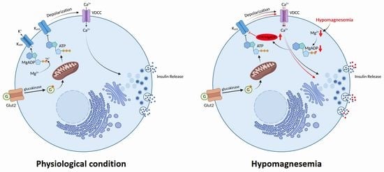
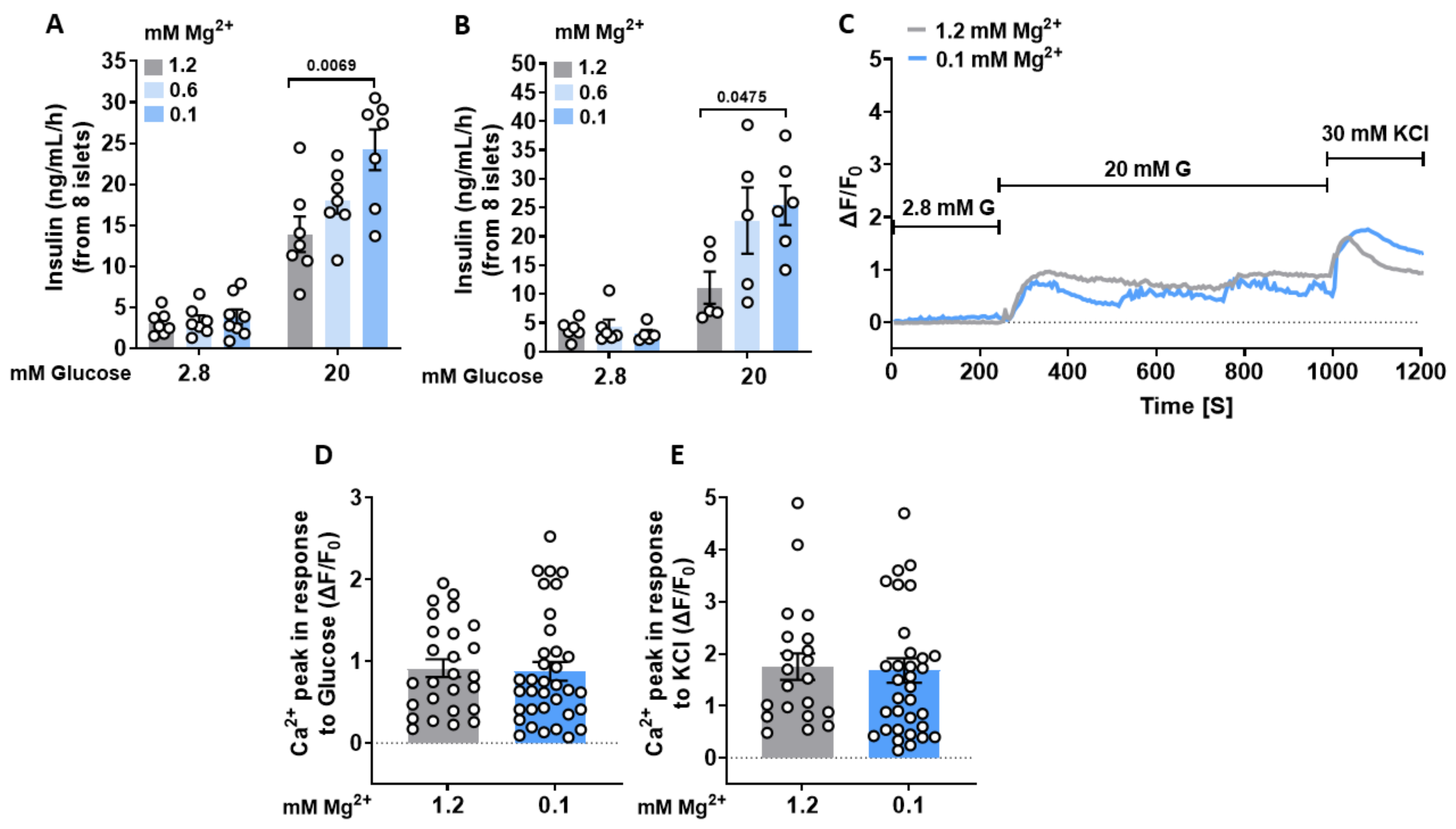
3.1. Lowering Extracellular Mg2+ Concentrations Enhances GIIS in Murine Islets

We assessed the short-term effect of low extracellular Mg2+ concentration on insulin secretion, after 3 h and 24 h preincubation with either 1.2, 0.6, or 0.1 mM MgCl2. Each of these declines in the Mg2+ concentration dose-dependently increased GIIS in pancreatic islets isolated from C57BL/6 mice. Increases in the glucose concentration from 2.8 to 20 mM enhanced insulin exocytosis about 4.5-fold in islets in the presence of a physiological concentration of Mg2+ (1.2 mM), whereas 3 h preincubation with 0.6 and 0.1 mM Mg2+ concentrations increased insulin exocytosis 5.3-fold and 6.2-fold, respectively (Figure 1A). Moreover, twenty-four-hour preincubation with either 0.6 or 0.1 mM MgCl2 increased insulin exocytosis nine-fold and eight-fold, respectively (Figure 1B).

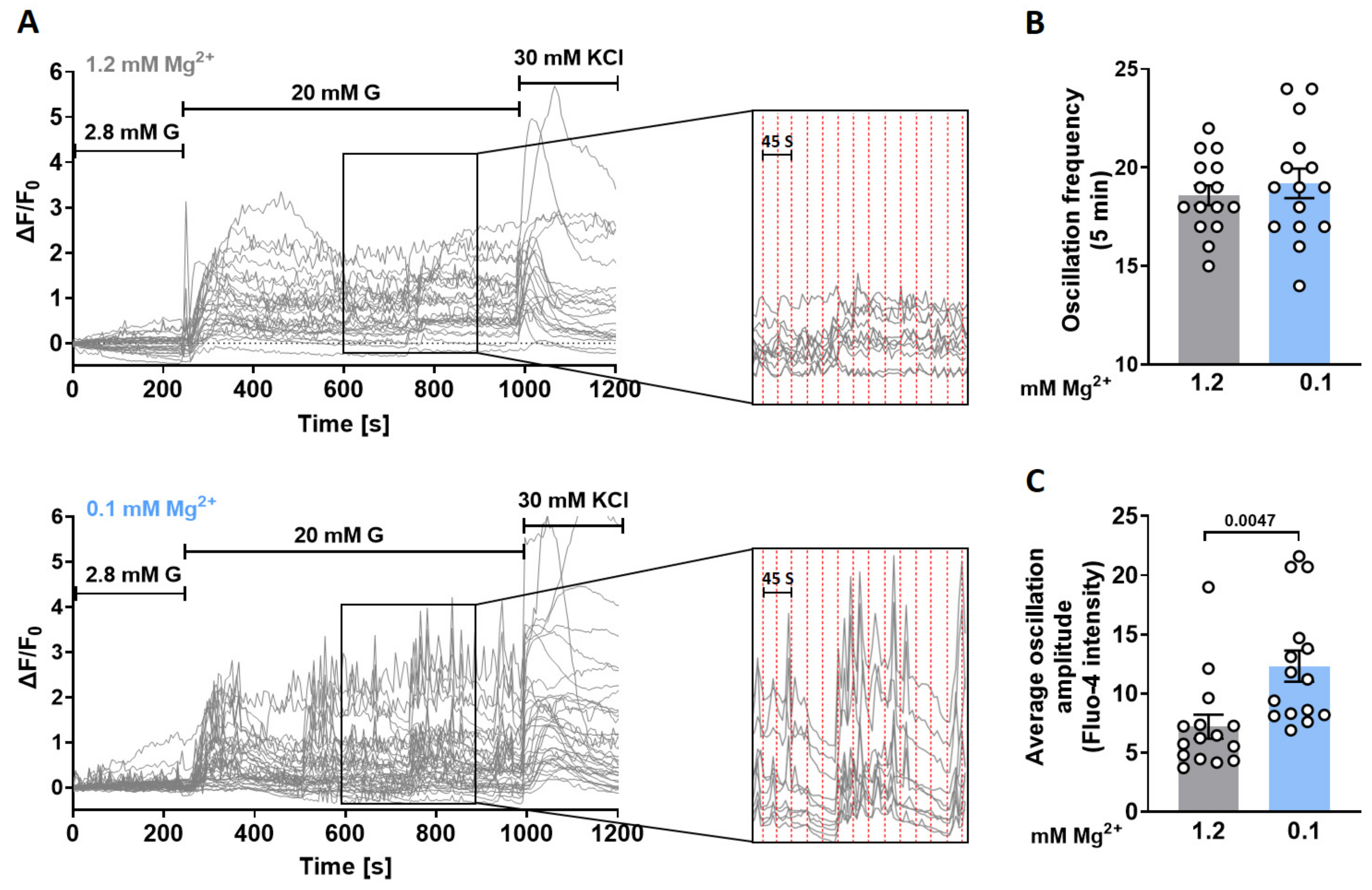
3.2. Lowering Extracellular Mg2+ Concentrations Augments Ca2+ Oscillatory Amplitudes in Murine Islets

To determine if exposure to a low Mg2+ concentration enhanced GIIS through increases in Ca2+ signaling, laser scanning confocal microscopy was used to monitor these Ca2+ responses in individual cells in intact islets. Twenty mM glucose rapidly induced [Ca2+]i transients that were similar to one another in the presence of either 1.2 mM or 0.1 mM Mg2+ (Figure 1C,D). The Ca2+ transient patterns induced by 30 mM KCl under identical conditions were also remarkably similar, suggesting that canonical KATP signaling is not affected by different Mg2+ concentrations (Figure 1E). Nevertheless, despite the comparable oscillation frequency (Figure 2A,B), the average oscillatory amplitudes were significantly larger in the presence of 0.1 mM than those in the presence of 1.2 mM Mg2+ (Figure 2A,C). This difference prompted us to hypothesize that a low Mg2+ concentration underlies the enhanced oscillatory responses and increased GIIS.

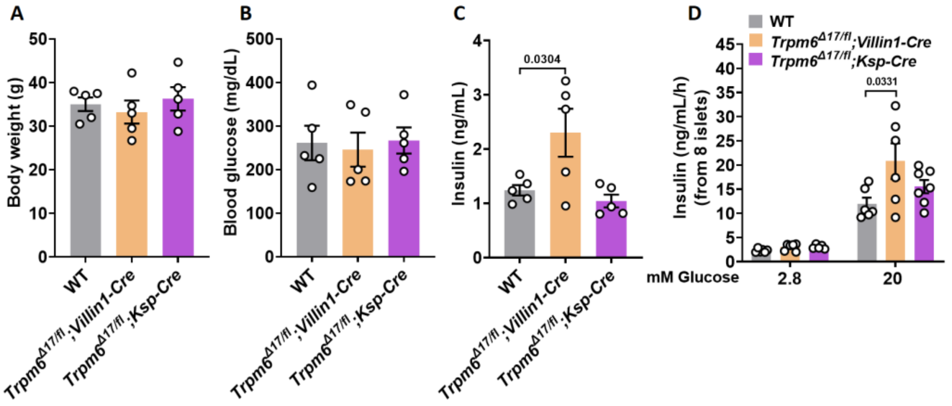
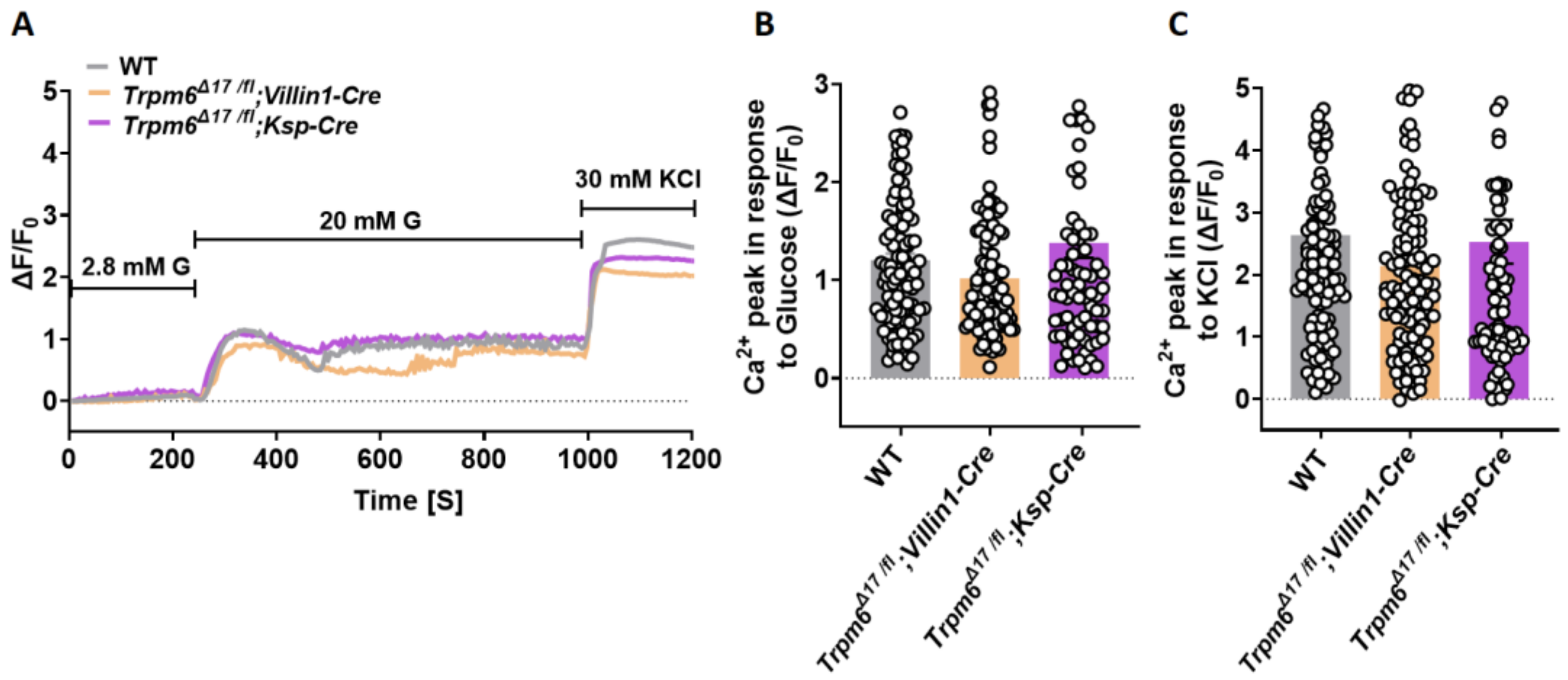
3.3. Plasma Levels of Insulin and GIIS Rise in Intestine-Specific Trpm6 KO Mice

To assess the effects of long-term hypomagnesemia on insulin secretion and glucose homeostasis, we determined if the loss of Trpm6 function in the intestine (Trpm6Δ17 /fl;Villin1-Cre) and kidney (Trpm6Δ17 /fl;Ksp-Cre) of mice alters glucose metabolism. The body weights of these two different genotypes that were fed a standard chow diet were similar to their wild-type counterpart (Figure 3A). Although the blood glucose concentrations remained unaffected in all three genotypes (Figure 3B), the plasma insulin levels were significantly higher in the Trpm6Δ17 /fl;Villin1-Cre mice (Figure 3C). Furthermore, a marked increase in GIIS was detected in isolated islets from Trpm6Δ17 /fl;Villin1-Cre mice. An increase in the glucose concentration from 2.8 to 20 mM enhanced insulin release about 5-fold in wild-type islets, whereas basal insulin exocytosis rose 6.8-fold in Trpm6Δ17 /fl;Villin1-Cre islets and 5.4-fold in Trpm6Δ17 /fl;Ksp-Cre islets (Figure 3D). Collectively, these data show that chronic hypomagnesemia in Trpm6Δ17 /fl;Villin1-Cre mice induces increases in insulin secretion, which we hypothesize are attributable to increases in insulin exocytosis. Importantly, the glycemic metabolic profile was not impaired in Trpm6Δ17 /fl;Villin1-Cre mice, indicating that chronic hypomagnesemia in this mouse model does not result in severe insulin resistance. Nevertheless, if we had access to a larger sample size, it would strengthen the robustness and reliability of these findings.

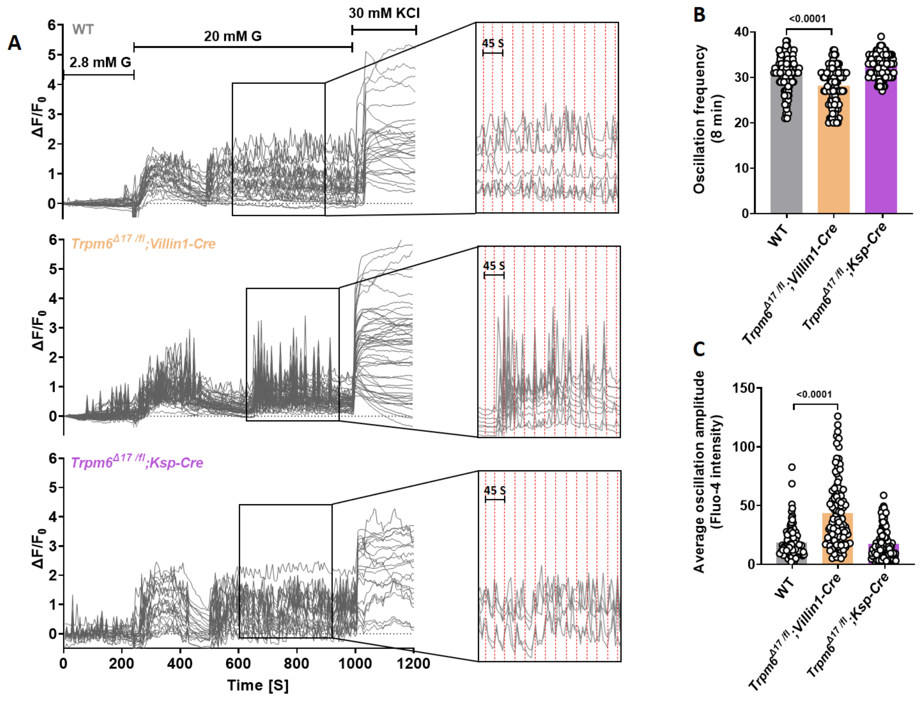
3.4. Intestine-Specific Trpm6 KO Islets Exhibit an Increased Ca2+ Oscillation Amplitude

Twenty mM glucose induced similar Ca2+ transients in islets isolated from Trpm6Δ17 /fl;Villin1-Cre, Trpm6Δ17 /fl;Ksp-Cre, and control littermates (Figure 4A,B). Furthermore, KCl (30 mM) induced similar responses under identical conditions in all three genotypes (Figure 4C).
Nevertheless, Trpm6Δ17 /fl;Villin1-Cre islets showed a remarkable alteration in oscillatory responses relative to control islets. They displayed a diminished oscillation frequency (Figure 5A,B) and an elevated average oscillation amplitude during the plateau phase (Figure 5C). This association between the loss of Trpm6 function and augmentation of the oscillatory response suggests that Mg2+ concentration affects mechanisms underlying extracellular insulin exocytosis.

4. Discussion

Overt clinical hypomagnesemia is particularly common in T2D patients afflicted with poorly controlled glycemic profiles [22]. Dietary Mg2+ supplementation is reported to be beneficial in various clinical studies in diabetic patients [23,24]. However, the cause-and-effect relationship is unclear between hypomagnesemia and this disease. Although clinical studies suggested that there is a positive correlation between Mg2+ concentration and insulin secretion [25], in vivo and in vitro experimental data disagree with this notion.
A recent study demonstrated that exposure of wild-type islets to a low extracellular Mg2+ concentration increased insulin exocytosis when the islets were cultured for 24 h under a high glucose condition that mimicked the T2D hyperglycemic condition [11]; furthermore, knockdown of transient receptor potential melastatin 7 (Trpm7) channels, the most abundant Mg2+ channel in β-cells, increased GIIS in rat insulinoma cell line (INS-1) [11]. A similar inverse relationship exists in another rat insulinoma cell line (RIN m5F), in which the depletion of extracellular Mg2+ stimulates insulin secretion [26,27]. Our results confirmed this inverse relationship between declines in intracellular Mg2+ concentration and increases in insulin secretion and linked them to increases in peak amplitudes of Ca2+ oscillation in pancreatic β-cells.
Even though we found that short-term Mg2+ deficiency does not impair insulin secretion, this finding might not contravene the relationship between hypomagnesemia and T2D since diabetic patients usually experience chronic hypomagnesemia. Thus, we hypothesize that impaired insulin secretion in diabetic patients may be attributed to long-term Mg2+ deficiency in these individuals. An earlier study demonstrated that rats fed a Mg2+-deficient diet for 6 weeks did not show any overt phenotype; however, after a period longer than 6 weeks on this diet, glucose tolerance and GIIS both improved in isolated islets relative to the control [28]. On the contrary, another study indicated an impaired glucose tolerance in rats fed a Mg2+-deficient diet for 6 weeks [29]. Consistent with this finding, glucose tolerance was improved in Zucker Diabetic Fatty (ZDF) rats after 6 weeks of Mg2+ supplementation [30].
In this current study, we used genetically modified animal models to assess the effects of long-term hypomagnesemia on β-cell function in mice. Recently, we generated tamoxifen-inducible β-cell-specific Trpm7 KO mice (βTrpm7 KO) and monitored the metabolic phenotype of this mouse model within 28 weeks. Notably, GIIS was severely diminished in isolated islets from βTrpm7 KO mice after 28 weeks of tamoxifen-induced recombination [21]. Here, we hypothesized that the progressive impairment in insulin secretion might be attributable to the chronic Mg2+ deficiency in β-cell-specific Trpm7 KO mice; however, this difference may instead stem from the fact that TRPM7 is a bifunctional protein consisting of a serine/threonine protein kinase linked to the channel moiety [31,32]. Importantly, we recently demonstrated the indispensable role of the kinase moiety of TRPM7 in mediating the expression of key β-cell markers and the major transcription factors involved in insulin biosynthesis and maintaining β-cell identity [21]; furthermore, TRPM7 can also function as a Ca2+ channel [33]. Hence, βTrpm7 KO mice might not be a suitable mouse model for delineating the role of Mg2+ in insulin secretion and β-cell function. We took another approach to evaluate if there is an association between hypomagnesemia and β-cell function. Here, we used an established mouse model of hypomagnesemia with organismal Mg2+ deficiency known as Trpm6Δ17 /fl;Villin1-Cre [18]. Our results indicate that conditional Trpm6 inactivation in the intestine increased the plasma levels of insulin. Nevertheless, the body weight and blood glucose levels remain similar among the Trpm6Δ17 /fl;Villin1-Cre and the control littermates. Furthermore, the high glucose condition in isolated Trpm6Δ17 /fl;Villin1-Cre pancreatic islets increased insulin secretion to levels that were higher than in the wild-type. This result is in agreement with an observation in a Canadian T2D cohort in which HOMA-β, an index of functional pancreatic β-cell mass, was inversely correlated with serum Mg2+ levels [34].
Mg2+ homeostasis is regulated by Mg2+ reabsorption from pro-urine in DCT [19]. Given the critical role of TRPM6 in controlling Mg2+ reabsorption [17], we characterized the metabolic phenotype of a conditional Trpm6 KO in the kidney (Trpm6Δ17 /fl;Ksp-Cre). Our result demonstrated that Trpm6 inactivation in the kidney had no impact on body weight, blood glucose, and plasma levels of insulin.
An increase in intracellular Ca2+ influx is the final trigger of insulin exocytosis. We found that chronic hypomagnesemia increased the average amplitudes of the Ca2+ oscillatory responses in Trpm6Δ17 /fl;Villin1-Cre islets. Here, we suggest that Mg2+ deficiency attenuates intracellular levels of MgADP in β-cells. Since the KATP channel activity is dependent on a balance between ATP-mediated suppression (at Kir6.2) and MgADP activation (at SUR1), a decline in MgADP content enhances ATP-induced K+ channel inhibition. Furthermore, Mg-nucleotides increase the number of functional KATP channels in the plasma membrane [35]. Consequently, a Mg2+ deficiency prolongs the depolarization phase in β-cells, resulting in larger increases in the amplitudes of Ca2+ oscillations and ultimately enhancing insulin release. Importantly, rises in MgADP content have been shown to reduce the interburst intervals [10]. Thus, we conclude that chronic hypomagnesemia reduced the MgADP content and, in turn, reduced the Ca2+ transient oscillation frequency in Trpm6Δ17 /fl;Villin1-Cre islets.
Various studies have demonstrated that declines in both intracellular and extracellular Mg2+ inhibit L-type Ca2+ channel activity [36,37]; therefore, it can be speculated that reduced intracellular Mg2+ concentrations due to hypomagnesemia increase L-type channel activity that, in turn, increases Ca2+ oscillatory amplitudes. However, it has also been reported that long-term hypomagnesemia instead reduces the expression of L-type Ca2+ channels and reduces Ca2+ responses [38]. Nevertheless, these findings were solely obtained in cardiomyocytes rather than in pancreatic β-cells.
Continuous exposure to increased insulin levels downregulates plasma membrane insulin receptor expression, which in turn augments insulin resistance [39]; however, we did not obtain any evidence suggesting that prolonged exposure to elevated insulin levels downregulates insulin receptor expression. This is supported by the absence of any detectable impairment in the metabolic phenotype of Trpm6Δ17 /fl;Villin1-Cre mice. This negative effect rules out a major impediment in insulin sensitivity in this mouse model of chronic hypomagnesemia.

5. Conclusions

The role of Mg2+ in controlling insulin secretion has only been investigated in a limited number of clinical studies [25,34]; furthermore, the beneficial effect of Mg2+ supplementation on β-cell function in individuals without diabetes was only observed in a small randomized clinical trial [40]. Hence, in contrast to current thinking, our findings strongly suggest that long-term hypomagnesemia does not negatively impact the metabolic phenotype, insulin secretion, and β-cell function. Accordingly, it is more likely that hypomagnesemia is the consequence of diabetes rather than a causative factor. One possible explanation for the association between hypomagnesemia and T2D is that insulin plays a vital role in controlling the reabsorption of Mg2+ in the kidney through modulating TRPM6 channel activity. The increased renal excretion of Mg2+ induced by a decrease in insulin may contribute to the development of hypomagnesemia in individuals with T2D [41].

Author Contributions

N.K. designed and conducted experiments, analyzed, and interpreted data, prepared figures, and wrote the manuscript. K.R. and P.B. conducted experiments, analyzed, and interpreted data, and edited the manuscript. P.C.F.S., T.G. (Tanja Gentz), E.C., A.B. and P.S.R. interpreted data and edited the manuscript. T.G. (Thomas Gudermann) directed the project, designed experiments, interpreted data, and edited the manuscript. All authors have read and agreed to the published version of the manuscript.

Funding

N.K. and T.G. (Thomas Gudermann) were supported by the Deutsche Forschungsgemeinschaft (German Research Foundation, DFG)—Project-ID 239283807—TRR-152 (P23, P15).

Institutional Review Board Statement

Experiments involving animals were done in accordance with the EU Animal Welfare Act and were approved by the local councils on animal care (permit No 55.2-1-54-2532-134-13 from the Government of Oberbayern, Germany, and permit No 2347-15-2014 from the State Ministry of Brandenburg, Germany).

Data Availability Statement

The data that support the findings of this study are available on request from the corresponding author.

Acknowledgments

The authors especially appreciate the support of Alin-Mihai Postu (Bioinformatic) for the algorithms required for the data evaluation and for very helpful discussions. Trpm6Δ17 /fl;Villin1-Cre, Trpm6Δ17 /fl;Ksp-Cre mice were kindly provided by Vladimir Chubanov (Walther Straub Institute of Pharmacology and Toxicology, LMU, Munich). The authors also appreciate the input of Johann Schredelseker (Walther Straub Institute of Pharmacology and Toxicology, LMU, Munich).

Conflicts of Interest

The authors declare no conflict of interest.

References

  1. Alswat, K. Type 2 diabetes control and complications and their relation to serum magnesium level. Arch. Med. Sci. 2022, 18, 307–313. [Google Scholar] [CrossRef] [PubMed]
  2. Pham, P.-C.T.; Pham, P.-M.T.; Pham, S.V.; Miller, J.M.; Pham, P.-T.T. Hypomagnesemia in patients with type 2 diabetes. Clin. J. Am. Soc. Nephrol. 2007, 2, 366–373. [Google Scholar] [CrossRef] [PubMed] [Green Version]
  3. Garfinkel, L.; Garfinkel, D. Magnesium regulation of the glycolytic pathway and the enzymes involved. Magnesium 1985, 4, 60–72. [Google Scholar] [PubMed]
  4. Matschinsky, F.M. Regulation of pancreatic beta-cell glucokinase: From basics to therapeutics. Diabetes 2002, 51 (Suppl. S3), S394–S404. [Google Scholar] [CrossRef] [Green Version]
  5. Molnes, J.; Teigen, K.; Aukrust, I.; Bjørkhaug, L.; Søvik, O.; Flatmark, T.; Njølstad, P.R. Binding of ATP at the active site of human pancreatic glucokinase-nucleotide-induced conformational changes with possible implications for its kinetic cooperativity. FEBS J. 2011, 278, 2372–2386. [Google Scholar] [CrossRef] [Green Version]
  6. Storer, A.C.; Cornish-Bowden, A. Kinetics of rat liver glucokinase. Co-operative interactions with glucose at physiologically significant concentrations. Biochem. J. 1976, 159, 7–14. [Google Scholar] [CrossRef] [Green Version]
  7. Braun, M.; Ramracheya, R.; Bengtsson, M.; Zhang, Q.; Karanauskaite, J.; Partridge, C.; Johnson, P.R.; Rorsman, P. Voltage-gated ion channels in human pancreatic beta-cells: Electrophysiological characterization and role in insulin secretion. Diabetes 2008, 57, 1618–1628. [Google Scholar] [CrossRef] [Green Version]
  8. Cook, D.L.; Hales, C.N. Intracellular ATP directly blocks K+ channels in pancreatic B-cells. Nature 1984, 311, 271–273. [Google Scholar] [CrossRef]
  9. Dunne, M.J.; Petersen, O.H. Intracellular ADP activates K+ channels that are inhibited by ATP in an insulin-secreting cell line. FEBS Lett. 1986, 208, 59–62. [Google Scholar] [CrossRef] [Green Version]
  10. Kakei, M.; Kelly, R.P.; Ashcroft, S.J.; Ashcroft, F.M. The ATP-sensitivity of K+ channels in rat pancreatic B-cells is modulated by ADP. FEBS Lett. 1986, 208, 63–66. [Google Scholar] [CrossRef] [Green Version]
  11. Gommers, L.M.M.; Hill, T.G.; Ashcroft, F.M.; De Baaij, J.H.F. Low extracellular magnesium does not impair glucose-stimulated insulin secretion. PLoS ONE 2019, 14, e0217925. [Google Scholar] [CrossRef] [PubMed] [Green Version]
  12. Gommers, L.M.M.; Hoenderop, J.G.J.; Bindels, R.J.M.; De Baaij, J.H.F. Hypomagnesemia in Type 2 Diabetes: A Vicious Circle? Diabetes 2016, 65, 3–13. [Google Scholar] [CrossRef] [PubMed] [Green Version]
  13. Simon, D.B.; Lu, Y.; Choate, K.A.; Velazquez, H.; Al-Sabban, E.; Praga, M.; Casari, G.; Bettinelli, A.; Colussi, G.; Rodriguez-Soriano, J.; et al. Paracellin-1, a renal tight junction protein required for paracellular Mg2+ resorption. Science 1999, 285, 103–106. [Google Scholar] [CrossRef] [PubMed]
  14. Walder, R.Y.; Landau, D.; Meyer, P.; Shalev, H.; Tsolia, M.; Borochowitz, Z.; Boettger, M.B.; Beck, G.E.; Englehardt, R.K.; Carmi, R.; et al. Mutation of TRPM6 causes familial hypomagnesemia with secondary hypocalcemia. Nat. Genet. 2002, 31, 171–174. [Google Scholar] [CrossRef] [PubMed]
  15. Arjona, F.; de Baaij, J.; Schlingmann, K.P.; Lameris, A.L.L.; Van Wijk, E.; Flik, G.; Regele, S.; Korenke, G.C.; Neophytou, B.; Rust, S.; et al. CCNNM2 mutations cause impaired brain development and seizures in patients with hypomagnesemia. PLoS Genet. 2014, 10, e1004267. [Google Scholar] [CrossRef] [PubMed]
  16. Schlingmann, K.P.; Weber, S.; Peters, M.; Niemann Nejsum, L.; Vitzthum, H.; Klingel, K.; Kratz, M.; Haddad, E.; Ristoff, E.; Dinour, D.; et al. Hypomagnesemia with secondary hypocalcemia is caused by mutations in TRPM6, a new member of the TRPM gene family. Nat. Genet. 2002, 31, 166–170. [Google Scholar] [CrossRef]
  17. Voets, T.; Nilius, B.; Hoefs, S.; van der Kemp, A.W.C.M.; Droogmans, G.; Bindels, R.J.; Hoenderop, J.G. TRPM6 forms the Mg2+ influx channel involved in intestinal and renal Mg2+ absorption. J. Biol. Chem. 2004, 279, 19–25. [Google Scholar] [CrossRef] [Green Version]
  18. Chubanov, V.; Ferioli, S.; Wisnowsky, A.; Simmons, D.G.; Leitzinger, C.; Einer, C.; Jonas, W.; Shymkiv, Y.; Bartsch, H.; Braun, A.; et al. Epithelial magnesium transport by TRPM6 is essential for prenatal development and adult survival. eLife 2016, 5, e20914. [Google Scholar] [CrossRef]
  19. De Baaij, J.H.; Hoenderop, J.G.; Bindels, R.J. Magnesium in man: Implications for health and disease. Physiol. Rev. 2015, 95, 1–46. [Google Scholar] [CrossRef]
  20. Khajavi, N.; Finan, B.; Kluth, O.; Müller, T.D.; Mergler, S.; Schulz, A.; Kleinau, G.; Scheerer, P.; Schürmann, A.; Gudermann, T.; et al. An incretin-based tri-agonist promotes superior insulin secretion from murine pancreatic islets via PLC activation. Cell. Signal. 2018, 51, 13–22. [Google Scholar] [CrossRef]
  21. Khajavi, N.; Beck, A.; Riçku, K.; Beyerle, P.; Jacob, K.; Syamsul, S.F.; Belkacemi, A.; Reinach, P.S.; Schreier, P.C.; Salah, H.; et al. TRPM7 kinase is required for insulin production and compensatory islet responses during obesity. JCI Insight 2023, 8, e163397. [Google Scholar] [CrossRef] [PubMed]
  22. Barbagallo, M.; Dominguez, L.J. Magnesium and type 2 diabetes. World J. Diabetes 2015, 6, 1152–1157. [Google Scholar] [CrossRef] [Green Version]
  23. ELDerawi, W.A.; Naser, I.A.; Taleb, M.H.; Abutair, A.S. The Effects of Oral Magnesium Supplementation on Glycemic Response among Type 2 Diabetes Patients. Nutrients 2018, 11, 44. [Google Scholar] [CrossRef] [PubMed] [Green Version]
  24. Jamilian, M.; Mirhosseini, N.; Eslahi, M.; Bahmani, F.; Shokrpour, M.; Chamani, M.; Asemi, Z. The effects of magnesium-zinc-calcium-vitamin D co-supplementation on biomarkers of inflammation, oxidative stress and pregnancy outcomes in gestational diabetes. BMC Pregnancy Childbirth 2019, 19, 107. [Google Scholar] [CrossRef] [PubMed] [Green Version]
  25. Rodríguez-Morán, M.; Guerrero-Romero, F. Insulin secretion is decreased in non-diabetic individuals with hypomagnesaemia. Diabetes Metab. Res. Rev. 2011, 27, 590–596. [Google Scholar] [CrossRef]
  26. Murakami, M.; Ishizuka, J.; Sumi, S.; Nickols, G.A.; Cooper, C.W.; Townsend, C.M.; Thompson, J.C. Role of extracellular magnesium in insulin secretion from rat insulinoma cells. Proc. Soc. Exp. Biol. Med. 1992, 200, 490–494. [Google Scholar] [CrossRef]
  27. Ishizuka, J.; Bold, R.J.; Townsend, C.M.; Thompson, J.C. In vitro relationship between magnesium and insulin secretion. Magnes. Res. 1994, 7, 17–22. [Google Scholar]
  28. Reis, M.A.; Latorraca, M.Q.; Carneiro, E.M.; Boschero, A.C.; Saad, M.J.; Velloso, L.A.; Reyes, F.G. Magnesium deficiency improves glucose homeostasis in the rat: Studies in vivo and in isolated islets in vitro. Br. J. Nutr. 2001, 85, 549–552. [Google Scholar] [CrossRef] [Green Version]
  29. Legrand, C.; Okitolonda, W.; Pottier, A.; Lederer, J.; Henquin, J. Glucose homeostasis in magnesium-deficient rats. Metabolism 1987, 36, 160–164. [Google Scholar] [CrossRef]
  30. Balon, T.W.; Gu, J.L.; Tokuyama, Y.; Jasman, A.P.; Nadler, J.L. Magnesium supplementation reduces development of diabetes in a rat model of spontaneous NIDDM. Am. J. Physiol. 1995, 269 Pt 1, E745–E752. [Google Scholar] [CrossRef]
  31. Ryazanova, L.V.; Dorovkov, M.V.; Ansari, A.; Ryazanov, A.G. Characterization of the protein kinase activity of TRPM7/ChaK1, a protein kinase fused to the transient receptor potential ion channel. J. Biol. Chem. 2004, 279, 3708–3716. [Google Scholar] [CrossRef] [PubMed] [Green Version]
  32. Runnels, L.W.; Yue, L.; Clapham, D.E. TRP-PLIK, a bifunctional protein with kinase and ion channel activities. Science 2001, 291, 1043–1047. [Google Scholar] [CrossRef] [PubMed]
  33. Monteilh-Zoller, M.K.; Hermosura, M.C.; Nadler, M.J.; Scharenberg, A.M.; Penner, R.; Fleig, A. TRPM7 provides an ion channel mechanism for cellular entry of trace metal ions. J. Gen. Physiol. 2003, 121, 49–60. [Google Scholar] [CrossRef] [Green Version]
  34. Randell, E.W.; Mathews, M.; Gadag, V.; Zhang, H.; Sun, G. Relationship between serum magnesium values, lipids and anthropometric risk factors. Atherosclerosis 2008, 196, 413–419. [Google Scholar] [CrossRef] [PubMed]
  35. Proks, P.; de Wet, H.; Ashcroft, F.M. Activation of the K(ATP) channel by Mg-nucleotide interaction with SUR1. J. Gen. Physiol. 2010, 136, 389–405. [Google Scholar] [CrossRef] [Green Version]
  36. Wang, M.; Berlin, J.R. Channel phosphorylation and modulation of L-type Ca2+ currents by cytosolic Mg2+ concentration. Am. J. Physiol. Cell Physiol. 2006, 291, C83–C92. [Google Scholar] [CrossRef] [PubMed]
  37. Zhang, J.; Berra-Romani, R.; Sinnegger-Brauns, M.J.; Striessnig, J.; Blaustein, M.P.; Matteson, D.R. Role of Cav1.2 L-type Ca2+ channels in vascular tone: Effects of nifedipine and Mg2+. Am. J. Physiol. Heart Circ. Physiol. 2007, 292, H415–H425. [Google Scholar] [CrossRef] [PubMed]
  38. Shimaoka, T.; Wang, Y.; Morishima, M.; Miyamoto, S.; Ono, K. Hypomagnesemic down-regulation of L-type Ca(2+) channel in cardiomyocyte as an arrhythmogenic substrate in rats. Pathophysiology 2015, 22, 87–93. [Google Scholar] [CrossRef]
  39. Gavin, J.R., III; Roth, J.; Neville, D.M., Jr.; De Meyts, P.; Buell, D.N. Insulin-dependent regulation of insulin receptor concentrations: A direct demonstration in cell culture. Proc. Natl. Acad. Sci. USA 1974, 71, 84–88. [Google Scholar] [CrossRef]
  40. Dasgupta, A.; Sarma, D.; Saikia, U.K. Hypomagnesemia in type 2 diabetes mellitus. Indian J. Endocrinol. Metab. 2012, 16, 1000–1003. [Google Scholar] [CrossRef]
  41. McNair, P.; Christensen, M.S.; Christiansen, C.; Madsbad, S.; Transbøl, I. Renal hypomagnesaemia in human diabetes mellitus: Its relation to glucose homeostasis. Eur. J. Clin. Investig. 1982, 12, 81–85. [Google Scholar] [CrossRef] [PubMed]
Figure 1. Lowering extracellular Mg2+ concentrations increases GIIS in murine islets. Islets were isolated from 8-week-old mice. (A,B) Insulin secretion was measured in intact wild-type (n = 8, from 5 mice) after 3 h (A), and (n ≥ 5, from 6 mice) after 24 h (B) of incubation with three different Mg2+ concentrations (1.2, 0.6, and 0.1 mM) in the presence of 2.8 or 20 mM glucose. (C) Intact wild-type islets (n ≥ 20, from 5 mice) were loaded with 4 µM fluo-4 AM, and alterations in [Ca2+]i of individual cells were monitored using confocal microscopy after increasing the extracellular glucose concentration from 2.8 to 20 mM in the presence of 1.2 mM or 0.1 mM Mg2+. KCl (30 mM) was used as a positive control. Mean traces for 1.2 mM and 0.1 mM Mg2+ are shown in gray and blue, respectively. (D,E) Average Ca2+ influx peaks assessed from baseline after glucose (D) and KCl (E) stimulation in wild-type β-cells in the presence of 1.2 or 0.1 mM Mg2+ (n ≥ 20, from 5 mice). The cells that displayed no increase in [Ca2+]i in response to high glucose concentration are excluded from the results. Data are shown as means ± S.E.M. (circles in bar graphs represent single values) and statistical differences were assessed by one-way ANOVA (A,B) or unpaired two-tailed Student’s t-test (D,E). p value is shown above the bar.
Cells 12 01790 g001
Figure 2. Lowering extracellular Mg2+ concentrations increases Ca2+ oscillatory amplitudes in murine islets. Islets were isolated from 8-week-old mice. (A) Intact wild-type islets (n ≥ 15, from 5 mice) were loaded with 4 µM fluo-4 AM, and alterations in [Ca2+]i of individual cells were monitored using confocal microscopy after increasing the extracellular glucose concentration from 2.8 to 20 mM in the presence of 1.2 mM or 0.1 mM Mg2+. KCl (30 mM) was used as a positive control. Inserts show amplified sections indicating the regions from which data in the right panels were taken. (B,C) Comparison of oscillation frequency and average oscillation amplitudes in the presence of 1.2 or 0.1 mM Mg2+. Data are shown as means ± S.E.M. (circles in bar graphs represent single values) and statistical differences were assessed by unpaired two-tailed Student’s t-test (B,C). p value is shown above the bar.
Cells 12 01790 g002
Figure 3. Tissue-specific Trpm6 deletion in intestine increases plasma levels of insulin and GIIS. (A) Body weight, (B) blood glucose levels, and (C) plasma insulin levels (n = 5) in 24-week-old male and female Trpm6Δ17 /fl;Villin1-Cre, Trpm6Δ17 /fl;Ksp-Cre, and wild-type mice on standard chow diet. (D) Insulin secretion (ng/mL/h/8 islets) in isolated islets of 24-week-old male and female Trpm6Δ17 /fl;Villin1-Cre, Trpm6Δ17 /fl;Ksp-Cre, and wild-type mice. Islets were incubated for 1 h in the presence of low glucose (2.8 mM) or high glucose (20 mM) (n = 5 mice per genotype). Data are shown as means ± S.E.M., and statistical differences were assessed by one-way ANOVA. Circles in bar graphs represent single values. p values are shown above the bars.
Cells 12 01790 g003
Figure 4. Tissue-specific Trpm6 deletion in intestine and kidney has no effect on glucose- and KCl-induced Ca2+ transients in murine islets. Islets were isolated from 24-week-old mice. (A) Intact WT (n ≥ 100, from 5 mice), Trpm6Δ17 /fl;Villin1-Cre (n ≥ 100, from 5 mice), and Trpm6Δ17 /fl;Ksp-Cre (n ≥ 100, from 5 mice) islets were loaded with 4 µM fluo-4 AM, and alterations in [Ca2+]i of individual cells were monitored using confocal microscopy after increasing the extracellular glucose concentration from 2.8 to 20 mM. KCl (30 mM) was used as a positive control. Mean traces are shown for every mouse model. (B,C) Average Ca2+ influx peaks assessed from baseline after glucose (B) and KCl (C) stimulation in WT, Trpm6Δ17 /fl;Villin1-Cre, and Trpm6Δ17 /fl;Ksp-Cre β-cells. The cells that displayed no increase in [Ca2+]i in response to a high glucose concentration are excluded from the results. Data are shown as means ± S.E.M. (circles in bar graphs represent single values). Statistical differences were assessed by one-way ANOVA.
Cells 12 01790 g004
Figure 5. Tissue-specific Trpm6 deletion in intestine increases Ca2+ oscillation amplitude. Islets were isolated from 24-week-old mice. (A) Intact wild-type (n ≥ 100, from 5 mice), Trpm6Δ17 /fl;Villin1-Cre (n ≥ 100, from 5 mice), and Trpm6Δ17 /fl;Ksp-Cre (n ≥ 100, from 5 mice) islets were loaded with 4 µM fluo-4 AM, and alterations in [Ca2+]i of individual cells were monitored using confocal microscopy after increasing the extracellular glucose concentration from 2.8 to 20 mM in the presence of 1.2 mM or 0.1 mM Mg2+. KCl (30 mM) was used as a positive control. Inserts show amplified sections indicating the regions from which data in the right panels were taken. (B,C) Comparison of oscillation frequency and average oscillation amplitudes between intact wild-type, Trpm6Δ17 /fl;Villin1-Cre, and Trpm6Δ17 /fl;Ksp-Cre islets. Data are shown as means ± S.E.M. (circles in bar graphs represent single values) and statistical differences were assessed by one-way ANOVA. p values are shown above the bar.
Cells 12 01790 g005
Disclaimer/Publisher’s Note: The statements, opinions and data contained in all publications are solely those of the individual author(s) and contributor(s) and not of MDPI and/or the editor(s). MDPI and/or the editor(s) disclaim responsibility for any injury to people or property resulting from any ideas, methods, instructions or products referred to in the content.

