Article

Identifying Genetic Mutation Status in Patients with Colorectal Cancer Liver Metastases Using Radiomics-Based Machine-Learning Models

by
Nina Wesdorp
1,2,†,
Michiel Zeeuw
1,2,*,†,
Delanie van der Meulen
1,2,
Iris van ‘t Erve
3,
Zuhir Bodalal
4,
Joran Roor
5,
Jan Hein van Waesberghe
2,6,
Shira Moos
2,6,
Janneke van den Bergh
2,6,
Irene Nota
2,6,
Susan van Dieren
2,7,
Jaap Stoker
2,8,
Gerrit Meijer
3,
Rutger-Jan Swijnenburg
2,7,
Cornelis Punt
9,10,
Joost Huiskens
1,2,
Regina Beets-Tan
4,
Remond Fijneman
3,
Henk Marquering
2,8,11,
Geert Kazemier
1,2 and
on behalf of the Dutch Colorectal Cancer Group Liver Expert Panel
add Show full author list remove Hide full author list
1
Department of Surgery, Amsterdam UMC, Vrije Universiteit Amsterdam, 1081 HV Amsterdam, The Netherlands
2
Cancer Center Amsterdam, 1081 HV Amsterdam, The Netherlands
3
Department of Pathology, The Netherlands Cancer Institute, 1066 CX Amsterdam, The Netherlands
4
Department of Radiology, The Netherlands Cancer Institute, 1066 CX Amsterdam, The Netherlands
5
Department of Health, SAS Institute B.V., 1272 PC Huizen, The Netherlands
6
Department of Radiology and Nuclear Medicine, Amsterdam UMC, Vrije Universiteit Amsterdam, 1081 HV Amsterdam, The Netherlands
7
Department of Surgery, Amsterdam UMC, University of Amsterdam, 1105 AZ Amsterdam, The Netherlands
8
Department of Radiology and Nuclear Medicine, Amsterdam UMC, University of Amsterdam, 1105 AZ Amsterdam, The Netherlands
9
Department of Medical Oncology, Amsterdam UMC, University of Amsterdam, 1105 AZ Amsterdam, The Netherlands
10
Department of Epidemiology, Julius Center for Health Sciences and Primary Care, University Medical Center Utrecht, 3584 CG Utrecht, The Netherlands
11
Department of Biomedical Engineering and Physics, Amsterdam UMC, University of Amsterdam, 1081 HV Amsterdam, The Netherlands
*
Author to whom correspondence should be addressed.
These authors contributed equally to this work.
Dutch Colorectal Cancer Group Liver Expert Panel are listed in acknowledgments.
Cancers 2023, 15(23), 5648; https://doi.org/10.3390/cancers15235648
Submission received: 20 September 2023 / Revised: 16 November 2023 / Accepted: 28 November 2023 / Published: 29 November 2023
(This article belongs to the Section Cancer Metastasis)

Abstract

:

Simple Summary

For patients with colorectal cancer with liver metastases, it is important to determine the genetic mutations (e.g., KRAS mutations) of the liver metastases. Around 35–45% of patients with colorectal cancer liver metastases (CRLM) have a KRAS mutation, and genetic mutations are used in treatment planning and prognostication. The aim of this study was to assess if KRAS mutations could be identified on CT scans using radiomics. In the discovery cohort of 255 patients, KRAS mutations could be identified with a good accuracy. In the external validation cohort consisting of 129 patients, the radiomics model performed poorly. These results indicate that radiomics might be used to determine genetic mutations such as KRAS, but foremost emphasize the importance of the external validation of radiomics models. External validation is crucial for the assessment of clinical applicability and should be mandatory in all future studies in the field of radiomics.

Abstract

For patients with colorectal cancer liver metastases (CRLM), the genetic mutation status is important in treatment selection and prognostication for survival outcomes. This study aims to investigate the relationship between radiomics imaging features and the genetic mutation status (KRAS mutation versus no mutation) in a large multicenter dataset of patients with CRLM and validate these findings in an external dataset. Patients with initially unresectable CRLM treated with systemic therapy of the randomized controlled CAIRO5 trial (NCT02162563) were included. All CRLM were semi-automatically segmented in pre-treatment CT scans and radiomics features were calculated from these segmentations. Additionally, data from the Netherlands Cancer Institute (NKI) were used for external validation. A total of 255 patients from the CAIRO5 trial were included. Random Forest, Gradient Boosting, Gradient Boosting + LightGBM, and Ensemble machine-learning classifiers showed AUC scores of 0.77 (95%CI 0.62–0.92), 0.77 (95%CI 0.64–0.90), 0.72 (95%CI 0.57–0.87), and 0.86 (95%CI 0.76–0.95) in the internal test set. Validation of the models on the external dataset with 129 patients resulted in AUC scores of 0.47–0.56. Machine-learning models incorporating CT imaging features could identify the genetic mutation status in patients with CRLM with a good accuracy in the internal test set. However, in the external validation set, the models performed poorly. External validation of machine-learning models is crucial for the assessment of clinical applicability and should be mandatory in all future studies in the field of radiomics.

1. Introduction

Colorectal cancer (CRC) is the second-leading cause of cancer-related deaths worldwide [1] and almost half of these patients develop colorectal cancer liver metastases (CRLM) [2,3]. Over the past few decades, substantial advances have occurred in personalized treatment, selecting the optimal treatment based on individual patient and tumor characteristics. The addition of targeted agents to chemotherapy increased response rates to around 60% [4,5,6,7,8]. Targeted agents selectively target the molecular pathways of cancer cells, such as the epidermal growth factor receptor (EGFR) pathway that regulates functions related to cell proliferation, growth, and apoptosis [9]. Overexpression of this pathway leads to a more aggressive disease course and poor prognosis in patients with metastasized CRC; therefore, anti-EGFR agents have emerged as a treatment option [7,8]. The genetic mutation status has been shown to be a promising biomarker for patients with CRLM [10]. Approximately 35–45% of patients with CRLM have a KRAS mutation, which is associated with a poorer prognosis than non-mutated (i.e., wild-type) CRLM [11]. A mutation of KRAS has been shown to lead to a poor response to anti-EGFR-targeted agents [12,13,14]. Pre-treatment determination of the genetic mutations status is therefore an important step in the personalized treatment strategy of patients with CRLM [15]. Nonetheless, the current methods of tumor genomic profiling require invasive methods, such as biopsies. Therefore, it is not feasible to determine the mutation status per individual CRLM. Nowadays, the standard care of patients with CRLM involves non-invasive medical imaging, such as computed tomography (CT), at diagnosis and follow-up. This large number of medical images may potentially be used to find non-invasive biomarkers using advanced imaging analytics, such as radiomics [16]. Radiomics is an advanced method to calculate numerous imaging features from medical images that cannot be quantified by the human eye [16,17]. These imaging features can be used in predictive models using machine-learning classifiers [16]. Research in radiomics has evolved exponentially and is increasingly used to predict clinical outcomes in patients with gastrointestinal cancer, including CRLM [18,19,20]. Radiomics is also applied to identify genetic mutations, which is called radiogenomics [17,21]. More broadly, radiogenomics aims to examine the relationship between imaging features and changes in gene expression. A review by Badic et al. reported that genetic mutations, such as KRAS and microsatellite instability, could be identified using radiomics models in patients with CRC in multiple studies [22]. Likewise, Shi et al. were able to classify the genetic mutation status (KRAS/BRAF/NRAS mutation versus wild-type) of patients with CRLM by making use of a neural network based on CT-based radiomics features [23]. Specifically for patients with mutation status heterogeneity between CRLM [24,25], the mutation status determination via the radiogenomics of individual CRLM could be of additional clinical value. The positive findings of previous studies suggest a relationship between the tumor genotype and its imaging features in metastatic CRC. However, other research also emphasized the methodological shortcomings of studies applying radiogenomics in patients with CRC, using small datasets and lacking external validation [23,26]. This study aims to investigate the relationship between radiomics imaging features and the genetic mutation status (KRAS versus wild-type) in a large dataset of patients with CRLM and validate these findings in an external dataset.

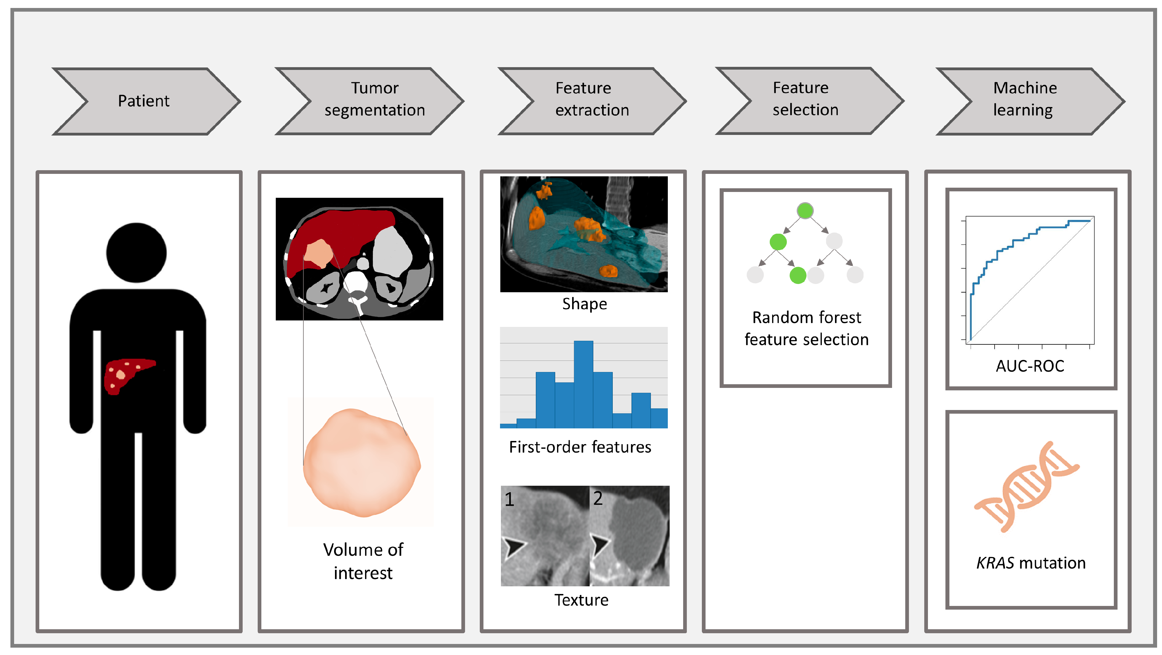
2. Materials and Methods

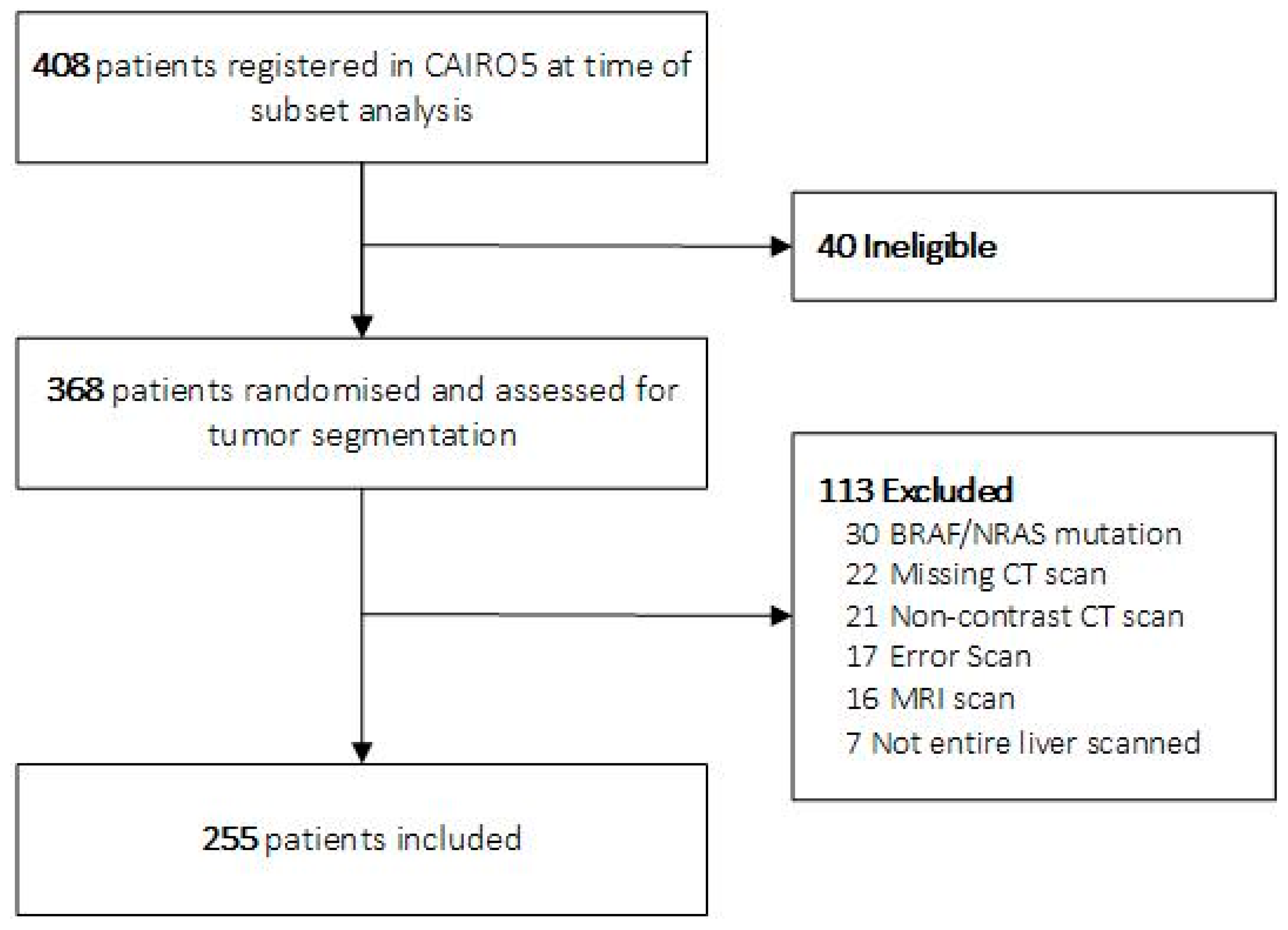
2.1. Study Population

The study population consisted of patients registered between 2014 and 2019 in the multicenter randomized clinical trial of the Dutch Colorectal Cancer Group (DCCG), CAIRO5 (NCT02162563) [27]. In this trial, patients with initially unresectable liver-only CRLM were randomized between different systemic therapies based on the primary tumor site and genetic mutation status (KRAS/NRAS/BRAF mutation vs. wild-type) [27]. All patients signed a written consent form, also allowing side-studies. In the current study, only patients with KRAS-mutated or wild-type CRLM were included. Patients from the Netherlands Cancer Institute (NKI) were included as an external validation cohort. This cohort comprised a routine retrospective cohort of patients with CRLM. These patients all underwent biopsy with subsequent next-generation sequencing between 2013 and 2020. Baseline contrast-enhanced CT images were collected with a maximum time difference between the biopsy date and scan date of 90 days.

2.2. Data Collection

2.2.1. Genetic Mutation Analysis

Tissue DNA mutation analyses for KRAS (exon 2, 3, and 4) mutations were performed in CCKL-accredited laboratories of participating hospitals following national pathologist guidelines. Tissue mutation analyses were performed mostly on DNA isolated from the primary tumor since tissue from metastases was rarely available (90% versus 10%, respectively).

2.2.2. Imaging Data

All CT scans were performed in one of the 66 centers responsible for trial inclusion using imaging protocols of the local hospitals. The current study only included patients with contrast-enhanced abdominal CT scans in the portal venous phase. Acquisition parameters of CT scans are summarized in Supplementary Materials S1.

2.3. Radiomics Analysis

This study utilized the Radiomics Quality Score (Supplementary Materials S2) [17].

2.3.1. Tumor Segmentation

All CRLM were semi-automatically segmented in the pre-treatment CT scans to determine the volume of interest (VOI) using IntelliSpace Portal 9.0® (Philips, Best, The Netherlands), by one of three trained members of the research team (NW, MZ, DvdM). All segmentations performed by the research team were verified and if needed, adjusted by an abdominal radiologist (JHvW, IN, SM, JvdB). All segmented CRLM per patient were combined into one segmentation as one VOI.

2.3.2. Feature Extraction and Feature Selection

Radiomics imaging features were calculated from all segmented tumors combined into one VOI per patient using the open-source Pyradiomics package (v3.0.1) [28]. These features were extracted from the original VOIs in the CT scans and as filtered VOIs on which a wavelet filter was applied to suppress possible noise in the image. All feature extraction settings can be found in Supplementary Materials S3. In order to transform features to a similar scale, all feature values were standardized. Subsequently, the random forest feature selection method was applied in the training set to define the top predictive features based on relative importance. Using validation within the training set, an optimal importance cut-off point of 0.6 was determined. All features with a relative importance above the cut-off were included as input for the classification models.

2.3.3. Data Pre-Processing and Modeling

Model training and testing were performed on the SAS® Viya Analytical platform. The CAIRO5 patients were divided into training and test sets with an 80:20 ratio. The sets were divided using stratification to ensure the similar distribution of the genetic mutation status in the two sets. Hyperparameter tuning was performed on the training set using 5-fold grid-search cross validation (Supplementary Materials S4). Three machine-learning classification algorithms were used: Random Forest (RF), Gradient Boosting (GB), and Gradient Boosting + LightGBM (GBM). In addition, the three models were joined into an Ensemble classifier, which was created using a voting averaging method. Figure 1 summarizes all steps of the radiomics workflow.
(1) Patient diagnosed with CRLM. (2) Segmentation of the CRLM to determine the VOI on a CT scan. (3) Radiomics feature extraction from the VOI: shape features, first-order features, and texture features (1 texture before systemic therapy; 2 texture after systemic therapy). (4) Random forest feature selection to determine top predictive features. (5) Development of machine-learning models to determine KRAS status, compared with AUC-ROC.

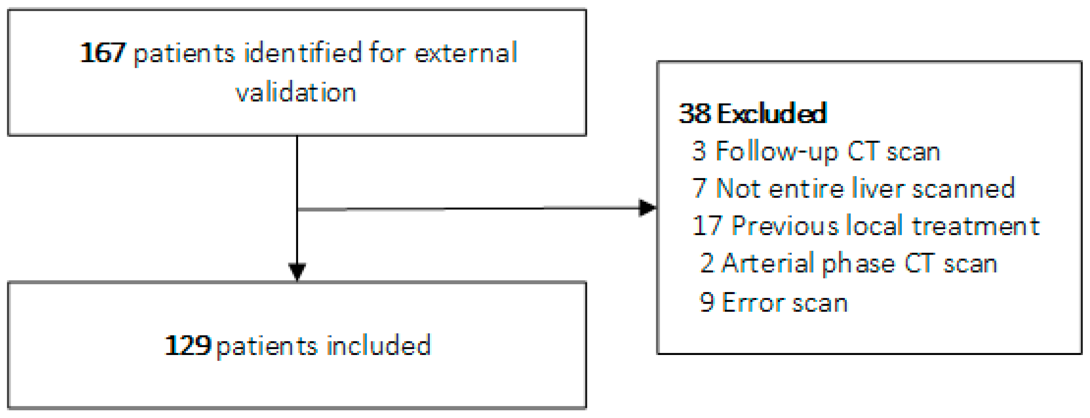
2.4. External Validation

Baseline CT scans in the portal venous phase of patients with CRLM from the NKI were collected. All CRLM were segmented by one of the members of our research team, verified, and if needed, adjusted by one abdominal radiologist (JHvW, IN, SM, JvdB) using using IntelliSpace Portal 9.0® (Philips, Best, The Netherlands)in the same manner as the CAIRO5 cohort. Similar data pre-processing and modeling were applied. In addition, ComBat harmonization was applied to the extracted features of the external validation cohort to reduce the center effect.

2.5. Statistics

The discriminative performance of the machine-learning models was assessed using the receiver operating curve (ROC), supported by the area under the receiver operating curve (AUC), accuracy, sensitivity, and specificity measures. The optimal cut-off value was determined by taking the point with the greatest difference between the sensitivity and 1—specificity values in the ROC curve (the Youden index). The 95% confidence intervals (CI) of the AUC scores were determined using the Delong test to give an indication of their robustness. The calibration was evaluated using the locally estimated scatterplot smoothing (LOESS) calibration curve and the Brier score, demonstrating the agreement between the observed and predicted probabilities. Baseline characteristics of the discovery cohort and external validation cohort were compared. The relation between the baseline variables and genetic mutation status was assessed using univariable linear regression models. Categorical variables were reported as frequencies and percentages and compared with Chi-square test or Fisher exact test, as appropriate. Continuous variables were displayed as median with interquartile range (IQR) and compared with Mann–Whitney U-test or t-test based on the distribution of data. Tests were considered statistically significant with a two-sided p-value < 0.05. Statistical analyses were performed using SAS® Studio (version 5.2, SAS® Viya®.03.05, SAS Institute Inc., Cary, NC, USA).

3. Results

3.1. Study Population

In total, 255 of 368 eligible patients from the CAIRO5 trial were included in the discovery cohort of this study (Figure 2). The 80:20 train/test ratio resulted in a training cohort of 204 patients and a test cohort of 51 patients. Of the 167 eligible patients for external validation, 129 were included (Figure 3).
The median number of CRLM per patient within the discovery cohort was 12 versus 5 in the external validation cohort (p < 0.0001) and the median slice thickness within the discovery cohort was 5.0 mm versus 1.0 mm in the external validation cohort (Supplementary Materials S1). All other baseline characteristics of the discovery and external validation cohort were similar (Table 1). Age (p = 0.21), sex (p = 0.93), and number of liver metastases (p = 0.61) were not significantly associated with the KRAS mutational status in the discovery cohort.

3.2. Feature Selection and Modeling

In total, 851 radiomics features were extracted. Using random forest feature selection, 10 features were selected as the input variables for the classification models (Supplement S5). The optimal model parameters were found using cross-validated hyperparameter tuning (Supplement S4).

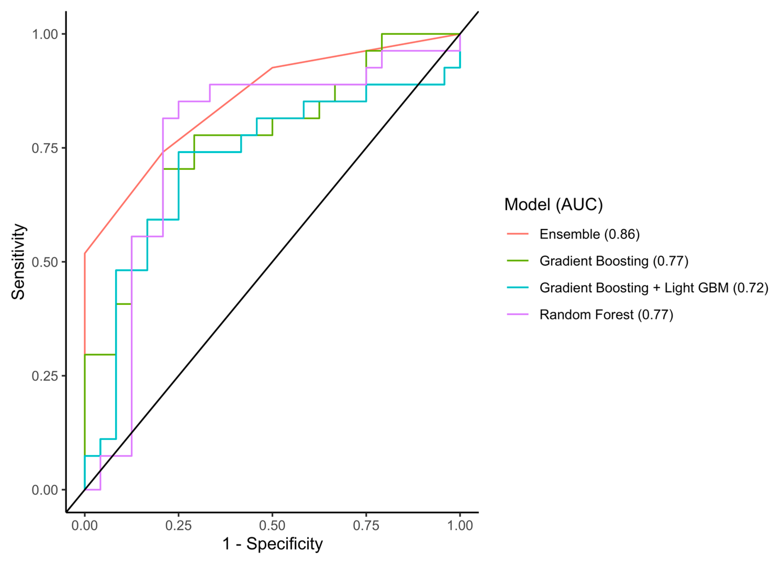
3.3. Performance of the Models

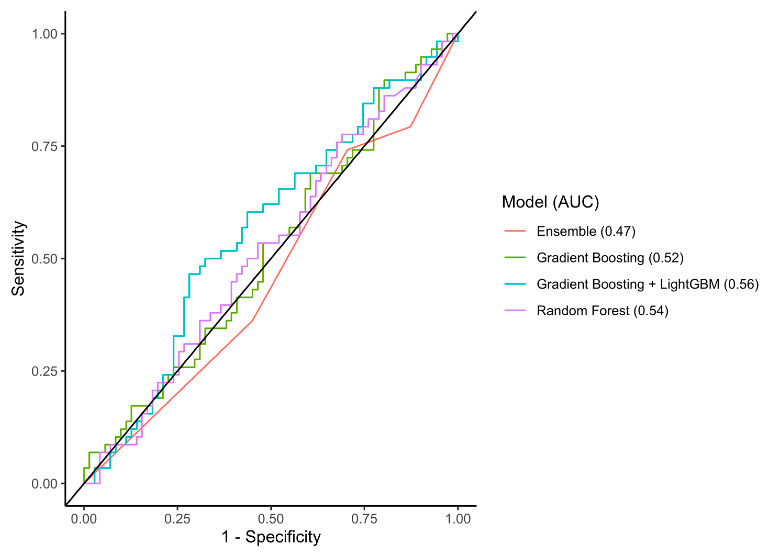
3.3.1. Discrimination Accuracy
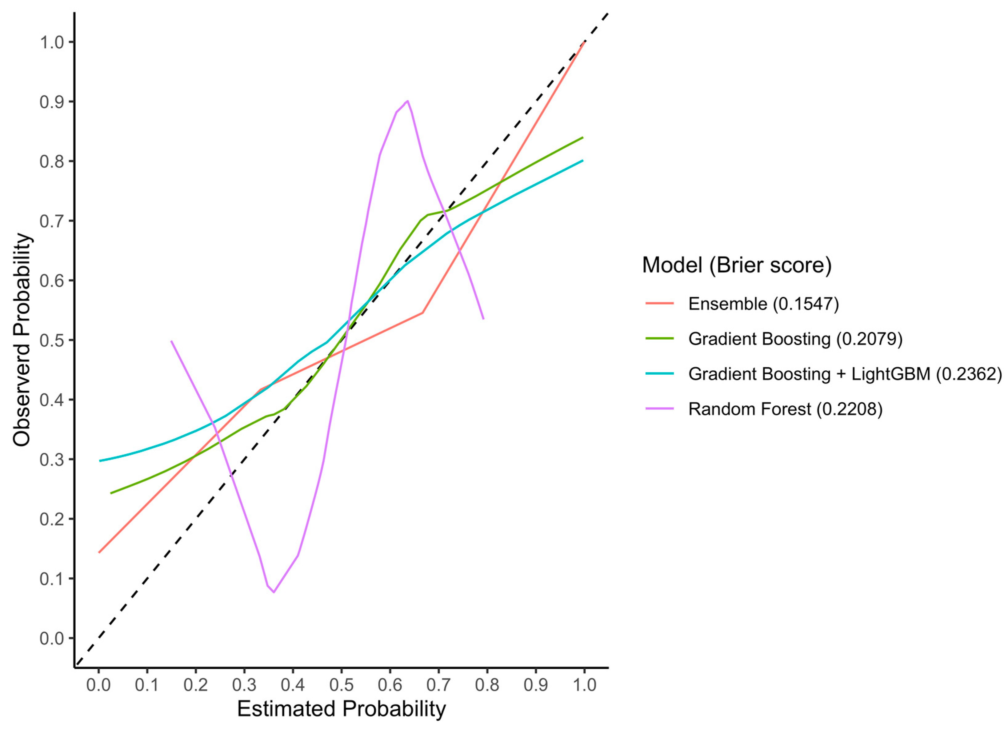
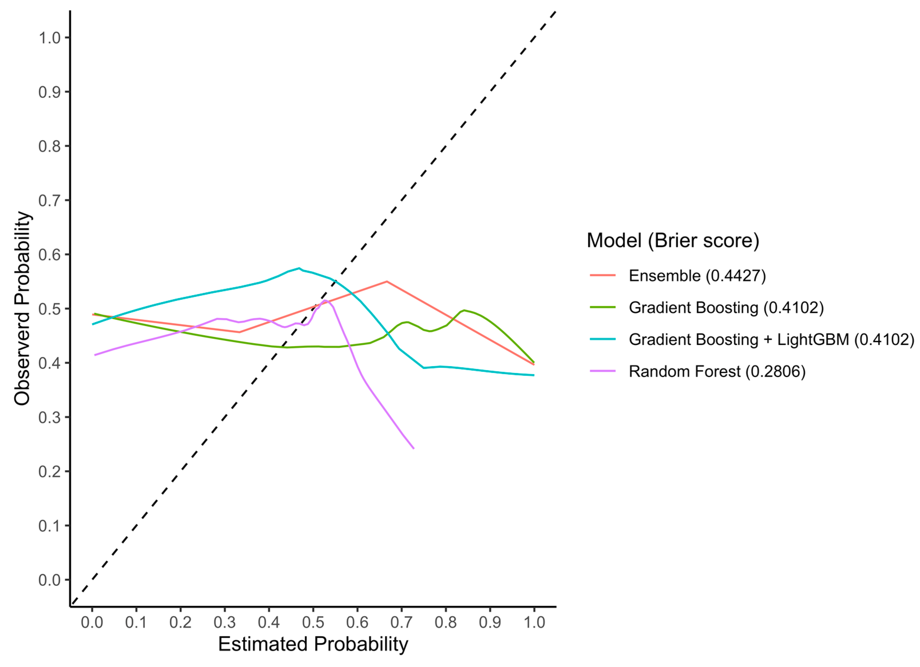
In the test set, the RF, GB, GBM, and Ensemble models reached AUCs of 0.77 (95%CI 0.62–0.93), 0.77 (95%CI 0.64–0.90), 0.72 (95%CI 0.57–0.87), and 0.86 (95%CI 0.76–0.95) with an accuracy of 0.80, 0.73, 0.71, and 0.77, respectively (Figure 4). In the external validation cohort, the RF, GB, GBM, and Ensemble models reached AUCs of 0.54 (95%CI 0.44–0.64), 0.52 (95%CI 0.42–0.62), 0.56 (95%CI 0.46–0.67), and 0.47 (95%CI 0.37–0.56), respectively (Figure 5). An overview of all the evaluation metrics is shown in Table 2.

3.3.2. Calibration

In the test set, the RF, GB, GBM, and Ensemble models reached Brier scores of 0.22, 0.21, 0.24, and 0.15, respectively (Figure 6). In the external validation cohort, the RF, GB, GBM, and Ensemble models reached Brier scores of 0.28, 0.41, 0.41, and 0.44, respectively (Figure 7).

4. Discussion

This study demonstrates that the genetic mutation status (KRAS mutation versus no mutation) of patients with CRLM could be identified with a good accuracy in the discovery cohort, using machine-learning models incorporating pre-treatment CT-based imaging features. However, the machine-learning models showed poor AUC scores in the external validation cohort, indicating no relationship between the genetic mutation status and radiomics imaging features.
If radiomics could prove to be predictive of the mutation status on a lesion level, it could facilitate personalized cancer treatment on a lesion level in the future. Brudvik et al. have reported that RAS mutations are associated with a higher positive margin resection rate [29]. Thus, for RAS-mutated CRLM, a more careful intraoperative assessment of the resection margin is recommended [29].
In previous studies, genetically driven physiological functions related to venous invasion in liver cancer were correlated with imaging features, suggesting that biological or morphological changes following genetic mutations are detected on medical imaging with radiomics [30]. But following the results in the external validation cohort of this study, the question remains if there is any relationship between radiomics and genetic mutations in patients with CRLM.
The models’ accuracy in this study’s test set was higher than a similar study, in which an AUC of 0.74 was reached in predicting the RAS/BRAF versus wild-type mutation status in patients with CRLM based on radiomics features [23]. Two other studies predicting the genetic mutation status in patients with colorectal cancer achieved comparable AUC scores of 0.82 and 0.83 [31,32]. However, all of these studies were conducted using smaller datasets and more importantly, the models were not externally validated [23,31] and therefore at risk of overfitting.
External validation of machine-learning models is crucial for the assessment of clinical applicability in daily clinical practice. However, it is a major challenge to develop models that also perform successfully in unknown external datasets [33,34]. Hence, machine-learning models should be trained on data that are as realistic and robust as possible. The variety in CT acquisition parameters could be considered a strength concerning external validity, as it is a good representation of different CT scans in daily practice since the CT scans were acquired from 66 hospitals. However, the developed models still performed poorly in the external validation set. This could imply that the selection of patients used for model development had a large influence on the generalizability of the models, as the population consisted of selected patients with initially unresectable CRLM and a median number of 12 metastases. Additionally, a difference in slice thickness was observed between the discovery cohort and external validation cohort.
In addition to patient selection, tumor segmentation also has a considerable influence on model performance, as this segmentation determines which voxels are analyzed and thus can influence the extracted feature values [17]. In this study, the CRLM were validated by one expert radiologist, which could have introduced interobserver bias [35], and therefore the stability of the radiomics features between different segmentations was not investigated. The inability to reject instable features may have affected the generalizability of the models [17]. The use of fully automatic segmentation models in the future would be valuable in increasing reproducibility and constructing robust models.
Another limitation is that the models were constructed based on imaging features calculated from the segmentation of all the CRLM combined. As a result, it was assumed that the mutation status was uniform amongst all the CRLM in one patient and intertumor heterogeneity was not taken into account. Two studies have shown intertumoral heterogeneity in 2/97 (2.1%) and 11/94 (11.7%) of patients with multiple CRLM, respectively [24,25]. Only the first study, however, used highly sensitive mutation analyses. Finally, it would be interesting to investigate if radiomics models can distinguish between RAS and BRAF mutations or even between specific KRAS mutation variants. Research has shown that patients with CRLM with a KRAS A146-mutated tumor have a higher tumor burden, associated with worse clinical outcomes, compared with patients with other KRAS mutation variants [36]. Due to high imbalance ratios in terms of both RAS/BRAF mutations and specific KRAS mutation variants in the current study, these analyses could not be performed without a high risk of bias.
In an in-depth analysis to explain the poor performance in the external validation cohort, it was firstly observed that the correlation between the individual features and the outcome was weak. Moreover, the correlation coefficient between the internal train, internal test, and external validation set varied significantly. The most predictive features in the internal test were not predictive in the external validation set. Secondly, the distribution of the KRAS mutation and wild-type was distributed similarly between the internal and external dataset. Both were around half of the patient population, which is an adequate representation of the distribution in the daily patient population. Hence, the misclassified samples were not under-represented in the training data.
We advocate for more standardized and robust radiomics research, as the lack of generalizability has to be addressed before a conclusion can be drawn about the potential clinical value of radiomics. External validation is crucial in testing the generalizability of radiomics models, which many previous studies have lacked. Our study has shown that despite promising internal results, the radiomics models showed an inadequate performance in the external validation cohort. In an attempt to build radiomics models that perform well across different, unseen patient cohorts, it is important to have a standardized approach in their development. This could include multiple patient cohorts in the development phase, and also external validation across multiple patient cohorts.

5. Conclusions

In conclusion, machine-learning models incorporating pre-treatment CT imaging features could identify the genetic mutation status (KRAS mutation and no mutation) in patients with CRLM with a good accuracy in the test set. However, in the external validation set, the models performed poorly. This study demonstrates the importance of external validation, which should be mandatory in all future studies in this field. Automation and standardization are equally necessary to further explore the potential of radiomics.

Supplementary Materials

The following supporting information can be downloaded at: https://www.mdpi.com/article/10.3390/cancers15235648/s1, S1. CT acquisition parameters; S2. Radiomics Quality Score; S3. Pyradiomics feature extraction settings; S4. Model configurations; S5. Selected features.

Author Contributions

Conceptualization, N.W., M.Z., I.v.‘t.E., J.S., R.-J.S., C.P., J.H., R.F. and G.K.; data curation, N.W., M.Z., I.v.‘t.E., Z.B., J.H.v.W., S.M., J.v.d.B., I.N. and Dutch Colorectal Cancer Group Liver Expert Panel; formal analysis, N.W., M.Z., D.v.d.M. and J.R.; funding acquisition, N.W., J.S., R.-J.S., C.P., J.H. and G.K.; investigation, N.W., M.Z. and J.R.; methodology, N.W., M.Z., D.v.d.M., I.v.‘t.E., Z.B., J.R., S.v.D., J.S., R.-J.S., C.P., J.H., H.M. and G.K.; project administration, N.W. and M.Z.; resources, I.v.‘t.E., Z.B., J.S., G.M., R.-J.S., C.P., J.H., R.B.-T., R.F. and G.K.; software, N.W., D.v.d.M. and J.R.; supervision, J.S., R.-J.S., C.P., J.H., H.M. and G.K.; validation, M.Z.; visualization, N.W., M.Z., D.v.d.M. and J.R.; writing—original draft, N.W., M.Z., D.v.d.M. and J.R.; writing—review and editing, I.v.‘t.E., Z.B., J.H.v.W., S.M., J.v.d.B., I.N., S.v.D., J.S., G.M., R.-J.S., C.P., J.H., R.B.-T., R.F., H.M. and G.K. All authors have read and agreed to the published version of the manuscript.

Funding

This research has received funding by the Dutch Cancer Society (KWF Kankerbestrijding), project number 14002/2021-2 and by KWF project number 10438, and an unrestricted grant from the Cancer Center Amsterdam Foundation. The CAIRO5 study is supported by unrestricted scientific grants from Roche and Amgen.

Institutional Review Board Statement

This study was conducted in accordance with the Declaration of Helsinki, and approved by the Ethics Committee of Amsterdam UMC (2019.236 at 24 April 2019).

Informed Consent Statement

Informed consent was obtained from all subjects involved in the CAIRO5 trial. Due to the retrospective nature of this study, informed consent was waived by the Ethical Committee.

Data Availability Statement

We closely collaborate with international partners in the Pancreatobiliary and Hepatic Artificial Intelligence Research (PHAIR) consortium. A PHAIR github will be created to share all results and data.

Acknowledgments

We thank all patients who consented to participate in the CAIRO5 study. SAS supported the study in kind by providing advanced analytics and deep-learning expertise. Martinus van Amerongen, Thiery Chapelle, Marc Engelbrecht, Michael Gerhards, Dirk Grunhagen, Thomas van Gulik, Koert de Jong, Joost Klaase, Mike Liem, Krijn van Lienden, Quintus Molenaar, Gijs Patijn, Arjen Rijken, Theo Ruers, Cornelis Verhoef, Johannes de Wilt, all participated actively in the Dutch Colorectal Cancer Group Liver Expert Panel.

Conflicts of Interest

C.J.A.P. has an advisory role for Nordic Pharma. H.A.M. is co-founder and shareholder of Nicolab. The funders had no role in the design, conduct, analysis of the study, writing of the manuscript, nor in the decision to submit the manuscript for publication.

References

  1. Bray, F.; Ferlay, J.; Soerjomataram, I.; Siegel, R.L.; Torre, L.A.; Jemal, A. Global cancer statistics 2018: GLOBOCAN estimates of incidence and mortality worldwide for 36 cancers in 185 countries. CA Cancer J. Clin. 2018, 68, 394–424. [Google Scholar] [CrossRef] [PubMed]
  2. Abdalla, E.K.; Adam, R.; Bilchik, A.J.; Jaeck, D.; Vauthey, J.N.; Mahvi, D. Improving resectability of hepatic colorectal metastases: Expert consensus statement. Ann. Surg. Oncol. 2006, 13, 1271–1280. [Google Scholar] [CrossRef] [PubMed]
  3. Donadon, M.; Ribero, D.; Morris-Stiff, G.; Abdalla, E.K.; Vauthey, J.N. New paradigm in the management of liver-only metastases from colorectal cancer. Gastrointest. Cancer Res. GCR 2007, 1, 20–27. [Google Scholar] [PubMed]
  4. Adam, R.; Kitano, Y. Multidisciplinary approach of liver metastases from colorectal cancer. Ann. Gastroenterol. Surg. 2019, 3, 50–56. [Google Scholar] [CrossRef] [PubMed]
  5. Lam, V.W.; Spiro, C.; Laurence, J.M.; Johnston, E.; Hollands, M.J.; Pleass, H.C.; Richardson, A.J. A systematic review of clinical response and survival outcomes of downsizing systemic chemotherapy and rescue liver surgery in patients with initially unresectable colorectal liver metastases. Ann. Surg. Oncol. 2012, 19, 1292–1301. [Google Scholar] [CrossRef] [PubMed]
  6. Abdalla, E.K.; Vauthey, J.N.; Ellis, L.M.; Ellis, V.; Pollock, R.; Broglio, K.R.; Hess, K.; Curley, S.A. Recurrence and outcomes following hepatic resection, radiofrequency ablation, and combined resection/ablation for colorectal liver metastases. Ann. Surg. 2004, 239, 818–825; discussion 825–827. [Google Scholar] [CrossRef] [PubMed]
  7. Venook, A.P.; Niedzwiecki, D.; Lenz, H.J.; Innocenti, F.; Fruth, B.; Meyerhardt, J.A.; Schrag, D.; Greene, C.; O’Neil, B.H.; Atkins, J.N.; et al. Effect of First-Line Chemotherapy Combined with Cetuximab or Bevacizumab on Overall Survival in Patients With KRAS Wild-Type Advanced or Metastatic Colorectal Cancer: A Randomized Clinical Trial. J. Am. Med. Assoc. 2017, 317, 2392–2401. [Google Scholar] [CrossRef]
  8. Van Cutsem, E.; Köhne, C.H.; Láng, I.; Folprecht, G.; Nowacki, M.P.; Cascinu, S.; Shchepotin, I.; Maurel, J.; Cunningham, D.; Tejpar, S.; et al. Cetuximab plus irinotecan, fluorouracil, and leucovorin as first-line treatment for metastatic colorectal cancer: Updated analysis of overall survival according to tumor KRAS and BRAF mutation status. J. Clin. Oncol. 2011, 29, 2011–2019. [Google Scholar] [CrossRef]
  9. Piawah, S.; Venook, A.P. Targeted therapy for colorectal cancer metastases: A review of current methods of molecularly targeted therapy and the use of tumor biomarkers in the treatment of metastatic colorectal cancer. Cancer 2019, 125, 4139–4147. [Google Scholar] [CrossRef]
  10. Adam, R.; de Gramont, A.; Figueras, J.; Kokudo, N.; Kunstlinger, F.; Loyer, E.; Poston, G.; Rougier, P.; Rubbia-Brandt, L.; Sobrero, A.; et al. Managing synchronous liver metastases from colorectal cancer: A multidisciplinary international consensus. Cancer Treat. Rev. 2015, 41, 729–741. [Google Scholar] [CrossRef]
  11. Yamashita, S.; Chun, Y.S.; Kopetz, S.E.; Vauthey, J.N. Biomarkers in colorectal liver metastases. Br. J. Surg. 2018, 105, 618–627. [Google Scholar] [CrossRef] [PubMed]
  12. Peeters, M.; Oliner, K.S.; Price, T.J.; Cervantes, A.; Sobrero, A.F.; Ducreux, M.; Hotko, Y.; André, T.; Chan, E.; Lordick, F.; et al. Analysis of KRAS/NRAS Mutations in a Phase III Study of Panitumumab with FOLFIRI Compared with FOLFIRI Alone as Second-line Treatment for Metastatic Colorectal Cancer. Clin. Cancer Res. An Off. J. Am. Assoc. Cancer Res. 2015, 21, 5469–5479. [Google Scholar] [CrossRef] [PubMed]
  13. Eklöf, V.; Wikberg, M.L.; Edin, S.; Dahlin, A.M.; Jonsson, B.A.; Öberg, Å.; Rutegård, J.; Palmqvist, R. The prognostic role of KRAS, BRAF, PIK3CA and PTEN in colorectal cancer. Br. J. Cancer 2013, 108, 2153–2163. [Google Scholar] [CrossRef] [PubMed]
  14. Peeters, M.; Kafatos, G.; Taylor, A.; Gastanaga, V.M.; Oliner, K.S.; Hechmati, G.; Terwey, J.H.; Van Krieken, J.H. Prevalence of RAS mutations and individual variation patterns among patients with metastatic colorectal cancer: A pooled analysis of randomised controlled trials. Eur. J. Cancer 2015, 51, 1704–1713. [Google Scholar] [CrossRef]
  15. Van Cutsem, E.; Cervantes, A.; Adam, R.; Sobrero, A.; Van Krieken, J.H.; Aderka, D.; Aguilar, E.A.; Bardelli, A.; Benson, A.; Bodoky, G.; et al. ESMO consensus guidelines for the management of patients with metastatic colorectal cancer. Ann. Oncol. 2016, 27, 1386–1422. [Google Scholar] [CrossRef]
  16. Gillies, R.J.; Kinahan, P.E.; Hricak, H. Radiomics: Images Are More than Pictures, They Are Data. Radiology 2016, 278, 563–577. [Google Scholar] [CrossRef]
  17. Lambin, P.; Leijenaar, R.T.H.; Deist, T.M.; Peerlings, J.; de Jong, E.E.C.; van Timmeren, J.; Sanduleanu, S.; Larue, R.T.; Even, A.J.; Jochems, A.; et al. Radiomics: The bridge between medical imaging and personalized medicine. Nat. Rev. Clin. Oncol. 2017, 14, 749–762. [Google Scholar] [CrossRef]
  18. Wesdorp, N.J.; Hellingman, T.; Jansma, E.P.; van Waesberghe, J.T.M.; Boellaard, R.; Punt, C.J.A.; Huiskens, J.; Kazemier, G. Advanced analytics and artificial intelligence in gastrointestinal cancer: A systematic review of radiomics predicting response to treatment. Eur. J. Nucl. Med. Mol. Imaging 2020, 48, 1785–1794. [Google Scholar] [CrossRef]
  19. Wesdorp, N.J.; van Goor, V.J.; Kemna, R.; Jansma, E.P.; van Waesberghe, J.H.T.M.; Swijnenburg, R.J.; Punt, C.J.A.; Huiskens, J.; Kazemier, G. Advanced image analytics predicting clinical outcomes in patients with colorectal liver metastases: A systematic review of the literature. Surg. Oncol. 2021, 38, 101578. [Google Scholar] [CrossRef]
  20. Staal, F.C.R.; van der Reijd, D.J.; Taghavi, M.; Lambregts, D.M.J.; Beets-Tan, R.G.H.; Maas, M. Radiomics for the Prediction of Treatment Outcome and Survival in Patients with Colorectal Cancer: A Systematic Review. Clin. Color. Cancer 2021, 20, 52–71. [Google Scholar] [CrossRef]
  21. Aerts, H.J.; Velazquez, E.R.; Leijenaar, R.T.; Parmar, C.; Grossmann, P.; Carvalho, S.; Bussink, J.; Monshouwer, R.; Haibe-Kains, B.; Rietveld, D.; et al. Decoding tumour phenotype by noninvasive imaging using a quantitative radiomics approach. Nat. Commun. 2014, 5, 4006. [Google Scholar] [CrossRef] [PubMed]
  22. Badic, B.; Tixier, F.; Cheze Le Rest, C.; Hatt, M.; Visvikis, D. Radiogenomics in Colorectal Cancer. Cancers 2021, 13, 973. [Google Scholar] [CrossRef]
  23. Shi, R.; Chen, W.; Yang, B.; Qu, J.; Cheng, Y.; Zhu, Z.; Gao, Y.; Wang, Q.; Liu, Y.; Li, Z.; et al. Prediction of KRAS, NRAS and BRAF status in colorectal cancer patients with liver metastasis using a deep artificial neural network based on radiomics and semantic features. Am. J. Cancer Res. 2020, 10, 4513–4526. [Google Scholar] [PubMed]
  24. Brunsell, T.H.; Sveen, A.; Bjornbeth, B.A.; Rosok, B.I.; Danielsen, S.A.; Brudvik, K.W.; Berg, K.C.; Johannessen, B.; Cengija, V.; Abildgaard, A.; et al. High Concordance and Negative Prognostic Impact of RAS/BRAF/PIK3CA Mutations in Multiple Resected Colorectal Liver Metastases. Clin. Color. Cancer 2020, 19, e26–e47. [Google Scholar] [CrossRef] [PubMed]
  25. Loes, I.M.; Immervoll, H.; Sorbye, H.; Angelsen, J.H.; Horn, A.; Knappskog, S.; Lønning, P.E. Impact of KRAS, BRAF, PIK3CA, TP53 status and intraindividual mutation heterogeneity on outcome after liver resection for colorectal cancer metastases. Int. J. Cancer 2016, 139, 647–656. [Google Scholar] [CrossRef] [PubMed]
  26. Jia, L.L.; Zhao, J.X.; Zhao, L.P.; Tian, J.H.; Huang, G. Current status and quality of radiomic studies for predicting KRAS mutations in colorectal cancer patients: A systematic review and meta-analysis. Eur. J. Radiol. 2023, 158, 110640. [Google Scholar] [CrossRef]
  27. Huiskens, J.; van Gulik, T.M.; van Lienden, K.P.; Engelbrecht, M.R.; Meijer, G.A.; van Grieken, N.C.; Schriek, J.; Keijser, A.; Mol, L.; Molenaar, I.Q.; et al. Treatment strategies in colorectal cancer patients with initially unresectable liver-only metastases, a study protocol of the randomised phase 3 CAIRO5 study of the Dutch Colorectal Cancer Group (DCCG). BMC Cancer 2015, 15, 365. [Google Scholar] [CrossRef] [PubMed]
  28. van Griethuysen, J.J.M.; Fedorov, A.; Parmar, C.; Hosny, A.; Aucoin, N.; Narayan, V.; Beets-Tan, R.G.; Fillion-Robin, J.C.; Pieper, S.; Aerts, H.J. Computational Radiomics System to Decode the Radiographic Phenotype. Cancer Res. 2017, 77, e104. [Google Scholar] [CrossRef]
  29. Brudvik, K.W.; Mise, Y.; Chung, M.H.; Chun, Y.S.; Kopetz, S.E.; Passot, G.; Conrad, C.; Maru, D.M.; Aloia, T.A.; Vauthey, J.N. RAS Mutation Predicts Positive Resection Margins and Narrower Resection Margins in Patients Undergoing Resection of Colorectal Liver Metastases. Ann. Surg. Oncol. 2016, 23, 2635–2643. [Google Scholar] [CrossRef]
  30. Segal, E.; Sirlin, C.B.; Ooi, C.; Adler, A.S.; Gollub, J.; Chen, X.; Chan, B.K.; Matcuk, G.R.; Barry, C.T.; Chang, H.Y.; et al. Decoding global gene expression programs in liver cancer by noninvasive imaging. Nat. Biotechnol. 2007, 25, 675–680. [Google Scholar] [CrossRef]
  31. Taguchi, N.; Oda, S.; Yokota, Y.; Yamamura, S.; Imuta, M.; Tsuchigame, T.; Nagayama, Y.; Kidoh, M.; Nakaura, T.; Shiraishi, S.; et al. CT texture analysis for the prediction of KRAS mutation status in colorectal cancer via a machine learning approach. Eur. J. Radiol. 2019, 118, 38–43. [Google Scholar] [CrossRef] [PubMed]
  32. Yang, L.; Dong, D.; Fang, M.; Zhu, Y.; Zang, Y.; Liu, Z.; Zhang, H.; Ying, J.; Zhao, X.; Tian, J. Can CT-based radiomics signature predict KRAS/NRAS/BRAF mutations in colorectal cancer? Eur. Radiol. 2018, 28, 2058–2067. [Google Scholar] [CrossRef] [PubMed]
  33. van de Sande, D.; van Genderen, M.E.; Huiskens, J.; Gommers, D.; van Bommel, J. Moving from bytes to bedside: A systematic review on the use of artificial intelligence in the intensive care unit. Intensive Care Med. 2021, 47, 750–760. [Google Scholar] [CrossRef] [PubMed]
  34. van de Sande, D.; Van Genderen, M.E.; Smit, J.M.; Huiskens, J.; Visser, J.J.; Veen, R.E.R.; Van Unen, E.; Hilgers, O.; Gommers, D.; van Bommel, J. Developing, implementing and governing artificial intelligence in medicine: A step-by-step approach to prevent an artificial intelligence winter. BMJ Health Care Inform. 2022, 29, e100495. [Google Scholar] [CrossRef]
  35. Marshall, C.; Thirion, P.; Mihai, A.; Armstrong, J.G.; Cournane, S.; Hickey, D.; McClean, B.; Quinn, J. Interobserver Variability of Gross Tumor Volume Delineation for Colorectal Liver Metastases Using Computed Tomography and Magnetic Resonance Imaging. Adv. Radiat. Oncol. 2023, 8, 101020. [Google Scholar] [CrossRef]
  36. van ‘t Erve, I.; Wesdorp, N.J.; Medina, J.E.; Ferreira, L.; Leal, A.; Huiskens, J.; Bolhuis, K.; van Waesberghe, J.H.T.; Swijnenburg, R.J.; van den Broek, D.; et al. KRAS A146 Mutations Are Associated with Distinct Clinical Behavior in Patients With Colorectal Liver Metastases. JCO Precis. Oncol. 2021, PO2100223. [Google Scholar] [CrossRef]
Figure 1. Illustration of the radiomics workflow. Abbreviations: AUC-ROC, area under the receiver operating characteristic curve; CRLM, colorectal liver metastases; CT, computed tomography; VOI, volume of interest.
Cancers 15 05648 g001
Figure 2. Flowchart of CAIRO5 patient inclusion. Abbreviations: CT, computed tomography; MRI, magnetic resonance imaging.
Cancers 15 05648 g002
Figure 3. Flowchart of patient inclusion external validation cohort. Abbreviations: CT, computed tomography.
Cancers 15 05648 g003
Figure 4. AUC-ROC of the three machine-learning models in the internal test set. Abbreviations: AUC-ROC, area under the receiver operating characteristic curve.
Cancers 15 05648 g004
Figure 5. AUC-ROC of the three machine-learning models in the external validation set. Abbreviations: AUC-ROC, area under the receiver operating characteristic curve.
Cancers 15 05648 g005
Figure 6. LOESS calibration curves and Brier scores of the three machine-learning models in the internal test set. Abbreviations: LOESS, locally estimated scatterplot smoothing.
Cancers 15 05648 g006
Figure 7. LOESS calibration curves and Brier scores of the three machine-learning models in the external validation set. Abbreviations: LOESS, locally estimated scatterplot smoothing.
Cancers 15 05648 g007
Table 1. Baseline patient characteristics.
Baseline CharacteristicsDiscovery Cohort
(n = 255)
External Validation Set (n = 129)p-Value
Age (years)62 (55–70)62 (53–69)0.607
Sex 0.068
 Male170 (66.7)73 (56.6)
 Female85 (33.3)56 (43.4)
KRAS mutational status 0.149
 Mutation136 (53.3)58 (45.0)
 Wild-type119 (46.7)71 (55.0)
Number of liver metastases12 (7–23) 5 (2–11)<0.0001
Note—values are shown as median (interquartile range) or number of participants (percentage).
Table 2. Model performance in discriminating KRAS mutant versus wild-type mutation status.
Machine-Learning ClassifierCohortAUC (95% CI)SensitivitySpecificityAccuracy
Random ForestTrain0.97 (0.95–0.99)0.820.950.89
Test0.77 (0.62–0.93)0.850.750.80
External validation0.54 (0.44–0.64)0.440.540.50
Gradient BoostingTrain0.96 (0.94–1.00)0.950.980.98
Test0.77 (0.64–0.90)0.670.790.73
External validation0.52 (0.42–0.62)0.460.570.52
Gradient Boosting (LightGBM)Train0.98 (0.97–1.00)0.980.990.98
Test0.72 (0.57–0.87)0.750.670.71
External validation0.56 (0.46–0.67)0.280.590.42
EnsembleTrain0.97 (0.94–1.00)0.970.930.95
Test0.86 (0.76–0.95)0.930.580.77
External validation0.47 (0.37–0.56)0.300.740.50
Disclaimer/Publisher’s Note: The statements, opinions and data contained in all publications are solely those of the individual author(s) and contributor(s) and not of MDPI and/or the editor(s). MDPI and/or the editor(s) disclaim responsibility for any injury to people or property resulting from any ideas, methods, instructions or products referred to in the content.

