
Dedifferentiated Endometrial Carcinoma: A Rare Aggressive Neoplasm-Clinical, Morphological and Immunohistochemical Features

Abstract
:Simple Summary
Abstract
1. Introduction
1.1. Clinical Features
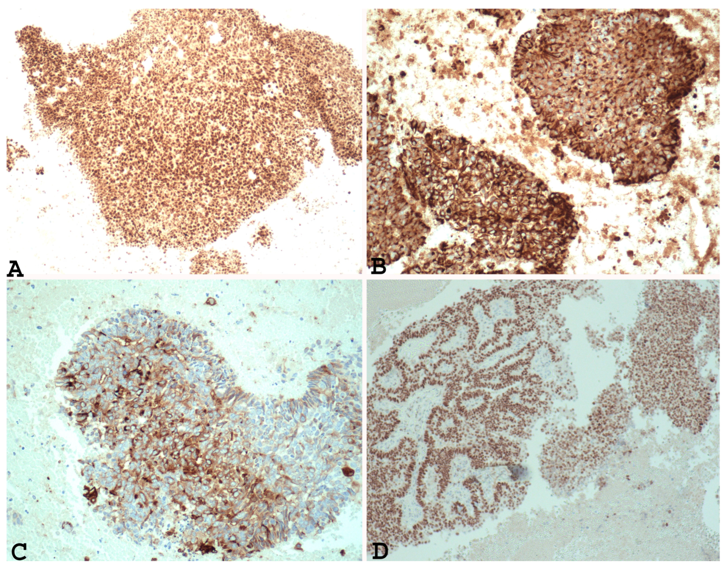
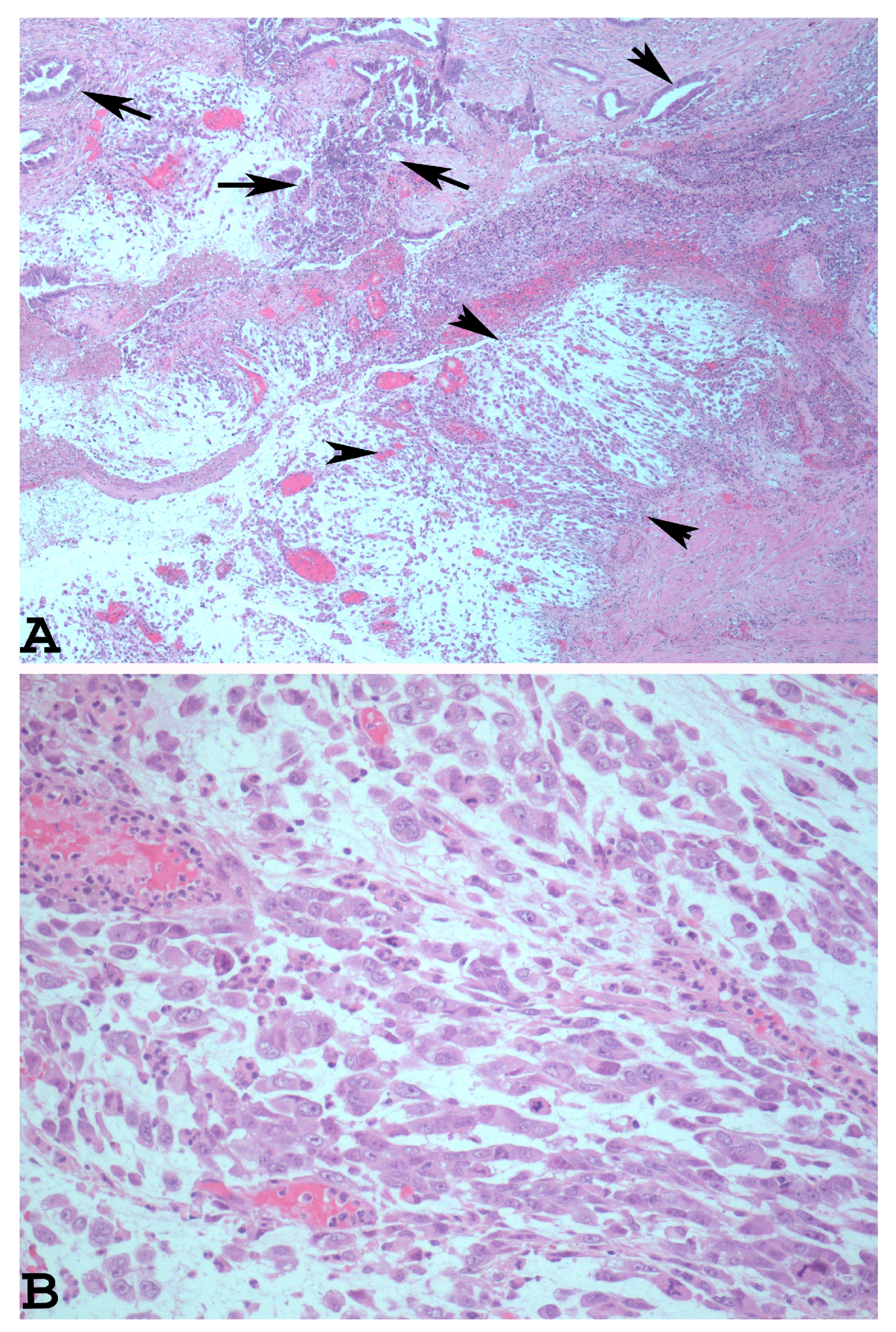
1.2. The Main Morphological and Immunohistochemical Features of EDC Used for Correct Pathological Diagnosis
1.3. Genetic Alterations of EDC: Comparison with Other Subtypes of Endometrial Carcinoma and Impact on Prognosis and Treatment
1.4. Molecular Alterations of EDC and Impact on Prognosis and Therapy
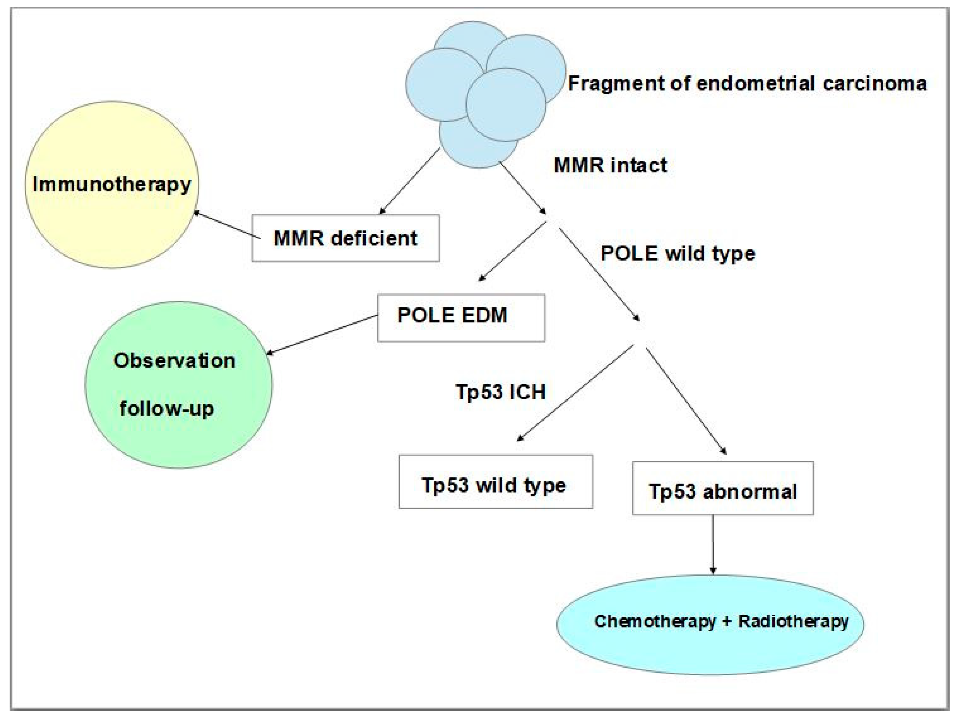
2. Mismatch Repair (MMR) Gene Mutations
3. POLE Domain Mutations
4. Switch/Sucrose Non-Fermentable (SWI/SNF) Complex
5. Tp53 Mutations
6. Conclusions
Author Contributions
Funding
Acknowledgments
Conflicts of Interest
References
- Friedmann-Morvinski, D.; Verma, I.M. Dedifferentiation and reprogramming: Origins of cancer stem cells. EMBO Rep. 2014, 15, 244–253. [Google Scholar] [CrossRef] [PubMed]
- Yuan, S.; Norgard, R.J.; Stanger, B.Z. Cellular Plasticity in Cancer. Cancer Discov. 2019, 9, 837–851. [Google Scholar] [CrossRef] [PubMed]
- Hölzel, M.; Bovier, A.; Tüting, T. Plasticity of tumour and immune cells: A source of heterogeneity and a cause for therapy resistance? Nat. Rev. Cancer 2013, 13, 365–376. [Google Scholar] [CrossRef] [PubMed]
- Boumahdi, S.; de Sauvage, F.J. The great escape: Tumour cell plasticity in resistance to targeted therapy. Nat. Rev. Drug Discov. 2020, 19, 39–56. [Google Scholar] [CrossRef]
- Skalova, A.; Leivo, I.; Hellquist, H.; Agaimy, A.; Simpson, R.H.W.; Stenman, G.; Vander Poorten, V.; Bishop, J.A.; Franchi, A.; Hernandez-Prera, J.C.; et al. High-grade Transformation/Dedifferentiation in Salivary Gland Carcinomas: Occurrence across Subtypes and Clinical Significance. Adv. Anat. Pathol. 2021, 28, 107–118. [Google Scholar] [CrossRef]
- Huang, S.C.; Chen, K.H.; Ng, K.F.; Lin, I.C.; Chao, Y.C.; Yeh, T.S.; Chuang, H.C.; Chen, T.C. Dedifferentiation-like tubular and solid carcinoma of the stomach shows phenotypic divergence and association with deficient SWI/SNF complex. Virchows Arch. 2022, 480, 771–781. [Google Scholar] [CrossRef]
- Fukunaga, Y.; Fukuda, A.; Omatsu, M.; Namikawa, M.; Sono, M.; Masuda, T.; Araki, O.; Nagao, M.; Yoshikawa, T.; Ogawa, S.; et al. Loss of Arid1a and Pten in Pancreatic Ductal Cells Induces Intraductal Tubulopapillary Neoplasm via the YAP/TAZ Pathway. Gastroenterology 2022, 163, 466–480.e6. [Google Scholar] [CrossRef]
- Agaimy, A.; Bertz, S.; Cheng, L.; Hes, O.; Junker, K.; Keck, B.; Lopez-Beltran, A.; Stöckle, M.; Wullich, B.; Hartmann, A. Loss of expression of the SWI/SNF complex is a frequent event in undifferentiated/dedifferentiated urothelial carcinoma of the urinary tract. Virchows Arch. 2016, 469, 321–330. [Google Scholar] [CrossRef]
- Tenti, P.; Carnevali, L.; Paulli, M.; Franchi, M.; Babilonti, L. Dedifferentiating endometrial adenocarcinoma. Report of a case. Eur. J. Gynaecol. Oncol. 1989, 10, 292–294. [Google Scholar]
- Silva, E.G.; Deavers, M.T.; Bodurka, D.C.; Malpica, A. Association of Low-Grade Endometrioid carcinoma of the uterus and ovary with carcinoma: A new type of dedifferentiated carcinoma? Int. J. Gynecol. Pathol. 2006, 25, 52–58. [Google Scholar] [CrossRef]
- Silverberg, S.G.; Nogales, F.; Tavassoli, F.A.; Devilee, P. Tumours of the Uterine Corpus. Pathology and Genetics: Tumours of the Breast and Female Genital Organs; World Health Organization Classification of Tumours; IARC Press: Lyon, France, 2003; pp. 217–257. [Google Scholar]
- Zaino, R.; Carinelli, S.G.; Eng, C.; Kurman, R.J.; Carcangiu, M.L.; Herrington, C.S.; Young, R.H. Tumours of the Uterine Corpus. In WHO Classification of Tumours of Female Reproductive Organs: World Health Organization Classification of Tumours; IARC Press: Lyon, France, 2014; pp. 121–154. [Google Scholar]
- Altrabulsi, B.; Malpica, A.; Deavers, M.T.; Deavers, M.T.; Bodurka, D.C.; Broaddus, R.; Silva, E.G. Undifferentiated carcinoma of the endometrium. Am. J. Surg. Pathol. 2005, 29, 1316–1321. [Google Scholar] [CrossRef] [PubMed]
- Ronnett, B.M.; Zaino, R.J.; Ellenson, L.H.; Kurman, R.J. Endometrial carcinoma. In Blaustein’s Pathology of the Female Genital Tract, 5th ed.; Kurman, R.J., Ed.; Springer: New York, NY, USA, 2002; pp. 501–559. [Google Scholar]
- Tafe, L.J.; Garg, K.; Chew, I.; Tornos, C.; Soslow, R.A. Endometrial and ovarian carcinomas with undifferentiated components: Clinically aggressive and frequently underrecognized neoplasms. Mod. Pathol. 2010, 23, 781–789. [Google Scholar] [CrossRef] [PubMed]
- Shen, Y.; Wang, Y.; Shi, Y.; Liu, J.; Liu, Y. Clinicopathologic study of endometrial dedifferentiated endometrioid adenocarcinoma: A case report. Int. J. Clin. Exp. Pathol. 2012, 5, 77–82. [Google Scholar] [PubMed]
- Murali, R.; Davidson, B.; Fadare, O.; Carlson, J.A.; Crum, C.P.; Gilks, C.B.; Irving, J.A.; Malpica, A.; Matias-Guiu, X.; McCluggage, W.G.; et al. High-grade Endometrial Carcinomas: Morphologic and Immunohistochemical Features, Diagnostic Challenges and Recommendations. Int. J. Gynecol. Pathol. 2019, 38, S40–S63. [Google Scholar] [CrossRef] [PubMed]
- Onder, S.; Taskin, O.C.; Sen, F.; Topuz, S.; Kucucuk, S.; Sozen, H.; Ilhan, R.; Tuzlali, S.; Yavuz, E. High expression of SALL4 and fascin, and loss of E-cadherin expression in undifferentiated/dedifferentiated carcinomas of the endometrium: An immunohistochemical and clinicopathologic study. Medicine 2017, 96, e6248. [Google Scholar] [CrossRef]
- Stewart, C.J.; Crook, M.L. Fascin expression in undifferentiated and dedifferentiated endometrial carcinoma. Hum. Pathol. 2015, 46, 1514–1520. [Google Scholar] [CrossRef]
- Taraif, S.H.; Deavers, M.T.; Malpica, A.; Silva, E.G. The significance of neuroendocrine expression in undifferentiated carcinoma of the endometrium. Int. J. Gynecol. Pathol. 2009, 28, 142–147. [Google Scholar] [CrossRef]
- Silva, E.G.; Deavers, M.T.; Malpica, A. Undifferentiated carcinoma of the endometrium: A review. Pathology 2007, 39, 134–138. [Google Scholar] [CrossRef]
- Li, Z.; Zhao, C. Clinicopathologic and Immunohistochemical Characterization of Dedifferentiated Endometrioid Adenocarcinoma. Appl. Immunohistochem. Mol. Morphol. 2016, 24, 562–568. [Google Scholar] [CrossRef]
- Morioka, S.; Tanase, Y.; Kawaguchi, R.; Uchiyama, T.; Kobayash, H. Two Cases of Dedifferentiated Endometrioid Carcinoma: Case Presentation and Brief Review of the Literature. Case Rep. Obstet. Gynecol. 2018, 2018, 7624785. [Google Scholar] [CrossRef]
- Han, J.; Ki, E.Y.; Rha, S.E.; Hur, S.; Lee, A. Dedifferentiated endometrioid carcinoma of the uterus: Report of four cases and review of literature. World J. Surg. Oncol. 2017, 15, 17. [Google Scholar] [CrossRef]
- Berretta, R.; Patrelli, T.S.; Faioli, R.; Mautone, D.; Gizzo, S.; Mezzogiorno, A.; Giordano, G.; Modena, A.B. Dedifferentiated endometrial cancer: An atypical case diagnosed from cerebellar and adrenal metastasis: Case presentation and review of literature. Int. J. Clin. Exp. Pathol. 2013, 6, 1652–1657. [Google Scholar] [PubMed]
- Wu, E.S.; Shih, I.M.; Díaz-Montes, T.P. Dedifferentiated endometrioid adenocarcinoma: An under-recognized but aggressive tumor? Gynecol. Oncol. Case Rep. 2013, 5, 25–27. [Google Scholar] [CrossRef] [PubMed]
- Goh, C.; Farah, B.L.; Ho, W.Y.; Wong, S.L.; Goh, C.H.R.; Chew, S.H.; Nadarajah, R.; Lim, Y.K.; Ho, T.H. Dedifferentiated endometrioid adenocarcinoma of the uterus: A case series and review of literature. Gynecol. Oncol. Rep. 2020, 32, 100538. [Google Scholar] [CrossRef]
- Yokomizo, R.; Yamada, K.; Iida, Y.; Kiyokawa, T.; Ueda, K.; Saito, M.; Yanaihara, N.; Nakamura, M.; Okamoto, A. Dedifferentiated endometrial carcinoma: A report of three cases and review of the literature. Mol. Clin. Oncol. 2017, 7, 1008–1012. [Google Scholar] [CrossRef] [PubMed]
- Lynch, H.T.; de la Chapelle, A. Genetic susceptibility to non-polyposis colorectal cancer. J. Med. Genet. 1999, 36, 801–818. [Google Scholar]
- Giordano, G.; D’Adda, T.; Bottarelli, L.; Lombardi, M.; Brigati, F.; Berretta, R.; Merisio, C. Two cases of low-grade endometriod carcinoma associated with undifferentiated carcinoma of the uterus (dedifferentiated carcinoma): A molecular study. Pathol. Oncol. Res. 2012, 18, 523–528. [Google Scholar] [CrossRef]
- Yiğit, S.; Ekinci, N.; Hayrullah, L.; Öcal, İ.; Bezircioğlu, İ. Dedifferentiated endometrioid adenocarcinoma; clinicopathologic and immunohistochemical features of five cases. J. Turk. Ger. Gynecol. Assoc. 2018, 19, 132–136. [Google Scholar] [CrossRef]
- Espinosa, I.; De Leo, A.; D’Angelo, E.; Rosa-Rosa, J.M.; Corominas, M.; Gonzalez, A.; Palacios, J.; Prat, J. Dedifferentiated endometrial carcinomas with neuroendocrine features: A clinicopathologic, immunohistochemical, and molecular genetic study. Hum. Pathol. 2018, 72, 100–106. [Google Scholar] [CrossRef]
- Zhou, F.; Zhang, X.; Chen, H.; Zheng, W. Dedifferentiated Endometrioid Carcinomas with Neuroendocrine Differentiation: A Clinicopathological and Immunohistochemical Study of Three Cases. Cancer Manag. Res. 2020, 12, 11623–11629. [Google Scholar] [CrossRef]
- Ramalingam, P.; Masand, R.P.; Euscher, E.D.; Malpica, A. Undifferentiated Carcinoma of the Endometrium: An Expanded Immunohistochemical Analysis Including PAX-8 and Basal-Like Carcinoma Surrogate Markers. Int. J. Gynecol. Pathol. 2016, 35, 410–418. [Google Scholar] [CrossRef]
- Strehl, J.D.; Wachter, D.L.; Fiedler, J.; Heimerl, E.; Beckmann, M.W.; Hartmann, A.; Agaimy, A. Pattern of SMARCB1 (INI1) and SMARCA4 (BRG1) in poorly differentiated endometrioid adenocarcinoma of the uterus: Analysis of a series with emphasis on a novel SMARCA4-deficient dedifferentiated rhabdoid variant. Ann. Diagn. Pathol. 2015, 19, 198–202. [Google Scholar] [CrossRef]
- Codorniz, A.; Cunha, R.; Fernandes, F.; Pais, M.J.; Neves, T.; Quintana, C. Uterine Extramedullary Plasmacytoma as a Primary Manifestation of Multiple Myeloma. Rev. Bras. Ginecol. Obstet. 2017, 39, 516–520. [Google Scholar] [CrossRef] [PubMed]
- Wang, J.; Jiang, L.; Ma, X.; Li, T.; Liu, H.; Chen, X.; Li, S. Case Report: Solitary Extramedullary Plasmacytoma in the Cervix Misdiagnosed as Cervical Cancer. Front. Oncol. 2021, 11, 685070. [Google Scholar] [CrossRef] [PubMed]
- Sardinha, R.; Hernández, T.; Fraile, S.; Tresserra, F.; Vidal, A.; Gómez, M.C.; Astudillo, A.; Hernández, N.; Saenz de Santamaría, J.; Ordi, J.; et al. Endometrial stromal tumors: Immunohistochemical and molecular analysis of potential targets of tyrosine kinase inhibitors. Clin. Sarcoma Res. 2013, 3, 3. [Google Scholar] [CrossRef] [PubMed]
- Kurihara, S.; Oda, Y.; Ohishi, Y.; Iwasa, A.; Takahira, T.; Kaneki, E.; Kobayashi, H.; Wake, N.; Tsuneyoshi, M. Endometrial stromal sarcomas and related high-grade sarcomas: Immunohistochemical and molecular genetic study of 31 cases. Am. J. Surg. Pathol. 2008, 32, 1228–1238. [Google Scholar] [CrossRef]
- Jakate, K.; Azimi, F.; Ali, R.H.; Lee, C.H.; Clarke, B.A.; Rasty, G.; Shaw, P.A.; Melnyk, N.; Huntsman, D.G.; Laframboise, S.; et al. Endometrial sarcomas: An immunohistochemical and JAZF1 re-arrangement study in low-grade and undifferentiated tumors. Mod. Pathol. 2013, 26, 95–105. [Google Scholar] [CrossRef]
- Shah, V.I.; McCluggage, W.G. Cyclin D1 does not distinguish YWHAE-NUTM2 high-grade endometrial stromal sarcoma from undifferentiated endometrial carcinoma. Am. J. Surg. Pathol. 2015, 39, 722–724. [Google Scholar] [CrossRef]
- Kosari, F.; Daneshbod, Y.; Parwaresch, R.; Krams, M.; Wacker, H.H. Lymphomas of the female genital tract: A study of 186 cases and review of the literature. Am. J. Surg. Pathol. 2005, 29, 1512–1520. [Google Scholar] [CrossRef]
- Ahmad, A.K.; Hui, P.; Litkouhi, B.; Azodi, M.; Rutherford, T.; McCarthy, S.; Xu, M.L.; Schwartz, P.E.; Ratner, E. Institutional review of primary non-hodgkin lymphoma of the female genital tract: A 33-year experience. Int. J. Gynecol. Cancer 2014, 24, 1250–1255. [Google Scholar] [CrossRef]
- Mandato, V.D.; Palermo, R.; Falbo, A.; Capodanno, I.; Capodanno, F.; Gelli, M.C.; Aguzzoli, L.; Abrate, M.; La Sala, G.B. Primary diffuse large B-cell lymphoma of the uterus: Case report and review. Anticancer. Res. 2014, 34, 4377–4390. [Google Scholar]
- Wadee, R. Diffuse large B-cell lymphoma of the endometrium: An unusual site for primary presentation. S. Afr. J. Gynaecol. Oncol. 2019, 11, 11–14. [Google Scholar] [CrossRef]
- Creasman, W.T.; Morrow, C.P.; Bundy, B.N.; Homesley, H.D.; Graham, J.E.; Heller, P.B. Surgical pathologic spread patterns of endometrial cancer. A Gynecologic Oncology Group Study. Cancer 1987, 60, 2035–2041. [Google Scholar] [CrossRef]
- Guntupalli, S.R.; Zighelboim, I.; Kizer, N.T.; Zhang, Q.; Powell, M.A.; Thaker, P.H.; Goodfellow, P.J.; Mutch, D.G. Lymphovascular space invasion is an independent risk factor for nodal disease and poor outcomes in endometrioid endometrial cancer. Gynecol. Oncol. 2012, 124, 31–35. [Google Scholar] [CrossRef] [PubMed]
- Keys, H.M.; Roberts, J.A.; Brunetto, V.L.; Zaino, R.J.; Spirtos, N.M.; Bloss, J.D.; Pearlman, A.; Maiman, M.A.; Bell, J.G. Gynecologic Oncology Group. A phase III trial of surgery with or without adjunctive external pelvic radiation therapy in intermediate risk endometrial adenocarcinoma: A Gynecologic Oncology Group study. Gynecol. Oncol. 2004, 92, 744–751, Erratum in Gynecol. Oncol. 2004, 94, 241–242. [Google Scholar] [CrossRef] [PubMed]
- Kizer, N.T.; Gao, F.; Guntupalli, S.; Thaker, P.H.; Powell, M.A.; Goodfellow, P.J.; Mutch, D.G.; Zighelboim, I. Lower uterine segment involvement is associated with poor outcomes in early-stage endometrioid endometrial carcinoma. Ann. Surg. Oncol. 2011, 18, 1419–1424. [Google Scholar] [CrossRef] [PubMed]
- de Boer, S.M.; Powell, M.E.; Mileshkin, L.; Katsaros, D.; Bessette, P.; Haie-Meder, C.; Ottevanger, P.B.; Ledermann, J.A.; Khaw, P.; Colombo, A.; et al. PORTEC study group. Adjuvant chemoradiotherapy versus radiotherapy alone for women with high-risk endometrial cancer (PORTEC-3): Final results of an international, open-label, multicentre, randomised, phase 3 trial. Lancet Oncol. 2018, 19, 295–309, Erratum in Lancet Oncol. 2018, 19, e184. [Google Scholar] [CrossRef]
- Matei, D.; Filiaci, V.; Randall, M.E.; Mutch, D.; Steinhoff, M.M.; DiSilvestro, P.A.; Moxley, K.M.; Kim, Y.M.; Powell, M.A.; O’Malley, D.M.; et al. Adjuvant Chemotherapy plus Radiation for Locally Advanced Endometrial Cancer. N. Engl. J. Med. 2019, 380, 2317–2326. [Google Scholar] [CrossRef]
- Miller, D.S.; Filiaci, V.L.; Mannel, R.S.; Cohn, D.E.; Matsumoto, T.; Tewari, K.S.; DiSilvestro, P.; Pearl, M.L.; Argenta, P.A.; Powell, M.A.; et al. Carboplatin and Paclitaxel for Advanced Endometrial Cancer: Final Overall Survival and Adverse Event Analysis of a Phase III Trial (NRG Oncology/GOG0209). J. Clin. Oncol. 2020, 38, 3841–3850. [Google Scholar] [CrossRef]
- Cancer Genome Atlas Research Network; Kandoth, C.; Schultz, N.; Cherniack, A.D.; Akbani, R.; Liu, Y.; Shen, H.; Robertson, A.G.; Pashtan, I.; Shen, R.; et al. Integrated genomic characterization of endometrial carcinoma. Nature 2013, 497, 67–73. [Google Scholar] [CrossRef]
- Raffone, A.; Travaglino, A.; Mascolo, M.; Carbone, L.; Guida, M.; Insabato, L.; Zullo, F. TCGA molecular groups of endometrial cancer: Pooled data about prognosis. Gynecol. Oncol. 2019, 155, 374–383. [Google Scholar] [CrossRef] [PubMed]
- Stelloo, E.; Bosse, T.; Nout, R.A.; MacKay, H.J.; Church, D.N.; Nijman, H.W.; Leary, A.; Edmondson, R.J.; Powell, M.E.; Crosbie, E.J.; et al. Refining prognosis and identifying targetable pathways for high-risk endometrial cancer; a TransPORTEC initiative. Mod. Pathol. 2015, 28, 836–844. [Google Scholar] [CrossRef] [PubMed]
- Talhouk, A.; McAlpine, J.N. New classification of endometrial cancers: The development and potential applications of genomic-based classification in research and clinical care. Gynecol. Oncol. Res. Pract. 2016, 3, 14. [Google Scholar] [CrossRef] [PubMed]
- Talhouk, A.; McConechy, M.K.; Leung, S.; Yang, W.; Lum, A.; Senz, J.; Boyd, N.; Pike, J.; Anglesio, M.; Kwon, J.S.; et al. Confirmation of ProMisE: A simple, genomics-based clinical classifier for endometrial cancer. Cancer 2017, 123, 802–813. [Google Scholar] [CrossRef] [PubMed]
- Santoro, A.; Angelico, G.; Travaglino, A.; Inzani, F.; Arciuolo, D.; Valente, M.; D’Alessandris, N.; Scaglione, G.; Fiorentino, V.; Raffone, A.; et al. New Pathological and Clinical Insights in Endometrial Cancer in View of the Updated ESGO/ESTRO/ESP Guidelines. Cancers 2021, 13, 2623. [Google Scholar] [CrossRef]
- Sloan, E.A.; Ring, K.L.; Willis, B.C.; Modesitt, S.C.; Mills, A.M. PD-L1 Expression in Mismatch Repair-deficient Endometrial Carcinomas, Including Lynch Syndrome-associated and MLH1 Promoter Hypermethylated Tumors. Am. J. Surg. Pathol. 2017, 41, 326–333. [Google Scholar] [CrossRef]
- Bregar, A.; Deshpande, A.; Grange, C.; Zi, T.; Stall, J.; Hirsch, H.; Reeves, J.; Sathyanarayanan, S.; Growdon, W.B.; Rueda, B.R. Characterization of immune regulatory molecules B7-H4 and PD-L1 in low and high grade endometrial tumors. Gynecol. Oncol. 2017, 145, 446–452. [Google Scholar] [CrossRef]
- Amarin, J.Z.; Mansour, R.; Al-Ghnimat, S.; Al-Hussaini, M. Differential Characteristics and Prognosis of PD-L1-Positive Endometrial Carcinomas: A Retrospective Chart Review. Life 2021, 11, 1047. [Google Scholar] [CrossRef]
- Sun, C.; Mezzadra, R.; Schumacher, T.N. Regulation and Function of the PD-L1 Checkpoint. Immunity 2018, 48, 434–452. [Google Scholar] [CrossRef]
- Le, D.T.; Durham, J.N.; Smith, K.N.; Wang, H.; Bartlett, B.R.; Aulakh, L.K.; Lu, S.; Kemberling, H.; Wilt, C.; Luber, B.S.; et al. Mismatch repair deficiency predicts response of solid tumors to PD-1 blockade. Science 2017, 357, 409–413. [Google Scholar] [CrossRef]
- O’Malley, D.; Bariani, G.M.; Cassier, P.A.; Marabelle, A.; Hansen, A.R.; De Jesus Acosta, A.; Miller, W.H., Jr.; Safra, T.; Italiano, A.; Mileshkin, L.; et al. 795MO Pembrolizumab (pembro) in patients with microsatellite instability-high (MSI-H) advanced endometrial cancer (EC): Updated results from KEYNOTE-158. Ann. Oncol. 2021, 32, S730–S731. [Google Scholar] [CrossRef]
- Corr, B.; Cosgrove, C.; Spinosa, D.; Guntupalli, S. Endometrial cancer: Molecular classification and future treatments. BMJ Med. 2022, 1, e000152. [Google Scholar] [CrossRef] [PubMed]
- Lujan, S.A.; Williams, J.S.; Kunkel, T.A. DNA Polymerases Divide the Labor of Genome Replication. Trends Cell Biol. 2016, 26, 640–654. [Google Scholar] [CrossRef] [PubMed]
- Johnson, R.E.; Klassen, R.; Prakash, L.; Prakash, S. A major role of DNA polymerase δ in replication of both the leading and lagging DNA strands. Mol. Cell 2015, 59, 163–175. [Google Scholar] [CrossRef] [PubMed]
- Rayner, E.; van Gool, I.C.; Palles, C.; Kearsey, S.E.; Bosse, T.; Tomlinson, I.; Church, D.N. A panoply of errors: Polymerase proofreading domain mutations in cancer. Nat. Rev. Cancer 2016, 16, 71–81. [Google Scholar] [CrossRef] [PubMed]
- Church, D.N.; Stelloo, E.; Nout, R.A.; Valtcheva, N.; Depreeuw, J.; ter Haar, N.; Noske, A.; Amant, F.; Tomlinson, I.P.; Wild, P.; et al. Prognostic significance of POLE proofreading mutations in endometrial cancer. J. Natl. Cancer Inst. 2014, 107, 402. [Google Scholar] [CrossRef]
- Meng, B.; Hoang, L.N.; McIntyre, J.B.; Duggan, M.A.; Nelson, G.S.; Lee, C.H.; Köbel, M. POLE exonuclease domain mutation predicts long progression-free survival in grade 3 endometrioid carcinoma of the endometrium. Gynecol. Oncol. 2014, 134, 15–19. [Google Scholar] [CrossRef]
- McConechy, M.K.; Talhouk, A.; Leung, S.; Chiu, D.; Yang, W.; Senz, J.; Reha-Krantz, L.J.; Lee, C.H.; Huntsman, D.G.; Gilks, C.B.; et al. Endometrial Carcinomas with POLE Exonuclease Domain Mutations Have a Favorable Prognosis. Clin. Cancer Res. 2016, 22, 2865–2873. [Google Scholar] [CrossRef]
- Piulats, J.M.; Guerra, E.; Gil-Martín, M.; Roman-Canal, B.; Gatius, S.; Sanz-Pamplona, R.; Velasco, A.; Vidal, A.; Matias-Guiu, X. Molecular approaches for classifying endometrial carcinoma. Gynecol. Oncol. 2017, 145, 200–207. [Google Scholar] [CrossRef]
- Stelloo, E.; Nout, R.A.; Osse, E.M.; Jürgenliemk-Schulz, I.J.; Jobsen, J.J.; Lutgens, L.C.; van der Steen-Banasik, E.M.; Nijman, H.W.; Putter, H.; Bosse, T.; et al. Improved Risk Assessment by Integrating Molecular and Clinicopathological Factors in Early-stage Endometrial Cancer-Combined Analysis of the PORTEC Cohorts. Clin. Cancer Res. 2016, 22, 4215–4224. [Google Scholar] [CrossRef]
- Plotkin, A.; Kuzeljevic, B.; De Villa, V.; Thompson, E.F.; Gilks, C.B.; Clarke, B.A.; Köbel, M.; McAlpine, J.N. Interlaboratory Concordance of ProMisE Molecular Classification of Endometrial Carcinoma Based on Endometrial Biopsy Specimens. Int. J. Gynecol. Pathol. 2020, 39, 537–545. [Google Scholar] [CrossRef] [PubMed]
- Travaglino, A.; Raffone, A.; Mascolo, M.; Guida, M.; Insabato, L.; Zannoni, G.F.; Zullo, F. TCGA Molecular Subgroups in Endometrial Undifferentiated/Dedifferentiated Carcinoma. Pathol. Oncol. Res. 2020, 26, 1411–1416. [Google Scholar] [CrossRef] [PubMed]
- Rosa-Rosa, J.M.; Leskelä, S.; Cristóbal-Lana, E.; Santón, A.; López-García, M.Á.; Muñoz, G.; Pérez-Mies, B.; Biscuola, M.; Prat, J.; Oliva, E.; et al. Molecular genetic heterogeneity in undifferentiated endometrial carcinomas. Mod. Pathol. 2016, 29, 1390–1398, Erratum in Mod. Pathol. 2016, 29, 1594. [Google Scholar] [CrossRef] [PubMed]
- Zhang, K.; Liu, Y.; Liu, X.; Du, J.; Wang, Y.; Yang, J.; Li, Y.; Liu, C. Clinicopathological significance of multiple molecular features in undifferentiated and dedifferentiated endometrial carcinomas. Pathology 2021, 53, 179–186. [Google Scholar] [CrossRef]
- Taira, Y.; Shimoji, Y.; Arakaki, Y.; Nakamoto, T.; Kudaka, W.; Aoki, Y. Comprehensive Genomic Profiling for Therapeutic and Identification of Gene Mutation in Uterine Endometrial Dedifferentiated Carcinoma. Case Rep. Oncol. 2022, 15, 46–55. [Google Scholar] [CrossRef]
- Travaglino, A.; Raffone, A.; Gencarelli, A.; Saracinelli, S.; Riccardi, C.; Mollo, A.; Zullo, F.; Insabato, L. Clinico-pathological features associated with mismatch repair deficiency in endometrial undifferentiated/dedifferentiated carcinoma: A systematic review and meta-analysis. Gynecol. Oncol. 2021, 160, 579–585. [Google Scholar] [CrossRef]
- Kihara, A.; Amano, Y.; Matsubara, D.; Fukushima, N.; Fujiwara, H.; Niki, T. BRG1, INI1, and ARID1B Deficiency in Endometrial Carcinoma: A Clinicopathologic and Immunohistochemical Analysis of a Large Series from a Single Institution. Am. J. Surg. Pathol. 2020, 44, 1712–1724. [Google Scholar] [CrossRef]
- Kuhn, E.; Ayhan, A.; Bahadirli-Talbott, A.; Zhao, C.; Shih, I.M. Molecular characterization of undifferentiated carcinoma associated with endometrioid carcinoma. Am. J. Surg. Pathol. 2014, 38, 660–665. [Google Scholar] [CrossRef]
- Al-Hussaini, M.; Lataifeh, I.; Jaradat, I.; Abdeen, G.; Otay, L.; Badran, O.; Abu Sheikha, A.; Dayyat, A.; El Khaldi, M.; Ashi Al-Loh, S. Undifferentiated Endometrial Carcinoma, an Immunohistochemical Study Including PD-L1Testing of a Series of Cases from a Single Cancer Center. Int. J. Gynecol. Pathol. 2018, 37, 564–574. [Google Scholar] [CrossRef]
- Liu, X.; Liu, X.; Wang, X.; Wu, R.; Zhang, K.; Liu, B.; Liu, Q.; Shao, Y.; Tang, R.; You, J.; et al. A novel case of endometrial dedifferentiated adenocarcinoma associated with MLH1 promotor hypermethylation and microsatellite instability. Pathol. Res. Pract. 2018, 214, 1904–1908. [Google Scholar] [CrossRef]
- Ono, R.; Nakayama, K.; Nakamura, K.; Yamashita, H.; Ishibashi, T.; Ishikawa, M.; Minamoto, T.; Razia, S.; Ishikawa, N.; Otsuki, Y.; et al. Dedifferentiated Endometrial Carcinoma Could be A Target for Immune Checkpoint Inhibitors (Anti PD-1/PD-L1 Antibodies). Int. J. Mol. Sci. 2019, 20, 3744. [Google Scholar] [CrossRef]
- Le, D.T.; Uram, J.N.; Wang, H.; Bartlett, B.R.; Kemberling, H.; Eyring, A.D.; Skora, A.D.; Luber, B.S.; Azad, N.S.; Laheru, D.; et al. PD-1 Blockade in Tumors with Mismatch-Repair Deficiency. N. Engl. J. Med. 2015, 372, 2509–2520. [Google Scholar] [CrossRef]
- Overman, M.J.; McDermott, R.; Leach, J.L.; Lonardi, S.; Lenz, H.J.; Morse, M.A.; Desai, J.; Hill, A.; Axelson, M.; Moss, R.A.; et al. Nivolumab in patients with metastatic DNA mismatch repair-deficient or microsatellite instability-high colorectal cancer (CheckMate 142): An open-label, multicentre, phase 2 study. Lancet Oncol. 2017, 18, 1182–1191. [Google Scholar] [CrossRef] [PubMed]
- Makker, V.; Taylor, M.H.; Aghajanian, C.; Oaknin, A.; Mier, J.; Cohn, A.L.; Romeo, M.; Bratos, R.; Brose, M.S.; DiSimone, C.; et al. Lenvatinib Plus Pembrolizumab in Patients with Advanced Endometrial Cancer. J. Clin. Oncol. 2020, 38, 2981–2992. [Google Scholar] [CrossRef]
- Oaknin, A.; Tinker, A.V.; Gilbert, L.; Samouëlian, V.; Mathews, C.; Brown, J.; Barretina-Ginesta, M.P.; Moreno, V.; Gravina, A.; Abdeddaim, C.; et al. Clinical Activity and Safety of the Anti-Programmed Death 1 Monoclonal Antibody Dostarlimab for Patients with Recurrent or Advanced Mismatch Repair-Deficient Endometrial Cancer: A Nonrandomized Phase 1 Clinical Trial. JAMA Oncol. 2020, 6, 1766–1772. [Google Scholar] [CrossRef] [PubMed]
- Espinosa, I.; D’Angelo, E.; Palacios, J.; Prat, J. Mixed and Ambiguous Endometrial Carcinomas: A Heterogenous Group of Tumors with Different Clinicopathologic and Molecular Genetic Features. Am. J. Surg. Pathol. 2016, 40, 972–981. [Google Scholar] [CrossRef] [PubMed]
- Espinosa, I.; Lee, C.H.; D’Angelo, E.; Palacios, J.; Prat, J. Undifferentiated and Dedifferentiated Endometrial Carcinomas with POLE Exonuclease Domain Mutations Have a Favorable Prognosis. Am. J. Surg. Pathol. 2017, 41, 1121–1128. [Google Scholar] [CrossRef]
- Concin, N.; Matias-Guiu, X.; Vergote, I.; Cibula, D.; Mirza, M.R.; Marnitz, S.; Ledermann, J.; Bosse, T.; Chargari, C.; Fagotti, A.; et al. ESGO/ESTRO/ESP guidelines for the management of patients with endometrial carcinoma. Int. J. Gynecol. Cancer. 2021, 31, 12–39. [Google Scholar] [CrossRef]
- Yu, S.; Sun, Z.; Zong, L.; Yan, J.; Yu, M.; Chen, J.; Lu, Z. Clinicopathological and molecular characterization of high-grade endometrial carcinoma with POLE mutation: A single center study. J. Gynecol. Oncol. 2022, 33, e38. [Google Scholar] [CrossRef]
- Clapier, C.R.; Iwasa, J.; Cairns, B.R.; Peterson, C.L. Mechanisms of action and regulation of ATP-dependent chromatin-remodelling complexes. Nat. Rev. Mol. Cell Biol. 2017, 18, 407–422. [Google Scholar] [CrossRef]
- Mathur, R.; Alver, B.H.; San Roman, A.K.; Wilson, B.G.; Wang, X.; Agoston, A.T.; Park, P.J.; Shivdasani, R.A.; Roberts, C.W. ARID1A loss impairs enhancer-mediated gene regulation and drives colon cancer in mice. Nat. Genet. 2017, 49, 296–302. [Google Scholar] [CrossRef] [PubMed]
- Varela, I.; Tarpey, P.; Raine, K.; Huang, D.; Ong, C.K.; Stephens, P.; Davies, H.; Jones, D.; Lin, M.L.; Teague, J.; et al. Exome sequencing identifies frequent mutation of the SWI/SNF complex gene PBRM1 in renal carcinoma. Nature 2011, 469, 539–542, Erratum in Nature 2012, 484, 130. [Google Scholar] [CrossRef] [PubMed]
- Isakoff, M.S.; Sansam, C.G.; Tamayo, P.; Subramanian, A.; Evans, J.A.; Fillmore, C.M.; Wang, X.; Biegel, J.A.; Pomeroy, S.L.; Mesirov, J.P.; et al. Inactivation of the Snf5 tumor suppressor stimulates cell cycle progression and cooperates with p53 loss in oncogenic transformation. Proc. Natl. Acad. Sci. USA 2005, 102, 17745–17750. [Google Scholar] [CrossRef] [PubMed]
- Hodges, H.C.; Stanton, B.Z.; Cermakova, K.; Chang, C.Y.; Miller, E.L.; Kirkland, J.G.; Ku, W.L.; Veverka, V.; Zhao, K.; Crabtree, G.R. Dominant-negative SMARCA4 mutants alter the accessibility landscape of tissue-unrestricted enhancers. Nat. Struct. Mol. Biol. 2018, 25, 61–72. [Google Scholar] [CrossRef] [PubMed]
- Li, M.; Zhao, H.; Zhang, X.; Wood, L.D.; Anders, R.A.; Choti, M.A.; Pawlik, T.M.; Daniel, H.D.; Kannangai, R.; Offerhaus, G.J.; et al. Inactivating mutations of the chromatin remodeling gene ARID2 in hepatocellular carcinoma. Nat. Genet. 2011, 43, 828–829. [Google Scholar] [CrossRef]
- Karnezis, A.N.; Hoang, L.N.; Coatham, M.; Ravn, S.; Almadani, N.; Tessier-Cloutier, B.; Irving, J.; Meng, B.; Li, X.; Chow, C.; et al. Loss of switch/sucrose non-fermenting complex protein expression is associated with dedifferentiation in endometrial carci-nomas. Mod. Pathol. 2016, 29, 302–314. [Google Scholar] [CrossRef]
- Tessier-Cloutier, B.; Coatham, M.; Carey, M.; Nelson, G.S.; Hamilton, S.; Lum, A.; Soslow, R.A.; Stewart, C.J.; Postovit, L.M.; Köbel, M.; et al. SWI/SNF-deficiency defines highly aggressive undifferentiated endometrial carcinoma. J. Pathol. Clin. Res. 2021, 7, 144–153. [Google Scholar] [CrossRef]
- Hoang, L.N.; Lee, Y.S.; Karnezis, A.N.; Tessier-Cloutier, B.; Almandani, N.; Coatham, M.; Gilks, C.B.; Soslow, R.A.; Stewart, C.J.; Köbel, M.; et al. Immunophenotypic features of dedifferentiated endometrial carcinomainsights from BRG1/INI1-deficient tumors. Histopathology 2016, 69, 560–569. [Google Scholar] [CrossRef]
- Ramalingam, P.; Croce, S.; McCluggage, W.G. Loss of expression of SMARCA4 (BRG1), SMARCA2 (BRM)and SMARCB1 (INI1) in undifferentiated carcinoma of the endometrium is not uncommon and is not always associated with rhabdoid morphology. Histopathology 2017, 70, 359–366. [Google Scholar] [CrossRef]
- Yang, C.; Wang, Y.; Sims, M.M.; He, Y.; Miller, D.D.; Pfeffer, L.M. Targeting the Bromodomain of BRG-1/BRM Subunit of the SWI/SNF Complex Increases the Anticancer Activity of Temozolomide in Glioblastoma. Pharmaceuticals 2021, 14, 904. [Google Scholar] [CrossRef]
- Xiao, L.; Parolia, A.; Qiao, Y.; Bawa, P.; Eyunni, S.; Mannan, R.; Carson, S.E.; Chang, Y.; Wang, X.; Zhang, Y.; et al. Targeting SWI/SNF ATPases in enhancer-addicted prostate cancer. Nature 2022, 601, 434–439. [Google Scholar] [CrossRef]
- Rago, F.; Elliott, G.; Li, A.; Sprouffske, K.; Kerr, G.; Desplat, A.; Abramowski, D.; Chen, J.T.; Farsidjani, A.; Xiang, K.X.; et al. The Discovery of SWI/SNF Chromatin Remodeling Activity as a Novel and Targetable Dependency in Uveal Melanoma. Mol. Cancer Ther. 2020, 19, 2186–2195. [Google Scholar] [CrossRef] [PubMed]
- Wu, C.; Lyu, J.; Yang, E.J.; Liu, Y.; Zhang, B.; Shim, J.S. Targeting AURKA-CDC25C axis to induce synthetic lethality in ARID1A-deficient colorectal cancer cells. Nat. Commun. 2018, 9, 3212. [Google Scholar] [CrossRef] [PubMed]
- Gounder, M.; Schöffski, P.; Jones, R.L.; Agulnik, M.; Cote, G.M.; Villalobos, V.M.; Attia, S.; Chugh, R.; Chen, T.W.; Jahan, T.; et al. Tazemetostat in advanced epithelioid sarcoma with loss of INI1/SMARCB1: An international, open-label, phase 2 basket study. Lancet Oncol. 2020, 21, 1423–1432. [Google Scholar] [CrossRef] [PubMed]
- Murnyák, B.; Hortobágyi, T. Immunohistochemical correlates of TP53 somatic mutations in cancer. Oncotarget 2016, 7, 64910–64920. [Google Scholar] [CrossRef]
- Hwang, H.J.; Nam, S.K.; Park, H.; Park, Y.; Koh, J.; Na, H.Y.; Kwak, Y.; Kim, W.H.; Lee, H.S. Prediction of TP53 mutations by p53 immunohistochemistry and their prognostic significance in gastric cancer. J. Pathol. Transl. Med. 2020, 54, 378–386. [Google Scholar] [CrossRef]
- Sallman, D.A.; DeZern, A.E.; Garcia-Manero, G.; Steensma, D.P.; Roboz, G.J.; Sekeres, M.A.; Cluzeau, T.; Sweet, K.L.; McLemore, A.; McGraw, K.L.; et al. Eprenetapopt (APR-246) and Azacitidine in TP53-Mutant Myelodysplastic Syndromes. J. Clin. Oncol. 2021, 39, 1584–1594. [Google Scholar] [CrossRef]
- Embaby, A.; Kutzera, J.; Geenen, J.J.; Pluim, D.; Hofland, I.; Sanders, J.; Lopez-Yurda, M.; Beijnen, J.H.; Huitema, A.D.R.; Witteveen, P.O.; et al. WEE1 inhibitor adavosertib in combination with carboplatin in advanced TP53 mutated ovarian cancer: A biomarker-enriched phase II study. Gynecol. Oncol. 2023, 174, 239–246. [Google Scholar] [CrossRef]


| Genetic Alterations | Treatment for EDC or Other Neoplasms with Same Mutations |
|---|---|
| MMR mutations [15,28,76,82,83,84] | Immune-checkpoint inhibitors (anti PD-1/antibodies) [28]. AntiPD-L1antibodies with other chemotherapeutic agents [84]. Pembrolizumab plus Lenvatinib or Dostarlimab (anti PD-1 antibody) in recurrent or advanced MMrd Ecs [KEYNOTE-146] [87,88]. |
| POLE domain mutations [76,89,90] | In EDC and EUC with concomitant POLEMut p53abn the same treatment of patients with POLEmut neoplas, since the prognosis remained good [91,92] |
| SWI/SNF complex mutations [17,35,99,100,101,102] | AU-15330 in xenograft models of prostate cancer [104]. Aurora A (ARIDIA-deficit colorectl cncer cells) [106]. Tazemetostat epithelioid sarcoma with loss of INII/SMARCCB1, with doxorubicin (Clinical Trials.gov. identifier: NCT04204944) [107]. |
| Tp53 mutations [75,76,82] | APR-246 (Eprenetapop) in Tp53-mutant myeloproliferative neoplasms such as acute myeloid leukaemis (Clinical Trials.gov.identifier NCT03072043) [75,76,81,110]. APR-246 (Eprenetapop) with carboplatin in advanced Tp53 mutated ovarian cancer at phase II (Clinical Trials.gov. identifier: NCT01164995) [111]. |
Disclaimer/Publisher’s Note: The statements, opinions and data contained in all publications are solely those of the individual author(s) and contributor(s) and not of MDPI and/or the editor(s). MDPI and/or the editor(s) disclaim responsibility for any injury to people or property resulting from any ideas, methods, instructions or products referred to in the content. |
© 2023 by the authors. Licensee MDPI, Basel, Switzerland. This article is an open access article distributed under the terms and conditions of the Creative Commons Attribution (CC BY) license (https://creativecommons.org/licenses/by/4.0/).
Share and Cite
Giordano, G.; Ferioli, E.; Guareschi, D.; Tafuni, A. Dedifferentiated Endometrial Carcinoma: A Rare Aggressive Neoplasm-Clinical, Morphological and Immunohistochemical Features. Cancers 2023, 15, 5155. https://doi.org/10.3390/cancers15215155
Giordano G, Ferioli E, Guareschi D, Tafuni A. Dedifferentiated Endometrial Carcinoma: A Rare Aggressive Neoplasm-Clinical, Morphological and Immunohistochemical Features. Cancers. 2023; 15(21):5155. https://doi.org/10.3390/cancers15215155
Chicago/Turabian StyleGiordano, Giovanna, Elena Ferioli, Debora Guareschi, and Alessandro Tafuni. 2023. "Dedifferentiated Endometrial Carcinoma: A Rare Aggressive Neoplasm-Clinical, Morphological and Immunohistochemical Features" Cancers 15, no. 21: 5155. https://doi.org/10.3390/cancers15215155