Review

Effects of Hyperthermia and Hyperthermic Intraperitoneal Chemoperfusion on the Peritoneal and Tumor Immune Contexture

1
Department of Surgery, National University Hospital, National University Health System, Singapore 119074, Singapore
2
Department of Human Structure and Repair, Experimental Surgery Lab, Ghent University, 9052 Ghent, Belgium
3
Cancer Research Institute Ghent, 9000 Ghent, Belgium
4
Department of GI Surgery, Ghent University Hospital, 9000 Ghent, Belgium
*
Author to whom correspondence should be addressed.
These authors contributed equally to this work.
Cancers 2023, 15(17), 4314; https://doi.org/10.3390/cancers15174314
Submission received: 22 July 2023 / Revised: 12 August 2023 / Accepted: 22 August 2023 / Published: 29 August 2023
(This article belongs to the Collection Hyperthermia in Cancer Therapy)

Abstract

:

Simple Summary

Cancer that spreads to the lining of the abdomen, the peritoneum, is currently treated with heated chemotherapy and surgery. However, the effects of the high temperature on the cancer cells and the immune cells are still unclear. In this review, we summarize the available data to show that high temperatures may have both positive and negative effects, and that further study is necessary to distinguish these effects.

Abstract

Hyperthermia combined with intraperitoneal (IP) drug delivery is increasingly used in the treatment of peritoneal metastases (PM). Hyperthermia enhances tumor perfusion and increases drug penetration after IP delivery. The peritoneum is increasingly recognized as an immune-privileged organ with its own distinct immune microenvironment. Here, we review the immune landscape of the healthy peritoneal cavity and immune contexture of peritoneal metastases. Next, we review the potential benefits and unwanted tumor-promoting effects of hyperthermia and the associated heat shock response on the tumor immune microenvironment. We highlight the potential modulating effect of hyperthermia on the biomechanical properties of tumor tissue and the consequences for immune cell infiltration. Data from translational and clinical studies are reviewed. We conclude that (mild) hyperthermia and HIPEC have the potential to enhance antitumor immunity, but detailed further studies are required to distinguish beneficial from tumor-promoting effects.

1. Introduction

Peritoneal metastases (PM) commonly arise from gastrointestinal, appendiceal and gynecological malignancies and are inadequately treated by systemic chemotherapy alone due to the peritoneal-plasma barrier and poor blood supply of PM [1]. However, this peritoneal barrier was also recognized as a potential therapeutic strategy to exploit by delivering intraperitoneal (IP) therapy, particularly drugs that remain sequestered within the peritoneum while limiting systemic toxicity. This was demonstrated in an early randomized trial by Sugarbaker et al. showing the superiority of IP over systemic chemotherapy for advanced colorectal cancer in 1985 [2]. The addition of hyperthermia to IP chemotherapy has shown to be feasible since the 1980s, and thought to have direct anti-tumor cytotoxic effects, synergize with chemotherapeutic agents and increase the depth of tissue penetration, becoming mainstream in the management of PM, collectively known as hyperthermic intraperitoneal chemotherapy (HIPEC) [3,4,5,6]. However, studies evaluating the impact of hyperthermia alone are lacking; while shown to be safe, few studies have evaluated the direct benefit of hyperthermic IP chemotherapy over normothermia for PM [7,8]. Studies investigating hyperthermic vs. normothermic IP chemotherapy for PM have found no difference in local tissue concentration of chemotherapy, apoptosis rates or pathological response in the hyperthermic group, failing to substantiate the assumptions regarding the benefits of hyperthermia [8].
In current practice, the role of hyperthermia remains contentious in the treatment of PM. RCTs investigating the use of heated IP chemotherapy in gastric and colorectal PM have failed to demonstrate improvement in overall survival [9,10]. In epithelial ovarian cancer, RCTs have reported mixed results with two European RCTs reporting superior OS in patients undergoing CRS + HIPEC compared with CRS alone [11,12]. These findings were unfortunately not corroborated in a recent Korean RCT, which reported superior OS in patients undergoing interval CRS + HIPEC but not primary resection [13].
While the benefits of hyperthermia remain to be proven, concurrent negative effects on the peritoneum and its immune microenvironment may be possible. For instance, hyperthermia has been reported to impair cytolytic activity of cytotoxic T lymphocytes and impair the functions and counts of key immune cells including NK cells and monocytes [14,15]. Given the increasing role of immunotherapy in treating metastatic cancer, this may have deleterious effects on survival outcomes that remain poorly understood. With the increasing role of hyperthermia in novel peritoneal-directed therapies like pressurized intra-peritoneal aerosol chemotherapy (PIPAC) and nanoparticle-bound chemotherapeutic agents, there is an urgent need to better understand the advantages and limitations of hyperthermia in IP treatment for PM [16,17,18]. In this review, we outline the relevant peritoneal anatomy and immune environment, mechanisms of PM and the effects of hyperthermia on both tumor and normal peritoneum.

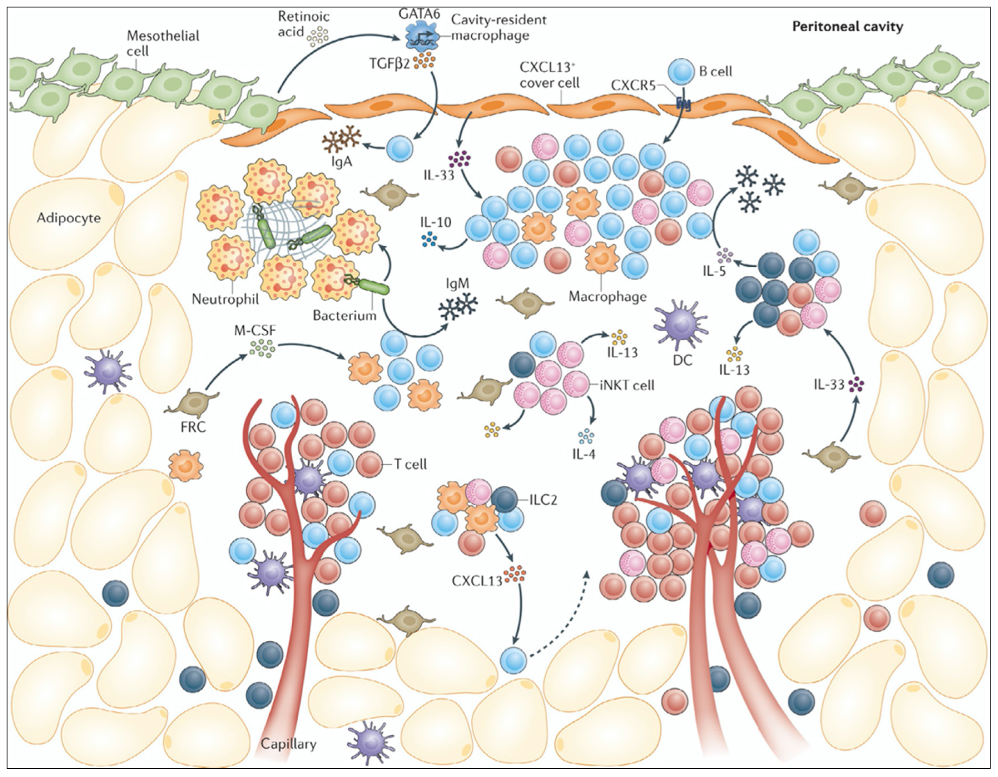
2. Immune Contexture of the Healthy Peritoneal Cavity

The peritoneal cavity holds the abdominal viscera and is lined by the peritoneum, a one-cell-layer-thick serous membrane of mesothelial cells (MC) with an underlying extracellular matrix. Anatomically, the peritoneum can be subdivided into the parietal peritoneum, which lines the abdominal wall, and the visceral peritoneum covering the viscera [19]. The MCs provide a smooth non-adhesive surface for the viscera by the production of peritoneal fluid (PF), surfactant, proteoglycans and glycosaminoglycans. Also, MCs facilitate transport of fluid and cells across the peritoneal cavity, form a physical barrier for pathogens and cancer cells, and engage a response to inflammation, tissue damage, and malignancy [20,21]. Mesothelial cells present antigens to CD4+ T cells, and express pattern recognition receptors (PRRs) that can recognize pathogens and damage-associated molecules [20]. Combined with their capacity to secrete cytokines, chemokines, growth factors, and extracellular matrix (ECM) components, they are essential in communication, tissue repair, and activation of the immune system in response to threats within the abdominal cavity. An important means for this communication is the PF, which contains both a molecular and cellular fraction and as such creates a peritoneal ecosystem across the abdominal cavity.
Throughout the peritoneal cavity, the peritoneum is fenestrated by stomata, openings in the mesothelial serous membrane connected to the lymphatic system. The main function of these stomata is to filter the PF and drain it to the lymph nodes [22,23]. Stomata located on the omentum and mesentery that are associated with fibroblasts and clusters of immune cells are termed milky spots or fat-associated lymphoid clusters (FALCs) and are of importance for immune cell turnover in the peritoneal cavity [23]. These tertiary lymphoid clusters are rich in both innate and adaptive immune cells, as well as lymphoid and myeloid cells, and have as main functions the maintenance of homeostasis, defense against threats, and tissue repair [23,24]. Very commonly found in these FALCs are neutrophils, a type of granulocyte capable of capturing circulating pathogens through phagocytosis and the use of their neutrophil extracellular traps (NETs). Upon inflammation, neutrophils can be recruited very promptly by the MC expression of the neutrophil recruitment chemokine, CXCL1, to help surmount the inflammatory cause [25]. Other phagocyting innate immune cells highly abundant in the peritoneal cavity are macrophages. Mice studies have shown the existence of two main macrophage populations in the peritoneum: the tissue resident GATA6-dependent large peritoneal macrophages (LPM) and the monocyte-derived IRF4-dependent small peritoneal macrophages (SPM) [26,27,28]. Whether these distinct peritoneal macrophage populations are also present in humans is yet to be confirmed.
One of the most common peritoneal immune cell types in the adaptive immune compartment are B cells. Specifically, the B1-cell subset, an innate-like B-cell type, rapidly produces high levels of IgM antibodies as a first response to peritoneal threats [29]. These B1 cells are also potent antigen presenting cells capable of activating CD4+ T cells. Among the T-cell population, both CD4+ T helper cells and CD8+ cytotoxic T cells prevail in the peritoneal cavity (Figure 1) [24].

3. Immune Contexture of Peritoneal Metastases

In peritoneal metastases (PM), a majority of the peritoneal immune microenvironment exhibits immunosuppressive phenotypes (e.g., regulatory T cells, exhausted T cells, tumor-associated macrophages and MDSCs) and thus support cancer growth [31]. The pathophysiology of PM can be described as a stepwise process, beginning with the detachment of cells from the primary tumor, transcoelomic transport with the peritoneal fluid, and attachment to the peritoneal surface or the underlying stroma. Subsequently, invasion into the subperitoneal space, proliferation of tumor cells, and angiogenesis lead to PM [31,32]. While these distinctive steps are part of a process shared by all abdominal malignancies metastasizing to the peritoneum, different cancer types may use unique mechanisms [32,33]. Moreover, PM can arise from primary tumors outside of the abdominal cavity such as melanoma, lung cancer, or lobular breast cancer, indicating a systemic route resulting in PM [34].
Recent insights suggest the existence of a pre-metastatic niche that precedes the formation of PM [31,33]. The peritoneal microenvironment is primed for PM by soluble tumor-secreted factors including cytokines such as TGF-β, VEGF, and exosomes [35,36]. As part of this process, the continuity of mesothelial cells is disrupted, and mesothelial-to-mesenchymal transition (MMT) occurs [37,38,39]. In addition, certain components of the peritoneal microenvironment such as macrophages and other immune cells are reprogrammed to allow tumor cells to establish as PM [31,33,40]. Integrating these concepts has resulted in a deeper understanding of the hallmarks of PM—namely paracrine factors, tumor-related factors, biomechanical forces, and the peritoneal microenvironment—that are intertwined in the formation of PM [41].

4. Non-Immune Related Effects of HIPEC

4.1. Effects on Peritoneal Blood Flow and Pharmacokinetics

Under physiological conditions, the estimated magnitude of the (visceral) peritoneal blood flow is 60–100 mL/min, representing 1–2% of the cardiac output [42]. Heat stress causes reflex vasodilation of skin vessels, promoting convective heat transfer from the body core to the skin. It is not known whether peritoneal microvessels exhibit a similar temperature-dependent vasodilation. However, since HIPEC causes an increased cardiac output and a lowered peripheral vascular resistance, it is likely that peritoneal blood flow increases with (mild) hyperthermia [43]. Due to vasodilation, hyperthermia may increase systemic uptake of chemotherapy, potentiating side effects. However, experimental results concerning the effect of hyperthermia on IP pharmacokinetics are inconclusive [44]. In a porcine oxaliplatin HIPEC model, tissue concentrations of platinum were increased in response to hyperthermia, with no increase in systemic absorption [45]. Other pharmacokinetic data in small animal models are conflicting, with some authors describing no significant effect of hyperthermia [46,47,48], while others demonstrated higher tissue platinum concentrations and decreased systemic absorption, which would indicate increased drug uptake [49,50]. However, pharmacokinetic analyses in animal models as reported by Sørensen et al. showed no effect of hyperthermia on MMC pharmacokinetics [51], while Klaver et al. did not demonstrate a survival benefit for HIPEC with MMC compared to normothermic chemoperfusion [52]. In an in vivo analysis of 50 patients, Xie et al. reported only a slight increase in plasma peak concentration of cisplatin when administered at 41 °C, with no increase in adverse events [53]. Multiple pre-clinical animal studies have demonstrated the increased depth of tissue penetration resulting from the addition of heat to IP chemotherapy [54,55,56,57]. This is explained by the temperature dependence of the diffusion process:
J = D 0 e Q d R T d C d x
With J the diffusion flux (mass transfer per unit area per unit time), D the diffusion coefficient (m2/s), Qd the activation energy (J/mol), R the gas constant, T the absolute temperature, and dC/dx the concentration gradient.

4.2. Effect on Chemotherapy Cytotoxicity

The three most common chemotherapeutic agents used in the context for HIPEC are mitomycin C (MMC), and the platinum agents cisplatin and oxaliplatin. All three of these agents have been described as being “thermally enhanced”, meaning their efficacy is increased by hyperthermic conditions [58]. However, most of these results are derived from in vitro or animal studies.
MMC combined with hyperthermia demonstrated increased cell-killing capacity of cancer cell lines in vitro [59,60], while a recent study by Helderman et al. disputes this [61]. Oxaliplatin and cisplatin efficacy has been described as augmented by hyperthermia, with increased apoptosis, drug uptake, and DNA damage reported in vitro in several colorectal and ovarian cancer cell lines, but these effects might be cell-line dependent [44,49,61,62,63].

4.3. Effects on Peritoneal and Gut Integrity

At temperatures higher than 43 °C, the risk of scald injury to the peritoneal surface increases [64]. Normal tissues in the vicinity of cancers are susceptible to thermal injury induced by hyperthermic IP treatment as has been verified by in vitro and in vivo studies [65]. In animal experiments exploring thermal dosimetry, significant histopathological damage to the bowel (rabbit, pig) was noted with exposure to 43 °C during 21–40 min, while liver injury was noted in rabbits from 41 min onward [65]. Pavel et al. reported small bowel damage induced by ≤20 mins of exposure to hyperthermia at 43 °C [66,67]. In animal models investigating intestinal injury, intestinal epithelial injury was seen at temperatures as low as 41.5 °C [67]. In addition, hyperthermia may affect the integrity of the mucosal intestinal barrier function. In animal studies, heat stress damaged mechanical and mucosal immune gut barriers reduces immune function of the intestinal mucosa and mesenteric lymphoid tissues, and leads to bacterial translocation [68,69,70]. Also, changes in core temperature have been linked to altered microbiome composition and function, and the effect of HIPEC on the integrity and function of the gut microbiome is an important question for future research [71].

5. Immune Effects of HIPEC

The potential immune stimulating effects of hyperthermia have been recognized since the observations of spontaneous tumor regression in patients with persistent fever. Endogenous hyperthermia (fever) stimulates the innate and adaptive immune system through multiple pathways, with interleukin-6 (IL-6) playing a central role [72]. The effects of hyperthermia on the peritoneal immune environment have not been studied in detail, and the mechanisms described below are not specific for HIPEC.
Hyperthermia alters the expression of thousands of genes involved in protein folding, cell cycle, mitosis, and cell death [73]. A prominent master regulator of the heat shock response is heat shock factor 1 (HSF1), which affects various cellular responses associated with tumorigenesis, including alteration of the tumor microenvironment, genome repair, and regulation of several cell death pathways including autophagy and ferroptosis [74]. Together with hypoxia-inducible factor 1 (HIF-1) [12], matrix metalloproteinase 3 (MMP-3), and heterochromatin protein 1, HSF1 triggers the production of heat shock proteins (HSPs) [75]. Heat shock proteins are molecular chaperones which protect intracellular proteins from misfolding or aggregation, inhibit cell death signaling cascades, and preserve essential intracellular signaling pathways. According to their molecular weight, they are classified as HSP110 (HSPH), HSP90 (HSPC), HSP70 (HSPA), HSP60 (HSPD), HSP40 (DNAJ), and small HSPs (HSPB). Although originally described in the setting of heat shock, they are known to be constitutively expressed and subject to stimulated expression by other stressors including cold. Overexpression of HSPs is common in a variety of solid cancer types, and they have been implicated in several oncogenic pathways. However, the effects seem to be tumor-type dependent, and efforts to develop HSP targeted therapies in clinical trials were unsuccessful [76]. Similarly, the effects of hyperthermia and HSPs on cancer immune surveillance are double edged. On the one hand, HSPs can bind tumor antigens and form complexes recognized by monocytes, macrophages, B cells, and dendritic cells, leading to cytotoxic T-cell activation [76]. On the other hand, HSF1 was shown to enhance the expression of PD-L1 in breast cancer. Additionally, compared to intra-cellular HSPs that are released by normal cells in response to trauma or stress states, HSPs are also transported into the extracellular compartment by non-canonical secretory pathways and may play a crucial role in promoting and sustaining key hallmarks of peritoneal carcinogenesis [77,78,79]. For instance, extracellular HSPs (eHSPs) have been shown to promote ECM remodeling to increase production of collagen and fibronectin production as scaffolding for tumor expansion [77]. eHSPs also contribute to stromal cell activation such as cancer-associated fibroblasts, which further secrete cytokines to maintain a pro-tumor environment [77,80]. eHSPs have also been shown to protect cancer cells from apoptosis or cell death by a variety of differing pathways [81]. eHSPs also directly interact with cancer cells via the paracrine effect on surface receptors such as the EGF receptor family, toll-like receptors and lipoprotein receptor-related proteins, which are implicated in cancer proliferation, apoptosis inhibition, and cancer progression [82] (Figure 2).
Of note, the effects are dependent on temperature: thermal ablation methods including microwave ablation result in the release of neoantigens and DAMPs, and provide the rationale for ongoing clinical studies that combine local ablation with immune checkpoint inhibitors [83]. Clearly, these mechanisms do not apply to the mild hyperthermia used during HIPEC.
Mild hyperthermia enhances immune cell recruitment by stimulating vascular perfusion and upregulation of vascular adhesion molecules including ICAM-1, which promote immune cell infiltration [84]. Several preclinical studies showed that mild hyperthermia results in reduced tumor growth and improved animal survival, in parallel with enhanced infiltration and activation of immune cell populations including natural killer (NK), CD4+ T, and CD8+ T cells. As an example, Toraya-Brown et al. found that treatment of murine melanoma with mild hyperthermia (43 °C during 30 min) using iron oxide nanoparticles combined with an alternating magnetic field resulted in activated dendritic cells (DCs) and subsequently CD8+ T cells in the draining lymph node, and conferred resistance against rechallenge [85]. However, these effects were not observed when using higher temperatures (45 °C).
Several authors have investigated the effects of HIPEC on tumor immune cell infiltration in animal models. Nevo et al. found that HIPEC with mitomycin C led to increased infiltration of CD4+, CD8+, CD68+, and CD20+ cells into omental and visceral metastases in a mouse model of colorectal PM (MC38/C57BL) [86]. In the same model, the authors showed that combining HIPEC with anti-PD-1 treatment resulted in improved survival compared with HIPEC alone [87]. Wu et al. administered IP chemohyperthermia in a mouse ovarian cancer model, and observed that the addition of hyperthermia restored the number of intraperitoneal macrophages and dendritic cells, which were depleted in the chemotherapy alone group [87]. Zunino et al. performed vaccination experiments in a CT26/Balb/c murine colorectal cancer model [88]. They found that after vaccination with mitomycin C or hyperthermia exposed CT26 cells, tumor growth was inhibited after injection of CT26 cells in the opposite flank. Mechanistically, they showed that the antitumor vaccination was mediated by HSP90 exposure on the cell surface of the dying cells.

Clinical and Translational Studies

Published data regarding the clinical benefits of hyperthermia in the context of PM has thus far been limited and inconsistent. One clinical trial of prophylactic HIPEC in gastric cancer was performed with mitomycin C (MMC) and oxaliplatin at normothermic or hyperthermic conditions, demonstrating a survival benefit for the hyperthermia group [89]. However, these findings were not confirmed in a larger meta-analysis comparing hyperthermic vs. normothermic IP chemotherapy for gastric cancer patients with PM [90]. The landmark CYTO-CHIP study evaluating a retrospective cohort of patients with PM from gastric cancer demonstrated a survival benefit with the addition of HIPEC to cytoreductive surgery, although it is unclear whether the benefit arose from hyperthermia, IP chemotherapy, or a synergistic combination of both factors [91]. Similarly, the RCT by Casado-Adam et al. evaluated patients with ovarian cancer undergoing normothermic vs. hyperthermic IP paclitaxel. In addition to finding no significant increase in local tissue concentrations of chemotherapy in patients receiving heated treatments, markers of apoptosis (caspase-3), pathological response, and cellular proliferation (p53, p27, p21, Ki67) were also not significantly different between normothermic and hyperthermic treatments, casting doubt on the purported benefits of adding heat to IP treatment [8]. To address some of these gaps in current knowledge, a number of ongoing RCTs aim to re-examine fundamentals of hyperthermic IP therapy. For instance, the OvIP1 trial (NCT02567253) aims to investigate the efficacy of heated vs. normothermic IP cisplatin in patients with ovarian cancer [92]. Ongoing and completed RCTs are summarized in Table 1.
While the clinical benefit of hyperthermia for PM treatment has yet to be demonstrated, several translational studies have alluded to a biological basis for hyperthermic treatment at a molecular level. Dellinger et al. examined pre- and post-treatment samples from patients with ovarian cancer undergoing HIPEC and reported heat shock proteins as one of the most upregulated genes after treatment. In addition, an increase in programmed cell death protein 1 expression on CD8+ T cells and downregulated DNA repair pathways within cancer islands were associated with significantly increased progression-free survival after hyperthermic IP treatment and could serve as a predictive biomarker in the future [93]. In the same study, the gene expression changes seen post-HIPEC were similar in tumor and normal tissue, albeit with a decreased magnitude of change seen in normal tissues. Metabolic pathways were downregulated and immune pathways were upregulated to a greater extent in tumor compared to normal tissue as well, suggesting that hyperthermia enabled a certain specificity for targeted treatment of the tumor [93]. This was further validated with post-HIPEC PM tumors demonstrating significant apoptosis and inflammatory change on immunohistochemistry while normal tissue only exhibited the latter [93]. Similarly, Moukarzel et al. examined transcriptomic changes after HIPEC in ovarian tumors and normal tissues, and demonstrated differential gene expression between tumors (heat response- and protein folding-related genes) and normal tissues where genes relating to immune response were altered [94]. A difference in upregulated HSPs was noted between tumors and normal tissue, with an overall increase of HSP90, GRP75, and Ubiquitin+1 seen in normal tissues compared to a decrease in tumor tissues; an increase in HSP27, HSP32, HSP40, HSP60 protein expression levels were observed in tumor tissues as compared to normal tissue and only HSP70 expression was increased in both normal and tumor tissue after HIPEC exposure [94]. However, the consequence of these changes in transcriptome are not apparent. In colorectal and gastrointestinal related PM, exposure to HIPEC resulted in upregulation of intracellular heat shock proteins (HSP) and resultant amplification of proliferation markers and antiapoptotic proteins, as well as chemoresistance against fluoropyrimidine, MMC, and oxaliplatin agents (Figure 3) [77,82,95,96,97]. A recent meta-analysis of published transcriptomic changes in human cancer cells undergoing heat stress evaluated a total of 18 datasets (2 unpublished) and identified considerable inter-study variability, as well as an apparent absence of a “universal” heat stress response, highlighting the need for further in-depth collaborative efforts to standardize clinical and experimental protocols crucial to understanding the response of PM to hyperthermia [73].
Lastly, the impact of hyperthermia on the resident immune microenvironment in the peritoneum needs further evaluation in the era of immunotherapy. Moukarzel et al. reported similar immune cell compositions between normal tissue and ovarian tumors, with normal tissue demonstrating increased CD8 T cells, activated and resting mast cells, activated NK cells and neutrophils while no change in immune cell composition was noted in tumor samples before and after HIPEC [73]. This suggests that there was no significant impact of hyperthermic IP chemotherapy on the proportions of immune cells in PM. In contrast, Dellinger et al. reported a significant increase in % PD-1 expression in CD8+ tumor-infiltrating lymphocytes and CD4+ conventional T cells after HIPEC, with the magnitude of change strongly correlated with overall survival [93]. Understanding the impact of hyperthermia on peritoneal immune cells is crucial to harness its potential synergy with immunotherapy.
Taken together, current data does suggest a biological impact of heated IP treatment with transcriptomic and proteomic changes that may serve as a predictive or prognostic biomarker. However, much more rigor is required to reduce inter-study variance and identify putative pathways for prognostic or therapeutic studies. Additionally, both tissue- and treatment type-specific interactions with hyperthermia might exist, underscoring the need for more rigorous study. The potential benefits and adverse effects of hyperthermia are summarized in Table 2.

6. Biomechanical Effects of Hyperthermia

Biomechanical properties of solid tumors have a major role in the outcome of the therapy [98]. Drug penetration in tumor tissue during HIPEC is driven by convection (pressure gradient) and diffusion (concentration gradient) [58,99]. Depending on several properties including the drug, one mechanism can be dominant over the other. Tumors are characterized by an elevated interstitial fluid pressure (IFP), generating an outward flow (interstitial fluid flow; IFV) that opposes drug penetration [98]. Therefore, the depth of drug penetration is determined by the balance between inward diffusion and outward convection (Figure 4). Also, the combination of mechanical stress and fibroblast-derived TGF-β induces the transformation of fibroblasts to CAFs, which in turn produce collagen and other ECM molecules, leading to increased stromal stiffness, which hampers the infiltration of immune cells.
As noted above, diffusion is temperature-dependent, and a higher temperature is correlated with enhanced drug penetration. This was recently confirmed in a computational model of HIPEC [100].
Next to the effects of temperature on drug penetration, reduced matrix stiffness may facilitate the migration of immune cells in the tumor stroma. It has been observed that the permeability of tumor tissue decreases due to solid matrix heat treatment [101,102,103]. This may be due to the thermal transition of collagen. In acidic solution, reversible thermal transition occurs at 31–37 °C and is due to depolymerization of the smallest collagen fibrils. Irreversible thermal transition occurs between 37 °C and 55 °C, and is related to the complete defibrillation and unfolding of the native triple helical structure of collagen [104]. In a murine model of nanoparticle-mediated hyperthermia, the collagen within the tumor stroma changed from a mesh, fiber-like structure to a completely unstructured fibril [105]. It is well-documented that T-cell migration is hampered by increased stiffness and higher collagen density [106,107]. Also, the alignment of collagen fibers, which is generally perpendicular to the tumor surface, was shown to affect lymphocyte migration [108]. Other immune cell types are affected by stromal composition: collagen density was shown to modulate the immunosuppressive function of macrophages [109]. Taken together, these data suggest that hyperthermia may facilitate infiltration of immune cell populations by altered mechanical stromal properties.

7. Conclusions

Hyperthermia increases anticancer drug penetration and may promote infiltration by immune cells. However, the heat shock response elicited by HIPEC may prove to be double-edged, as enhanced expression of heat shock proteins might provoke unwanted, tumor-promoting effects next to reported antitumoral effects. Despite the increasing use of heated locoregional IP chemotherapy, there remains a lack of empirical and clinical evidence that clearly demonstrates the advantages of hyperthermia when treating peritoneal metastases and additional well-designed basic science; translational and clinical studies are urgently required to clarify the future treatment of PM.

Author Contributions

Conceptualization, W.C., D.K.A.C. and J.D.; writing—original draft preparation, D.K.A.C., J.D., S.E., H.S. and W.C.; writing—review and editing, W.C., J.D. and D.K.A.C.; supervision, W.C. All authors have read and agreed to the published version of the manuscript.

Funding

This research received no external funding.

Conflicts of Interest

The authors declare no conflict of interest.

References

  1. Jacquet, P.; Sugarbaker, P.H. Peritoneal-plasma barrier. Cancer Treat. Res. 1996, 82, 53–63. [Google Scholar] [CrossRef] [PubMed]
  2. Sugarbaker, P.H.; Gianola, F.J.; Speyer, J.C.; Wesley, R.; Barofsky, I.; Meyers, C.E. Prospective, randomized trial of intravenous versus intraperitoneal 5-fluorouracil in patients with advanced primary colon or rectal cancer. Surgery 1985, 98, 414–422. [Google Scholar] [PubMed]
  3. Spratt, J.S.; Adcock, R.A.; Sherrill, W.; Travathen, S. Hyperthermic peritoneal perfusion system in canines. Cancer Res. 1980, 40, 253–255. [Google Scholar] [PubMed]
  4. Koga, S.; Hamazoe, R.; Maeta, M.; Shimizu, N.; Kanayama, H.; Osaki, Y. Treatment of implanted peritoneal cancer in rats by continuous hyperthermic peritoneal perfusion in combination with an anticancer drug. Cancer Res. 1984, 44, 1840–1842. [Google Scholar] [PubMed]
  5. Fujimoto, S.; Shrestha, R.D.; Kokubun, M.; Ohta, M.; Takahashi, M.; Kobayashi, K.; Kiuchi, S.; Okui, K.; Miyoshi, T.; Arimizu, N.; et al. Intraperitoneal hyperthermic perfusion combined with surgery effective for gastric cancer patients with peritoneal seeding. Ann. Surg. 1988, 208, 36–41. [Google Scholar] [CrossRef] [PubMed]
  6. Witkamp, A.J.; de Bree, E.; Van Goethem, R.; Zoetmulder, F.A. Rationale and techniques of intra-operative hyperthermic intraperitoneal chemotherapy. Cancer Treat. Rev. 2001, 27, 365–374. [Google Scholar] [CrossRef] [PubMed]
  7. Gremonprez, F.; Gossye, H.; Ceelen, W. Use of hyperthermia versus normothermia during intraperitoneal chemoperfusion with oxaliplatin for colorectal peritoneal carcinomatosis: A propensity score matched analysis. Eur. J. Surg. Oncol. 2019, 45, 366–370. [Google Scholar] [CrossRef]
  8. Casado-Adam, A.; Rodriguez-Ortiz, L.; Rufian-Peña, S.; Muñoz-Casares, C.; Caro-Cuenca, T.; Ortega-Salas, R.; Fernandez-Peralbo, M.A.; Luque-de-Castro, M.D.; Sanchez-Hidalgo, J.M.; Hervas-Martinez, C.; et al. The Role of Intraperitoneal Intraoperative Chemotherapy with Paclitaxel in the Surgical Treatment of Peritoneal Carcinomatosis from Ovarian Cancer-Hyperthermia versus Normothermia: A Randomized Controlled Trial. J. Clin. Med. 2022, 11, 5785. [Google Scholar] [CrossRef]
  9. Rau, B.; Lang, H.; Königsrainer, A.; Gockel, I.; Rau, H.G.; Seeliger, H.; Lerchenmüller, C.; Reim, D.; Wahba, R.; Angele, M.; et al. 1376O The effect of hyperthermic intraperitoneal chemotherapy (HIPEC) upon cytoreductive surgery (CRS) in gastric cancer (GC) with synchronous peritoneal metastasis (PM): A randomized multicentre phase III trial (GASTRIPEC-I-trial). Ann. Oncol. 2021, 32, S1040. [Google Scholar] [CrossRef]
  10. Quénet, F.; Elias, D.; Roca, L.; Goéré, D.; Ghouti, L.; Pocard, M.; Facy, O.; Arvieux, C.; Lorimier, G.; Pezet, D.; et al. Cytoreductive surgery plus hyperthermic intraperitoneal chemotherapy versus cytoreductive surgery alone for colorectal peritoneal metastases (PRODIGE 7): A multicentre, randomised, open-label, phase 3 trial. Lancet Oncol. 2021, 22, 256–266. [Google Scholar] [CrossRef]
  11. van Driel, W.J.; Koole, S.N.; Sikorska, K.; Schagen van Leeuwen, J.H.; Schreuder, H.W.R.; Hermans, R.H.M.; de Hingh, I.; van der Velden, J.; Arts, H.J.; Massuger, L.; et al. Hyperthermic Intraperitoneal Chemotherapy in Ovarian Cancer. N. Engl. J. Med. 2018, 378, 230–240. [Google Scholar] [CrossRef]
  12. Antonio, C.C.P.; Alida, G.G.; Elena, G.G.; Rocío, G.S.; Jerónimo, M.G.; Luis, A.R.J.; Aníbal, N.D.; Francisco, B.V.; Jesús, G.; Pablo, R.R.; et al. Cytoreductive Surgery with or without HIPEC after Neoadjuvant Chemotherapy in Ovarian Cancer: A Phase 3 Clinical Trial. Ann. Surg. Oncol. 2022, 29, 2617–2625. [Google Scholar] [CrossRef] [PubMed]
  13. Lim, M.C.; Chang, S.J.; Park, B.; Yoo, H.J.; Yoo, C.W.; Nam, B.H.; Park, S.Y. Survival after Hyperthermic Intraperitoneal Chemotherapy and Primary or Interval Cytoreductive Surgery in Ovarian Cancer: A Randomized Clinical Trial. JAMA Surg. 2022, 157, 374–383. [Google Scholar] [CrossRef] [PubMed]
  14. Harris, J.W.; Meneses, J.J. Effects of hyperthermia on the production and activity of primary and secondary cytolytic T-lymphocytes in vitro. Cancer Res. 1978, 38, 1120–1126. [Google Scholar] [PubMed]
  15. Agarwal, S.S.; Katz, E.J.; Loeb, L.A. Effect of hyperthermia on the survival of normal human peripheral blood mononuclear cells. Cancer Res. 1983, 43, 3124–3126. [Google Scholar] [PubMed]
  16. Jung, D.H.; Son, S.Y.; Oo, A.M.; Park, Y.S.; Shin, D.J.; Ahn, S.H.; Park, D.J.; Kim, H.H. Feasibility of hyperthermic pressurized intraperitoneal aerosol chemotherapy in a porcine model. Surg. Endosc. 2016, 30, 4258–4264. [Google Scholar] [CrossRef]
  17. Bachmann, C.; Sautkin, I.; Nadiradze, G.; Archid, R.; Weinreich, F.J.; Königsrainer, A.; Reymond, M.A. Technology development of hyperthermic pressurized intraperitoneal aerosol chemotherapy (hPIPAC). Surg. Endosc. 2021, 35, 6358–6365. [Google Scholar] [CrossRef]
  18. Nowacki, M.; Peterson, M.; Kloskowski, T.; McCabe, E.; Guiral, D.C.; Polom, K.; Pietkun, K.; Zegarska, B.; Pokrywczynska, M.; Drewa, T.; et al. Nanoparticle as a novel tool in hyperthermic intraperitoneal and pressurized intraperitoneal aerosol chemotheprapy to treat patients with peritoneal carcinomatosis. Oncotarget 2017, 8, 78208–78224. [Google Scholar] [CrossRef]
  19. van Baal, J.O.; Van de Vijver, K.K.; Nieuwland, R.; van Noorden, C.J.; van Driel, W.J.; Sturk, A.; Kenter, G.G.; Rikkert, L.G.; Lok, C.A. The histophysiology and pathophysiology of the peritoneum. Tissue Cell 2017, 49, 95–105. [Google Scholar] [CrossRef]
  20. Mutsaers, S.E.; Prêle, C.M.; Pengelly, S.; Herrick, S.E. Mesothelial cells and peritoneal homeostasis. Fertil. Steril. 2016, 106, 1018–1024. [Google Scholar] [CrossRef]
  21. Yung, S.; Li, F.K.; Chan, T.M. Peritoneal mesothelial cell culture and biology. Perit. Dial. Int. 2006, 26, 162–173. [Google Scholar] [CrossRef] [PubMed]
  22. Wang, Z.B.; Li, M.; Li, J.C. Recent advances in the research of lymphatic stomata. Anat. Rec. 2010, 293, 754–761. [Google Scholar] [CrossRef] [PubMed]
  23. Meza-Perez, S.; Randall, T.D. Immunological Functions of the Omentum. Trends Immunol. 2017, 38, 526–536. [Google Scholar] [CrossRef] [PubMed]
  24. Liu, M.; Silva-Sanchez, A.; Randall, T.D.; Meza-Perez, S. Specialized immune responses in the peritoneal cavity and omentum. J. Leukoc. Biol. 2021, 109, 717–729. [Google Scholar] [CrossRef] [PubMed]
  25. Jackson-Jones, L.H.; Smith, P.; Portman, J.R.; Magalhaes, M.S.; Mylonas, K.J.; Vermeren, M.M.; Nixon, M.; Henderson, B.E.P.; Dobie, R.; Vermeren, S.; et al. Stromal Cells Covering Omental Fat-Associated Lymphoid Clusters Trigger Formation of Neutrophil Aggregates to Capture Peritoneal Contaminants. Immunity 2020, 52, 700–715.e706. [Google Scholar] [CrossRef] [PubMed]
  26. Jayakumar, P.; Laganson, A.; Deng, M. GATA6(+) Peritoneal Resident Macrophage: The Immune Custodian in the Peritoneal Cavity. Front. Pharmacol. 2022, 13, 866993. [Google Scholar] [CrossRef] [PubMed]
  27. Rosas, M.; Davies, L.C.; Giles, P.J.; Liao, C.T.; Kharfan, B.; Stone, T.C.; O’Donnell, V.B.; Fraser, D.J.; Jones, S.A.; Taylor, P.R. The transcription factor Gata6 links tissue macrophage phenotype and proliferative renewal. Science 2014, 344, 645–648. [Google Scholar] [CrossRef]
  28. Kim, K.W.; Williams, J.W.; Wang, Y.T.; Ivanov, S.; Gilfillan, S.; Colonna, M.; Virgin, H.W.; Gautier, E.L.; Randolph, G.J. MHC II+ resident peritoneal and pleural macrophages rely on IRF4 for development from circulating monocytes. J. Exp. Med. 2016, 213, 1951–1959. [Google Scholar] [CrossRef]
  29. Haas, K.M. B-1 lymphocytes in mice and nonhuman primates. Ann. N. Y Acad. Sci. 2015, 1362, 98–109. [Google Scholar] [CrossRef]
  30. Trim, W.V.; Lynch, L. Immune and non-immune functions of adipose tissue leukocytes. Nat. Rev. Immunol. 2022, 22, 371–386. [Google Scholar] [CrossRef]
  31. Demuytere, J.; Ernst, S.; van Ovost, J.; Cosyns, S.; Ceelen, W. The tumor immune microenvironment in peritoneal carcinomatosis. Int. Rev. Cell Mol. Biol. 2022, 371, 63–95. [Google Scholar] [CrossRef] [PubMed]
  32. Lemoine, L.; Sugarbaker, P.; Van der Speeten, K. Pathophysiology of colorectal peritoneal carcinomatosis: Role of the peritoneum. World J. Gastroenterol. 2016, 22, 7692–7707. [Google Scholar] [CrossRef] [PubMed]
  33. Mikuła-Pietrasik, J.; Uruski, P.; Tykarski, A.; Książek, K. The peritoneal “soil” for a cancerous “seed”: A comprehensive review of the pathogenesis of intraperitoneal cancer metastases. Cell. Mol. Life Sci. 2018, 75, 509–525. [Google Scholar] [CrossRef] [PubMed]
  34. Cortés-Guiral, D.; Hübner, M.; Alyami, M.; Bhatt, A.; Ceelen, W.; Glehen, O.; Lordick, F.; Ramsay, R.; Sgarbura, O.; Van Der Speeten, K.; et al. Primary and metastatic peritoneal surface malignancies. Nat. Rev. Dis. Primers 2021, 7, 91. [Google Scholar] [CrossRef]
  35. Liu, Y.; Cao, X. Characteristics and Significance of the Pre-metastatic Niche. Cancer Cell 2016, 30, 668–681. [Google Scholar] [CrossRef] [PubMed]
  36. Chen, X.; Wang, H.; Huang, Y.; Chen, Y.; Chen, C.; Zhuo, W.; Teng, L. Comprehensive Roles and Future Perspectives of Exosomes in Peritoneal Metastasis of Gastric Cancer. Front. Oncol. 2021, 11, 684871. [Google Scholar] [CrossRef] [PubMed]
  37. Deng, G.; Qu, J.; Zhang, Y.; Che, X.; Cheng, Y.; Fan, Y.; Zhang, S.; Na, D.; Liu, Y.; Qu, X. Gastric cancer-derived exosomes promote peritoneal metastasis by destroying the mesothelial barrier. FEBS Lett. 2017, 591, 2167–2179. [Google Scholar] [CrossRef]
  38. Demuytere, J.; Ceelen, W.; Van Dorpe, J.; Hoorens, A. The role of the peritoneal microenvironment in the pathogenesis of colorectal peritoneal carcinomatosis. Exp. Mol. Pathol. 2020, 115, 104442. [Google Scholar] [CrossRef]
  39. Wei, M.; Yang, T.; Chen, X.; Wu, Y.; Deng, X.; He, W.; Yang, J.; Wang, Z. Malignant ascites-derived exosomes promote proliferation and induce carcinoma-associated fibroblasts transition in peritoneal mesothelial cells. Oncotarget 2017, 8, 42262–42271. [Google Scholar] [CrossRef]
  40. Song, H.; Wang, T.; Tian, L.; Bai, S.; Chen, L.; Zuo, Y.; Xue, Y. Macrophages on the Peritoneum are involved in Gastric Cancer Peritoneal Metastasis. J. Cancer 2019, 10, 5377–5387. [Google Scholar] [CrossRef]
  41. Gwee, Y.X.; Chia, D.K.A.; So, J.; Ceelen, W.; Yong, W.P.; Tan, P.; Ong, C.J.; Sundar, R. Integration of Genomic Biology into Therapeutic Strategies of Gastric Cancer Peritoneal Metastasis. J. Clin. Oncol. 2022, 40, 2830. [Google Scholar] [CrossRef] [PubMed]
  42. Solass, W.; Horvath, P.; Struller, F.; Königsrainer, I.; Beckert, S.; Königsrainer, A.; Weinreich, F.J.; Schenk, M. Functional vascular anatomy of the peritoneum in health and disease. Pleura Peritoneum 2016, 1, 145–158. [Google Scholar] [CrossRef] [PubMed]
  43. Bezu, L.; Raineau, M.; Deloménie, M.; Cholley, B.; Pirracchio, R. Haemodynamic management during hyperthermic intraperitoneal chemotherapy: A systematic review. Anaesth. Crit. Care Pain. Med. 2020, 39, 531–542. [Google Scholar] [CrossRef] [PubMed]
  44. Vos, L.M.C.; Aronson, S.L.; van Driel, W.J.; Huitema, A.D.R.; Schagen van Leeuwen, J.H.; Lok, C.A.R.; Sonke, G.S. Translational and pharmacological principles of hyperthermic intraperitoneal chemotherapy for ovarian cancer. Best Pract. Res. Clin. Obstet. Gynaecol. 2022, 78, 86–102. [Google Scholar] [CrossRef] [PubMed]
  45. Facy, O.; Al Samman, S.; Magnin, G.; Ghiringhelli, F.; Ladoire, S.; Chauffert, B.; Rat, P.; Ortega-Deballon, P. High pressure enhances the effect of hyperthermia in intraperitoneal chemotherapy with oxaliplatin: An experimental study. Ann. Surg. 2012, 256, 1084–1088. [Google Scholar] [CrossRef]
  46. Pestieau, S.R.; Belliveau, J.F.; Griffin, H.; Stuart, O.A.; Sugarbaker, P.H. Pharmacokinetics of intraperitoneal oxaliplatin: Experimental studies. J. Surg. Oncol. 2001, 76, 106–114. [Google Scholar] [CrossRef] [PubMed]
  47. Zeamari, S.; Floot, B.; Van der Vange, N.; Stewart, F.A. Pharmacokinetics and pharmacodynamics of cisplatin after Intraoperative Hyperthermic Intraperitoneal Chemoperfusion (HIPEC). Anticancer Res. 2003, 23, 1643–1648. [Google Scholar] [PubMed]
  48. Mas-Fuster, M.I.; Ramon-Lopez, A.; Lacueva, F.J.; Arroyo, A.; Más-Serrano, P.; Nalda-Molina, R. Population pharmacokinetics of oxaliplatin after intraperitoneal administration with hyperthermia in Wistar rats. Eur. J. Pharm. Sci. Off. J. Eur. Fed. Pharm. Sci. 2018, 119, 22–30. [Google Scholar] [CrossRef]
  49. Los, G.; Sminia, P.; Wondergem, J.; Mutsaers, P.H.; Havemen, J.; ten Bokkel Huinink, D.; Smals, O.; Gonzalez-Gonzalez, D.; McVie, J.G. Optimisation of intraperitoneal cisplatin therapy with regional hyperthermia in rats. Eur. J. Cancer 1991, 27, 472–477. [Google Scholar] [CrossRef]
  50. Piché, N.; Leblond, F.A.; Sidéris, L.; Pichette, V.; Drolet, P.; Fortier, L.-P.; Mitchell, A.; Dubé, P. Rationale for heating oxaliplatin for the intraperitoneal treatment of peritoneal carcinomatosis: A study of the effect of heat on intraperitoneal oxaliplatin using a murine model. Ann. Surg. 2011, 254, 138–144. [Google Scholar] [CrossRef]
  51. Sørensen, O.; Andersen, A.M.; Kristian, A.; Giercksky, K.-E.; Flatmark, K. Impact of hyperthermia on pharmacokinetics of intraperitoneal mitomycin C in rats investigated by microdialysis. J. Surg. Oncol. 2014, 109, 521–526. [Google Scholar] [CrossRef] [PubMed]
  52. Klaver, Y.L.B.; Hendriks, T.; Lomme, R.M.L.M.; Rutten, H.J.T.; Bleichrodt, R.P.; de Hingh, I.H.J.T. Hyperthermia and Intraperitoneal Chemotherapy for the Treatment of Peritoneal Carcinomatosis: An Experimental Study. Ann. Surg. 2011, 254, 125–130. [Google Scholar] [CrossRef] [PubMed]
  53. Xie, F.; Van Bocxlaer, J.; Colin, P.; Carlier, C.; Van Kerschaver, O.; Weerts, J.; Denys, H.; Tummers, P.; Willaert, W.; Ceelen, W.; et al. PKPD Modeling and Dosing Considerations in Advanced Ovarian Cancer Patients Treated with Cisplatin-Based Intraoperative Intraperitoneal Chemotherapy. AAPS J. 2020, 22, 96. [Google Scholar] [CrossRef]
  54. Benoit, L.; Duvillard, C.; Rat, P.; Chauffert, B. The effect of intra-abdominal temperature on the tissue and tumor diffusion of intraperitoneal cisplatin in a model of peritoneal carcinomatosis in rats. Chirurgie 1999, 124, 375–379. [Google Scholar] [CrossRef] [PubMed]
  55. Jacquet, P.; Averbach, A.; Stuart, O.A.; Chang, D.; Sugarbaker, P.H. Hyperthermic intraperitoneal doxorubicin: Pharmacokinetics, metabolism, and tissue distribution in a rat model. Cancer Chemother. Pharmacol. 1998, 41, 147–154. [Google Scholar] [CrossRef] [PubMed]
  56. Sticca, R.P.; Dach, B.W. Rationale for hyperthermia with intraoperative intraperitoneal chemotherapy agents. Surg. Oncol. Clin. N. Am. 2003, 12, 689–701. [Google Scholar] [CrossRef] [PubMed]
  57. Panteix, G.; Guillaumont, M.; Cherpin, L.; Cuichard, J.; Gilly, F.N.; Carry, P.Y.; Sayag, A.; Salle, B.; Brachet, A.; Bienvenu, J.; et al. Study of the pharmacokinetics of mitomycin C in humans during intraperitoneal chemohyperthermia with special mention of the concentration in local tissues. Oncology 1993, 50, 366–370. [Google Scholar] [CrossRef]
  58. Ceelen, W.; Demuytere, J.; de Hingh, I. Hyperthermic Intraperitoneal Chemotherapy: A Critical Review. Cancers 2021, 13, 3114. [Google Scholar] [CrossRef]
  59. Sakaguchi, Y.; Kohnoe, S.; Emi, Y.; Maehara, Y.; Kusumoto, T.; Sugimachi, K. Cytotoxicity of mitomycin C and carboquone combined with hyperthermia against hypoxic tumor cells in vitro. Oncology 1992, 49, 227–232. [Google Scholar] [CrossRef]
  60. Teicher, B.A.; Kowal, C.D.; Kennedy, K.A.; Sartorelli, A.C. Enhancement by hyperthermia of the in vitro cytotoxicity of mitomycin C toward hypoxic tumor cells. Cancer Res. 1981, 41, 1096–1099. [Google Scholar]
  61. Helderman, R.; Loke, D.R.; Verhoeff, J.; Rodermond, H.M.; van Bochove, G.G.W.; Boon, M.; van Kesteren, S.; Garcia Vallejo, J.J.; Kok, H.P.; Tanis, P.J.; et al. The Temperature-Dependent Effectiveness of Platinum-Based Drugs Mitomycin-C and 5-FU during Hyperthermic Intraperitoneal Chemotherapy (HIPEC) in Colorectal Cancer Cell Lines. Cells 2020, 9, 1775. [Google Scholar] [CrossRef] [PubMed]
  62. Roy, P.; Canet-Jourdan, C.; Annereau, M.; Zajac, O.; Gelli, M.; Broutin, S.; Mercier, L.; Paci, A.; Lemare, F.; Ducreux, M.; et al. Organoids as preclinical models to improve intraperitoneal chemotherapy effectiveness for colorectal cancer patients with peritoneal metastases: Preclinical models to improve HIPEC. Int. J. Pharm. 2017, 531, 143–152. [Google Scholar] [CrossRef] [PubMed]
  63. de Jong, L.A.W.; Elekonawo, F.M.K.; de Reuver, P.R.; Bremers, A.J.A.; de Wilt, J.H.W.; Jansman, F.G.A.; ter Heine, R.; van Erp, N.P. Hyperthermic intraperitoneal chemotherapy with oxaliplatin for peritoneal carcinomatosis: A clinical pharmacological perspective on a surgical procedure. Br. J. Clin. Pharmacol. 2019, 85, 47–58. [Google Scholar] [CrossRef] [PubMed]
  64. Fujimoto, S.; Takahashi, M.; Kobayashi, K.; Mutou, T.; Toyosawa, T.; Izawa, E.; Numai, T.; Kondoh, F.; Ohkubo, H. Histologic evaluation of preventive measures for scald injury on the peritoneo-serosal surface due to intraoperative hyperthermic chemoperfusion for patients with gastric cancer and peritoneal metastasis. Int. J. Hyperth. 1998, 14, 75–83. [Google Scholar] [CrossRef] [PubMed]
  65. Dewhirst, M.W.; Viglianti, B.L.; Lora-Michiels, M.; Hanson, M.; Hoopes, P.J. Basic principles of thermal dosimetry and thermal thresholds for tissue damage from hyperthermia. Int. J. Hyperth. 2003, 19, 267–294. [Google Scholar] [CrossRef] [PubMed]
  66. Yarmolenko, P.S.; Moon, E.J.; Landon, C.; Manzoor, A.; Hochman, D.W.; Viglianti, B.L.; Dewhirst, M.W. Thresholds for thermal damage to normal tissues: An update. Int. J. Hyperth. 2011, 27, 320–343. [Google Scholar] [CrossRef] [PubMed]
  67. Lambert, G.P.; Gisolfi, C.V.; Berg, D.J.; Moseley, P.L.; Oberley, L.W.; Kregel, K.C. Selected contribution: Hyperthermia-induced intestinal permeability and the role of oxidative and nitrosative stress. J. Appl. Physiol. 2002, 92, 1750–1761; discussion 1749. [Google Scholar] [CrossRef] [PubMed]
  68. Liu, X.; Li, H.; Lu, A.; Zhong, Y.; Hou, X.; Wang, N.; Jia, D.; Zan, J.; Zhao, H.; Xu, J.; et al. Reduction of intestinal mucosal immune function in heat-stressed rats and bacterial translocation. Int. J. Hyperth. 2012, 28, 756–765. [Google Scholar] [CrossRef]
  69. Bozer, M.; Türkçapar, N.; Bayar, S.; Kocaoglu, H. Intraperitoneal hyperthermic perfusion may induce bacterial translocation. Hepatogastroenterology. 2005, 52, 111–114. [Google Scholar]
  70. Ghulam Mohyuddin, S.; Khan, I.; Zada, A.; Qamar, A.; Arbab, A.A.I.; Ma, X.B.; Yu, Z.C.; Liu, X.X.; Yong, Y.H.; Ju, X.H.; et al. Influence of Heat Stress on Intestinal Epithelial Barrier Function, Tight Junction Protein, and Immune and Reproductive Physiology. Biomed. Res. Int. 2022, 2022, 8547379. [Google Scholar] [CrossRef]
  71. Hylander, B.L.; Repasky, E.A. Temperature as a modulator of the gut microbiome: What are the implications and opportunities for thermal medicine? Int. J. Hyperth. 2019, 36, 83–89. [Google Scholar] [CrossRef] [PubMed]
  72. Evans, S.S.; Repasky, E.A.; Fisher, D.T. Fever and the thermal regulation of immunity: The immune system feels the heat. Nat. Rev. Immunol. 2015, 15, 335–349. [Google Scholar] [CrossRef] [PubMed]
  73. Scutigliani, E.M.; Lobo-Cerna, F.; Mingo Barba, S.; Scheidegger, S.; Krawczyk, P.M. The Effects of Heat Stress on the Transcriptome of Human Cancer Cells: A Meta-Analysis. Cancers 2022, 15, 113. [Google Scholar] [CrossRef] [PubMed]
  74. Chin, Y.; Gumilar, K.E.; Li, X.G.; Tjokroprawiro, B.A.; Lu, C.H.; Lu, J.; Zhou, M.; Sobol, R.W.; Tan, M. Targeting HSF1 for cancer treatment: Mechanisms and inhibitor development. Theranostics 2023, 13, 2281–2300. [Google Scholar] [CrossRef] [PubMed]
  75. Minnaar, C.A.; Szasz, A. Forcing the Antitumor Effects of HSPs Using a Modulated Electric Field. Cells 2022, 11, 1838. [Google Scholar] [CrossRef] [PubMed]
  76. Cyran, A.M.; Zhitkovich, A. Heat Shock Proteins and HSF1 in Cancer. Front. Oncol. 2022, 12, 860320. [Google Scholar] [CrossRef] [PubMed]
  77. Seclì, L.; Fusella, F.; Avalle, L.; Brancaccio, M. The dark-side of the outside: How extracellular heat shock proteins promote cancer. Cell. Mol. Life Sci. 2021, 78, 4069–4083. [Google Scholar] [CrossRef] [PubMed]
  78. Calderwood, S.K.; Gong, J. Heat Shock Proteins Promote Cancer: It’s a Protection Racket. Trends Biochem. Sci. 2016, 41, 311–323. [Google Scholar] [CrossRef]
  79. Chakafana, G.; Shonhai, A. The Role of Non-Canonical Hsp70s (Hsp110/Grp170) in Cancer. Cells 2021, 10, 254. [Google Scholar] [CrossRef]
  80. Bohonowych, J.E.; Hance, M.W.; Nolan, K.D.; Defee, M.; Parsons, C.H.; Isaacs, J.S. Extracellular Hsp90 mediates an NF-κB dependent inflammatory stromal program: Implications for the prostate tumor microenvironment. Prostate 2014, 74, 395–407. [Google Scholar] [CrossRef]
  81. Li, D.Y.; Liang, S.; Wen, J.H.; Tang, J.X.; Deng, S.L.; Liu, Y.X. Extracellular HSPs: The Potential Target for Human Disease Therapy. Molecules 2022, 27, 2361. [Google Scholar] [CrossRef] [PubMed]
  82. Javid, H.; Hashemian, P.; Yazdani, S.; Sharbaf Mashhad, A.; Karimi-Shahri, M. The role of heat shock proteins in metastatic colorectal cancer: A review. J. Cell. Biochem. 2022, 123, 1704–1735. [Google Scholar] [CrossRef] [PubMed]
  83. Adnan, A.; Muñoz, N.M.; Prakash, P.; Habibollahi, P.; Cressman, E.N.K.; Sheth, R.A. Hyperthermia and Tumor Immunity. Cancers 2021, 13, 2507. [Google Scholar] [CrossRef] [PubMed]
  84. Scutigliani, E.M.; Liang, Y.; Crezee, H.; Kanaar, R.; Krawczyk, P.M. Modulating the Heat Stress Response to Improve Hyperthermia-Based Anticancer Treatments. Cancers 2021, 13, 1243. [Google Scholar] [CrossRef] [PubMed]
  85. Toraya-Brown, S.; Sheen, M.R.; Zhang, P.; Chen, L.; Baird, J.R.; Demidenko, E.; Turk, M.J.; Hoopes, P.J.; Conejo-Garcia, J.R.; Fiering, S. Local hyperthermia treatment of tumors induces CD8(+) T cell-mediated resistance against distal and secondary tumors. Nanomedicine 2014, 10, 1273–1285. [Google Scholar] [CrossRef] [PubMed]
  86. Nevo, N.; Lee Goldstein, A.; Bar-David, S.; Abu-Abeid, A.; Dayan, D.; Lahat, G.; Nizri, E. Immunological effects of heated intraperitoneal chemotherapy can be augmented by thymosin α1. Int. Immunopharmacol. 2023, 116, 109829. [Google Scholar] [CrossRef] [PubMed]
  87. Geva, R.; Alon, G.; Nathanson, M.; Bar-David, S.; Nevo, N.; Aizic, A.; Peles-Avraham, S.; Lahat, G.; Nizri, E. PD-1 Blockade Combined with Heated Intraperitoneal Chemotherapy Improves Outcome in Experimental Peritoneal Metastases from Colonic Origin in a Murine Model. Ann. Surg. Oncol. 2023, 30, 2657–2663. [Google Scholar] [CrossRef] [PubMed]
  88. Zunino, B.; Ricci, J.E. Hyperthermic intra-peritoneal chemotherapy and anticancer immune response. Oncoimmunology 2016, 5, e1060392. [Google Scholar] [CrossRef]
  89. Yonemura, Y.; de Aretxabala, X.; Fujimura, T.; Fushida, S.; Katayama, K.; Bandou, E.; Sugiyama, K.; Kawamura, T.; Kinoshita, K.; Endou, Y.; et al. Intraoperative chemohyperthermic peritoneal perfusion as an adjuvant to gastric cancer: Final results of a randomized controlled study. Hepatogastroenterology 2001, 48, 1776–1782. [Google Scholar]
  90. Huang, J.Y.; Xu, Y.Y.; Sun, Z.; Zhu, Z.; Song, Y.X.; Guo, P.T.; You, Y.; Xu, H.M. Comparison different methods of intraoperative and intraperitoneal chemotherapy for patients with gastric cancer: A meta-analysis. Asian Pac. J. Cancer Prev. 2012, 13, 4379–4385. [Google Scholar] [CrossRef]
  91. Bonnot, P.E.; Piessen, G.; Kepenekian, V.; Decullier, E.; Pocard, M.; Meunier, B.; Bereder, J.M.; Abboud, K.; Marchal, F.; Quenet, F.; et al. Cytoreductive Surgery with or without Hyperthermic Intraperitoneal Chemotherapy for Gastric Cancer with Peritoneal Metastases (CYTO-CHIP study): A Propensity Score Analysis. J. Clin. Oncol. 2019, 37, 2028–2040. [Google Scholar] [CrossRef] [PubMed]
  92. Farrell, R.; Burling, M.; Lee, Y.C.; Pather, S.; Robledo, K.; Mercieca-Bebber, R.; Stockler, M. Clinical Trial Protocol for HyNOVA: Hyperthermic and Normothermic intraperitoneal chemotherapy following interval cytoreductive surgery for stage III epithelial OVArian, fallopian tube and primary peritoneal cancer (ANZGOG1901/2020). J. Gynecol. Oncol. 2022, 33, e1. [Google Scholar] [CrossRef] [PubMed]
  93. Dellinger, T.H.; Han, E.S.; Raoof, M.; Lee, B.; Wu, X.; Cho, H.; He, T.F.; Lee, P.; Razavi, M.; Liang, W.S.; et al. Hyperthermic Intraperitoneal Chemotherapy-Induced Molecular Changes in Humans Validate Preclinical Data in Ovarian Cancer. JCO Precis. Oncol. 2022, 6, e2100239. [Google Scholar] [CrossRef] [PubMed]
  94. Moukarzel, L.A.; Ferrando, L.; Dopeso, H.; Stylianou, A.; Basili, T.; Pareja, F.; Da Cruz Paula, A.; Zoppoli, G.; Abu-Rustum, N.R.; Reis-Filho, J.S.; et al. Hyperthermic intraperitoneal chemotherapy (HIPEC) with carboplatin induces distinct transcriptomic changes in ovarian tumor and normal tissues. Gynecol. Oncol. 2022, 165, 239–247. [Google Scholar] [CrossRef] [PubMed]
  95. Wang, J.; Cui, S.; Zhang, X.; Wu, Y.; Tang, H. High expression of heat shock protein 90 is associated with tumor aggressiveness and poor prognosis in patients with advanced gastric cancer. PLoS ONE 2013, 8, e62876. [Google Scholar] [CrossRef] [PubMed]
  96. Abi Zamer, B.; El-Huneidi, W.; Eladl, M.A.; Muhammad, J.S. Ins and Outs of Heat Shock Proteins in Colorectal Carcinoma: Its Role in Carcinogenesis and Therapeutic Perspectives. Cells 2021, 10, 2862. [Google Scholar] [CrossRef] [PubMed]
  97. Ge, H.; Yan, Y.; Guo, L.; Tian, F.; Wu, D. Prognostic role of HSPs in human gastrointestinal cancer: A systematic review and meta-analysis. Onco Targets Ther. 2018, 11, 351–359. [Google Scholar] [CrossRef]
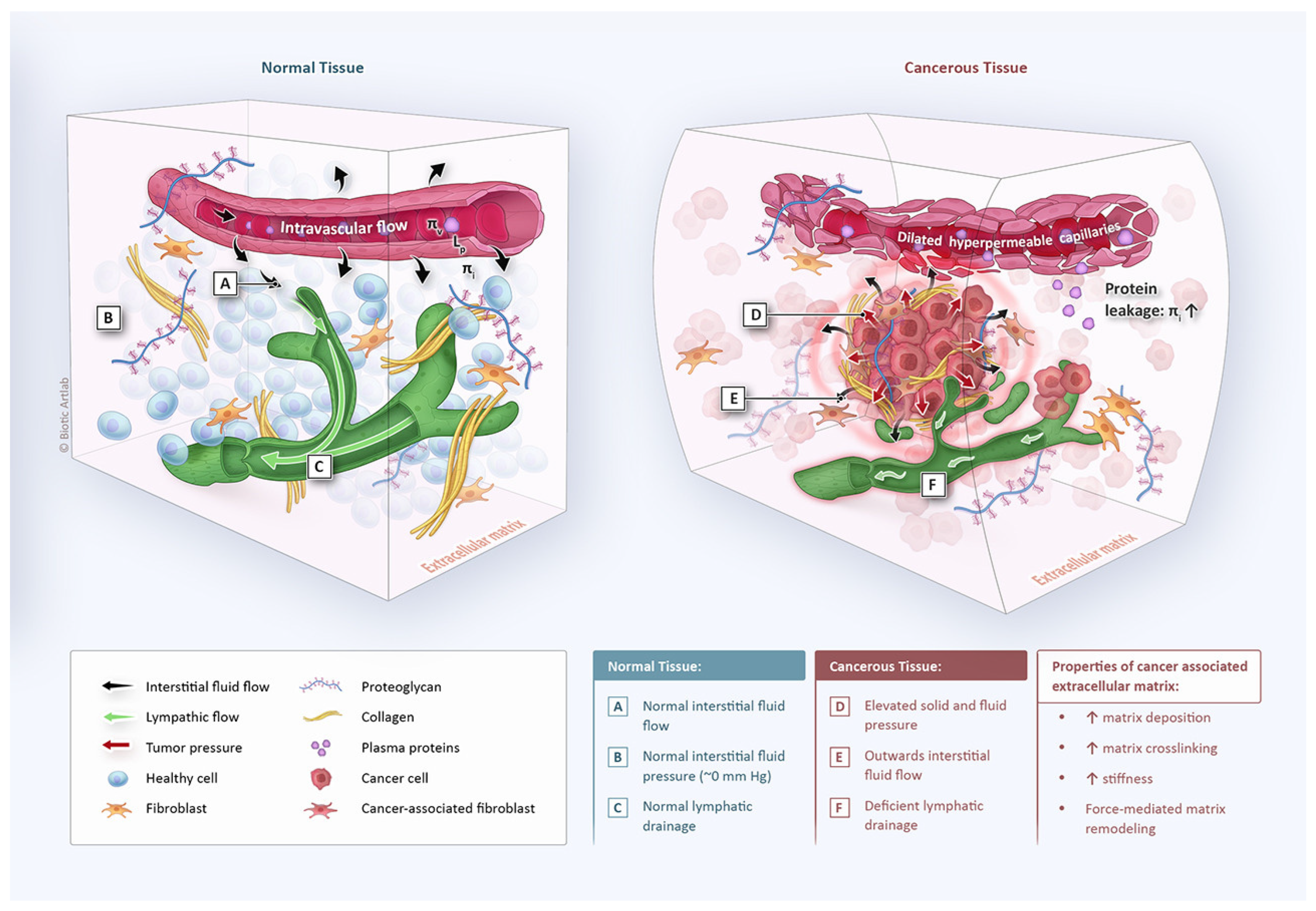
  98. Salavati, H.; Debbaut, C.; Pullens, P.; Ceelen, W. Interstitial fluid pressure as an emerging biomarker in solid tumors. Biochim. Biophys. Acta Rev. Cancer 2022, 1877, 188792. [Google Scholar] [CrossRef]
  99. Salavati, H.; Pullens, P.; Ceelen, W.; Debbaut, C. Drug transport modeling in solid tumors: A computational exploration of spatial heterogeneity of biophysical properties. Comput. Biol. Med. 2023, 163, 107190. [Google Scholar] [CrossRef]
  100. Löke, D.R.; Helderman, R.F.C.P.A.; Franken, N.A.P.; Oei, A.L.; Tanis, P.J.; Crezee, J.; Kok, H.P. Simulating drug penetration during hyperthermic intraperitoneal chemotherapy. Drug Deliv. 2021, 28, 145–161. [Google Scholar] [CrossRef]
  101. Kerch, G. Tissue Integrity and COVID-19. Encyclopedia 2021, 1, 206–219. [Google Scholar] [CrossRef]
  102. Costa, A. Permeability-porosity relationship: A reexamination of the Kozeny-Carman equation based on a fractal pore-space geometry assumption. Geophys. Res. Lett. 2006, 33. [Google Scholar] [CrossRef]
  103. Hannon, G.; Tansi, F.L.; Hilger, I.; Prina-Mello, A. The Effects of Localized Heat on the Hallmarks of Cancer. Adv. Ther. 2021, 4, 2000267. [Google Scholar] [CrossRef]
  104. Liu, Y.; Liu, L.; Chen, M.; Zhang, Q. Double thermal transitions of type I collagen in acidic solution. J. Biomol. Struct. Dyn. 2013, 31, 862–873. [Google Scholar] [CrossRef] [PubMed]
  105. Kolosnjaj-Tabi, J.; Marangon, I.; Nicolas-Boluda, A.; Silva, A.K.A.; Gazeau, F. Nanoparticle-based hyperthermia, a local treatment modulating the tumor extracellular matrix. Pharmacol. Res. 2017, 126, 123–137. [Google Scholar] [CrossRef] [PubMed]
  106. Sadjadi, Z.; Zhao, R.; Hoth, M.; Qu, B.; Rieger, H. Migration of Cytotoxic T Lymphocytes in 3D Collagen Matrices. Biophys. J. 2020, 119, 2141–2152. [Google Scholar] [CrossRef] [PubMed]
  107. Tabdanov, E.; Rodríguez-Merced, N.; Cartagena-Rivera, A.; Puram, V.; Callaway, M.; Ensminger, E.; Pomeroy, E.; Yamamoto, K.; Lahr, W.; Webber, B. Engineering T cells to enhance 3D migration through structurally and mechanically complex tumor microenvironments. Nat. Commun. 2021, 12, 2815. [Google Scholar] [CrossRef]
  108. Bougherara, H.; Mansuet-Lupo, A.; Alifano, M.; Ngô, C.; Damotte, D.; Le Frère-Belda, M.A.; Donnadieu, E.; Peranzoni, E. Real-Time Imaging of Resident T Cells in Human Lung and Ovarian Carcinomas Reveals How Different Tumor Microenvironments Control T Lymphocyte Migration. Front. Immunol. 2015, 6, 500. [Google Scholar] [CrossRef]
  109. Larsen, A.M.H.; Kuczek, D.E.; Kalvisa, A.; Siersbæk, M.S.; Thorseth, M.L.; Johansen, A.Z.; Carretta, M.; Grøntved, L.; Vang, O.; Madsen, D.H. Collagen Density Modulates the Immunosuppressive Functions of Macrophages. J. Immunol. 2020, 205, 1461–1472. [Google Scholar] [CrossRef]
Figure 1. Immune environment in the healthy peritoneal cavity. Fat-associated lymphoid clusters (FALCs) are dense immunological structures found within the omentum. FALCs are capped by a layer of CXCL13 + FALC cover cells interspersed with the mesothelial cells. FALCs also contain a network of fibroblastic reticular cells (FRCs), which recruit and modulate immune cells. Mesothelial cells also release retinoic acid, which sustains GATA6-expressing cavity-resident macrophages in the peritoneum, which induce B-cell class-switching to IgA via transforming growth factor-β2 (TGFβ2). Neutrophils also are home to FALCs during acute inflammation and form neutrophil extracellular traps that capture contaminants or cancer cells. Reprinted with permission from [30].
Cancers 15 04314 g001
Figure 2. Effects of hyperthermia-related paracrine factors on carcinogenesis.
Cancers 15 04314 g002
Figure 3. Mechanisms of carcinogenesis and pro-tumor effects of heat shock protein (HSP) 110. (A) Heat shock proteins have a chaperone function and lead to STAT3 phosphorylation. (B) Pro-tumoral effects of heat shock proteins (specifically HSP110) include angiogenesis, EMT, and immune evasion. Reprinted with permission from [79].
Cancers 15 04314 g003
Figure 4. Key differences in tissue architecture and biophysics between normal and cancerous tissue. In normal tissue (left), fluid hemostasis is preserved through balancing fluid inflow and outflow, resulting in a low or even negative interstitial fluid pressure (IFP). Cancer tissue (right) is characterized by morphologically and functionally deficient blood and lymphatic microvessels, increased matrix deposition, and cancer cell proliferation. These changes result in increased solid and fluid pressure and elevated matrix stiffness, leading to a radially outward leakage of interstitial fluid. Reprinted with permission from [98].
Cancers 15 04314 g004
Table 1. Summary of ongoing and completed clinical trials.
Completed and Ongoing Clinical Trials of Hyperthermia for the Treatment of PM
StudyCancer TypeTreatmentStatusResults
OVIP1 (NCT02567253) Ovarian CancerNormothermic (37 °C) vs. Hyperthermic (41 °C) Cisplatin (H)IPECCompletedHyperthermia increased the absorption rate of cisplatin by 16.3% [53]; clinical results not yet published.
NCT02739698Ovarian CancerNormothermic (37 °C) vs. Hyperthermic (42–43 °C) Paclitaxel (H)IPECCompletedNo difference observed in tissue penetration, pathological response, or apoptosis [8]
HyNOVA (ACTRN12621000269831)Ovarian CancerNormothermic (37 °C) vs. Hyperthermic (42 °C) Cisplatin (H)IPECOngoing
Table 2. Potential benefits and adverse effects of hyperthermia.
Potential Benefits and Adverse Effects of Hyperthermia
BenefitsAdverse Effects
Direct cytotoxic effectsIncreased systemic uptake of chemotherapy
Synergism with some chemotherapeutic compoundsHeat shock response
Increased tissue penetrationImmunosuppressive effects
Immune-enhancing effectsScald injury to the peritoneal surface
Increased blood flow and decreased interstitial fluid pressure → improved drug deliveryIncreased bacterial translocation
Disclaimer/Publisher’s Note: The statements, opinions and data contained in all publications are solely those of the individual author(s) and contributor(s) and not of MDPI and/or the editor(s). MDPI and/or the editor(s) disclaim responsibility for any injury to people or property resulting from any ideas, methods, instructions or products referred to in the content.

