Review

Recent Advances in Molecular Mechanisms of Cancer Immunotherapy

by
Mateusz Kciuk
1,2,*,
Esam Bashir Yahya
3,*,
Montaha Mohamed Ibrahim Mohamed
4,
Summya Rashid
5,
Muhammad Omer Iqbal
6,
Renata Kontek
1,
Muhanad A. Abdulsamad
7 and
Abdulmutalib A. Allaq
8
1
Department of Molecular Biotechnology and Genetics, University of Lodz, Banacha Street 12/16, 90-237 Lodz, Poland
2
Doctoral School of Exact and Natural Sciences, University of Lodz, Banacha Street 12/16, 90-237 Lodz, Poland
3
Bioprocess Technology Division, School of Industrial Technology, Universiti Sains Malaysia, Penang 11800, Malaysia
4
Faculty of Applied Medical Sciences, Mohail Aseer, King Khalid University, Abha 62529, Saudi Arabia
5
Department of Pharmacology & Toxicology, College of Pharmacy, Prince Sattam bin Abdulaziz University, Al-Kharj 11942, Saudi Arabia
6
Shandong Provincial Key Laboratory of Glycoscience and Glycoengineering, School of Medicine and Pharmacy, Ocean University of China, Qingdao 266003, China
7
Department of Molecular Biology, Faculty of Science, Sabratha University, Sabratha 00218, Libya
8
Faculty of Applied Science, Universiti Teknologi MARA, Shah Alam 40450, Malaysia
*
Authors to whom correspondence should be addressed.
Cancers 2023, 15(10), 2721; https://doi.org/10.3390/cancers15102721
Submission received: 24 April 2023 / Revised: 9 May 2023 / Accepted: 10 May 2023 / Published: 11 May 2023
(This article belongs to the Special Issue Mechanisms of Immunosuppression in Cancer)

Abstract

:

Simple Summary

The cancer-immunity cycle is characterized by various stimulatory and inhibitory factors, which together regulate the immune response and halt the extreme response that may lead to autoimmune disease. Immunotherapy of cancer has rejuvenated the field of tumor immunology and revolutionized treatment options. In the present review, we discussed the recent advances in different molecular mechanisms of cancer immunotherapy.

Abstract

Cancer is among the current leading causes of death worldwide, despite the novel advances that have been made toward its treatment, it is still considered a major public health concern. Considering both the serious impact of cancer on public health and the significant side effects and complications of conventional therapeutic options, the current strategies towards targeted cancer therapy must be enhanced to avoid undesired toxicity. Cancer immunotherapy has become preferable among researchers in recent years compared to conventional therapeutic options, such as chemotherapy, surgery, and radiotherapy. The understanding of how to control immune checkpoints, develop therapeutic cancer vaccines, genetically modify immune cells as well as enhance the activation of antitumor immune response led to the development of novel cancer treatments. In this review, we address recent advances in cancer immunotherapy molecular mechanisms. Different immunotherapeutic approaches are critically discussed, focusing on the challenges, potential risks, and prospects involving their use.

1. Introduction

The therapeutic options for cancer treatment have been constantly evolving, resulting in the prolongation of life expectancy among many cancer patients [1]. Nonetheless, cancer is still among the leading causes of death worldwide and is considered a major public health concern [2]. The lack of effective cancer treatments capable of overcoming the natural acquisition of tumor resistance is the main reason for high mortality among cancer patients. Conventional therapies such as surgery, chemotherapy, radiotherapy, or hormone therapy are either not available or highly toxic for patients [3]. The recent breakthroughs in the development of new generations of cancer immunotherapies and understanding of tumor immune biology have opened a brand new era of the war against cancer. Immunotherapy aims to improve the natural immune responses directed against tumor cells with fewer off-target effects compared to the generalized chemotherapies and other agents designed to directly kill cancer cells [4,5]. Thus, immunotherapy has been recognized in the scientific community and is now considered a promising approach for the treatment and even cure of various types of tumors. A significant acceleration in the number of scientific publications each year confirms the promising potential of immunotherapy in cancer treatment. Figure 1 present the number of publications in the Science Direct database (https://www.sciencedirect.com/, accessed on 30 March 2023) related to cancer immunotherapy in the past two decades.
Cancer immunotherapy focuses on shifting the target from tumor cells themselves to the patient’s immune system to enable its mobilization and to enhance the activation of the antitumor immune response. This helps the immune cells recognize, attack, and eventually eliminate the tumor cells [6]. The interest in cancer immunotherapy has gained increasing attention in recent years compared to the other treatment options. The discovery of different cancer immune checkpoints, the extensive understanding of the role of cytokines in the activation of antitumor response, advances in genetic engineering technology, and the generation of genetically modified immune cells changed the landscape of cancer treatment and made the promise of the final cure for this disease nearer than ever. Numerous review articles have been published recently discussing the concept of immunotherapy [7], brain tumors immunotherapy [8], and the role of certain immune cells in cancer therapy such as macrophages [9] and dendritic cells (DCs) [10]. Fukumura et al. [11] discussed the opportunities and challenges of enhancing cancer immunotherapy using antiangiogenics, without referring to the types and different mechanisms of cancer immunotherapy. Riley et al. [5] further explained delivery technologies in cancer immunotherapy.
The current review extensively covers the chronological development of cancer immunotherapy, from the first attempts to develop a cancer vaccine to the current state, going through the natural immune response and immune surveillance. Different techniques of cancer immunotherapy will be furtherly discussed including regulation of immune checkpoints and their inhibitors, oncolytic virus therapies, cancer vaccines, cytokine therapies, and adoptive cell transfer. Finally, the challenges regarding immunotherapy and potential risks as well as future directions are addressed in the present review.

2. Cancer and Immunotherapy

2.1. Incidence, Progression, and Metastasis of Cancer

For decades, scientists have been extensively studying the biological nature of cancer cells to uncover the basis of the origin of human cancers [12]. Many studies have confirmed the causative roles of chemical (tobacco smoke, medications), physical (UV-radiation, ionizing radiation), and biological (bacteria, viruses, parasites, etc.) carcinogens as well as genetic and epigenetic abnormalities (Figure 2) [13,14,15].
The development of tumors from single cells that start to proliferate in an aberrant manner, also known as tumor clonality is among the fundamental characteristics of cancer. However, just because a tumor has a clonal origin does not mean that the precursor cell from which it originated had already acquired the other characteristic of malignant cells. Cancer development is a multistep process characterized by unrestricted cell proliferation, invasion, and metastasis resulting from the accumulation of genetic and epigenetic alterations. Furthermore, cancer cells release signaling molecules to support the formation of a network of capillaries and blood vessels, to provide nutrients and energy for the growing tumor, or to facilitate migration from the site of the origin to the distant locations of the body [16]. These features accompanied by avoidance of immune destruction and generation of tumor-promoting inflammation constitute the hallmarks of cancer introduced by Hanahan and Weinberg in 2000 [17,18,19,20]. However, even after the successful treatment or removal of tumor, circulating tumor cells (metastatic cancer cells) have been observed in the blood circulation of some individuals not presenting any symptoms of cancer and suggesting that these individuals may still harbor dormant cancer cells in some tissues of their body [21]. Furthermore, the development of malignant lesions from normal cells requires a long-term dormancy time, even after direct exposure to known carcinogens [22,23]. Metastatic dormancy is a late stage in cancer progression that occurs after cell extravasation at a secondary site, where metastatic cells cease proliferation but remain dormant. When the microenvironment is favorable, they re-initiate proliferation and colonization, sometimes years after the original tumor has been treated [21].
The tumor microenvironment is a complex ecosystem that consists of various cells and non-cellular components surrounding a tumor. These cells play critical roles in tumor growth, invasion, and response to therapy [24]. Cancer cells, which are the primary cells within the tumor itself. They have acquired genetic and epigenetic alterations that allow them to proliferate uncontrollably, invade surrounding tissues, and evade the immune system. Cancer cells secrete various factors that influence the behavior of other cells in the microenvironment [25]. Cancer-associated fibroblasts (CAFs) are a type of activated fibroblast that are commonly found in the tumor stroma. They produce extracellular matrix (ECM) proteins, growth factors, and cytokines that promote tumor growth, angiogenesis (formation of new blood vessels), and invasion. CAFs can also remodel the ECM, creating a physical barrier that limits immune cell infiltration into the tumor [26]. Various immune cells also infiltrate the tumor microenvironment, either attracted by signals released by the tumor or recruited by other immune cells. These include; Tumor-infiltrating lymphocytes are a diverse population of T cells that can include both cytotoxic CD8+ T cells, which directly kill cancer cells, and regulatory T cells (Tregs), which suppress immune responses. The balance between effector T cells and Tregs influences the immune response within the tumor [27]. Tumor-associated macrophages (TAMs) are a type of immune cell derived from circulating monocytes. They can have different functional phenotypes, including M1-like (pro-inflammatory) or M2-like (immunosuppressive). TAMs play roles in promoting angiogenesis, tissue remodeling, and suppressing immune responses. Other cells have also been reported to have critical role including dendritic cells, endothelial cells, mesenchymal stem cells and other stromal cells. The tumor microenvironment may also contain other stromal cells, such as adipocytes and pericytes, which can influence tumor behavior through various mechanisms, including the secretion of factors that promote tumor growth and invasion [28]. It is important to note that the composition and function of the tumor microenvironment can vary depending on the type of cancer and the stage of the disease. The interactions between these different cell types within the tumor microenvironment are highly complex and can have both promoting and inhibitory effects on tumor progression and response to therapy. Understanding these interactions is crucial for the development of effective cancer treatments.

2.2. Antitumor Natural Immune Response

Based on current knowledge of cancer immune response, three separate steps have been described that must be completed to generate effective antitumor immunity. Antitumor immunity initiates when DCs recognize and sample cancer antigens [29]. The DCs must receive a suitable activation signal for their maturation, to allow them to further differentiate [30]. Tumor cells have been reported to overexpress certain endoplasmic reticulum stress proteins including calreticulin (CRT) on their cell surface [31]. These proteins seem to promote their phagocytosis by the activated DCs, enabling their presentation on class I and class II molecules of major histocompatibility complex (MHC) [32]. Following antigen presentation, DCs migrate to the nearest lymphoid organs and generate protective T-cell responses depending on many factors such as the interaction between T-cell co-stimulatory molecules and DC surface receptors, DC maturation stimuli, and the type of presented antigens [33]. The interaction of the surface receptors: T-cell-specific surface glycoprotein CD28 (CD28) on T cells with CD80/86 present on antigen-presenting cells (APCs) such as DCs will lead to the promotion of antitumor T-cell response. However, other receptors on T cells such as cytotoxic T-lymphocyte protein 4 (CTLA4) may interact with CD80/86 and suppress the T-cell activation, proliferation, and cytokine production. Similarly, the interaction between the programmed cell death protein 1 (PD-1) receptor expressed on the surface of T cells and programmed cell death ligand 1 (PD-L1) expressed on the surface of APCs, tumor cells, and other cells will lead to the suppression of T-cell responses and will promote the formation of regulatory T cells (T regs) [32]. Calcium (Ca2+) signaling is of paramount importance to immunity. Increasing intracellular calcium ([Ca2+]i) levels play a pivotal role in the activation of the immune system [34]. Calcium signaling is a fundamental mechanism that regulates various aspects of immune cell function, including cell activation, proliferation, differentiation, and effector responses [35,36,37]. In a recent study, the authors found two mechanisms that may account for the Ca2+ optimum of cancer cell killing—migration velocity and persistence at a moderate optimum between 500 and 1000 μm [Ca2+]o in CTLs, and lytic granule release at the immune synapse between CTLs and cancer cells is increased at 146 μm compared to 3 or 800 μm [35]. Activated T cells will leave the lymph node and migrate to the tumor bed. The exact type of activated T-cell response varies, but many studies suggested the generation of cytotoxic T cells or CD8+ effector T cells, which possess cytotoxic potential and kill cancer cells [38,39]. Figure 3 presents the steps of cancer immunity.
A balanced and highly controlled cancer-immunity cycle, characterized by various stimulatory and inhibitory factors, regulates the immune response and halts the extreme response that may trigger an autoimmune reaction. DCs have been reported to stimulate antibody-mediated as well as natural killer (NK) cell responses, which contribute to antitumor activity [41]. DCs are thought to be the link between innate and adaptive immunity because they can sample antigenic information released by cancer and present it to naive CD8+ and CD4+ T cells in draining lymph nodes via MHC-I and MHC-II molecules [42]. Many mechanisms have been proposed for tumor cells to avoid the immune response when cancer-specific T cells enter the tumor bed. These include the production of various surface molecules that suppress T-cell receptor signaling (CTLA-4 or PD-L1), localized accumulation of Treg cells, or down-regulation of MHC-I expression on the tumor cell’s surface [43]. The expression of such suppressive ligands has been linked to oncogenic mutations found in many malignancies (e.g., phosphatidylinositol 3,4,5-trisphosphate 3-phosphatase and dual-specificity protein phosphatase (PTEN) loss). Tumors can also produce immunosuppressive molecules such as indoleamine 2,3-dioxygenase (IDO), which limits tryptophan accessibility and restricts T-cell activity. Finally, stroma cells of the tumor can also hamper T-cell activity by blocking their adhesion and preventing the colonization of tumor cells in distant sites [44].
As a result, the success of cancer immunotherapy relies on its ability to overcome all of these major obstacles. Moreover, tumor-associated antigens are identical to self-antigens because they are part of the host’s tissues, making their use extremely difficult. Otherwise, the treatment could trigger autoimmune responses dangerous for patients. However, as will be discussed later, there appears to be a path to clinical success for cancer immunotherapy via the development of numerous novel approaches.

2.3. Chronological Development of Cancer Immunotherapy

Following various microbial infections, spontaneous tumor regression has been observed inspiring the first immunotherapeutic approach [45]. W. B. Coley found in the 1800s that inducing fever aids the immune system’s fight against the tumor, resulting in its regression. Coley’s approach to tumor therapy at the time, and even now, represents a once-in-a-lifetime opportunity to develop a safe, relatively inexpensive, and yet effective cancer treatment [46]. A few years later, the events regarding cancer immunotherapy have steadily developed as presented in Table 1.

3. Mechanisms of Cancer Immunotherapy

The human immune system comprises various defense mechanisms against all potential threats including arising cancer cells. Immune surveillance continuously recognizes and eliminates any transformed tumor cells through numerous mechanisms [75]. Cancer immunotherapy has been described as the fourth pillar of therapy against the tumor, which may surpass the effectiveness of conventional therapies such as surgery, radiotherapy, and chemotherapy [76]. Particularly, it has been listed by Science in 2013 among the top ten annual significantly valuable scientific breakthroughs [77]. Zhang et al. [78] classified the mechanisms of cancer immune therapies into five major groups (Figure 4)—the regulation of immune checkpoints, oncolytic virus therapies, cancer vaccines, cytokine therapies, and adoptive cell transfer.

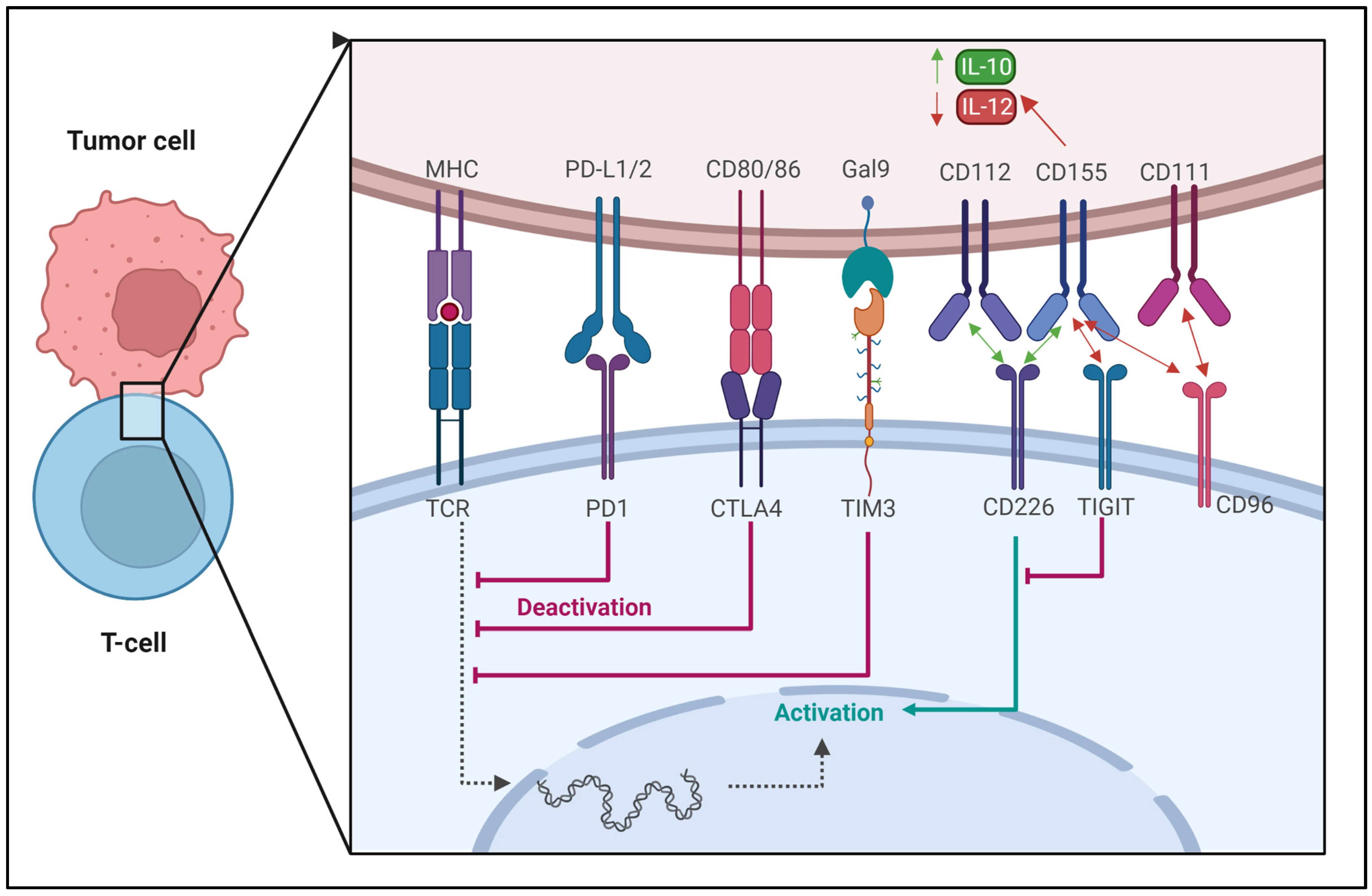
3.1. Regulation of Immune Checkpoints

Cancer therapy by regulation of immune checkpoints is an approach that aims to activate the immune system against cancer cells by targeting proteins that act as negative regulators of immune responses. Cancer cells can evade immune surveillance by expressing multiple immune-checkpoint proteins, such as programmed cell death ligand 1 (PD-L1), CD80/86, and Gal9, which inhibit T-cell activation and proliferation [79,80]. Therapies that target these immune-checkpoint proteins, such as immune-checkpoint inhibitors, have shown remarkable success in the treatment of various types of cancer [81,82,83]. These drugs block the interaction between immune-checkpoint proteins and their ligands, thereby releasing the inhibition of T-cell function.
PD-1 is an immunoglobulin (Ig) class protein that is expressed in T, B, myeloid, and NK cells. It has been demonstrated that PD-1 has two primary interacting partners: PD-L1 and PD-L2. PD-L1 is expressed by the APCs, hematopoietic and non-hematopoietic cells, whereas PD-L2 is expressed by activated macrophages and DCs. When PD-1 interacts with its ligands on other T cells, T-cell activation is hindered. The efficacy of TCR signaling is correlated with PD-1 expression in T cells. Its normal physiological function is to prevent the overactivation of T cells in the absence of the antigen. However, chronic viral infection and malignancy can trigger a constitutive PD-1/PD-L1 cell surface expression, which in turn inhibits the immune response and impairs T-cell function. This topic was reviewed by other authors [84,85,86,87,88] and the promises and limitations of the PD-1/PD-L1 blockage were recently described by our group and will not be discussed in detail [89].
CTLA-4 is a protein receptor found on the surface of T cells (CD4+ and CD8+). It is essential for immune response control, particularly in the context of T-cell activation and tolerance. CTLA-4 has a similar structure to CD28, another protein receptor produced on T cells that plays an important role in T-cell activation. CTLA-4, on the other hand, offers an inhibitory signal that reduces T-cell activation and proliferation, whilst CD28 gives a co-stimulatory signal that increases T-cell activation. CTLA-4 competes with CD28 for binding to APCs co-stimulatory molecules CD80 (B7-1) and CD86 (B7-2). When CD28 attaches to these molecules, it provides a stimulating signal, whereas CTLA-4 delivers a negative signal, limiting T-cell activation and proliferation [90,91,92].
Despite both of these molecules contribute to the inhibition of anticancer immunity, the timing of down-regulation, the underlying signaling mechanisms, and the anatomic locations of immunological suppression by these two immune checkpoints are different. PD-1 is more widely expressed on activated T cells, B cells, and myeloid cells than CTLA-4, which is only found on T cells. While CTLA-4 is active during the T-cell activation’s priming phase, PD-1 is active during the T-cell activation’s effector phase, largely within peripheral tissues. These characteristics suggest that anti-CTLA-4 and anti-PD-1 treatments may have cumulative and synergistic benefits in the treatment of the disease [93,94,95]. PD-1 inhibitors, such as pembrolizumab and nivolumab, have been approved for the treatment of melanoma, non-small-cell lung cancer, bladder cancer, and other types of cancer. CTLA-4 inhibitors, such as ipilimumab, have been approved for the treatment of melanoma, and combinations of PD-1 and CTLA-4 inhibitors are being investigated for the treatment of several types of cancer. Other immune-checkpoint inhibitors, such as those targeting T-cell immunoglobulin and mucin-domain containing-3 (TIM-3), are currently under investigation in clinical trials [96,97].
TIM-3 is another cell surface immune-checkpoint molecule expressed in immunological cells such as T cells, Tregs, DCs, B cells, macrophages, NK cells, and mast cells. TIM-3 binds to four different ligands: galectin-9 (Gal-9), CEACAM-1 (carcinoembryonic antigen cell adhesion molecule 1), HMGB1 (high-mobility group protein B1), and PS (phosphatidylserine). TIM-3 mediates T-cell exhaustion, which suppresses antitumor immunity. The selective blockage of TIM-3 enhances anticancer immunity through increased IFN-γ production by T cells. TIM-3 inhibitors have been studied in animal models and clinical trials [98,99,100,101]. The blockade of a single checkpoint in many cases may not achieve the desired therapeutic effect [102,103]. Figure 5 presents the mutual regulation by various immune checkpoints between cancer cells and T cells.
TIGIT (T-cell immunoreceptor with Ig and ITIM domains) is a protein that is expressed on various types of immune cells, including T cells and NK cells. TIGIT is up-regulated on activated T cells, particularly on Tregs, follicular helper T cells (Tfh), and memory T cells [104]. TIGIT is a co-inhibitory receptor that interacts with its ligands, CD155 (poliovirus receptor) and CD112 (nectin-2), which are expressed on APCs and some tumor cells. The binding of TIGIT to its ligands leads to the inhibition of T-cell activation and proliferation, as well as the suppression of cytokine production. Therefore, TIGIT plays an important role in the regulation of the immune response and maintenance of immune homeostasis. Targeting TIGIT has emerged as a potential immunotherapy strategy for cancer and autoimmune diseases [105]. On the other hand, CD96 (also known as TACTILE) is a protein that is primarily expressed on activated T cells, including CD8+ T cells, Tregs, NKT cells, and some subsets of memory T cells. CD96 is also expressed in some tumor cells [106]. CD96 interacts with its ligand, CD155, which is widely expressed in various cell types, including T cells, DCs, tumor cells, and many other cell types. CD96 has been shown to play a role in T-cell activation and effector functions, as well as in the regulation of immune tolerance and autoimmunity. CD226 (also known as DNAM-1) and CD112 (nectin-2) are also cell surface receptors that are expressed on various immune cells, including T cells, NK cells, and DCs, as well as on many tumor cells. CD226 interacts with its ligands, CD155 and CD112, and plays a role in the activation and cytotoxicity of NK cells and CD8+ T cells [107,108]. TIGIT and CD96 are more likely to bind and suppress T-cell activation than CD226, which has a lower affinity for CD155. TIGIT binding to CD155 on the tumor cells leads to the up-regulation of IL-10 and down-regulation of IL-12 [106].
Vascular endothelial growth factor receptor-1 (VEGFR-1) is a crucial receptor that plays a major role in tumor progression as well as in the resistance to treatment methods based on immune-checkpoint inhibitors [109]. Activation of VEGFR is involved in myeloid progenitors mobilization from the bone marrow which later infiltrates the tumor. Lacal et al. [110] recently developed an anti-VEGFR monoclonal antibody (anti-VEGFR-1 mAb) able to inhibit the growth and proliferation of melanoma in preclinical in vivo models. In addition, the reduction in macrophage progenitor mobilization and tumor cell infiltration by myeloid cells was observed. The same authors indicated significant up-regulation in the expression of VEGFR-1 in human-activated macrophages 2 (M2) compared to activated macrophage 1 (M1) cells. The exposure to the designed monoclonal antibody (D16F7 mAb) decreases the chemotaxis of macrophages in vitro. The results of in vivo treatment in melanoma-bearing mice resulted in significant inhibition of tumor growth due to the alterations in the tumor microenvironment, which lead to a decrease in melanoma infiltration by M2 as well as PD-1+ and FoxP3+ cells. These modifications resulted in an increase in M1/M2 and CD8+/FoxP3+ cell ratios in favor of antitumor immune response [110]. Figure 6 presents the mechanism of using VEGFR-mediated signaling inhibition for the reduction in tumor infiltration by improving antitumor immune response and enhancing the efficacy of immunotherapy using immune-checkpoint inhibitors. Studies by Chauvin et al. [111] revealed the overexpression of PD-1 and TIGIT on most tumor-infiltrating CD8+ T cells and tumor antigen-specific circulating CD8+ T cells. Combined blockade of both checkpoints (TIGIT and PD-1) has shown significant enhancement in the antitumor function of cytotoxic CD8+ T cells compared to blocking PD-1 alone [112], this dual blockade seems to depend on CD226 signal transduction [113]. The combination of specific checkpoint blockade can be used to improve immune response and constitutes a promising direction for cancer immunotherapy.
Immune-checkpoint inhibition can be used to treat various malignancies including melanoma, lung cancer, bladder cancer, kidney cancer, and Hodgkin’s lymphoma. Unlike standard cancer therapies such as chemotherapy, immune-checkpoint inhibition can result in long-term remission and even tumor eradication in some individuals. Because they exclusively target the immune system rather than all rapidly dividing cells in the body, immune-checkpoint inhibitors have fewer side effects than standard chemotherapy. Despite increased safety profile, they can still induce substantial consequences such as lung, liver, and other organ inflammation. Additionally, the inhibition success rate depends on the type of cancer and other factors such as the patient’s immune system status. Additionally, certain cancer cells may acquire resistance to immune-checkpoint inhibitors, resulting in therapy failure. Because immune-checkpoint inhibitors are a relatively new type of cancer treatment, long-term data on their efficacy and potential side effects is also lacking. This topic was previously described by other authors [114,115,116,117].

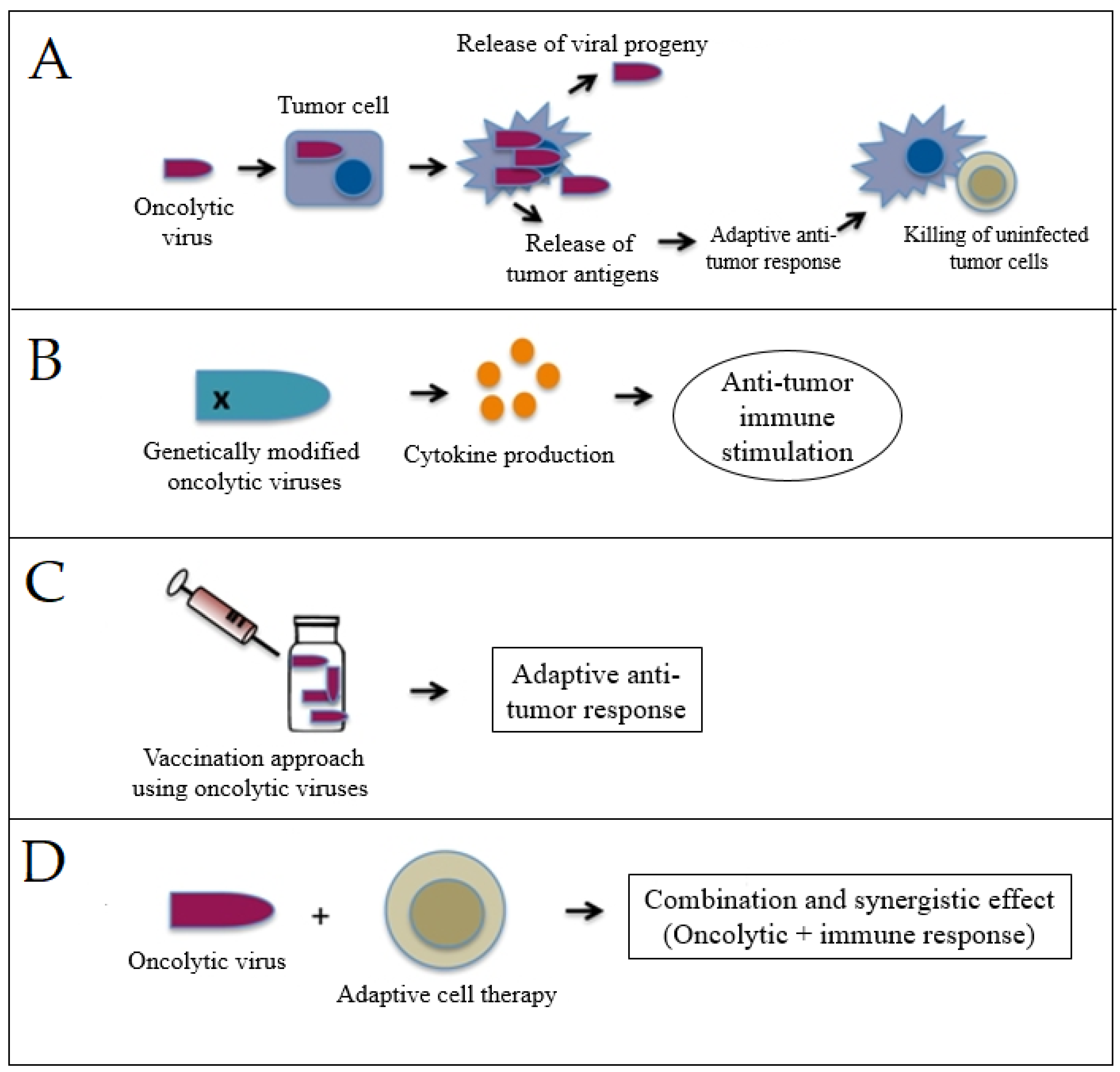
3.2. Viro-Immunotherapy

Viruses have been widely investigated for being the ideal therapeutic option for various types of cancers in combination with immune-checkpoint inhibitors and many other types of immunotherapeutic approaches [118,119]. Oncolytic virotherapy focuses on the development of highly specific genetically engineered viruses for targeting only cancer cells. Following the recognition of cancer cells expressing the particular molecules, these viruses infect and kill cancer cells through their lysis. Owing to their ability to accommodate gene insertions, oncolytic viruses have been armed with transgenes to produce immune-stimulatory signals to improve antitumor immune response [120,121]. Numerous clinical and preclinical studies on the use of different oncolytic viruses in cancer immunotherapy have been conducted and reported promising antitumor potential. Table 2 presents the most commonly used oncolytic viruses for viro-immunotherapy.
Vesicular stomatitis viruses (VSV) have been reported to have several beneficial properties that favor their use for immunotherapy, including inherent tumor specificity, rapid replication kinetics, and the capacity to elicit a wide variety of immune responses [129]. Melzer et al. [130] summarized the strategies of this novel platform for the enhancement of the immune-stimulating potential as presented in Figure 7. Modification of the virus’s endogenous genes to stimulate more interferon induction and virus-mediated expression of immune-stimulatory cytokines are two strategies that have been widely used with VSVs-based viro-immunotherapy or employed with other viruses to boost antitumor immune responses [131,132]. Vaccination approaches based on viruses have also been used to stimulate and even improve adaptive immune responses against particular tumor antigens [133].
In comparison to chemotherapies, oncolytic viro-immunotherapy has many advantages [134,135] including (a) selective targeting of cancer cells. Cancer cells can be specifically targeted and destroyed with viro-immunotherapy while healthy cells are unharmed. This is because the viruses have been engineered to specifically target cancer cells, reducing the likelihood of unintended effects. (b) Induction of immunogenic cell death: Immunogenic cell death triggered by oncolytic viruses primes the immune system to target and destroy cancer cells. As a result, the immune system has an increased likelihood of killing cancer cells even if they have metastasized. (c) Synergistic effect with other therapies: Combining viro-immunotherapy with standard cancer therapies such as chemotherapy and radiation has been shown to improve patient outcomes. Some clinical and preclinical research indicates that combination therapy is more effective than either medication alone. (d) Minimal systemic toxicity: As the viruses used in viro-immunotherapy are engineered to specifically target and destroy cancer cells, they are found to have low systemic toxicity. This lessens the likelihood of side effects that are usually associated with conventional anticancer therapies. (e) Potential to overcome drug resistance: Drug resistance is a serious problem impacting the usefulness of cancer therapies. Oncolytic viruses can specifically target and kill cancer cells that have developed resistance to other therapies. (f) Ability to target cancer stem cells: Cancer stem cells are a subpopulation of cancer cells that are believed to be responsible for tumor initiation, upkeep, and resurgence. Viro-immunotherapy may be able to target these cells. Treatment outcomes may improve and last longer if cancer stem cells are targeted [121,136,137,138]. However, their use can be also associated with some limitations such as (1) Limited efficacy in some types of cancer: The presence of a protective stromal barrier that blocks the virus from accessing the cancer cells has restricted the effectiveness of viro-immunotherapy in certain kinds of cancer. This may reduce the efficacy of viro-immunotherapy in some conditions. (2) Potential for immune-related adverse events: Viro-immunotherapy raises the risk of immunological-related side effects because it stimulates the immune system. Symptoms including the flu, inflammation, and autoimmune disorders are all potential side effects. (3) Pre-existing immunity to the virus: The therapeutic efficacy of the oncolytic virus may be reduced in patients who already have developed an immune response to the virus. This may occur as a result of previous contact with viruses or a tendency to mount an immune response against the virus as a result of a person’s genetic makeup. (4) Limited availability of clinical trials: Since viro-immunotherapy is still in its experimental stages, there are not many clinical trials available, that determine its safety and efficacy. Additionally, due to the novelty of viro-immunotherapy patients’ accessibility to this treatment option may be limited. (5) Potential for viral shedding: Viruses used in viro-immunotherapy can spread to other people especially patients with compromised immune systems. (6) Cost: Because of its complexity and high expense, viro-immunotherapy may be out of reach for many individuals. As indicated by the previous authors [119,121,136,137,138].

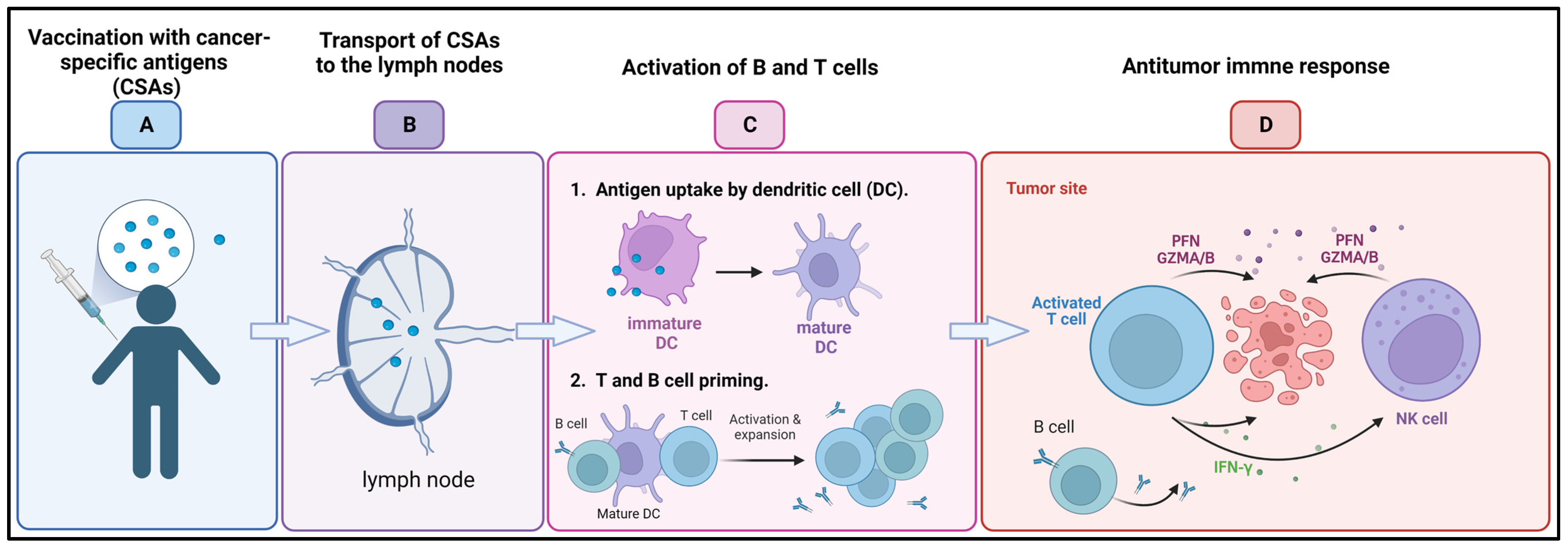
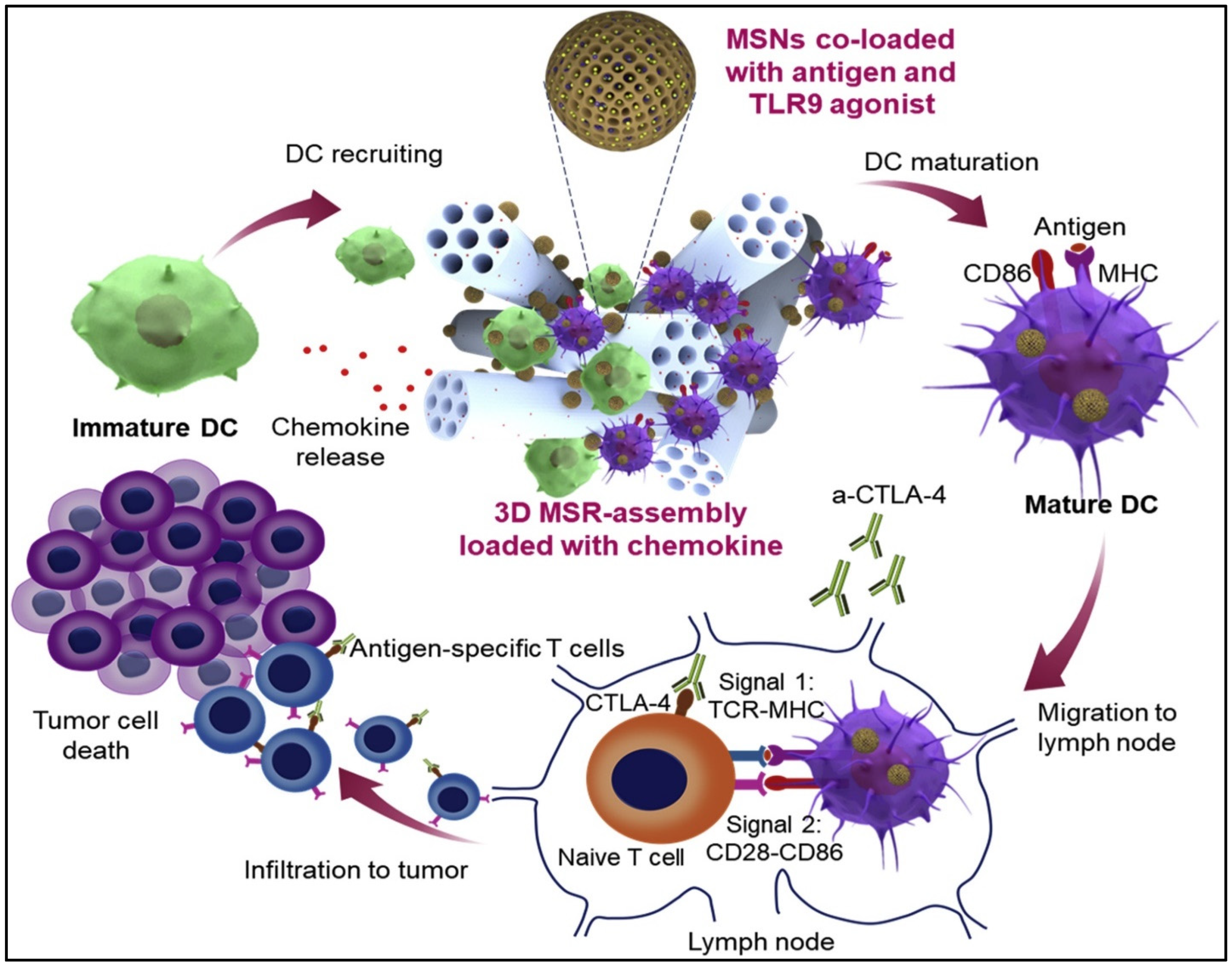
3.3. Cancer Vaccines

Cancer-preventive vaccines are intended to thwart the progression of cancer by either focusing on cancer-specific antigens that are expressed on precancerous or early-stage cancer cells or on viruses that are known to cause cancer [139]. On the other hand, therapeutic cancer vaccines (Figure 8) are intended to cure tumors that have already developed by enhancing the immune system’s capacity to recognize and destroy cancer cells. In recent decades, tremendous effort has been devoted to the development of cancer vaccines. One strategy that showed promise as a potential therapeutic option is the use of cancer-specific antigens (CSAs). CSAs are proteins or peptides that are exclusive to cancer cells and are identified by the immune system as being of a foreign origin. Several CSAs have been investigated, and they are now being incorporated into cancer vaccines to elicit an immune response against cancer cells [140,141].
Carcinoembryonic Antigen (CEA): CEA is a glycoprotein that is overexpressed in many malignancies, especially colorectal cancer. Clinical trials using a CEA-based vaccination for the treatment of several human cancers have been designed based on experimental results achieved using animal models. DCs- and recombinant virus-based vaccinations appear to be the most reliable among the several CEA-based cancer vaccines. Vaccination against CEA has been shown to induce a strong immune response, leading to a slower rate of tumor progression and improved survival among certain cancer patients. However, in most cases, vaccination has failed to eradicate the tumor, in part because of the detrimental impact exerted by the tumor microenvironment on the immune response [142,143].
Melanoma-associated antigen-A3 (MAGE-A3): MAGE-A3 is highly expressed in a variety of malignancies, including melanoma, non-small-cell lung cancer, and head and neck cancer. MAGE-A3 vaccination has been studied in multiple clinical studies for its safety, and potential to increase survival rates and trigger an immunological response in patients with the above-mentioned cancers [144,145,146,147]. The exact cellular function of this protein has not been revealed. Yang et al. suggested that MAGE protein expression may actively contribute to the formation of malignancies and increase tumor survival through inhibition of the tumor suppressor TP53 protein and decrease in cell apoptosis [148].
Mucin 1 (MUC1): Glycoprotein MUC1 is overexpressed in several cancers, such as breast, pancreatic, and lung cancers. The MUC1 vaccination has shown promise in boosting survival rates and eliciting immune responses in patients with pancreatic cancer and non-small-cell lung cancer in clinical studies [149,150].
New York esophageal squamous cell carcinoma-1 antigen (NY-ESO-1): Several kinds of cancer, such as melanoma, lung cancer, and ovarian cancer, express the antigen. Because of its capacity to trigger spontaneous humoral and cellular immune responses, as well as its restricted expression pattern, it is a promising candidate target for cancer immunotherapy [151].
Prostate-specific antigen (PSA): PSA is widely utilized in the identification and monitoring of prostate cancer. Vaccines based on PSA have been the subject of many clinical trials, some of which have shown encouraging results while others have been ambiguous [152,153,154].
Wilms Tumor 1 (WT1): WT1 is a transcription factor up-regulated in a wide variety of malignancies, including solid tumors, lymphomas, and leukemias. Patients with leukemia and mesothelioma who received WT1 vaccination in clinical trials experienced a significant increase in immune responses and survival rates [155,156].
Other include melanoma-associated antigen 1 (MZ2-E) and melanoma-associated antigen 3 (MZ2-D) from the melanoma-associated antigen gene family. They were effectively recognized by many immune cells, including cytotoxic T cells, and have been shown to elicit antitumor immune responses in melanoma-bearing cancer patients [157].
Dendritic cell vaccines, which use DCs to trigger an immune response against cancer cells, are another potential treatment option. DCs are specialized immune cells capable of presenting antigens to T cells. DC vaccines have shown promise in clinical trials for a range of malignancies, including melanoma and prostate cancer [158]. Furthermore, several different kinds of macroporous scaffolds were created to release DC-recruiting chemokines, which would trigger the activation and maturation of these DCs. In a recent study, Nguyen et al. [42] developed an injectable dual-scale mesoporous silica vaccine enclosing chemokine, thereby facilitating the recruitment of numerous DCs into the scaffold composed of Toll-like receptor 9 (TLR-9) agonist and an antigen, which internalized by the recruited DCs became effectively presented [42]. The use of aerogels and mesoporous silica nanoparticles attracted tremendous attention in recent years [159,160]. Figure 9 presents efficient DC recruitment and activation by injectable dual-scale mesoporous silica microrod–mesoporous silica nanoparticle vaccine.
Exosomes are small vesicles that are connected to membranes and are produced by the majority of cells for intercellular exchange and regulation. These exosomes deliver materials or messages to the cells they are targeting for a variety of physiologically significant purposes. Exosomes that were derived from M1-polarized, proinflammatory macrophages were the subject of the research that was conducted by Cheng et al. The researchers were interested in the possibility of using M1 exosomes as an immunopotentiator for a cancer vaccine. After being injected subcutaneously, the M1 exosomes showed a tropism toward lymph nodes. They were predominantly uptaken by the local macrophages and DCs and triggered the secretion of a pool of Th1 cytokines. M1, but not M2, exosomes increased the activity of a lipid calcium phosphate (LCP) nanoparticle-encapsulated tyrosinase-related protein 2 (TRP2) vaccine and elicited a stronger antigen-specific cytotoxic T-cell response. In a melanoma growth inhibition study, M1 exosomes showed to be a more potent immunopotentiator than an adjuvant, such as CpG oligonucleotides (ODNs) when combined with LCP nanoparticle vaccine in a melanoma. This study demonstrated the potential of M1-polarized macrophage-derived exosomes as a vaccine adjuvant [161].
Despite the large number of vaccine strategies that are being developed regularly, the construction of safe and successful therapeutic cancer vaccines remains a challenge [140,162,163]. Advantages of their use include the prevention of cancer—vaccines against cancer-causing viruses including human papillomavirus (HPV) and hepatitis B virus (HBV) can be used to protect against the onset of cancer. Cancers such as cervical and liver cancer have been proven to decrease in frequency after vaccination against these viruses. Fewer side effects are another advantage due to cancer vaccines target only cancer cells, leaving healthy cells unharmed, making them safer than standard cancer therapies such as chemotherapy and radiation. On the other hand, the disadvantages include limited efficacy: not all individuals who receive a vaccine will experience a positive response. Cancer vaccines’ success rates may differ among patients with different cancer stages. High cost: cancer vaccines can be pricey, especially if they are customized for each patient, and may not be affordable for everyone. Additionally, development challenges: it can be difficult and time-consuming to develop a vaccine against cancer since accomplishing it requires finding the right target antigens, refining the vaccine’s formulation, and testing it in humans to make sure it is safe and effective.

3.4. Cytokine Therapies

Cytokines are chemical messengers secreted by immune and non-immune cells in reaction to cellular stresses such as bacterial infections, inflammation, and tumorigenesis to control cellular interactions [164]. The first use of cytokines in cancer treatment occurred in 1976, following the discovery of interleukin-2 (IL-2), initially known as a T-cell growth factor, which appeared to have the capacity to activate T cells and thus exert immune-stimulatory properties [164,165]. In vitro incubation of inactive lymphoid cells with recombinant IL-2 results in the production of lymphokine-activated killer (LAK) cells, which are cells that are capable to lyse tumor cells. The administration of LAKs and large doses of IL-2 in patients with various types of cancer have been reported to cause cancer regressions and significantly enhance antitumor response in preclinical studies and clinical trials [166,167].
More than half a century ago, type I interferons (IFNs), were identified for the first time as the factors responsible for virus inhibition. However, in the last ten years, researchers have only just started to understand the precise function that type I IFNs play in the body’s natural immune response to cancer. IFNs have been implicated in two primary functions in tumor control—induction of tumor cell senescence and stimulation of DC maturation to boost the T-cell cytotoxicity [168]. Cauwels et al. [169] developed a novel strategy for cancer treatment using Activity-on-Target cytokines, which are modified immune-cytokines that possess up to 1000-fold more potency for target cells. The authors reported that modified type I interferon designed to target Clec9A+ DCs was able to limit the progression of tumors and showed strong antitumor activity in breast carcinoma, murine melanoma, and lymphoma models, without toxic side effects. IFN activation of immune cells may inhibit tumor angiogenesis, whereas ILs promote the activation and growth of helper and cytotoxic T lymphocytes (CD4+ and CD8+ T cells) [170,171]. Granulocyte-macrophage colony-stimulating factor (GM-CSF) is another cytokine that, in reaction to stress, infections, and malignancies, stimulates the formation of myeloid cell subgroups including neutrophils, monocytes, macrophages, and DCs. GM-CSF has a global effect on the host immune surveillance system when pathologic conditions are present. When compared to other soluble immune mediators, it has been discovered that either an excess of or a deficiency in GM-CSF can encourage the aggressiveness of cancer. An insufficient amount of GM-CSF inhibits the proper production of innate immune cells, which in turn prevents the resulting induction of adaptive anticancer immune responses. On the other hand, too much GM-CSF can exhaust immune cells and encourage the growth of cancer [172,173]. GM-CSF has been also used to accelerate and augment granulocyte recovery after chemotherapy of cancers [174,175]. Cytokines such as INFγ or tumor necrosis factors (TNFs) are essential in the development of an immunogenic microenvironment. Anthracyclines as well as other treatments such as photodynamic therapy (PDT) exert their impacts on cancer cells in a way that stimulates the body’s immune system. This process, also known as immunogenic cell death (ICD), is distinguished by the release of membrane-bound and soluble factors that boost the function of immune cells [176] and can act as pro-inflammatory factors (TNFs, IL-1β, and IL-6) or can lead to increase in the expression of MHC class I on the surface of antigen-presenting cells. This leads to improved immune response via the differentiation promotion or the activation of both T cells and NK cells [177].
Immunogenic cell death inducers trigger the release of damage-associated molecular patterns (DAMPs) such as high mobility group box protein 1 (HMGBP1), CRT, and ATP, as well as inflammatory cytokines from cancer cells. Both DAMPs and cytokines send activating signals for DCs and NK cells, which respond instantly by releasing effector cytokines [178]. Dying tumor cells treated with ICD inducers trigger the release of various cytokines, which modulate an effective immune response, such as IL-6 and IL-8, [179]. NK cells functionality appears to be increased by cytokines released from activated DCs, such as IL-12, as well as cytokines produced by other innate immune cells, such as IFN-α/β, leading to the secretion of IFN-γ and TNFα (Figure 10) [177].
Ekeke et al. [180] used the vaccinia virus to up-regulate the expression of IL-2 to activate antitumor response against malignant pleural disease and to trigger the immune modulatory effects. Local administration of an IL-2-expressing vaccinia virus (VV-IL-2) significantly reduced tumor burden, due to the increase in the activity of immune cells including CD4+ and CD8+ T cells and α,β T-cell receptor diversity in a malignant pleural disease model (Figure 11).
While cytokine treatments have the potential to improve cancer treatment, they also pose considerable risks [181,182,183,184,185,186]. Below are the pros and negatives of their use:
  • Stimulation of the immune system: the immune system can be stimulated with cytokine treatments to better target and destroy cancer cells.
  • Reduced toxicity: compared to conventional treatments such as chemotherapy and radiation, cytokine therapies are safer and have fewer adverse effects.
  • Versatility: cytokine treatments are adaptable because they can be used to treat a wide variety of cancers.
  • May provide long-term benefits: some cytokine therapies have been demonstrated to produce lasting advantages in cancer patients, increasing the likelihood of a long-lasting response to treatment.
  • Can be used in combination with other treatments: combining cytokine therapies with standard cancer treatments such as chemotherapy, radiation therapy, and immunotherapy has been shown to improve patient outcomes.
  • May induce remission: some cytokine therapies, such as high-dose IL-2 and IFN-α, have been shown to induce complete or partial remission in some cancer patients.
  • Long-term immune memory: long-term immunological memory can be induced by some cytokine therapies, such as IL-2, meaning that the immune system will continue to detect and fight cancer cells even after treatment has ended.
  • Fewer treatment sessions: patients may find cytokine therapies more convenient because they often need fewer treatment sessions than treatments such as chemotherapy.
Although cytokine treatments have shown promise in the treatment of cancer, further study is needed to determine their full potential. It is important to overcome the potential limitations of this therapeutic approach:
  • Limited effectiveness: some patients may only have a partial response to cytokine treatments, and they may not work for all forms of cancer.
  • Side effects: side effects from cytokine therapies include fever, exhaustion, and muscle aches, but they are typically less severe than those from chemotherapy and radiation. Some cytokine therapies, such as high-dose IL-2, can cause damage to organs such as the kidneys and liver, which can be life-threatening.
  • Expensive: some patients may not be able to afford cytokine therapy due to their high cost.
  • Can cause autoimmune reactions: there are risks associated with cytokine treatments, including autoimmune reactions and tissue damage.

3.5. Adoptive Cell Transfer

Adoptive cell transfer (ACT) is a cancer treatment method that involves injecting immune cells, such as T cells, into a patient’s body, where they seek out and destroy cancer cells. Before being administered to a patient, T cells are usually modified or enhanced in the lab to strengthen their ability to recognize and target cancer cells. CAR T-cell therapy is a very successful form of ACT that involves engineering T cells to express chimeric antigen receptors (CARs) that can recognize and adhere to specific proteins on the surface of cancer cells (Figure 12) [187,188].
Rosenberg et al. [166] revealed that the administration of modified autologous LAK cells in addition to recombination-derived IL-2 was effective in the treatment of patients with metastatic cancers. High response rates in patients with follicular lymphoma and diffuse large B-cell lymphoma have been reported with modified T cell-based therapy, using CAR able to target CD19 receptors on the surface of B-cell cancers [189]. Schuster et al. [190] used autologous modified T cells expressing a CD19-directed CAR to treat patients with follicular lymphoma or diffuse large B-cell lymphoma. The authors revealed that the engineered cells were effective in the treatment. High rates of durable disease reduction were observed, with significant recovery of B cells and immunoglobulins in some patients. Similarly, Wang et al. [191] conducted a trail on seven patients suffering from advanced diffuse large B cell lymphomas and concluded that anti-CD20 CAR T cells caused prolonged tumor regression. In a different study, Mohammed et al. [192] generated chimeric antigen receptor T cells directed against a specific antigen located on prostate stem cells and revealed specific tumor lysis. The authors generated an inverted receptor for the cytokine to protect the modified cells from the immunosuppressive cytokines, resulting in significant enhancement in antitumor activity. For the determination of whether the superior modified T-cell expansion and antitumor effects of chimeric antigen receptor/inverted cytokine receptor (CAR/ICR) could be recapitulated in vivo, the authors used mice models as presented in Figure 13, and treated animals intravenously with CAR/ICR T cells. Superior antitumor effects were observed with CAR/ICR T cells-based treatment, resulting in 100% overall survival for the treated group compared to the control group [192].
Similarly to the previously discussed immunotherapeutic approaches CAR T-therapies possess significant advantages over conventional chemotherapy and radiotherapy. In contrast to conventional chemotherapy, which is harmful to normal cells as well as cancerous ones, CAR T-cell therapy is a targeted technique that can selectively destroy cancer cells while sparing healthy ones. This lowers the toxicity and adverse effects of CAR T-cell treatment. CAR T cells may also persist in the body for an extended time and continue to attack cancer cells, offering protection against the disease even after the cells have been eliminated. The elimination of cancer cells using CAR T-cell therapy is curative in many patients. However, similar to other novel immunotherapies, the CAR T-cell approach is an expensive treatment. This can be attributed to the sophisticated manufacturing procedure for CAR T cells that can cause treatment delays for some patients. Moreover, it is crucial to determine which patients could benefit the most form the treatment and those with increased risk of detrimental side effects such as cytokine release syndrome (CRS) and immune effector cell-associated neurotoxicity syndrome (ICANS) [193,194].

3.6. Other Immune Therapies

Lag-3 (Lymphocyte Activation Gene 3) is a protein receptor expressed on the surface of certain immune cells, including T cells, B cells, and natural killer (NK) cells. Lag-3 plays a role in regulating immune responses and is considered an immune-checkpoint protein [195]. In the context of immunotherapy, Lag-3 has gained attention as a potential target for cancer treatment. Immune-checkpoint inhibitors, such as antibodies targeting PD-1 (programmed cell death protein 1) and CTLA-4 (cytotoxic T-lymphocyte-associated protein 4), have shown success in unleashing the immune system against cancer cells. However, not all patients respond to these therapies, highlighting the need for additional targets [196]. Several clinical trials are underway to investigate the efficacy of Lag-3 inhibitors either as monotherapy or in combination with other immunotherapies. Preliminary data from these trials suggest that Lag-3 inhibitors may provide clinical benefit in certain types of cancers, such as melanoma, lung cancer, and renal cell carcinoma [197,198]. It is important to note that the field of Lag-3 and immunotherapy is still evolving, and more research is needed to fully understand its role and potential in cancer treatment. Clinical trials will continue to provide valuable insights into the efficacy and safety of Lag-3-targeted therapies, as well as their optimal use in different cancer types and patient populations.
Nanoparticles and biomaterials have been employed to enable programmed localization, improve pharmacokinetics, and co-deliver immune-modulatory substances that were not previously possible with direct administration of these compounds in solution [199]. In a recent investigation, Ji et al. [200] designed a biopolymer immune implant for post-surgical therapy of colorectal cancer and revealed that an immune implant was able to eradicate residual tumors post-surgery for more than 150 days. The gradual release of the loaded resiquimod (Toll-like receptor 7/8 (TLR7/TLR8) agonist) was achieved and the anti-CD134 (OX-40) antibody elicited immune memory and inhibited the growth of distal tumors when used in the implant [200]. In previous studies, the OX40 and OX40 ligand interaction was shown to promote the growth and division of T cells while attenuating the immunosuppression of Treg cells. The OX40 plays a crucial part in immunity, hence it is the subject of numerous clinical trials to assess its therapeutic effects in the treatment of cancer [201]. Immunological studies demonstrated that treating patients with a biopolymer immune implant has a two-phase effect: initially, a rise in the infiltration of NK cells and DCs is observed in the first few days, followed by an increase in the infiltration of T cells and the establishment of immune memory in the following weeks [200]. Peptide-based materials constitute another approach to cancer immunotherapies that have been used to solve several traditional treatment challenges and have demonstrated specific and considerable efficacy as cancer immunotherapies [202]. Peptide-based cancer vaccines and delivery systems have been the subject of extensive research. These vaccines and systems aim to replicate the functional domains of proteins with highly specialized immuno-regulatory capabilities [202,203]. Shen et al. [204] reported a mono-palmitoylated peptide able to induce an anticancer immune response without any addition of an adjuvant. The authors also developed a long peptide of a di-palmitic acid conjugated with TLR2 agonist and achieved significant improvement in antitumor immunity by diminishing the function of tumor-associated macrophages (TAMs). Using a different approach, Choi et al. [205] investigated different peptide antigens from cancer stem cells to compose DC vaccination for hepatocellular carcinoma and human breast cancer. By pulsing dendritic cells with CD44 and epithelial cell adhesion molecule-based peptides, the authors reported an effective stimulation and mature DC production in addition to enhanced T-cell stimulation and significant increase in the number of CTLs [205].

4. Challenges and Prospects of Cancer Immunotherapy

Improving the immune response against cancer can be accomplished by suppressing or activating immunological checkpoint proteins on T cells. Anti-CTLA-4 and anti-PD-1/PD-L1 checkpoint inhibitors have shown promise in the treatment of melanoma, non-small-cell lung cancer, and bladder cancer. These drugs, however, have been linked to immune-related side effects such as colitis, hepatitis, and pneumonitis [93,206]. The goal of oncolytic viral therapy is to stimulate an immune response against the tumor by selectively infecting and destroying cancer cells. These treatments have shown promise in studies on a variety of cancers, including melanoma, glioblastoma, and pancreatic cancer. However, because of difficulties with virus transmission to tumor locations and preexisting immunity, they may not be as effective as intended [119,121,136,137,138,207]. Cancer vaccines work by inducing an immune response against cancer-causing antigens to prevent or treat the disease. Cancer vaccines that have been developed and tested include peptide vaccines, DC vaccines, and whole-cell vaccinations. Despite encouraging achievements in generating an immune response, most vaccinations still have low clinical effectiveness. Some issues with the use of cancer therapeutic vaccines to treat various types of malignancies have been described, such as the nonresponsiveness of activated T cells due to reduced antigen presentation and/or increased suppression of self-reactivity by Tregs [140,141,208]. Another concern with therapeutic vaccines is that they are inadequately immunogenic, resulting in self-derived antigens that limit the immune response. However, certain attempts to boost such responses have resulted in the development of autoimmune reactions in patients [209,210]. Cytokine treatments include the injection of patients with cytokines, which are signaling molecules produced by immune cells, to improve the immune system’s defenses against cancer. However, these treatments can have serious adverse effects and are only partially successful against certain types of cancer [184]. T cells are extracted from a patients, grown and manipulated to attack cancer-related antigens, after their reintroduction to the patient. CAR- T-cell therapy, which involves engineering T cells to express CARs that identify cancer cells, has shown promising results in hematological malignancies such as leukemia and lymphoma. Although CAR- T-cell therapy has shown promise, its success in treating solid tumors is currently limited. Any of these strategies, used alone or in combination, can help to boost the immunological response against cancer [211,212].
The kind, stage, and location of cancer, as well as the patient’s overall health and immune status, are all important factors to consider when deciding on an immunotherapeutic strategy. The inability to anticipate the clinical efficacy of some novel techniques, such as the use of immunotherapy combinations, must still be evaluated at various phases of clinical studies. Many patients obtain considerable remission and prolonged survival after receiving cancer immunotherapies, but the long-term implications of these treatments are still being investigated [190,213]. The optimization of treatment duration, therapeutic dose or schedules and establishment of novel surrogate endpoints that specifically capture the overall survival benefit of treatment are needed [214]. To ensure the safety of these breakthrough therapies, specific biomarkers capable of providing evidence-based decisions for the success of immunotherapy need to be established [215,216].

5. Conclusions

The past two decades have witnessed a significant understanding of the role of the immune system in the control of malignancies as well as methods employed by cancer cells to avoid immunosurveillance. Cancer immunotherapy has shown remarkable success in treating various types of cancer, including melanoma, lung cancer, and bladder cancer, and certain types of leukemia and lymphoma. Many types of cancer immunotherapy have been developed including immune-checkpoint inhibitors, oncolytic viro-therapies, cancer vaccines, cytokine therapies, and adoptive cell transfer technology, with significant clinical improvements in patients’ survival and quality of life. This has revolutionized cancer treatment and has the potential to provide long-term remission or even cure in some cases. However, it is important to note that not all patients respond to immunotherapy, and further research is ongoing to improve its effectiveness and identify biomarkers that can predict response to treatment. More investigations on long-term effects and the possibility of developing autoimmune illnesses are needed to ensure the safety of these therapies. The next generation of cancer immunotherapies will transform the battle against cancer, making the disease preventable, controllable, or even curable.

Author Contributions

Conceptualization, E.B.Y. and M.K.; methodology, E.B.Y.; software, M.K. and M.M.I.M.; validation, M.A.A. and A.A.A.; formal analysis, S.R. and M.O.I.; investigation, E.B.Y. and R.K.; resources, E.B.Y. and A.A.A.; data curation, M.A.A.; writing—original draft preparation, E.B.Y. and M.K.; writing—review and editing, E.B.Y. and M.K.; visualization, M.M.I.M.; supervision, E.B.Y.; project administration, E.B.Y.; funding acquisition, M.K. and S.R. All authors have read and agreed to the published version of the manuscript.

Funding

This study is supported via funding from King Khalid University through a large group Research Project under grant number RGP2/388/44 and founded by the University of Lodz: order no. 18 of the Rector of the University of Lodz of 23 October 2019.

Institutional Review Board Statement

Not applicable.

Informed Consent Statement

Not applicable.

Data Availability Statement

Not applicable.

Acknowledgments

The authors extend their appreciation to the Deanship of Scientific Research at King Khalid University for funding this work through a large group Research Project under grant number RGP2/388/44.

Conflicts of Interest

The authors declare no conflict of interest.

References

  1. Ahmed, S.A.; Parama, D.; Daimari, E.; Girisa, S.; Banik, K.; Harsha, C.; Dutta, U.; Kunnumakkara, A.B. Rationalizing the therapeutic potential of apigenin against cancer. Life Sci. 2020, 267, 118814. [Google Scholar] [CrossRef] [PubMed]
  2. Bray, F.; Ferlay, J.; Soerjomataram, I.; Siegel, R.L.; Torre, L.A.; Jemal, A. Global cancer statistics 2018: GLOBOCAN estimates of incidence and mortality worldwide for 36 cancers in 185 countries. CA Cancer J. Clin. 2018, 68, 394–424. [Google Scholar] [CrossRef] [PubMed]
  3. Padmanabhan, R.; Meskin, N.; Al Moustafa, A.-E. Control Strategies for Cancer Therapy. In Mathematical Models of Cancer and Different Therapies; Springer: Berlin/Heidelberg, Germany, 2021; pp. 215–247. [Google Scholar]
  4. Rosenberg, S.A. IL-2: The first effective immunotherapy for human cancer. J. Immunol. 2014, 192, 5451–5458. [Google Scholar] [CrossRef] [PubMed]
  5. Riley, R.S.; June, C.H.; Langer, R.; Mitchell, M.J. Delivery technologies for cancer immunotherapy. Nat. Rev. Drug Discov. 2019, 18, 175–196. [Google Scholar] [CrossRef] [PubMed]
  6. Miller, J.F.; Sadelain, M. The journey from discoveries in fundamental immunology to cancer immunotherapy. Cancer Cell 2015, 27, 439–449. [Google Scholar] [CrossRef] [PubMed]
  7. Jackson, C.M.; Choi, J.; Lim, M. Mechanisms of immunotherapy resistance: Lessons from glioblastoma. Nat. Immunol. 2019, 20, 1100–1109. [Google Scholar] [CrossRef]
  8. Sampson, J.H.; Gunn, M.D.; Fecci, P.E.; Ashley, D.M. Brain immunology and immunotherapy in brain tumours. Nat. Rev. Cancer 2020, 20, 12–25. [Google Scholar] [CrossRef]
  9. DeNardo, D.G.; Ruffell, B. Macrophages as regulators of tumour immunity and immunotherapy. Nat. Rev. Immunol. 2019, 19, 369–382. [Google Scholar] [CrossRef]
  10. Wculek, S.K.; Cueto, F.J.; Mujal, A.M.; Melero, I.; Krummel, M.F.; Sancho, D. Dendritic cells in cancer immunology and immunotherapy. Nat. Rev. Immunol. 2020, 20, 7–24. [Google Scholar] [CrossRef]
  11. Fukumura, D.; Kloepper, J.; Amoozgar, Z.; Duda, D.G.; Jain, R.K. Enhancing cancer immunotherapy using antiangiogenics: Opportunities and challenges. Nat. Rev. Clin. Oncol. 2018, 15, 325. [Google Scholar] [CrossRef]
  12. Bukhari, N.; Joseph, J.P.; Hussain, J.; Adeeb, M.A.M.; Wakim, M.J.Y.; Yahya, E.B.; Arif, A.; Saleem, A.; Sharif, N. Prevalence of human papilloma virus sub genotypes following head and neck squamous cell carcinomas in Asian continent, a systematic review Article. Asian Pac. J. Cancer Prev. APJCP 2019, 20, 3269. [Google Scholar] [CrossRef] [PubMed]
  13. Jones, P.A.; Baylin, S.B. The epigenomics of cancer. Cell 2007, 128, 683–692. [Google Scholar] [CrossRef] [PubMed]
  14. Yahya, E.B.; Alqadhi, A.M. Recent trends in cancer therapy: A review on the current state of gene delivery. Life Sci. 2021, 119087. [Google Scholar] [CrossRef] [PubMed]
  15. Blackadar, C.B. Historical review of the causes of cancer. World J. Clin. Oncol. 2016, 7, 54. [Google Scholar] [CrossRef]
  16. Levine, M.S.; Bakker, B.; Boeckx, B.; Moyett, J.; Lu, J.; Vitre, B.; Spierings, D.C.; Lansdorp, P.M.; Cleveland, D.W.; Lambrechts, D. Centrosome amplification is sufficient to promote spontaneous tumorigenesis in mammals. Dev. Cell 2017, 40, 313–322.e315. [Google Scholar] [CrossRef]
  17. Hanahan, D. Hallmarks of cancer: New dimensions. Cancer Discov. 2022, 12, 31–46. [Google Scholar] [CrossRef]
  18. Hanahan, D.; Weinberg, R.A. The hallmarks of cancer. Cell 2000, 100, 57–70. [Google Scholar] [CrossRef]
  19. Hanahan, D.; Weinberg, R.A. Hallmarks of cancer: The next generation. Cell 2011, 144, 646–674. [Google Scholar] [CrossRef]
  20. Fouad, Y.A.; Aanei, C. Revisiting the hallmarks of cancer. Am. J. Cancer Res. 2017, 7, 1016. [Google Scholar]
  21. Osisami, M.; Keller, E.T. Mechanisms of metastatic tumor dormancy. J. Clin. Med. 2013, 2, 136–150. [Google Scholar] [CrossRef]
  22. Loomans-Kropp, H.A.; Umar, A. Cancer prevention and screening: The next step in the era of precision medicine. NPJ Precis. Oncol. 2019, 3, 3. [Google Scholar] [CrossRef] [PubMed]
  23. Yahya, E.B.; Alfallous, K.A.; Wali, A.; Hameid, S.; Zwaid, H. Growth rate and antibiotic sensitivity effect of some natural and petroleum based materials on Staphylococcus aureus. Int. J. Res. Appl. Sci. Biotechnol. 2020, 7, 7–11. [Google Scholar] [CrossRef]
  24. Arneth, B. Tumor microenvironment. Medicina 2019, 56, 15. [Google Scholar] [CrossRef] [PubMed]
  25. Caras, I.; Tucureanu, C.; Lerescu, L.; Pitica, R.; Melinceanu, L.; Neagu, S.; Salageanu, A. Influence of tumor cell culture supernatants on macrophage functional polarization: In vitro models of macrophage-tumor environment interaction. Tumori J. 2011, 97, 647–654. [Google Scholar] [CrossRef]
  26. Sahai, E.; Astsaturov, I.; Cukierman, E.; DeNardo, D.G.; Egeblad, M.; Evans, R.M.; Fearon, D.; Greten, F.R.; Hingorani, S.R.; Hunter, T. A framework for advancing our understanding of cancer-associated fibroblasts. Nat. Rev. Cancer 2020, 20, 174–186. [Google Scholar] [CrossRef]
  27. Paijens, S.T.; Vledder, A.; de Bruyn, M.; Nijman, H.W. Tumor-infiltrating lymphocytes in the immunotherapy era. Cell. Mol. Immunol. 2021, 18, 842–859. [Google Scholar] [CrossRef]
  28. Blonska, M.; Agarwal, N.K.; Vega, F. Shaping of the tumor microenvironment: Stromal cells and vessels. In Seminars in Cancer Biology; Academic Press: Cambridge, MA, USA, 2015; pp. 3–13. [Google Scholar]
  29. Abogmaza, A.F.; Keer, K.F.; Ayad, A.T.; Yahya, E.B. A Review on the Medicinal and Aromatic Plants Growing in Libya and Their Therapeutic Properties. Int. Res. J. Sci. Technol. 2020, 2. [Google Scholar] [CrossRef]
  30. Trombetta, E.S.; Mellman, I. Cell biology of antigen processing in vitro and in vivo. Annu. Rev. Immunol. 2005, 23, 975–1028. [Google Scholar] [CrossRef]
  31. Almashgab, A.M.; Yahya, E.B.; Banu, A. The Cytotoxicity Effects of Outer Membrane Vesicles Isolated from Hospital and Laboratory Strains of Pseudomonas Aeruginosa on Human Keratinocyte Cell Line. Malays. J. Sci. 2020, 39, 45–53. [Google Scholar] [CrossRef]
  32. Rosenberg, S.A.; Yang, J.C.; Restifo, N.P. Cancer immunotherapy: Moving beyond current vaccines. Nat. Med. 2004, 10, 909–915. [Google Scholar] [CrossRef]
  33. Keenan, B.P.; Fong, L. Conditional Cancer Immunotherapy as a Safer Way to Step on the Gas. Cancer Discov. 2021, 11, 20–22. [Google Scholar] [CrossRef] [PubMed]
  34. Faris, P.; Rumolo, A.; Tapella, L.; Tanzi, M.; Metallo, A.; Conca, F.; Negri, S.; Lefkimmiatis, K.; Pedrazzoli, P.; Lim, D. Store-Operated Ca2+ Entry Is Up-Regulated in Tumour-Infiltrating Lymphocytes from Metastatic Colorectal Cancer Patients. Cancers 2022, 14, 3312. [Google Scholar] [CrossRef] [PubMed]
  35. Zhou, X.; Friedmann, K.S.; Lyrmann, H.; Zhou, Y.; Schoppmeyer, R.; Knörck, A.; Mang, S.; Hoxha, C.; Angenendt, A.; Backes, C.S. A calcium optimum for cytotoxic T lymphocyte and natural killer cell cytotoxicity. J. Physiol. 2018, 596, 2681–2698. [Google Scholar] [CrossRef] [PubMed]
  36. Espie, D.; Barrin, S.; Rajnpreht, I.; Vimeux, L.; Donnadieu, E. Live Imaging of CAR T Cell Ca2+ Signals in Tumor Slices Using Confocal Microscopy. In The Immune Synapse: Methods and Protocols; Springer: Berlin/Heidelberg, Germany, 2023; pp. 453–462. [Google Scholar]
  37. Trebak, M.; Kinet, J.-P. Calcium signalling in T cells. Nat. Rev. Immunol. 2019, 19, 154–169. [Google Scholar] [CrossRef] [PubMed]
  38. Sung, W.W.; Chow, J.C.; Cho, W.C. Tumor mutational burden as a tissue-agnostic biomarker for cancer immunotherapy. Expert Rev. Clin. Pharmacol. 2020, 14, 141–143. [Google Scholar] [CrossRef]
  39. Bagheri, Y.; Barati, A.; Aghebati-Maleki, A.; Aghebati-Maleki, L.; Yousefi, M. Current progress in cancer immunotherapy based on natural killer cells. Cell Biol. Int. 2021, 45, 2–17. [Google Scholar] [CrossRef]
  40. Chen, D.S.; Mellman, I. Oncology meets immunology: The cancer-immunity cycle. Immunity 2013, 39, 1–10. [Google Scholar] [CrossRef]
  41. Kunimasa, K.; Goto, T. Immunosurveillance and immunoediting of lung cancer: Current perspectives and challenges. Int. J. Mol. Sci. 2020, 21, 597. [Google Scholar] [CrossRef]
  42. Nguyen, T.L.; Cha, B.G.; Choi, Y.; Im, J.; Kim, J. Injectable dual-scale mesoporous silica cancer vaccine enabling efficient delivery of antigen/adjuvant-loaded nanoparticles to dendritic cells recruited in local macroporous scaffold. Biomaterials 2020, 239, 119859. [Google Scholar] [CrossRef]
  43. Adamaki, M.; Zoumpourlis, V. Immunotherapy as a Precision Medicine Tool for the Treatment of Prostate Cancer. Cancers 2021, 13, 173. [Google Scholar] [CrossRef]
  44. Mellman, I.; Coukos, G.; Dranoff, G. Cancer immunotherapy comes of age. Nature 2011, 480, 480–489. [Google Scholar] [CrossRef] [PubMed]
  45. Coley, W.B. A Preliminary Note on the Treatment of Inoperable Sarcoma by the Toxic Products of Erysipelas; Meriden Gravure Company: Meriden, CT, USA, 1893. [Google Scholar]
  46. Romero, P.; Banchereau, J.; Bhardwaj, N.; Cockett, M.; Disis, M.L.; Dranoff, G.; Gilboa, E.; Hammond, S.A.; Hershberg, R.; Korman, A.J. The Human Vaccines Project: A roadmap for cancer vaccine development. Sci. Transl. Med. 2016, 8, 334ps339. [Google Scholar] [CrossRef] [PubMed]
  47. Hericourt, J.; Richet, C. Remarques a propos de la note de M. Boureau sur la serotherapie des neoplasms compte rend. Acad. Sc. 1895, 21, 373. [Google Scholar]
  48. Coley, W. The treatment of inoperable cancer. Practitioner 1899, 9, 510. [Google Scholar]
  49. Von Leyden, E.; Blumenthal, F. Attempts to immunize humans by inoculation of their own cancer. Dtsch. Med. Wochens. 1902, 28, 637–638. [Google Scholar]
  50. Murphy, J.B. Factors of resistance to heteroplastic tissue-grafting: Studies in Tissue Specificity. III. J. Exp. Med. 1914, 19, 513–522. [Google Scholar] [CrossRef]
  51. Pérez-Ibave, D.C.; Burciaga-Flores, C.H.; Elizondo-Riojas, M.-Á. Prostate-specific antigen (PSA) as a possible biomarker in non-prostatic cancer: A review. Cancer Epidemiol. 2018, 54, 48–55. [Google Scholar] [CrossRef]
  52. Gicobi, J.K.; Barham, W.; Dong, H. Immune resilience in response to cancer therapy. Cancer Immunol. Immunother. 2020, 69, 2165–2167. [Google Scholar] [CrossRef]
  53. Sjögren, H.; Hellström, I.; Bansal, S.; Hellström, K. Suggestive evidence that the “blocking antibodies” of tumor-bearing individuals may be antigen-antibody complexes. Proc. Natl. Acad. Sci. USA 1971, 68, 1372–1375. [Google Scholar] [CrossRef]
  54. Carswell, E.; Old, L.J.; Kassel, R.; Green, S.; Fiore, N.; Williamson, B. An endotoxin-induced serum factor that causes necrosis of tumors. Proc. Natl. Acad. Sci. USA 1975, 72, 3666–3670. [Google Scholar] [CrossRef]
  55. Morgan, D.A.; Ruscetti, F.W.; Gallo, R. Selective in vitro growth of T lymphocytes from normal human bone marrows. Science 1976, 193, 1007–1008. [Google Scholar] [CrossRef] [PubMed]
  56. Stevenson, G.; Elliott, E.; Stevenson, F. Idiotypic determinants on the surface immunoglobulin of neoplastic lymphocytes: A therapeutic target. Fed. Proc. 1977, 36, 2268. [Google Scholar] [PubMed]
  57. Miller, R.; Levy, R. Response of cutaneous T cell lymphoma to therapy with hybridoma monoclonal antibody. Lancet 1981, 318, 226–229. [Google Scholar] [CrossRef] [PubMed]
  58. Taniguchi, T.; Matsui, H.; Fujita, T.; Takaoka, C.; Kashima, N.; Yoshimoto, R.; Hamuro, J. Structure and expression of a cloned cDNA for human interleukin-2. Nature 1983, 302, 305–310. [Google Scholar] [CrossRef] [PubMed]
  59. Haskins, K.; Kubo, R.; White, J.; Pigeon, M.; Kappler, J.; Marrack, P. The major histocompatibility complex-restricted antigen receptor on T cells. I. Isolation with a monoclonal antibody. J. Exp. Med. 1983, 157, 1149–1169. [Google Scholar] [CrossRef]
  60. Knuth, A.; Danowski, B.; Oettgen, H.F.; Old, L.J. T-cell-mediated cytotoxicity against autologous malignant melanoma: Analysis with interleukin 2-dependent T-cell cultures. Proc. Natl. Acad. Sci. USA 1984, 81, 3511–3515. [Google Scholar] [CrossRef]
  61. Rosenberg, S.A.; Packard, B.S.; Aebersold, P.M.; Solomon, D.; Topalian, S.L.; Toy, S.T.; Simon, P.; Lotze, M.T.; Yang, J.C.; Seipp, C.A. Use of tumor-infiltrating lymphocytes and interleukin-2 in the immunotherapy of patients with metastatic melanoma. N. Engl. J. Med. 1988, 319, 1676–1680. [Google Scholar] [CrossRef]
  62. Gross, G.; Waks, T.; Eshhar, Z. Expression of immunoglobulin-T-cell receptor chimeric molecules as functional receptors with antibody-type specificity. Proc. Natl. Acad. Sci. USA 1989, 86, 10024–10028. [Google Scholar] [CrossRef]
  63. Dranoff, G.; Jaffee, E.; Lazenby, A.; Golumbek, P.; Levitsky, H.; Brose, K.; Jackson, V.; Hamada, H.; Pardoll, D.; Mulligan, R.C. Vaccination with irradiated tumor cells engineered to secrete murine granulocyte-macrophage colony-stimulating factor stimulates potent, specific, and long-lasting anti-tumor immunity. Proc. Natl. Acad. Sci. USA 1993, 90, 3539–3543. [Google Scholar] [CrossRef]
  64. Leach, D.R.; Krummel, M.F.; Allison, J.P. Enhancement of antitumor immunity by CTLA-4 blockade. Science 1996, 271, 1734–1736. [Google Scholar] [CrossRef]
  65. Weiner, L.M. Monoclonal antibody therapy of cancer. Semin. Oncol. 1999, 26, 43–51. [Google Scholar] [PubMed]
  66. Hurwitz, A.A.; Foster, B.A.; Kwon, E.D.; Truong, T.; Choi, E.M.; Greenberg, N.M.; Burg, M.B.; Allison, J.P. Combination immunotherapy of primary prostate cancer in a transgenic mouse model using CTLA-4 blockade. Cancer Res. 2000, 60, 2444–2448. [Google Scholar] [PubMed]
  67. Hirano, F.; Kaneko, K.; Tamura, H.; Dong, H.; Wang, S.; Ichikawa, M.; Rietz, C.; Flies, D.B.; Lau, J.S.; Zhu, G. Blockade of B7-H1 and PD-1 by monoclonal antibodies potentiates cancer therapeutic immunity. Cancer Res. 2005, 65, 1089–1096. [Google Scholar] [CrossRef] [PubMed]
  68. Kershaw, M.H.; Westwood, J.A.; Parker, L.L.; Wang, G.; Eshhar, Z.; Mavroukakis, S.A.; White, D.E.; Wunderlich, J.R.; Canevari, S.; Rogers-Freezer, L. A phase I study on adoptive immunotherapy using gene-modified T cells for ovarian cancer. Clin. Cancer Res. 2006, 12, 6106–6115. [Google Scholar] [CrossRef] [PubMed]
  69. Rosenberg, S.A.; Restifo, N.P.; Yang, J.C.; Morgan, R.A.; Dudley, M.E. Adoptive cell transfer: A clinical path to effective cancer immunotherapy. Nat. Rev. Cancer 2008, 8, 299–308. [Google Scholar] [CrossRef] [PubMed]
  70. Geyer, M.B.; Rivière, I.; Sénéchal, B.; Wang, X.; Wang, Y.; Purdon, T.J.; Hsu, M.; Devlin, S.M.; Halton, E.; Lamanna, N. Autologous CD19-targeted CAR T cells in patients with residual CLL following initial purine analog-based therapy. Mol. Ther. 2018, 26, 1896–1905. [Google Scholar] [CrossRef] [PubMed]
  71. McGranahan, N.; Furness, A.J.; Rosenthal, R.; Ramskov, S.; Lyngaa, R.; Saini, S.K.; Jamal-Hanjani, M.; Wilson, G.A.; Birkbak, N.J.; Hiley, C.T. Clonal neoantigens elicit T cell immunoreactivity and sensitivity to immune checkpoint blockade. Science 2016, 351, 1463–1469. [Google Scholar] [CrossRef] [PubMed]
  72. Wallace, J.; Hu, R.; Mosbruger, T.L.; Dahlem, T.J.; Stephens, W.Z.; Rao, D.S.; Round, J.L.; O’Connell, R.M. Genome-wide CRISPR-Cas9 screen identifies microRNAs that regulate myeloid leukemia cell growth. PLoS ONE 2016, 11, e0153689. [Google Scholar] [CrossRef]
  73. Freedman, J.D.; Duffy, M.R.; Lei-Rossmann, J.; Muntzer, A.; Scott, E.M.; Hagel, J.; Campo, L.; Bryant, R.J.; Verrill, C.; Lambert, A. An oncolytic virus expressing a T-cell engager simultaneously targets cancer and immunosuppressive stromal cells. Cancer Res. 2018, 78, 6852–6865. [Google Scholar] [CrossRef]
  74. Zhang, M.; Shi, Y.; Zhang, Y.; Wang, Y.; Alotaibi, F.; Qiu, L.; Wang, H.; Peng, S.; Liu, Y.; Li, Q. miRNA-5119 regulates immune checkpoints in dendritic cells to enhance breast cancer immunotherapy. Cancer Immunol. Immunother. 2020, 69, 951–967. [Google Scholar] [CrossRef]
  75. Yahya, E.B.; Amirul, A.; HPS, A.K.; Olaiya, N.G.; Iqbal, M.O.; Jummaat, F.; AK, A.S.; Adnan, A. Insights into the Role of Biopolymer Aerogel Scaffolds in Tissue Engineering and Regenerative Medicine. Polymers 2021, 13, 1612. [Google Scholar] [CrossRef] [PubMed]
  76. Shi, T.; Ma, Y.; Yu, L.; Jiang, J.; Shen, S.; Hou, Y.; Wang, T. Cancer immunotherapy: A focus on the regulation of immune checkpoints. Int. J. Mol. Sci. 2018, 19, 1389. [Google Scholar] [CrossRef] [PubMed]
  77. Couzin-Frankel, J. Cancer immunotherapy. Science 2013, 342, 1432–1433. [Google Scholar] [CrossRef] [PubMed]
  78. Zhang, Y.; Zhang, Z. The history and advances in cancer immunotherapy: Understanding the characteristics of tumor-infiltrating immune cells and their therapeutic implications. Cell. Mol. Immunol. 2020, 17, 807–821. [Google Scholar] [CrossRef]
  79. Witkowska, M.; Smolewski, P. Immune checkpoint inhibitors to treat malignant lymphomas. J. Immunol. Res. 2018, 2018, 1982423. [Google Scholar] [CrossRef]
  80. Zhang, C.; Peng, Y.; Hublitz, P.; Zhang, H.; Dong, T. Genetic abrogation of immune checkpoints in antigen-specific cytotoxic T-lymphocyte as a potential alternative to blockade immunotherapy. Sci. Rep. 2018, 8, 5549. [Google Scholar] [CrossRef]
  81. Akhbariyoon, H.; Azizpour, Y.; Esfahani, M.F.; Firoozabad, M.S.M.; Rad, M.R.; Esfahani, K.S.; Khoshavi, N.; Karimi, N.; Shirinisaz, A.; Abedi, F. Immune checkpoint inhibition for the treatment of cancers: An update and critical review of ongoing clinical trials. Clin. Immunol. 2021, 232, 108873. [Google Scholar] [CrossRef]
  82. Gadducci, A.; Guerrieri, M.E. Immune checkpoint inhibitors in gynecological cancers: Update of literature and perspectives of clinical research. Anticancer Res. 2017, 37, 5955–5965. [Google Scholar]
  83. Kudo, M. Immune checkpoint inhibition in hepatocellular carcinoma: Basics and ongoing clinical trials. Oncology 2017, 92, 50–62. [Google Scholar] [CrossRef]
  84. Munari, E.; Mariotti, F.R.; Quatrini, L.; Bertoglio, P.; Tumino, N.; Vacca, P.; Eccher, A.; Ciompi, F.; Brunelli, M.; Martignoni, G. PD-1/PD-L1 in cancer: Pathophysiological, diagnostic and therapeutic aspects. Int. J. Mol. Sci. 2021, 22, 5123. [Google Scholar] [CrossRef]
  85. Chang, E.; Pelosof, L.; Lemery, S.; Gong, Y.; Goldberg, K.B.; Farrell, A.T.; Keegan, P.; Veeraraghavan, J.; Wei, G.; Blumenthal, G.M. Systematic review of PD-1/PD-L1 inhibitors in oncology: From personalized medicine to public health. Oncol. 2021, 26, e1786–e1799. [Google Scholar] [CrossRef] [PubMed]
  86. Akinleye, A.; Rasool, Z. Immune checkpoint inhibitors of PD-L1 as cancer therapeutics. J. Hematol. Oncol. 2019, 12, 92. [Google Scholar] [CrossRef] [PubMed]
  87. Liu, J.; Chen, Z.; Li, Y.; Zhao, W.; Wu, J.; Zhang, Z. PD-1/PD-L1 checkpoint inhibitors in tumor immunotherapy. Front. Pharmacol. 2021, 12, 731798. [Google Scholar] [CrossRef] [PubMed]
  88. Gong, J.; Chehrazi-Raffle, A.; Reddi, S.; Salgia, R. Development of PD-1 and PD-L1 inhibitors as a form of cancer immunotherapy: A comprehensive review of registration trials and future considerations. J. Immunother. Cancer 2018, 6, 8. [Google Scholar] [CrossRef] [PubMed]
  89. Kciuk, M.; Kołat, D.; Kałuzińska-Kołat, Ż.; Gawrysiak, M.; Drozda, R.; Celik, I.; Kontek, R. PD-1/PD-L1 and DNA Damage Response in Cancer. Cells 2023, 12, 530. [Google Scholar] [CrossRef]
  90. Rowshanravan, B.; Halliday, N.; Sansom, D.M. CTLA-4: A moving target in immunotherapy. Blood J. Am. Soc. Hematol. 2018, 131, 58–67. [Google Scholar] [CrossRef]
  91. Sobhani, N.; Tardiel-Cyril, D.R.; Davtyan, A.; Generali, D.; Roudi, R.; Li, Y. CTLA-4 in regulatory T cells for cancer immunotherapy. Cancers 2021, 13, 1440. [Google Scholar] [CrossRef]
  92. Raskov, H.; Orhan, A.; Christensen, J.P.; Gögenur, I. Cytotoxic CD8+ T cells in cancer and cancer immunotherapy. Br. J. Cancer 2021, 124, 359–367. [Google Scholar] [CrossRef]
  93. Buchbinder, E.I.; Desai, A. CTLA-4 and PD-1 pathways: Similarities, differences, and implications of their inhibition. Am. J. Clin. Oncol. 2016, 39, 98. [Google Scholar] [CrossRef]
  94. De Silva, P.; Aiello, M.; Gu-Trantien, C.; Migliori, E.; Willard-Gallo, K.; Solinas, C. Targeting CTLA-4 in cancer: Is it the ideal companion for PD-1 blockade immunotherapy combinations? Int. J. Cancer 2021, 149, 31–41. [Google Scholar] [CrossRef]
  95. Wojtukiewicz, M.Z.; Rek, M.M.; Karpowicz, K.; Górska, M.; Polityńska, B.; Wojtukiewicz, A.M.; Moniuszko, M.; Radziwon, P.; Tucker, S.C.; Honn, K.V. Inhibitors of immune checkpoints—PD-1, PD-L1, CTLA-4—New opportunities for cancer patients and a new challenge for internists and general practitioners. Cancer Metastasis Rev. 2021, 40, 949–982. [Google Scholar] [CrossRef] [PubMed]
  96. Rotte, A. Combination of CTLA-4 and PD-1 blockers for treatment of cancer. J. Exp. Clin. Cancer Res. 2019, 38, 1–12. [Google Scholar] [CrossRef] [PubMed]
  97. Alsaab, H.O.; Sau, S.; Alzhrani, R.; Tatiparti, K.; Bhise, K.; Kashaw, S.K.; Iyer, A.K. PD-1 and PD-L1 checkpoint signaling inhibition for cancer immunotherapy: Mechanism, combinations, and clinical outcome. Front. Pharmacol. 2017, 8, 561. [Google Scholar] [CrossRef]
  98. He, Y.; Cao, J.; Zhao, C.; Li, X.; Zhou, C.; Hirsch, F.R. TIM-3, a promising target for cancer immunotherapy. OncoTargets Ther. 2018, 7005–7009. [Google Scholar] [CrossRef]
  99. Saleh, R.; Toor, S.M.; Elkord, E. Targeting TIM-3 in solid tumors: Innovations in the preclinical and translational realm and therapeutic potential. Expert Opin. Ther. Targets 2020, 24, 1251–1262. [Google Scholar] [CrossRef] [PubMed]
  100. Rezaei, M.; Tan, J.; Zeng, C.; Li, Y.; Ganjalikhani-Hakemi, M. TIM-3 in leukemia; immune response and beyond. Front. Oncol. 2021, 11, 753677. [Google Scholar] [CrossRef]
  101. Gomes de Morais, A.L.; Cerdá, S.; de Miguel, M. New checkpoint inhibitors on the road: Targeting TIM-3 in solid tumors. Curr. Oncol. Rep. 2022, 24, 651–658. [Google Scholar] [CrossRef]
  102. Johnston, R.J.; Comps-Agrar, L.; Hackney, J.; Yu, X.; Huseni, M.; Yang, Y.; Park, S.; Javinal, V.; Chiu, H.; Irving, B. The immunoreceptor TIGIT regulates antitumor and antiviral CD8+ T cell effector function. Cancer Cell 2014, 26, 923–937. [Google Scholar] [CrossRef] [PubMed]
  103. Kwon, E.D.; Drake, C.G.; Scher, H.I.; Fizazi, K.; Bossi, A.; Van den Eertwegh, A.J.; Krainer, M.; Houede, N.; Santos, R.; Mahammedi, H. Ipilimumab versus placebo after radiotherapy in patients with metastatic castration-resistant prostate cancer that had progressed after docetaxel chemotherapy (CA184-043): A multicentre, randomised, double-blind, phase 3 trial. Lancet Oncol. 2014, 15, 700–712. [Google Scholar] [CrossRef]
  104. Chauvin, J.-M.; Zarour, H.M. TIGIT in cancer immunotherapy. J. Immunother. Cancer 2020, 8, e000957. [Google Scholar] [CrossRef]
  105. Stanietsky, N.; Simic, H.; Arapovic, J.; Toporik, A.; Levy, O.; Novik, A.; Levine, Z.; Beiman, M.; Dassa, L.; Achdout, H. The interaction of TIGIT with PVR and PVRL2 inhibits human NK cell cytotoxicity. Proc. Natl. Acad. Sci. USA 2009, 106, 17858–17863. [Google Scholar] [CrossRef] [PubMed]
  106. Dougall, W.C.; Kurtulus, S.; Smyth, M.J.; Anderson, A.C. TIGIT and CD 96: New checkpoint receptor targets for cancer immunotherapy. Immunol. Rev. 2017, 276, 112–120. [Google Scholar] [CrossRef] [PubMed]
  107. Meyer, D.; Seth, S.; Albrecht, J.; Maier, M.K.; du Pasquier, L.; Ravens, I.; Dreyer, L.; Burger, R.; Gramatzki, M.; Schwinzer, R. CD96 interaction with CD155 via its first Ig-like domain is modulated by alternative splicing or mutations in distal Ig-like domains. J. Biol. Chem. 2009, 284, 2235–2244. [Google Scholar] [CrossRef]
  108. Harjunpää, H.; Guillerey, C. TIGIT as an emerging immune checkpoint. Clin. Exp. Immunol. 2020, 200, 108–119. [Google Scholar] [CrossRef] [PubMed]
  109. Lisi, L.; Pia Ciotti, G.M.; Chiavari, M.; Ruffini, F.; Lacal, P.M.; Graziani, G.; Navarra, P. Vascular endothelial growth factor receptor 1 in glioblastoma-associated microglia/macrophages. Oncol. Rep. 2020, 43, 2083–2092. [Google Scholar] [CrossRef] [PubMed]
  110. Lacal, P.M.; Atzori, M.G.; Ruffini, F.; Scimeca, M.; Bonanno, E.; Cicconi, R.; Mattei, M.; Bernardini, R.; D’Atri, S.; Tentori, L. Targeting the vascular endothelial growth factor receptor-1 by the monoclonal antibody D16F7 to increase the activity of immune checkpoint inhibitors against cutaneous melanoma. Pharmacol. Res. 2020, 159, 104957. [Google Scholar] [CrossRef]
  111. Chauvin, J.-M.; Pagliano, O.; Fourcade, J.; Sun, Z.; Wang, H.; Sander, C.; Kirkwood, J.M.; Chen, T.-h.T.; Maurer, M.; Korman, A.J. TIGIT and PD-1 impair tumor antigen–specific CD8+ T cells in melanoma patients. J. Clin. Investig. 2015, 125, 2046–2058. [Google Scholar] [CrossRef]
  112. Hung, A.L.; Maxwell, R.; Theodros, D.; Belcaid, Z.; Mathios, D.; Luksik, A.S.; Kim, E.; Wu, A.; Xia, Y.; Garzon-Muvdi, T. TIGIT and PD-1 dual checkpoint blockade enhances antitumor immunity and survival in GBM. Oncoimmunology 2018, 7, e1466769. [Google Scholar] [CrossRef]
  113. Yadav, M.; Green, C.; Ma, C.; Robert, A.; Glibicky, A.; Nakamura, R.; Sumiyoshi, T.; Meng, R.; Chu, Y.-W.; Wu, J. Tigit, CD226 and PD-L1/PD-1 are highly expressed by marrow-infiltrating T cells in patients with multiple myeloma. Blood 2016, 128, 2102. [Google Scholar] [CrossRef]
  114. Robert, C. A decade of immune-checkpoint inhibitors in cancer therapy. Nat. Commun. 2020, 11, 3801. [Google Scholar] [CrossRef]
  115. Zhang, Y.; Chikata, T.; Kuse, N.; Murakoshi, H.; Gatanaga, H.; Oka, S.; Takiguchi, M. Immunological Control of HIV-1 Disease Progression by Rare Protective HLA Allele. J. Virol. 2022, 96, e01248-01222. [Google Scholar] [CrossRef] [PubMed]
  116. Naimi, A.; Mohammed, R.N.; Raji, A.; Chupradit, S.; Yumashev, A.V.; Suksatan, W.; Shalaby, M.N.; Thangavelu, L.; Kamrava, S.; Shomali, N. Tumor immunotherapies by immune checkpoint inhibitors (ICIs); the pros and cons. Cell Commun. Signal. 2022, 20, 1–31. [Google Scholar] [CrossRef] [PubMed]
  117. Sanmamed, M.; Chester, C.; Melero, I.; Kohrt, H. Defining the optimal murine models to investigate immune checkpoint blockers and their combination with other immunotherapies. Ann. Oncol. 2016, 27, 1190–1198. [Google Scholar] [CrossRef] [PubMed]
  118. Dyer, A.; Frost, S.; Fisher, K.; Seymour, L. The role of cancer metabolism in defining the success of oncolytic viro-immunotherapy. Cytokine Growth Factor Rev. 2020, 56, 115–123. [Google Scholar] [CrossRef] [PubMed]
  119. Lichty, B.D.; Breitbach, C.J.; Stojdl, D.F.; Bell, J.C. Going viral with cancer immunotherapy. Nat. Rev. Cancer 2014, 14, 559–567. [Google Scholar] [CrossRef]
  120. Choi, K.-J.; Zhang, S.-N.; Choi, I.-K.; Kim, J.-S.; Yun, C.-O. Strengthening of antitumor immune memory and prevention of thymic atrophy mediated by adenovirus expressing IL-12 and GM-CSF. Gene Ther. 2012, 19, 711–723. [Google Scholar] [CrossRef]
  121. Chiocca, E.A.; Rabkin, S.D. Oncolytic viruses and their application to cancer immunotherapy. Cancer Immunol. Res. 2014, 2, 295–300. [Google Scholar] [CrossRef]
  122. Gong, J.; Sachdev, E.; Mita, A.C.; Mita, M.M. Clinical development of reovirus for cancer therapy: An oncolytic virus with immune-mediated antitumor activity. World J. Methodol. 2016, 6, 25. [Google Scholar] [CrossRef]
  123. Bergmann, M.; Romirer, I.; Sachet, M.; Fleischhacker, R.; García-Sastre, A.; Palese, P.; Wolff, K.; Pehamberger, H.; Jakesz, R.; Muster, T. A genetically engineered influenza A virus with ras-dependent oncolytic properties. Cancer Res. 2001, 61, 8188–8193. [Google Scholar]
  124. Eissa, I.R.; Naoe, Y.; Bustos-Villalobos, I.; Ichinose, T.; Tanaka, M.; Zhiwen, W.; Mukoyama, N.; Morimoto, T.; Miyajima, N.; Hitoki, H. Genomic signature of the natural oncolytic herpes simplex virus HF10 and its therapeutic role in preclinical and clinical trials. Front. Oncol. 2017, 7, 149. [Google Scholar] [CrossRef]
  125. Yu, W.; Fang, H. Clinical trials with oncolytic adenovirus in China. Curr. Cancer Drug Targets 2007, 7, 141–148. [Google Scholar] [CrossRef]
  126. Fujiyuki, T.; Amagai, Y.; Shoji, K.; Kuraishi, T.; Sugai, A.; Awano, M.; Sato, H.; Hattori, S.; Yoneda, M.; Kai, C. Recombinant SLAMblind Measles Virus Is a Promising Candidate for Nectin-4-Positive Triple Negative Breast Cancer Therapy. Mol. Ther. Oncolytics 2020, 19, 127–135. [Google Scholar] [CrossRef] [PubMed]
  127. Deng, L.; Fan, J.; Ding, Y.; Zhang, J.; Zhou, B.; Zhang, Y.; Huang, B.; Hu, Z. Oncolytic cancer therapy with a vaccinia virus strain. Oncol. Rep. 2019, 41, 686–692. [Google Scholar] [CrossRef] [PubMed]
  128. Shiau, A.-L.; Lin, Y.-P.; Shieh, G.-S.; Su, C.-H.; Wu, W.-L.; Tsai, Y.-S.; Cheng, C.-W.; Lai, M.-D.; Wu, C.-L. Development of a conditionally replicating pseudorabies virus for HER-2/neu-overexpressing bladder cancer therapy. Mol. Ther. 2007, 15, 131–138. [Google Scholar] [CrossRef] [PubMed]
  129. Smedberg, J.R.; Westcott, M.M.; Ahmed, M.; Lyles, D.S. Signaling pathways in murine dendritic cells that regulate the response to vesicular stomatitis virus vectors that express flagellin. J. Virol. 2014, 88, 777–785. [Google Scholar] [CrossRef]
  130. Melzer, M.K.; Lopez-Martinez, A.; Altomonte, J. Oncolytic vesicular stomatitis virus as a viro-immunotherapy: Defeating cancer with a “hammer” and “anvil”. Biomedicines 2017, 5, 8. [Google Scholar] [CrossRef]
  131. Pikor, L.A.; Bell, J.C.; Diallo, J.-S. Oncolytic viruses: Exploiting cancer’s deal with the devil. Trends Cancer 2015, 1, 266–277. [Google Scholar] [CrossRef] [PubMed]
  132. Atherton, M.J.; Evgin, L.; Keller, B.A.; Shenouda, M.M.; Stephenson, K.B.; Vile, R.G.; Bell, J.C.; Evans, D.H.; Lichty, B.D. Infectious Optimism following the 10th International Oncolytic Virus Meeting. Mol. Ther. Oncolytics 2017, 7, 12–16. [Google Scholar] [CrossRef]
  133. Sultan, H.; Fesenkova, V.I.; Addis, D.; Fan, A.E.; Kumai, T.; Wu, J.; Salazar, A.M.; Celis, E. Designing therapeutic cancer vaccines by mimicking viral infections. Cancer Immunol. Immunother. 2017, 66, 203–213. [Google Scholar] [CrossRef]
  134. Fukuhara, H.; Ino, Y.; Todo, T. Oncolytic virus therapy: A new era of cancer treatment at dawn. Cancer Sci. 2016, 107, 1373–1379. [Google Scholar] [CrossRef]
  135. Davola, M.E.; Mossman, K.L. Oncolytic viruses: How “lytic” must they be for therapeutic efficacy? Oncoimmunology 2019, 8, e1581528. [Google Scholar] [CrossRef] [PubMed]
  136. Andtbacka, R.H.; Kaufman, H.L.; Collichio, F.; Amatruda, T.; Senzer, N.; Chesney, J.; Delman, K.A.; Spitler, L.E.; Puzanov, I.; Agarwala, S.S. Talimogene laherparepvec improves durable response rate in patients with advanced melanoma. J. Clin. Oncol. 2015, 33, 2780–2788. [Google Scholar] [CrossRef] [PubMed]
  137. Breitbach, C.J.; Burke, J.; Jonker, D.; Stephenson, J.; Haas, A.R.; Chow, L.Q.; Nieva, J.; Hwang, T.-H.; Moon, A.; Patt, R. Intravenous delivery of a multi-mechanistic cancer-targeted oncolytic poxvirus in humans. Nature 2011, 477, 99–102. [Google Scholar] [CrossRef] [PubMed]
  138. Holmes, M.; Scott, G.B.; Heaton, S.; Barr, T.; Askar, B.; Müller, L.M.; Jennings, V.A.; Ralph, C.; Burton, C.; Melcher, A. Efficacy of Coxsackievirus A21 against drug-resistant neoplastic B cells. Mol. Ther. Oncolytics 2023, 29, 17–29. [Google Scholar] [CrossRef]
  139. Enokida, T.; Moreira, A.; Bhardwaj, N. Vaccines for immunoprevention of cancer. J. Clin. Investig. 2021, 131, e146956. [Google Scholar] [CrossRef]
  140. Grimmett, E.; Al-Share, B.; Alkassab, M.B.; Zhou, R.W.; Desai, A.; Rahim, M.M.A.; Woldie, I. Cancer vaccines: Past, present and future; a review article. Discov. Oncol. 2022, 13, 31. [Google Scholar] [CrossRef]
  141. Hargadon, K.M. Tumor microenvironmental influences on dendritic cell and T cell function: A focus on clinically relevant immunologic and metabolic checkpoints. Clin. Transl. Med. 2020, 10, 374–411. [Google Scholar] [CrossRef]
  142. Kitadani, J.; Ojima, T.; Iwamoto, H.; Tabata, H.; Nakamori, M.; Nakamura, M.; Hayata, K.; Katsuda, M.; Miyajima, M.; Yamaue, H. Cancer vaccine therapy using carcinoembryonic antigen-expressing dendritic cells generated from induced pluripotent stem cells. Sci. Rep. 2018, 8, 4569. [Google Scholar] [CrossRef]
  143. Turriziani, M.; Fantini, M.; Benvenuto, M.; Izzi, V.; Masuelli, L.; Sacchetti, P.; Modesti, A.; Bei, R. Carcinoembryonic antigen (CEA)-based cancer vaccines: Recent patents and antitumor effects from experimental models to clinical trials. Recent Pat. Anti Cancer Drug Discov. 2012, 7, 265–296. [Google Scholar] [CrossRef]
  144. Adam, V.; Wauters, I.; Vansteenkiste, J. Melanoma-associated antigen-A3 vaccination in the treatment of non-small-cell lung cancer. Expert Opin. Biol. Ther. 2014, 14, 365–376. [Google Scholar] [CrossRef]
  145. Schäfer, P.; Paraschiakos, T.; Windhorst, S. Oncogenic activity and cellular functionality of melanoma associated antigen A3. Biochem. Pharmacol. 2021, 192, 114700. [Google Scholar] [CrossRef] [PubMed]
  146. Esfandiary, A.; Ghafouri-Fard, S. MAGE-A3: An immunogenic target used in clinical practice. Immunotherapy 2015, 7, 683–704. [Google Scholar] [CrossRef] [PubMed]
  147. Peled, N.; Oton, A.B.; Hirsch, F.R.; Bunn, P. MAGE A3 antigen-specific cancer immunotherapeutic. Immunotherapy 2009, 1, 19–25. [Google Scholar] [CrossRef] [PubMed]
  148. Yang, B.; O’Herrin, S.M.; Wu, J.; Reagan-Shaw, S.; Ma, Y.; Bhat, K.M.; Gravekamp, C.; Setaluri, V.; Peters, N.; Hoffmann, F.M. MAGE-A, mMage-b, and MAGE-C proteins form complexes with KAP1 and suppress p53-dependent apoptosis in MAGE-positive cell lines. Cancer Res. 2007, 67, 9954–9962. [Google Scholar] [CrossRef] [PubMed]
  149. Gao, T.; Cen, Q.; Lei, H. A review on development of MUC1-based cancer vaccine. Biomed. Pharmacother. 2020, 132, 110888. [Google Scholar] [CrossRef]
  150. Guo, M.; You, C.; Dou, J. Role of transmembrane glycoprotein mucin 1 (MUC1) in various types of colorectal cancer and therapies: Current research status and updates. Biomed. Pharmacother. 2018, 107, 1318–1325. [Google Scholar] [CrossRef] [PubMed]
  151. Thomas, R.; Al-Khadairi, G.; Roelands, J.; Hendrickx, W.; Dermime, S.; Bedognetti, D.; Decock, J. NY-ESO-1 based immunotherapy of cancer: Current perspectives. Front. Immunol. 2018, 9, 947. [Google Scholar] [CrossRef]
  152. Madan, R.A.; Gulley, J.L.; Arlen, P.M. PSA-based vaccines for the treatment of prostate cancer. Expert Rev. Vaccines 2006, 5, 199–209. [Google Scholar] [CrossRef]
  153. Karan, D. Formulation of the bivalent prostate cancer vaccine with surgifoam elicits antigen-specific effector T cells in PSA-transgenic mice. Vaccine 2017, 35, 5794–5798. [Google Scholar] [CrossRef]
  154. Doehn, C.; Böhmer, T.; Kausch, I.; Sommerauer, M.; Jocham, D. Prostate cancer vaccines: Current status and future potential. BioDrugs 2008, 22, 71–84. [Google Scholar] [CrossRef]
  155. Oka, Y.; Udaka, K.; Tsuboi, A.; Elisseeva, O.A.; Ogawa, H.; Aozasa, K.; Kishimoto, T.; Sugiyama, H. Cancer immunotherapy targeting Wilms’ tumor gene WT1 product. J. Immunol. 2000, 164, 1873–1880. [Google Scholar] [CrossRef] [PubMed]
  156. Hein, K.Z.; Yao, S.; Fu, S. Wilms’ Tumor 1 (WT1): The Vaccine for Cancer. J. Immunother. Precis. Oncol. 2020, 3, 165–171. [Google Scholar] [CrossRef] [PubMed]
  157. Gaugler, B.; Van den Eynde, B.; van der Bruggen, P.; Romero, P.; Gaforio, J.J.; De Plaen, E.; Lethe, B.; Brasseur, F.; Boon, T. Human gene MAGE-3 codes for an antigen recognized on a melanoma by autologous cytolytic T lymphocytes. J. Exp. Med. 1994, 179, 921–930. [Google Scholar] [CrossRef] [PubMed]
  158. Palucka, K.; Banchereau, J. Dendritic-cell-based therapeutic cancer vaccines. Immunity 2013, 39, 38–48. [Google Scholar] [CrossRef]
  159. Khalil, H.A.; Adnan, A.; Yahya, E.B.; Olaiya, N.; Safrida, S.; Hossain, M.; Balakrishnan, V.; Gopakumar, D.A.; Abdullah, C.; Oyekanmi, A. A Review on plant cellulose nanofibre-based aerogels for biomedical applications. Polymers 2020, 12, 1759. [Google Scholar] [CrossRef]
  160. Yahya, E.B.; Jummaat, F.; Amirul, A.; Adnan, A.; Olaiya, N.; Abdullah, C.; Rizal, S.; Mohamad Haafiz, M.; Khalil, H. A review on revolutionary natural biopolymer-based aerogels for antibacterial delivery. Antibiotics 2020, 9, 648. [Google Scholar] [CrossRef]
  161. Cheng, L.; Wang, Y.; Huang, L. Exosomes from M1-polarized macrophages potentiate the cancer vaccine by creating a pro-inflammatory microenvironment in the lymph node. Mol. Ther. 2017, 25, 1665–1675. [Google Scholar] [CrossRef]
  162. Emens, L.A. Cancer vaccines: On the threshold of success. Expert Opin. Emerg. Drugs 2008, 13, 295–308. [Google Scholar] [CrossRef]
  163. Donninger, H.; Li, C.; Eaton, J.W.; Yaddanapudi, K. Cancer vaccines: Promising therapeutics or an unattainable dream. Vaccines 2021, 9, 668. [Google Scholar] [CrossRef]
  164. Waldmann, T.A. Cytokines in cancer immunotherapy. Cold Spring Harb. Perspect. Biol. 2018, 10, a028472. [Google Scholar] [CrossRef]
  165. Yron, I.; Wood, T.; Spiess, P.; Rosenberg, S. In vitro growth of murine T cells. V. The isolation and growth of lymphoid cells infiltrating syngeneic solid tumors. J. Immunol. 1980, 125, 238–245. [Google Scholar] [CrossRef] [PubMed]
  166. Rosenberg, S.A.; Lotze, M.T.; Muul, L.M.; Leitman, S.; Chang, A.E.; Ettinghausen, S.E.; Matory, Y.L.; Skibber, J.M.; Shiloni, E.; Vetto, J.T. Observations on the systemic administration of autologous lymphokine-activated killer cells and recombinant interleukin-2 to patients with metastatic cancer. N. Engl. J. Med. 1985, 313, 1485–1492. [Google Scholar] [CrossRef] [PubMed]
  167. Rosenberg, S.A.; Mulé, J.J.; Spiess, P.J.; Reichert, C.M.; Schwarz, S.L. Regression of established pulmonary metastases and subcutaneous tumor mediated by the systemic administration of high-dose recombinant interleukin 2. J. Exp. Med. 1985, 161, 1169–1188. [Google Scholar] [CrossRef]
  168. Zitvogel, L.; Galluzzi, L.; Kepp, O.; Smyth, M.J.; Kroemer, G. Type I interferons in anticancer immunity. Nat. Rev. Immunol. 2015, 15, 405–414. [Google Scholar] [CrossRef] [PubMed]
  169. Cauwels, A.; Van Lint, S.; Paul, F.; Garcin, G.; De Koker, S.; Van Parys, A.; Wueest, T.; Gerlo, S.; Van der Heyden, J.; Bordat, Y. Delivering type I interferon to dendritic cells empowers tumor eradication and immune combination treatments. Cancer Res. 2018, 78, 463–474. [Google Scholar] [CrossRef] [PubMed]
  170. Enomoto, H.; Tao, L.; Eguchi, R.; Sato, A.; Honda, M.; Kaneko, S.; Iwata, Y.; Nishikawa, H.; Imanishi, H.; Iijima, H. The in vivo antitumor effects of type I-interferon against hepatocellular carcinoma: The suppression of tumor cell growth and angiogenesis. Sci. Rep. 2017, 7, 12189. [Google Scholar] [CrossRef]
  171. Cox, M.A.; Harrington, L.E.; Zajac, A.J. Cytokines and the inception of CD8 T cell responses. Trends Immunol. 2011, 32, 180–186. [Google Scholar] [CrossRef]
  172. Yan, W.-L.; Shen, K.-Y.; Tien, C.-Y.; Chen, Y.-A.; Liu, S.-J. Recent progress in GM-CSF-based cancer immunotherapy. Immunotherapy 2017, 9, 347–360. [Google Scholar] [CrossRef]
  173. Kumar, A.; Khani, A.T.; Ortiz, A.S.; Swaminathan, S. GM-CSF: A double-edged sword in cancer immunotherapy. Front. Immunol. 2022, 13, 901277. [Google Scholar] [CrossRef]
  174. Bohlius, J.; Reiser, M.; Schwarzer, G.; Engert, A. Impact of granulocyte colony-stimulating factor (CSF) and granulocyte–macrophage CSF in patients with malignant lymphoma: A systematic review. Br. J. Haematol. 2003, 122, 413–423. [Google Scholar] [CrossRef]
  175. Chen, Y.; Zhao, Z.; Chen, Y.; Lv, Z.; Ding, X.; Wang, R.; Xiao, H.; Hou, C.; Shen, B.; Feng, J. An epithelial-to-mesenchymal transition-inducing potential of granulocyte macrophage colony-stimulating factor in colon cancer. Sci. Rep. 2017, 7, 8265. [Google Scholar] [CrossRef] [PubMed]
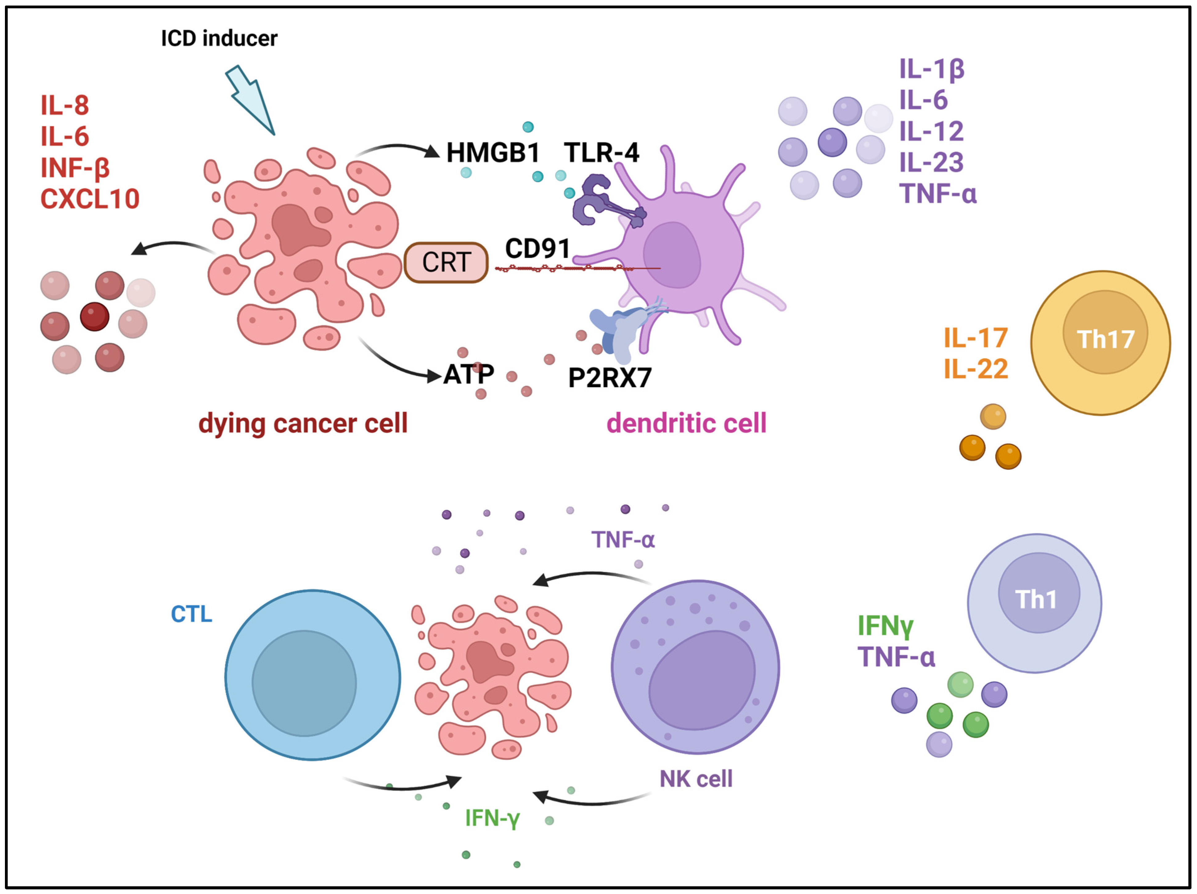
  176. Showalter, A.; Limaye, A.; Oyer, J.L.; Igarashi, R.; Kittipatarin, C.; Copik, A.J.; Khaled, A.R. Cytokines in immunogenic cell death: Applications for cancer immunotherapy. Cytokine 2017, 97, 123–132. [Google Scholar] [CrossRef] [PubMed]
  177. Gao, X.; Mi, Y.; Guo, N.; Xu, H.; Xu, L.; Gou, X.; Jin, W. Cytokine-induced killer cells as pharmacological tools for cancer immunotherapy. Front. Immunol. 2017, 8, 774. [Google Scholar] [CrossRef] [PubMed]
  178. Galdiero, M.R.; Marone, G.; Mantovani, A. Cancer inflammation and cytokines. Cold Spring Harb. Perspect. Biol. 2018, 10, a028662. [Google Scholar] [CrossRef]
  179. Green, D.R.; Ferguson, T.; Zitvogel, L.; Kroemer, G. Immunogenic and tolerogenic cell death. Nat. Rev. Immunol. 2009, 9, 353–363. [Google Scholar] [CrossRef]
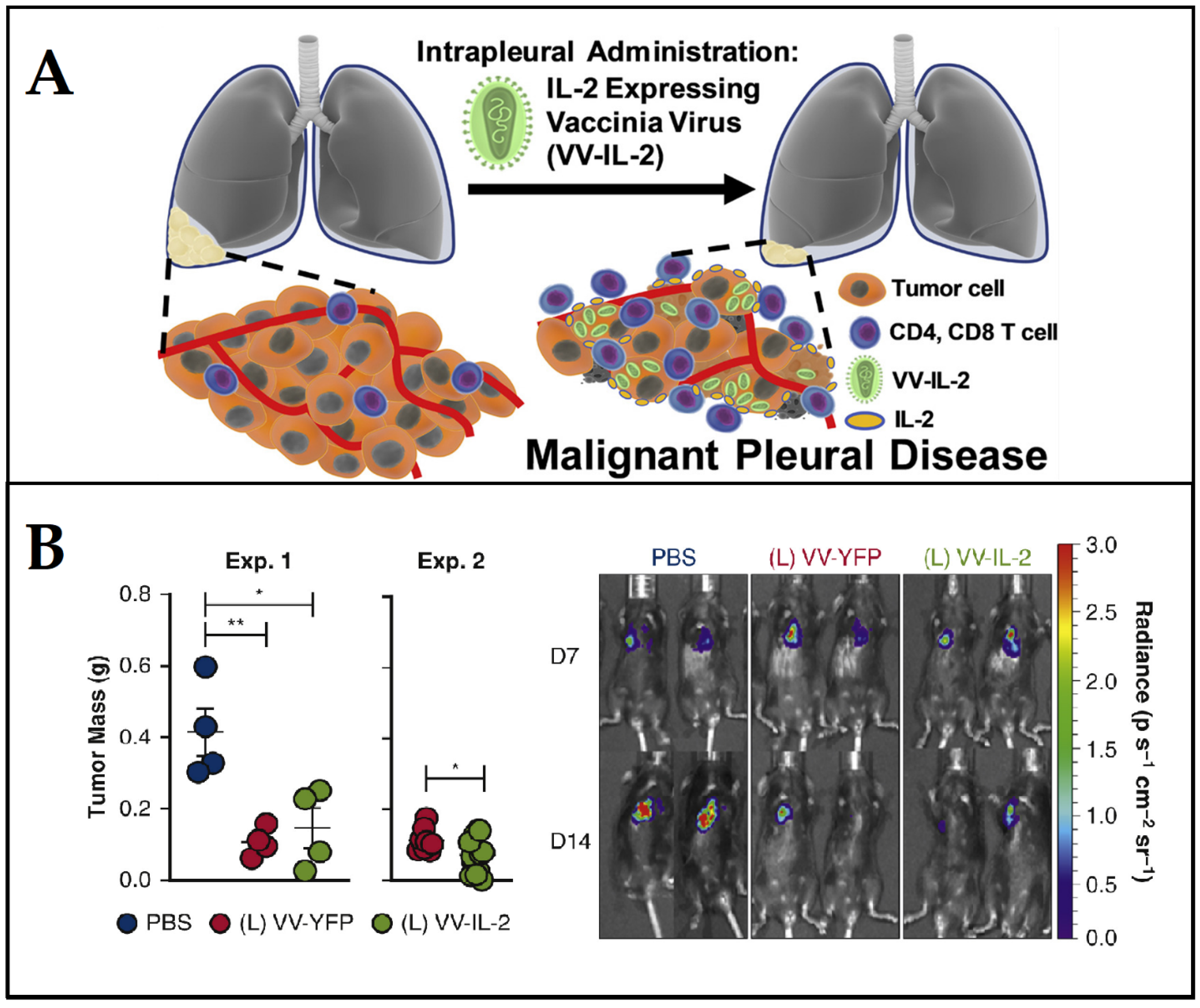
  180. Ekeke, C.N.; Russell, K.L.; Murthy, P.; Guo, Z.S.; Soloff, A.C.; Weber, D.; Pan, W.; Lotze, M.T.; Dhupar, R. Intrapleural IL-2 Expressing Oncolytic Virotherapy Enhances Acute Antitumor Effects and T Cell Receptor Diversity in Malignant Pleural Disease. J. Thorac. Cardiovasc. Surg. 2020, 163, e313–e328. [Google Scholar] [CrossRef]
  181. Conlon, K.C.; Miljkovic, M.D.; Waldmann, T.A. Cytokines in the treatment of cancer. J. Interferon Cytokine Res. 2019, 39, 6–21. [Google Scholar] [CrossRef]
  182. Silk, A.W.; Margolin, K. Cytokine therapy. Hematol. Oncol. Clin. 2019, 33, 261–274. [Google Scholar] [CrossRef]
  183. Xue, D.; Hsu, E.; Fu, Y.-X.; Peng, H. Next-generation cytokines for cancer immunotherapy. Antibody Ther. 2021, 4, 123–133. [Google Scholar] [CrossRef]
  184. Qiu, Y.; Su, M.; Liu, L.; Tang, Y.; Pan, Y.; Sun, J. Clinical application of cytokines in cancer immunotherapy. Drug Des. Dev. Ther. 2021, 2269–2287. [Google Scholar] [CrossRef]
  185. Donnelly, R.P.; Young, H.A.; Rosenberg, A.S. An overview of cytokines and cytokine antagonists as therapeutic agents. Ann. N. Y. Acad. Sci. 2009, 1182, 1–13. [Google Scholar] [CrossRef] [PubMed]
  186. Tagawa, M. Cytokine therapy for cancer. Curr. Pharm. Des. 2000, 6, 681–699. [Google Scholar] [CrossRef] [PubMed]
  187. Rosenberg, S.A.; Restifo, N.P. Adoptive cell transfer as personalized immunotherapy for human cancer. Science 2015, 348, 62–68. [Google Scholar] [CrossRef] [PubMed]
  188. Rapoport, A.P.; Stadtmauer, E.A.; Binder-Scholl, G.K.; Goloubeva, O.; Vogl, D.T.; Lacey, S.F.; Badros, A.Z.; Garfall, A.; Weiss, B.; Finklestein, J. NY-ESO-1-specific TCR-engineered T cells mediate sustained antigen-specific antitumor effects in myeloma. Nat. Med. 2015, 21, 914–921. [Google Scholar] [CrossRef]
  189. Zhang, W.-y.; Wang, Y.; Guo, Y.-l.; Dai, H.-r.; Yang, Q.-m.; Zhang, Y.-j.; Zhang, Y.; Chen, M.-x.; Wang, C.-m.; Feng, K.-c. Treatment of CD20-directed chimeric antigen receptor-modified T cells in patients with relapsed or refractory B-cell non-Hodgkin lymphoma: An early phase IIa trial report. Signal Transduct. Target. Ther. 2016, 1, 16002. [Google Scholar] [CrossRef] [PubMed]
  190. Schuster, S.J.; Svoboda, J.; Chong, E.A.; Nasta, S.D.; Mato, A.R.; Anak, Ö.; Brogdon, J.L.; Pruteanu-Malinici, I.; Bhoj, V.; Landsburg, D. Chimeric antigen receptor T cells in refractory B-cell lymphomas. N. Engl. J. Med. 2017, 377, 2545–2554. [Google Scholar] [CrossRef] [PubMed]
  191. Wang, Y.; Zhang, W.-y.; Han, Q.-w.; Liu, Y.; Dai, H.-r.; Guo, Y.-l.; Bo, J.; Fan, H.; Zhang, Y.; Zhang, Y.-j. Effective response and delayed toxicities of refractory advanced diffuse large B-cell lymphoma treated by CD20-directed chimeric antigen receptor-modified T cells. Clin. Immunol. 2014, 155, 160–175. [Google Scholar] [CrossRef]
  192. Mohammed, S.; Sukumaran, S.; Bajgain, P.; Watanabe, N.; Heslop, H.E.; Rooney, C.M.; Brenner, M.K.; Fisher, W.E.; Leen, A.M.; Vera, J.F. Improving chimeric antigen receptor-modified T cell function by reversing the immunosuppressive tumor microenvironment of pancreatic cancer. Mol. Ther. 2017, 25, 249–258. [Google Scholar] [CrossRef]
  193. Siegler, E.L.; Kenderian, S.S. Neurotoxicity and cytokine release syndrome after chimeric antigen receptor T cell therapy: Insights into mechanisms and novel therapies. Front. Immunol. 2020, 11, 1973. [Google Scholar] [CrossRef]
  194. Murthy, H.; Iqbal, M.; Chavez, J.C.; Kharfan-Dabaja, M.A. Cytokine release syndrome: Current perspectives. ImmunoTargets Ther. 2019, 8, 43–52. [Google Scholar] [CrossRef]
  195. Lythgoe, M.P.; Liu, D.S.K.; Annels, N.E.; Krell, J.; Frampton, A.E. Gene of the month: Lymphocyte-activation gene 3 (LAG-3). J. Clin. Pathol. 2021, 74, 543–547. [Google Scholar] [CrossRef] [PubMed]
  196. Yu, X.; Huang, X.; Chen, X.; Liu, J.; Wu, C.; Pu, Q.; Wang, Y.; Kang, X.; Zhou, L. Characterization of a novel anti-human lymphocyte activation gene 3 (LAG-3) antibody for cancer immunotherapy. mAbs 2019, 11, 1139–1148. [Google Scholar] [CrossRef] [PubMed]
  197. Atkinson, V.; Khattak, A.; Haydon, A.; Eastgate, M.; Roy, A.; Prithviraj, P.; Mueller, C.; Brignone, C.; Triebel, F. Eftilagimod alpha, a soluble lymphocyte activation gene-3 (LAG-3) protein plus pembrolizumab in patients with metastatic melanoma. J. Immunother. Cancer 2020, 8, e001681. [Google Scholar] [CrossRef] [PubMed]
  198. Zhao, L.; Wang, H.; Xu, K.; Liu, X.; He, Y. Update on lymphocyte-activation gene 3 (LAG-3) in cancers: From biological properties to clinical applications. Chin. Med. J. 2022, 135, 1203–1212. [Google Scholar] [CrossRef] [PubMed]
  199. Goldberg, M.S. Improving cancer immunotherapy through nanotechnology. Nat. Rev. Cancer 2019, 19, 587–602. [Google Scholar] [CrossRef]
  200. Ji, G.; Zhang, Y.; Si, X.; Yao, H.; Ma, S.; Xu, Y.; Zhao, J.; Ma, C.; He, C.; Tang, Z. Biopolymer Immune Implants’ Sequential Activation of Innate and Adaptive Immunity for Colorectal Cancer Postoperative Immunotherapy. Adv. Mater. 2021, 33, 2004559. [Google Scholar] [CrossRef]
  201. Deng, J.; Zhao, S.; Zhang, X.; Jia, K.; Wang, H.; Zhou, C.; He, Y. OX40 (CD134) and OX40 ligand, important immune checkpoints in cancer. OncoTargets Ther. 2019, 12, 7347. [Google Scholar] [CrossRef]
  202. Sun, H.; Dong, Y.; Feijen, J.; Zhong, Z. Peptide-decorated polymeric nanomedicines for precision cancer therapy. J. Control. Release 2018, 290, 11–27. [Google Scholar] [CrossRef]
  203. Song, H.; Yang, P.; Huang, P.; Zhang, C.; Kong, D.; Wang, W. Injectable polypeptide hydrogel-based co-delivery of vaccine and immune checkpoint inhibitors improves tumor immunotherapy. Theranostics 2019, 9, 2299. [Google Scholar] [CrossRef]
  204. Shen, K.-Y.; Song, Y.-C.; Chen, I.-H.; Chong, P.; Liu, S.-J. Depletion of tumor-associated macrophages enhances the anti-tumor immunity induced by a Toll-like receptor agonist-conjugated peptide. Hum. Vaccines Immunother. 2014, 10, 3241–3250. [Google Scholar] [CrossRef]
  205. Choi, Y.J.; Park, S.-J.; Park, Y.-S.; Park, H.S.; Yang, K.M.; Heo, K. EpCAM peptide-primed dendritic cell vaccination confers significant anti-tumor immunity in hepatocellular carcinoma cells. PLoS ONE 2018, 13, e0190638. [Google Scholar] [CrossRef] [PubMed]
  206. Lisi, L.; Lacal, P.M.; Martire, M.; Navarra, P.; Graziani, G. Clinical experience with CTLA-4 blockade for cancer immunotherapy: From the monospecific monoclonal antibody ipilimumab to probodies and bispecific molecules targeting the tumor microenvironment. Pharmacol. Res. 2022, 175, 105997. [Google Scholar] [CrossRef] [PubMed]
  207. Russell, S.J.; Peng, K.-W.; Bell, J.C. Oncolytic virotherapy. Nat. Biotechnol. 2012, 30, 658–670. [Google Scholar] [CrossRef] [PubMed]
  208. Mesa, C.; Fernández, L.E. Challenges facing adjuvants for cancer immunotherapy. Immunol. Cell Biol. 2004, 82, 644–650. [Google Scholar] [CrossRef] [PubMed]
  209. Yang, S.; Yu, K.H.; Palmer, N.; Fox, K.; Kou, S.; Kohane, I.S. Autoimmune effects of lung cancer immunotherapy revealed by data-driven analysis on a nationwide cohort. Clin. Pharmacol. Ther. 2020, 107, 388–396. [Google Scholar] [CrossRef]
  210. Amos, S.M.; Duong, C.P.; Westwood, J.A.; Ritchie, D.S.; Junghans, R.P.; Darcy, P.K.; Kershaw, M.H. Autoimmunity associated with immunotherapy of cancer. Blood 2011, 118, 499–509. [Google Scholar] [CrossRef]
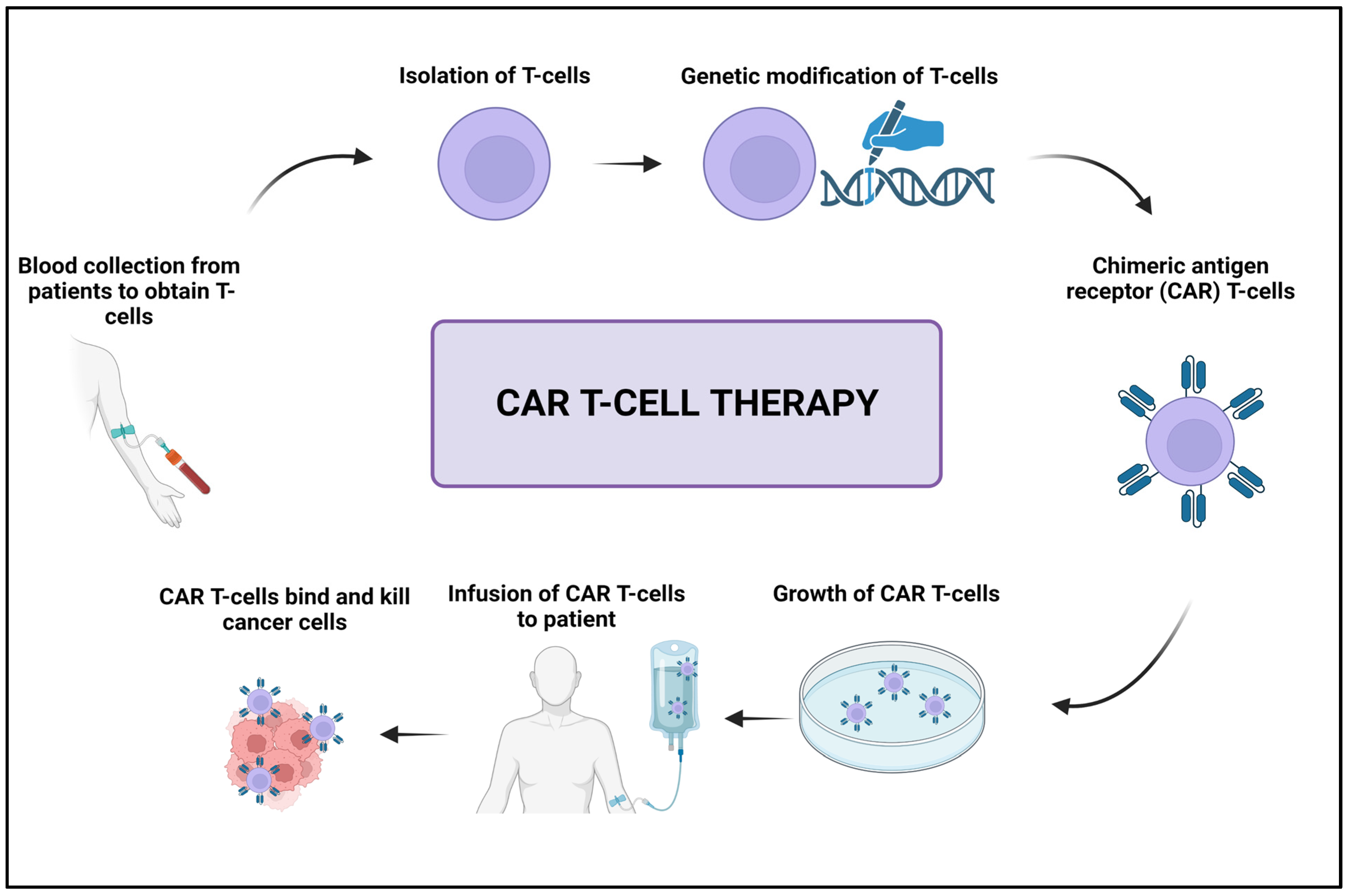
  211. Miliotou, A.N.; Papadopoulou, L.C. CAR T-cell therapy: A new era in cancer immunotherapy. Curr. Pharm. Biotechnol. 2018, 19, 5–18. [Google Scholar] [CrossRef]
  212. Sterner, R.C.; Sterner, R.M. CAR-T cell therapy: Current limitations and potential strategies. Blood Cancer J. 2021, 11, 69. [Google Scholar] [CrossRef]
  213. Porter, D.L.; Hwang, W.-T.; Frey, N.V.; Lacey, S.F.; Shaw, P.A.; Loren, A.W.; Bagg, A.; Marcucci, K.T.; Shen, A.; Gonzalez, V. Chimeric antigen receptor T cells persist and induce sustained remissions in relapsed refractory chronic lymphocytic leukemia. Sci. Transl. Med. 2015, 7, 303ra139. [Google Scholar] [CrossRef]
  214. Emens, L.A.; Ascierto, P.A.; Darcy, P.K.; Demaria, S.; Eggermont, A.M.; Redmond, W.L.; Seliger, B.; Marincola, F.M. Cancer immunotherapy: Opportunities and challenges in the rapidly evolving clinical landscape. Eur. J. Cancer 2017, 81, 116–129. [Google Scholar] [CrossRef]
  215. Quinn, C.; Garrison, L.P.; Pownell, A.K.; Atkins, M.B.; De Pouvourville, G.; Harrington, K.; Ascierto, P.A.; McEwan, P.; Wagner, S.; Borrill, J. Current challenges for assessing the long-term clinical benefit of cancer immunotherapy: A multi-stakeholder perspective. J. ImmunoTherapy Cancer 2020, 8, e000648. [Google Scholar] [CrossRef] [PubMed]
  216. Berz, A.M.; Dromain, C.; Vietti-Violi, N.; Boughdad, S.; Duran, R. Tumor response assessment on imaging following immunotherapy. Front. Oncol. 2022, 12, 982983. [Google Scholar] [CrossRef] [PubMed]
Figure 1. The number of scientific publications related to cancer immunotherapy (Search done through Science Direct database on 30 March 2023).
Cancers 15 02721 g001
Figure 2. The main causes of human cancers and hallmarks of cancer established by Hanahan and Weinberg. Created with BioRender.com, accessed on 23 April 2023.
Cancers 15 02721 g002
Figure 3. Schematic illustration of the cancer-immunity cycle. (CTL: cytotoxic t lymphocytes; APC: an antigen-presenting cell). Adapted with permission from Chen et al. [40].
Cancers 15 02721 g003
Figure 4. Cancer immunotherapy types include the use of immune-checkpoint inhibitors, cancer vaccines, cytokines, viruses, and adoptive cell transfer. Created with BioRender.com, accessed on 23 April 2023.
Cancers 15 02721 g004
Figure 5. Schematic illustration of the mutual regulation by various immune checkpoints between cancer cells and T cells. The interaction between Major Histocompatibility Complex class (MHC) molecules and the T-cell receptor (TCR) is a critical step in the activation of T cells. The interaction between PD-1 (programmed cell death protein 1) on the surface of T cells and PD-L1/2 (programmed cell death ligand 1/2) expressed on tumor cells inhibits TCR signaling. A similar effect is observed when CD80/CD86 molecules present in cancer cells bind with cytotoxic T-lymphocyte-associated protein 4 (CTLA4). Inhibitory effects on TCR signaling and T-cell activity have been attributed to the interaction between TIM-3 (T-cell immunoglobulin and mucin-domain containing-3) and its ligand Galectin-9 (Gal-9). In turn, T-cell immunoreceptor with Ig and ITIM domains (TIGIT) and CD96 can suppress T-cell activation by binding to CD155 with a higher affinity than CD226, which in turn outcompetes CD226 for the binding site. The binding of TIGIT with CD155 induces an inhibitory signal within tumor cells. This signal up-regulates IL-10 while simultaneously down-regulating IL-12. TIGIT hinders CD226 and CD155 from engaging in interaction by disrupting the homodimerization of CD226. It is currently unknown whether or not TIGIT can directly provide co-inhibitory signals in T cells once it has bound to CD155. CD226 is responsible for delivering co-stimulatory signals by binding to CD112 and CD155. CD96 is responsible for delivering co-inhibitory signals when it binds to CD111 and CD155. Created with BioRender.com, accessed on 23 April 2023. Based on [76].
Cancers 15 02721 g005
Figure 6. Vascular endothelial growth factor receptor-1 (VEGFR-1)-mediated approach for improving antitumor immune response. (A) Schematic illustration of the principal; (B) antitumor activity of the anti-VEGFR-1 monoclonal antibody (mAb D16F7) in combination with anti-CTLA-4 or anti-PD-1 mAbs. Adapted with permission from Lacal et al. [110].
Cancers 15 02721 g006
Figure 7. Schematic representation of oncolytic viro-immunotherapy strategies: (A) modification of endogenous genes of the virus for stimulating interferon induction, (B) virus-mediated expression of immune-stimulatory cytokines, (C) oncolytic cancer vaccine approach, and (D) synergistic effect of adaptive cell therapy and oncolytic viruses. Adapted with permission from Melzer et al. [130].
Cancers 15 02721 g007
Figure 8. Cancer vaccine: (A) Following administration of cancer-specific antigens (CSAs) through vaccination (B) antigens are transported to the lymph nodes, where (C) they are uptaken by dendritic cells (DCs) and activate the B and T cells. (D) B cells produce tumor-targeted antibodies, while activated T cells and natural killer (NK) recruited to the tumor microenvironment release perforins (PFNs) and granzyme A/B (GZMA/B) and interferon γ (IFN-γ) leading to cancer cell death. Created with BioRender.com, accessed on 23 April 2023.
Cancers 15 02721 g008
Figure 9. Efficient dendritic cell (DC) recruitment and activation by injectable dual-scale mesoporous silica microrod–mesoporous silica nanoparticle vaccine. Adapted with permission from Nguyen et al. [42].
Cancers 15 02721 g009
Figure 10. Cytokines that are involved in immunogenic cell death induction. Damage-associated molecular patterns (DAMPs) are released from dying cells during immunogenic cell death (ICD) and facilitate tumor antigen presentation and augment adaptive immunity. Cancer cells secrete calreticulin (CRT), which binds to the CD91 receptor on dendritic cells (DCs), and high-mobility group protein 1 (HMGB1), which is a ligand for the Toll-like receptor (TLR-4) on DCs. In addition, DSc’s P2X purinoceptor 7 (P2RX7) engages with ATP that cancer cells secrete. Created with BioRender.com, accessed on 23 April 2023. Modified from Showalter et al. [176].
Cancers 15 02721 g010
Figure 11. Antitumor effect of vaccinia virus expressing interleukin-2 (VV-IL-2) against malignant pleural disease. (A) The principal of the approach; (B) antitumor effect upon the administration of VV-IL-2, which reduced tumor burden and increased PD-1+ tumor-infiltrating T lymphocytes and survival. Phosphate-buffered saline (PBS) was used as a control (* p < 0.05, ** p < 0.01). Adapted from Ekeke et al. [180].
Cancers 15 02721 g011
Figure 12. Schematic illustration of the CAR T-cell therapy. Created with BioRender.com, accessed on 23 April 2023.
Cancers 15 02721 g012
Figure 13. Inverted cytokine receptor-modified chimeric antigen receptor T cells exhibit noticeably antitumor activity. (A) Schematic drawing of mice engrafted with cancer cells and treated with either CAR/ICR T cells or CAR. (B) CAR and CAR/ICR T cells expansion and persistence as measured by bioluminescence imaging. (C) The reduction in tumor volume for CAR/ICR T cell-based treatment. (D) Superimposition of tumor volume and T-cell signal in CAR/ICR-based treated mice. (E) Images of mice administered with two treatments: CAR, CAR/ICR T cells, or control. Adapted with permission from Mohammed et al. [192].
Cancers 15 02721 g013
Table 1. Summary of the chronological events of cancer immunotherapy.
Scientist/s and YearRemarkRef
W. B. Coley in 1893The first use of immunotherapy to treat a cancer patient.[45]
Hericourt and Richet in 1895Serotherapy in cancer treatment.[47]
W. B. Coley in 1899The first vaccine against sarcoma.[48]
Leyden et al. in 1902The first cancer vaccine was created using a patient’s tumor cells.[49]
J. B. Murphy in 1914Understanding the function of lymphocytes in the rejection of grafted tumors.[50]
Ernest Witebsky in 1929Tumor molecular markers are used as antigens.[51]
Burnet and Lewis in 1957The concept of the natural anticancer immune response. [52]
Sjögren et al. in 1971The role of antibodies in shielding tumor cells from the anticancer immune reaction.[53]
Carswell et al. in 1975Discovery of tumor necrosis factor (TNF-α) with antitumor effects.[54]
Morgan et al. in 1976The first discovery of IL-2 as a T-cell growth factor.[55]
Stevenson and Elliott in 1977First anti-idiotype antibodies activate immune cells against the tumor.[56]
Ronald Levy in 1981First successful cancer treatment with anti-idiotype antibody.[57]
Taniguchi et al. in 1983First cloning of T-cell growth factor (IL-2).[58]
Kappler et al. in 1983First identification of T-cell antigen receptor.[59]
Knuth et al. in 1984First clinical trial on the potential use of T cells to fight off tumors.[60]
S A Rosenberg in 1988Successful application of IL-2-based cancer therapy.[61]
Gross et al. in 1989The first use of genetically engineered T cells for tumor targeting.[62]
Dranoff et al. in 1993Using macrophage colony-stimulating factor (M-CSF) to improve cancer immunity.[63]
Leach et al. in 1996Inhibition of the immune cells CTLA-4 molecule in cancer treatment.[64]
Levy and Rastetter in 1997First FDA-approved monoclonal antibody as an anticancer drug.[65]
J. P. Allison in 2000First immune-checkpoint inhibitor drug against CTLA-4.[66]
Hirano et al. in 2005Development of fully human anti-PD-1 antibody for cancer therapy.[67]
Kershaw et al. in 2006Development of adoptive cell-mediated immunotherapy.[68]
Rosenberg et al. in 2008Anti-PD-1 antibody started phase 1 of the clinical trial for cancer[69]
June and Whitehead in 2012First leukemia treatment by adoptive cell (CAR-T based therapy).[70]
Quezada et al. in 2016Genetic modification of immune cells to prompt antitumor response.[71]
Wallace et al. in 2016A first clinical trial of gene editing tool CRISPR/Cas 9 for cancer.[72]
Freedman et al. in 2018Genetically engineered virus to selectively kill cancer cells.[73]
Zhang et al. in 2020The use of miRNA to regulate immune checkpoints.[74]
Table 2. Important and commonly used oncolytic viruses in cancer immunotherapy.
Virus (Family)Nucleic Acid Type (Group)Study RemarksRef
Reovirus (Reoviridae)dsRNA (G III)Viruses exhibited a safe and tolerable toxicity profile in early clinical trials, with minimal viral shedding, replication, localization, and cytotoxic effects.[122]
Influenza A Virus
(Orthomyxoviridae)
ssRNA-ve (G V)Engineered viruses exhibited tumor-ablative potential and do not replicate in normal cell lines.[123]
Herpes Simplex Virus1 (Herpesviridae)dsDNA (G I)A virus exhibited high antitumor efficacy, has a high safety margin, is well tolerated and its use is associated with a low frequency of adverse effects in most of the treated patients. [124]
Adenovirus (Adenoviridae)dsDNA (G I)Clinical data revealed that the virus was tolerable and effective even when combined with other therapeutic approaches.[125]
Measles virus (Paramyxoviridae)ssRNA-ve (G V)The virus replicated selectively in the tumor and significantly suppressed its development. It was completely safe and did not result in any measles-like symptoms. [126]
Vaccinia virus (Poxviridae)dsDNA (G I)The virus replicated rapidly in tumor cells with significant antitumor effects, but its cytotoxicity varied based on cell lines. [127]
Pseudorabies (Herpesviridae)dsDNA (G I)Selective replication of viruses has been observed in many kinds of cancers, with tumor growth being significantly slowed by more than 50%.[128]
Disclaimer/Publisher’s Note: The statements, opinions and data contained in all publications are solely those of the individual author(s) and contributor(s) and not of MDPI and/or the editor(s). MDPI and/or the editor(s) disclaim responsibility for any injury to people or property resulting from any ideas, methods, instructions or products referred to in the content.

