
How Should We Treat Meningeal Melanocytoma? A Retrospective Analysis of Potential Treatment Strategies

Department of Neurosurgery, University Hospital Muenster, 48149 Muenster, Germany
*
Author to whom correspondence should be addressed.
†
These authors contributed equally to this work.
Cancers 2022, 14(23), 5851; https://doi.org/10.3390/cancers14235851
Submission received: 3 October 2022
/
Revised: 11 November 2022
/
Accepted: 20 November 2022
/
Published: 27 November 2022
(This article belongs to the Section Cancer Therapy)
Abstract
:Simple Summary
As a rare tumor disease, only single case reports and small case series have been published on meningeal melanocytomas. In the case of complete surgical resection, there is a shallow risk of recurrence, whereas the benefit of radiotherapy or chemotherapy, both as single or combined therapy, is unclear. This work aims to analyze and summarize previously published cases and proposes a therapeutic algorithm.
Abstract
Background: Meningeal melanocytomas (MM) are rare primary melanocytic tumors of the leptomeninges with an incidence of 1:10,000,000. Until now, there has been only sparse information about this tumor entity. Here, we provide a meta-analysis of all cases published in the English language since 1972. Methods: A literature review was performed using PubMed and Web of Science. All published cases were evaluated for location, sex, age, therapeutic approach, and outcome. In total, we included 201 patient cases in our meta–analysis. Results: The majority of MM was diagnosed more frequently in men between the third and fifth decade of life. Surgery is the preferred therapeutic approach, and total resection is associated with the best outcome. Patients with partial resection or tumor recurrence benefit from adjuvant radiotherapy, whereas chemo- or immunotherapies do not improve the disease course. Malignant transformation was described in 18 patients. Of these, 11 patients developed metastasis. Conclusions: We present the first retrospective meta-analysis of all MM cases published in the English language, including an evaluation of different treatment strategies allowing us to suggest a novel treatment guideline highlighting the importance of total resection for recurrence–free survival and characterizing those cases which benefit from adjuvant radiotherapy.
1. Introduction
Meningeal melanocytomas (MM) are rare primary melanocytic tumors of the leptomeninges. They can be divided into circumscribed or diffuse, benign or malignant lesions. Well–differentiated, circumscribed tumors are called meningeal melanocytomas, while their malignant counterparts are meningeal melanomas. Additionally, MM with increased mitotic activity or invasion of the CNS parenchyma are considered intermediate-grade lesions. Furthermore, diffuse melanocytic tumors are characterized by invasion into subarachnoid space. Depending on the dignity of the histological phenotype, the lesion is called meningeal melanocytosis or meningeal melanomatosis [1]. Since their first description in 1972 [2], about 201 cases have been reported in English worldwide. They can be found anywhere along the neuraxis, and clinical presentation depends on tumor location and size.
Depending on the amount of melanin [3], MM appear isointense to hyperintense on T1-weighted and isointense to hypointense on T2-weighted MRI and show heterogenous contrast enhancement. In CT, MM appear as well–defined, isodense to hyperdense with homogeneous, contrast-enhancing lesions [4,5].
Macroscopically the tumor is encapsulated without infiltration of the surrounding tissue [6,7]. The color of the tumor appears dark and varies between coal black, reddish brown, and dark blue [8]. Often, the dura appears darker than usual [9,10]. Microscopically, MM are characterized by nets composed of spindle-shaped cells [11] that express characteristically S–100, a typical calcium-binding protein [12,13], Melanoma Antigen (Melan–A), a melanocytic differentiation marker, and show positive reactivity for homatropine methyl bromide-45 (HMB–45) [10], a monoclonal antibody that interacts with GP-100. Furthermore, they show variable expression of neuron-specific enolase and vimentin [14] (Figure 1).
Although several case reports have been published, so far, it remains unclear which is the best treatment approach for MM, in particular for those cases in which only incomplete surgical resection could be performed. Therefore, the aim of this study was to reanalyze all published cases, as well as one unpublished case from our department, concerning the applied treatment strategies and their outcome.
2. Materials and Methods
Search Strategy and Statistics
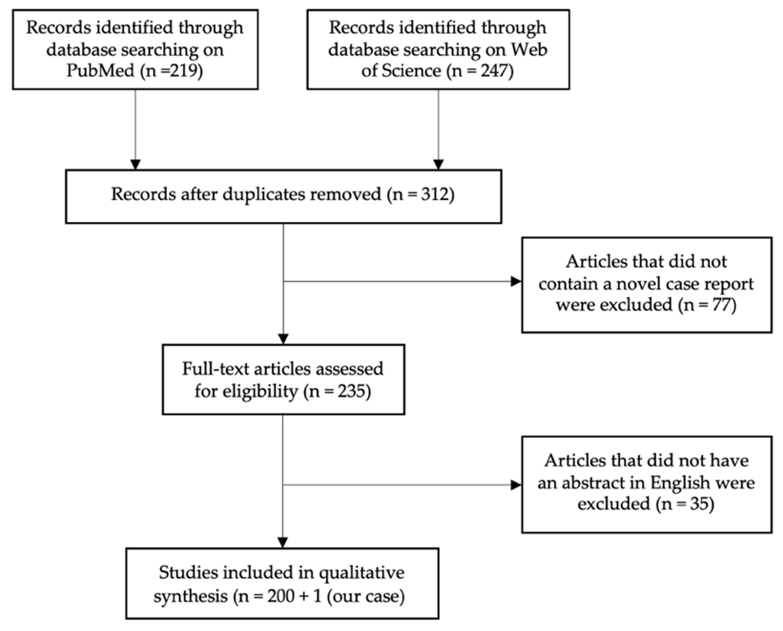
MM cases were identified by using PubMed (Medline) and Web of Science (Clarivate). As the keyword, “meningeal melanocytoma” was used. In total, 219 (PubMed) and 247 (Web of Science) results were found, which were published between 1972 and 2022. After subtracting duplicates, the total data covered 312 items. Articles were excluded if they did not provide a novel case report, if the abstract did not include MM as a topic (n = 77), or if written in another language than English (n = 35), so in total, 201 cases were included in this analysis, as we also included one unpublished MM case from our department (Figure 1). All published cases were evaluated for age, gender, tumor location, and therapeutic approach as well as postoperative outcome (recurrence, metastasis, recurrence-free survival) (Figure 2).
All patient cases were included in our analysis, of which at least one of the above variables could be collected. For this reason, the population groups for the different analyses also differed in size (indicated each time in the text). Histological diagnoses of tumors described here were adopted from the original manuscripts. No reevaluation of histological specimens was performed by local neuropathologists according to the 2021 WHO classification. Therefore, the lesions depicted here were classified according to the current WHO classification in each case. Statistical analysis was performed using the statistics software SPSS (IBM, version 28). Descriptive data included the calculation of the mean or median and standard deviation. Pearson Chi–Square testing was performed to compare categorical variables. p values less than 0.05 were considered statistically significant.
3. Results
3.1. Population
3.1.1. Age and Gender
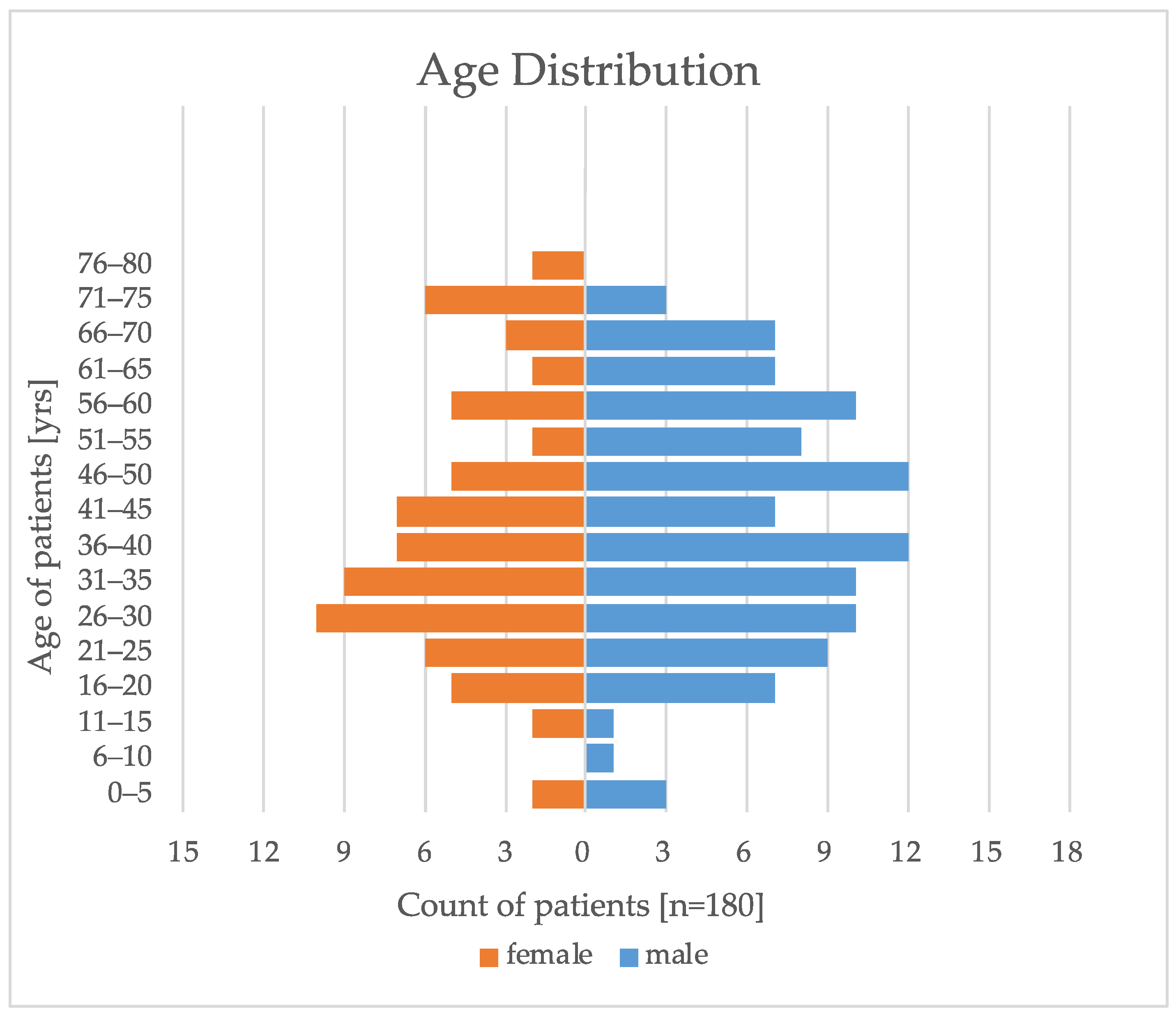
Since 1972, 201 cases of MM were reported (Table S1). In 89.6% (180/201) of the reported cases, information concerning gender and age was provided. MM usually was diagnosed between the 3rd and 5th decade of life, with a median age of onset of the disease at 38 years. The youngest patient was only 28 weeks old, and the oldest patient was diagnosed at the age of 79 years (Figure 3). MM occurred more frequently in men (107/180, 59.4%) than in women (73/180, 40.6%). Females were diagnosed earlier (median, 37 years) than males (median, 42 years).
3.1.2. Location
The location of the tumor was reported in 189 out of 201 cases. About half of the MM were found intracranially (101/189, 52.6%). The predominant location was the posterior fossa (57/101, 56.4%), followed by the middle cranial fossa with 11 cases (11/101, 10.9%) and the sellar region with 9 cases (9/101, 8.9%). Within the posterior fossa, MM occurred most often at the cerebellopontine–angle (CPA) (18/57, 31.6%). In the spine, MM occurred predominantly in the thoracic spine (39/78, 50%) and cervical spine (26/78, 33.3%). Furthermore, in eight cases, tumors grew in orbit. Two cases reported multifocal location within the spine as well as intracranially.
3.2. Treatment Strategies
3.2.1. Total Resection and Partial Resection
Treatment strategies were reported in 186 out of 201 cases (92.5%). As the primary therapeutic approach, surgery was performed in 179 out of 186 patients (96.2%) of the cases. In the remaining cases, radiotherapy was used in two, and no therapy was applied in five others. Total resection was achieved in 89 out of 179 (49.7%) of cases and partial resection in 73 out of 179 cases (40.7%), while in 9.5% of surgical procedures, the extent of resection was not documented (17/179).
3.2.2. Adjuvant Therapy
Total resection was performed without any further therapy in 81 out of 179 patients (45.3%). The combination of total resection and radiotherapy was applied in 8 (4.5%), partial resection alone in 45 cases (25.1%), and adjuvant radiotherapy after partial resection in 24 out of 179 cases (13.4%). In two cases, radiosurgery was used after partial resection so that the remaining tumor could also be targeted [15,16]. Due to the severity and progression of the disease, five patients did not receive any therapy, and three of them died; one developed tumor progression, and for the last one, follow–up data were not available. In fact, one of those patients was diagnosed by autopsy.
Chemo- or immunotherapy such as Temozolomide [17], Cisplatin and Fotemustine [18,19], Methotrexate [20], Nivolumab [21], or Ipilimumab [22] were applied in 11 patients combined with radiotherapy (Table 1) [18,21,23]. However, in all patients but one, tumor growth was observed, and all patients but four died.
3.2.3. Definite Radiotherapy and Radiosurgery
Due to tumor localization in an eloquent region, resection could not be performed in three patients. In two of these patients’ definite radiotherapy was chosen as a treatment option with a tumor-free follow-up of 42 months in 1 patient [24]. The other patient died due to pneumonia, unrelated to the MM [24]. In one case, radiosurgery alone was used as initial treatment after biopsy of the tumor [25].

Table 1.
Overview of all cases in which adjuvant chemo- or immunotherapy was applied: In all cases, the adjuvant therapy failed, and tumor progression was observed. Ten patients died because of MM growth and its consequences.
Table 1.
Overview of all cases in which adjuvant chemo- or immunotherapy was applied: In all cases, the adjuvant therapy failed, and tumor progression was observed. Ten patients died because of MM growth and its consequences.
| Case | Age | Sex | Location | Treatment | Outcome |
|---|---|---|---|---|---|
| [17] | 20 | M | Intracranial | PR + RT, Reop + RT + Temolozomide + Cisplatin + Fotemustine | Death |
| [18] | 46 | F | Intracranial | TR, Reop + Fotemustine + Temolozomide | Death |
| [19] | 38 | M | Intracranial | TR, RT + Temolozomide | Death |
| [20] | 79 | F | Spine | PR, RT + Methotrexat | Death |
| [21] | 70 | M | Spine | PR + RT + Nivolumab, Reop + Temozolomide | Death |
| [22] | 43 | F | Intracranial | TR + RT, Reop + RT + Temozolomide + Ipilimumab | Death |
| [23] | 71 | F | Spine | TR + RT, PR + RT + C. parvum + Dactinomycin + Dacarbazine | Tumor progression |
| [25] | 32 | M | Orbita | Radiosurgery + Immunotherapy | Tumor progression |
| [26] | 37 | F | Intracranial | PR, Reop + RT + Temozolomide | Death |
| [27] | 19 | F | Spine | TR, PR + RT + Pembrolizumab + Bevacizumab + Temozolomide | Death |
| [28] | 71 | F | Spine | PR + RT, Reop + C. parvum, Dimethyl Triazeno Imidazole Carboxamide + Actinomycin | Death |
| [29] | 36 | F | Spine | TR, Reop + RT + Nivolumab | Death |
| [30] | 35 | M | Orbita | PR + RT + BCNU + DTIC + Cisplatin | Tumor progression |
| [31] | 49 | M | Orbita | PR + Dacarbazine + Vincristine + Nimustine Hydrochloride | No recurrence |
Re–operation was the main therapy chosen in cases of tumor regrowth in 14 out of 44 cases (31.8%), followed by combination of re-operation and radiotherapy, applied in 7 (15.9%), re–operation combined with radiochemotherapy in another 4 (9.1%), while radiotherapy alone was used in 4 out of 44 cases (9.1%). In addition, three cases in which radiosurgery was used as follow-up therapy after recurrence of the tumor were also reported [32,33,34].
3.3. Outcome
For outcome analysis, we were able to evaluate 147 out of 201 data sets comprising information about initial therapy and follow-up (73.1%). Total resection was the most efficient therapy and showed a tumor-free interval without recurrence in 68.1% of cases (p = 0.001). The median tumor-free interval was 18 months, with a minimum of 1 month and a maximum of 35 years. If, alternatively, only partial resection was performed, a better outcome was shown in 61.9% of the patients by the additional use of adjuvant radiotherapy (see Table 2 and Table 3). Tumor progression was recorded here at a median of 24 months, ranging from 1 month to 16 years before recurrence developed.
When resection was combined with chemotherapy, tumor–free follow–up was not recorded in 10 out of 11 cases for total resection, partial resection, or with additional radiotherapy. Only one case was noted to have a tumor-free interval [31] (Table 1).
For radiosurgery, the outcome in all but one case was found to result in a tumor–free or progression–free interval. For this, it has been regardless of whether radiosurgery was used initially or subsequently at recurrence [15,16,25,32,33,34].
Of all patients, 29 patients died. Eight patients died from causes other than the underlying tumor disease, such as ischemic heart disease [35], urinary tract infection [36], pulmonary embolism [21], pneumonia [37,38], renal cell carcinoma [39], cerebellar hemorrhage related to anticoagulation [40], and other unrelated reasons [41].
3.4. Intermediate-Grade and Malignant Transformation
MM with a MIB–1/Ki-67 of 5–10% are defined as intermediate–grade MM with potential for development into malignant melanoma (>10% Ki-67). In total, 16 intermediate cases were reported, 14 showed a high recurrence rate without adjuvant radiotherapy after resection, and 2 were treated by radiotherapy without recurrence [42].
Furthermore, another 17 cases of MM reported malignant transformation [26,27,29,34,43,44]. Thirteen of these patients died, and four patients developed tumor progression. In 11 of these patients, MM metastases were found. All but three of these patients died of disease progression. Regarding the localization of metastasis, we were able to highlight that MM metastasize both within the central nervous system but also can occur in other tissues, as metastases were observed in the liver [8,22], pancreas [22], and skeleton [8] (Table 4).
Regarding the temporal component at follow–up, we were able to highlight a median of 18 months (range: few days–35 years).
4. Discussion
Since its first description in 1972 by Limas and Tio [2], several case descriptions of MM have been published; however, a meta-analysis of MM and evaluation of treatment strategies is missing but urgently needed. Our aim of this study was to analyze all available data from all MM cases published so far and evaluate potential treatment strategies.
Interestingly, our data demonstrated that MM occurs at a younger age in females than in males and is more frequent in males than in females, which contradicts the impression from smaller studies where female patients were thought to develop MM more often [50,51]. We could confirm the impression that MM is a disease of the adult, and in the majority of the cases independent of gender, MM occurred between the 3rd and 5th decade [51], while children were only rarely affected.
Furthermore, our analysis shows that the predominant location is the posterior fossa, as well as the cervical and thoracic spine, which is in line with the embryological development of melanocytes [52]. Melanocytes originate from the neural crest, which in turn gives rise, among other tissues, also to the leptomeninges. This may explain the preferred location of MM in the posterior fossa as well as the cervical and thoracic spine [52].
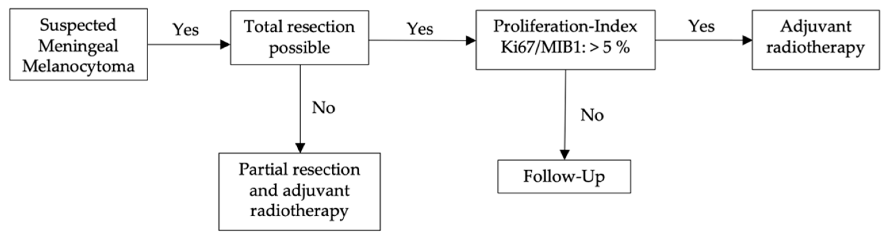
We evaluated treatment strategies and their outcome, showing the best recurrence-free follow–up after total resection (Figure 4). In these cases, no clear evidence was found that adjuvant therapy is beneficial. Combined treatment with total resection and adjuvant radiotherapy may be considered only for those cases where tumors show high mitotic activity, i.e., intermediate MM, which has been observed to be associated with tumor recurrence. The differentiation to intermediate–grade (Ki67/MIB1–proliferation index of 5–10%) MM and malignant transformation into melanoma (Ki67/MIB1–proliferation index of >10%) is particularly important [53,54,55,56].
Only in those cases that first received a total resection but developed MM recurrence, radiotherapy appears to be an advantage for recurrence-free survival (Figure 5).
In patients where only partial resection was achieved, radiotherapy showed a clear beneficial effect, while other therapeutic approaches, such as chemotherapy or immunotherapy, did not show any advantages. Furthermore, some case reports indicate a potential benefit of radiosurgery.
When MM is considered as a diagnosis, we do not suggest deciding on conservative follow-up observations, as we learned that MM appear capable of developing metastases. When the tumor is inoperable, definite radiotherapy has also been used successfully.
Therefore, we suggest the following treatment approach for patients that are diagnosed or considered to have MM (Figure 4):
Although chemo– and immunotherapy has failed so far, several mutations, such as in Guanine-nucleotide binding protein G subunit alpha (GNAQ, GNA11), have been identified [57]. Both GNAQ and GNA11 are g–proteins that share responsibility for activating the MAP–kinase pathway. Both have been suspected to be potential therapeutic targets. In mouse models, GNAQ and GNA11 have been shown to lead to hyperpigmentation and proliferation of melanocytes [58]. Additionally, molecular analysis of PLCB4 and CYSLTR2 [59,60,61] and methylation profiling are especially useful in discriminating these lesions from other pigmented CNS tumors [1,59]. The presence of SF3B1–, EIF1AX–, or BAP1–mutation or complex copy number variations indicate aggressive behavior consistent with meningeal melanoma [62,63,64]. Especially in children, meningeal melanocytosis and melanomatosis are characterized by NRAS- and occasionally BRAF mutations [65,66,67].
Beyond that, novel immunotherapies applied for malignant melanoma therapy may be beneficial also for MM. Both share common antigens, such as Melan-A or S–100 [11,12,13]. Therefore, further studies investigating novel potential therapies are needed.
Limitations of the Study
As MM is a rare entity, the composition of our multi–center derived data set was very inhomogeneous due to different authors and unrelated case reports. For that reason, this study can only serve as a meta–analysis of all published case reports, which intuitively cannot fulfill the criteria for a real comprehensive study. Thus, future multi–center prospective studies with fixed follow-up periods are now needed in order to achieve precise outcome and prognosis and verify our treatment suggestions derived from our retrospective analysis.
The fact that we were unable to reanalyze the tissue samples in this study is a methodological limitation of studies such as this and can bias conclusions. Hopefully, future reports will be able to use more up–to–date histopathological classifications.
5. Conclusions
Based on the first retrospective meta–analysis of all MM cases published in the English language, we propose a novel guideline for the treatment of MM, highlighting the importance of total resection for recurrence-free follow-up and suggesting in which cases adjuvant radiotherapy may have beneficial effects of the patient’s disease course.
Supplementary Materials
The following supporting information can be downloaded at: https://www.mdpi.com/article/10.3390/cancers14235851/s1, Table S1: All data of 201 case reports since 1972. RT = Radiotherapy; Surgery = Surgery without report of extent of resection. References [68,69,70,71,72,73,74,75,76,77,78,79,80,81,82,83,84,85,86,87,88,89,90,91,92,93,94,95,96,97,98,99,100,101,102,103,104,105,106,107,108,109,110,111,112,113,114,115,116,117,118,119,120,121,122,123,124,125,126,127,128,129,130,131,132,133,134,135,136,137,138,139,140,141,142,143,144,145,146,147,148,149,150,151,152,153,154,155,156,157,158,159,160,161,162,163,164,165,166,167,168,169,170,171,172,173,174,175,176,177,178,179,180,181,182,183,184,185,186,187,188,189,190,191,192,193,194,195,196,197,198] are mentioned in the supplementary materials.
Author Contributions
Conceptualization, S.R., M.G. and M.H.; methodology, S.R., M.G., W.S. and M.H.; software, S.R. and M.G.; validation, M.G. and M.H.; formal analysis, S.R., M.G. and M.H.; investigation, S.R. and M.G.; resources, S.R., M.G. and M.H.; data curation, S.R. and M.G.; writing—original draft preparation, S.R. and M.G.; writing—review and editing, M.G., W.S. and M.H.; visualization, S.R., M.G. and M.H.; supervision, W.S. and M.H.; project administration, M.G. and M.H. All authors have read and agreed to the published version of the manuscript.
Funding
This research received no external funding.
Institutional Review Board Statement
Ethical review and approval were waived for this study due to it´s anonymized retrospective character.
Informed Consent Statement
Patient consent was waived due to the usage of external anonymized studies, which were already published.
Data Availability Statement
The data presented in this study are available in the supplementary material.
Acknowledgments
We thank the Institute of Neuropathology Münster, in particular Astrid Jeibmann, for the histological evaluation and provision of the images.
Conflicts of Interest
The authors declare no conflict of interest.
References
- Küsters-Vandevelde, H.V.N.; Küsters, B.; van Engen-van Grunsven, A.C.H.; Groenen, P.J.T.A.; Wesseling, P.; Blokx, W.A.M. Primary Melanocytic Tumors of the Central Nervous System: A Review with Focus on Molecular Aspects. Brain Pathol. 2015, 25, 209–226. [Google Scholar] [CrossRef] [PubMed]
- Limas, C.; Tio, F.O. Meningeal Melanocytoma (“melanotic Meningioma”). Its Melanocytic Origin as Revealed by Electron Microscopy. Cancer 1972, 30, 1286–1294. [Google Scholar] [CrossRef] [PubMed]
- Demirci, A.; Kawamura, Y.; Sze, G.; Duncan, C. MR of Parenchymal Neurocutaneous Melanosis. AJNR Am. J. Neuroradiol. 1995, 16, 603–606. [Google Scholar] [PubMed]
- Malik, A.; Goyal, M.; Gambhir, M.; Patir, R.; Mishra, N.K.; Gaikwad, S.; Sharma, M.C. Imaging Appearances of Meningeal Melanocytoma. J. Clin. Neurosci. 1999, 6, 264–265. [Google Scholar] [CrossRef] [PubMed]
- Gill Naul, L.; Hise, J.H.; Bauserman, S.C.; Todd, F.D. CT and MR of Meningeal Melanocytoma. AJNR Am. J. Neuroradiol. 1991, 12, 315–316. [Google Scholar]
- Marwaha, N.; Batanian, J.R.; Coppens, J.R.; Pierson, M.J.; Richards-Yutz, J.; Ebrahimzadeh, J.; Ganguly, A.; Guzman, M.A. Subcutaneous Melanocytoma Mimicking a Lipoma: A Rare Presentation of a Rare Neoplasm with Histological, Immunohistochemical, Cytogenetic and Molecular Characterization. J. Cutan. Pathol. 2016, 43, 1186–1196. [Google Scholar] [CrossRef] [PubMed]
- Oruckaptan, H.H.; Soylemezoglu, F.; Kutluk, T.; Akalan, N. Benign Melanocytic Tumor in Infancy: Discussion on a Rare Case and Review of the Literature. Pediatr. Neurosurg. 2000, 32, 240–247. [Google Scholar] [CrossRef]
- Wang, F.; Li, X.; Chen, L.; Pu, X. Malignant Transformation of Spinal Meningeal Melanocytoma. Case Report and Review of the Literature. J. Neurosurg. Spine 2007, 6, 451–454. [Google Scholar] [CrossRef] [PubMed]
- Fagundes-Pereyra, W.J.; de Sousa, L.; Carvalho, G.T.C.; Pittella, J.E.H.; de Sousa, A.A. Meningeal Melanocytoma of the Posterior Fossa: Case Report and Literature Review. Surg. Neurol. 2005, 63, 269–274. [Google Scholar] [CrossRef] [PubMed]
- Hino, K.; Nagane, M.; Fujioka, Y.; Shiokawa, Y. Meningeal Melanocytoma Associated with Ipsilateral Nevus of Ota Presenting as Intracerebral Hemorrhage: Case Report. Neurosurgery 2005, 56, E1376. [Google Scholar] [CrossRef]
- Alameda, F.; Lloreta, J.; Galitó, E.; Roquer, J.; Serrano, S. Meningeal Melanocytoma: A Case Report and Literature Review. Ultrastruct. Pathol. 1998, 22, 349–356. [Google Scholar] [CrossRef]
- Wen, D.-R.; Bhuta, S.; Herschman, H.R.; Gaynor, R.B.; Cochran, A.J. S-100 Protein: A Marker for Melanocytic Tumors. Ann. N. Y. Acad. Sci. 1983, 420, 261–266. [Google Scholar] [CrossRef] [PubMed]
- Donato, R. S-100 Proteins. Cell Calcium 1986, 7, 123–145. [Google Scholar] [CrossRef] [PubMed]
- Smith, S.H.; Goldschmidt, M.H.; McManus, P.M. A Comparative Review of Melanocytic Neoplasms. Vet. Pathol. 2002, 39, 651–678. [Google Scholar] [CrossRef]
- Pierson, M.; Marwaha, N.; Guzman, M.; Mikulec, A.A.; Coppens, J.R. Multifocal Melanocytoma of the Posterior Fossa and Subcutaneous Scalp in the Absence of Neurocutaneous Melanosis. Surg. Neurol. Int. 2016, 7, S591–S595. [Google Scholar] [CrossRef] [PubMed] [Green Version]
- Lee, N.K.; Lee, J.Y.; Wang, K.C.; Kim, D.G.; Park, S.H.; Cheon, J.E.; Phi, J.H.; Kim, S.K. Primary Atypical Melanocytoma Arising from the Cavernous Sinus in a Child. Child’s Nerv. Syst. 2015, 31, 1577–1582. [Google Scholar] [CrossRef] [PubMed]
- Doglietto, F.; Colosimo, C.; Lauriola, L.; Balducci, M.; De Bonis, P.; Montano, N.; Zadeh, G.; Maira, G.; Pallini, R. Intracranial melanocytic meningeal tumours and melanosis oculi: Case report and literature review. BMC Cancer 2012, 12, 220. [Google Scholar] [CrossRef] [PubMed] [Green Version]
- San-Miguel, T.; Navarro, L.; Sanchez-Sendra, B.; Megıas, J.; Muñoz-Hidalgo, L.; Santonja, N.; Lopez-Gines, C.; Cerda-Nicolas, M. Identification of a Novel BRCA1 Alteration in Recurrent Melanocytoma Resulting in Increased Proliferation. J. Neuropathol. Exp. Neurol. 2020, 79, 1233–1238. [Google Scholar] [CrossRef] [PubMed]
- Koch, H.J.; Roeber, S.; Zimmermann, U.W.; Schäfer, C.; Villarrubia, V.; Kuchelmeister, K.; Schachenmayr, W.; Bogdahn, U.; Steinbrecher, A. Spinale Und Zerebrale Meningeosis Eines Sekundär Malignisierten Melanozytoms Des Kleinhirnbrückenwinkels. Wien. Med. Wochenschr. 2005, 155, 360–364. [Google Scholar] [CrossRef] [PubMed]
- Bydon, A.; Gutierrez, J.A.; Mahmood, A. Meningeal Melanocytoma: An Aggressive Course for a Benign Tumor. J. Neurooncol. 2003, 64, 259–263. [Google Scholar] [CrossRef] [PubMed]
- Hean, V.; Bouleftour, W.; Ramirez, C.; Forest, F.; Boutet, C.; Rivoirard, R. Nivolumab as Adjuvant Treatment for a Spinal Melanocytoma: A Case Report. Medicine 2021, 100, e25862. [Google Scholar] [CrossRef] [PubMed]
- Küsters-Vandevelde, H.V.N.; Kruse, V.; van Maerken, T.; Boterberg, T.; Pfundt, R.; Creytens, D.; van den Broecke, C.; Machielsen, T.C.; Koelsche, C.; von Deimling, A.; et al. Copy Number Variation Analysis and Methylome Profiling of a GNAQ-Mutant Primary Meningeal Melanocytic Tumor and Its Liver Metastasis. Exp. Mol. Pathol. 2017, 102, 25–31. [Google Scholar] [CrossRef] [PubMed]
- Verma, D.S.; Spitzer, G.; Legha, S.; McCredie, K.B. Chemoimmunotherapy for Meningeal Melanocytoma of the Thoracic Spinal Cord. Report of a Case. JAMA 1979, 242, 2435–2436. [Google Scholar] [CrossRef] [PubMed]
- Classen, J.; Hehr, T.; Paulus, W.; Plate, K.; Bamberg, M. Suprasellar Melanocytoma: A Case of Primary Radiotherapy and Review of the Literature. J. Neuro-Oncol. 2002, 58, 39–46. [Google Scholar] [CrossRef]
- Fernandez, C.; Hoeltzel, G.; Werner-Wasik, M.; Kenyon, L.C.; Shi, W. Definitive Radiotherapy for Meningeal Brainstem Melanocytoma: A Case Report. Br. J. Neurosurg. 2020, 1–4. [Google Scholar] [CrossRef] [PubMed]
- Zhou, H.W.; Tran, A.Q.; North, V.S.; Zagzag, D.; Sen, C.; Kazim, M. GNA11 Mutation in an Intracranial Melanocytoma with Orbital Involvement and Nevus of Ota. Ophthalmic. Plast. Reconstr. Surg. 2021, 38, e47–e49. [Google Scholar] [CrossRef] [PubMed]
- Roser, F.; Nakamura, M.; Brandis, A.; Hans, V.; Vorkapic, P.; Samii, M. Transition from Meningeal Melanocytoma to Primary Cerebral Melanoma. Case Report. J. Neurosurg. 2004, 101, 528–531. [Google Scholar] [CrossRef]
- Deng, S.L.; Wang, Y.B.; Wang, D.H.; Zhan, S.; Jing, Y.; Guan, Y. Malignant Transformation and Metastatic Spread of Dumbbell-Shaped Meningeal Melanocytoma of the Cervical Spine: A Case Report and Literature Review. Front. Surg. 2022, 9, 789256. [Google Scholar] [CrossRef] [PubMed]
- Steinberg, J.M.; Gillespie, J.J.; MacKay, B.; Benjamin, R.S.; Leavens, M.E. Meningeal Melanocytoma with Invasion of the Thoracic Spinal Cord. Case Report. J. Neurosurg. 1978, 48, 818–824. [Google Scholar] [CrossRef] [Green Version]
- Shaikh, S.; Gupta, G.; Mohanty, C.; Deopujari, C. Spinal Nerve Root Extradural Melanocytoma Progressing to Malignant Melanoma: A Case Report with Review of Literature. Asian J. Neurosurg. 2021, 16, 394. [Google Scholar] [CrossRef]
- de Tella, O.I.; Agner, C.; Aguiar, P.H.; Herculano, M.A.; Prandini, M.N.; Stavile, J.N. Aggressive Management of Orbital Meningeal Melanocytoma. Acta Neurochir. 2003, 145, 1121–1126. [Google Scholar] [CrossRef] [PubMed]
- Sato, K.; Kubota, T.; Kodera, T.; Kitai, R.; Takeuchi, H.; Yoshida, K. Melanocytoma in the Orbital Apex. J. Neurooncol. 2009, 92, 107–110. [Google Scholar] [CrossRef] [PubMed]
- Hamasaki, O.; Nakahara, T.; Sakamoto, S.; Kutsuna, M.; Sakoda, K. Intracranial Meningeal Melanocytoma-Case Report. Neurol. Med. Chir. 2002, 42, 504–509. [Google Scholar] [CrossRef] [PubMed] [Green Version]
- Kurita, H.; Segawa, H.; Shin, M.; Ueki, K.; Ichi, S.; Sasaki, T.; Tago, M.; Kirino, T. Radiosurgery of Meningeal Melanocytoma. J. Neuro-Oncol. 2000, 46, 57–61. [Google Scholar] [CrossRef] [PubMed]
- Uozumi, Y.; Kawano, T.; Kawaguchi, T.; Kaneko, Y.; Ooasa, T.; Ogasawara, S.; Yoshida, H.; Yoshida, T. Malignant Transformation of Meningeal Melanocytoma: A Case Report. Brain Tumor Pathol. 2003, 20, 21–25. [Google Scholar] [CrossRef]
- Gardiman, M.; Altavilla, G.; Marchioro, L.; Boscolo, L.; Alessio, L.; Piazza, M. Meningeal Melanocytoma: A Rare Lesion of The Central Nervous System. Tumori J. 1996, 82, 494–496. [Google Scholar] [CrossRef]
- Iida, M.; Llena, J.F.; Suarez, M.A.; Malik, S.; Weidenheim, K.M.; Lasala, P.; Hirano, A. Two Cases of Spinal Meningeal Melanocytoma. Brain Tumor Pathol. 2002, 19, 41–45. [Google Scholar] [CrossRef] [PubMed]
- Das, A.; Ratnagopal, P.; Puvanendran, K.; Teo, J.G. Spinal Meningeal Melanocytoma with Hydrocephalus and Intracranial Superficial Siderosis. Intern. Med. J. 2001, 31, 562–564. [Google Scholar] [CrossRef]
- Hirose, T.; Horiguchi, H.; Kaneko, F.; Kusaka, K.; Morizumi, H.; Seki, K.; Sano, T. Melanocytoma of the Foramen Magnum. Pathol. Int. 1997, 47, 155–160. [Google Scholar] [CrossRef] [PubMed]
- Clarke, D.B.; Leblanc, R.; Bertrand, G.; Quartey, G.R.; Snipes, G.J. Meningeal Melanocytoma. Report of a Case and a Historical Comparison. J. Neurosurg. 1998, 88, 116–121. [Google Scholar] [CrossRef] [PubMed]
- Phang, I.; Elashaal, R.; Ironside, J.; Eljamel, S. Primary Cerebellopontine Angle Melanocytoma: Review. J. Neurol. Surg. Rep. 2012, 73, 025–031. [Google Scholar] [CrossRef] [PubMed]
- Sakar, M.; Erdogan, O.; Bozkurt, S.U.; Dagcinar, A. Intermediate-Grade Meningeal Melanocytoma in a 19 Months Old Child with Difficulties in Differential Diagnosis and Management. Turk. Neurosurg. 2022, 32, 166–170. [Google Scholar] [CrossRef] [PubMed]
- Perrini, P.; Caniglia, M.; Pieroni, M.; Castagna, M.; Parenti, G.F. Malignant Transformation of Intramedullary Melanocytoma: Case Report. Neurosurgery 2010, 67, E867–E869. [Google Scholar] [CrossRef]
- Gempt, J.; Buchmann, N.; Grams, A.E.; Zoubaa, S.; Schlegel, J.; Meyer, B.; Ringel, F. Black Brain: Transformation of a Melanocytoma with Diffuse Melanocytosis into a Primary Cerebral Melanoma. J. Neurooncol. 2011, 102, 323–328. [Google Scholar] [CrossRef] [PubMed]
- Yang, C.; Fang, J.; Li, G.; Yang, J.; Xu, Y. Primary Scattered Multifocal Melanocytomas in Spinal Canal Mimicking Neurofibromatosis. Spine J. 2016, 16, e553–e559. [Google Scholar] [CrossRef] [PubMed]
- Ferracini, R.; Gardini, G.; Brisigotti, M.; Lanzanova, G.; Manetto, V.; Lorenzini, P. Metastasizing Meningeal Melanocytoma. Tumori 1980, 66, 405–408. [Google Scholar] [CrossRef]
- Barth, A.; Pizzolato, G.P.; Berney, J. Intramedullary Meningeal Melanocytoma. Neurochirurgie 1993, 39, 188–194. [Google Scholar]
- Rades, D.; Heidenreich, F.; Tatagiba, M.; Brandis, A.; Karstens, J.H. Therapeutic Options for Meningeal Melanocytoma. Case Report. J. Neurosurg. 2001, 95, 225–231. [Google Scholar] [CrossRef] [PubMed]
- Horn, E.M.; Nakaji, P.; Coons, S.W.; Dickman, C.A. Surgical Treatment for Intramedullary Spinal Cord Melanocytomas. J. Neurosurg. Spine 2008, 9, 48–54. [Google Scholar] [CrossRef]
- Ibáñez, J.; Weil, B.; Ayala, A.; Jimenez, A.; Acedo, C.; Rodrigo, I. Meningeal Melanocytoma: Case Report and Review of the Literature. Histopathology 1997, 30, 576–581. [Google Scholar] [CrossRef]
- Rahimi-Movaghar, V.; Karimi, M. Meningeal Melanocytoma of the Brain and Oculodermal Melanocytosis (Nevus of Ota): Case Report and Literature Review. Surg. Neurol. 2003, 59, 200–210. [Google Scholar] [CrossRef]
- Painter, T.J.; Chaljub, G.; Sethi, R.; Singh, H.; Gelman, B. Intracranial and Intraspinal Meningeal Melanocytosis. Am. J. Neuroradiol. 2000, 21, 1349–1353. [Google Scholar] [PubMed]
- Louis, D.N.; Perry, A.; Wesseling, P.; Brat, D.J.; Cree, I.A.; Figarella-Branger, D.; Hawkins, C.; Ng, H.K.; Pfister, S.M.; Reifenberger, G.; et al. The 2021 WHO Classification of Tumors of the Central Nervous System: A Summary. Neuro. Oncol. 2021, 23, 1231–1251. [Google Scholar] [CrossRef] [PubMed]
- Navas, M.; Pascual, J.M.; Fraga, J.; Pedrosa, M.; Shakur, S.; Carrasco, R.; Martínez, P.; Manzanares, R.; Sola, R.G. Intracranial Intermediate-Grade Meningeal Melanocytoma with Increased Cellular Proliferative Index: An Illustrative Case Associated with a Nevus of Ota. J. Neurooncol. 2009, 95, 105–115. [Google Scholar] [CrossRef] [PubMed]
- Quatresooz, P.; Piérard-Franchimont, C.; Piérard, G.E. Highlighting the Immunohistochemical Profile of Melanocytomas: Review. Oncol. Rep. 2008, 19, 1367–1372. [Google Scholar]
- El-Khashab, M.; Koral, K.; Bowers, D.C.; Johnson-Welch, S.; Swift, D.; Nejat, F. Intermediate Grade Meningeal Melanocytoma of Cervical Spine. Childs Nerv. Syst. 2009, 25, 407–410. [Google Scholar] [CrossRef] [PubMed]
- Palicelli, A.; Disanto, M.G.; Panzarasa, G.; Veggiani, C.; Galizia, G.; Dal Cin, S.; Gruppioni, E.; Boldorini, R. Orbital Meningeal Melanocytoma: Histological, Immunohistochemical and Molecular Characterization of a Case and Review of the Literature. Pathol. Res. Pract. 2016, 212, 946–953. [Google Scholar] [CrossRef] [PubMed]
- van Raamsdonk, C.D.; Bezrookove, V.; Green, G.; Bauer, J.; Gaugler, L.; O’Brien, J.M.; Simpson, E.M.; Barsh, G.S.; Bastian, B.C. Frequent Somatic Mutations of GNAQ in Uveal Melanoma and Blue Naevi. Nature 2009, 457, 599–602. [Google Scholar] [CrossRef] [PubMed] [Green Version]
- Küsters-Vandevelde, H.V.N.; van Engen-van Grunsven, I.A.C.H.; Küsters, B.; van Dijk, M.R.C.F.; Groenen, P.J.T.A.; Wesseling, P.; Blokx, W.A.M. Improved Discrimination of Melanotic Schwannoma from Melanocytic Lesions by Combined Morphological and GNAQ Mutational Analysis. Acta Neuropathol. 2010, 120, 755–764. [Google Scholar] [CrossRef] [Green Version]
- Griewank, K.G.; Koelsche, C.; van de Nes, J.A.P.; Schrimpf, D.; Gessi, M.; Möller, I.; Sucker, A.; Scolyer, R.A.; Buckland, M.E.; Murali, R.; et al. Integrated Genomic Classification of Melanocytic Tumors of the Central Nervous System Using Mutation Analysis, Copy Number Alterations, and DNA Methylation Profiling. Clin. Cancer Res. 2018, 24, 4494–4504. [Google Scholar] [CrossRef] [Green Version]
- Koelsche, C.; Hovestadt, V.; Jones, D.T.W.; Capper, D.; Sturm, D.; Sahm, F.; Schrimpf, D.; Adeberg, S.; Böhmer, K.; Hagenlocher, C.; et al. Melanotic Tumors of the Nervous System Are Characterized by Distinct Mutational, Chromosomal and Epigenomic Profiles. Brain Pathol. 2015, 25, 202–208. [Google Scholar] [CrossRef]
- Kuznetsov, J.N.; Aguero, T.H.; Owens, D.A.; Kurtenbach, S.; Field, M.G.; Durante, M.A.; Rodriguez, D.A.; King, M.L.; Harbour, J.W. BAP1 Regulates Epigenetic Switch from Pluripotency to Differentiation in Developmental Lineages Giving Rise to BAP1-Mutant Cancers. Sci. Adv. 2019, 5, eaax1738. [Google Scholar] [CrossRef] [Green Version]
- Küsters-Vandevelde, H.V.N.; Creytens, D.; van Engen-van Grunsven, A.C.H.; Jeunink, M.; Winnepenninckx, V.; Groenen, P.J.T.A.; Küsters, B.; Wesseling, P.; Blokx, W.A.M.; Prinsen, C.F.M. SF3B1 and EIF1AX Mutations Occur in Primary Leptomeningeal Melanocytic Neoplasms; yet Another Similarity to Uveal Melanomas. Acta Neuropathol. Commun. 2016, 4, 5. [Google Scholar] [CrossRef]
- van de Nes, J.; Wrede, K.; Ringelstein, A.; Stiller, M.; Horn, S.; Sucker, A.; Möller, I.; Scholz, S.L.; Murali, R.; Gessi, M.; et al. Diagnosing a Primary Leptomeningeal Melanoma by Gene Mutation Signature. J. Invest. Dermatol. 2016, 136, 1526–1528. [Google Scholar] [CrossRef] [Green Version]
- Kinsler, V.A.; Thomas, A.C.; Ishida, M.; Bulstrode, N.W.; Loughlin, S.; Hing, S.; Chalker, J.; McKenzie, K.; Abu-Amero, S.; Slater, O.; et al. Multiple Congenital Melanocytic Nevi and Neurocutaneous Melanosis Are Caused by Postzygotic Mutations in Codon 61 of NRAS. J. Investig. Dermatol. 2013, 133, 2229–2236. [Google Scholar] [CrossRef] [Green Version]
- Pedersen, M.; Küsters-Vandevelde, H.V.N.; Viros, A.; Groenen, P.J.T.A.; Sanchez-Laorden, B.; Gilhuis, J.H.; van Engen-van Grunsven, I.A.; Renier, W.; Schieving, J.; Niculescu-Duvaz, I.; et al. Primary Melanoma of the CNS in Children Is Driven by Congenital Expression of Oncogenic NRAS in Melanocytes. Cancer Discov. 2013, 3, 458–469. [Google Scholar] [CrossRef] [PubMed] [Green Version]
- Salgado, C.M.; Basu, D.; Nikiforova, M.; Bauer, B.S.; Johnson, D.; Rundell, V.; Grunwaldt, L.J.; Reyes-Múgica, M. BRAF Mutations Are Also Associated with Neurocutaneous Melanocytosis and Large/Giant Congenital Melanocytic Nevi. Pediatr. Dev. Pathol. 2015, 18, 1–9. [Google Scholar] [CrossRef] [PubMed] [Green Version]
- Botticelli, A.R.; Villani, M.; Angiari, P.; Peserico, L. Meningeal Melanocytoma of Meckel’s Cave Associated with Ipsilateral Ota’s Nevus. Cancer 1983, 51, 2304–2310. [Google Scholar] [CrossRef] [PubMed]
- O’Brien, T.F.; Moran, M.; Miller, J.H.; Hensley, S.D. Meningeal Melanocytoma. An Uncommon Diagnostic Pitfall in Surgical Neuropathology. Arch. Pathol. Lab. Med. 1995, 119, 542–546. [Google Scholar]
- Fan, M.; Wang, J.; Fu, W.; Liu, K.; Li, L.; Sun, P. Primary Meningeal Melanocytoma Located in Foramen Magnum: A Case Report and Review of the Literatures. Chin. Med. Sci. J. 2012, 27, 115–120. [Google Scholar]
- Châabane, M.; Ellouze, S.; Hamrouni, A.; Mlika, N.; ben Hammouda, M.; Khoudja, F. Meningeal Melanocytoma: A Rare Meningeal Tumor. J. Radiol. 2003, 84, 415–416. [Google Scholar] [PubMed]
- Nakahara, K.; Morota, N.; Ihara, S.; Oka, H.; Matsuoka, K.; Nakagawa, A. Meningeal Melanocytoma Extruded From the Skull of a Neonate-Case Report. Neurol. Med. Chir. 2010, 50, 240–242. [Google Scholar] [CrossRef] [PubMed] [Green Version]
- Aissaoui, A.; Mosrati, M.A.; Moussa, A.; Belhaj, M.; Bougattas, M.; Zakhama, A.; Chadly, A. Sudden Death and Primary Leptomeningeal Melanocytosis: A Case Report with an Autopsy Diagnosis. Am. J. Forensic. Med. Pathol. 2015, 36, 199–201. [Google Scholar] [CrossRef] [PubMed]
- Maaloul, I.; Moussaoui, M.; Salah, A.; Feki, W.; Fourati, H.; Charfi, N.; Mnif, Z. Suprasellar Melanocytoma with Leptomeningeal Seeding: An Aggressive Clinical Course for a Histologically Benign Tumor. Case Rep. Radiol. 2021, 2021, 7306432. [Google Scholar] [CrossRef]
- Mitchell, P.J.; Funt, S.A.; Gonzales, M.F.; Popovic, E.A. Primary pineal and meningeal malignant melanomatosis. J. Clin. Neurosci. 1998, 5, 353–356. [Google Scholar] [CrossRef]
- Zeiler, F.A.; Krcek, J.P. Plaque-Type Blue Nevus with Meningeal Melanocytomas. Can. J. Neurol. Sci. 2012, 39, 117–120. [Google Scholar] [CrossRef] [Green Version]
- Rousseau, A.; Bernier, M.; Kujas, M.; Varlet, P. Primary Intracranial Melanocytic Tumor Simulating Pituitary Macroadenoma: Case Report and Review of the Literature. Neurosurgery 2005, 57, E369. [Google Scholar] [CrossRef]
- Matsuno, H.; Takasu, S.; Seki, Y. Usefulness of Neuromelanin Sensitive MRI for En Plaque Meningeal Melanocytoma Involving the Cavernous Sinus: A Case Report. NMC Case Rep. J. 2019, 6, 43–46. [Google Scholar] [CrossRef] [Green Version]
- Gupta, A.; Ahmad, F.U.; Sharma, M.C.; Garg, A.; Mehta, V.S. Cerebellopontine Angle Meningeal Melanocytoma: A Rare Tumor in an Uncommon Location. Case Report. J. Neurosurg. 2007, 106, 1094–1097. [Google Scholar] [CrossRef] [PubMed]
- Al-Abdulwahhab, A.H.; Al-Sharydah, A.M.; Al-Suhibani, S.S.; Al-Shayji, H.; Al-Saad, I.; Al-Issawi, W. Primary Pigmented Meningeal Melanocytoma Originating in Meckel Cave in a Patient with Carney Complex: A Case Report. Medicine 2020, 99, e18783. [Google Scholar] [CrossRef]
- Samimi, K.; Gharib, M.H.; Rezaei-Kalantari, K.; Jafari, M. Unusual Tonsillar Herniation in Meningeal Melanocytoma: A Case Report. Iran. J. Radiol. 2012, 9, 227–230. [Google Scholar] [CrossRef] [PubMed] [Green Version]
- Sun, J.; Wang, C.; Shu, Q.; Liu, J.; Mao, G. Case Report A Case Report on Intermediate-Grade Malignant Meningeal Melanocytoma with Elevated Cell Proliferation Index. Int. J. Clin. Exp. Pathol. 2015, 8, 11698. [Google Scholar]
- Maiuri, F.; Iaconetta, G.; Benvenuti, D.; Lamaida, E.; de Caro, M.L. Intracranial Meningeal Melanocytoma: Case Report. Surg. Neurol. 1995, 44, 504–509. [Google Scholar] [CrossRef]
- Sakata, K.; Miyoshi, J.; Takeshige, N.; Komaki, S.; Miyagi, N.; Nakashima, S.; Morioka, M.; Sugita, Y. Primary Meningeal Melanocytoma of the Sellar Region: Review of the Literature and Differential Diagnosis with Special Reference to Angiographical Features. Pituitary 2015, 18, 685–694. [Google Scholar] [CrossRef]
- Kang, S.-G.; Yoo, D.S.; Cho, K.S.; Kim, D.S.; Chang, E.D.; Huh, P.W.; Kim, M.C. Coexisting Intracranial Meningeal Melanocytoma, Dermoid Tumor, and Dandy-Walker Cyst in a Patient with Neurocutaneous Melanosis. Case Report. J. Neurosurg. 2006, 104, 444–447. [Google Scholar] [CrossRef] [PubMed] [Green Version]
- Adib, S.D.; Ebner, F.H.; Bornemann, A.; Hempel, J.-M.; Tatagiba, M. Surgical Management of Primary Cerebellopontine Angle Melanocytoma: Outcome, Recurrence and Additional Therapeutic Options. World Neurosurg. 2019, 128, e835–e840. [Google Scholar] [CrossRef]
- O’Brien, D.F.; Crooks, D.; Mallucci, C.; Javadpour, M.; Williams, D.; du Plessis, D.; Broome, J.; Foy, P.; Pizer, B. Meningeal Melanocytoma. Childs Nerv. Syst. 2006, 22, 556–561. [Google Scholar] [CrossRef] [PubMed]
- Ren, Y.; Xiao, A.; Wu, X.; Zhang, Y. Meningeal Melanocytoma of the Middle Cranial Fossa (the Meckel’s Cave). Neurol. India 2015, 63, 260. [Google Scholar] [CrossRef]
- Donofrio, C.A.; Roncaroli, F.; Riccio, L.; Pereira, M.; O’Sullivan, J.; Mayers, H.; Potter, G.M.; Djoukhadar, I.; Rutherford, S.A. A Challenging Case of Sporadic Melanocytoma of the Jugular Foramen. Neurochirurgie 2021, 68, 453–457. [Google Scholar] [CrossRef]
- Franken, S.P.G.; Setz-Pels, W.; Smink-Bol, M.; Gijtenbeek, J.M.M.; Nanda, D.; van der Maazen, R.W.M.; van der Vliet, T.; Bussink, J. Unusual Case of Bifocal Leptomeningeal Melanocytoma in the Posterior Fossa with Seeding in the Spinal Canal. Br. J. Radiol. 2009, 82, e182–e188. [Google Scholar] [CrossRef]
- Pan, H.; Wang, H.; Fan, Y. Intracranial Meningeal Melanocytoma Associated with Nevus of Ota. J. Clin. Neurosci. 2011, 18, 1548–1550. [Google Scholar] [CrossRef]
- Beseoglu, K.; Knobbe, C.B.; Reifenberger, G.; Steiger, H.-J.; Stummer, W. Supratentorial Meningeal Melanocytoma Mimicking a Convexity Meningioma. Acta Neurochir. 2006, 148, 485–490. [Google Scholar] [CrossRef] [PubMed]
- Lee, S.-U.; Kim, H.-J.; Choi, J.-Y.; Kim, J.-S. Lower Brainstem Melanocytoma Masquerading as Vestibular Paroxysmia. J. Neurol. 2018, 265, 1222–1225. [Google Scholar] [CrossRef] [PubMed]
- Aimar, E.; Debernardi, A.; Tancioni, F.; di Ieva, A.; Bossi, P.; Gaetani, P.; Rodriguez y Baena, R. Meningeal Melanocytoma of the Temporal Lobe. An Uncommon Tumor in an Unusual Location. Case Report. J. Neurosurg. Sci. 2003, 47, 211. [Google Scholar] [PubMed]
- Albano, L.; Losa, M.; Spatola, G.; Panni, P.; Terreni, M.R.; Barzaghi, L.R.; Mortini, P. Primary Sellar Melanocytoma: Report of Two Cases Treated at the Same Institution and Their Long-Term Outcome. Br. J. Neurosurg. 2019, 1–5. [Google Scholar] [CrossRef]
- Prabhu, S.S.; Lynch, P.G.; Keogh, A.J.; Parekh, H.C. Intracranial Meningeal Melanocytoma: A Report of Two Cases and a Review of the Literature. Surg. Neurol. 1993, 40, 516–521. [Google Scholar] [CrossRef]
- Vreto, G.; Rroji, A.; Xhumari, A.; Leka, L.; Rakacolli, M.; Petrela, M. Meningeal Melanocytoma of the Cerebellopontine Angle as the Unusual Cause of Superficial Siderosis. Neuroradiology 2011, 53, 927–930. [Google Scholar] [CrossRef]
- Cusumano, S.; Marchiofi, G.; Trincia, G.; Barotto, V.; Tonetto, G. Malignant Meningeal Melanoma; Springer: Berlin/Heidelberg, Germany, 1998; Volume 19. [Google Scholar]
- Shin, D.; Lee, K.J.; Adeluwa, T.; Hur, J. Machine Learning-Based Predictive Modeling of Postpartum Depression. J. Clin. Med. 2020, 9, 2899. [Google Scholar] [CrossRef] [PubMed]
- Chen, H.; Liu, W.; Zhang, S.; Xu, J.; Hui, X. Cerebellar Meningeal Melanocytoma Associated with Nevus of Ota: An Extremely Rare Case. Neurology 2015, 85, 555–556. [Google Scholar] [CrossRef] [Green Version]
- Rai, S.; Sharma, M.; Naik, R.; Sinha, R.; Philipose, R.; Verghese, R. Melanocytoma of Cerebellum. Indian J. Pathol. Microbiol. 2008, 51, 47. [Google Scholar] [CrossRef] [PubMed]
- Elbadry, R.; Elazim, A.A.; Mohamed, K.; Issa, M.; Ayyad, A. Primary Meningeal Melanocytoma of the Cerebellopontine Angle Associated with Ipsilateral Nevus of Ota: A Case Report. Surg. Neurol. Int. 2018, 9, 245. [Google Scholar] [CrossRef] [PubMed]
- González-Tortosa, J.; Ros de San Pedro, J.; Ferri-Ñíguez, B. Melanocitoma Meníngeo Del Ángulo Pontocerebeloso: ¿Un Tumor Benigno? Neurocirugia 2009, 20, 372–379. [Google Scholar] [CrossRef] [PubMed] [Green Version]
- Chan, I.Y.M.; Li, H.; Shi, T.; Hammond, R.R.; Jurkiewicz, M.T. Parietal Convexity Meningeal Melanocytoma: Radiologic-Pathologic Correlation. Can. J. Neurol. Sci. 2021, 48, 719–721. [Google Scholar] [CrossRef] [PubMed]
- Offiah, C.J.; Laitt, R.D. Intracranial Meningeal Melanocytoma: A Cause of High. Signal on T1-and Low Signal on T2-Weighted MRI. Clin. Radiol. 2006, 61, 294–298. [Google Scholar] [CrossRef] [PubMed]
- Yukawa, H.; Seki, H.; Sugawara, T.; Boku, N.; Higuchi, H.; Ono, S. A Case of Primary Meningeal Melanocytoma of the Left Middle Fossa. No Shinkei Geka 2003, 31, 1023–1028. [Google Scholar] [PubMed]
- Faro, S.H.; Koenigsberg, R.A.; Turtz, A.R.; Croul, S.E. Melanocytoma of the Cavernous Sinus: CT and MR Findings. Am. J. Neuroradiol. 1996, 17, 1087–1090. [Google Scholar]
- Wang, F.; Ling, S. Primary Meningeal Melanocytoma in Sellar Region, Simulating a Nonfunctioning Pituitary Adenoma: Case Report and Literature Review. World Neurosurg. 2018, 112, 209–213. [Google Scholar] [CrossRef]
- Shinoda, K.; Hayasaka, S.; Nagaki, Y.; Kadoi, C.; Kurimoto, M.; Okada, E. Melanocytoma of the Left Optic Nerve Head and Right Retrobulbar Optic Neuropathy Compressed by a Tuberculum Sellae Meningioma. Ophthalmologica 2000, 214, 161–163. [Google Scholar] [CrossRef] [PubMed]
- Chen, C.J.; Hsu, Y.I.; Ho, Y.S.; Hsu, Y.H.; Wang, L.J.; Wong, Y.C. Intracranial Meningeal Melanocytoma: CT and MRI. Neuroradiology 1997, 39, 811–814. [Google Scholar] [CrossRef] [PubMed]
- Chow, M.; Clarke, D.B.; Maloney, W.J.; Sangalang, V. Meningeal Melanocytoma of the Planum Sphenoidale. Case Report and Review of the Literature. J. Neurosurg. 2001, 94, 841–845. [Google Scholar] [CrossRef] [PubMed]
- Kuo, K.L.; Lin, C.L.; Wu, C.H.; Chang, C.H.; Tsai, H.P.; Loh, J.K.; Lieu, A.S.; Su, Y.F. Meningeal Melanocytoma Associated with Nevus of Ota: Analysis of Twelve Reported Cases. World Neurosurg. 2019, 127, e311–e320. [Google Scholar] [CrossRef] [PubMed]
- Muñoz-Hidalgo, L.; Lopez-Gines, C.; Navarro, L.; Callaghan, R.C.; San Miguel, T.; Gil-Benso, R.; Quilis, V.; Botella, L.; Gonzalez-Darder, J.; Cerda-Nicolas, M. BRAF V600E Mutation in Two Distinct Meningeal Melanocytomas Associated with a Nevus of Ota. J. Clin. Oncol. Off. J. Am. Soc. Clin. Oncol. 2014, 32, e72–e75. [Google Scholar] [CrossRef] [Green Version]
- Ahluwalia, S.; Ashkan, K.; Casey, A.T.H. Meningeal Melanocytoma: Clinical Features and Review of the Literature. Br. J. Neurosurg. 2003, 17, 347–351. [Google Scholar] [CrossRef] [PubMed]
- Das, K.; Nair, A.; Jaiswal, S.; Sahu, R.; Srivastava, A.; Kumar, R.; Mehrotra, A. Supratentorial Intermediate Grade Meningeal Melanocytoma with Intratumoral Bleed in the Background of Neurocutaneous Melanosis: Report of an Unusual Case and Review of Literature. Asian J. Neurosurg. 2017, 12, 98. [Google Scholar] [CrossRef] [PubMed] [Green Version]
- Samadian, M.; Nejad, A.M.; Bakhtevari, M.H.; Sabeti, S.; Sharifi, G.; Jabbari, R.; Rezaei, O. Primary Meningeal Melanocytoma in the Left Temporal Lobe Associated with Nevus Ota: A Case Report and Review of the Literature. World Neurosurg. 2015, 84, 567–573. [Google Scholar] [CrossRef] [PubMed]
- Piercecchi-Marti, M.-D.; Mohamed, H.; Liprandi, A.; Gambarelli, D.; Grisoli, F.; Pellissier, J.-F. Intracranial Meningeal Melanocytoma Associated with Ipsilateral Nevus of Ota. Case Report. J. Neurosurg. 2002, 96, 619–623. [Google Scholar] [CrossRef]
- Jellinger, K.; Böck, F.; Brenner, H. Meningeal Melanocytoma. Report of a Case and Review of the Literature. Acta Neurochir. 1988, 94, 78–87. [Google Scholar] [CrossRef]
- Litofsky, N.S.; Zee, C.S.; Breeze, R.E.; Chandrasoma, P.T. Meningeal Melanocytoma: Diagnostic Criteria for a Rare Lesion. Neurosurgery 1992, 31, 945–950. [Google Scholar] [CrossRef] [PubMed]
- Kini, J.R.; Jeyraj, V.; Jayaprakash, C.S.; Indira, S.; Naik, C.N.R.; Rao, D. Intraoperative Smear Cytology of Meningeal Melanocytoma of the Posterior Fossa. Cytopathology 2009, 20, 59–62. [Google Scholar] [CrossRef]
- Schindler, C.U.; Kuchelmeister, K.; Richter, H.P.; Schachenmayr, W. Meningeal Melanocytoma. Pathologe 1998, 94, 78–87. [Google Scholar] [CrossRef]
- Wang, F.; Qiao, G.; Lou, X.; Song, X.; Chen, W. Malignant Transformation of Intracranial Meningeal Melanocytoma. Case Report and Review of the Literature. Neuropathology 2011, 31, 414–420. [Google Scholar] [CrossRef] [PubMed]
- Uramaru, K.; Sakata, K.; Shimohigoshi, W.; Kawasaki, T.; Manaka, H. Primary Meningeal Melanocytoma Located in the Craniovertebral Junction: A Case Report and Literature Review. NMC Case Rep. J. 2021, 8, 349–354. [Google Scholar] [CrossRef] [PubMed]
- Czirják, S.; Vitanovic, D.; Slowik, F.; Magyar, A. Primary Meningeal Melanocytoma of the Pineal Region. Case Report. J. Neurosurg. 2000, 92, 461–465. [Google Scholar] [CrossRef] [PubMed]
- Uematsu, Y.; Yukawa, S.; Yokote, H.; Itakura, T.; Hayashi, S.; Komai, N. Meningeal Melanocytoma: Magnetic Resonance Imaging Characteristics and Pathological Features. Case Report. J. Neurosurg. 1992, 76, 705–710. [Google Scholar] [CrossRef] [PubMed] [Green Version]
- Leonardi, M.A.; Lumenta, C.B.; Èlzle, A.S.; Mu, J.; Ècker, È.-H. Unusual Clinical Presentation of a Meningeal Melanocytoma with Seizures: Case Report and Review of the Literature. Acta Neurochir. 1998, 140, 621–628. [Google Scholar] [CrossRef] [PubMed]
- Winston, K.R.; Sotrel, A.; Schnitt, S.J. Meningeal Melanocytoma. Case Report and Review of the Clinical and Histological Features. J. Neurosurg. 1987, 66, 50–57. [Google Scholar] [CrossRef]
- Koenigsmann, M.; Jautzke, G.; Unger, M.; Théallier-Janko, A.; Wiegel, T.; Stoltenburg-Didinger, G. June 2002: 57-Year-Old Male with Leptomeningeal and Liver Tumors Clinical History, Radiology and Microscopic Description. Brain Pathol. 2002, 12, 519. [Google Scholar] [CrossRef]
- Bir, H.; Sodhi, S.; Salunke, P.; Sahoo, S.K.; Radotra, B.D.; Kumar, N. Primary Ventral Foramen Magnum Meningeal Melanocytoma. Neurology 2014, 62, 230. [Google Scholar]
- Lin, B.; Yang, H.; Qu, L.; Li, Y.; Yu, J. Primary Meningeal Melanocytoma of the Anterior Cranial Fossa: A Case Report and Review of the Literature. World J. Surg. Oncol. 2012, 10, 135. [Google Scholar] [CrossRef] [Green Version]
- Gamoh, S.; Tsuno, T.; Akiyama, H.; Kotaki, S.; Nakanishi, T.; Tsuji, K.; Yoshida, H.; Shimizutani, K. Intracranial Meningeal Melanocytoma Diagnosed Using an Interdisciplinary Approach: A Case Report and Review of the Literature. J. Med. Case Rep. 2018, 12, 177. [Google Scholar] [CrossRef] [PubMed]
- Ali, Y.; Rahme, R.; Moussa, R.; Abadjian, G.; Menassa-Moussa, L.; Samaha, E. Multifocal Meningeal Melanocytoma: A New Pathological Entity or the Result of Leptomeningeal Seeding? J. Neurosurg. 2009, 111, 488–491. [Google Scholar] [CrossRef] [Green Version]
- Merciadri, P.; Secci, F.; Sbaffi, P.F.; Zona, G. Multifocal Meningeal Melanocytoma of the Conus Medullaris. Acta Neurochir. 2011, 153, 2283–2285. [Google Scholar] [CrossRef]
- Bourhis, A.; Quintin-Roué, I.; Redon, S.; Bourhis, M.; Magro, E.; Seizeur, R.; Marcorelles, P.; Uguen, A. Transformation d’un Mélanocytome Méningé En Mélanome: Étude Clinique, Histopathologique et Cytogénétique. Ann. Pathol. 2019, 39, 352–356. [Google Scholar] [CrossRef] [PubMed]
- Tewari, M.K.; Radotra, B.D.; Sharma, B.S.; Mathuriya, S.N.; Pathak, A.; Banerjee, A.K.; Kak, V.K. Meningeal Melanocytoma: Report of Two Cases. Indian J. Cancer 1990, 27. [Google Scholar]
- Balmaceda, C.M.; Fetell, M.R.; O’Brien, J.L.; Housepian, E.H. Nevus of Ota and Leptomeningeal Melanocytic Lesions. Neurology 1993, 43, 381. [Google Scholar] [CrossRef]
- Brito, A.B.; Rogerio, F.; Reis, F.; Garmes, H.M.; Vassallo, J.; Lima, C.S. Primary Meningeal Melanocytoma Mimicking a Nonfunctioning Pituitary Adenoma. Clin. Neuropathol. 2016, 35, 158. [Google Scholar] [CrossRef]
- Hou, G.Q.; Sun, J.C.; Zhang, X.J.; Shen, B.X.; Zhu, X.J.; Liang, L.; Zhang, X.L. MR Imaging Findings of the Intraspinal Meningeal Melanocytoma: Correlation with Histopathologic Findings. Am. J. Neuroradiol. 2012, 33, 1525–1529. [Google Scholar] [CrossRef] [PubMed] [Green Version]
- Turhan, T.; Oner, K.; Yurtseven, T.; Akalin, T.; Ovul, I. Spinal Meningeal Melanocytoma. Report of Two Cases and Review of the Literature. J. Neurosurg. 2004, 100, 287–290. [Google Scholar]
- Tregnago, A.C.; Furlan, M.v.; Bezerra, S.M.; Porto, G.C.L.M.; Mendes, G.G.; Henklain, J.V.R.; Pinto, C.A.L.; Kowalski, L.P.; de Carvalho, G.B.; Costa, F.D. Orbital Melanocytoma Completely Resected with Conservative Surgery in Association with Ipsilateral Nevus of Ota: Report of a Case and Review of the Literature. Head Neck 2015, 37, E49–E55. [Google Scholar] [CrossRef] [PubMed]
- Mathai, A.M.; Naik, R.; Pai, M.R.; Kini, J.R.; Kumar, S.; Ballal, C.K. Orbital Melanocytoma. Orbit 2008, 27, 383–387. [Google Scholar] [CrossRef] [PubMed]
- Tsugu, H.; Nabeshima, K.; Matsumoto, S.; Omura, T.; Yahiro, T.; Oshiro, S.; Komatsu, F.; Abe, H.; Fukushima, T.; Inoue, T.; et al. A Case of a Heavily Pigmented Orbital Melanocytoma. Brain Tumor Pathol. 2009, 26, 25–29. [Google Scholar] [CrossRef] [PubMed]
- Rutten, I.; Bolle, S.; Kaschten, B.; Stevenaert, A.; Deneufbourg, J.M.; Deprez, M. Recurrent Intracranial Melanocytoma Associated with a Nevus of Ota. Acta Neurochir. 2005, 147, 313–315. [Google Scholar] [CrossRef] [PubMed]
- Kraft Roverea, R.; Dagnonia, C.; Gomes de Oliveiraa, G.; Sapellia, J. Meningeal Melanocytoma: Case Report and Literature Review. Bol. De La Asoc. Med. De Puerto Rico 2014, 106, 30–32. [Google Scholar]
- Khadilkar, U.N.; Agarwal, N.; Bhat, V. Primary Meningeal Melanocytoma. Kathmandu Univ. Med. J. 2008, 6, 245–247. [Google Scholar]
- Crouzet, J.; Richard, S.; Pillet, G.; Muckensturm, B.; Srour, A.; Pradat, P. Meningeal Melanocytoma or Multiple Pigmented Meningioma of the Spinal Canal. Report of a Case. Review of the Literature. Rev. Rhum. Mal. Osteoartic. 1992, 59, 738–743. [Google Scholar] [PubMed]
- Hioki, A.; Miyamoto, K.; Kato, H.; Hatano, Y.; Asano, N.; Hirose, Y.; Fushimi, K.; Shimizu, K. Sudden Onset of Paraplegia Caused by Subarachnoid Hemorrhage Associated with Meningeal Melanocytoma of the Conus Medullaris: A Case Report of Intraoperative Identification of These Two Pathological Conditions. Eur. J. Orthop. Surg. Traumatol. 2012, 22, 593–596. [Google Scholar] [CrossRef]
- Vaidya, M.; Dhake, R.; Parikh, R.; Sabnis, M.; Sabnis, J. Recurrent Meningeal Melanocytoma of Cervical Spine: A Rare Case. Asian J. Neurosurg. 2021, 16, 159. [Google Scholar] [CrossRef]
- Yang, C.; Fang, J.; Li, G.; Jia, W.; Liu, H.; Qi, W.; Xu, Y. Spinal Meningeal Melanocytomas: Clinical Manifestations, Radiological and Pathological Characteristics, and Surgical Outcomes. J. Neurooncol. 2016, 127, 279–286. [Google Scholar] [CrossRef] [PubMed]
- Sankhla, S.K.; Lynch, P.G.; Davis, C.H. Spinal Meningeal Melanocytoma: A Case Report and Review of the Literature. Br. J. Neurosurg. 1996, 10, 205–210. [Google Scholar] [CrossRef]
- Chacko, G.; Rajshekhar, V. Thoracic Intramedullary Melanocytoma with Long-Term Follow-up: Case Report. J. Neurosurg. Spine 2008, 9, 589–592. [Google Scholar] [CrossRef]
- Tateyama, M.; Fujimoto, T.; Nakamura, T.; Miyamoto, T. Meningeal Melanocytoma Occurring at Epidural Region of the Cervical Spine. Spine Surg. Relat. Res. 2020, 4, 377–379. [Google Scholar] [CrossRef]
- Liu, Z.-Q. World Journal of Clinical Cases. Contents Thrice Mon. 2021, 9, 8280–8626. [Google Scholar]
- Wang, C.; Shao, X.; Zou, Y. Primary Intramedullary Melanocytoma in the Thoracic Cord: A Case Report and Literature Review. Transl. Cancer Res. 2022, 11, 928–934. [Google Scholar] [CrossRef]
- Kinnen, F.; Fleck, S.K.; Baldauf, J.; Hans, V.; Daeschlein, G.; Rathmann, E.; Schroeder, H.W.S.; Marx, S. Primary Leptomeningeal Melanocytic Tumors of the Spine: Report of Two Cases and Review of the Literature. World Neurosurg. 2019, 124, 228–236. [Google Scholar] [CrossRef] [PubMed]
- Eskandari, R.; Schmidt, M.H. Intramedullary Spinal Melanocytoma. Rare Tumors 2010, 2, 64–67. [Google Scholar] [CrossRef] [Green Version]
- Sung Hye Park, M.D.; Heum Rye Park, M.D.; Yong Ko, M.D. Spinal Meningeal Melanocytoma. J. Korean Med. Sci. 1992, 7, 364–368. [Google Scholar] [CrossRef] [PubMed] [Green Version]
- Shimoda, H.; Oka, K.; Naoi, Y.; Nishida, S.; Oka, T.; Nakazato, Y.; Mori, N. Primary Melanocytoma Arising from the Thoracic Leptomeninges Case. Clin. Neuropathol. 1999, 18, 80–83. [Google Scholar]
- Salah El-Din, A.M.; Aboul-Ela, H.M.; Alsawy, M.F.; Koheil, A.; Ashry, A.H. Spinal Meningeal Melanocytoma in a 5-Year-Old Child: A Case Report and Review of Literature. Egypt. J. Neurol. Psychiatry Neurosurg. 2018, 54, 13. [Google Scholar] [CrossRef]
- Bhargava, P.; Grewal, S.S.; Dewan, Y.; Jhawar, S.S.; Jain, V.; Gupta, B. Craniovertebral Junction Melanocytoma: A Case Report. Turk. Neurosurg. 2013, 23, 539–542. [Google Scholar] [CrossRef] [Green Version]
- Flores, A.; Gadot, R.; Noorbhai, I.; Hall, H.; Heck, K.A.; Raper, D.M.S.; Xu, D.; Karas, P.; Mandel, J.J.; Ropper, A.E. S-100-Negative, GNA11 Mutation-Positive Intramedullary Meningeal Melanocytoma of the Thoracic Spine: A Radiographic Challenge and Histologic Anomaly. Surg. Neurol. Int. 2021, 12, 315. [Google Scholar] [CrossRef]
- Kim, O.H.; Kim, S.J.; Choo, H.J.; Lee, S.J.; Lee, I.S.; Kim, J.Y.; Kim, H. Spinal Meningeal Melanocytoma with Benign Histology Showing Leptomeningeal Spread: Case Report. Korean J. Radiol. 2013, 14, 470–476. [Google Scholar] [CrossRef] [Green Version]
- Mangels, K.J.; Johnson, M.D.; Weil, R.J. 35-Year-Old Woman with Progressive Bilateral Leg Weakness. Brain Pathol. 2006, 16, 183–184. [Google Scholar] [CrossRef]
- Foit, N.A.; Neidert, M.C.; Woernle, C.M.; Rushing, E.J.; Krayenbü Hl, N. Bifocal Extra- and Intradural Melanocytoma of the Spine: Case Report and Literature Review. Eur. Spine J. 2013, 22, 521–525. [Google Scholar] [CrossRef] [Green Version]
- Goyal, A.; Sinha, S.; Singh, A.K.; Tatke, M.; Kansal, A. Lumbar Spinal Meningeal Melanocytoma of the L3 Nerve Root with Paraspinal Extension: A Case Report. Spine 2003, 28, E140–E142. [Google Scholar] [CrossRef]
- Chen, K.T.K. Crush Cytology of Melanocytoma of the Spinal Cord. A Case Report. Acta Cytol. 2003, 47, 1091–1094. [Google Scholar] [CrossRef]
- Sharma, V.; Bhaskar, S.; Kumar, A.; Bhardwaj, M. Conus Melanocytoma: A Rare Spinal Tumor. Neurology 2019, 67, 591. [Google Scholar] [CrossRef]
- Shownkeen, H.N.; Harmath, C.; Thomas, C. Multiform Cervical Melanocytoma: A Case Report. Neuroradiology 2002, 44, 1008–1010. [Google Scholar] [CrossRef]
- Schembri, M.; Kok, H.K.; Brennan, P.; O’Hare, A.; Thornton, J.; Looby, S.; Asadi, H. Mystery Case: Dural Melanocytoma with Leptomeningeal Melanocytosis. Neurology 2017, 88, e70–e71. [Google Scholar] [CrossRef] [Green Version]
- Srirama Jayamma, S.; Sud, S.; Buxi, T.; Madan, V.; Goyal, A.; Dhawan, S. Cervical Spinal Meningeal Melanocytoma Presenting as Intracranial Superficial Siderosis. Case Rep. Radiol. 2015, 2015, 674868. [Google Scholar] [CrossRef] [PubMed] [Green Version]
- Delhaye, M.; Menei, P.; Rousselet, M.C.; Diabira, S.; Mercier, P. A Case of Intramedullary Primary Melanocytic Tumor: Meningeal Melanocytoma or Malignant Melanoma? Neurochirurgie 2001, 47, 133–136. [Google Scholar]
- Sethi, D.; Duhan, A.; Sen, R.; Goyal, V.; Modi, S. Spinal Meningeal Melanocytoma. Asian J. Neurosurg. 2011, 6, 111. [Google Scholar] [CrossRef] [Green Version]
- Nicolotto, E.; Presas-Rodriguez, S.; Morra, I.; Franchino, F.; Magistrello, M.; Pellerino, A.; Massaro, F.; Pinessi, L.; Rudà, R.; Soffietti, R. Spinal Melanocytoma with Leptomeningeal Spread and Long Survival Following Surgery and Chemotherapy. J. Neurosurg. Sci. 2018, 62, 375–378. [Google Scholar] [CrossRef]
- Gonçjalves, J.; Díaz, P.; Maíllo, A.; Blanco, A.; Subhi-Issa, I. Melanocitoma Meníngeo Cervical Simulando Un Neurinoma de La Raíz C7: Caso Clínico. Neurocirugia 2002, 13, 393–396. [Google Scholar] [CrossRef]
- Shanthi, V.; Ramakrishna, B.A.; Bheemaraju, V.V.; Rao, N.M.; Murthy Athota, V.R. Spinal Meningeal Melanocytoma: A Rare Meningeal Tumor. Ann. Indian Acad. Neurol. 2010, 13, 308–310. [Google Scholar] [CrossRef] [PubMed]
- Lach, B.; Russell, N.; Benoit, B.; Atack, D. Cellular Blue Nevus (“melanocytoma”) of the Spinal Meninges: Electron Microscopic and Immunohistochemical Features. Neurosurgery 1988, 22, 773–780. [Google Scholar] [CrossRef]
- Akgun, M.Y.; Isler, C.; Ulu, M.O. C6-T1 Intradural Extramedullary Ventral Meningeal Melanocytoma Resected Via Anterior Corpectomy with Reconstruction. World Neurosurg. 2020, 138, 457–460. [Google Scholar] [CrossRef]
- Dorwal, P.; Gautam, D.; Mohapatra, I.; Gupta, A. Intramedullary Melanocytoma of Thoracic Spine: A Rare Case Report. Asian J. Neurosurg. 2014, 9, 36. [Google Scholar] [CrossRef] [Green Version]
- Yin, M.; Ma, J.; Ye, J.; Xu, H.; Mo, W. 8-Year Follow-up for Woman with Spinal Meningeal Melanocytoma in S1 Nerve Root: Case Report and Literature Review. World Neurosurg. 2019, 129, 143–147. [Google Scholar] [CrossRef]
- Czarnecki, E.J.; Silbergleit, R.; Gutierrez, J.A. MR of Spinal Meningeal Melanocytoma. Am. J. Neuroradiol. 1997, 18, 180–182. [Google Scholar]
- Muthappan, M.; Muthu, T.; Hussain, Z.; Lamont, D.; Balakrishnan, V. Cervical Intramedullary Melanocytoma: A Case Report and Review of Literature. J. Clin. Neurosci. 2012, 19, 1450–1453. [Google Scholar] [CrossRef]
- Ganesan, S.; Acharya, S.; Kalra, K.; Chahal, R. Intradural Intramedullary Primary Spinal Melanocytoma: A Rare Case Report. Indian J. Neurosurg. 2017, 6, 055–058. [Google Scholar] [CrossRef] [Green Version]
- Tatagiba, M.; Böker, D.K.; Brandis, A.; Samii, M.; Ostertag, H.; Babu, R. Meningeal Melanocytoma of the C8 Nerve Root: Case Report. Neurosurgery 1992, 31, 958–961. [Google Scholar] [CrossRef] [PubMed]
- Asanuma, K.; Kasai, Y.; Takegami, K.; Ito, H.; Yoshikawa, T.; Uchida, A. Spinal Neurocutaneous Melanosis without Cutaneous Nevi. Spine 2008, 33, E798–E801. [Google Scholar] [CrossRef] [PubMed]
- Miura, I.; Kubota, M.; Momosaki, O.; Nyui, M.; Takebayashi, K.; Kawamata, T.; Yuzurihara, M. A Rapidly Growing Cervical Meningeal Melanocytoma with a Dumbbell-Shaped Extension. World Neurosurg. 2020, 134, 90–93. [Google Scholar] [CrossRef] [PubMed]
- Reddy, R.; Krishna, V.; Prasad Sahu, B.; Uppin, M.; Sundaram, C. Multifocal Spinal Meningeal Melanocytoma: An Illustrated Case Review. Turk. Neurosurg. 2012, 22, 791–794. [Google Scholar] [CrossRef] [PubMed]
- Bejarano, B.; Isla, A.; Morales, C.; Paz, J.; Blázquez, M.G. Melanocitoma Meníngeo Espinal. Neurocirugia 1995, 6, 230–232. [Google Scholar] [CrossRef]
- Lee, J.K.; Rho, Y.J.; Jeong, D.M.; Rhim, S.C.; Kim, S.J. Diagnostic Clue of Meningeal Melanocytoma: Case Report and Review of Literature. Yonsei Med. J. 2017, 58, 467–470. [Google Scholar] [CrossRef] [PubMed] [Green Version]
- Matsumoto, S.; Kang, Y.; Sato, S.; Kawakami, Y.; Oda, Y.; Araki, M.; Kawamura, J.; Uchida, H. Spinal Meningeal Melanocytoma Presenting with Superficial Siderosis of the Central Nervous System. Case Report and Review of the Literature. J. Neurosurg. 1998, 88, 890–894. [Google Scholar] [CrossRef]
- Eun, S.S.; Kim, H.S.; Lee, S.H.; Liu, W.C.; Lee, J.H. Spinal Meningeal Melanocytoma in the S-1 Nerve Root Sheath with Paraspinal Extension Mimicking Schwannoma. World Neurosurg. 2011, 75, 303–306. [Google Scholar] [CrossRef]
- Seo, J.S.; Ahn, S.S.; Choi, J.H.; Choi, H.J. Primary Meningeal Melanocytoma in the Thoracic Spine—A Case Report. Korean J. Spine 2011, 8, 121–124. [Google Scholar] [CrossRef]
- Armocida, D.; Pesce, A.; Berra, L.V.; Marzetti, F.; Antonelli, M.; Santoro, A. Intradural Extramidullary Dorsal Melanocytoma in the Adult: Case Report and Review of the Literature. J. Clin. Neurosci. 2019, 62, 248–253. [Google Scholar] [CrossRef]
- Caruso, R.; Marrocco, L.; Wierzbicki, V.; Salvati, M. Intramedullary Melanocytoma: Case Report and Review of Literature. Tumori 2009, 95, 389–393. [Google Scholar] [CrossRef]
- Ruelle, A.; Tunesi, G.; Andrioli, G. Spinal Meningeal Melanocytoma. Case Report and Analysis of Diagnostic Criteria. Neurosurg. Rev. 1996, 19, 39–42. [Google Scholar] [CrossRef]
- Tsai, M.-H.; Lin, W.-P.; Liao, W.-A.; Chiang, P.-Y.; Lin, Y.-C. Recurrent Spinal Meningeal Melanocytoma at Lumbar Spine Level: A Case Report. Br. J. Neurosurg. 2021. [Google Scholar] [CrossRef] [PubMed]
- Xie, S.; Jiang, Z. Primary Spinal Cord Melanocytoma: A Case Report and Review of Literature. Int. J. Clin. Exp. Pathol. 2019, 12, 669–673. [Google Scholar] [PubMed]
- Hoffmann, M.; Koelsche, C.; Seiz-Rosenhagen, M.; Mai, S.; Lohr, F.; Reuss, D.; Wenz, F.; Gebhardt, C.; Giordano, F.A. The GNAQ in the Haystack: Intramedullary Meningeal Melanocytoma of Intermediate Grade at T9-10 in a 58-Year-Old Woman. J. Neurosurg. 2016, 125, 53–56. [Google Scholar] [CrossRef]
- Kordás, M.; Czirják, S.; Slowik, F. Primary Meningeal Melanocytoma of the Spinal Cord: Report of a Paediatric Case with Benign Course and Review of the Literature. Eur. J. Neurol. 1996, 3, 141–145. [Google Scholar] [CrossRef]
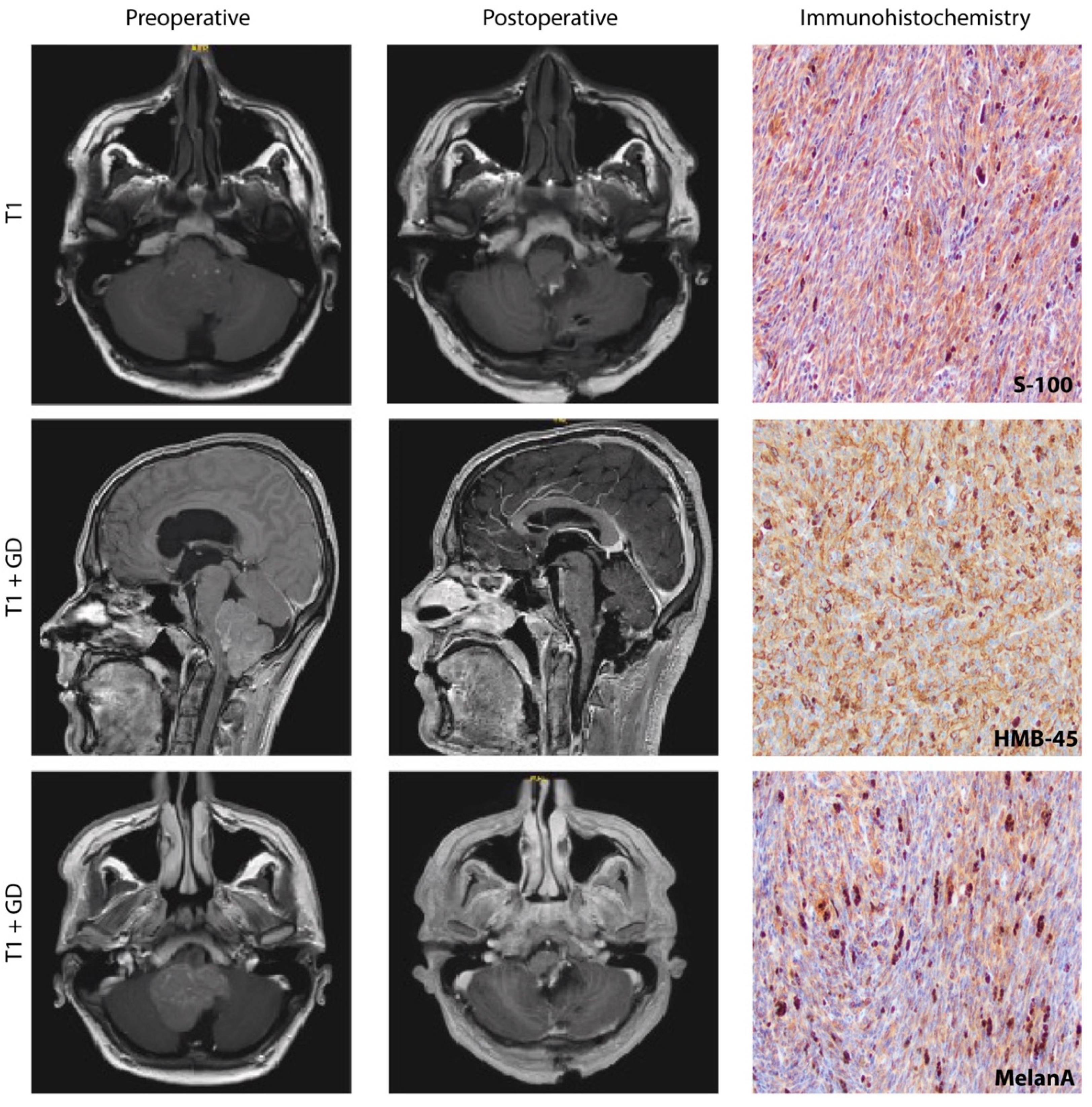
Figure 1.
Exemplary perioperative MRI sequences of a 25-year-old Meningeal Melanocytoma (MM) patient that underwent surgery for a cerebellar lesion at our department. Axial T1 MRI showing a grey matter isointense posterior fossa mass shifting the brain stem forward, compressing cerebellum and brainstem, thereby leading to obstruction of the fourth ventricle. Preoperative (left row) T1 imaging after gadolinium application shows sagittal and axial inhomogeneous contrast enhancement. Postoperative (middle row) T1-weighted MRI without contrast agent (axial) and following gadolinium application (sagittal, axial), showing total resection of the mass with decompressed 4th ventricle and without secondary bleeding. Histopathological analysis of our case revealed spindle-shaped cells that showed expression of S-100, HMB45, and MelanA (right row).

Figure 2.
PRISMA Flow Chart visualizing the literature selection process. As database PubMed and Web of Science were used. As keyword “meningeal melanocytoma” was applied. In total, 219 results were found using PubMed and 247 using Web of Science published between the first description in 1972 and 2022. After subtracting duplicates, the total data covered 312 items. A total of n = 77 were excluded because no novel case report of MM was provided, and n = 35 were excluded as no abstract was available in English language. Therefore, in total, 201 published cases, as well as 1 unpublished case from our department, were included for further analysis.

Figure 3.
Age–dependent incidence of MM in females (red) and males (blue). The y–axis represents the age of the patients, and the x–axis is the count of female or male patients that were diagnosed with MM. The youngest patient was only 28 weeks old, and the oldest patient was diagnosed at the age of 79 years (Figure 2). The median age of disease onset of female patients was 37 years, and for male patients, it was 42 years. A higher frequency of MM was found in men compared to women (1.4:1).

Figure 4.
Treatment strategies at initial therapy: When MM is diagnosed, total resection should be the primary goal. Depending on the Ki–67 index, it should then be evaluated whether adjuvant radiotherapy is necessary.
Figure 4.
Treatment strategies at initial therapy: When MM is diagnosed, total resection should be the primary goal. Depending on the Ki–67 index, it should then be evaluated whether adjuvant radiotherapy is necessary.

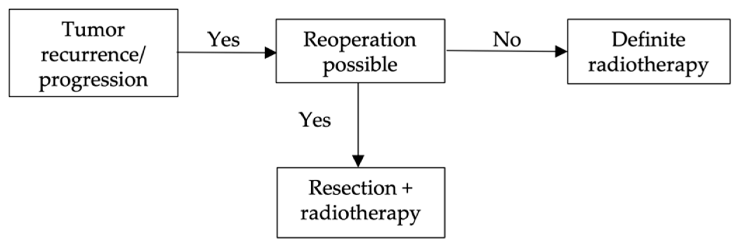
Figure 5.
Treatment strategies after tumor recurrence/progression: Reoperation is the most used treatment option for tumor progression. However, in almost no case report, a second follow–up after the first recurrence is recorded, so no further evaluation can be made here as to how successful the respective therapy options are. These are only the therapies that are most frequently used.

Table 2.
Total Resection and Follow–Up Data: Total resection alone showed the best outcome when it came to therapy options. Regarding adjuvant therapy, the data are very limited.
Table 2.
Total Resection and Follow–Up Data: Total resection alone showed the best outcome when it came to therapy options. Regarding adjuvant therapy, the data are very limited.
| Therapy | Frequency | Follow–Up No Recurrence |
|---|---|---|
| Total Resection only | 69 (90.8 %) | 47/69 (68.1%) |
| Total Resection + RT | 7 (9.2%) | 5/7 (71.4%) |
| Total | 76 (100%) | 52/76 (68.4%) |
Table 3.
Partial Resection and Follow–Up Data: Partial Resection alone shows a high recurrence rate. The combination with adjuvant radiotherapy leads to more promising results.
Table 3.
Partial Resection and Follow–Up Data: Partial Resection alone shows a high recurrence rate. The combination with adjuvant radiotherapy leads to more promising results.
| Therapy | Frequency | Follow–Up No Recurrence |
|---|---|---|
| Partial Resection only | 33 (55.9%) | 18/33 (54.5%) |
| Partial Resection + RT | 21 (35.6%) | 13/21 (61.9%) |
| Partial Resection + Chemo | 1 (1.7%) | 1/1 (100%) |
| Partial Resection + RT + Chemo | 2 (3.4%) | 0/2 (0%) |
| Partial Resection + Radiosurgery | 2 (3.4%) | 2/2 (100%) |
| Total | 59 (100%) | 34/59 (57.6%) |
Table 4.
Metastasis: Metastasis led to either tumor progression or death. TR = Total Resection, PR = Partial Resection, RT = Radiotherapy, Reop = Reoperation.
Table 4.
Metastasis: Metastasis led to either tumor progression or death. TR = Total Resection, PR = Partial Resection, RT = Radiotherapy, Reop = Reoperation.
| Case | Primary Location MM | Location Metastasis | Therapy | Outcome |
|---|---|---|---|---|
| [8] | Spine | Liver, rib | TR, Reop + RT | Tumor progression |
| [17] | Intracranial | Thoracic | PR + RT, Reop + RT + Temolozomide + Cisplatin + Fotemustine | Death |
| [19] | Intracranial | Spine | TR, RT + Temozolomide | Death |
| [22] | Intracranial | Intracranial, Liver, pancreas | TR + RT, Reop + RT; Temozolomide; Ipilimumab; | Death |
| [27] | Spine | Intracranial | TR, Reop + RT + Pembrolizumab + Bevacizumab + Temozolomide | Death |
| [44] | Intracranial | Intracranial | TR, RT | Tumor progression |
| [45] | Spine | Intracranial | PR | Death |
| [46] | Spine | Intracranial | TR | Death |
| [47] | Spine | Spine | Resection | Death |
| [48] | Spine | Intracranial | PR, Reop + RT | Death |
| [49] | Spine | No data | TR, Reop | Tumor progression |
Publisher’s Note: MDPI stays neutral with regard to jurisdictional claims in published maps and institutional affiliations. |
© 2022 by the authors. Licensee MDPI, Basel, Switzerland. This article is an open access article distributed under the terms and conditions of the Creative Commons Attribution (CC BY) license (https://creativecommons.org/licenses/by/4.0/).
Share and Cite
MDPI and ACS Style
Ricchizzi, S.; Gallus, M.; Stummer, W.; Holling, M. How Should We Treat Meningeal Melanocytoma? A Retrospective Analysis of Potential Treatment Strategies. Cancers 2022, 14, 5851. https://doi.org/10.3390/cancers14235851
AMA Style
Ricchizzi S, Gallus M, Stummer W, Holling M. How Should We Treat Meningeal Melanocytoma? A Retrospective Analysis of Potential Treatment Strategies. Cancers. 2022; 14(23):5851. https://doi.org/10.3390/cancers14235851
Chicago/Turabian StyleRicchizzi, Sarah, Marco Gallus, Walter Stummer, and Markus Holling. 2022. "How Should We Treat Meningeal Melanocytoma? A Retrospective Analysis of Potential Treatment Strategies" Cancers 14, no. 23: 5851. https://doi.org/10.3390/cancers14235851
Note that from the first issue of 2016, this journal uses article numbers instead of page numbers. See further details here.