Article

Is there an Exposure–Response Relationship for Nivolumab in Real-World NSCLC Patients?

by
Audrey Bellesoeur
1,
Edouard Ollier
2,3,
Marie Allard
4,
Laure Hirsch
1,
Pascaline Boudou-Rouquette
1,
Jennifer Arrondeau
1,
Audrey Thomas-Schoemann
5,6,
Manuela Tiako
4,
Nihel Khoudour
4,
Jeanne Chapron
7,
Frédérique Giraud
7,
Marie Wislez
7,
Diane Damotte
8,9,
Audrey Lupo
8,9,
Michel Vidal
4,6,
Jérôme Alexandre
1,10,
François Goldwasser
1,10,
Michel Tod
11,12,13 and
Benoit Blanchet
4,5,*
1
Department of Medical Oncology, Cochin Hospital, AP-HP, CARPEM, 75014 Paris, France
2
Clinical Research Unity, Innovation, Pharmacology, Hôpital Nord, 42000 Saint-Etienne, France
3
INSERM, U1059, Dysfonction Vasculaire et Hémostase, 42000 Saint-Etienne, France
4
Department of Pharmacokinetics and Pharmacochemisty, Cochin Hospital, AP-HP, CARPEM, 75014 Paris, France
5
Department of Clinical Pharmacy, Cochin Hospital, AP-HP, 75014 Paris, France
6
UMR8038 CNRS, U1268 INSERM, Faculty of Pharmacy, University Paris Descartes, PRES Sorbonne Paris Cité, 75006 Paris, France
7
Department of Pneumology, Cochin Hospital, AP-HP, 75014 Paris, France
8
Department of Pathology, Cochin Hospital, AP-HP, 75014 Paris, France
9
UMRS U1138, Centre de Recherche des Cordeliers, Université Paris Descartes, 75005 Paris, France
10
Cochin Institute, INSERM U1016, 75014 Paris, France
11
Department of Clinical Pharmacy, la Croix Rousse Hospital, Hospices civils de Lyon, 69002 Lyon, France
12
Department of clinical Pharmacology, Claude Bernard Lyon 1 University, 69100 Villeurbane, France
13
EMR 3738, Lyon-sud Medical School, Lyon 1 University, BP 12 Chemin du grand revoyet, 69921 Oullins, France
*
Author to whom correspondence should be addressed.
Cancers 2019, 11(11), 1784; https://doi.org/10.3390/cancers11111784
Submission received: 28 October 2019 / Accepted: 8 November 2019 / Published: 13 November 2019
(This article belongs to the Special Issue Cancer Biomarkers)

Abstract

:
Pharmacokinetic/pharmacodynamic data from real-world cohort are sparse in non small–cell lung cancer (NSCLC) patients treated with nivolumab. The aim of this prospective observational study was to explore the exposure-response relationship for effectiveness and toxicity of nivolumab in 81 outpatients with metastatic lung cancer. Nivolumab plasma trough concentrations (Cmin) were assayed at days 14, 28, and 42. Prognostic factors (including Cmin) regarding progression-free survival (PFS) and overall survival (OS) were explored using a multivariate Cox model. A Spearman’s rank test was used to investigate the relationship between Cmin and grade >2 immune-related adverse events (irAE). Mean nivolumab Cmin was 16.2 ± 6.0 µg/mL (n = 76), 25.6 ± 10.2 µg/mL (n = 64) and 33.4 ± 11.3 µg/mL (n = 53) at days 14, 28, and 42, respectively. No pharmacokinetic/pharmacodynamic (PK/PD) relationship was observed with either survival or onset of irAE. Multivariable Cox regression analysis identified Eastern Cooperative Oncology Group Performance Status (hazard ratio 1.85, 95%confidence interval 1.02–3.38, p-value = 0.043) and baseline use of corticosteroids (HR 8.08, 95%CI 1.78–36.62, p-value = 0.007) as independent risk factor for PFS and only baseline use of corticosteroids (HR 6.29, 95%CI 1.46–27.08, p-value = 0.013) for OS. No PK/PD relationship for nivolumab was observed in real-world NSCLC patients. This supports the recent use of flat dose regimens without plasma drug monitoring.

1. Introduction

In the last few years, immunotherapy has provided a major treatment advance for lung cancer patients. In particular, inhibitors of programmed cell death 1 (PD-1) pathway have significantly changed treatment algorithms. Anti-PD-1 monoclonal antibodies bind to the PD-1 receptor and prevent interactions with its ligands PD-L1 and PD-L2, thereby releasing PD-1 pathway-mediated inhibition of the immune tumor response [1]. Nivolumab, a fully human immunoglobulin G4, can help to reinstate the antitumor immune response by targeting PD-1 receptors located on lymphocytes’ surfaces. Nivolumab is currently approved by the Food and Drug Administration for the treatment of advanced solid cancers such as melanoma, renal cell carcinoma, head and neck squamous cell carcinoma, urothelial cancer, colorectal cancer with microsatellite instability, and hepatocellular carcinoma [2]. Besides, two phase III trials showed improved overall survival (OS), response rate, and progression-free survival (PFS) with nivolumab (3 mg/kg, every two weeks) versus docetaxel in patients with advanced, previously treated squamous-cell and non-squamous non-small cell lung cancer (NSCLC) [3,4]. Nivolumab is currently approved as second-line treatment after chemotherapy in NSCLC patients.
Despite significant advances in immuno-oncology, there is still a long way to go for NSCLC treatment. Only 20% of NSCLC patients respond to nivolumab as a second line therapy, and approximately half of these responses are durable [3,4]. In a context of personalized medicine, many questions remain about the unmet need for predictors of response and toxicity with nivolumab. Even though PD-L1 expression is commonly used in daily clinical practice in immuno-oncology, it is not mandatory for nivolumab prescription. A reliable baseline biomarker is still lacking to provide binary discrimination of responsiveness to nivolumab treatment [5], which could prevent both toxicities in non-responders and unnecessary costs. Questions regarding an optimal nivolumab dosing regimen also warrant consideration in NSCLC patients. Nivolumab was first approved for a 3 mg/kg dosing regimen every two weeks (Q2W). The relatively flat exposure–response relationships of safety and efficacy over a large range of plasma exposure (for dose levels of 1–10 mg/kg Q2W) argue for a wide therapeutic margin of nivolumab in NSCLC patients [6]. A quantitative clinical pharmacology approach has provided evidence of a similar risk- benefit profile of a 240-mg flat dose Q2W and a 480-mg flat dose every four weeks (Q4W) relative to 3 mg/kg Q2W [7,8], which recently resulted in the approval of these new dosing regimens in NSCLC patients. However, this approach, based on pharmacokinetic/pharmacodynamic (PK/PD) simulation, was conducted from a database including patients enrolled in different clinical trials (phase I, II, and III studies). In contrast with pembrolizumab [9], no phase III study is currently published to confirm these new dosing regimens of nivolumab in NSCLC patients. In this context, there can be concern, in particular at extreme weights. A better understanding of PK/PD relationship of nivolumab in NSCLC patients from the “real-world” could help physicians to optimize plasma drug exposure and the risk/benefit ratio in a daily clinical practice.
Regarding pharmacokinetic data and the exposure–response relationship, the main published data arise from clinical trials conducted with selected patients. In NSCLC nivolumab trials, Feng et al. identified no relation between the average concentration after the first injection (Cavg1) and survival [6]. They described a flat exposure-response relationship and a wide therapeutic index for doses ranging from 1 mg/kg to 10 mg/kg. Two clinical trials confirmed these results in metastatic melanoma patients treated with nivolumab [9,10]. On the contrary, Basak et al. [11] recently published a correlation between nivolumab exposure and effectiveness (response and survival) in NSCLC patients from daily clinical practice. Thus, PK/PD data for nivolumab are currently conflicting between clinical trials and “real world” cohort studies.
The aims of this prospective observational study were to explore the exposure–response relationship for effectiveness and toxicity of nivolumab in NSCLC patients in daily clinical practice and to characterize the interindividual variability in nivolumab plasma exposure.

2. Results

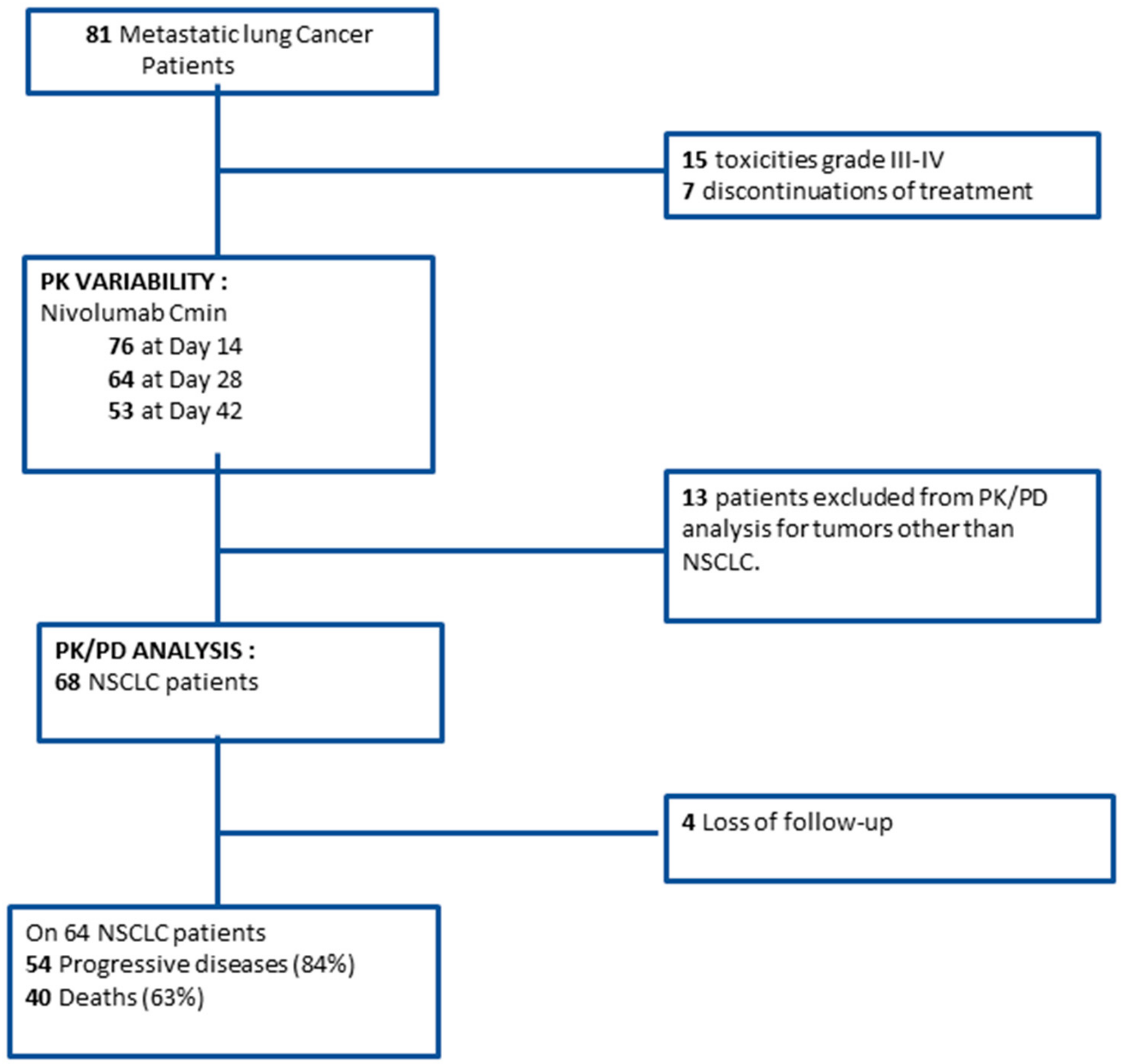
2.1. Study Population

Eighty-one patients with lung cancer were included in this study (Figure 1).
The demographical, biological, and clinical characteristics of the cohort are summarized in Table 1. Forty-nine patients (60%) were male, with a median age of 65 years; 68 patients (84%) had NSCLC, and 20 patients (25%) experienced cerebral metastasis. Fifty-eight patients (72%) received nivolumab as second-line treatment. The results of PD-L1 testing were available for 43 patients (53%), with a median of 5% PD-L1 expression (0–28).
Results are expressed as median (interquartile range) or frequency (percent).
In the whole population, the median duration of nivolumab treatment was 2.5 months (1.4–7.9). The median follow-up duration was 9.4 months (3.8—16.7). At data cut-off in January 2018, eight patients (10%) were still treated with nivolumab.

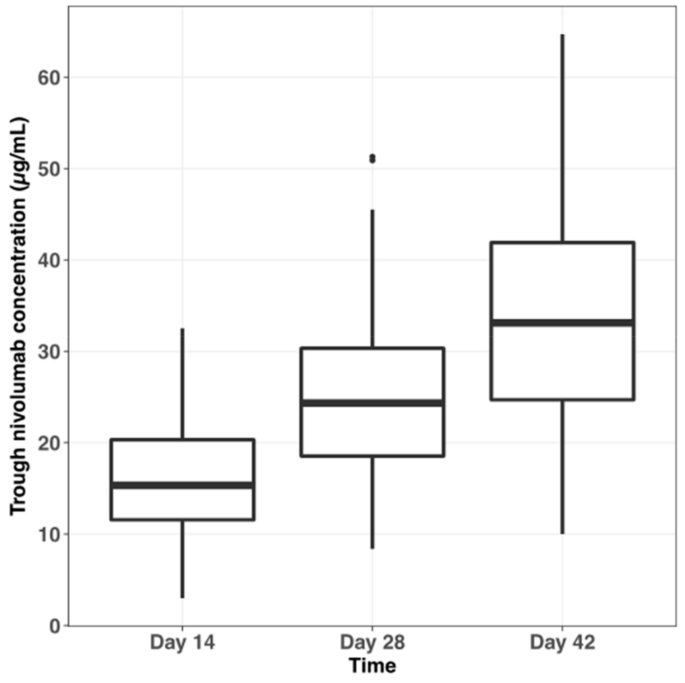
2.2. Pharmacokinetic Variability

Pharmacokinetic data of the whole cohort (n = 81 patients) before the second (Day 14), third (Day 28), and fourth (Day 42) infusions of nivolumab after treatment initiation were analyzed to investigate the interindividual variability in trough plasma concentration (Cmin) of nivolumab and its determinants. Overall, 193 plasma samples could be analyzed (mean of 2.4 samples per patient). Figure S1 presents individual nivolumab plasma Cmin within the 42 first days of treatment. Mean nivolumab plasma Cmin at days 14, 28, and 42 after treatment start was 16.2 ± 6.0 µg/mL (n = 76, coefficient of variation; CV = 37.0%), 25.6 ± 10.2 µg/mL (n = 64; CV = 39.8%), and 33.4 ± 11.3 µg/mL (n = 53; CV = 33.8%), respectively (Figure 2). Nivolumab plasma Cmin were statistically different (interaction test, p-value < 0.001) between different sampling occasions (i.e., days 14, 28, and 42).
All pharmacokinetic data were initially analyzed using a population pharmacokinetic approach. Given that only nivolumab plasma Cmin were available, the analysis was conducted by fitting a previously published population pharmacokinetic model to estimate individual parameters [12]. However, the shrinkage of the empirical bayesian estimates was large, precluding an analysis based on these estimates. In univariate analysis, gender (p-value = 0.078), total body weight (p-value = 0.005), body mass index (BMI) (p-value < 0.0001), C-reactive protein (CRP) level (p-value = 0.0089), cerebral metastasis (p = 0.013), and albumin level (p-value = 0.0068) were identified as factors influencing nivolumab plasma Cmin at day 14 (Table 2). In multivariate analysis, only BMI was identified as an independent variability factor of nivolumab plasma Cmin (p-value < 0.0001), with a strong positive correlation between these two parameters. Interestingly, baseline use of corticosteroids was statistically associated with nivolumab plasma Cmin at day 28 (p-value = 0.0117). Thus, an increase of 8.7 μg/mL (CI95% 2.0; 15.4) for nivolumab plasma Cmin is expected in patients treated with corticosteroids. By contrast, the relationship was not statistically significant at day 42 (p-value = 0.3).

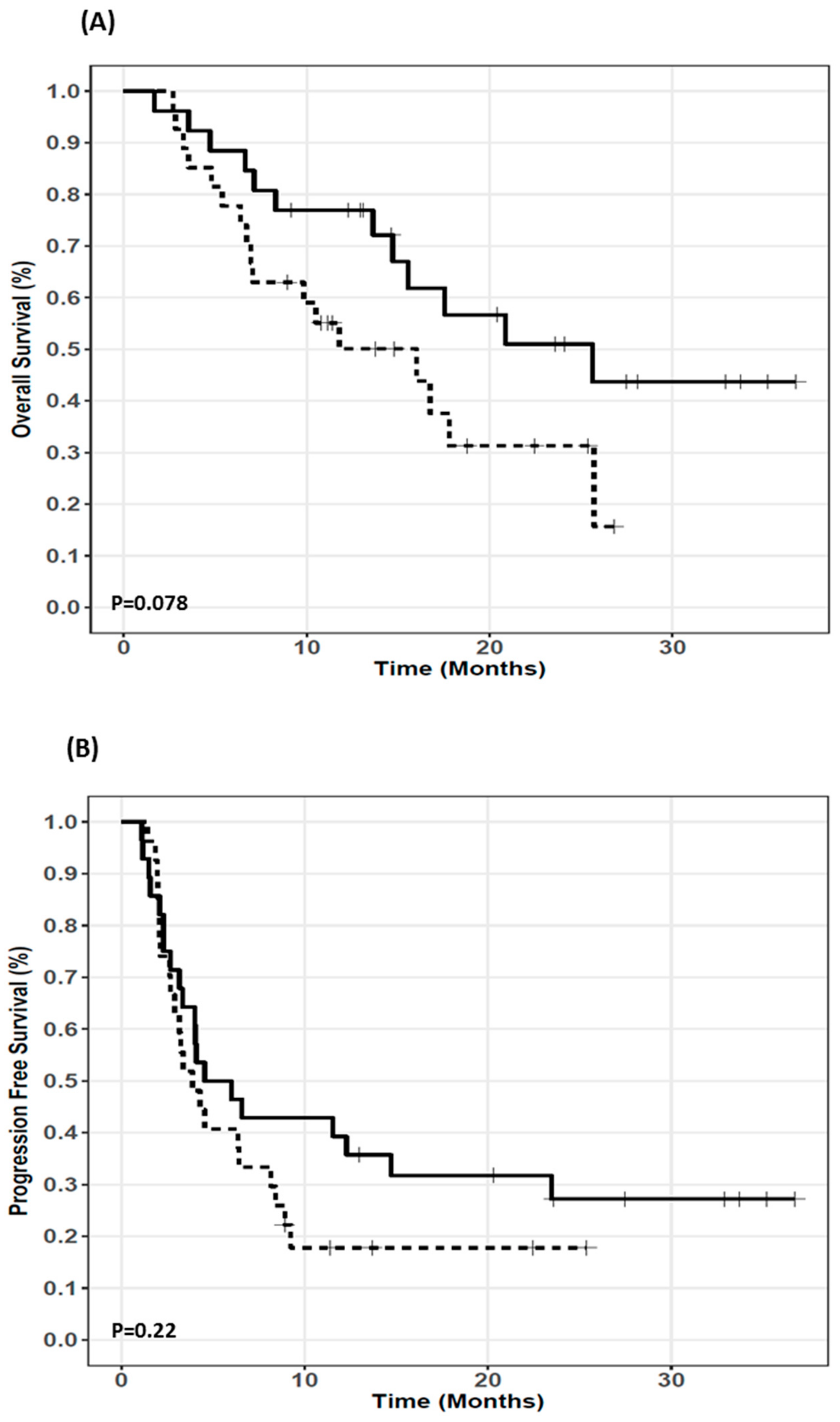
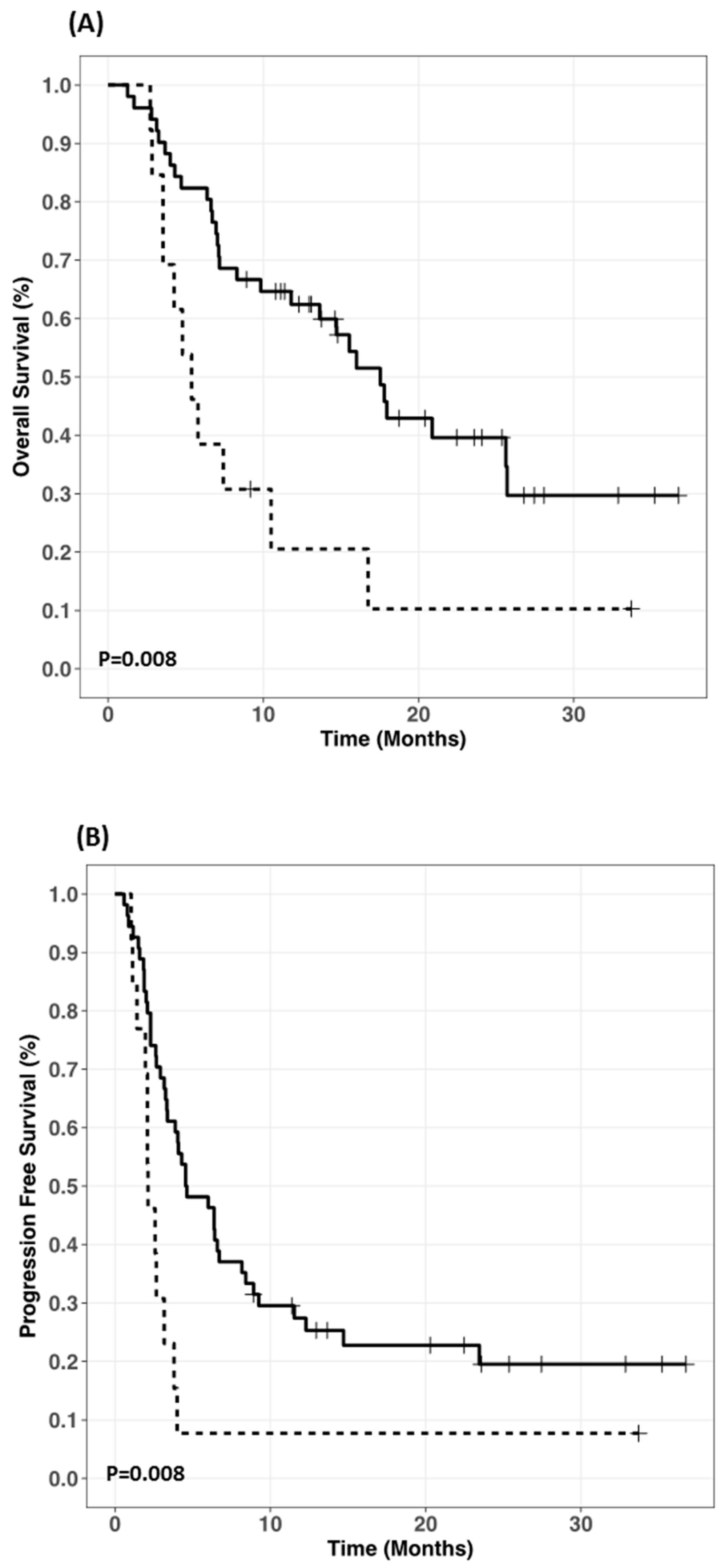
2.3. Exposure-Survival Relationship

PK/PD data of patients with other histological tumor types than NSCLC (n = 13) were excluded for exposure-survival relationship analysis. Among NSCLC patients, four patients were lost to follow-up (Figure 1). Overall, the median PFS and OS in the PK/PD analysis cohort (n = 68 patients) were 4.0 months (CI95%: 3.2–6.4) and 15.5 months (CI95%: 8.3–20.9), respectively. Nivolumab plasma Cmin (mean ± SD) at days 14, 28, and 42 after treatment start was 16.2 ± 6.0 µg/mL (n = 62, fifth to 95 th percentile: 6.5 to 25.1 µg/mL), 25.7 ± 10.2 µg/mL (n = 53; fifth to 95 th percentile: 10.5 to 42.6 µg/mL), and 33.4 ± 11.3 µg/mL (n = 43; fifth to 95th percentile: 16.4 to 50.9 µg/mL), respectively. NSCLC patients with Cmin ≤ 24.7 µg/mL (median value) at day 28 tended to have longer OS (25.6 (15.5–undetermined) vs 16.0 (7.0-undetermined) months; p-value = 0.0786) (Figure 3A). By contrast, low nivolumab plasma Cmin at day 28 (≤24.7 µg/mL) was not statistically associated with longer PFS (5.3 (3.3-undetermined) vs 3.9 (2.9–8.4) months, p-value = 0.22) (Figure 3B). There was no exposure-response relationship for Cmin at day 14 or 42.
Amongst 68 NSCLC patients, nine (13%) received systemic corticosteroids at the beginning of nivolumab therapy. Figure 4 shows that these patients had both shorter PFS and OS than corticosteroids free patients (PFS: 2.12 (1.96-undetermined) vs 4.58 (3.38–8.42) months; p-value = 0.008); (OS: 5.38 (3.54 undetermined) vs 17.54 (13.62 undetermined) months; p-value = 0.008).
Multivariable Cox regression analysis identified Eastern Cooperative Oncology Group (ECOG) performance status (HR 1.85, 95%CI 1.02–3.38, p-value = 0.043) and baseline corticosteroids (HR 8.08, 95%CI 1.78–36.62, p-value = 0.007) as independent risk factor for PFS, and only baseline corticosteroids (HR 6.29, 95%CI 1.46–27.08, p-value = 0.013) for OS (Table 3). Finally, no PK/PD relationship was observed for survival in 59 free-corticosteroids NSCLC patients. The univariate analysis did not show any relationship between nivolumab plasma Cmin at day 28 (≤24.7 µg/mL) and PFS (HR: 1.08 (0.54–2.16), p-value = 0.81) or OS (HR: 0.81 (0.34–1.90), p-value = 0.62).

2.4. Exposure-Toxicity Relationship

Of 81 lung cancer patients, nivolumab treatment was associated in 15 patients (18%) with grade III or IV immune-related adverse events (irAE): cardiopulmonary events (5/81, 6%, with myocarditis, pericarditis, pulmonary hypertension, interstitial lung disease), colitis (3/81, 4%), rheumatologic disorders (2/81, 2%, arthritis), neurological events (2/81, 2%, peripheral neuropathy and central nervous system disorder), endocrine disorders (2/81, 2%, hypophysitis and diabetes), and renal insufficiency (1/81, 1%). Nivolumab was discontinued in seven patients—two patients at day 14 with respective Cmin of 8.8 and 15.4 µg/mL, three patients at day 42 (Cmin of 42.1, 32.1, and 38.3 µg/mL at the time of treatment discontinuation), and the two others after more than six months of treatment. For five patients with plasma Cmin at the time of treatment discontinuation, Cmin was included in the 10-90th percentile range of Cmin observed in the whole cohort. Overall, no relationship was found between nivolumab plasma Cmin and the occurrence of grade III or IV irAE.

3. Discussion

Nivolumab treatment can enable durable responses and long-term overall survival in NSCLC patients [13]. However, predicting response to nivolumab remains currently difficult since some patients without or with low PDL-1 expression do have responses [5,14]. In this context, the identification of factors contributing to the interindividual variability in clinical outcomes of nivolumab is mandatory, especially in real world setting. The present study shows that use of corticosteroids at initiation of nivolumab therapy is a deleterious factor for NSCLC patients receiving nivolumab, while nivolumab plasma exposure does not seem to have any influence on both effectiveness and safety.
PK/PD data about PD-1 inhibitors remain sparse [2], especially in unselected NSCLC patients. Bajaj et al demonstrated that nivolumab clearance decreases over time [12]. Furthermore, this significant decrease in nivolumab clearance over time was identified as a predictive factor of response in patients with advanced solid tumors [10,12]. Baseline clearance has also been identified as a predictive factor of survival for anti-PD-1 antibodies [9,15]. The higher plasma exposure at steady state in responders could be the result rather than the cause of better response to nivolumab therapy. Indeed, decreased cachexia and improvement in disease state characterizing a lower tumor burden, normalization of protein turnover rate, and decreased inflammation state might contribute to the decrease in nivolumab clearance over time. Simulation has showed that exposure–response analysis results based on nivolumab plasma exposure after a single infusion was more consistent with the true exposure–response than with the steady-state plasma exposure [6,12,16].
In the present study, the multivariable Cox regression analysis failed to show a relationship between survival and nivolumab plasma Cmin at day 14 or 28. A recent study suggests that a threshold value of 10 µg/mL of nivolumab would allow for >90% of maximum achievable receptor occupancy (about 80% receptor occupancy) of circulating T lymphocytes T [17]. Another study proposes a target concentration of 4.5 µg/mL [18]. In the present study, eight patients (11.2%) and none exhibited nivolumab plasma Cmin below 10 µg/mL and 4.5 µg/mL at day 14, respectively. These results could explain in part the lack of relationship between nivolumab plasma Cmin and survival in our cohort. Nevertheless, one cannot exclude that this relationship is different between pharmacodynamic activity on infiltrated T lymphocytes and nivolumab concentration in the tumor microenvironment. Further investigations are required to address this issue. In the present study, the occurrence of grade III or IV irAE was not statistically associated with increased nivolumab plasma Cmin, which suggests that the onset of severe irAE would be more related to intrinsic immune characteristics than plasma drug exposure. Our results from “real-life” NSCLC patients support the lack of PK/PD relationship for both efficacy and safety, previously described from nivolumab clinical trials in melanoma, lung cancer, or Hodgkin patients [6,7,9,10,19,20]. Recently, Basak et al. reported exposure relationship for response and survival but not with toxicity [11]. These results are in contradiction with ours about effectiveness. Even though our population seems close to that of Basak et al., no information about corticosteroids exposure is available on their population. Furthermore, their analysis of the exposure–response relationship does not account for clinical and biological factors in multivariate analysis, which could be a limiting factor in the interpretation of their results. Overall, our results are in favor of a wide therapeutic index and consistent with flat-dose regimens recently approved for nivolumab [2]. Given the wide therapeutic index of nivolumab [2], our results suggest that an approach of pharmacokinetically guided dose adjustment of nivolumab should not be necessary to improve efficacy in daily clinical practice. However, recent in silico data suggest that drug monitoring could help to postpone forthcoming courses of nivolumab by simulating the time by which the patient is likely to be underexposed with regard to target engagement [18]. Overall, the extension of the administration interval guided by drug monitoring could improve the safety of nivolumab without compromising its efficacy.
The nivolumab exposure–response relationship may be confounded by several factors, including cachexia but also corticosteroids intake. The value of nivolumab clearance, which drives its plasma exposure, is the sum of the clearance in the tumor (target-mediated or proteolytic) [21], the hydrolytic clearance in other organs, and marginally, the clearance related to anti-drug antibodies (ADAs) [22] (Figure 5). Tumor-associated clearance is expected to be proportional to tumor size; hydrolytic clearance is known to decrease as inflammation regresses and ADA-mediated clearance increases over time if ADAs are produced [23]. Hypoalbuminemia is a well-recognized marker of cachexia and elevated protein turnover secondary to chronic systemic inflammatory conditions, as observed in many cancer indications. In contrast with other studies [12,24], we failed to identify albuminemia as an independent variability factor of nivolumab pharmacokinetics, probably because of the low interindividual variability in albuminemia. Depending on the time course of tumor size, inflammation status and ADAs production, nivolumab clearance may vary more or less. Moreover, the magnitude of the variation depends on the presence of glucocorticoids or not (Figure 5). On one hand, corticosteroids, by their anti-inflammatory and immunosuppressive properties [25], could decrease hydrolytic and ADA-mediated clearance. On the other hand, corticosteroids may antagonize the immunostimulant effect of nivolumab, resulting in a smaller antitumoral effect and a smaller reduction of the target-mediated clearance. In our data, corticosteroids tended to be associated with higher nivolumab plasma Cmin at D14, D28, and D42 (significantly at D28). The lack of significant association between nivolumab plasma Cmin at D42 and corticosteroids intake is unclear, it could be related to a lower statistical power because of many missing data. The statistical association between corticosteroids and higher nivolumab plasma Cmin at D28 is consistent with nivolumab clearance being reduced by corticosteroids. However, the immunosuppressive effect of corticosteroids may also limit nivolumab pharmacodynamics, resulting in less tumor shrinkage and less tumor-associated clearance reduction.
The second result of our study demonstrated that NSCLC patients treated with corticosteroids at the time of nivolumab initiation exhibited a shorter survival (PFS and OS) than in corticosteroid-free patients. Baseline use of corticosteroids, even at low dose (median dose in our population: 20 mg/d, range 10–25), was identified as an independent risk factor for PFS and OS in multivariate analysis, which confirms the results from recent studies [26,27]. Several hypotheses can be made to explain the detrimental effect of corticosteroid. First, baseline corticosteroid use could reflect a more severe condition associated with poorer prognosis independently of which anticancer drug is used. Thus, severe bone pain or intracranial hypertension are common indications of systemic corticosteroids. However, other parameters related to advanced disease, such as number of metastatic sites reflecting tumor burden, is associated neither with progression nor survival.
Systemic corticoids could impair efficacy of PD1/PDL1 inhibitors by a pharmacokinetic or a pharmacodynamic interaction. In our study, corticosteroids use is not clearly associated with nivolumab exposure variability, which seems not to favor a pharmacokinetic interaction. The shorter survival in patients concomitantly treated with corticosteroids could be further related to PD interaction. As previously shown in transplant recipients, corticosteroids are able to decrease the immune reaction [25], which represents a pharmacodynamical antagonism for nivolumab. However, mechanisms of how corticosteroids prevent response to nivolumab remain ill-defined, and in vivo data are lacking. Different hypothesis among others can be proposed. Granzyme B (GranB) is a main component of the cytotoxic activity of CD8+ T cells and natural killer (NK) cells during the cellular immune response against cancer cells. Thus, recent studies have suggested potential benefit of GranB positron emission tomography (PET) imaging to predict response to cancer immunotherapy [28,29]. In renal transplant recipients treated with corticosteroids, Hruba et al. reported a significantly lower cytotoxic T lymphocytes and NK cells–derived GranB expression than in corticosteroid-free recipients at day 14 post-transplantation [30]. These data suggest that a lower baseline GranB expression is expected in NSCLC patients under corticosteroids at the time of nivolumab initiation, which could compromise the immunological effect of nivolumab. Costantini et al. recently showed an association between low plasma level of soluble GranB at nivolumab initiation and poor response, PFS and OS in 43 NSCLC patients treated with nivolumab [31], which supports our hypothesis. Finally, corticosteroids use is known to significantly decrease NK cells and/or impair their function in transplant recipients [30,32]. A high baseline level of circulating NK cells has been recently identified as a predictive factor of prolonged survival outcome in 31 NSCLC patients treated with nivolumab [33]. In the present study, we did not find any difference in baseline levels of circulating NK cells between NSCLC patients treated or not with corticosteroids (data not shown). Further investigations are warranted to elucidate how corticosteroids prevent nivolumab efficacy. However, our results suggest prudent use of corticosteroids at the time of nivolumab initiation.

4. Materials and Methods

4.1. Participants

From July 2015 to June 2017, 81 outpatients treated for metastatic lung cancer in the CERTIM cohort (Immunomodulatory Therapies Multidisciplinary Study group) were included in this prospective observational monocentric study. All patients were treated and had follow-up visits in the oncology department of Cochin Hospital (Paris, France). Patients were eligible if they had a histologically confirmed lung cancer classified as stage IV or stage III without any curative treatment possible, progressive after at least one line of chemotherapy. A minimal age of 18-years-old, ECOG performance status ≤2, and adequate renal and hepatic functions according to manufacturer recommendations were also required. Patients with small-cell lung cancer who underwent nivolumab therapy as part of a clinical trial were eligible for pharmacokinetic analysis and exposure–toxicity analysis only. This prospective project was in compliance with the Declaration of Helsinki and approved by the local medical ethical board on 22 December 2016 with approval number CLEC N2442. All patients gave their written informed consent to participate in the study.

4.2. Treatment

All patients were treated with the recommended dose of nivolumab (3 mg/kg every two weeks) administered intravenously over 60 minutes. All adverse events were graded using the National Cancer Institute Common Toxicity Criteria, version 4.0. Nivolumab treatment was continued until disease progression (either based on clinical or radiological criteria) or unacceptable toxicity. However, treatment discontinuation was authorized in the cases of complete response; the timing of such interruption was left to the discretion of physician.

4.3. Pharmacokinetic Measurements

Nivolumab Cmin in plasma was determined just prior to next infusion at Day 14, Day 28 and Day 42. The plasma was separated by centrifugation (4000 rpm, 10 min, 4 °C) within two hours after blood collection and then stored at −80 °C until the analysis. Nivolumab plasma Cmin were assayed using a validated home-made enzyme-linked immunosorbent assay (ELISA) [34]. The calibration range for nivolumab assay was 5–100 μg/mL with a limit of detection at 3 μg/mL. The within- and between-day accuracy for internal quality controls (5, 20 and 75 μg/mL) was included between –7.5% and 3.5%; within- and between-day imprecision were less than 5% and 12% for three levels of internal quality control, respectively. Nivolumab concentration is stable over a three-month period when plasma is stored at −80 °C [34]. All plasma samples were analyzed within the month after blood collection.

4.4. PD-L1 Expression Analysis

Tumor PD-L1 protein expression was evaluated retrospectively in pretreatment (archival or recent) tumor-biopsy or surgical resection specimens with the use of an automated immunohistochemistry assay that used rabbit monoclonal antihuman PD-L1 antibody (E1L3N Cell Signaling Technology—automat Leica; Leica Biosystems, Nanterre, France). Tumors were defined as PD-L1 positive when staining of the tumor-cell membrane (at any intensity) was observed at pre-specified expression levels of 5% or higher in a section that included at least 100 tumor cells for evaluation. As PD-L1 status is not mandatory for nivolumab prescription, results were not available for all patients.

4.5. Study Endpoints

The main objective was to explore the relationship between nivolumab plasma exposure and survival in NSCLC patients. The primary endpoint was OS, defined as the time from the first infusion of nivolumab to death from any cause. The secondary endpoint was PFS, defined as the time from nivolumab treatment initiation to documented progression event (either clinical or radiological progression) or death from any cause. Radiographic evidence of progression was defined according to modified Response Evaluation Criteria in Solid Tumors [35]. The secondary objectives were to investigate the exposure-response relationship for grade III or IV irAE and to characterize the magnitude of interindividual variability in nivolumab pharmacokinetics in “real-world” lung cancer patients.

4.6. Statistical Analysis

Descriptive statistics used median (interquartile range) for quantitative variables and percentages for qualitative ones. Patients with small-cell lung cancer were included in the pharmacokinetic analysis and exposure-toxicity analysis but were excluded from the exposure–survival analysis (Figure 1). The relationships between the nivolumab Cmin at day 14 and different variables were tested using two sample Wilcoxon tests or Kruskal-Wallis tests for qualitative variables with two modalities or more than two modalities, respectively, and by Spearman rank correlation coefficient tests for quantitative ones. The following variables were tested: age, age >70 years old, gender, total body weight, body mass index (BMI), lean body mass [36], C-reactive protein (CRP), albumin, immunoglobulin G, hepatic enzymes (AST and ALT), and baseline use of corticosteroids (i.e., patients who received systemic corticosteroids at the time of nivolumab initiation), PD-L1 expression, cerebral metastasis, tumor histology (adenocarcinoma vs squamous cell carcinoma), stage disease (III vs IV). A stepwise multiple linear regression was then performed with variables significant with p = 0.10 in the first step included in the regression and variables significant with p = 0.05 kept in the final model. Both OS and PFS functions were estimated using the Kaplan-Meier method. Survival data were analyzed using a Cox proportional hazard regression model adjusted for established clinical and biological risk factors. Following biological and clinical parameters were studied as prognostic factors: nivolumab plasma Cmin at day 14 (<median value), nivolumab plasma Cmin at day 28 (<median value), ECOG performance status [37,38], number of metastasis [38,39], neutrophil-to-lymphocyte ratio (NLR) (<median value) [39,40], and baseline use of corticosteroids and tumor PD-L1 protein expression (>median value) [14,41]. A Spearman’s rank test was used to investigate the relationship between nivolumab plasma Cmin and grade >2 irAE. All computations were performed using R statistical software v3.4.4 (R Stats Package, R Foundation, Vienna, Austria).

5. Conclusions

Our results show a lack of PK/PD relationship, suggesting a safe use of flat-dose regimens. The wide therapeutic index of nivolumab supports a close benefit-safety profile between new dosing regimens (240-mg flat dose Q2W and 480-mg flat dose Q4W) and 3 mg/kg Q2W in unselected NSCLC outpatients. By contrast, corticosteroid use at the time of nivolumab initiation, even in small amounts, was associated with a significantly shorter survival (OS, PFS). Thus, we argue for limiting corticosteroid use during nivolumab initiation.

Supplementary Materials

The following are available online at https://www.mdpi.com/2072-6694/11/11/1784/s1, Figure S1: Individual plasma trough concentration of nivolumab (n = 81 patients) within the 42 first days of treatment.

Author Contributions

Conceptualization: A.B., B.B., and M.T. (Michel Tod); data curation: A.B., E.O., B.B., and M.V.; formal analysis: A.B., E.O.; B.B., and M.T. (Michel Tod); funding acquisition: F.G. and B.B.; investigation: A.B., M.A., L.H., P.B.R., J.A. (Jennifer Arrondeau), A.T.S., M.T. (Manuela Tiako), N.K., J.C., F.G., M.W., D.D., A.L., J.A. (Jérôme Alexandre), F.G., and B.B.; Methodology: F.G., B.B., and M.T. (Michel Tod); project administration/ supervision: F.G., B.B., and M.T. (Michel Tod); resources: F.G., B.B., and M.T. (Michel Tod); visualization: A.B., E.O., J.A. (Jérôme Alexandre), F.G., B.B., and M.T. (Michel Tod); writing (draft) : A.B., E.O., M.T. (Michel Tod), and B.B.; writing (review and editing): A.B., E.O., M.A., L.H., P.B.R., J.A. (Jennifer Arrondeau), A.T.S., M.T. (Manuela Tiako), N.K., J.C., F.G., M.W., D.D., A.L., J.A. (Jérôme Alexandre), F.G., M.T. (Michel Tod), and B.B.

Funding

This research received no external funding.

Acknowledgments

E.O. thanks the Nuovo Soldati Fellowship Fund for its support.

Conflicts of Interest

B.B. has served on advisory boards and received honoraria AstraZeneca, Bayer, Boehringer Ingelheim, Bristol-Myers Squibb, Leo Pharma, and Pfizer. D.D. has received fees for consulting and educational activities from Bristol-Myers Squibb, Merck Sharp and Dohme, AstraZeneca Medimmune, and Roche/Genentech. F.G. has received travel accommodation and research grant from Bristol-Myers Squibb. J.A.L. has received grants and honoraria from AstraZeneca, Roche/Genentech, Novartis, Ipsen, and Jansen. P.B.R. has served on advisory board for and received honoraria from Bristol-Myers Squibb. M.W. has received fees for consulting, advisory board and educational activities from Boehringer Ingelheim, Roche, MSD, Amgen, BMS, and Astra Zeneca, and her institution received clinical trials support from AstraZeneca. A.B., E.O., M.A., L.H., J.A.R., A.T.-S., M.T., N.K., J.C., F.G.I., M.T., M.V., and A.L. have no conflicts of interest to declare.

References

  1. Pardoll, D.M. The blockade of immune checkpoints in cancer immunotherapy. Nat. Rev. Cancer 2012, 12, 252–264. [Google Scholar] [CrossRef]
  2. Sheng, J.; Srivastava, S.; Sanghavi, K.; Lu, Z.; Schmidt, B.J.; Bello, A.; Gupta, M. Clinical Pharmacology Considerations for the Development of Immune Checkpoint Inhibitors. J. Clin. Pharmacol. 2017, 57, S26–S42. [Google Scholar] [CrossRef]
  3. Brahmer, J.; Reckamp, K.L.; Baas, P.; Crinò, L.; Eberhardt, W.E.E.; Poddubskaya, E.; Antonia, S.; Pluzanski, A.; Vokes, E.E.; Holgado, E.; et al. Nivolumab versus Docetaxel in Advanced Squamous-Cell Non-Small-Cell Lung Cancer. N. Engl. J. Med. 2015, 373, 123–135. [Google Scholar] [CrossRef]
  4. Borghaei, H.; Paz-Ares, L.; Horn, L.; Spigel, D.R.; Steins, M.; Ready, N.E.; Chow, L.Q.; Vokes, E.E.; Felip, E.; Holgado, E.; et al. Nivolumab versus Docetaxel in Advanced Nonsquamous Non-Small-Cell Lung Cancer. N. Engl. J. Med. 2015, 373, 1627–1639. [Google Scholar] [CrossRef]
  5. Bassanelli, M.; Sioletic, S.; Martini, M.; Giacinti, S.; Viterbo, A.; Staddon, A.; Liberati, F.; Ceribelli, A. Heterogeneity of PD-L1 Expression and Relationship with Biology of NSCLC. Anticancer Res. 2018, 38, 3789–3796. [Google Scholar] [CrossRef]
  6. Feng, Y.; Wang, X.; Bajaj, G.; Agrawal, S.; Bello, A.; Lestini, B.; Finckenstein, F.G.; Park, J.-S.; Roy, A. Nivolumab Exposure-Response Analyses of Efficacy and Safety in Previously Treated Squamous or Nonsquamous Non-Small Cell Lung Cancer. Clin. Cancer Res. 2017, 23, 5394–5405. [Google Scholar] [CrossRef]
  7. Zhao, X.; Suryawanshi, S.; Hruska, M.; Feng, Y.; Wang, X.; Shen, J.; Vezina, H.E.; McHenry, M.B.; Waxman, I.M.; Achanta, A.; et al. Assessment of nivolumab benefit-risk profile of a 240-mg flat dose relative to a 3-mg/kg dosing regimen in patients with advanced tumors. Ann. Oncol. 2017, 28, 2002–2008. [Google Scholar] [CrossRef]
  8. Long, G.V.; Tykodi, S.S.; Schneider, J.G.; Garbe, C.; Gravis, G.; Rashford, M.; Agrawal, S.; Grigoryeva, E.; Bello, A.; Roy, A.; et al. Assessment of nivolumab exposure and clinical safety of 480 mg every 4 weeks flat-dosing schedule in patients with cancer. Ann. Oncol. 2018, 29, 2208–2213. [Google Scholar] [CrossRef] [PubMed]
  9. Bajaj, G.; Gupta, M.; Feng, Y.; Statkevich, P.; Roy, A. Exposure-Response Analysis of Nivolumab in Patients with Previously Treated or Untreated Advanced Melanoma. J. Clin. Pharmacol. 2017, 57, 1527–1533. [Google Scholar] [CrossRef] [PubMed]
  10. Wang, X.; Feng, Y.; Bajaj, G.; Gupta, M.; Agrawal, S.; Yang, A.; Park, J.-S.; Lestini, B.; Roy, A. Quantitative Characterization of the Exposure-Response Relationship for Cancer Immunotherapy: A Case Study of Nivolumab in Patients with Advanced Melanoma. CPT Pharmacomet. Syst. Pharmacol. 2017, 6, 40–48. [Google Scholar] [CrossRef] [PubMed]
  11. Basak, E.A.; Koolen, S.L.W.; Hurkmans, D.P.; Schreurs, M.W.J.; Bins, S.; Oomen-de Hoop, E.; Wijkhuijs, A.J.M.; den Besten, I.; Sleijfer, S.; Debets, R.; et al. Correlation between nivolumab exposure and treatment outcomes in non-small-cell lung cancer. Eur. J. Cancer 2019, 109, 12–20. [Google Scholar] [CrossRef] [PubMed]
  12. Bajaj, G.; Wang, X.; Agrawal, S.; Gupta, M.; Roy, A.; Feng, Y. Model-Based Population Pharmacokinetic Analysis of Nivolumab in Patients with Solid Tumors. CPT Pharmacomet. Syst. Pharmacol. 2017, 6, 58–66. [Google Scholar] [CrossRef] [PubMed]
  13. Gettinger, S.; Horn, L.; Jackman, D.; Spigel, D.; Antonia, S.; Hellmann, M.; Powderly, J.; Heist, R.; Sequist, L.V.; Smith, D.C.; et al. Five-Year Follow-Up of Nivolumab in Previously Treated Advanced Non-Small-Cell Lung Cancer: Results from the CA209-003 Study. J. Clin. Oncol. 2018, 36, 1675–1684. [Google Scholar] [CrossRef] [PubMed]
  14. Brody, R.; Zhang, Y.; Ballas, M.; Siddiqui, M.K.; Gupta, P.; Barker, C.; Midha, A.; Walker, J. PD-L1 expression in advanced NSCLC: Insights into risk stratification and treatment selection from a systematic literature review. Lung Cancer 2017, 112, 200–215. [Google Scholar] [CrossRef] [PubMed]
  15. Turner, D.C.; Kondic, A.G.; Anderson, K.M.; Robinson, A.G.; Garon, E.B.; Riess, J.W.; Jain, L.; Mayawala, K.; Kang, J.; Ebbinghaus, S.W.; et al. Pembrolizumab Exposure-Response Assessments Challenged by Association of Cancer Cachexia and Catabolic Clearance. Clin. Cancer Res. 2018, 24, 5841–5849. [Google Scholar] [CrossRef] [PubMed]
  16. Liu, C.; Yu, J.; Li, H.; Liu, J.; Xu, Y.; Song, P.; Liu, Q.; Zhao, H.; Xu, J.; Maher, V.E.; et al. Association of time-varying clearance of nivolumab with disease dynamics and its implications on exposure response analysis. Clin. Pharmacol. Ther. 2017, 101, 657–666. [Google Scholar] [CrossRef] [PubMed]
  17. Ogungbenro, K.; Patel, A.; Duncombe, R.; Nuttall, R.; Clark, J.; Lorigan, P. Dose Rationalization of Pembrolizumab and Nivolumab Using Pharmacokinetic Modeling and Simulation and Cost Analysis. Clin. Pharmacol. Ther. 2018, 103, 582–590. [Google Scholar] [CrossRef]
  18. Peer, C.J.; Goldstein, D.A.; Ratain, M.J.; Figg, W.D. A modeling and simulation study of less frequent dosing of nivolumab 480 mg. J. Clin. Oncol. 2019. [Google Scholar] [CrossRef]
  19. Agrawal, S.; Feng, Y.; Roy, A.; Kollia, G.; Lestini, B. Nivolumab dose selection: Challenges, opportunities, and lessons learned for cancer immunotherapy. J. Immunother. Cancer 2016, 4, 72. [Google Scholar] [CrossRef]
  20. Wang, X.; Ludwig, E.A.; Passarell, J.; Bello, A.; Roy, A.; Hruska, M.W. Population Pharmacokinetics and Exposure—Safety Analyses of Nivolumab in Patients with Relapsed or Refractory Classical Hodgkin Lymphoma. J. Clin. Pharmacol. 2019, 59, 364–373. [Google Scholar] [CrossRef]
  21. Brezski, R.J.; Jordan, R.E. Cleavage of IgGs by proteases associated with invasive diseases: An evasion tactic against host immunity? MAbs 2010, 2, 212–220. [Google Scholar] [CrossRef] [PubMed]
  22. Agrawal, S.; Statkevich, P.; Bajaj, G.; Feng, Y.; Saeger, S.; Desai, D.D.; Park, J.-S.; Waxman, I.M.; Roy, A.; Gupta, M. Evaluation of Immunogenicity of Nivolumab Monotherapy and Its Clinical Relevance in Patients with Metastatic Solid Tumors. J. Clin. Pharmacol. 2017, 57, 394–400. [Google Scholar] [CrossRef] [PubMed]
  23. Ryman, J.T.; Meibohm, B. Pharmacokinetics of Monoclonal Antibodies. CPT Pharmacomet. Syst. Pharmacol. 2017, 6, 576–588. [Google Scholar] [CrossRef] [PubMed]
  24. Hurkmans, D.P.; Basak, E.A.; van Dijk, T.; Mercieca, D.; Schreurs, M.W.J.; Wijkhuijs, A.J.M.; Bins, S.; Hoop, E.O.; Debets, R.; Joerger, M.; et al. A prospective cohort study on the pharmacokinetics of nivolumab in metastatic non-small cell lung cancer, melanoma, and renal cell cancer patients. J. Immunother. Cancer 2019, 7, 192. [Google Scholar] [CrossRef]
  25. Almawi, W.Y.; Hess, D.A.; Rieder, M.J. Multiplicity of glucocorticoid action in inhibiting allograft rejection. Cell Transpl. 1998, 7, 511–523. [Google Scholar] [CrossRef]
  26. Arbour, K.C.; Mezquita, L.; Long, N.; Rizvi, H.; Auclin, E.; Ni, A.; Martínez-Bernal, G.; Ferrara, R.; Lai, W.V.; Hendriks, L.E.L.; et al. Impact of Baseline Steroids on Efficacy of Programmed Cell Death-1 and Programmed Death-Ligand 1 Blockade in Patients with Non-Small-Cell Lung Cancer. J. Clin. Oncol. 2018, 36, 2872–2878. [Google Scholar] [CrossRef] [PubMed]
  27. Scott, S.C.; Pennell, N.A. Early Use of Systemic Corticosteroids in Patients with Advanced NSCLC Treated with Nivolumab. J. Thorac. Oncol. 2018, 13, 1771–1775. [Google Scholar] [CrossRef]
  28. Larimer, B.M.; Bloch, E.; Nesti, S.; Austin, E.E.; Wehrenberg-Klee, E.; Boland, G.; Mahmood, U. The Effectiveness of Checkpoint Inhibitor Combinations and Administration Timing Can Be Measured by Granzyme B PET Imaging. Clin. Cancer Res. 2019, 25, 1196–1205. [Google Scholar] [CrossRef]
  29. Larimer, B.M.; Wehrenberg-Klee, E.; Dubois, F.; Mehta, A.; Kalomeris, T.; Flaherty, K.; Boland, G.; Mahmood, U. Granzyme B PET Imaging as a Predictive Biomarker of Immunotherapy Response. Cancer Res. 2017, 77, 2318–2327. [Google Scholar] [CrossRef]
  30. Hruba, P.; Tycova, I.; Krepsova, E.; Girmanova, E.; Sekerkova, A.; Slatinska, J.; Striz, I.; Honsova, E.; Viklicky, O. Steroid free immunosuppression is associated with enhanced Th1 transcripts in kidney transplantation. Transpl. Immunol. 2017, 42, 18–23. [Google Scholar] [CrossRef]
  31. Costantini, A.; Julie, C.; Dumenil, C.; Hélias-Rodzewicz, Z.; Tisserand, J.; Dumoulin, J.; Giraud, V.; Labrune, S.; Chinet, T.; Emile, J.-F.; et al. Predictive role of plasmatic biomarkers in advanced non-small cell lung cancer treated by nivolumab. Oncoimmunology 2018, 7, e1452581. [Google Scholar] [CrossRef] [PubMed] [Green Version]
  32. Ohira, M.; Nishida, S.; Tryphonopoulos, P.; Ruiz, P.; Ohdan, H.; Tzakis, A.G. Impact of Steroids on Natural Killer Cells Against Cytotoxicity and Hepatitis C Virus Replication. Transpl. Proc. 2017, 49, 1160–1164. [Google Scholar] [CrossRef] [PubMed]
  33. Mazzaschi, G.; Facchinetti, F.; Missale, G.; Canetti, D.; Madeddu, D.; Zecca, A.; Veneziani, M.; Gelsomino, F.; Goldoni, M.; Buti, S.; et al. The circulating pool of functionally competent NK and CD8+ cells predicts the outcome of anti-PD1 treatment in advanced NSCLC. Lung Cancer 2019, 127, 153–163. [Google Scholar] [CrossRef] [PubMed]
  34. Puszkiel, A.; Noé, G.; Boudou-Rouquette, P.; Cossec, C.L.; Arrondeau, J.; Giraud, J.-S.; Thomas-Schoemann, A.; Alexandre, J.; Vidal, M.; Goldwasser, F.; et al. Development and validation of an ELISA method for the quantification of nivolumab in plasma from non-small-cell lung cancer patients. J Pharm Biomed. Anal. 2017, 139, 30–36. [Google Scholar] [CrossRef]
  35. Therasse, P.; Arbuck, S.G.; Eisenhauer, E.A.; Wanders, J.; Kaplan, R.S.; Rubinstein, L.; Verweij, J.; Van Glabbeke, M.; van Oosterom, A.T.; Christian, M.C.; et al. New guidelines to evaluate the response to treatment in solid tumors. European Organization for Research and Treatment of Cancer, National Cancer Institute of the United States, National Cancer Institute of Canada. J. Natl. Cancer Inst. 2000, 92, 205–216. [Google Scholar] [CrossRef] [Green Version]
  36. Janmahasatian, S.; Duffull, S.B.; Ash, S.; Ward, L.C.; Byrne, N.M.; Green, B. Quantification of lean bodyweight. Clin. Pharmacokinet. 2005, 44, 1051–1065. [Google Scholar] [CrossRef]
  37. Facchinetti, F.; Veneziani, M.; Buti, S.; Gelsomino, F.; Squadrilli, A.; Bordi, P.; Bersanelli, M.; Cosenza, A.; Ferri, L.; Rapacchi, E.; et al. Clinical and hematologic parameters address the outcomes of non-small-cell lung cancer patients treated with nivolumab. Immunotherapy 2018, 10, 681–694. [Google Scholar] [CrossRef]
  38. Shah, S.; Wood, K.; Labadie, B.; Won, B.; Brisson, R.; Karrison, T.; Hensing, T.; Kozloff, M.; Bao, R.; Patel, J.D.; et al. Clinical and molecular features of innate and acquired resistance to anti-PD-1/PD-L1 therapy in lung cancer. Oncotarget 2018, 9, 4375–4384. [Google Scholar] [CrossRef] [Green Version]
  39. Diem, S.; Schmid, S.; Krapf, M.; Flatz, L.; Born, D.; Jochum, W.; Templeton, A.J.; Früh, M. Neutrophil-to-Lymphocyte ratio (NLR) and Platelet-to-Lymphocyte ratio (PLR) as prognostic markers in patients with non-small cell lung cancer (NSCLC) treated with nivolumab. Lung Cancer 2017, 111, 176–181. [Google Scholar] [CrossRef]
  40. Bagley, S.J.; Kothari, S.; Aggarwal, C.; Bauml, J.M.; Alley, E.W.; Evans, T.L.; Kosteva, J.A.; Ciunci, C.A.; Gabriel, P.E.; Thompson, J.C.; et al. Pretreatment neutrophil-to-lymphocyte ratio as a marker of outcomes in nivolumab-treated patients with advanced non-small-cell lung cancer. Lung Cancer 2017, 106, 1–7. [Google Scholar] [CrossRef]
  41. El-Osta, H.; Jafri, S. Predictors for clinical benefit of immune checkpoint inhibitors in advanced non-small-cell lung cancer: A meta-analysis. Immunotherapy 2019, 11, 189–199. [Google Scholar] [CrossRef] [PubMed]
Figure 1. Study flowchart. PK/PD: pharmacokinetic/pharmacodynamic; NSCLC: non-small cell lung cancer.
Cancers 11 01784 g001
Figure 2. Plasma trough concentration of nivolumab in the whole cohort (n = 81 patients) within the first 42 days of treatment.
Cancers 11 01784 g002
Figure 3. Kaplan-Meier curves of (A) overall survival and (B) progression-free survival according to plasma trough concentration of nivolumab at day 28 (≤24.7 µg/mL vs >24.7 µg/mL). The plain and dotted lines represent trough plasma concentration of nivolumab at day 28 ≤24.7 µg/mL (median value) and >24.7 µg/mL, respectively.
Cancers 11 01784 g003
Figure 4. Kaplan-Meier curves of (A) overall survival and (B) progression-free survival according to systemic use of corticosteroids at initiation of nivolumab therapy. The dotted line represents non small–cell lung cancer patients concomitantly treated with corticosteroids.
Cancers 11 01784 g004
Figure 5. Factors influencing the time-course of nivolumab clearance during treatment. Total clearance is the sum of target-mediated clearance, hydrolysis clearance, and clearance due to antidrug antibodies. Each component varies with time, but the magnitude of the variation depends on the presence of glucocorticoids or not. See the discussion for further explanation. Abbreviations: ADA, antidrug antibody; CL, clearance; TMDD, target-mediated drug disposition.
Cancers 11 01784 g005
Table 1. Demographic and baseline characteristics of patients.
Characteristicsn = 81
Demographic data
Sex, n (%)
Male49 (60)
Female32 (40)
Age (years)65.0 (57.0–69.0)
Body weight (kg)69.0 (62.0–78.0)
BMI (kg/m2)23.7 (21.7–26.5)
Lean body mass (kg)51.7 (41.3–57.2)
ECOG performance status, n (%)
0–145 (56)
236 (44)
Corticosteroids therapy at nivolumab initiation, n (%)14 (17)
Corticosteroids daily dose (mg)20 (10–25)
Disease characteristics
Histological tumor type, n (%)
Adenocarcinoma52 (64)
Squamous cell carcinoma16 (20)
Other13 (16)
Number of previous treatment line, n (%)
158 (72)
213 (16)
≥310 (12)
Metastasis, n (%)
Synchronous55 (68)
Metachronous26 (32)
Number of extrathoracic metastatic sites, n (%)
020 (25)
129 (36)
217 (21)
≥315 (18)
Cerebral metastasis, n (%)
Yes20 (25)
No61 (75)
Tumor cells PD-L1 expression (%)5 (0–28)
Baseline Biological data
Hemoglobin (g/dL) (n = 80)12.2 (11.2–13.3)
Platelets (×109/L) (n = 80)244 (199–322)
Lymphocytes (×109/L) (n = 74)1.18 (0.85–1.72)
Neutrophils (×109/L) (n = 80)5.31 (3.89–6.84)
NLR (n = 74)4.31 (2.99–5.67)
IgG (UI/mL) (n = 77)10.2 (7.6–13.2)
PAL (UI/L) (n = 78)87 (71–109)
AST (UI/L) (n = 78)25 (21–31)
ALT (UI/L) (n = 78)22 (18–35)
Total bilirubin (µmol/L) (n = 79)5.9 (4.3–7.1)
Albumin (g/L) (n = 81)38 (34–42)
CRP (mg/L) (n = 77)11.2 (3.4–31.9)
Creatinine (µmol/L) (n = 81)78 (64–90)
ALT, alanine amino transferase; AST, aspartate amino transferase; BMI, body mass index; CrCl, creatinine clearance; CRP, C-reactive protein, ECOG, Eastern Cooperative Oncology Group; IgG, immunoglobulin G; NLR, neutrophil to lymphocyte ratio; PD-L1, Programmed death-ligand 1; PAL, phosphatase alkaline.
Table 2. Determinants influencing nivolumab plasma trough concentration at day 14 after treatment initiation.
UnivariateMultivariate
Linear regression coefficient estimate
(95% CI)
p-valuep-value
Age (year) (n = 75)0.016 (−0.084; 0.117)0.746
Age > 70 years old (n = 75)1.54 (−1.77; 4.85)0.357
Sex (male) (n=75)−2.48 (−5.25; 0.28)0.07770.36
Total body weight (kg) (n = 75)0.146 (0.045; 0.247)0.005
BMI (kg.m-2) (n = 75)0.78 (0.48; 1.09)0.0000016<0.0001
Lean body mass (kg) (n = 75)0.012 (−0.13; 0.15)0.862
CRP (mg/L) (n = 72)−0.054 (−0.094; −0.014)0.00890.69
Albumin (g/L) (n = 75)0.42 (0.12; 0.72)0.00680.10
IgG (UI) (n = 71)−0.277 (−0.661; 0.108)0.156
AST (UI) (n = 72)−0.043 (−0.159; 0.074)0.47
ALT(UI) (n = 72)0.044 (−0.050;0.138)0.352
Creatinine clearance (mL/min) a0.019 (−0.025; 0.064)0.388
PD-L1 expression−0.0163 (−0.0643; 0.0318)0.498
Stage III vs stage IV (reference = stage III)−1.571 (−4.882; 1.740)0.348
Adenocarcinoma vs SCC−2.036 (−5.578; 1.506)0.255
Cerebral metastasis (reference = no)−3.946 (0.873; 7.018)0.0130.145
Baseline use of corticosteroids2.1 (−1.55; 5.76)0.255
ALT, alanine amino transferase; AST, aspartate amino transferase; BMI, body mass index; CRP, C-reactive protein; PD-L1, Programmed death-ligand 1; SCC, Squamous Cell Carcinoma. a Creatinine clearance was estimated using the Cockcroft-Gault equation. In multivariate analysis, bold value denotes statistical significance at the p < 0.05 level.
Table 3. Univariate and multivariate Cox proportional hazard analysis of risk factors for death and progression.
Univariate Model HR (95%CI)p-ValueMultivariate Model HR
(95%CI)
(n = 46)
p-value
Risk of death
Cmin D14 ≤median (n = 62)1.03 (0.55–1.96)0.9145
Cmin D28 ≤median (n = 53)0.51 (0.24–1.09)0.07861.37 (0.44-4.32)0.586
ECOG Performance Status (n = 64)2.06 (1.28–3.30)0.00341.75 (1.86–3.57)0.122
Number of metastasis (n = 64)1.04 (0.80–1.35)0.7987
NLR > median (n = 58)2.38 (1.05–5.55)0.00361.82 (0.74–4.54)0.189
PD-L1 > 5% (n = 37) a0.43 (0.18–1.02)0.055
Baseline use of corticosteroids2.62 (1.29–5.31)0.0086.29 (1.46–27.08)0.013
Risk of progression
Cmin D14 ≤ median (n = 65)1.14 (0.66–1.96)0.646
Cmin D28 ≤ median (n = 55)0.68 (0.37–1.27)0.2231.30 (0.49–3.39)0.59
ECOG Performance Status (n = 67)1.97 (1.28–3.02)0.0231.85 (1.02–3.38)0.043
Number of metastasis (n = 67)1.09 (0.87–1.37)0.4521.14 (0.70–1.88)0.58
NLR > median (n = 60)1.82 (1.02–3.22)0.042
PD-L1 > 5% (n = 38) a0.44 (0.20–0.96)0.041
Baseline use of corticosteroids2.51 (1.30–4.89)0.0088.08 [1.78–36.62)0.007
95%CI, 95% confidence interval; Cmin, plasma trough concentration of nivolumab; D14, day 14 after treatment start; D28, day 28 after treatment start; HR, hazard ratio; ECOG, Eastern Cooperative Oncology Group; NLR, neutrophil to lymphocyte ratio; NT, not tested; PD-L1, Programmed death-ligand 1. a PD-L1 was not included in the multivariate analysis because of the high proportion of missing values. In multivariate analysis, bold values denote statistical significance at the p < 0.05 level.

