Survival and Prognostic Factors for Outcome after Radiotherapy for T2 Glottic Carcinoma

Abstract
:1. Introduction
2. Methods
2.1. Patients
2.2. Follow-Up
2.3. Statistical Analysis
3. Results
3.1. Patients and Treatment Characteristics
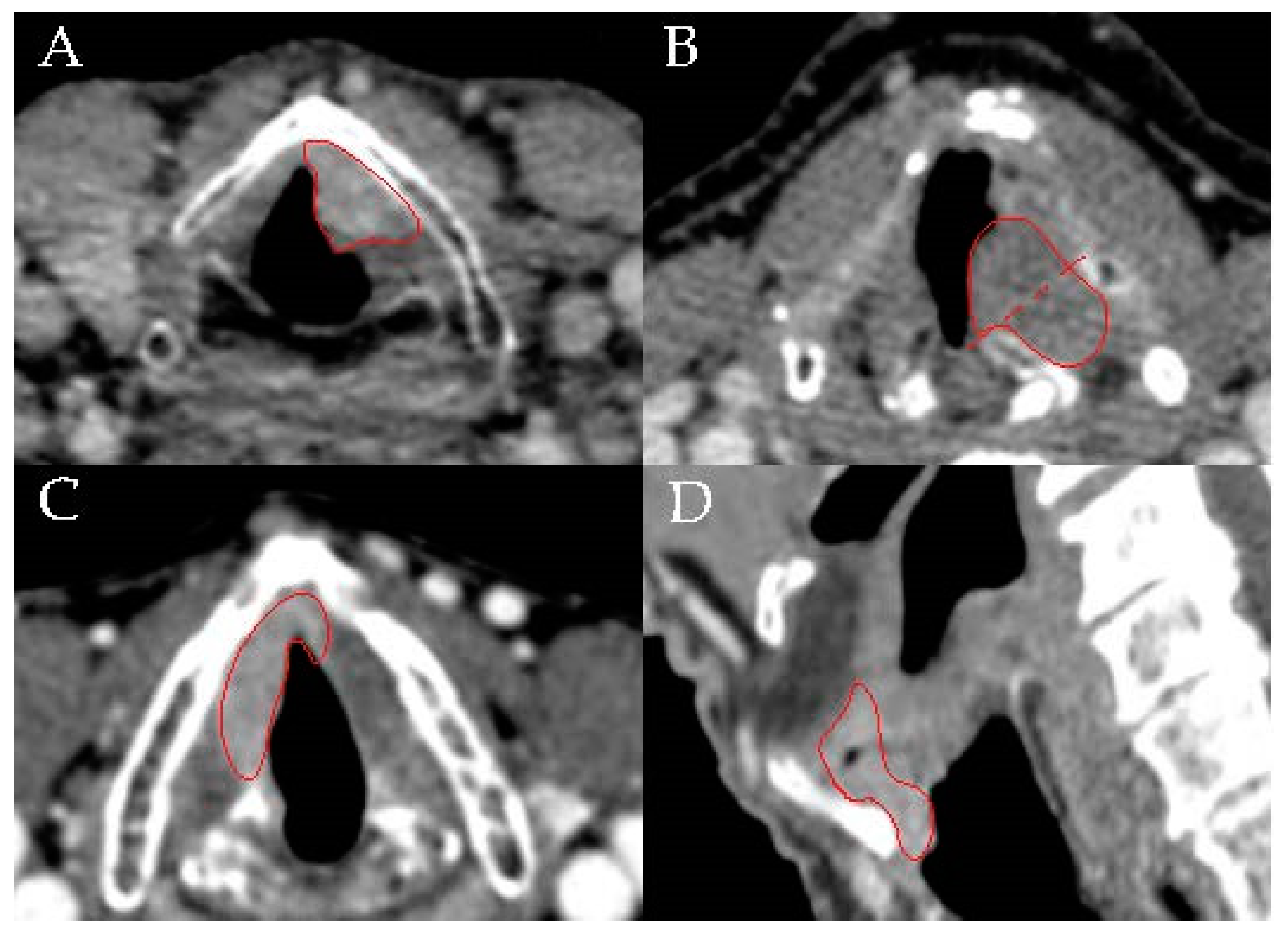
3.2. Radiological Characteristics
3.3. Follow-Up
3.4. Survival and Local Control
3.5. Toxicity
4. Discussion
5. Limitations
6. Conclusions
Author Contributions
Funding
Conflicts of Interest
References
- Nederlandse Kankerregistratie (NKR), IKNL. Available online: https://www.cijfersoverkanker.nl/selecties/Dataset_1/img5b756230c37f1 (accessed on 20 August 2018).
- Sjögren, E.V. Epidemiology of head and neck squamous cell carcinoma. In Prognosis in Head and Neck Cancer, 1st ed.; Taylor and Francis: London, UK, 2006; ISBN 9781135174286. [Google Scholar]
- Nederlandse Werkgroep Hoofd-Halstumoren NWHHT. Larynxcarcinoom Inhoudsopgave; Nederlandse Werkgroep Hoofd-Halstumoren NWHHT: Nieuwegein, The Netherlands, 2010. [Google Scholar]
- Warner, L.; Chudasama, J.; Kelly, C.G.; Loughran, S.; McKenzie, K.; Wight, R.; Dey, P.; Arnold, D.; Wight, R.; MacKenzie, K.; et al. Radiotherapy versus open surgery versus endolaryngeal surgery (with or without laser) for early laryngeal squamous cell cancer. Cochrane Database Syst. Rev. 2014. [Google Scholar] [CrossRef] [PubMed]
- Warner, L.; Lee, K.; Homer, J.J. Transoral laser microsurgery versus radiotherapy for T2 glottic squamous cell carcinoma: A systematic review of local control outcomes. Clin. Otolaryngol. 2017, 42, 629–636. [Google Scholar] [CrossRef] [PubMed]
- Mo, H.L.; Li, J.; Yang, X.; Zhang, F.; Xiong, J.W.; Yang, Z.L.; Tan, J.; Li, B. Transoral laser microsurgery versus radiotherapy for T1 glottic carcinoma: A systematic review and meta-analysis. Lasers Med. Sci. 2017, 32, 461–467. [Google Scholar] [CrossRef] [PubMed]
- Abdurehim, Y.; Hua, Z.; Yasin, Y.; Xukurhan, A.; Imam, I.; Yuqin, F. Transoral laser surgery versus radiotherapy: Systematic review and meta-analysis for treatment options of T1a glottic cancer. Head Neck 2012, 34, 23–33. [Google Scholar] [CrossRef] [PubMed]
- Schrijvers, M.L.; van Riel, E.L.; Langendijk, J.A.; Dikkers, F.G.; Schuuring, E.; van der Wal, J.E.; van der Laan, B.F. Higher laryngeal preservation rate after CO2 laser surgery compared with radiotherapy in T1a glottic laryngeal carcinoma. Head Neck 2009, 31, 759–764. [Google Scholar] [CrossRef]
- Hendriksma, M.; Heijnen, B.J.; Sjögren, E.V. Oncologic and functional outcomes of patients treated with transoral CO2 laser microsurgery or radiotherapy for T2 glottic carcinoma: A systematic review of the literature. Curr. Opin. Otolaryngol. Head Neck Surg. 2018, 26, 84–93. [Google Scholar] [CrossRef]
- Succo, G.; Crosetti, E.; Bertolin, A.; Piazza, C.; Molteni, G.; Cirillo, S.; Petracchini, M.; Tascone, M.; Sprio, A.E.; Berta, G.N.; et al. Treatment for T3 to T4a laryngeal cancer by open partial horizontal laryngectomies: Prognostic impact of different pathologic tumor subcategories. Head Neck 2018, 40, 1897–1908. [Google Scholar] [CrossRef]
- Peretti, G.; Piazza, C.; Mensi, M.C.; Magnoni, L.; Bolzoni, A. Endoscopic treatment of cT2 glottic carcinoma: Prognostic impact of different pT subcategories. Ann. Otol. Rhinol. Laryngol. 2005, 114, 579–586. [Google Scholar] [CrossRef]
- Chen, M.F.; Chang, J.T.; Tsang, N.M.; Liao, C.T.; Chen, W.C. Radiotherapy of early-stage glottic cancer: Analysis of factors affecting prognosis. Ann. Otol. Rhinol. Laryngol. 2003, 112, 904–911. [Google Scholar] [CrossRef]
- Dagan, R.; Morris, C.G.; Bennett, J.A.; Mancuso, A.A.; Amdur, R.J.; Hinerman, R.W.; Mendenhall, W.M. Prognostic significance of paraglottic space invasion in T2N0 glottic carcinoma. Am. J. Clin. Oncol. Cancer Clin. Trials 2007, 30, 186–190. [Google Scholar] [CrossRef]
- Sakata, K.; Oouchi, A.; Nagakura, H.; Akiba, H.; Tamakawa, M.; Koito, K.; Hareyama, M.; Asakura, K.; Satoh, M.; Ohtani, S. Accelerated radiotherapy for T1, 2 glottic carcinoma: Analysis of results with KI-67 index. Int. J. Radiat. Oncol. Biol. Phys. 2000, 47, 81–88. [Google Scholar] [CrossRef]
- Smee, R.; Bridger, G.P.; Williams, J.; Fisher, R. Early glottic carcinoma: Results of treatment by radiotherapy. Australas. Radiol. 2000, 44, 53–59. [Google Scholar] [CrossRef] [PubMed]
- Garden, A.S.; Forster, K.; Wong, P.F.; Morrison, W.H.; Schechter, N.R.; Ang, K.K. Results of radiotherapy for T2N0 glottic carcinoma: Does the “2” stand for twice-daily treatment? Int. J. Radiat. Oncol. Biol. Phys. 2003, 55, 322–328. [Google Scholar] [CrossRef]
- Chera, B.S.; Amdur, R.J.; Morris, C.G.; Kirwan, J.M.; Mendenhall, W.M. T1N0 to T2N0 squamous cell carcinoma of the glottic larynx treated with definitive radiotherapy. Int. J. Radiat. Oncol. Biol. Phys. 2010, 78, 461–466. [Google Scholar] [CrossRef] [PubMed]
- Jones, D.A.; Mendenhall, C.M.; Kirwan, J.; Morris, C.G.; Donnan, A.; Holwerda, S.; Kraus, S.T.; Mann, C.J.; Grant, J.R.; Donnan, B.; et al. Radiation therapy for management of t1-t2 glottic cancer at a private practice. Am. J. Clin. Oncol. 2010, 33, 587–590. [Google Scholar] [CrossRef] [PubMed]
- Tong, C.C.; Au, K.H.; Ngan, R.K.; Cheung, F.Y.; Chow, S.M.; Fu, Y.T.; Au, J.S.; Law, S.C. Definitive radiotherapy for early stage glottic cancer by 6 MV photons. Head Neck Oncol. 2012, 4, 23. [Google Scholar] [CrossRef] [PubMed]
- Hoebers, F.; Rios, E.; Troost, E.; van den Ende, P.; Kross, K.; Lacko, M.; Lalisang, R.; Kremer, B.; De, J.J. Definitive radiation therapy for treatment of laryngeal carcinoma: Impact of local relapse on outcome and implications for treatment strategies. Strahlenther. Onkol. 2013, 189, 834–841. [Google Scholar] [CrossRef] [PubMed]
- Furusaka, T.; Matsuda, H.; Saito, T.; Katsura, Y.; Ikeda, M. Long-term follow-up and salvage surgery in patients with T2N0M0 squamous cell carcinoma of the glottic larynx who received concurrent chemoradiation therapy with carboplatin (CBDCA) AUC 1.5 vs AUC 2.0. Acta Otolaryngol. 2012, 132, 1215–1223. [Google Scholar] [CrossRef] [PubMed]
- Gorphe, P.; Blanchard, P.; Breuskin, I.; Temam, S.; Tao, Y.; Janot, F. Vocal fold mobility as the main prognostic factor of treatment outcomes and survival in stage II squamous cell carcinomas of the glottic larynx. J. Laryngol. Otol. 2015, 129, 903–909. [Google Scholar] [CrossRef]
- Harada, A.; Sasaki, R.; Miyawaki, D.; Yoshida, K.; Nishimura, H.; Ejima, Y.; Kitajima, K.; Saito, M.; Otsuki, N.; Nibu, K. Treatment outcomes of the patients with early glottic cancer treated with initial radiotherapy and salvaged by conservative surgery. Jpn. J. Clin. Oncol. 2015, 45, 248–255. [Google Scholar] [CrossRef]
- Motegi, A.; Kawashima, M.; Arahira, S.; Zenda, S.; Toshima, M.; Onozawa, M.; Hayashi, R.; Akimoto, T. Accelerated radiotherapy for T1 to T2 glottic cancer. Head Neck 2015, 37, 579–584. [Google Scholar] [CrossRef] [PubMed]
- Murakami, R.; Nishimura, R.; Baba, Y.; Furusawa, M.; Ogata, N.; Yumoto, E.; Yamashita, Y. Prognostic factors of glottic carcinomas treated with radiation therapy: Value of the adjacent sign on radiological examinations in the sixth edition of the UICC TNM staging system. Int. J. Radiat. Oncol. Biol. Phys. 2005, 61, 471–475. [Google Scholar] [CrossRef] [PubMed]
- Shor, S.; Krawitz, H.; Macann, A.; West, T.; Morton, R.P.; Mcivor, N.P.; Chaplin, J.; Simcock, P.; Gathercole, J.; Dorman, B.; et al. T1N0/T2N0glottic carcinoma: A comparison of two fractionation schedules. Australas. Radiol. 2006, 50, 152–157. [Google Scholar] [CrossRef] [PubMed]
- Stoeckli, S.J.; Schnieper, I.; Huguenin, P.; Schmid, S. Early glottic carcinoma: Treatment according patient’s preference? Head Neck 2003, 25, 1051–1056. [Google Scholar] [CrossRef] [PubMed]
- Raitiola, H.; Wigren, T.; Pukander, J. Radiotherapy outcome and prognostic factors in early glottic carcinoma. Auris Nasus Larynx 2000, 27, 153–159. [Google Scholar] [CrossRef]
- Khan, M.K.; Koyfman, S.A.; Hunter, G.K.; Reddy, C.A.; Saxton, J.P. Definitive radiotherapy for early (T1-T2) glottic squamous cell carcinoma: A 20 year Cleveland Clinic experience. Radiat. Oncol. 2012, 7, 193. [Google Scholar] [CrossRef] [PubMed]
- Le, Q.T.X.; Fu, K.K.; Kroll, S.; Ryu, J.K.; Quivey, J.M.; Meyler, T.S.; Krieg, R.M.; Phillips, T.L. Influence of fraction size, total dose, and overall time on local control of T1-T2 glottic carcinoma. Int. J. Radiat. Oncol. Biol. Phys. 1997, 39, 115–126. [Google Scholar] [CrossRef]
- Frata, P.; Cellai, E.; Magrini, S.M.; Bonetti, B.; Vitali, E.; Tonoli, S.; Buglione, M.; Paiar, F.; Barca, R.; Fondelli, S.; et al. Radical radiotherapy for early glottic cancer: Results in a series of 1087 patients from two Italian radiation oncology centers. II. The case of T2N0 disease. Int. J. Radiat. Oncol. Biol. Phys. 2005, 63, 1387–1394. [Google Scholar] [CrossRef] [PubMed]
- Eskiizmir, G.; Baskin, Y.; Yalcin, F.; Ellidokuz, H.; Ferris, R.L. Risk factors for radiation failure in early-stage glottic carcinoma: A systematic review and meta-analysis. Oral Oncol. 2016, 62, 90–100. [Google Scholar] [CrossRef] [PubMed]
- Bron, L.P.; Soldati, D.; Zouhair, A.; Ozsahin, M.; Brossard, E.; Monnier, P.; Pasche, P. Treatment of early stage squamous-cell carcinoma of the glottic larynx: Endoscopic surgery or cricohyoidoepiglottopexy versus radiotherapy. Head Neck 2001, 23, 823–829. [Google Scholar] [CrossRef]
- Zouhair, A.; Azria, D.; Coucke, P.; Matzinger, O.; Bron, L.; Moeckli, R.; Do, H.P.; Mirimanoff, R.O.; Ozsahin, M. Decreased local control following radiation therapy alone in early-stage glottic carcinoma with anterior commissure extension. Strahlenther. Onkol. 2004, 180, 84–90. [Google Scholar] [CrossRef] [PubMed]
- Nur, D.A.; Oguz, C.; Kemal, E.T.; Ferhat, E.; Sulen, S.; Emel, A.; Munir, K.; Ann, C.S.; Mehmet, S. Prognostic factors in early glottic carcinoma implications for treatment. Tumori 2005, 91, 182–187. [Google Scholar] [CrossRef] [PubMed]
- Marshak, G.; Brenner, B.; Shvero, J.; Shapira, J.; Ophir, D.; Hochman, I.; Marshak, G.; Sulkes, A.; Rakowsky, E. Prognostic factors for local control of early glottic cancer: The Rabin Medical Center retrospective study on 207 patients. Int. J. Radiat. Oncol. Biol. Phys. 1999, 43, 1009–1013. [Google Scholar] [CrossRef]
- Burke, L.S.; Greven, K.M.; McGuirt, W.T.; Case, D.; Hoen, H.M.; Raben, M. Definitive radiotherapy for early glottic carcinoma: Prognostic factors and implications for treatment. Int. J. Radiat. Oncol. Biol. Phys. 1997, 38, 1001–1006. [Google Scholar] [CrossRef]
- Al-Mamgani, A.; van Rooij, P.H.; Woutersen, D.P.; Mehilal, R.; Tans, L.; Monserez, D.; Baatenburg de Jong, R.J. Radiotherapy for T1-2N0 glottic cancer: A multivariate analysis of predictive factors for the long-term outcome in 1050 patients and a prospective assessment of quality of life and voice handicap index in a subset of 233 patients. Clin. Otolaryngol. 2013, 38, 306–312. [Google Scholar] [CrossRef] [PubMed]
- Bignardi, M.; Antognoni, P.; Sanguineti, G.; Magli, A.; Molteni, M.; Merlotti, A.; Richetti, A.; Tordiglione, M.; Conte, L.; Magno, L. Hyperfractionated radiotherapy for T2N0 glottic carcinoma: A retrospective analysis at 10 years follow-up in a series of 60 consecutive patients. Tumori 2004, 90, 317–323. [Google Scholar] [CrossRef] [PubMed]
- Hendriksma, M.; Sjogren, E.V. Involvement of the Anterior Commissure in Early Glottic Cancer (Tis-T2): A Review of the Literature. Cancers 2019, 11, 1234. [Google Scholar] [CrossRef] [PubMed]
- Piazza, C.; Filauro, M.; Paderno, A.; Marchi, F.; Perotti, P.; Morello, R.; Taboni, S.; Parrinello, G.; Incandela, F.; Iandelli, A.; et al. Three-Dimensional Map of Isoprognostic Zones in Glottic Cancer Treated by Transoral Laser Microsurgery as a Unimodal Treatment Strategy. Front. Oncol. 2018, 8, 175. [Google Scholar] [CrossRef] [PubMed]
- Carta, F.; Bandino, F.; Olla, A.M.; Chuchueva, N.; Gerosa, C.; Puxeddu, R. Prognostic value of age, subglottic, and anterior commissure involvement for early glottic carcinoma treated with CO2 laser transoral microsurgery: A retrospective, single-center cohort study of 261 patients. Eur. Arch. Otorhinolaryngol. 2018, 275, 1199–1210. [Google Scholar] [CrossRef] [PubMed]
- Nonoshita, T.; Shioyama, Y.; Nakamura, K.; Nakashima, T.; Ohga, S.; Yoshitake, T.; Ohnishi, K.; Terashima, K.; Asai, K.; Honda, H. Concurrent chemoradiotherapy with S-1 for T2N0 Glottic squamous cell carcinoma. J. Radiat. Res. 2010, 51, 481–484. [Google Scholar] [CrossRef] [PubMed]
- Kimura, K.; Itoh, Y.; Okada, T.; Nakahara, R.; Kawamura, M.; Kubota, S.; Itoh, J.; Hiramatsu, M.; Fujimoto, Y.; Shibata, T.; et al. Critical evaluation of a prospective study of concurrent chemoradiotherapy with S-1 for early glottic carcinoma. Anticancer Res. 2015, 35, 2385–2390. [Google Scholar]
- Bhateja, P.; Ward, M.C.; Hunter, G.H.; Greskovich, J.F.; Reddy, C.A.; Nwizu, T.I.; Lamarre, E.; Burkey, B.B.; Adelstein, D.J.; Koyfman, S.A. Impaired vocal cord mobility in T2N0 glottic carcinoma: Suboptimal local control with Radiation alone. Head Neck 2016, 38, 1832–1836. [Google Scholar] [CrossRef] [PubMed]
- Kimura, K.; Itoh, Y.; Okada, T.; Kubota, S.; Kawamura, M.; Nakahara, R.; Oie, Y.; Kozai, Y.; Takase, Y.; Tsuzuki, H.; et al. Optimized treatment strategy of radiotherapy for early glottic squamous cell carcinomas: An initial analysis. Nagoya J. Med. Sci. 2017, 79, 331–338. [Google Scholar] [PubMed]
- Saitoh, J.-I.; Shirai, K.; Imaeda, M.; Musha, A.; Abe, T.; Shino, M.; Takayasu, Y.; Takahashi, K.; Chikamatsu, K.; Nakano, T. Concurrent chemoradiotherapy with conventional fractionated radiotherapy and low-dose daily cisplatin plus weekly docetaxel for T2N0 glottic cancer. Radiat. Oncol. 2017, 12. [Google Scholar] [CrossRef] [PubMed]
- Akimoto, T.; Nonaka, T.; Kitamoto, Y.; Ishikawa, H.; Ninomiya, H.; Chikamatsu, K.; Furuya, N.; Hayakawa, K.; Mitsuhashi, N.; Nakano, T. Radiation therapy for T2N0 laryngeal cancer: A retrospective analysis for the impact of concurrent chemotherapy on local control. Int. J. Radiat. Oncol. Biol. Phys. 2006, 64, 995–1001. [Google Scholar] [CrossRef]
- Kitani, Y.; Kubota, A.; Furukawa, M.; Sato, K. Prognostic factors for local control in patients receiving radiation therapy for early glottic cancer: Anterior commissure involvement and effect of chemoradiotherapy. Eur. Arch. Otorhinolaryngol. 2016, 273, 1011–1017. [Google Scholar] [CrossRef]


| Characteristics | No. of Patients (%) | 5-Year Local Control (%) | p-Value |
|---|---|---|---|
| Clinical characteristics (n = 94) | |||
| Involvement AC | 0.597 | ||
| Yes | 61 (64.9) | 68.1 | |
| No | 33 (35.1) | 74.8 | |
| Mobility | 0.438 | ||
| Normal | 76 (80.9) | 69.1 | |
| Impaired | 18 (19.1) | 77.4 | |
| Radiological characteristics (n = 46) | |||
| Tumor infiltration in VM | 0.077 | ||
| Superficial | 16 (34.8) | 92.9 | |
| Deep | 30 (65.2) | 68 | |
| Position relative to M-line | 0.875 | ||
| Anterior | 37 (82.2) | 76.6 | |
| Posterior | 1 (2.2) | 100 | |
| Both | 7 (15.6) | 71.4 | |
| Horizontal involvement AC | |||
| Yes | 13 (28.3) | 59.2 | 0.047 |
| No | 33 (71.7) | 83 | |
| Vertical involvement AC | <0.0001 | ||
| No | 32 (71.1) | 81.8 | |
| Supraglottic | 5 (11.1) | 100 | |
| Subglottic | 4 (8.9) | 75 | |
| Both | 4 (8.9) | 0 | |
| Treatment characteristics (n = 94) | |||
| Type of radiotherapy | 0.277 | ||
| Normal | 79 (84.0) | 71.6 | |
| IMRT | 15 (16.0) | 65.2 | |
| Elective neck irradiation | 0.827 | ||
| Yes | 37 (39.4) | 68.6 | |
| No | 57 (60.6) | 72 | |
| Total dose | 0.965 | ||
| ≤68 Gy | 49 (52.1) | 71.8 | |
| >68 Gy | 45 (47.9) | 69.1 | |
| Fraction size | 0.77 | ||
| ≤2.0 Gy | 84 (89.4) | 71 | |
| >2.0 Gy | 10 (10.6) | 67.5 | |
| Overall treatment time | 0.331 | ||
| ≤42 days | 73 (77.7) | 68.2 | |
| >42 days | 21 (22.3) | 79 | |
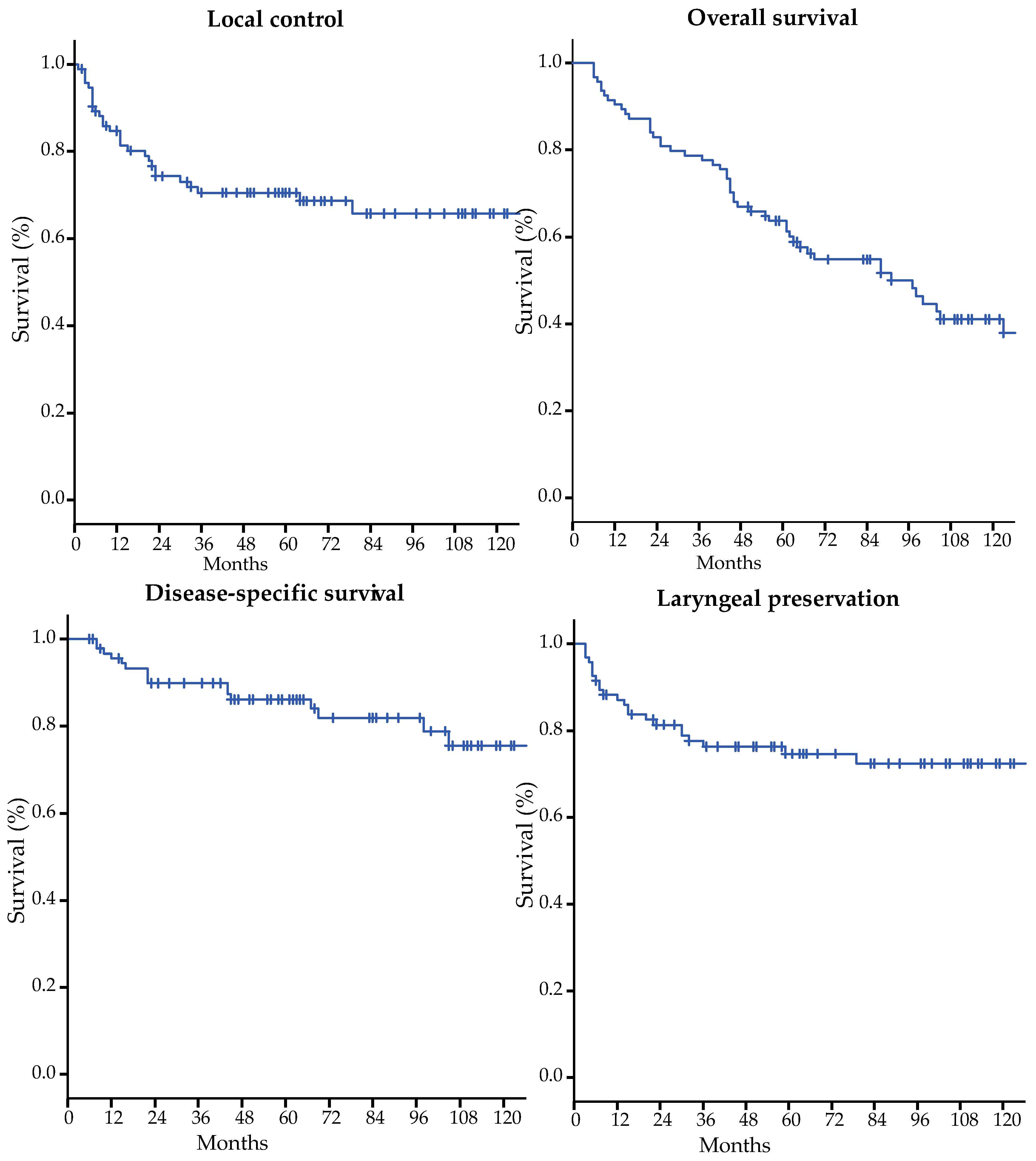
| Outcomes | Five-Year Survival (%) | 10-Year Survival (%) |
|---|---|---|
| Local control | 70.5 | 65.8 |
| Overall survival | 63.7 | 41.0 |
| Disease-specific survival | 86.0 | 75.6 |
| Laryngeal preservation | 74.7 | 72.4 |
© 2019 by the authors. Licensee MDPI, Basel, Switzerland. This article is an open access article distributed under the terms and conditions of the Creative Commons Attribution (CC BY) license (http://creativecommons.org/licenses/by/4.0/).
Share and Cite
Hendriksma, M.; Ruler, M.A.P.v.; Verbist, B.M.; Jong, M.A.d.; Langeveld, T.P.M.; Benthem, P.P.G.v.; Sjögren, E.V. Survival and Prognostic Factors for Outcome after Radiotherapy for T2 Glottic Carcinoma. Cancers 2019, 11, 1319. https://doi.org/10.3390/cancers11091319
Hendriksma M, Ruler MAPv, Verbist BM, Jong MAd, Langeveld TPM, Benthem PPGv, Sjögren EV. Survival and Prognostic Factors for Outcome after Radiotherapy for T2 Glottic Carcinoma. Cancers. 2019; 11(9):1319. https://doi.org/10.3390/cancers11091319
Chicago/Turabian StyleHendriksma, Martine, Marc A.P. van Ruler, Berit M. Verbist, Martin A. de Jong, Ton P.M Langeveld, Peter Paul G. van Benthem, and Elisabeth V. Sjögren. 2019. "Survival and Prognostic Factors for Outcome after Radiotherapy for T2 Glottic Carcinoma" Cancers 11, no. 9: 1319. https://doi.org/10.3390/cancers11091319