Article

Etomidate versus Propofol for Electroconvulsive Therapy in Patients with Major Depressive Disorders in Terms of Clinical Responses to Treatment: A Retrospective Analysis

1
Department of Psychiatry, Seoul National University Bundang Hospital, Seongnam 13620, Republic of Korea
2
Department of Psychiatry, Seoul National University College of Medicine, Seoul 03080, Republic of Korea
3
Department of Anesthesia and Pain Medicine, Seoul National University Bundang Hospital, Seongnam 13620, Republic of Korea
4
Department of Anesthesia and Pain Medicine, Seoul National University College of Medicine, Seoul 03080, Republic of Korea
5
Chung Psychiatric Clinic, Seoul 06614, Republic of Korea
*
Author to whom correspondence should be addressed.
These authors contributed equally to this work.
Brain Sci. 2023, 13(7), 1023; https://doi.org/10.3390/brainsci13071023
Submission received: 9 June 2023 / Revised: 25 June 2023 / Accepted: 30 June 2023 / Published: 3 July 2023
(This article belongs to the Section Psychiatric Diseases)

Abstract

:
General anesthetic agents may be associated with the clinical efficacy of electroconvulsive therapy (ECT), as they may influence seizure quality and duration. Hence, a retrospective study was conducted to compare the clinical effects and seizure variables of etomidate and propofol during ECT. Patients treated with ECT under anesthesia with etomidate (n = 43) or propofol (n = 12) were retrospectively analyzed. Seizure variables (seizure duration, intensity, and threshold) and hemodynamic changes during ECT were assessed and recorded. Clinical responses to treatment were evaluated using the Clinical Global Impression scale and mood at discharge after the course of ECT. Adverse effects were also recorded. The demographic characteristics were similar between the two groups. There were no significant differences in the Clinical Global Impression scale scores, mood at discharge, and adverse effects between the two groups (p > 0.05); however, etomidate was associated with a significantly longer motor (42.0 vs. 23.65 s, p < 0.001) and electroencephalogram (51.8 vs. 33.5 s, p < 0.001) seizure duration than propofol. In conclusion, etomidate showed more favorable seizure profiles than propofol during ECT; however, both agents (etomidate and propofol) were associated with similar clinical efficacy profiles at discharge.

1. Introduction

Electroconvulsive therapy (ECT) artificially induces seizures by delivering electrical stimulation through electrodes attached to the scalp. This procedure is commonly performed to alleviate the symptoms of patients with severe or refractory depressive disorders that are uncontrolled by medications. It is performed under general anesthesia to ensure patient safety and tolerability. Various anesthetic agents, such as etomidate, propofol, ketamine, and thiopental, can be used for ECT anesthesia.
Anesthetic agents may affect seizure quality [1], and in some studies, seizure duration has also been associated with the clinical efficacy of ECT [2,3]. Most anesthetic agents, including propofol, thiopental, and midazolam, have anticonvulsant properties [4,5]; etomidate has been found to reduce the seizure threshold [6] and provide significantly longer seizure duration compared to other anesthetic agents in ECT [7].
Several studies comparing the effects of anesthetic agents during ECT in patients with depressive disorders have focused on seizure-related variables rather than on clinical improvements. We hypothesized that patients administered etomidate during ECT would achieve better clinical outcomes than those administered propofol. Therefore, this retrospective analysis aimed to compare the clinical effects of etomidate and propofol based on seizure variables and hemodynamic responses in patients with depressive disorder undergoing ECT.

2. Materials and Methods

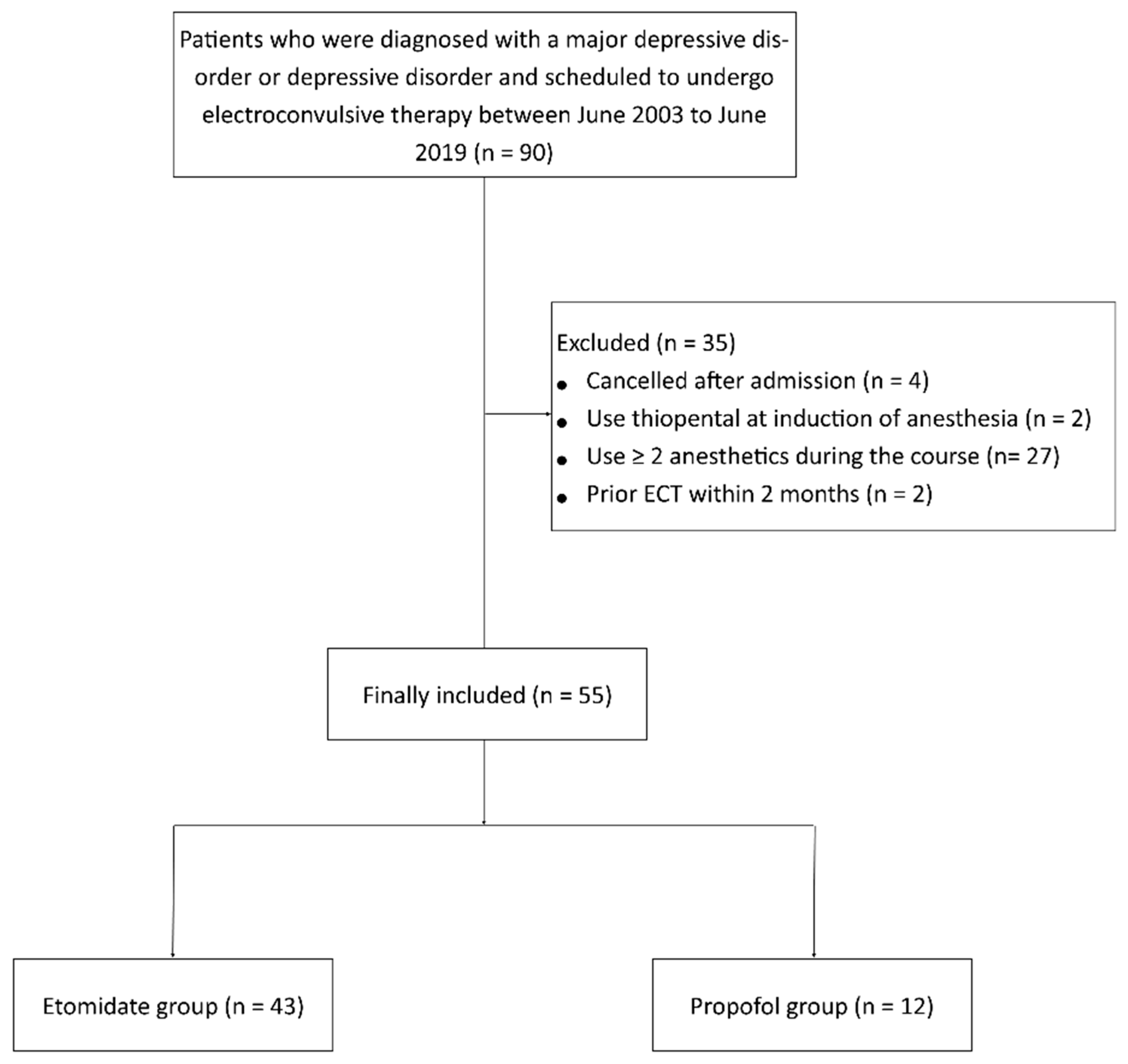
2.1. Study Design and Study Participants

In this retrospective study, patients who were diagnosed with major depressive disorder or depressive disorder and underwent ECT at Seoul National University Bundang Hospital between June 2003 and June 2019 were enrolled. The exclusion criteria were as follows: (1) patients who did not complete the course of ECT or had undergone ECT in the past 2 months; (2) patients who received both etomidate and propofol; (3) and patients who received other anesthetic agents during the course of ECT.

2.2. Anesthesia for ECT

All patients were monitored using pulse oximetry, electrocardiography, and noninvasive arterial pressure measurements throughout the ECT procedures. After premedication with 0.2 mg of glycopyrrolate, anesthesia was induced with 0.2–0.3 mg/kg of etomidate or 1–2 mg/kg of propofol. Succinylcholine (1 mg/kg) was used for neuromuscular blockade. Subsequently, ECT was performed using the Thymatron IV system (Somatics, LLC, Lake Bluff, IL, USA). The stimulus dosage was selected by the attending psychiatrist based on the guidelines of the institution. The stimulus charge starts from 32 mC for female patients and from 48 mC for male patients [8]. If there was no seizure or its duration was shorter than 20 s, an additional stimulation (about 50% increased energy) was adjusted according to the protocol of the guideline. In the subsequent session, the energy of the last stimulation in the previous session was chosen as the dose of the first stimulation.

2.3. Outcomes

This study compared the effects of etomidate and propofol on clinical outcomes using the Clinical Global Impression (CGI) scale. For all patients undergoing ECT, the CGI scale was used by experienced psychiatrists to assess the severity of the illness. The CGI consists of a 7-point Likert scale with scores as follows: 1, normal, not at all ill; 2, borderline mentally ill; 3, mildly ill; 4, moderately ill; 5, markedly ill; 6, severely ill; and 7, extremely ill. CGI analysis was conducted at the time of admission to the hospital (before ECT) and discharge from the hospital (after ECT). The CGI score was determined based on the overall clinical appearance, including symptoms, behaviors, and functional impairments.
Secondary outcomes, including mood at discharge, full or partial remission, length of hospital stay, and adverse events related to ECT, were also analyzed. Adverse events, including amnesia, headache, anxiety, and insomnia, were recorded if any of these occurred.
ECT-related variables (doses and types of anesthetics, number of ECT sessions, and stimulus charges) and seizure-related variables (motor and electroencephalogram (EEG) seizure duration) were recorded. In terms of stimulus charges and seizure durations, the total values were divided by the number of ECT sessions because different numbers of ECT sessions could produce biased results.
Hemodynamic variables during the ECT (heart rate and blood pressure) were recorded. Hemodynamic responses were defined as maximal changes during ECT from baseline hemodynamic parameters, which were measured prior to the induction of anesthesia. The mean values of hemodynamic responses were calculated and compared.

2.4. Statistical Analyses
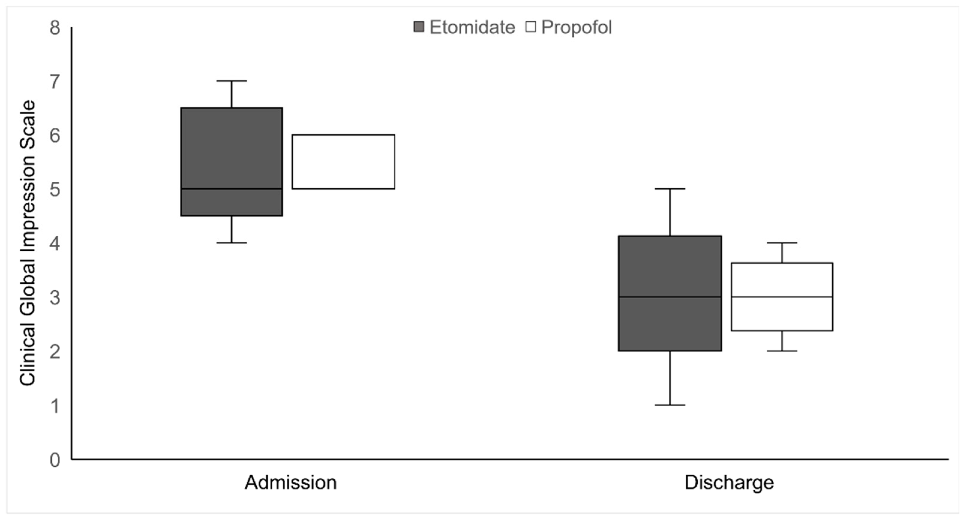
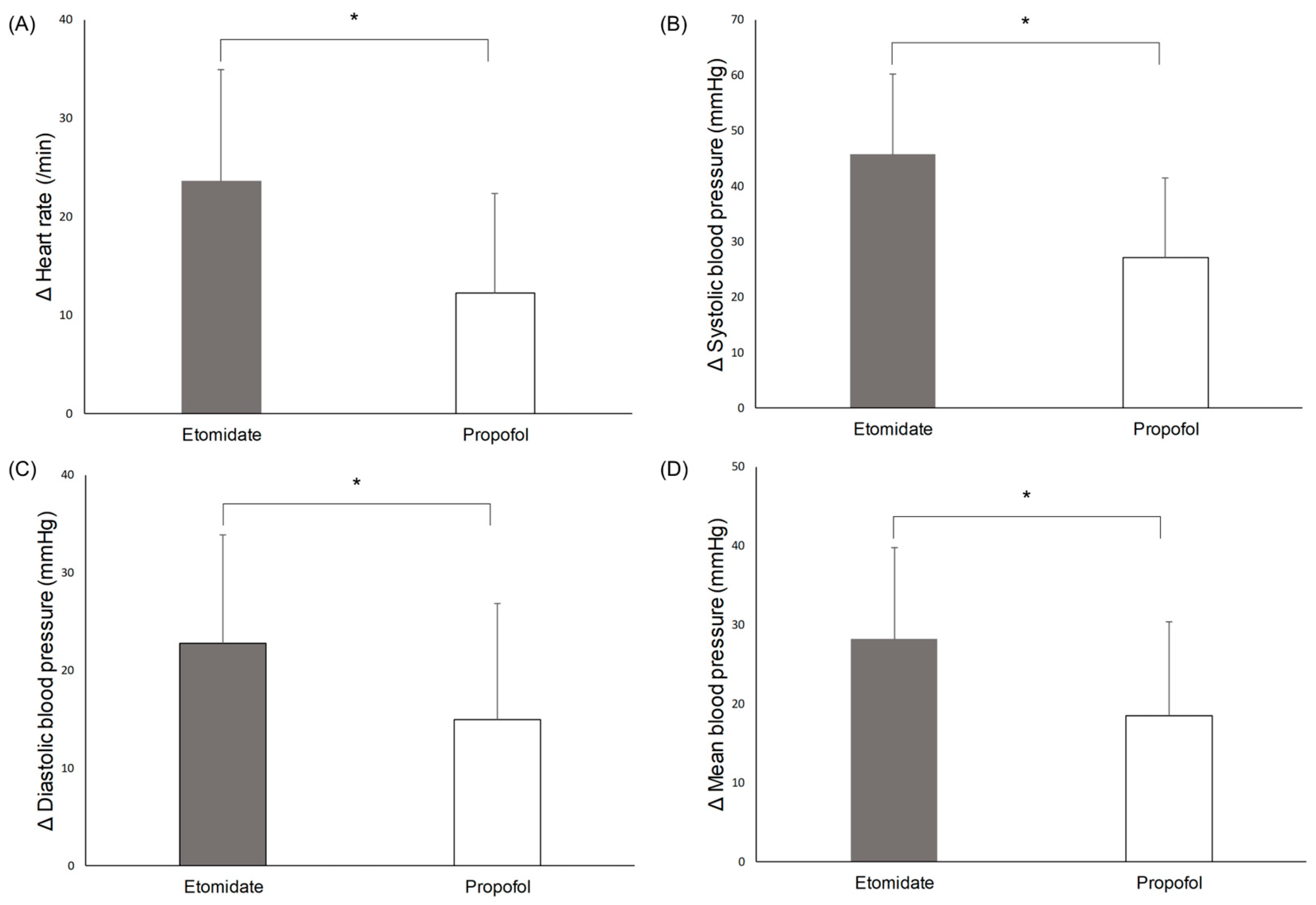
All statistical analyses were performed using the Statistical Package for the Social Sciences (SPSS) software version 25.0 (SPSS Inc., Chicago, IL, USA). Continuous variables are presented as mean with standard deviation or median with interquartile range, depending on the normality of distribution. Student’s t-test or Mann–Whitney U test was used for comparison. Categorical variables are presented as numbers with percentages, and the chi-squared test or Fisher’s exact test was used for comparison. For missing values, mean values were substituted for missing values by using unconditional mean imputation.

3. Results

In total, 90 patients were scheduled to undergo ECT between June 2003 and June 2019. Of these, four patients were excluded from the analysis because they canceled ECT after admission. Two patients who received thiopental during anesthesia induction and 27 patients who received more than two anesthetics during the ECT course were excluded. Two patients who had undergone ECT within the last 2 months were also excluded. Hence, 55 patients were included in the final analysis, of whom 43 were administered etomidate, whereas 12 received propofol during anesthetic induction (Figure 1).
The demographic characteristics and baseline hemodynamic parameters are shown in Table 1. Based on the anesthetic administered, the patients were divided into etomidate and propofol groups.
The CGI scores at admission and discharge are shown in Figure 2. CGI scores at admission were not available for six patients (three in each group); thus, they were excluded from the CGI analysis.
The CGI scores were comparable between the two groups (at admission, 5 [5–6] and 6 [5–6], p = 0.091; at discharge, 3 [3–3.25] and 3 [2.75–3.25], p = 0.352).
The other clinical outcomes at discharge are summarized in Table 2. No significant differences in mood at discharge, remission, and length of hospital stay were found between the two groups. No patient in the etomidate group experienced anxiety, while two in the propofol group experienced anxiety after the ECT (p = 0.044), which was statistically significant.
The ECT parameters, including the number of ECT sessions, seizure duration, and stimulus charges, are described in Table 3. There were some missing data regarding seizure duration (seven patients in the etomidate group and two patients in the propofol group) and stimulus charge (one patient in the etomidate group) because of lack of records. Hence, missing data were imputed using the unconditional mean imputation.
Significant differences were observed in ECT-related parameters between the two groups. Patients in the etomidate group received less sessions of ECT than those in the propofol group (7.6 vs. 10, p = 0.004). A significant difference in seizure duration and stimulus charges between the groups was seen. The motor (42.0 vs. 23.65 s, p < 0.001) and EEG (51.8 vs. 33.5, p < 0.001) seizure durations per session were significantly longer in the etomidate group than in the propofol group. The mean stimulus charge per session was significantly lower in the etomidate group than in the propofol group (211.2 vs. 394.75 mC, p < 0.001).
Significant hemodynamic changes during the procedure were seen in the two groups (Figure 3). The mean increase in heart rate (23.6 ± 11.3 vs. 12.3 ± 10.1 /min, p = 0.003), systolic blood pressure (45.7 ± 14.5 vs. 27.2 ± 14.3 mmHg, p < 0.001), diastolic blood pressure (22.8 ± 11.0 vs. 15 ± 11.8 mmHg, p = 0.036), and mean blood pressure (28.2 ± 11.6 vs. 18.5 ± 11.9 mmHg, p = 0.014) were significantly higher in the etomidate group than in the propofol group.

4. Discussion

This retrospective analysis showed that etomidate demonstrated similar clinical outcomes to propofol, despite more favorable seizure profiles (longer seizure duration and lower stimulus charge) in patients with depressive disorder undergoing ECT. Hemodynamic responses to the ECT stimulus were significantly higher in the etomidate group than in the propofol group.
To the best of our knowledge, this is the first study using the CGI scale to validate the efficacy of ECT in patients with depression. CGI is a quick, simple, and easy tool for experienced physicians to evaluate a patient’s current condition based on an overall assessment of mental status, symptoms, behaviors, and functional impairment [9]. It is a reliable tool for the evaluation of psychiatric disorder [10]. There are two types of CGI: CGI-severity (CGI-S) and CGI-improvement (CGI-I) [11]. CGI-S assesses the current severity of illness, while CGI-I evaluates the degree of change before and after treatment [12]. Both CGI types use a seven-point Likert scale. In our institution, CGI-S was routinely used and recorded when the patients were discharged, but not CGI-I. This can be attributed to patient characteristics and medical systems. Patients were hospitalized for approximately 30 days in this study, but resident physicians in charge of the medical records were rotated every month. Therefore, it seemed difficult to trace and compare the severity of illness during their hospital stay.
No significant difference in the CGI scores on discharge was seen between the two groups. These results are consistent with those of previous studies [13,14]. Eranti et al. investigated the effects of anesthetic agents on the therapeutic response to ECT [13]. The responses were classified as complete recovery, major improvement, minor improvement, no change, and worsening. The choice of anesthetic agents, including etomidate and propofol, has no influence on the response rate after ECT. Graveland et al. [14] analyzed patients with depression undergoing ECT using the Hamilton Rating Scale for Depression (HAM-D) and Montgomery–Asberg Depression Rating Scale (MADRS). No significant differences in remission and response rates and reduction in HAM-D or MADRS scores were found, regardless of the anesthetic agent used. To assess the severity of depression, the authors employed specific tools, the HAM-D and MADRS, which focused on depressive symptoms. In contrast, the CGI used in our study assesses the overall and general conditions [15]. However, CGI shows similar performance to the HAM-D and MADRS [16], although it is a more conservative tool than the HAM-D and MADRS [15].
Seizure durations were significantly longer in the etomidate group compared with the propofol group. This is consistent with previous studies, which reported that seizure duration was prolonged with etomidate compared to propofol [17,18]. Among various anesthetic agents, etomidate induces the longest seizure duration, whereas propofol induces the shortest seizure duration [19]. This concurs well with orders of anesthetic agents reducing seizure threshold and confirms our findings. Etomidate may act as a proconvulsant [20], whereas propofol may act as an anticonvulsant [21]. This maybe the reason why the stimulus charges were significantly lower in the etomidate group than in the propofol group. The findings of this study imply that seizure duration is not correlated with clinical improvement in patients with depression undergoing ECT.
It is worth noting that patients in the etomidate group significantly required less ECT sessions than those in the propofol group. It may imply patients who were administered propofol need more treatment sessions to achieve the comparable clinical improvement compared with those who were administered etomidate. However, these findings need to be interpreted with caution due to relatively smaller number of patients included in the propofol group. The difference might be a statistical bias resulting from the smaller sample size, rather than a true difference.
The increase in hemodynamic parameters, such as heart rate and blood pressure, were more prominent after etomidate administration. This is in agreement with several studies, which concluded that propofol provided hemodynamic stability in comparison to etomidate during ECT procedures [22,23,24]. ECT stimulates the sympathetic nervous system, inducing unstable hemodynamic responses, such as tachycardia and hypertension [25]. Etomidate has a limited effect on cardiovascular function, leading to unstable hemodynamic responses associated with ECT [26]. In contrast, given that cardiovascular depression is seen with propofol, the hemodynamic response to ECT can be attenuated after propofol administration [26].

Limitations

The present study has some limitations. First, since this was a retrospective study, we could not control for all confounding factors that might have affected the clinical outcomes. Second, the selection of anesthetics was not unified during the course of the ECT in several patients. In our institution, anesthetic induction agents are selected at the discretion of anesthesiologists or psychiatrists. Consequently, a quarter of the patients were excluded from the analysis. Furthermore, etomidate was preferred due to its advantage-related seizure duration, which resulted in a difference in sample sizes between the groups. Uneven sample size could potentially lead to an over- or under-interpretation of the data, reducing the reliability of our results. Third, CGI was rated by several psychiatrists; hence, there is a possibility of inter-rater variability in the assessment of the CGI. In addition, CGI was the only scale used to evaluate the patients in this retrospective study. Future studies including additional evaluation scales, such as subjective scales, and the incorporation of metrics, like time to recovery, are needed to ensure a more detailed comparison between the impacts of etomidate and propofol. A large-scale prospective randomized clinical trial should be conducted to evaluate the clinical effects of etomidate use during ECT in patients with major psychiatric disorders.

5. Conclusions

This retrospective study analyzed and compared the clinical outcomes of etomidate and propofol during ECT in patients with major depressive disorders. Etomidate showed clinical outcomes similar to those of propofol, despite having more favorable seizure profiles.

Author Contributions

Conceptualization, J.-H.R. and I.-Y.Y.; Data curation, S.-H.D., I.-Y.Y. and C.-H.K.; Formal analysis, J.-H.R., B.M., I.-Y.Y. and C.-H.K.; Funding acquisition, J.-H.R.; Investigation, J.-H.R.; Methodology, S.-H.D. and B.M.; Supervision, I.-Y.Y. and C.-H.K.; Validation, S.-H.D., B.M., and C.-H.K.; Visualization, J.-H.R. and C.-H.K.; Writing—original draft, I.-Y.Y., J.-H.R. and C.-H.K.; Writing—review and editing, S.-H.D., B.M. and I.-Y.Y. All authors have read and agreed to the published version of the manuscript.

Funding

This research was funded by the Seoul National University Bundang Hospital, grant number 02-2017-0033.

Institutional Review Board Statement

The study was approved by the Institutional Review Board (IRB) of Seoul National University Bundang Hospital (approval no. B-1908-558-107).

Informed Consent Statement

Considering the retrospective design of this study, electronic medical records were reviewed, and the need for informed consent was waived by the IRB.

Data Availability Statement

The datasets generated and analyzed during this study are available on request to the corresponding author on reasonable request.

Conflicts of Interest

The authors declare no conflict of interest.

References

  1. Hoyer, C.; Kranaster, L.; Janke, C.; Sartorius, A. Impact of the anesthetic agents ketamine, etomidate, thiopental, and propofol on seizure parameters and seizure quality in electroconvulsive therapy: A retrospective study. Eur. Arch. Psychiatry Clin. Neurosci. 2014, 264, 255–261. [Google Scholar] [CrossRef] [PubMed]
  2. MacEwan, T. An audit of seizure duration in electroconvulsive therapy. Psychiatr. Bull. 2002, 26, 337–339. [Google Scholar] [CrossRef] [Green Version]
  3. McClintock, S.M.; Choi, J.; Deng, Z.D.; Appelbaum, L.G.; Krystal, A.D.; Lisanby, S.H. Multifactorial determinants of the neurocognitive effects of electroconvulsive therapy. J. ECT 2014, 30, 165–176. [Google Scholar] [CrossRef] [PubMed] [Green Version]
  4. Abdollahi, M.H.; Izadi, A.; Hajiesmaeili, M.R.; Ghanizadeh, A.; Dastjerdi, G.; Hosseini, H.A.; Ghiamat, M.M.; Abbasi, H.R. Effect of etomidate versus thiopental on major depressive disorder in electroconvulsive therapy, a randomized double-blind controlled clinical trial. J. ECT 2012, 28, 10–13. [Google Scholar] [CrossRef] [PubMed]
  5. Reddy, S.D.; Reddy, D.S. Midazolam as an anticonvulsant antidote for organophosphate intoxication--A pharmacotherapeutic appraisal. Epilepsia 2015, 56, 813–821. [Google Scholar] [CrossRef] [Green Version]
  6. Uppal, V.; Dourish, J.; Macfarlane, A. Anaesthesia for electroconvulsive therapy. Contin. Educ. Anaesth. Crit. Care Pain. 2010, 10, 192–196. [Google Scholar] [CrossRef] [Green Version]
  7. Singh, P.M.; Arora, S.; Borle, A.; Varma, P.; Trikha, A.; Goudra, B.G. Evaluation of etomidate for seizure duration in electroconvulsive therapy: A systematic review and meta-analysis. J. ECT 2015, 31, 213–225. [Google Scholar] [CrossRef]
  8. Coffey, C.E.; Lucke, J.; Weiner, R.D.; Krystal, A.D.; Aque, M. Seizure threshold in electroconvulsive therapy: I. Initial seizure threshold. Biol. Psychiatry 1995, 37, 713–720. [Google Scholar] [CrossRef]
  9. Busner, J.; Targum, S.D. The clinical global impressions scale: Applying a research tool in clinical practice. Psychiatry 2007, 4, 28–37. [Google Scholar]
  10. Pinna, F.; Deriu, L.; Diana, E.; Perra, V.; Randaccio, R.P.; Sanna, L.; Tusconi, M.; Carpiniello, B.; Cagliari Recovery Study Group. Clinical Global impression-severity score as a reliable measure for routine evaluation of remission in schizophrenia and schizoaffective disorders. Ann. Gen. Psychiatry 2015, 14, 6. [Google Scholar] [CrossRef] [Green Version]
  11. Kolevzon, A.; Ventola, P.; Keary, C.J.; Heimer, G.; Neul, J.L.; Adera, M.; Jaeger, J. Development of an adapted Clinical Global impression scale for use in Angelman syndrome. J. Neurodev. Disord. 2021, 13, 3. [Google Scholar] [CrossRef]
  12. Forkmann, T.; Scherer, A.; Boecker, M.; Pawelzik, M.; Jostes, R.; Gauggel, S. The Clinical Global impression Scale and the influence of patient or staff perspective on outcome. BMC Psychiatry 2011, 11, 83. [Google Scholar] [CrossRef] [Green Version]
  13. Eranti, S.V.; Mogg, A.J.; Pluck, G.C.; Landau, S.; McLoughlin, D.M. Methohexitone, propofol and etomidate in electroconvulsive therapy for depression: A naturalistic comparison study. J. Affect. Disord. 2009, 113, 165–171. [Google Scholar] [CrossRef]
  14. Graveland, P.E.; Wierdsma, A.I.; van den Broek, W.W.; Birkenhäger, T.K. A retrospective comparison of the effects of propofol and etomidate on stimulus variables and efficacy of electroconvulsive therapy in depressed inpatients. Prog. Neuropsychopharmacol. Biol. Psychiatry 2013, 45, 230–235. [Google Scholar] [CrossRef]
  15. Spielmans, G.I.; McFall, J.P. A comparative meta-analysis of Clinical Global Impressions change in antidepressant trials. J. Nerv. Ment. Dis. 2006, 194, 845–852. [Google Scholar] [CrossRef]
  16. Khan, A.; Khan, S.R.; Shankles, E.B.; Polissar, N.L. Relative sensitivity of the Montgomery-Asberg Depression Rating Scale, the Hamilton Depression rating scale and the Clinical Global Impressions rating scale in antidepressant clinical trials. Int. Clin. Psychopharmacol. 2002, 17, 281–285. [Google Scholar] [CrossRef]
  17. Jindal, S.; Sidhu, G.K.; Kumari, S.; Kamboj, P.; Chauhan, R. Etomidate versus Propofol for Motor Seizure Duration during Modified Electroconvulsive Therapy. Anesth. Essays Res. 2020, 14, 62–67. [Google Scholar]
  18. Gurel, S.C.; Ozden, H.C.; Karahan, S.; Ayhan, Y. The superiority of ketofol and etomidate against propofol or thiopental anesthesia for ECT. Asian J Psychiatr. 2022, 72, 103090. [Google Scholar] [CrossRef]
  19. Kadiyala, P.K.; Kadiyala, L.D. Anaesthesia for electroconvulsive therapy: An overview with an update on its role in potentiating electroconvulsive therapy. Indian J. Anaesth. 2017, 61, 373–380. [Google Scholar] [CrossRef]
  20. Voss, L.J.; Sleigh, J.W.; Barnard, J.P.M.; Kirsch, H.E. The howling cortex: Seizures and general anesthetic drugs. Anesth. Analg. 2008, 107, 1689–1703. [Google Scholar] [CrossRef] [Green Version]
  21. Baker, M.T. The anticonvulsant effects of propofol and a propofol analog, 2,6-diisopropyl-4-(1-hydroxy-2,2,2-trifluoroethyl) phenol, in a 6-Hz partial seizure model. Anesth. Analg. 2011, 112, 340–344. [Google Scholar] [CrossRef] [PubMed]
  22. Erdil, F.; Demirbilek, S.; Begec, Z.; Ozturk, E.; Ersoy, M.O. Effects of propofol or etomidate on QT interval during electroconvulsive therapy. J. ECT 2009, 25, 174–177. [Google Scholar] [CrossRef] [PubMed]
  23. Gazdag, G.; Kocsis, N.; Tolna, J.; Iványi, Z. Etomidate versus propofol for electroconvulsive therapy in patients with schizophrenia. J. ECT 2004, 20, 225–229. [Google Scholar] [CrossRef] [PubMed]
  24. Mehta, D.; Palta, S.; Gupta, N.; Saroa, R. Comparison of effect of etomidate with propofol on hemodynamics during modified electroconvulsive therapy. J. Anaesthesiol. Clin. Pharmacol. 2022, 38, 104–110. [Google Scholar]
  25. Ding, Z.; White, P.F. Anesthesia for electroconvulsive therapy. Anesth. Analg. 2002, 94, 1351–1364. [Google Scholar] [CrossRef]
  26. Su, P.; Pan, J.Z. Anesthesia for Electroconvulsive Therapy. In Anesthesiology: A Practical Approach; Goudra, B.G., Ed.; Springer International Publishing: Cham/Geneva, Switzerland, 2018; pp. 229–238. [Google Scholar] [CrossRef]
Figure 1. Flow diagram of included and excluded patients.
Brainsci 13 01023 g001
Figure 2. Clinical Global Impression scale scores of the two groups at admission and discharge.
Brainsci 13 01023 g002
Figure 3. Hemodynamic changes during electroconvulsive therapy: (A) heart rate, (B) systolic blood pressure, (C) diastolic blood pressure, (D) mean blood pressure. * p < 0.05.
Brainsci 13 01023 g003
Table 1. Demographic data.
VariablesEtomidate (n = 43)Propofol (n = 12)p Value
Age, years66.2 ± 11.563.8 ± 10.40.516
Female sex, n (%)32 (74.4%)10 (83.3%)0.709
BMI, kg/m223.4 ± 3.621.7 ± 2.30.063
Drug treatment
SSRI, n (%)22 (51.2%)11 (91.7%)0.018 *
SNRI, n (%)17 (39.5%)5 (41.7%)0.894
TCA, n (%)15 (34.9%)6 (50.0%)0.341
NDRI, n (%)6 (14.0%)1 (8.3%)1.000
BDZ, n (%)34 (79.1%)11 (91.7%)0.430
Antipsychotics, n (%)31 (72.1%)10 (83.3 %)0.709
Baseline hemodynamic parameter
Heart rate, /min90.4 ± 10.493.7 ± 10.20.325
Systolic blood pressure, mmHg136.7 ± 12.8138.0 ± 12.20.863
Diastolic blood pressure, mmHg81.4 ± 9.779.7 ± 7.10.580
Mean blood pressure, mmHg96.2 ± 9.697.2 ± 8.30.740
The data are presented as mean ± standard deviation or number (percentage). Abbreviations: BMI = body mass index; SSRI = selective serotonin reuptake inhibitor; SNRI = serotonin–norepinephrine reuptake inhibitor; TCA = tricyclic antidepressant; NDRI = norepinephrine–dopamine reuptake inhibitor; BDZ = benzodiazepine. * p < 0.05.
Table 2. Clinical outcomes at discharge.
Etomidate (n = 43)Propofol (n = 12)p Value
Mood 0.530
Euthymic28 (65.1%)10 (83.3%)
Mild depressive12 (27.9%)2 (16.7%)
Moderate depressive3 (7.0%)0
Remission 0.199
Full remission6 (14.0%)4 (33.3%)
Partial remission37 (86.0%)8 (66.7%)
Length of hospital stay30 [24, 39]37 [23.5, 50.5]0.380
Side effect
Amnesia13 (30.2%)4 (33.3%)1.000
Headache1 (2.3%)1 (8.3%)0.392
Anxiety02 (16.7%)0.044 *
Insomnia01 (8.3%)0.218
The data are presented as number (%) or median [interquartile range]. * p < 0.05.
Table 3. ECT parameters.
VariablesEtomidate (n = 43)Propofol (n = 12)p Value
Number of ECT 7.6 ± 2.210.0 ± 3.20.004 *
Mean motor seizure (s)42.0 [36.8, 51.4] 23.65 [22.65, 28.5]<0.001 *
Mean EEG seizure (s)51.8 [44.15, 56.2]33.5 [31.3, 37.825]<0.001 *
Mean stimulus charge (mC)211.2 [165.4, 286.65]394.75 [293.5, 660.8]0.001 *
The data are presented as number (%) or median [interquartile range]. * p < 0.05
Disclaimer/Publisher’s Note: The statements, opinions and data contained in all publications are solely those of the individual author(s) and contributor(s) and not of MDPI and/or the editor(s). MDPI and/or the editor(s) disclaim responsibility for any injury to people or property resulting from any ideas, methods, instructions or products referred to in the content.

