Recent Prospects of Carbonaceous Nanomaterials-Based Laccase Biosensor for Electrochemical Detection of Phenolic Compounds


1
Department of Applied Chemistry, Delhi Technological University, Delhi 110042, India
2
Department of Chemistry, Faculty of Science, SGT University, Gurugram 122505, India
*
Authors to whom correspondence should be addressed.
Biosensors 2023, 13(3), 305; https://doi.org/10.3390/bios13030305
Submission received: 14 January 2023
/
Revised: 10 February 2023
/
Accepted: 16 February 2023
/
Published: 22 February 2023
(This article belongs to the Special Issue Nanomaterial-Based Biosensors and Their Applications)
Abstract
:Phenolic compounds (PhCs) are ubiquitously distributed phytochemicals found in many plants, body fluids, food items, medicines, pesticides, dyes, etc. Many PhCs are priority pollutants that are highly toxic, teratogenic, and carcinogenic. Some of these are present in body fluids and affect metabolism, while others possess numerous bioactive properties such as retaining antioxidant and antimicrobial activity in plants and food products. Therefore, there is an urgency for developing an effective, rapid, sensitive, and reliable tool for the analysis of these PhCs to address their environmental and health concern. In this context, carbonaceous nanomaterials have emerged as a promising material for the fabrication of electrochemical biosensors as they provide remarkable characteristics such as lightweight, high surface: volume, excellent conductivity, extraordinary tensile strength, and biocompatibility. This review outlines the current status of the applications of carbonaceous nanomaterials (CNTs, graphene, etc.) based enzymatic electrochemical biosensors for the detection of PhCs. Efforts have also been made to discuss the mechanism of action of the laccase enzyme for the detection of PhCs. The limitations, advanced emerging carbon-based material, current state of artificial intelligence in PhCs detection, and future scopes have also been summarized.
1. Introduction
Phenolic compounds (PhCs) are extensively used in the production of dyes, chemical analysis, disinfectants, synthesis of artificial resins, petrochemicals, pharmaceuticals, textiles, paints, and medical industries [1,2]. These PhCs are present in body fluids, and certain PhCs have potential antioxidant properties [3]. A diverse variety of PhCs display varied impacts on biotic and abiotic components.
PhCs used for industrial purposes and a few released from the body (e.g., estrogen) are ubiquitous pollutants that are toxic even at low concentrations. The level of PhCs toxicity is in the range of 9–25 mg L−1, while the water purity standards set by the US EPA allow a threshold value of approximately 1 ppb of phenol in the case of surface water [1]. The European Commission has prescribed a 0.001 mg L−1 concentration limit of PhCs in natural water, whereas the Central Pollution Control Board of India has restricted 1.0 mg L−1 of a phenolic compound as the benchmark for discharge of water to inland surfaces under the Environment (Protection) Rules, 1986. When discharged into the environment without prior treatment, these compounds lead to severe complications and long-term health issues for humans, animals, and marine systems [4]. Drinking water containing PhCs may cause diseases such as indigestion, skin burns, muscle tremor, liver problems and kidney damage in humans, and death of fish in aquatic regions [2]. In contrast, some other PhCs present in food and body fluid are responsible for preventing disease by balancing blood pressure and stress, reducing cardiovascular diseases, and providing healthy antioxidants [5,6]. The effects and sources of widely used PhCs have been summarized in Table 1.
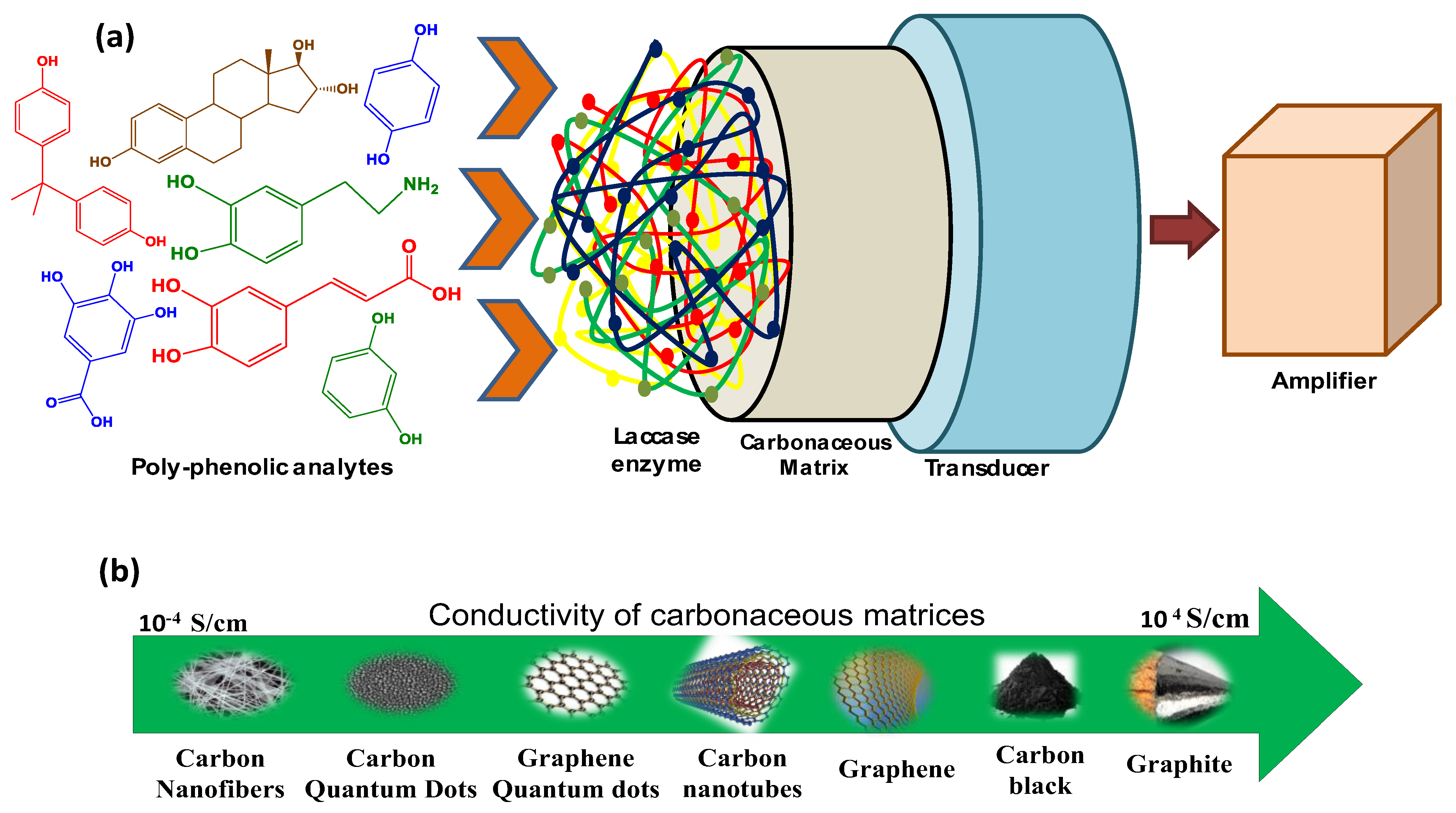
Various chromatographic and spectroscopic techniques have been used to detect and quantify PhCs. These laboratory techniques require tedious process of sample pretreatment from water bodies, which are time-consuming and costly [7]. For rapid and efficient detection, it is necessary to develop portable and more sensitive on-site tools comprising resourceful materials to reduce the devastating effects caused by PhCs. In this context, biosensors can be considered the most reliable techniques for on-site and specific detection of PhCs [8] (Figure 1a).

Table 1.
Harmful effects and sources of widely used PhCs on humans and animals.
| S.No. | PhCs | Effects | Sources | Ref. |
|---|---|---|---|---|
| 1. | Catechol |
|
| [9] |
| 2. | Bisphenol A |
|
| [10] |
| 3. | Caffeic and dihydrocaffeic acids |
|
| [11] |
| 4. | Chlorophenol |
|
| [12] |
| 5. | Hydroquinone |
|
| [13] |
| 6. | Para-cresol |
|
| [14] |
| 7. | Estrogen |
|
| [15] |
| 8. | Dopamine |
|
| [16] |
| 9. | Galliac acid |
|
| [17] |
The advantages of using biosensors include high sensitivity, accuracy, short response time, reliability, longer shelf life, and user-friendliness [7,18,19,20]. Biosensors can detect a very low amount of contaminant even from a composite medium, (wastewater) and are considered reliable for quantifying the total phenolic content in food by checking antioxidant properties of the foodstuff [21,22]. Several bio-recognition components have been used to detect polyphenols, such as microorganisms [23], DNA [24], whole cells [18], anti-bodies, and enzymes [25]. For monitoring PhCs, enzymatic biosensors based on laccase (Lac) [26], horseradish peroxidase [27], and tyrosinase [28,29] have proven to be the most effective due to their ability to directly catalyze electron transfer reactions without introducing cofactors into the reaction medium. Different supporting matrices such as conducting polymers, metal and metal oxide nanoparticles, carbon nano tubes (CNTs), graphite, silica gel, activated charcoal, reduced graphene oxide (RGO), glass surfaces, etc., have been used for the immobilization of these enzymes [30]. The comparative conductivities of such materials have been shown in Figure 1b.
In the current review article, we discuss:
- The recent progress in carbon-based nanomaterials (CNMs)-based Lac biosensors;
- The structure, mechanism of action, and immobilization methods of the Lac enzyme on CNMs;
- The application of CNMs-based Lac biosensors for the detection of PhCs present in food and body fluids;
- The limitations of highly utilized graphitic materials and challenges.
Recently, few articles have focused on discussing the importance of Lac enzymes for many applications. However, to the best of our knowledge, none of them focus on the application of CNMs, which can resolve the major challenge in the development of Lac-based biosensors for the detection of PhCs.
2. Transduction Principle for Monitoring PhCs
The biosensor has emerged as an interesting analytical approach for the detection of PhCs. It mainly consists of two components, including biological entities (analyte and receptor) and a transducer [31]. Enzyme-based sensors are prominently explored due to their ease of selectivity, sensitivity, reliability, and usage [32,33]. In this context, Lac has been considered a greener bio-recognition component supported on modified conducting carbon-based matrices for the detection of PhCs. An enzymatic biosensor produces signals targeted to the analyte’s concentration by combining an enzyme with the transducer, and the transducer converts the signal received into a quantifiable response [34]. Amperometric biosensors aim to electrochemically convert non-active analytes into products by catalysis depending on the enzyme system that performs oxidation and reduction at the surface of the working electrode. Das et al. [35] fabricated Lac-based amperometric biosensor based on an osmium tetroxide/poly 4-vinyl pyridine/multi-walled carbon nanotube (MWCNT)/Nafion/carbon black/GCE electrode for pyrocatechol detection in environmental samples. Kavetskyy et.al. [36] investigated an electroconductive immobilized matrix based on microporous carbon fibers and the Lac enzyme using the amperometric transduction principle. A successive assessment of biosensors was performed by detecting industrial pollutant catechol from actual communal wastewater samples. There is a limited number of optical biosensors reported in the literature that use CNMs for the detection of PhCs, one of which comprises carbon quantum dots (C-QDs) and the Lac enzyme [37]. An optical biosensor based on a sol-gel immobilized Lac was also developed for the detection of three isomeric PhCs (catechol, resorcinol, and hydroquinone) in real and tap water samples [38].
2.1. Electron Transfer and Reaction Mechanism in Lac
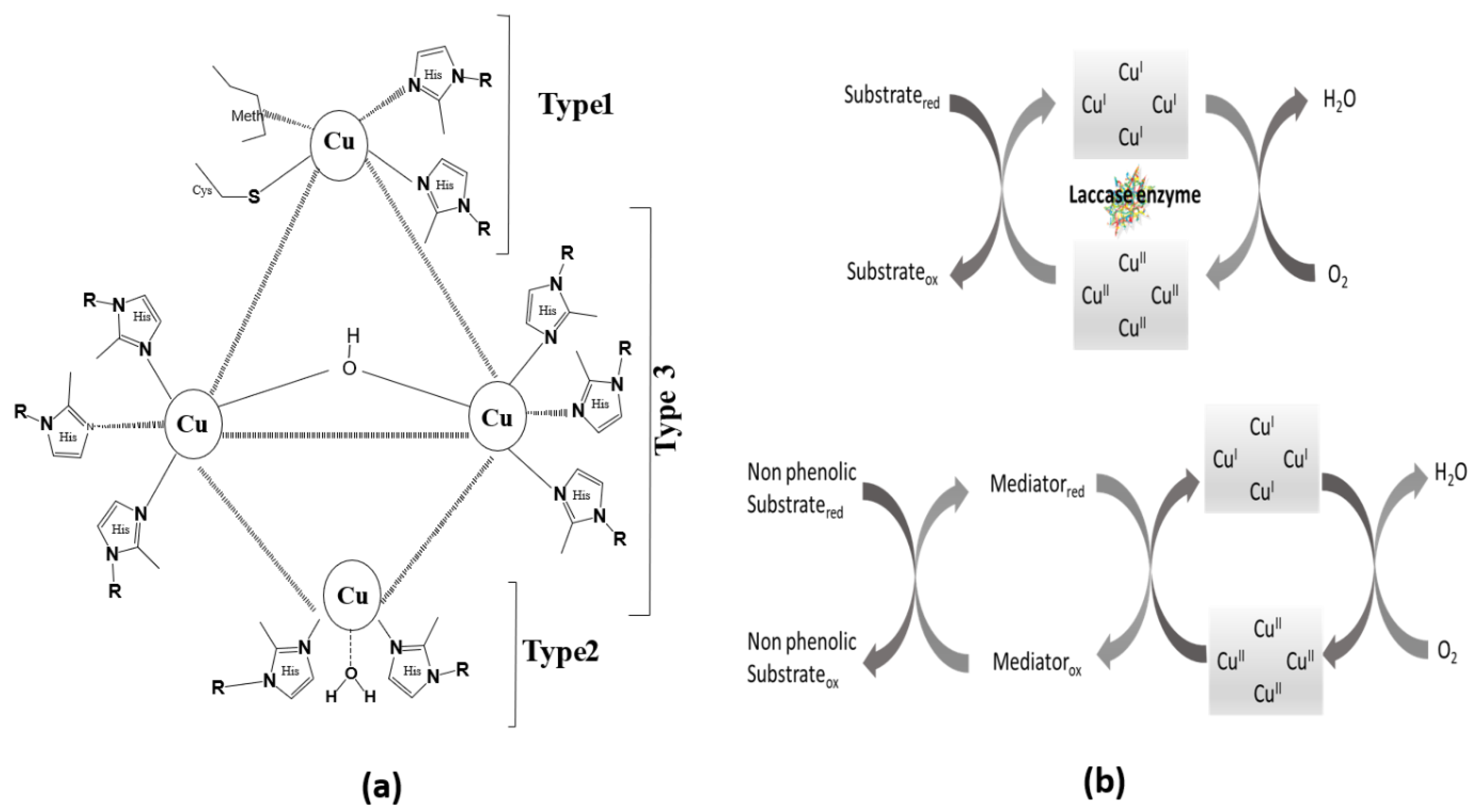
Lac is the largest subgroup of multi-copper oxidases, comprising four copper atoms in different oxidation states, as shown in Figure 2a. Lac has the capability to catalytically oxidize a variety of PhCs in the presence of molecular oxygen by performing a four-electron reduction of oxygen to water. The substrate undergoes reduction by the Lac enzyme conjoining its four-electron oxidation leading to the reductive cleavage of molecular oxygen (O=O) bonds by four electrons along with four Cu atoms. The Cu atoms forming the core of Lac are sub-divided into three types, based on the electron paramagnetic resonance (EPR) spectroscopic technique in the literature [39,40]:
- Type 1 (Cu C1) has trigonal coordination with one sulfur atom of cysteine (cys) and two nitrogen atoms of histidine imidazole units. The fourth coordination is with S of methionine attached axially and far more than the other three. Therefore, the structure seems distorted in the form of tetrahedral geometry with triagonal elongation [41]. It possesses intense blue color resulting from the strong electronic absorbance at 600 nm (charge transfer: S of cysteine to Cu, ε = 5000 M−1 cm−1) in UV/visible spectroscopy, and its paramagnetic nature has been confirmed using EPR spectroscopy.
- Type 2 (Cu C2) has been found to be coordinated with two nitrogen atoms of histidine units and one water ligand. Although it is colorless, EPR studies have revealed its paramagnetic nature via ultrafine splitting.
- Type 3 (2Cu C3) comprises two anti-ferromagnetically coupled Cu atoms each tetragonally coordinated with three nitrogen atoms of histidine units and one bridged hydroxide group. The bridged hydroxide leads to electron-paired Cu sites. The oxidized form shows weak absorbance under UV-visible spectrum having a shoulder at around 330 nm (charge transfer: OH to Cu), and EPR studies show no signal signifying their diamagnetic nature.
The histidine units are, therefore, coordinated in a 2:2:6 ratio for Cu C1, Cu C2, and 2Cu C3, respectively. The bonds connecting Cu and N of histidine are slightly different in bond lengths. The approximate Cu–N bond length for all categories of Cu is 2 Å A֯, some Cu–N bonds are of greater bond length, and some are found to be slightly smaller. The distance between Cu C1 and binuclear Cu atoms (2Cu C3) is found to be around 12 Å, whereas, in the trinuclear Cu cluster, the distance between Cu C2 and binuclear Cu atoms (2Cu C3) is observed at about 4 Å.
Figure 2.
Scheme showing the (a) assembly of the Lac enzyme, adapted from ref. [39] and (b) the activity of the Lac enzyme for a phenolic and non-phenolic substrate [42].

The Electron Paramagnetic Resonance (EPR) of Lac have been observed to be perturbed in the presence of strong anion binding, leading to the disturbance of the EPR signal intensity of Cu C2 and in the anti-ferromagnetic coupling of 2Cu C3. At the same time, the electron reduction of molecular oxygen and discharge of water takes place at a tri-nuclear cluster of Cu C2 and 2Cu C3 atoms. Based on the physical and chemical properties of oxidoreductase Lac, the positioning of copper atoms has different well-defined redox potentials (E0). Depending upon the chemical nature, the Lac enzymes obtained from basidiomycetes (mostly white rot fungi) are considered to have high redox potential, whereas low redox potential is found to be possessed by Lac obtained from bacterial and plant sources [40]. Other biotic sources of Lac enzymes have been listed in Table S1. However, the physical structure shows that Cu C1 and 2Cu C3 have variable low (+E0 = 0.4–0.5 V) and high (E0 = 0.7–0.8 V) redox potentials with respect to standard hydrogen electrodes. The low and high redox potential of Cu C2 appears at approximately 0.4 V [43].
Biosensors based on tyrosinase, horseradish peroxidase, Lac, and polyphenol oxidase enzymes have been broadly used for the detection of different PhCs. However, Lac possesses higher stability and better catalytic ability for electron transfer reactions [44]. Lac-based biosensors are modest in assembling over other reported enzymatic biosensor because they do not necessarily involve hydrogen peroxide as one of the co-substrates or some other co-factors for the catalytic process.
2.2. Activity of the Lac Enzyme
The catalytic activity of Lac depends on the copper (Cu) atoms. All the Cu sites show different functions. Cu C1 is the primary electron acceptor from the reduced substrate. The high redox potential (790 mV) of Cu C1 is the reason for substrate oxidation at its site [39]. Further, the electron received by Cu C1 is delivered via Cys-His pathways to the trinuclear Cu cluster that shows an inter-electron transfer mechanism, which takes place between Cu C1 and 2Cu C3 positions. Due to this transfer of an electron from one Cu site to another, the step where the oxygen molecule is reduced to water by four electrons replicates. It has been reported that H2O2 was not detected during the steady state [45]. At the same site, an oxidized form of the enzyme has been generated. During this aerobic oxidation process, 2Cu C3 accepts two electrons in the presence of Cu C2. The presence of Cu C2 is found to be necessary for the acceptance of electrons by 2Cu C3. In short, the Lac enzyme is oxidized by reducing oxygen to water followed by the oxidation of the substrate leading towards the terminus of the catalytic mechanism by reducing the oxidized Lac enzyme to Lac again.
The Lac enzyme does not possess the ability to oxidize non-phenolic substrates directly. To increase the range of compounds that can be oxidized using the Lac enzyme, several Lac-based mediator compounds have been reported. These Lac-based mediator compounds consider the substrate as an intermediate, exhibiting higher redox potential and indirectly oxidizing non-phenolic substrates [43] (Figure 2b). The mediators enhance the catalytic performance of the Lac enzyme, acting as a co-catalyst. The catalytic mechanism for non-phenolic compounds initiates with the conversion of Lac into oxidized Lac on the same side, leading to the reduction of oxygen to release water. The mediator works by oxidizing the substrate and reducing itself to the original mediator, indicating completion of the co-catalytic mechanism [42]. A standard mediator must be low in molecular weight to avoid hindrance due to size and should possess a high redox potential to enhance the catalytic oxidation process. An efficient mediator must perform several consecutive cycles deprived of deterioration. For example, (2,2′-azino-bis(3-ethylbenzothiazoline-6-sulfonic acid)) is one of the most frequently used mediator for oxidation of non-phenolic compounds [43].
Enzymes undergo specific interactions with certain chemical carriers and substrates to undergo genetic and chemical modifications to increase their catalytic activity. In this context, Zhang et.al. reported that flower-like Cu3(PO4)2.3H2O nanocrystals integrated with Lac shows 6 times activation in the activity of the Lac enzyme [46]. Whereas, Wu et al. found a 1.5 to 4 times increase in the activity of fungal Lac after its pre-incubation with different organic solvents such as methanol, acetone, dimethylformamide, and dimethyl sulfoxide [47]. On the other hand, the influence on enzymatic activity has also been studied based on mutagenesis focusing on the residue at certain position and pH. Another method to enhance activity is through the use of multienzyme, which involves channeling different substrates, kinetics matching, and 3-D distribution of the involved enzymes. These methods can be utilized to engineer the best enzyme–substrate relationship to provide superior catalytic performance [46].
2.3. Immobilization Matrix
In addition to the several merits of the Lac enzyme, the use of the Lac enzyme for biosensor application has not been explored much because of the denaturation of the enzyme by the external environment. In order to increase the stability, lifetime, efficiency, and reusability, and to equilibrate the cost of developed biosensors for detection of PhCs, the efficient immobilization of the Lac enzyme onto suitable matrices is very important [48]. Immobilization provides a way to increase the ability of enzymes by reducing the cost of production [49], leading to better stability of the biosensor [50], strategies for reproducibility, ease of recovery [49], etc. Moreover, the involvement of new bonding, entrapment, and crosslinking also enhances the shelf life of enzymes [51,52]. Immobilization maintains the structural stability and functional possessions of the immobilized enzyme by proper attachment to the surface of the appropriate matrix so that the required activity of the enzyme can be retained throughout the repetitive use of sensor [53,54]. Immobilization methods vary from one enzyme to another and depend on the type of application for which the enzymes are being used. An ideal matrix must be physically rigid, chemically inert, and insoluble in enzymes to preserve its catalytic properties. The immobilization matrix should be thermally and photo-chemically stable, showing effective charge transfer capability. Furthermore, there must be permissible diffusion of a bio-catalytic reaction between the substrate and matrices on which the enzyme has to be immobilized [55].
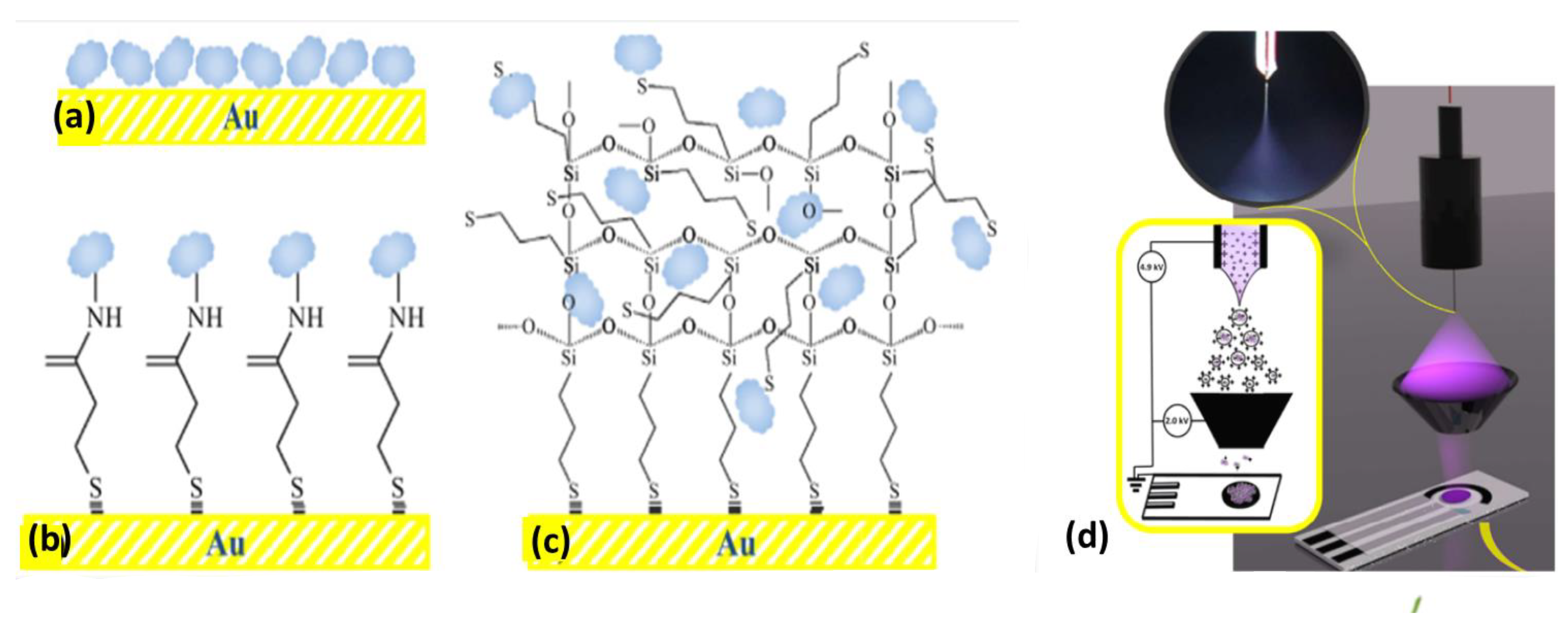
Effective immobilization also helps to restrict the gross movement of biomolecules, thus leading to the fabrication of a stable and accurate biosensor. The different immobilization techniques include (i) covalent bonding, (ii) adsorption, (iii) cross-linking, (iv) entrapment, and (v) encapsulation (Figure 3a–c) [56]. One more method, i.e., the electrospray deposition technique, has also been reported for Lac immobilization on carbon black-nanomodified screen-printed electrodes, as shown in Figure 3d [57]. Several physical and chemical factors, such as pH, temperature, solubility, concentration, etc., influence the immobilization strategies. Large numbers of organic and inorganic matrices have been used for immobilizing the Lac enzyme. In particular, conducting polymers [58], metal nanoparticles [59], metal oxides [60], silica [61,62], clay [63], CNMs, metal–organic frameworks [64], etc. Conducting polymers (CPs) have been widely explored for the fabrication of Lac-based biosensors because of their good compatibility with enzymes and electron conduction ability. In the case of CPs, conductivity can also be varied by hosting doping materials. Calitri et al. [65] evaluated the total phenolic content using CPs poly(aniline-co-2-aminobenzylamine) supported on MWCNT by immobilizing Lac. Yaropolov et al. [66] fabricated a Lac biosensor using three CPs electrodes, viz., Nafion, poly(ethyleneimine), and Eastman AQ 29D supported with GCE for amperometric determination of PhCs. The synthesis of polydopamine nanoparticles based on a bacterial cellulose composite was also accomplished as a compatible matrix for Lac immobilization. Meso-porous silica sieve matrices possess compatibility and uniformity with respect to well-ordered pore structure, size, density, and surface characteristics. Amperometric detection of catechol has been reported where magnesium-doped mesoporous silica sieve–polyvinyl alcohol composite has been used as a matrix [67]. Li et al. constructed a disposable biosensor using silica sphere on the surface of MWCNT with a screen printed electrode (SPE) for immobilizing Lac and detecting dopamine [68]. The major problem with this sensor is the film formation ability and lower conductivity.
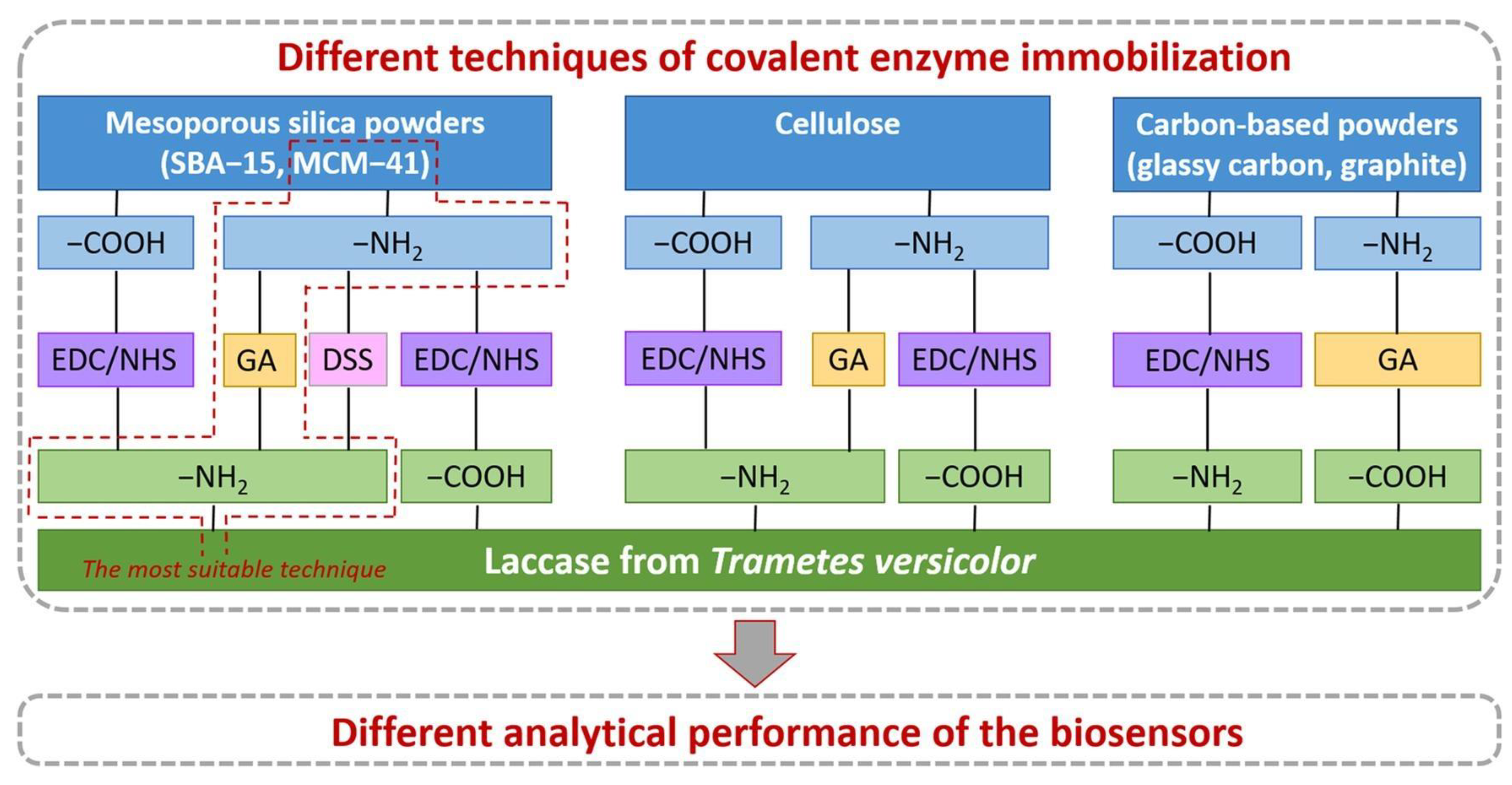
The recent era specifically focuses more on immobilization matrices based on nanomaterials. Although many other matrices have been used to support enzymes, nanomaterials-based matrices are widely explored because of their numerous benefits. Introducing nanomaterial matrices into the enzyme-based biosensors led to improved sensitivity, lower detection limit and higher response time, better stability and rigidity, a longer shelf life, and speeding up the fabrication process [69]. There is a broad spectrum of nanomaterials, including metal nanoparticles, metal oxide nanoparticles, nanostructured polymer composites, nanofibers, nanorods, etc., utilized in the field of biosensing for polyphenol detection. Sofia et al. demonstrated numerous methods for covalent immobilization of the Lac onto several matrices (Figure 4), and concluded that the approach based on the linkage of carbon-based materials through EDC-NHS to −COOH functionalized matrices was less effective than the Lac bonded with the -NH2 group using glutaraldehyde to support amminated carbon based matrices [70].
3. Carbonaceous Nanomaterial-Based Lac Biosensor
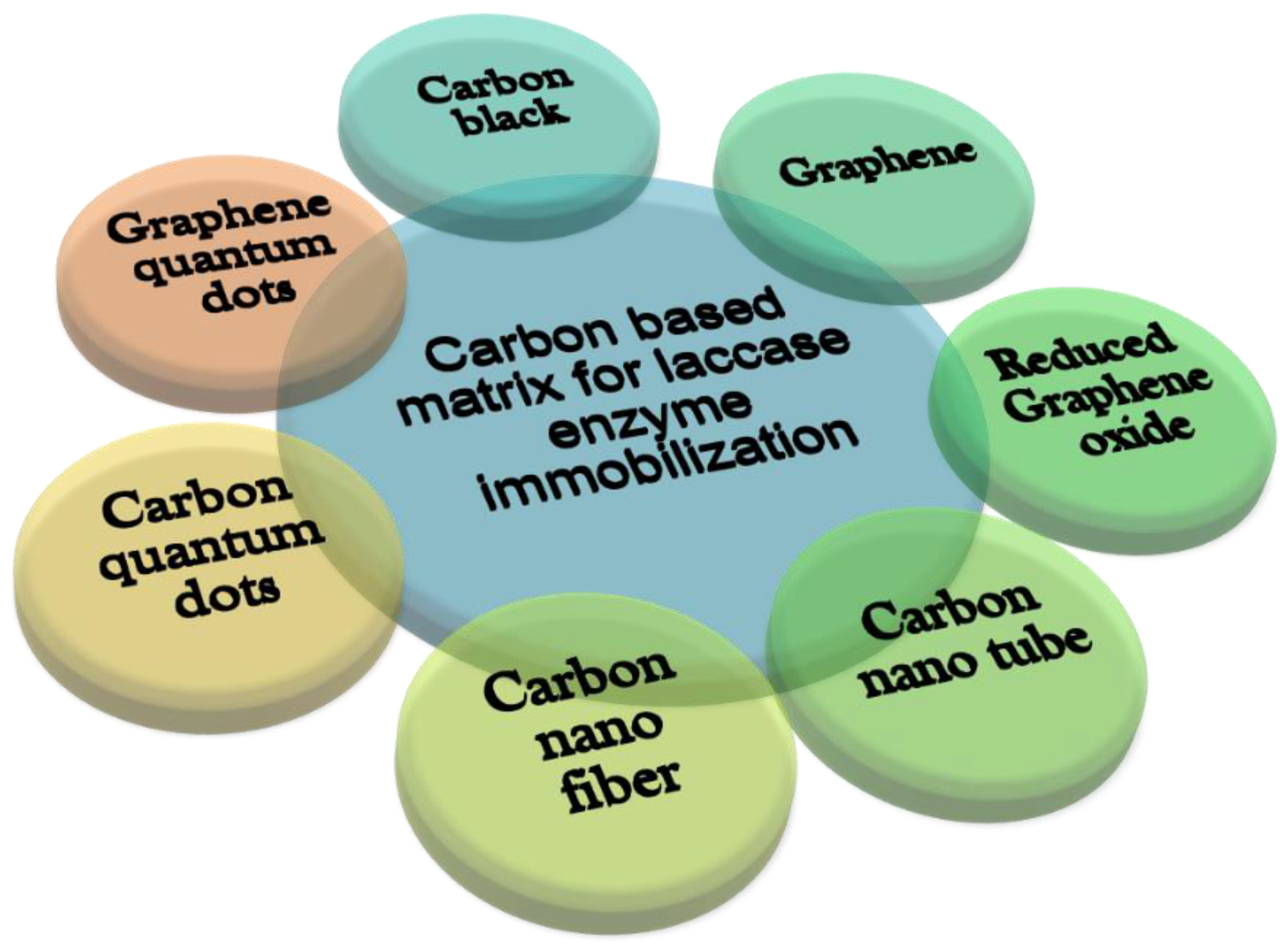
Carbon-based nanomaterials (CNMs) are one of the most explored nanomaterials for biosensor applications. Carbon nanomaterials have comparable dimensions to redox proteins, and thus can be used as an effective electrical connector with redox enzymes. The most common CNMs used to support the Lac enzyme in biosensing applications include graphene (Gr), reduced GO, carbon nanotubes, carbon quantum dots, and graphene quantum dots (Figure 5) as summarized in Table S2. These CNMs possess a tremendous ability to detect phenolic pollutants as they have a large surface-to-volume ratio, which in addition to sensing, helps in the adsorption of pollutants. Table 2 shows the application of different CNMs-based Lac biosensors for the detection of phenols.
3.1. Carbon Nano-Tube (CNT)-Based Lac Biosensors
The benzene-type hexagonal arrangement of carbon atoms forming hefty cylindrical molecules constitutes a quasi-one-dimensional CNT [110,111]. This allotrope of carbon has an enormous surface area in terms of a large length-to-diameter ratio and is observed to be just one atom thick [111]. CNTs exhibit extraordinary properties, such as being lightweight and stable, with excellent electronic, thermal, and mechanical properties [112]. A CNT lacks solubility in aqueous media, and the introduction of chemical functionalization increases their solubility in different solvents and increases their potential for biosensor applications [113]. CNTs are conjugated with many metal and metal oxide nanoparticles such as Au, Fe, Pd, Pt, Cu, Fe3O4, ZrO2, etc., for biosensing application [79,114]. However, certain factors such as the bundling effect, increment in the number of walls, and aggregation can cause problem while using CNTs.
3.2. Carbon Nanofibers (CNFs)-Based Lac Biosensors
A CNF is a one-dimensional nano range fiber with a hollow core with a diameter of 10 to 500 nm and a length in the range of 0.5 to 200 μm [115]. In CNFs, strands of layered stratified graphite sheets are stacked on a single molecule which have different variations such as platelets, ribbon, cones, herringbone, cups, etc., in the nano size range [116,117]. It has remarkable mechanical and chemical properties, due to which they are superior to other fibers. The presence of a graphitic structure enables their larger area and excellent thermal and electrical conductivities. In addition, they do not become oxidized easily. Other characteristics such as a low density, a high Young’s modulus, thermally stability, low defects, a more significant aspect ratio, and a condensed structure make CNFs suitable matrix materials [118]. These advantages lead to their applications in ceramics, fuel cells, cement composition, etc. [119]. Yang et al., used electrospinning, carbonization, and the solvothermal technique to fabricate a novel Lac-based biosensor for monitoring hydroquinone using TiO2 decorated copper and carbon composite nanofibers [120]. Recently, combined electrospinning and an in situ polymerization technique for synthesizing polyaniline-encapsulated carbon/copper composite nanofibers have been reported for the detection of hydroquinone in river water [87].
3.3. Carbon Quantum Dots (C-QDs)-Based Lac Biosensors
C-QDs are low-cost and water-soluble QDs that are easy to synthesize and have core carbon atoms organized in sp2 hybridization bearing oxygen-containing functional groups (hydroxyl, carboxyl, carbonyl) [121,122]. The biocompatibility, electronic properties, photostability, and photo-luminescent radiation emission of C-QDs have been explored for different applications such as photodegradation, sensing, bioimaging, drug delivery, catalysis, energy conversion, optical electronics, etc. [123].
C-QDs do not interact with all analytes directly, and very few biosensors have been reported based on C-QDs as a biosensor matrix for the detection of phenols. Recently, a fluorescent sensor has been reported utilizing the unique optical properties of C-QDs for the detection of dopamine using photo-luminescent [37]. However, the quantum yield of C-QDs is generally low, and they are difficult to reproduce due to their geometric arrangement and challenging structural characterization [124].
3.4. Graphite-Based Lac Biosensors
Graphite has been utilized as electrode material in amperometric biosensors due to its exceptional electronic and catalytic properties. Cato et al. used bare and modified graphite-epoxy decorated with either tyrosinase or the Lac enzyme and copper nanoparticles for the voltammetry detection of antioxidant activity phenols that are present in rosé cava wines [125,126]. Ibarra Escutia et al. also developed an amperometric biosensor, entrapping the fungal Lac enzyme onto polyvinyl alcohol photopolymer and graphitic SPE for sensing PhCs in tea infusions [127]. However, graphite lacs in scope for the modification of an electrode because of its lower functionality and poor solubility in organic solvents, due to which the derivatives of graphite offer more promising biosensing properties and are highly utilized.
3.5. Graphene (Gr) and Its Derivatives-Based Lac Biosensors
Gr is a single-layered sheet of carbon obtained after isolation from stacked layers of graphite having a thickness in atomic dimensions bearing a hexagonal layout of sp2 hybridized carbon atoms. Gr provides a large surface area, excellent electrical conductivity due to its highly conjugated structure, comparable tensile strength, and remarkable biocompatibility [128]. Gr-based materials provide an enhanced signal response in electrochemical biosensing [129]. Due to these properties, Gr sheets have been widely utilized as an immobilization matrix for immobilizing various enzymes, including Lac, for detecting various phenolic compounds. Palanisamy et al. fabricated a Lac enzyme-based biosensor to detect catechol using Gr cellulose microfiber composite-improved screen-printed carbon electrodes [44]. A biosensor based on the Lac enzyme immobilized on a polymer-like polyaniline (PANI) and magnetic graphene electrode has been introduced recently by Lou et al. for detecting hydroquinone [71].
Graphene Oxide (GO) is a derivative of Gr having abundant epoxy functional groups on the surface of Gr sheets with a large surface-to-volume ratio. Although the conductivity of GO is lower than graphene (presence of oxygen functionality), it has been reported as an immobilization matrix due to its solubility in water and its potential for the immobilization of enzymes on its surface without the use of crosslinker [130,131]. Maleki, et al. fabricated a biosensor using poly(3,4-methylenedioxy-thiophene), GO nano-sheets, and the Lac enzyme to detect catechol [75]. An electrochemical sensor utilizing GO and a molecularly imprinted polymer was developed for selective and sensitive detection of 2,4-dichlorophenol in water. It was also optimized to estimate 2,4-dichlorophenol in real water samples [132]. The introduction of oxygen functionality to GO led to a reduction in the conductivity of GO, while the debut of RGO led to better conductivity compared to that of pristine graphene. Moreover, it provides promising characteristics such as easy assembling, cost-effectiveness, higher mobility, biocompatibility, etc. [133]. A large array of materials and composites have been used for doping RGO sheets to synergistically enhance the catalytic activity and electron transfer process of biosensors for the detection of different phenolic pollutants such as 17β-estradiol [74], catechol [134], estriol [135], etc. Using RGO as a matrix for the Lac enzyme, Mei et al. fabricated RGO palladium–copper electrodes for the sensitive determination of catechol [134]. Eremia et al. developed a disposable biosensor using platinum nanoparticles/RGO composite supported on carbon screen-printed electrodes for the assessment of caffeic acid. They reported the kinetics of the detection of polyphenol electrochemically and claimed that the fabricated biosensor can be used for evaluating the total phenolic content in tea infusions [136]. Despite all the benefits, there are certain limitations of using GO and RGO such as quick agglomeration, reduction in conductivity after undergoing functionalization, and the use of toxic chemicals during synthesis. These can perhaps be overcome by the use of advanced 2D materials.
3.6. Graphene Quantum Dots (G-QDs)-Based Lac Biosensors
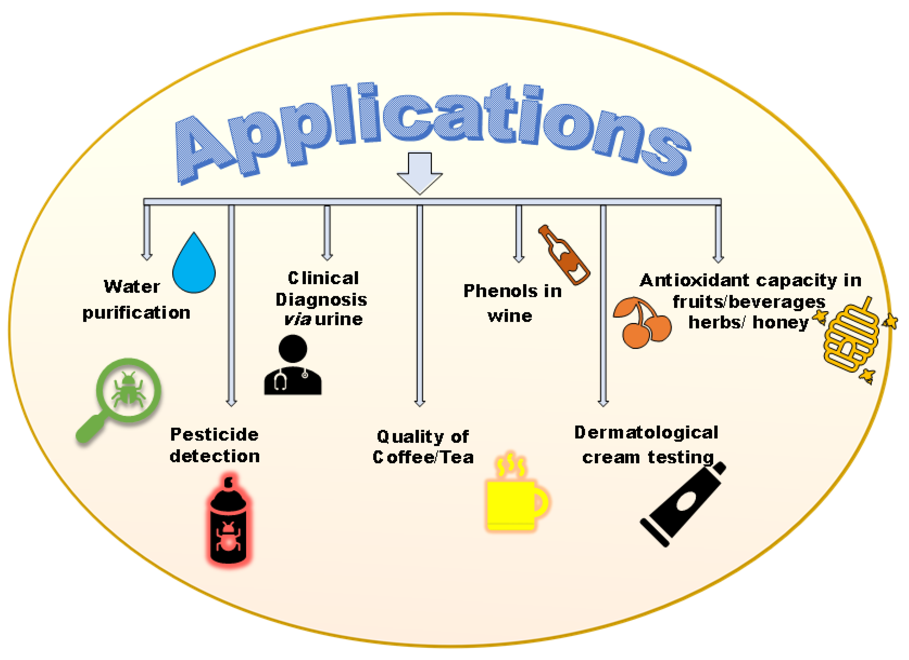
G-QDs are considered zero-dimensional nanomaterials which are derived from the 2D sheet of graphene [137]. G-QDs exhibit better solubility than CNTs and displays biocompatibility, low toxicity, and stability. High quantum yield, edge effects, and confinement make it different from other carbon nanomaterials [138,139]. Top-down synthesis of G-QDs from graphene is a multistep and cost-effective process, while bottom-up synthesis of G-QDs from polycyclic aromatic compounds proceeds via complex steps and requires costly chemicals and equipment. G-QDs demonstrate remarkable photoluminescence and robust quantum properties [140]. Baluta, et al. used the fluorescence technique to detect dopamine using a Lac-based ceramic biosensor supported by graphene QDs. The fabricated biosensor was also modified using a conducting polymer [78]. Unlike graphene, G-QDs have zero band gap and can be modified using functional groups due to the edge effect [97]. Vasilescu et al. modified G-QDs electrodes using molybdenum disulfide, and the fabricated Lac-based biosensor has been used for the detection of caffeic acid and were proven to be useful for estimating the total phenolic content in red wine [141]. Inspite of several applications, the use of G-QDs is still challenging as the formation of a single layer of high-quality G-QDs is difficult. In addition, its synthesis suffers from toxic organic solvents, a long reaction time, and low yield, which limits its use for industrial purposes. Different applications of CNMs to detect PhCs have been summarized in Figure 6.
4. Applications of Lac-Based Biosensors
4.1. Detection of Phenolic Pollutants in Wastewater
Water bodies contain waste from different sources in the form of dyes and harmful chemicals that affect various life forms (both flora and fauna). Toxic chemicals, dyes, and PhCs are major raw contributors from various textile, plastics, leather, and paper industries. PhCs also merge into water bodies from the oil, gas, and coal industries. Conventional methods such as photo-catalytic degradation, distillation, ozonation, extraction techniques, membrane processes, advanced oxidation process, and enzymatic biological methods have been used to remove these dyes and phenolic pollutants from wastewater [142,143]. Before their removal, it is important to implement a real-time monitoring device to determine which pollutant is to be treated and how much to reduce the cost and time for wastewater treatment methods is very crucial. The major water pollution-contributing phenolic compounds are catechol, hydroquinone, and Bisphenol-A.
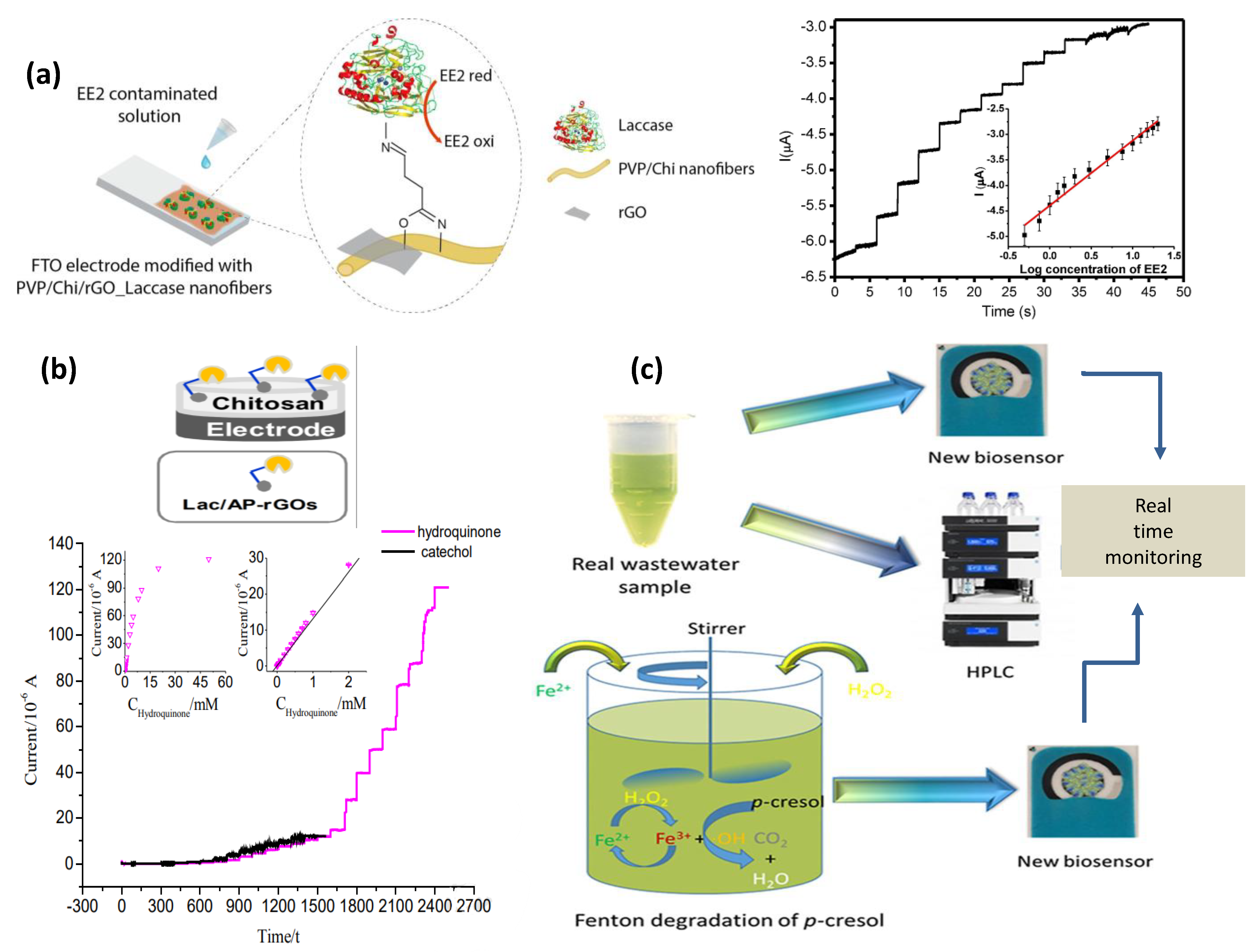
Hydroquinone (Hq) is a widely used raw material in chemical industries as a dye intermediate and black and white photo developer and in cosmetics as a skin-lightening agent as well as a polymerization inhibitor. Studies reveal that high exposure to Hq may lead to skin-irritating effects, and it is also harmful to soil microorganisms. Recently, PANI/magnetic graphene immobilized Lac enzyme-modified electrodes were developed that show superior biosensing activity for Hq in real water samples [71]. Upan, et al. introduced a flow-injection amperometric sensor using a glutaraldehyde cross-linked Lac enzyme onto CNT-modified SPE for estimation of Hq in water sample [144]. rGO-MoS2 nanocomposite has been used to detect Hq using chronoamperometry technique [109]. Synthetic catechol, on the other hand, has been utilized extensively in the production of pesticides, pharmaceuticals, chemical perfumes, etc. [145,146]. Similar to Hq, it is also used in the field of black-and-white photography. Several biosensors have been developed for the evaluation of catechol. A biosensor based on an artificial neural network-integrated system has been formulated for the assessment of real water samples containing catechol under a wider linear range using poly(3,4-ethylenedioxythiophene) (PEDOT), GO nano-sheets, and Lac [75]. Kong, et al. used a graphite electrode-based intercalated montmorillonite for the electrochemical determination of catechol [147]. One of the fabricated biosensor-based on Lac/Polyvinylpyrrolidone/chitosan/rGO electrospun has been shown in Figure 7a [73]. Also, simultaneous detection of Hq and catechol has been carried out using Lac/aminopyrine/RGO/GCE electrode in water samples d [148] (Figure 7b). Bisphenol A is a component of hard plastic that are being used daily in different forms such as bottles, medical equipment, household electronic items, sports equipment, etc. [149]. Schematic representation of Lac/CNT/SPCE electrode for detecting p−cresol in real water samples and p−cresol produced during the Fenton process has been depicted in Figure 7c [89].
Bisphenol A leaches into our bodies mainly through food containers as it is used to coat the inside of food containers and drink cans. Numerous studies reveal that it acts as an endocrine disrupter even at a very low dose [150]. Bisphenol A can cause DNA damage, so monitoring its amount in water is necessary. Jalalvand, et al. used biosensing electrodes as layer-by-layer modified GCE with methylene blue-DNA/MWCNTs-chitosan/palladium nanoparticles/fullerene C60 [151]. An rGO-(4-ferrocenylethyne) phenylamine/AuNPs/GCE electrode has been used for the detection of Bisphenol A infused in milk samples [152]. Lac–thionine–carbon black-modified SPE was formulated by Portaccio, M., et al. for the assessment of endocrine disruptors in tomato juice samples [153]. Paracresol is also a water-polluting phenol, as illustrated in Figure 6a, and quite recently, a Lac/CNT/SPCE electrode has been used for its detection in real water samples. More importantly, paracresol formed while performing the Fenton process has a good resemblance with that of the results obtained using the HPLC technique [89].
One of the main causes of the accumulation of phenolic compounds in water bodies is their heavy usage in the agricultural sector as insecticides, pesticides, and herbicides. Pesticides such as pentachlorophenol undergo degradation in water into different chlorophenols. Chlorophenols easily leach from soil into water bodies as they are water-soluble and have also been widely used for impregnating wood [154]. The washing away of byproducts from agricultural materials into water bodies plays a significant role in polluting water. Biosensors help in the detection of these pesticides with ease. Carbamates are among widely used pesticides in insecticides, nematicides, fungicides, acaricides, or herbicides for increasing the yield of crops as a prophylactic measure. However, for non-targeted organisms, carbamates are toxic due to their deliberate discharge into the soil and subsequently into water bodies, leading to ill effects on the environment and human health worldwide [155]. These are also among the list of endocrine disruptors as of Bisphenol A [156], and thus become an important analyte for intensive monitoring. A novel enzyme-based biosensor has been developed by Oliveira et al. for the estimation of carbamate pesticide applied on tomato and potato crops using direct immobilization of Prussian blue functionalized Lac on graphene-modified carbon paste electrodes [157]. They also detected pirimicarb pesticide by immobilizing Lac on composite carbon paste electrodes containing an MWCNTs paste electrode modified by the dispersion of Lac within the optimum composite matrix [158]. Pesticides cause a higher threat to vegetables and fruits, which easily carry food-borne microbes and pesticide residue into our bodies when eaten raw [159]. Therefore, it is necessary to investigate the amounts of pesticides to protect the endocrine system of human beings and animals. For governing food safety, Oliveira et al. constructed a dual enzyme (Lac and tyrosinase)-based biosensor immobilized on to Au nanoparticles, chitosan, and graphene-doped carbon paste electrodes in order to detect the number of different carbamate residues in fruits [160].
4.2. Detection of Phenolic Compounds in Food
PhCs from natural compounds are found in vegetables, fruits, cereals, beverages, and red wines. The nutritional value of plants, fruits, and vegetables can be determined by quantifying their PhCs content [161]. It has been reported that PhCs rich food items prevent oxidative damage leading to age-associated diseases by terminating free radicals generated after cell metabolism [162]. Biosensors have proven to be an important technique to check the monitoring of food quality by detecting PhCs in plants and food products. De Macêdo, et al. estimated the total phenolic content with the help of a Lac immobilized graphite electrode in dry extracts of red fruits [163]. Honey is commonly used as an alternative to normal sugar due to its good antioxidant properties [164]. The authenticity of honey can be evaluated by inspecting its total phenolic content and antioxidant capacity using a real-time biosensor. De Oliveira Neto reported an electroanalytical method for immobilizing the Lac enzyme onto a carbon paste electrode for detecting both the antioxidant nature and total phenolic content of honey. The results correlate with the data obtained using the spectroscopic method [165].
Among generally consumed beverages, tea contains various PhCs (phenolic acids, flavonoids, tannins, catechins, etc.), possessing a variety of biological activities that can lead to cancer prevention, anti-aging, cardiovascular diseases, and oxidative stress in the brain. Tea infusions have excellent antioxidant properties and contain a high percentage of caffeic acid. Caffeic acid was determined from a tea infusion by Eremia, who constructed disposable biosensors based on platinum nanoparticles-RGO-Lac biocomposite electrodes [136]. Rawal et al. constructed Lac/Fe3O4NPs/cMWCNT/PANI/Au, Lac/MnO2NPs/cMWCNT/PANI/Au, and Lac/AgNPs/cMWCNT/PANI/Au electrodes for measuring the total content of phenols in tea leaf extract [59,166]. The total phenolic content in tea leaf extract and in black radish root was also quantified by Ibarra-Escutia, P., et al. with the help of an amperometric biosensor based on carbon SPE [167]. On the other hand, coffee, a worldwide beloved and traded beverage, holds potential for reducing high blood pressure and high cholesterol, risk to pancreatic cancer, and bone loss. All varieties of coffee contain lipids, vitamins, and carbohydrates accompanied by variable amounts of PhCs including chlorogenic acid and caffeic acid that are responsible for forecasting their virtuous qualities and categories [168]. Chlorogenic acid is the major phenol in coffee. Moreover, it is also a chief antioxidant among this beverage. GO paste/PtNPs, Lac, and BOT (botryosphaeran) electrodes were fabricated by Salamanca-Neto, et al. to discriminate the exceptional and traditional brewed coffee beverages by measuring chlorogenic acid [81].
PhCs were found to contribute to the color, astringency, mouth feel, and oxidative stabilization of wine [169,170]. PhCs in wine give an advantage to hypertensive patients based on their antioxidant nature by decreasing blood pressure [171]. Montereali et al. reported the detection of polyphenols in wine using an amperometric biosensor by immobilizing tyrosinase and the Lac enzyme onto ferrocene-modified graphite screen printed electrodes [172]. A bioelectric tongue was fabricated to detect gallic acid, a tri-hydroxy group containing compound in white wine, rose wine, and red wine [173]. The differential pulse voltametric technique has been used for assessing gallic acid using a [Cu2tpmc](ClO4)4/PVC matrix coated on graphite or carbon rods [174]. However, Almeida et al. [85] used thin polydopamine film on carbon electrodes for the detection of gallic acid from the chestnut shell obtained from industrial waste extract [141].
Phenolic content can also be tested in medicinal plants. Lac-immobilized electrodeposited MWCNT and chitosan film were used to investigate phenols (caffeic acid, gallic acids, chlorogenic acid, and rosmarinic acid) present in the medicinal plants Salvia officinalis and Menthapiperita [175]. The oil of Menthapiperita (Pepperment) contains specific characteristic PhCs and is a well-known medicinal ingredient that is used in tea and herbs. It has strong antioxidant properties and possesses an excellent potential as a repellent of allergies, microbes, and tumors [176]. On the other hand, the herb Salvia officinalis (sage) is used as tonic in medicine to improve memory, learning power, and reduce stress [177]. PhCs present in sage (Salvia officinalis) show excellent antioxidant behavior. Rosmarinic acid is the chief phenol present on both herbs [178]. Eremia et al. determined rosmarinic acid from in vitro saliva culture with Lac–Nafion-based biosensors [179].
4.3. Detection of Analytes in Body Fluids
Dopamine is a significant neurotransmitter of the brain, which plays vital roles in the cardiovascular, central nervous, and endocrine systems [180]. Dopamine is found in lower concentrations in patients with dementia, Parkinson’s disease, and schizophrenia [181]. The normal concentration of dopamine in the brain is necessary for maintaining accurate blood pressure, learning, enthusiasm, physical activities, and cognizance before releasing dopamine into the brain [37]. Therefore, a rapid clinical and sensitive diagnosis of dopamine is in considerable demand for monitoring Parkinson’s disease and depression [182]. A fluorescence-based sensor has been developed using the Lac enzyme and polydopamine on the surface of G-QDs for dopamine detection [78]. Hua, et al. assessed the role of dopamine in human urine samples using a synthesized β-Cyclodextrin inclusion complex as the immobilization matrix onto RGO for the fabrication of a biosensor [183].
Estrogens are responsible for both environmental pollution and harmful effects on humans [184]. For example, 17 β-estradiol is an environmental pollutant that is of paramount concern amongst estrogens. It is also a natural steroid that regulates the female reproductive system by contributing to estrogen [185]. Studies reveal that the release of 17β-estradiol and estriol hormones is around 280–600 μg/day and 6000–10,000 μg/day for pregnant women [156]. The presence of pollutants in food such as meat and milk lead to severe diseases related to fertility and tumor issues in females [186,187]. Biosensors for detecting estrogens from blood and urine are considered superior over other conventional methods as they are simple, rapid, and sensitive [187,188]. Wang et al. developed an electrochemical Lac enzyme poly L-lysine/citric acid-graphene biosensor to detect 17β-estradiol in urine samples [77]. Urine contains conjugated forms of estriol accompanying water soluble sulfates that hydrolyze promptly, leading to regeneration of the free form of estriol, subsequently threatening human and marine lives and giving rise to water pollution [189]. A voltammetry sensor was constructed using cobalt-poly(methionine) modified GCE for determining estriol hormone in urine and pharmaceuticals [190]. Lac functionalized polyvinylpyrrolidone/chitosan/RGO hybrid nanofibers deposited onto fluorine doped tin oxide (FTO) were electrochemically able to diagnose 17α—Ethinylestradiol in synthetic and human urine samples [73] (Figure 6c).
5. Future Scope
It is believed that CNTs and graphitic materials offer faster response time and appreciable sensitivity compared to traditional electrodes at minimal working potentials. However, effort has been made to modify CNMs-based biosensors to have better control on chemical and physical properties. The limitations may be overcome using the integration of metal, conducting polymers, metal chalcogenides, etc., with CNMs, which can enhance the performance of biosensors to address future challenges.
A lot of interest is now been focused on advanced 2D materials such as MXenes (MXe), which was recently explored in the field of biosensors [191]. MXe has been found to be better than graphene-based nanomaterials in terms of improved electron transfer in the heterogeneous phase, low diffusion barrier, and stable dispersibility. This is due to the presence of metal at the center on MX, which does not reduce its conductivity or dispersibility even after functionalization and offers excellent biocompatibility. In this context, MXe based biosensors are progressing toward the development of lab-on-chip biosensing devices involving 5G communication [192].
Future generations demand quick and accurate detection of analytes [193]. New aspects such as the internet of things and machine learning involving artificial intelligence (AI) are being used in the advancement of point-of-care (POC) devices which are emerging more promising technologies as compared to conventional healthcare monitoring [194]. Emphasis on advanced biosensor fabrication must be given to use AI for detecting phenols and for their on-site and online monitoring in order to balance the content of phenol in diverse applications, as discussed in this review article.
6. Conclusions
The reliable detection of PhCs using Lac-based biosensors may result in maintaining permissible polyphenols in water, body fluids, and sustainable management of antioxidant quality. Developing a biosensor for PhCs detection will provide a way to overcome conventional techniques, which are time-consuming and expensive. The application of Lac-based biosensors is mainly focused on the electrochemical transduction method. Amperometric biosensors are among the most reported techniques for detecting PhCs using the Lac enzyme and carbonaceous matrices. The carbon-based matrix has shown enormous potential to support the Lac enzyme by providing enhanced surface area, superior conductivity, stability, and excellent mechanical strength. They have been used in diverse applications to detect polyphenols from wastewater, wine samples, urine, fruits, vegetables, juices, human serums, and beverages. The most significant limitation that remains is the leaching of the enzyme from the substrate. This can perhaps be improved using suitable immobilization techniques and incorporation of nanomaterials to maintain the enzyme activity. The integration of CNMs with Lac-based biosensors can be crucial in developing POC devices for application in food, pharmaceuticals, and environmental monitoring. The oxidizing characteristics of Lac make it an effective biocatalyst, which can also be utilized for other applications related to the same mechanism. Enzyme modification, the multienzyme approach, AI, and advanced 2D materials should be explored for the development of POC devices.
Supplementary Materials
Author Contributions
S.V.: Conceptualization, writing—original draft, and investigation. D.T.: validation and visualization. C.M.P.: supervision, validation, writing—reviewing and editing. D.K.: supervision, validation, writing—editing. All authors have read and agreed to the published version of the manuscript.
Funding
This research received no external funding.
Institutional Review Board Statement
Not applicable.
Informed Consent Statement
Not applicable.
Data Availability Statement
Data will be made available on request.
Acknowledgments
Sakshi Verma thanks UGC-India for the Senior Research Fellowship award. Deeksha Thakur acknowledges the Department of Science and Technology (DST), New Delhi, India, for the DST-INSPIRE SRF award. C.M. Pandey acknowledges the Department of Science and Technology (DST), New Delhi, India, for the DST-INSPIRE Faculty award.
Conflicts of Interest
The authors declare no conflict of interest.
Abbreviations
PANI: Polyaniline, NP: nanoparticle, SPE: screen printed electrode, SPCE: screen printed carbon electrode, TPC: total phenolic content, GCE: glassy carbon electrode, CS: chitosan, ZIF: zeoliticimid–azolate framework, BC: bacterial cellulose, Pc: phthalocyanine, CMB: carboxymethyl-botryosphaeran, CBPE: carbon black paste electrode, GNPl: graphenenanoplatelets, BMIMBF4: 1-butyl-3-methylimidazolium tetra-fluoroborate, PVP: polyvinylpyrrolidone, NF: nanofiber, GPE: graphene doped carbon paste electrode, Tyr: Tyrosinase, G: graphite, PANABA: poly-aniline-2-aminobenzylamine, OMC-SPE: organized mesoporous carbon-modified carbon screen-printed electrode, cMWCNT: carbonylated multi wall carbon nanotube, MG: magnetic graphene, PEDOT: poly(3,4-ethylenedioxythiophene), PPy: polypyrrole, G-QDs: graphene quantum dots, POXA1b: laccase Pleurotus ostreatus, DPV: Differential Pulse Voltametry, CV: Cyclic voltammetry, SWV: square wave voltammetry, Amp: Amperometry, ChronoAmp: Chronoamperometry
References
- Villegas, L.G.C.; Mashhadi, N.; Chen, M.; Mukherjee, D.; Taylor, K.E.; Biswas, N. A Short Review of Techniques for Phenol Removal from Wastewater. Curr. Pollut. Rep. 2016, 2, 157–167. [Google Scholar] [CrossRef] [Green Version]
- Anku, W.W.; Mamo, M.; Govender, P. Phenolic Compounds in Water: Sources, Reactivity, Toxicity and Treatment Methods. In Phenolic Compounds—Natural Sources, Importance and Applications; InTech Open: London, UK, 2017; pp. 420–443. [Google Scholar]
- Kulys, J.; Bratkovskaja, I. Antioxidants determination with laccase. Talanta 2007, 72, 526–531. [Google Scholar] [CrossRef]
- Michałowicz, J.; Duda, W. Phenols—Sources and Toxicity. Pol. J. Environ. Stud. 2007, 16, 347–362. [Google Scholar]
- Rice-Evans, C.; Miller, N.; Paganga, G. Antioxidant properties of phenolic compounds. Trends Plant Sci. 1997, 2, 152–159. [Google Scholar] [CrossRef]
- Meder, D.; Herz, D.M.; Rowe, J.B.; Lehéricy, S.; Siebner, H.R. The role of dopamine in the brain—Lessons learned from Parkinson’s disease. Neuroimage 2019, 190, 79–93. [Google Scholar] [CrossRef]
- Ejeian, F.; Etedali, P.; Mansouri-Tehrani, H.-A.; Soozanipour, A.; Low, Z.-X.; Asadnia, M.; Taheri-Kafrani, A.; Razmjou, A. Biosensors for wastewater monitoring: A review. Biosens. Bioelectron. 2018, 118, 66–79. [Google Scholar] [CrossRef]
- Thévenot, D.R.; Toth, K.; Durst, R.A.; Wilson, G.S. Electrochemical biosensors: Recommended definitions and classification. Biosens. Bioelectron. 2001, 16, 121–131. [Google Scholar] [CrossRef]
- Aghapour, A.A.; Moussavi, G.; Yaghmaeian, K. Biological degradation of catechol in wastewater using the sequencing continuous-inflow reactor (SCR). J. Environ. Health Sci. Eng. 2013, 11, 3. [Google Scholar] [CrossRef] [Green Version]
- Groff, T. Bisphenol A: Invisible pollution. Curr. Opin. Pediatr. 2010, 22, 524–529. [Google Scholar] [CrossRef]
- Li, Y.; Trush, M.A. Reactive oxygen-dependent DNA damage resulting from the oxidation of phenolic compounds by a copper-redox cycle mechanism. Cancer Res. 1994, 54, 1895s–1898s. [Google Scholar]
- Kadmi, Y.; Favier, L.; Yehya, T.; Soutrel, I.; Simion, A.I.; Vial, C.; Wolbert, D. Controlling contamination for determination of ultra-trace levels of priority pollutants chlorophenols in environmental water matrices. Arab. J. Chem. 2019, 12, 2905–2913. [Google Scholar] [CrossRef]
- Enguita, F.J.; Leitão, A.L. Hydroquinone: Environmental pollution, toxicity, and microbial answers. BioMed Res. Int. 2013, 2013, 542168. [Google Scholar] [CrossRef] [Green Version]
- Singh, T.; Bhatiya, A.K.; Mishra, P.K.; Srivastava, N. Chapter 11—An effective approach for the degradation of phenolic waste: Phenols and cresols. In Abatement of Environmental Pollutants; Singh, P., Kumar, A., Borthakur, A., Eds.; Elsevier: Amsterdam, The Netherlands, 2020; pp. 203–243. [Google Scholar] [CrossRef]
- Lecomte, S.; Habauzit, D.; Charlier, T.D.; Pakdel, F. Emerging Estrogenic Pollutants in the Aquatic Environment and Breast Cancer. Genes 2017, 8, 229. [Google Scholar] [CrossRef] [Green Version]
- Lin, T.-W.; Kuo, Y.-M. Exercise benefits brain function: The monoamine connection. Brain Sci. 2013, 3, 39–53. [Google Scholar] [CrossRef] [Green Version]
- Badhani, B.; Sharma, N.; Kakkar, R. Gallic acid: A versatile antioxidant with promising therapeutic and industrial applications. RSC Adv. 2015, 5, 27540–27557. [Google Scholar] [CrossRef]
- Ye, Y.; Guo, H.; Sun, X. Recent progress on cell-based biosensors for analysis of food safety and quality control. Biosens. Bioelectron. 2019, 126, 389–404. [Google Scholar] [CrossRef]
- Lu, X.; Sun, J.; Sun, X. Recent advances in biosensors for the detection of estrogens in the environment and food. TrAC Trends Anal. Chem. 2020, 127, 115882. [Google Scholar] [CrossRef]
- María Isabel, G.-A.; Juan, B.C.; Gustavo, A.P. Nanomaterial-based fluorescent biosensors for monitoring environmental pollutants: A critical review. Talanta Open 2020, 2, 100006. [Google Scholar] [CrossRef]
- Mello, L.D.; Kubota, L.T. Biosensors as a tool for the antioxidant status evaluation. Talanta 2007, 72, 335–348. [Google Scholar] [CrossRef]
- Marco, M.-P.; Barceló, D. Chapter 22 Fundamentals and applications of biosensors for environmental analysis. In Techniques and Instrumentation in Analytical Chemistry; BarcelÓ, D., Ed.; Elsevier: Amsterdam, The Netherlands, 2000; Volume 21, pp. 1075–1105. [Google Scholar]
- Makarewicz, M.; Drożdż, I.; Tarko, T.; Duda-Chodak, A. The Interactions between Polyphenols and Microorganisms, Especially Gut Microbiota. Antioxidants 2021, 10, 188. [Google Scholar] [CrossRef]
- Zheng, Y.; Yang, C.; Pu, W.; Zhang, J. Carbon nanotube-based DNA biosensor for monitoring phenolic pollutants. Microchim. Acta 2009, 166, 21–26. [Google Scholar] [CrossRef]
- Othman, A.; Karimi, A.; Andreescu, S. Functional nanostructures for enzyme based biosensors: Properties, fabrication and applications. J. Mater. Chem. B 2016, 4, 7178–7203. [Google Scholar] [CrossRef]
- Sekretaryova, A.N.; Volkov, A.V.; Zozoulenko, I.V.; Turner, A.P.F.; Vagin, M.Y.; Eriksson, M. Total phenol analysis of weakly supported water using a laccase-based microband biosensor. Anal. Chim. Acta 2016, 907, 45–53. [Google Scholar] [CrossRef]
- Cheng, H.; Hu, M.; Zhai, Q.; Li, S.; Jiang, Y. Polydopamine tethered CPO/HRP-TiO2 nano-composites with high bio-catalytic activity, stability and reusability: Enzyme-photo bifunctional synergistic catalysis in water treatment. Chem. Eng. J. 2018, 347, 703–710. [Google Scholar] [CrossRef]
- de Matos Morawski, F.; Deon, M.; Nicolodi, S.; Menezes, E.; Costa, T.; Dias, S.; Benvenutti, E.; Arenas, L. Magnetic silica/titania xerogel applied as electrochemical biosensor for catechol and catecholamines. Electrochim. Acta 2018, 264, 319–328. [Google Scholar] [CrossRef]
- Manan, F.A.A.; Hong, W.W.; Abdullah, J.; Yusof, N.A.; Ahmad, I. Nanocrystalline cellulose decorated quantum dots based tyrosinase biosensor for phenol determination. Mater. Sci. Eng. C 2019, 99, 37–46. [Google Scholar] [CrossRef]
- Durán, N.; Rosa, M.A.; D’Annibale, A.; Gianfreda, L. Applications of laccases and tyrosinases (phenoloxidases) immobilized on different supports: A review. Enzym. Microb. Technol. 2002, 31, 907–931. [Google Scholar] [CrossRef]
- Pandey, C. Biosensors: Fundamentals and Applications; Smithers Rapra: Shrewsbury, UK, 2017. [Google Scholar]
- Scheller, F.W.; Hintsche, R.; Pfeiffer, D.; Schubert, F.; Riedel, K.; Kindervater, R. Biosensors: Fundamentals, applications and trends. Sens. Actuators B Chem. 1991, 4, 197–206. [Google Scholar] [CrossRef]
- Sarkar, A.; Sarkar, K.D.; Amrutha, V.; Dutta, K. Chapter 15—An overview of enzyme-based biosensors for environmental monitoring. In Tools, Techniques and Protocols for Monitoring Environmental Contaminants; Kaur Brar, S., Hegde, K., Pachapur, V.L., Eds.; Elsevier: Amsterdam, The Netherlands, 2019; pp. 307–329. [Google Scholar] [CrossRef]
- Mulchandani, A. Principles of Enzyme Biosensors. In Enzyme and Microbial Biosensors: Techniques and Protocols; Mulchandani, A., Rogers, K.R., Eds.; Humana Press: Totowa, NJ, USA, 1998; pp. 3–14. [Google Scholar]
- Das, P.; Barbora, L.; Das, M.; Goswami, P. Highly sensitive and stable laccase based amperometric biosensor developed on nano-composite matrix for detecting pyrocatechol in environmental samples. Sens. Actuators B Chem. 2014, 192, 737–744. [Google Scholar] [CrossRef]
- Kavetskyy, T.; Smutok, O.; Demkiv, O.; Maťko, I.; Švajdlenková, H.; Šauša, O.; Novák, I.; Berek, D.; Čechová, K.; Pecz, M.; et al. Microporous carbon fibers as electroconductive immobilization matrixes: Effect of their structure on operational parameters of laccase-based amperometric biosensor. Mater. Sci. Eng. C 2020, 109, 110570. [Google Scholar] [CrossRef]
- Tammina, S.K.; Yang, D.; Koppala, S.; Cheng, C.; Yang, Y. Highly photoluminescent N, P doped carbon quantum dots as a fluorescent sensor for the detection of dopamine and temperature. J. Photochem. Photobiol. B Biol. 2019, 194, 61–70. [Google Scholar] [CrossRef] [PubMed]
- Lepore, M.; Portaccio, M. Optical detection of different phenolic compounds by means of a novel biosensor based on sol–gel immobilized laccase. Biotechnol. Appl. Biochem. 2017, 64, 782–792. [Google Scholar] [CrossRef]
- Claus, H. Laccases: Structure, reactions, distribution. Micron 2004, 35, 93–96. [Google Scholar] [CrossRef]
- Dwivedi, U.N.; Singh, P.; Pandey, V.P.; Kumar, A. Structure–function relationship among bacterial, fungal and plant laccases. J. Mol. Catal. B Enzym. 2011, 68, 117–128. [Google Scholar] [CrossRef]
- Jones, S.M.; Solomon, E.I. Electron transfer and reaction mechanism of laccases. Cell. Mol. Life Sci. CMLS 2015, 72, 869–883. [Google Scholar] [CrossRef] [Green Version]
- Chaurasia, P.; Yadav, R.; Yadava, S. A review on mechanism of laccase action. Res. Rev. Biosci. 2013, 7, 66–71. [Google Scholar]
- Rodríguez-Delgado, M.M.; Alemán-Nava, G.S.; Rodríguez-Delgado, J.M.; Dieck-Assad, G.; Martínez-Chapa, S.O.; Barceló, D.; Parra, R. Laccase-based biosensors for detection of phenolic compounds. TrAC Trends Anal. Chem. 2015, 74, 21–45. [Google Scholar] [CrossRef] [Green Version]
- Palanisamy, S.; Ramaraj, S.K.; Chen, S.-M.; Yang, T.C.K.; Yi-Fan, P.; Chen, T.-W.; Velusamy, V.; Selvam, S. A novel Laccase Biosensor based on Laccase immobilized Graphene-Cellulose Microfiber Composite modified Screen-Printed Carbon Electrode for Sensitive Determination of Catechol. Sci. Rep. 2017, 7, 41214. [Google Scholar] [CrossRef] [Green Version]
- Castrovilli, M.C.; Bolognesi, P.; Chiarinelli, J.; Avaldi, L.; Calandra, P.; Antonacci, A.; Scognamiglio, V. The convergence of forefront technologies in the design of laccase-based biosensors—An update. TrAC Trends Anal. Chem. 2019, 119, 115615. [Google Scholar] [CrossRef]
- Zhang, Y.; Ge, J.; Liu, Z. Enhanced Activity of Immobilized or Chemically Modified Enzymes. ACS Catal. 2015, 5, 4503–4513. [Google Scholar] [CrossRef]
- Wu, M.-H.; Lin, M.-C.; Lee, C.-C.; Yu, S.-M.; Wang, A.H.J.; Ho, T.-H.D. Enhancement of laccase activity by pre-incubation with organic solvents. Sci. Rep. 2019, 9, 9754. [Google Scholar] [CrossRef] [Green Version]
- Daronch, N.A.; Kelbert, M.; Pereira, C.S.; de Araújo, P.H.H.; de Oliveira, D. Elucidating the choice for a precise matrix for laccase immobilization: A review. Chem. Eng. J. 2020, 397, 125506. [Google Scholar] [CrossRef]
- Prado Barragán, L.A.; Buenrostro-Figueroa, J.J.; Aguilar González, C.N.; Marañon, I. Chapter 10—Production, Stabilization, and Uses of Enzymes From Fruit and Vegetable Byproducts. In Biotransformation of Agricultural Waste and By-Products; Poltronieri, P., D’Urso, O.F., Eds.; Elsevier: Amsterdam, The Netherlands, 2016; pp. 271–286. [Google Scholar] [CrossRef]
- Minteer, S.D. Chapter 16—Cell-Free Biotechnologies. In Biotechnology for Biofuel Production and Optimization; Eckert, C.A., Trinh, C.T., Eds.; Elsevier: Amsterdam, The Netherlands, 2016; pp. 433–448. [Google Scholar] [CrossRef]
- Alarcon-Angeles, G.; Álvarez-Romero, G.A.; Merkoçi, A. Electrochemical Biosensors: Enzyme Kinetics and Role of Nanomaterials. In Encyclopedia of Interfacial Chemistry; Wandelt, K., Ed.; Elsevier: Amsterdam, The Netherlands, 2018; pp. 140–155. [Google Scholar] [CrossRef]
- Salmon, S.; House, A. Chapter 2—Enzyme-catalyzed Solvents for CO2 Separation. In Novel Materials for Carbon Dioxide Mitigation Technology; Shi, F., Morreale, B., Eds.; Elsevier: Amsterdam, The Netherlands, 2015; pp. 23–86. [Google Scholar] [CrossRef]
- Sassolas, A.; Blum, L.J.; Leca-Bouvier, B.D. Immobilization strategies to develop enzymatic biosensors. Biotechnol. Adv. 2012, 30, 489–511. [Google Scholar] [CrossRef]
- Cacicedo, M.L.; Manzo, R.M.; Municoy, S.; Bonazza, H.L.; Islan, G.A.; Desimone, M.; Bellino, M.; Mammarella, E.J.; Castro, G.R. Chapter 7—Immobilized Enzymes and Their Applications. In Advances in Enzyme Technology; Singh, R.S., Singhania, R.R., Pandey, A., Larroche, C., Eds.; Elsevier: Amsterdam, The Netherlands, 2019; pp. 169–200. [Google Scholar] [CrossRef]
- Fernández-Fernández, M.; Sanromán, M.Á.; Moldes, D. Recent developments and applications of immobilized laccase. Biotechnol. Adv. 2013, 31, 1808–1825. [Google Scholar] [CrossRef]
- Casero, E.; Petit-Domínguez, M.D.; Vázquez, L.; Ramírez-Asperilla, I.; Parra-Alfambra, A.M.; Pariente, F.; Lorenzo, E. Laccase biosensors based on different enzyme immobilization strategies for phenolic compounds determination. Talanta 2013, 115, 401–408. [Google Scholar] [CrossRef] [Green Version]
- Castrovilli, M.C.; Bolognesi, P.; Chiarinelli, J.; Avaldi, L.; Cartoni, A.; Calandra, P.; Tempesta, E.; Giardi, M.T.; Antonacci, A.; Arduini, F.; et al. Electrospray deposition as a smart technique for laccase immobilisation on carbon black-nanomodified screen-printed electrodes. Biosens. Bioelectron. 2020, 163, 112299. [Google Scholar] [CrossRef]
- Mazlan, S.Z.; Lee, Y.H.; Hanifah, S.A. A New Laccase Based Biosensor for Tartrazine. Sensors 2017, 17, 2859. [Google Scholar] [CrossRef] [Green Version]
- Rawal, R.; Chawla, S.; Malik, P.; Pundir, C.S. An amperometric biosensor based on laccase immobilized onto MnO2NPs/cMWCNT/PANI modified Au electrode. Int. J. Biol. Macromol. 2012, 51, 175–181. [Google Scholar] [CrossRef]
- Tortolini, C.; Bollella, P.; Zumpano, R.; Favero, G.; Mazzei, F.; Antiochia, R. Metal Oxide Nanoparticle Based Electrochemical Sensor for Total Antioxidant Capacity (TAC) Detection in Wine Samples. Biosensors 2018, 8, 108. [Google Scholar] [CrossRef] [Green Version]
- Galliker, P.; Hommes, G.; Schlosser, D.; Corvini, P.; Shahgaldian, P. Laccase-modified silica nanoparticles efficiently catalyze the transformation of phenolic compounds. J. Colloid Interface Sci. 2010, 349, 98–105. [Google Scholar] [CrossRef]
- Zdarta, J.; Feliczak-Guzik, A.; Siwińska-Ciesielczyk, K.; Nowak, I.; Jesionowski, T. Mesostructured cellular foam silica materials for laccase immobilization and tetracycline removal: A comprehensive study. Microporous Mesoporous Mater. 2020, 291, 109688. [Google Scholar] [CrossRef]
- Cacciotti, I.; Lombardelli, C.; Benucci, I.; Esti, M. Clay/chitosan biocomposite systems as novel green carriers for covalent immobilization of food enzymes. J. Mater. Res. Technol. 2019, 8, 3644–3652. [Google Scholar] [CrossRef]
- Liang, S.; Wu, X.-L.; Xiong, J.; Zong, M.-H.; Lou, W.-Y. Metal-organic frameworks as novel matrices for efficient enzyme immobilization: An update review. Coord. Chem. Rev. 2020, 406, 213149. [Google Scholar] [CrossRef]
- Calitri, G.; Bollella, P.; Ciogli, L.; Tortolini, C.; Mazzei, F.; Antiochia, R.; Favero, G. Evaluation of different storage processes of passion fruit (Passiflora edulis Sims) using a new dual biosensor platform based on a conducting polymer. Microchem. J. 2020, 154, 104573. [Google Scholar] [CrossRef]
- Yaropolov, A.I.; Shleev, S.V.; Morozova, O.V.; Zaitseva, E.A.; Marko-Varga, G.; Emneus, J.; Gorton, L. An Amperometric Biosensor Based on Laccase Immobilized in Polymer Matrices for Determining Phenolic Compounds. J. Anal. Chem. 2005, 60, 553–557. [Google Scholar] [CrossRef] [Green Version]
- Dai, Z.; Guo, M.Q.; Wang, X.J.; Wang, H.F.; Chen, W.Y. Development of Amperometric Laccase Biosensor through Immobilizing Enzyme in Magnesium-Containing Mesoporous Silica Sieve (Mg-MCM-41)/Polyvinyl Alcohol Matrix. J. Nanomater. 2014, 2014, 458245. [Google Scholar] [CrossRef] [Green Version]
- Li, Y.; Zhang, L.; Li, M.; Pan, Z.; Li, D. A disposable biosensor based on immobilization of laccase with silica spheres on the MWCNTs-doped screen-printed electrode. Chem. Cent. J. 2012, 6, 103. [Google Scholar] [CrossRef] [Green Version]
- Kucherenko, I.S.; Soldatkin, O.O.; Kucherenko, D.Y.; Soldatkina, O.V.; Dzyadevych, S.V. Advances in nanomaterial application in enzyme-based electrochemical biosensors: A review. Nanoscale Adv. 2019, 1, 4560–4577. [Google Scholar] [CrossRef] [Green Version]
- Tvorynska, S.; Barek, J.; Josypcuk, B. Influence of different covalent immobilization protocols on electroanalytical performance of laccase-based biosensors. Bioelectrochemistry 2022, 148, 108223. [Google Scholar] [CrossRef]
- Lou, C.; Jing, T.; Zhou, J.; Tian, J.; Zheng, Y.; Wang, C.; Zhao, Z.; Lin, J.; Liu, H.; Zhao, C.; et al. Laccase immobilized polyaniline/magnetic graphene composite electrode for detecting hydroquinone. Int. J. Biol. Macromol. 2020, 149, 1130–1138. [Google Scholar] [CrossRef]
- Zrinski, I.; Pungjunun, K.; Martinez, S.; Zavašnik, J.; Stanković, D.; Kalcher, K.; Mehmeti, E. Evaluation of phenolic antioxidant capacity in beverages based on laccase immobilized on screen-printed carbon electrode modified with graphene nanoplatelets and gold nanoparticles. Microchem. J. 2020, 152, 104282. [Google Scholar] [CrossRef]
- Pavinatto, A.; Mercante, L.A.; Facure, M.H.M.; Pena, R.B.; Sanfelice, R.C.; Mattoso, L.H.C.; Correa, D.S. Ultrasensitive biosensor based on polyvinylpyrrolidone/chitosan/reduced graphene oxide electrospun nanofibers for 17α—Ethinylestradiol electrochemical detection. Appl. Surf. Sci. 2018, 458, 431–437. [Google Scholar] [CrossRef]
- Povedano, E.; Cincotto, F.H.; Parrado, C.; Díez, P.; Sánchez, A.; Canevari, T.C.; Machado, S.A.S.; Pingarrón, J.M.; Villalonga, R. Decoration of reduced graphene oxide with rhodium nanoparticles for the design of a sensitive electrochemical enzyme biosensor for 17β-estradiol. Biosens. Bioelectron. 2017, 89, 343–351. [Google Scholar] [CrossRef] [Green Version]
- Maleki, N.; Kashanian, S.; Maleki, E.; Nazari, M. A Novel Enzyme Based Biosensor for Catechol Detection in Water Samples Using Artificial Neural Network. Biochem. Eng. J. 2017, 128, 1–11. [Google Scholar] [CrossRef]
- Vlamidis, Y.; Gualandi, I.; Tonelli, D. Amperometric biosensors based on reduced GO and MWCNTs composite for polyphenols detection in fruit juices. J. Electroanal. Chem. 2017, 799, 285–292. [Google Scholar] [CrossRef]
- Wang, A.; Ding, Y.; Li, L.; Duan, D.; Mei, Q.; Zhuang, Q.; Cui, S.; He, X. A novel electrochemical enzyme biosensor for detection of 17β-estradiol by mediated electron-transfer system. Talanta 2019, 192, 478–485. [Google Scholar] [CrossRef]
- Baluta, S.; Malecha, K.; Zając, D.; Sołoducho, J.; Cabaj, J. Dopamine sensing with fluorescence strategy based on low temperature co-fired ceramic technology modified with conducting polymers. Sens. Actuators B Chem. 2017, 252, 803–812. [Google Scholar] [CrossRef]
- Othman, A.M.; Wollenberger, U. Amperometric biosensor based on coupling aminated laccase to functionalized carbon nanotubes for phenolics detection. Int. J. Biol. Macromol. 2020, 153, 855–864. [Google Scholar] [CrossRef]
- Li, X.; Li, D.; Zhang, Y.; Lv, P.; Feng, Q.; Wei, Q. Encapsulation of enzyme by metal-organic framework for single-enzymatic biofuel cell-based self-powered biosensor. Nano Energy 2020, 68, 104308. [Google Scholar] [CrossRef]
- Salamanca-Neto, C.A.R.; Marcheafave, G.G.; Scremin, J.; Barbosa, E.C.M.; Camargo, P.H.C.; Dekker, R.F.H.; Scarminio, I.S.; Barbosa-Dekker, A.M.; Sartori, E.R. Chemometric-assisted construction of a biosensing device to measure chlorogenic acid content in brewed coffee beverages to discriminate quality. Food Chem. 2020, 315, 126306. [Google Scholar] [CrossRef]
- Liu, T.; Zhao, Q.; Xie, Y.; Jiang, D.; Chu, Z.; Jin, W. In situ fabrication of aloe-like Au–ZnO micro/nanoarrays for ultrasensitive biosensing of catechol. Biosens. Bioelectron. 2020, 156, 112145. [Google Scholar] [CrossRef]
- Mattos, G.J.; Moraes, J.T.; Barbosa, E.C.M.; Camargo, P.H.C.; Dekker, R.F.H.; Barbosa-Dekker, A.M.; Sartori, E.R. Laccase stabilized on β-D-glucan films on the surface of carbon black/gold nanoparticles: A new platform for electrochemical biosensing. Bioelectrochemistry 2019, 129, 116–123. [Google Scholar] [CrossRef]
- Liu, L.; Anwar, S.; Ding, H.; Xu, M.; Yin, Q.; Xiao, Y.; Yang, X.; Yan, M.; Bi, H. Electrochemical sensor based on F,N-doped carbon dots decorated laccase for detection of catechol. J. Electroanal. Chem. 2019, 840, 84–92. [Google Scholar] [CrossRef]
- Almeida, L.C.; Correia, R.D.; Squillaci, G.; Morana, A.; La Cara, F.; Correia, J.P.; Viana, A.S. Electrochemical deposition of bio-inspired laccase-polydopamine films for phenolic sensors. Electrochim. Acta 2019, 319, 462–471. [Google Scholar] [CrossRef]
- Apetrei, R.-M.; Cârâc, G.; Bahrim, G.; Camurlu, P. Utilization of enzyme extract self-encapsulated within polypyrrole in sensitive detection of catechol. Enzym. Microb. Technol. 2019, 128, 34–39. [Google Scholar] [CrossRef]
- Fu, Y.; An, Q.; Ni, R.; Zhang, Y.; Li, Y.; Ke, H. Preparation of Polyaniline-Encapsulated Carbon/Copper Composite Nanofibers for Detection of Polyphenol Pollutant. Colloids Surf. A Physicochem. Eng. Asp. 2018, 559, 289–296. [Google Scholar] [CrossRef]
- Bravo, I.; Prata, M.; Torrinha, Á.; Delerue-Matos, C.; Lorenzo, E.; Morais, S. Laccase bioconjugate and multi-walled carbon nanotubes-based biosensor for bisphenol A analysis. Bioelectrochemistry 2022, 144, 108033. [Google Scholar] [CrossRef]
- Zhao, K.; Veksha, A.; Ge, L.; Lisak, G. Near real-time analysis of para-cresol in wastewater with a laccase-carbon nanotube-based biosensor. Chemosphere 2021, 269, 128699. [Google Scholar] [CrossRef]
- Chandran, M.; Aswathy, E.; Shamna, I.; Vinoba, M.; Kottappara, R.; Bhagiyalakshmi, M. Laccase immobilized on Au confined MXene based electrode for electrochemical detection of catechol. Mater. Today Proc. 2021, 46, 3136–3143. [Google Scholar] [CrossRef]
- Sorrentino, I.; Stanzione, I.; Piscitelli, A.; Giardina, P.; Le Goff, A. Carbon-nanotube-supported POXA1b laccase and its hydrophobin chimera for oxygen reduction and picomolar phenol biosensing. Biosens. Bioelectron. X 2021, 8, 100074. [Google Scholar] [CrossRef]
- Li, D.; Cheng, Y.; Zuo, H.; Zhang, W.; Pan, G.; Fu, Y.; Wei, Q. Dual-functional biocatalytic membrane containing laccase-embedded metal-organic frameworks for detection and degradation of phenolic pollutant. J. Colloid Interface Sci. 2021, 603, 771–782. [Google Scholar] [CrossRef]
- Gomes, A.; Mattos, G.J.; Coldibeli, B.; Dekker, R.F.H.; Barbosa Dekker, A.M.; Sartori, E.R. Covalent attachment of laccase to carboxymethyl-botryosphaeran in aqueous solution for the construction of a voltammetric biosensor to quantify quercetin. Bioelectrochemistry 2020, 135, 107543. [Google Scholar] [CrossRef]
- Dăscălescu, D.; Apetrei, C. Development of a Novel Electrochemical Biosensor Based on Organized Mesoporous Carbon and Laccase for the Detection of Serotonin in Food Supplements. Chemosensors 2022, 10, 365. [Google Scholar] [CrossRef]
- Ma Guadalupe, G.-R.; Mariana, R.-A.; Héctor Eduardo, M.-F. Characterization of nanostructures of TiO2 used as bioreceptor to immobilize laccase enzyme for detection of gallic acid. Mater. Res. Express 2022, 9, 095005. [Google Scholar] [CrossRef]
- Wang, Z.; Wang, Y.; Yang, S.; Xue, L.; Feng, W.; Liu, X.; Li, B.; Yin, M.; Jiao, J.; Chen, Q. Electrochemical sensor based on magnetic nanohybrids of multiple phthalocyanine doped ferrites/CMWCNTs for detection of rosmarinic acid. Talanta 2021, 226, 122165. [Google Scholar] [CrossRef]
- Đurđić, S.; Stanković, V.; Vlahović, F.; Ognjanović, M.; Kalcher, K.; Veličković, T.Ć.; Mutić, J.; Stanković, D.M. Laccase Polyphenolic Biosensor Supported on MnO2@GNP Decorated SPCE: Preparation, Characterization, and Analytical Application. J. Electrochem. Soc. 2021, 168, 037510. [Google Scholar] [CrossRef]
- Wang, J. Study on Enzymatic and Electrochemical Properties of Cellulase Immobilized with Multi-Walled Carbon Nanotubes as Sensor for Catechol. Int. J. Electrochem. Sci. 2021, 16, 210447. [Google Scholar] [CrossRef]
- Livia Alexandra Dinu, G.; Florina, P.; Sevinc, K.; Lucian-Barbu, T.; Andreea Bianca, S.; Irina, K.; Stela, P. Graphene-Gold Nanoparticles Nanozyme-Based Electrochemical Sensor with Enhanced Laccase-Like Activity for Determination of Phenolic Substrates. J. Electrochem. Soc. 2021, 168, 067523. [Google Scholar] [CrossRef]
- Fartas, F.; Abdullah, J.; Yusof, N.; Sulaiman, Y.; Saiman, M.; Mat Zaid, M. Laccase Electrochemical Biosensor Based on Graphene-Gold/ Chitosan Nanocomposite Film for Bisphenol A Detection. Curr. Anal. Chem. 2019, 15, 570–579. [Google Scholar] [CrossRef]
- Kunene, K.; Sabela, M.; Kanchi, S.; Bisetty, K. High Performance Electrochemical Biosensor for Bisphenol A Using Screen Printed Electrodes Modified with Multiwalled Carbon Nanotubes Functionalized with Silver-Doped Zinc Oxide. Waste Biomass Valorization 2020, 11, 1085–1096. [Google Scholar] [CrossRef]
- Fernandes, P.M.V.; Campiña, J.M.; Silva, A.F. A layered nanocomposite of laccase, chitosan, and Fe3O4 nanoparticles-reduced graphene oxide for the nanomolar electrochemical detection of bisphenol A. Microchim. Acta 2020, 187, 262. [Google Scholar] [CrossRef]
- Peng, C.; Tan, S.Y.; Li, M.T. Synthesis and Characterization of Iron Phthalocyanine Supported on Graphene Oxide and Catalysis of Adrenaline. J. Nanosci. Nanotechnol. 2020, 20, 2195–2204. [Google Scholar] [CrossRef]
- Garcia, L.F.; da Cunha, C.E.P.; Moreno, E.K.G.; Vieira Thomaz, D.; Lobón, G.S.; Luque, R.; Somerset, V.; de Souza Gil, E. Nanostructured TiO₂ Carbon Paste Based Sensor for Determination of Methyldopa. Pharmaceuticals 2018, 11, 99. [Google Scholar] [CrossRef] [Green Version]
- Baluta, S.; Lesiak, A.; Cabaj, J. Graphene Quantum Dots-based Electrochemical Biosensor for Catecholamine Neurotransmitters Detection. Electroanalysis 2018, 30, 1781–1790. [Google Scholar] [CrossRef]
- Lou, C.; Jing, T.; Tian, J.; Zheng, Y.; Zhang, J.; Dong, M.; Wang, C.; Hou, C.; Fan, J.; Guo, Z. 3-Dimensional graphene/Cu/Fe3O4 composites: Immobilized laccase electrodes for detecting bisphenol A. J. Mater. Res. 2019, 34, 2964–2975. [Google Scholar] [CrossRef] [Green Version]
- Albayati, S.A.R.; Kashanian, S.; Nazari, M.; Rezaei, S. Novel fabrication of a laccase biosensor to detect phenolic compounds using a carboxylated multiwalled carbon nanotube on the electropolymerized support. Bull. Mater. Sci. 2019, 42, 187. [Google Scholar] [CrossRef] [Green Version]
- Yashas, S.R.; Sandeep, S.; Shivakumar, B.P.; Swamy, N.K. A matrix of perovskite micro-seeds and polypyrrole nanotubes tethered laccase/graphite biosensor for sensitive quantification of 2,4-dichlorophenol in wastewater. Anal. Methods 2019, 11, 4511–4519. [Google Scholar] [CrossRef]
- Verma, S.; Pandey, C.; Kumar, D. A highly efficient rGO grafted MoS 2 nanocomposite for dye adsorption and electrochemical detection of hydroquinone in wastewater. New J. Chem. 2022, 46, 21190–21200. [Google Scholar] [CrossRef]
- Holban, A.M.; Grumezescu, A.M.; Andronescu, E. Chapter 10—Inorganic nanoarchitectonics designed for drug delivery and anti-infective surfaces. In Surface Chemistry of Nanobiomaterials; Grumezescu, A.M., Ed.; William Andrew Publishing: Oxford, UK, 2016; pp. 301–327. [Google Scholar] [CrossRef]
- Saifuddin, N.; Raziah, A.Z.; Junizah, A.R. Carbon Nanotubes: A Review on Structure and Their Interaction with Proteins. Journal of Chemistry 2013, 2013, 676815. [Google Scholar] [CrossRef] [Green Version]
- Yu, M.-F.; Files, B.S.; Arepalli, S.; Ruoff, R.S. Tensile Loading of Ropes of Single Wall Carbon Nanotubes and their Mechanical Properties. Phys. Rev. Lett. 2000, 84, 5552–5555. [Google Scholar] [CrossRef] [Green Version]
- Harik, V. Chapter 1—Nanotechnology of Carbon Nanotubes: Sensors, Transistors and Nanocomposites. In Mechanics of Carbon Nanotubes; Harik, V., Ed.; Academic Press: Cambridge, MA, USA, 2018; pp. 1–24. [Google Scholar] [CrossRef]
- Korri-Youssoufi, H.; Zribi, B.; Miodek, A.; Haghiri-Gosnet, A.-M. Chapter 4—Carbon-Based Nanomaterials for Electrochemical DNA Sensing. In Nanotechnology and Biosensors; Nikolelis, D.P., Nikoleli, G.-P., Eds.; Elsevier: Amsterdam, The Netherlands, 2018; pp. 113–150. [Google Scholar] [CrossRef]
- Han, B.; Yu, X.; Ou, J. Chapter 2—Compositions of Self-Sensing Concrete. In Self-Sensing Concrete in Smart Structures; Han, B., Yu, X., Ou, J., Eds.; Butterworth-Heinemann: Oxford, UK, 2014; pp. 13–43. [Google Scholar] [CrossRef]
- Karuppanan, K.K.; Panthalingal, M.K.; Biji, P. Chapter 26—Nanoscale, Catalyst Support Materials for Proton-Exchange Membrane Fuel Cells. In Handbook of Nanomaterials for Industrial Applications; Mustansar Hussain, C., Ed.; Elsevier: Amsterdam, The Netherlands, 2018; pp. 468–495. [Google Scholar] [CrossRef]
- Mohamed, A. Chapter Eight—Synthesis, Characterization, and Applications Carbon Nanofibers. In Carbon-Based Nanofillers and Their Rubber Nanocomposites; Yaragalla, S., Mishra, R., Thomas, S., Kalarikkal, N., Maria, H.J., Eds.; Elsevier: Amsterdam, The Netherland, 2019; pp. 243–257. [Google Scholar] [CrossRef]
- Feng, L.; Xie, N.; Zhong, J. Carbon Nanofibers and Their Composites: A Review of Synthesizing, Properties and Applications. Materials 2014, 7, 3919–3945. [Google Scholar] [CrossRef]
- Yazdani, N.; Brown, E. 3—Carbon nanofibers in cement composites: Mechanical reinforcement. In Innovative Developments of Advanced Multifunctional Nanocomposites in Civil and Structural Engineering; Loh, K.J., Nagarajaiah, S., Eds.; Woodhead Publishing: Oxford, UK, 2016; pp. 47–58. [Google Scholar] [CrossRef]
- Yang, J.; Li, D.; Fu, J.; Huang, F.; Wei, Q. TiO2-CuCNFs based laccase biosensor for enhanced electrocatalysis in hydroquinone detection. J. Electroanal. Chem. 2016, 766, 16–23. [Google Scholar] [CrossRef]
- Das, R.; Bandyopadhyay, R.; Pramanik, P. Carbon quantum dots from natural resource: A review. Mater. Today Chem. 2018, 8, 96–109. [Google Scholar] [CrossRef]
- Feng, H.; Qian, Z. Functional Carbon Quantum Dots: A Versatile Platform for Chemosensing and Biosensing. Chem. Rec. 2018, 18, 491–505. [Google Scholar] [CrossRef]
- Huang, S.; Li, W.; Han, P.; Zhou, X.; Cheng, J.; Wen, H.; Xue, W. Carbon quantum dots: Synthesis, properties, and sensing applications as a potential clinical analytical method. Anal. Methods 2019, 11, 2240–2258. [Google Scholar] [CrossRef]
- Wang, Y.; Zhu, Y.; Yu, S.; Jiang, C. Fluorescent carbon dots: Rational synthesis, tunable optical properties and analytical applications. RSC Adv. 2017, 7, 40973–40989. [Google Scholar] [CrossRef] [Green Version]
- Chung, D. Review Graphite. J. Mater. Sci. 2002, 37, 1475–1489. [Google Scholar] [CrossRef]
- Cetó, X.; Capdevila, J.; Mínguez, S.; del Valle, M. Voltammetric BioElectronic Tongue for the analysis of phenolic compounds in rosé cava wines. Food Res. Int. 2014, 55, 455–461. [Google Scholar] [CrossRef]
- Ibarra-Escutia, P.; Gómez, J.J.; Calas-Blanchard, C.; Marty, J.L.; Ramírez-Silva, M.T. Amperometric biosensor based on a high resolution photopolymer deposited onto a screen-printed electrode for phenolic compounds monitoring in tea infusions. Talanta 2010, 81, 1636–1642. [Google Scholar] [CrossRef]
- Krishnan, S.K.; Singh, E.; Singh, P.; Meyyappan, M.; Nalwa, H.S. A review on graphene-based nanocomposites for electrochemical and fluorescent biosensors. RSC Adv. 2019, 9, 8778–8881. [Google Scholar] [CrossRef]
- Peña-Bahamonde, J.; Nguyen, H.N.; Fanourakis, S.K.; Rodrigues, D.F. Recent advances in graphene-based biosensor technology with applications in life sciences. J. Nanobiotechnol. 2018, 16, 75. [Google Scholar] [CrossRef] [Green Version]
- Zhang, J.; Zhang, F.; Yang, H.; Huang, X.; Liu, H.; Zhang, J.; Guo, S. Graphene oxide as a matrix for enzyme immobilization. Langmuir ACS J. Surf. Colloids 2010, 26, 6083–6085. [Google Scholar] [CrossRef]
- Bolibok, P.; Wiśniewski, M.; Roszek, K.; Terzyk, A.P. Controlling enzymatic activity by immobilization on graphene oxide. Sci. Nat. 2017, 104, 36. [Google Scholar] [CrossRef] [Green Version]
- Liang, Y.; Yu, L.; Yang, R.; Li, X.; Qu, L.; Li, J. High sensitive and selective graphene oxide/molecularly imprinted polymer electrochemical sensor for 2,4-dichlorophenol in water. Sens. Actuators B Chem. 2017, 240, 1330–1335. [Google Scholar] [CrossRef]
- Lu, X.; Miodek, A.; Munief, W.-M.; Jolly, P.; Pachauri, V.; Chen, X.; Estrela, P.; Ingebrandt, S. Reduced graphene-oxide transducers for biosensing applications beyond the Debye-screening limit. Biosens. Bioelectron. 2019, 130, 352–359. [Google Scholar] [CrossRef]
- Mei, L.-P.; Feng, J.-J.; Wu, L.; Zhou, J.-Y.; Chen, J.-R.; Wang, A.-J. Novel phenol biosensor based on laccase immobilized on reduced graphene oxide supported palladium–copper alloyed nanocages. Biosens. Bioelectron. 2015, 74, 347–352. [Google Scholar] [CrossRef]
- Cincotto, F.H.; Canevari, T.C.; Machado, S.A.S.; Sánchez, A.; Barrio, M.A.R.; Villalonga, R.; Pingarrón, J.M. Reduced graphene oxide-Sb2O5 hybrid nanomaterial for the design of a laccase-based amperometric biosensor for estriol. Electrochim. Acta 2015, 174, 332–339. [Google Scholar] [CrossRef] [Green Version]
- Eremia, S.A.V.; Vasilescu, I.; Radoi, A.; Litescu, S.-C.; Radu, G.-L. Disposable biosensor based on platinum nanoparticles-reduced graphene oxide-laccase biocomposite for the determination of total polyphenolic content. Talanta 2013, 110, 164–170. [Google Scholar] [CrossRef]
- Ray, S.C. Chapter 1—Application and Uses of Graphene. In Applications of Graphene and Graphene-Oxide Based Nanomaterials; Ray, S.C., Ed.; William Andrew Publishing: Oxford, UK, 2015; pp. 1–38. [Google Scholar] [CrossRef]
- Sun, H.; Wu, L.; Gao, N.; Ren, J.; Qu, X. Improvement of Photoluminescence of Graphene Quantum Dots with a Biocompatible Photochemical Reduction Pathway and Its Bioimaging Application. ACS Appl. Mater. Interfaces 2013, 5, 1174–1179. [Google Scholar] [CrossRef]
- Tian, P.; Tang, L.; Teng, K.S.; Lau, S.P. Graphene quantum dots from chemistry to applications. Mater. Today Chem. 2018, 10, 221–258. [Google Scholar] [CrossRef]
- Ramezani, M.; Alibolandi, M.; Nejabat, M.; Charbgoo, F.; Taghdisi, S.M.; Abnous, K. Chapter 6—Graphene-Based Hybrid Nanomaterials for Biomedical Applications. In Biomedical Applications of Graphene and 2D Nanomaterials; Nurunnabi, M., McCarthy, J.R., Eds.; Elsevier: Amsterdam, The Netherlands, 2019; pp. 119–141. [Google Scholar] [CrossRef]
- Vasilescu, I.; Eremia, S.A.; Kusko, M.; Radoi, A.; Vasile, E.; Radu, G.L. Molybdenum disulphide and graphene quantum dots as electrode modifiers for laccase biosensor. Biosens. Bioelectron. 2016, 75, 232–237. [Google Scholar] [CrossRef]
- Preis, S.; Kamenev, S.; Kallas, J.; Munter, R. Advanced Oxidation Processes Against Phenolic Compounds In Wastewater Treatment. Ozone Sci. Eng. 1995, 17, 399–418. [Google Scholar] [CrossRef]
- Mohamed, A.; Yousef, S.; Nasser, W.S.; Osman, T.A.; Knebel, A.; Sánchez, E.P.V.; Hashem, T. Rapid photocatalytic degradation of phenol from water using composite nanofibers under UV. Environ. Sci. Eur. 2020, 32, 160. [Google Scholar] [CrossRef]
- Upan, J.; Reanpang, P.; Chailapakul, O.; Jakmunee, J. Flow injection amperometric sensor with a carbon nanotube modified screen printed electrode for determination of hydroquinone. Talanta 2016, 146, 766–771. [Google Scholar] [CrossRef]
- Lennard, L. 4.21—Methyltransferases. In Comprehensive Toxicology, 2nd ed.; McQueen, C.A., Ed.; Elsevier: Amsterdam, The Netherlands, 2010; pp. 435–457. [Google Scholar] [CrossRef]
- Prakash, D.; Pandey, J.; Tiwary, B.N.; Jain, R.K. A process optimization for bio-catalytic production of substituted catechols (3-nitrocatechol and 3-methylcatechol. BMC Biotechnol. 2010, 10, 49. [Google Scholar] [CrossRef] [Green Version]
- Kong, Y.; Xu, Y.; Mao, H.; Yao, C.; Ding, X. Expanded graphite modified with intercalated montmorillonite for the electrochemical determination of catechol. J. Electroanal. Chem. 2012, 669, 1–5. [Google Scholar] [CrossRef]
- Zhou, X.-H.; Liu, L.-H.; Bai, X.; Shi, H.-C. A reduced graphene oxide based biosensor for high-sensitive detection of phenols in water samples. Sens. Actuators B Chem. 2013, 181, 661–667. [Google Scholar] [CrossRef]
- Agrawal, V.; Ghaznavi, S.A.; Paschke, R. Environmental Goitrogens☆. In Encyclopedia of Endocrine Diseases, 2nd ed.; Huhtaniemi, I., Martini, L., Eds.; Academic Press: Oxford, UK, 2018; pp. 506–511. [Google Scholar] [CrossRef]
- Miyagawa, S.; Sato, T.; Iguchi, T. Subchapter 101C—Bisphenol A. In Handbook of Hormones; Takei, Y., Ando, H., Tsutsui, K., Eds.; Academic Press: San Diego, CA, USA, 2016; pp. 577–578. [Google Scholar] [CrossRef]
- Jalalvand, A.R.; Haseli, A.; Farzadfar, F.; Goicoechea, H.C. Fabrication of a novel biosensor for biosensing of bisphenol A and detection of its damage to DNA. Talanta 2019, 201, 350–357. [Google Scholar] [CrossRef]
- Huang, N.; Liu, M.; Li, H.; Zhang, Y.; Yao, S. Synergetic signal amplification based on electrochemical reduced graphene oxide-ferrocene derivative hybrid and gold nanoparticles as an ultra-sensitive detection platform for bisphenol A. Anal. Chim. Acta 2015, 853, 249–257. [Google Scholar] [CrossRef]
- Portaccio, M.; Di Tuoro, D.; Arduini, F.; Moscone, D.; Cammarota, M.; Mita, D.G.; Lepore, M. Laccase biosensor based on screen-printed electrode modified with thionine–carbon black nanocomposite, for Bisphenol A detection. Electrochim. Acta 2013, 109, 340–347. [Google Scholar] [CrossRef]
- Laine, M.; Jørgensen, K. Straw compost as an inoculum for the bioremediation of chlorophenol-contaminated soil. Appl. Environ. Microbiol. 1996, 62, 1507–1513. [Google Scholar] [CrossRef] [Green Version]
- Morais, S.; Dias, E.; Pereira, M. Carbamates: Human Exposure and Health Effects. . Academy Press: Cambridge, MA, USA, 2012; pp. 21–38. [Google Scholar]
- Nair, P.R.A.; Sujatha, C.H. Organic Pollutants as Endocrine Disruptors: Organometallics, PAHs, Organochlorine, Organophosphate and Carbamate Insecticides, Phthalates, Dioxins, Phytoestrogens, Alkyl Phenols and Bisphenol A. In Environmental Chemistry for a Sustainable World: Volume 1: Nanotechnology and Health Risk; Lichtfouse, E., Schwarzbauer, J., Robert, D., Eds.; Springer: Dordrecht, The Netherlands, 2012; pp. 259–309. [Google Scholar]
- Oliveira, T.; Barroso, M.F.; Morais, S.; Araújo, M.; Freire, C.; Lima-Neto, P.; Correia, A.; Oliveira, M.; Delerue-Matos, C. Laccase-Prussian blue film-graphene doped carbon paste modified electrode for carbamate pesticides quantification. Biosens. Bioelectron. 2013, 47C, 292–299. [Google Scholar] [CrossRef] [Green Version]
- Oliveira, T.M.B.F.; Fátima Barroso, M.; Morais, S.; de Lima-Neto, P.; Correia, A.N.; Oliveira, M.B.P.P.; Delerue-Matos, C. Biosensor based on multi-walled carbon nanotubes paste electrode modified with laccase for pirimicarb pesticide quantification. Talanta 2013, 106, 137–143. [Google Scholar] [CrossRef] [Green Version]
- Kapeleka, J.A.; Sauli, E.; Sadik, O.; Ndakidemi, P.A. Co-exposure risks of pesticides residues and bacterial contamination in fresh fruits and vegetables under smallholder horticultural production systems in Tanzania. PLoS ONE 2020, 15, e0235345. [Google Scholar] [CrossRef]
- Oliveira, T.M.B.F.; Barroso, M.F.; Morais, S.; Araújo, M.; Freire, C.; de Lima-Neto, P.; Correia, A.N.; Oliveira, M.B.P.P.; Delerue-Matos, C. Sensitive bi-enzymatic biosensor based on polyphenoloxidases–gold nanoparticles–chitosan hybrid film–graphene doped carbon paste electrode for carbamates detection. Bioelectrochemistry 2014, 98, 20–29. [Google Scholar] [CrossRef] [Green Version]
- Ignat, I.; Volf, I.; Popa, V.I. A critical review of methods for characterisation of polyphenolic compounds in fruits and vegetables. Food Chem. 2011, 126, 1821–1835. [Google Scholar] [CrossRef]
- Haminiuk, C.W.I.; Maciel, G.M.; Plata-Oviedo, M.S.V.; Peralta, R.M. Phenolic compounds in fruits—An overview. Int. J. Food Sci. Technol. 2012, 47, 2023–2044. [Google Scholar] [CrossRef]
- de Macêdo, I.Y.L.; Garcia, L.F.; Oliveira Neto, J.R.; de Siqueira Leite, K.C.; Ferreira, V.S.; Ghedini, P.C.; de Souza Gil, E. Electroanalytical tools for antioxidant evaluation of red fruits dry extracts. Food Chem. 2017, 217, 326–331. [Google Scholar] [CrossRef]
- Bogdanov, S.; Jurendic, T.; Sieber, R.; Gallmann, P. Honey for Nutrition and Health: A Review. J. Am. Coll. Nutr. 2008, 27, 677–689. [Google Scholar] [CrossRef]
- de Oliveira Neto, J.R.; Rezende, S.G.; Lobón, G.S.; Garcia, T.A.; Macedo, I.Y.L.; Garcia, L.F.; Alves, V.F.; Torres, I.M.S.; Santiago, M.F.; Schmidt, F.; et al. Electroanalysis and laccase-based biosensor on the determination of phenolic content and antioxidant power of honey samples. Food Chem. 2017, 237, 1118–1123. [Google Scholar] [CrossRef]
- Rawal, R.; Chawla, S.; Pundir, C.S. Polyphenol biosensor based on laccase immobilized onto silver nanoparticles/multiwalled carbon nanotube/polyaniline gold electrode. Anal. Biochem. 2011, 419, 196–204. [Google Scholar] [CrossRef]
- Verrastro, M.; Cicco, N.; Crispo, F.; Morone, A.; Dinescu, M.; Dumitru, M.; Favati, F.; Centonze, D. Amperometric biosensor based on Laccase immobilized onto a screen-printed electrode by Matrix Assisted Pulsed Laser Evaporation. Talanta 2016, 154, 438–445. [Google Scholar] [CrossRef]
- Bae, J.-H.; Park, J.-H.; Im, S.-S.; Song, D.-K. Coffee and health. Integr. Med. Res. 2014, 3, 189–191. [Google Scholar] [CrossRef] [Green Version]
- Cetó, X.; Gutiérrez, J.M.; Gutiérrez, M.; Céspedes, F.; Capdevila, J.; Mínguez, S.; Jiménez-Jorquera, C.; del Valle, M. Determination of total polyphenol index in wines employing a voltammetric electronic tongue. Anal. Chim. Acta 2012, 732, 172–179. [Google Scholar] [CrossRef] [Green Version]
- Kanner, J.; Frankel, E.; Granit, R.; German, B.; Kinsella, J.E. Natural antioxidants in grapes and wines. J. Agric. Food Chem. 1994, 42, 64–69. [Google Scholar] [CrossRef]
- Snopek, L.; Mlcek, J.; Sochorova, L.; Baron, M.; Hlavacova, I.; Jurikova, T.; Kizek, R.; Sedlackova, E.; Sochor, J. Contribution of Red Wine Consumption to Human Health Protection. Molecules 2018, 23, 1684. [Google Scholar] [CrossRef] [Green Version]
- Montereali, M.R.; Seta, L.D.; Vastarella, W.; Pilloton, R. A disposable Laccase–Tyrosinase based biosensor for amperometric detection of phenolic compounds in must and wine. J. Mol. Catal. B Enzym. 2010, 64, 189–194. [Google Scholar] [CrossRef]
- Cetó, X.; Céspedes, F.; del Valle, M. BioElectronic Tongue for the quantification of total polyphenol content in wine. Talanta 2012, 99, 544–551. [Google Scholar] [CrossRef]
- Petković, B.B.; Stanković, D.; Milčić, M.; Sovilj, S.P.; Manojlović, D. Dinuclear copper(II) octaazamacrocyclic complex in a PVC coated GCE and graphite as a voltammetric sensor for determination of gallic acid and antioxidant capacity of wine samples. Talanta 2015, 132, 513–519. [Google Scholar] [CrossRef] [Green Version]
- Diaconu, M.; Litescu, S.C.; Radu, G.L. Laccase–MWCNT–chitosan biosensor—A new tool for total polyphenolic content evaluation from in vitro cultivated plants. Sens. Actuators B Chem. 2010, 145, 800–806. [Google Scholar] [CrossRef]
- McKay, D.; Blumberg, J. A Review of the bioactivity and potential health benefits of peppermint tea (Mentha piperita L.). Phytotherapy Res. 2006, 20, 619–633. [Google Scholar] [CrossRef]
- Lopresti, A.L. Salvia (Sage): A Review of its Potential Cognitive-Enhancing and Protective Effects. Drugs R D 2017, 17, 53–64. [Google Scholar] [CrossRef] [Green Version]
- Wang, M.; Li, J.; Rangarajan, M.; Shao, Y.; LaVoie, E.J.; Huang, T.-C.; Ho, C.-T. Antioxidative Phenolic Compounds from Sage (Salvia officinalis). J. Agric. Food Chem. 1998, 46, 4869–4873. [Google Scholar] [CrossRef]
- Eremia, S.A.V.; Radu, G.-L.; Litescu, S.-C. Monitoring of Rosmarinic Acid Accumulation in Sage Cell Cultures using Laccase Biosensor. Phytochem. Anal. 2013, 24, 53–58. [Google Scholar] [CrossRef]
- Ko, J.H.; Strafella, A.P. Dopaminergic neurotransmission in the human brain: New lessons from perturbation and imaging. Neurosci. A Rev. J. Bringing Neurobiol. Neurol. Psychiatry 2012, 18, 149–168. [Google Scholar] [CrossRef] [Green Version]
- Schultz, W. Chapter 2—Electrophysiological Correlates of Reward Processing in Dopamine Neurons. In Decision Neuroscience; Dreher, J.-C., Tremblay, L., Eds.; Academic Press: San Diego, CA, USA, 2017; pp. 21–31. [Google Scholar] [CrossRef]
- Hou, S.; Kasner, M.L.; Su, S.; Patel, K.; Cuellari, R. Highly Sensitive and Selective Dopamine Biosensor Fabricated with Silanized Graphene. J. Phys. Chem. C 2010, 114, 14915–14921. [Google Scholar] [CrossRef]
- Hua, Z.; Qin, Q.; Bai, X.; Wang, C.; Huang, X. β-Cyclodextrin inclusion complex as the immobilization matrix for laccase in the fabrication of a biosensor for dopamine determination. Sens. Actuators B Chem. 2015, 220, 1169–1177. [Google Scholar] [CrossRef]
- Adeel, M.; Song, X.; Wang, Y.; Francis, D.; Yang, Y. Environmental impact of estrogens on human, animal and plant life: A critical review. Environ. Int. 2017, 99, 107–119. [Google Scholar] [CrossRef]
- Vardanyan, R.S.; Hruby, V.J. 28—Female Sex Hormones. In Synthesis of Essential Drugs; Vardanyan, R.S., Hruby, V.J., Eds.; Elsevier: Amsterdam, The Netherlands, 2006; pp. 365–379. [Google Scholar] [CrossRef]
- Das, A.; Sangaranarayanan, M.V. A sensitive electrochemical detection of progesterone using tin-nanorods modified glassy carbon electrodes: Voltammetric and computational studies. Sens. Actuators B Chem. 2018, 256, 775–789. [Google Scholar] [CrossRef]
- Dai, Y.; Liu, C.C. Detection of 17 β-Estradiol in Environmental Samples and for Health Care Using a Single-Use, Cost-Effective Biosensor Based on Differential Pulse Voltammetry (DPV). Biosensors 2017, 7, 15. [Google Scholar] [CrossRef] [Green Version]
- Yildirim-Tirgil, N.; Long, F.; Gao, C.; He, M.; Shi, H.; Gu, A. Aptamer-Based Optical Biosensor For Rapid and Sensitive Detection of 17β-Estradiol In Water Samples. Environ. Sci. Technol. 2012, 46, 3288–3294. [Google Scholar] [CrossRef]
- Manjunatha, J.G. Electroanalysis of estriol hormone using electrochemical sensor. Sens. Bio-Sens. Res. 2017, 16, 79–84. [Google Scholar] [CrossRef]
- Gomes, E.S.; Leite, F.R.F.; Ferraz, B.R.L.; Mourão, H.A.J.L.; Malagutti, A.R. Voltammetric sensor based on cobalt-poly(methionine)-modified glassy carbon electrode for determination of estriol hormone in pharmaceuticals and urine. J. Pharm. Anal. 2019, 9, 347–357. [Google Scholar] [CrossRef]
- Khan, R.; Andreescu, S. MXenes-Based Bioanalytical Sensors: Design, Characterization, and Applications. Sensors 2020, 20, 5434. [Google Scholar] [CrossRef]
- Chaudhary, V.; Kaushik, A.; Furukawa, H.; Khosla, A. Review—Towards 5th Generation AI and IoT Driven Sustainable Intelligent Sensors Based on 2D MXenes and Borophene. ECS Sens. Plus 2022, 1, 013601. [Google Scholar] [CrossRef]
- Manickam, P.; Mariappan, S.A.; Murugesan, S.M.; Hansda, S.; Kaushik, A.; Shinde, R.; Thipperudraswamy, S.P. Artificial Intelligence (AI) and Internet of Medical Things (IoMT) Assisted Biomedical Systems for Intelligent Healthcare. Biosensors 2022, 12, 562. [Google Scholar] [CrossRef]
- Chaudhary, V.; Khanna, V.; Ahmed Awan, H.T.; Singh, K.; Khalid, M.; Mishra, Y.K.; Bhansali, S.; Li, C.-Z.; Kaushik, A. Towards hospital-on-chip supported by 2D MXenes-based 5th generation intelligent biosensors. Biosens. Bioelectron. 2023, 220, 114847. [Google Scholar] [CrossRef]
- Kumar, V.; Sonkar, P. Laccases: Sources and Their Environmental Application. Int. J. Bioassays 2013, 2, 909–911. [Google Scholar]
- Viswanath, B.; Rajesh, B.; Janardhan, A.; Kumar, A.P.; Narasimha, G. Fungal Laccases and Their Applications in Bioremediation. Enzym. Res. 2014, 2014, 163242. [Google Scholar] [CrossRef] [Green Version]
- Shraddha; Shekher, R.; Sehgal, S.; Kamthania, M.; Kumar, A. Laccase: Microbial Sources, Production, Purification, and Potential Biotechnological Applications. Enzym. Res. 2011, 2011, 217861. [Google Scholar] [CrossRef] [Green Version]
- Ece, S.; Lambertz, C.; Fischer, R.; Commandeur, U. Heterologous expression of a Streptomyces cyaneus laccase for biomass modification applications. AMB Express 2017, 7, 86. [Google Scholar] [CrossRef] [Green Version]
- Blánquez, A.; Ball, A.S.; González-Pérez, J.A.; Jiménez-Morillo, N.T.; González-Vila, F.; Arias, M.E.; Hernández, M. Laccase SilA from Streptomyces ipomoeae CECT 3341, a key enzyme for the degradation of lignin from agricultural residues? PLoS ONE 2017, 12, e0187649. [Google Scholar] [CrossRef] [Green Version]
Figure 1.
Scheme representing (a) a biosensor for PhCs detection using a carbon-based matrix to support the laccase enzyme and (b) the comparative conductivity of carbon−based materials.
Figure 1.
Scheme representing (a) a biosensor for PhCs detection using a carbon-based matrix to support the laccase enzyme and (b) the comparative conductivity of carbon−based materials.

Figure 3.
Enzyme immobilization methodologies employed for biosensor fabrication using Lac onto a Au matrix: (a) direct adsorption, (b) covalent binding, (c) sol-gel encapsulation, Adapted with permission from Ref. [56]. Copyright © 2013 Elsevier and (d) the electrospray deposition technique for Lac immobilization on carbon black-nanomodified SPE. Adapted with permission from Ref. [57]. Copyright © 2020 Elsevier.

Figure 4.
Illustration of different methods of covalent immobilization of laccase onto a variety of matrices Adapted with Permission from Ref. [70]. Copyright © 2022 Elsevier.
Figure 4.
Illustration of different methods of covalent immobilization of laccase onto a variety of matrices Adapted with Permission from Ref. [70]. Copyright © 2022 Elsevier.

Figure 5.
Types of carbon-based immobilization matrices.

Figure 6.
Applications of Lac enzyme-based biosensors.

Figure 7.
(a) Illustration for Lac/Polyvinylpyrrolidone/chitosan/rGO electrospun nanofibers for 17α− Ethinylestradiol electrochemical detection in synthetic and human urine. Adapted with permission from Ref. [73]. Copyright © 2018 Elsevier, (b) Schematic representation of Lac/aminopyrene/RGO/GCE electrode for the detection of phenols in water samples. Adapted with permission from Ref. [148]. Copyright © 2013 Elsevier (c) Lac/CNT/SPCE electrode for detecting p−cresol in real water samples and p−cresol produced during the Fenton process. Adapted with permission from Ref. [89]. Copyright © 2021 Elsevier.

Table 2.
Biosensors used for the detection of phenols using the Lac enzyme.
| S.No. | Matrix | Method of Analysis | Analyte | Matrices | Linear Range | LOD | Ref. |
|---|---|---|---|---|---|---|---|
| 1. | PANI/MG composite | ChronoAmp | Hydroquinone | Gr | 0.4–337.2 μM | 2.94 μM | [71] |
| 2. | Microporous carbon fibers | ChronoAmp | Catechol | C F | 0.01–0.05 mM | <5 μM | [36] |
| 3. | Au NP/graphene NP-SPE | ChronoAmp | Hydroquinone | Gr | 4–130 µM | 1.5 µM | [72] |
| 4. | Polyvinylpyrrolidone/CS/RGO | Amp | 17 α-Ethinylestradiol | RGO | 0.25–20 pmol L−1 | 0.15 pM | [73] |
| 5. | Rh/GO | DPV | 17β-estradiol | GO | 0.9–11 pM | 0.54 pM | [74] |
| 6. | PEDOT, GO nano-sheets | DPV | Catechol | GO | 0.036–0.35 μM and 0.35–2.5 μM, | 0.032 μM | [75] |
| 7. | RGO-MWCNT | ChronoAmp | Epicatechin equivalents | RGO & MWCNT | 1–300 μM | 0.3 μM | [76] |
| 8. | Poly L-lysine/Citric acid-Gr/GCE | DPV | 17β-estradiol | Gr | 4 × 10⁻¹³–5.7 × 10⁻¹¹ M | 1.3 × 10⁻¹³ M | [77] |
| 9. | Poly(dithienotetraphenylsilane) | Fluorescence | Dopamine | G-QDs | 1–200 μM | 80 nM | [78] |
| 10. | SPCE/anthraquinone-COOH-MWCNT | Amp | Catechol | MWCNT | 0.002–0.061 μM | [79] | |
| 11. | G/PANABA/MWCNT | ChronoAmp | Phenol | G & MWCNT | 0.0005–0.4 mM | 0.5 μM | [65] |
| 12. | Bacterial Cellulose/cMWCNTs/ZIF-8 | Bisphenol A | MWCNT | 0.01–0.4 mM | 1.95 mM | [80] | |
| 13. | Pt NP/GO | SWV | Chlorogenic acid | GO | 0.56–7.3 µmol L−1 | 0.18 and 0.59 µM | [81] |
| 14. | Au–ZnO/NP/ITO | Amp | Catechol | NC | 75 nM–1100 μM | 25 nM | [82] |
| 15. | D glucan/carbon black paste/Au NP | SWV | Hydroquinone | C | 2.00–56.5 μM | 0.474 μM | [83] |
| 16. | F, N-doped carbon dots | CV | Catechol | F,N-CD | 0.1–0.45 mM | 0.014 μM | [84] |
| 17. | Thin polydopamine film/carbon surfaces | ChronoAmp | Caffeic acid, rosmarinic acid, and gallic acid | C | 1–150 μM | 0.29 μM | [85] |
| 18. | PPy/GCE | Amp | Catechol | GCE | 1–60 μM | [86] | |
| 19. | PANI/ CuCNFs | Amp | Hydroquinone | C/Cu NF | 500 nM–110 μM | 0.24 μM | [87] |
| 20. | BMIMBF4-CS and MWCNT | SWV | Bisphenol A | MWCNT | 8.4 ± 0.3 nM | [88] | |
| 21. | Plastic packaging waste derived CNTs/SPCE | Para cresol | CNT | 0.2–25 ppm | 0.05 ppm | [89] | |
| 22. | Au-MXene (Ti3C2) | Amp | Catechol | Ti3C2 | 0.05–0.15 µM | 0.05 µM | [90] |
| 23. | Enzyme POXA1b and POXA1b-Vmh2/MWCNT | ChronoAmp | Catechol and Dopamine | MWCNT | 2–30 pM, 0.1–800 μM and 0.015–90 μM | 2 pM and 15 nM | [91] |
| 24. | BC/c-MWCNTs/LAC@ZIF-90 membrane | DPV | Catechol | MWCNT | 20–400 μM | 1.86 µM | [92] |
| 25. | CMB/CBPE | SWV | Quercetin | Carbon black | 4.98–50.0 × 10⁻⁸ M | 2.6 × 10⁻⁸ M | [93] |
| 26. | OMC-SPE | SWV | Serotinin | Mesoporous carbon | 0.1–1.2 mu M | 316 nM | [94] |
| 27. | TiO2/nafion/graphitic | CV | Gallic acid | G | 0.125–175 mu M | 0.125 mu M | [95] |
| 28. | Fe3O4-Pc-cMWCNTs | DPV | Rosenmerinic acid | cMWCNT | 0.2–400 mu M | 0.182 mu M | [96] |
| 29. | MnO2/GNP decorated SPCE | Amp | Caffeic acid | GNP | 0.3 µM–0.4 mM | 1.9 mu M | [97] |
| 30. | Cellulase/c-MWCNTs | Amp | Catechol | cMWCNT | 10–160 mu M | 0.004 mu M | [98] |
| 31. | Au-RGO/SPE | DPV | Catechol | RGO | 1 mM–1 nM | 3.3 µM | [99] |
| 32. | SPCE modified Gr-AuNPs with CS | DPV | Bisphenol A | Gr | 0.05–12 µM | 0.023 mu M | [100] |
| 33. | Ag-ZnO/ MWCNTs/SPE | DPV | Bisphenol A | MWCNT | 0.5–2.99 mu M | 6 nM | [101] |
| 34. | CS-Fe2O3/RGO | ChronoAmp | Bisphenol A | RGO | 6–228 ppb | 18 nM | [102] |
| 35. | GO/Fe Pc composite | Adrenaline | GO | 1.8–92 µM | [103] | ||
| 36. | TiO2-GPE | DPV | Methyldopa | GPE | 10–180 mu M | 1 mu M | [104] |
| 37. | G-QDs | Catecholamine | G-QDs | 1–120 µM | 83 nM | [105] | |
| 38. | Gr/Cu/Fe3O4 composites | DPV | Bisphenol A | Gr | 7.2–18 µM | 1.7 µM | [106] |
| 39. | PEDOT/Au/cMWCNT | Catechol | cMWCNT | 0.1–0.5 and 11.99–94.11 µM | 0.11 and 12.26 µM | [107] | |
| 40. | Gr/PPy nanotubes/SrCuO2 | DPV | 2,4-Di chlorophenol | Gr | 1–50 mu M | 0.18 mu M | [108] |
| 41. | RGO-MoS2 | ChronoAmp | Hydroquinone | RGO | 1–100 µM | 0.1 µM | [109] |
Disclaimer/Publisher’s Note: The statements, opinions and data contained in all publications are solely those of the individual author(s) and contributor(s) and not of MDPI and/or the editor(s). MDPI and/or the editor(s) disclaim responsibility for any injury to people or property resulting from any ideas, methods, instructions or products referred to in the content. |
© 2023 by the authors. Licensee MDPI, Basel, Switzerland. This article is an open access article distributed under the terms and conditions of the Creative Commons Attribution (CC BY) license (https://creativecommons.org/licenses/by/4.0/).
Share and Cite
MDPI and ACS Style
Verma, S.; Thakur, D.; Pandey, C.M.; Kumar, D. Recent Prospects of Carbonaceous Nanomaterials-Based Laccase Biosensor for Electrochemical Detection of Phenolic Compounds. Biosensors 2023, 13, 305. https://doi.org/10.3390/bios13030305
AMA Style
Verma S, Thakur D, Pandey CM, Kumar D. Recent Prospects of Carbonaceous Nanomaterials-Based Laccase Biosensor for Electrochemical Detection of Phenolic Compounds. Biosensors. 2023; 13(3):305. https://doi.org/10.3390/bios13030305
Chicago/Turabian StyleVerma, Sakshi, Deeksha Thakur, Chandra Mouli Pandey, and Devendra Kumar. 2023. "Recent Prospects of Carbonaceous Nanomaterials-Based Laccase Biosensor for Electrochemical Detection of Phenolic Compounds" Biosensors 13, no. 3: 305. https://doi.org/10.3390/bios13030305
Note that from the first issue of 2016, this journal uses article numbers instead of page numbers. See further details here.