
Novel Approaches in Chronic Renal Failure without Renal Replacement Therapy: A Review
 , , and
Abstract
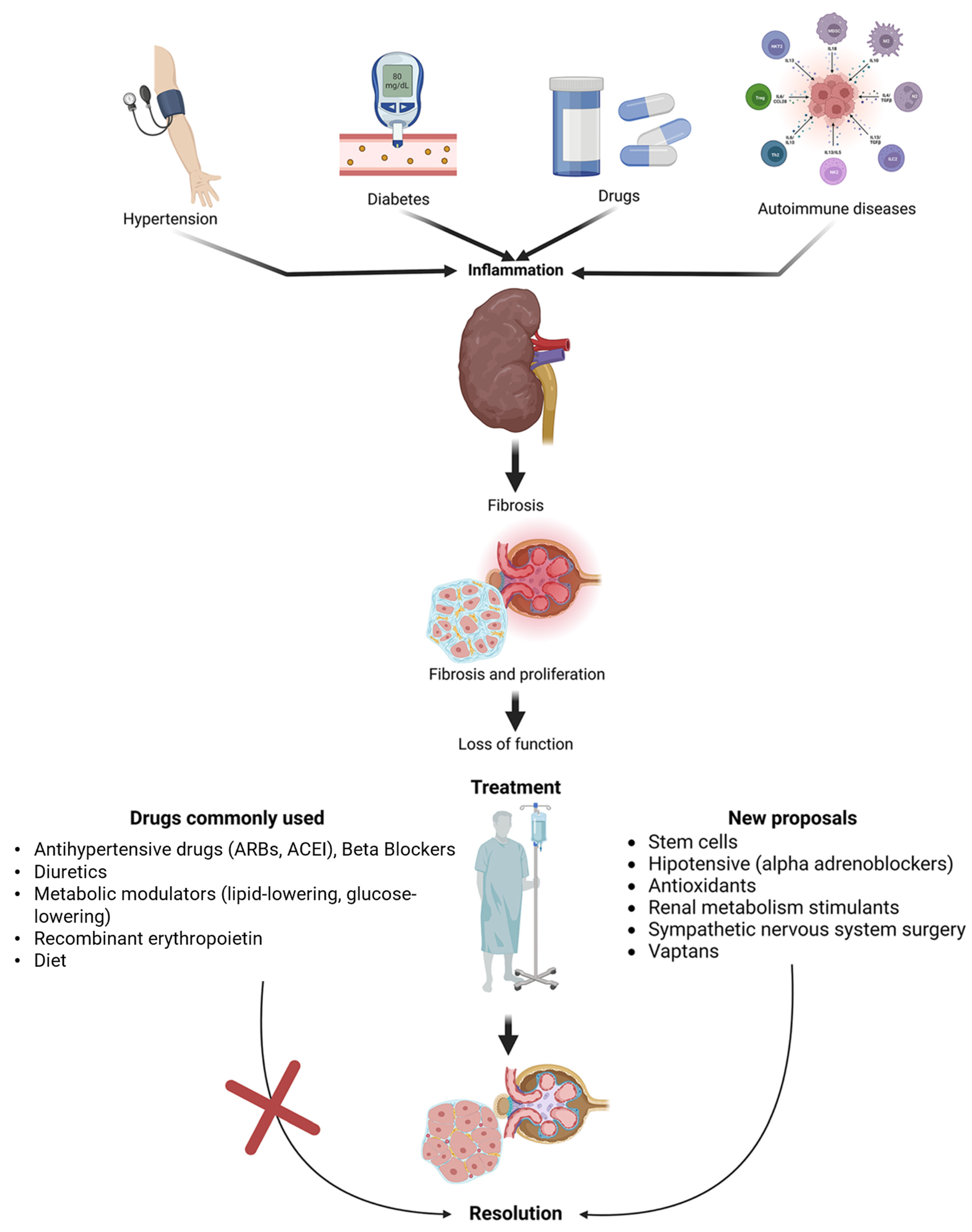
:1. Introduction
2. Main Etiologies of CKD
2.1. Arterial Hypertension
2.2. Diabetes Mellitus
2.2.1. Microangiopathic Complications
2.2.2. Formation of Late Glycation End Products
2.2.3. Activation of the Hexosamine Pathway
2.3. Pathophysiology of Fibrosis
3. CKD Treatment
3.1. General Considerations
3.2. Antihypertensives
3.2.1. ACE Inhibitors
3.2.2. Angiotensin Receptor Blockers (ARBs)
3.2.3. Renin Inhibitors
3.2.4. Calcium Channel Blockers
3.2.5. Adrenoblockers
3.3. Diuretics
3.4. Mineralocorticoid Receptor
3.5. Hypoglycemic Agents
3.5.1. Insulin Sensitizers
3.5.2. Dipeptidylpeptidase-4 (DPP-4i) Enzyme Inhibitors
3.5.3. Sodium–Glucose Cotransporter 2 Inhibitor (SGLT2i)
3.6. Diet
3.7. Treatment with Vitamins and Minerals
3.7.1. Potassium
3.7.2. Iron
3.7.3. Vitamin D
3.8. Calcimimetics
4. Novel Proposals
4.1. Probiotic in CKD
4.2. Metabolic Modulators
4.3. Novel Antihypertensives
4.3.1. Vasopressin Inhibitors
4.3.2. Treatments Based on Angiotensin 1–7 Inhibitor
4.3.3. Endothelin Inhibitors
4.4. Antifibrotics
4.5. Antioxidants
4.6. Retinoic Acid (RA) (Vitamin A)
4.7. Oral Carbon Absorbent (AST-120)
4.8. Glucagon-like Peptide-1 Receptor Agonist (GLP-1RA)
4.9. Demethylating Agents
4.10. Novel Potassium Binders
4.11. Hypoxia-Inducible Factor (HIF) Stabilizers
4.12. Stem Cells of Mesenchymal and Adipose Tissue Origins
4.13. Antagonism of the Sympathetic Nervous System or Sympathectomy
Sympathectomy
5. Benefit of the Treatment in Population with CKD
Future Direction
6. Conclusions
Author Contributions
Funding
Data Availability Statement
Acknowledgments
Conflicts of Interest
References
- Lameire, N.H.; Levin, A.; Kellum, J.A.; Cheung, M.; Jadoul, M.; Winkelmayer, W.C.; Stevens, P.E. Conference Participants Harmonizing Acute and Chronic Kidney Disease Definition and Classification: Report of a Kidney Disease: Improving Global Outcomes (KDIGO) Consensus Conference. Kidney Int. 2021, 100, 516–526. [Google Scholar] [CrossRef]
- Chawla, L.S.; Bellomo, R.; Bihorac, A.; Goldstein, S.L.; Siew, E.D.; Bagshaw, S.M.; Bittleman, D.; Cruz, D.; Endre, Z.; Fitzgerald, R.L.; et al. Acute Kidney Disease and Renal Recovery: Consensus Report of the Acute Disease Quality Initiative (ADQI) 16 Workgroup. Nat. Rev. Nephrol. 2017, 13, 241–257. [Google Scholar] [CrossRef]
- Jha, V.; Garcia-Garcia, G.; Iseki, K.; Li, Z.; Naicker, S.; Plattner, B.; Saran, R.; Wang, A.Y.-M.; Yang, C.-W. Chronic Kidney Disease: Global Dimension and Perspectives. Lancet 2013, 382, 260–272. [Google Scholar] [CrossRef]
- Weckmann, G.F.C.; Stracke, S.; Haase, A.; Spallek, J.; Ludwig, F.; Angelow, A.; Emmelkamp, J.M.; Mahner, M.; Chenot, J.-F. Diagnosis and Management of Non-Dialysis Chronic Kidney Disease in Ambulatory Care: A Systematic Review of Clinical Practice Guidelines. BMC Nephrol. 2018, 19, 258. [Google Scholar] [CrossRef] [PubMed]
- Wheeler, D.C.; Winkelmayer, W.C. KDIGO 2017 Clinical Practice Guideline Update for the Diagnosis, Evaluation, Prevention, and Treatment of Chronic Kidney Disease–Mineral and Bone Disorder (CKD-MBD). Kidney Int. Suppl. 2017, 7, 1–59. [Google Scholar] [CrossRef]
- Ameh, O.I.; Ekrikpo, U.; Bello, A.; Okpechi, I. Current Management Strategies of Chronic Kidney Disease in Resource-Limited Countries. Int. J. Nephrol. Renov. Dis. 2020, 13, 239–251. [Google Scholar] [CrossRef] [PubMed]
- McCarty, M.F. Adjuvant Strategies for Prevention of Glomerulosclerosis. Med. Hypotheses 2006, 67, 1277–1296. [Google Scholar] [CrossRef]
- Kluth, D.C.; Erwig, L.-P.; Rees, A.J. Multiple Facets of Macrophages in Renal Injury. Kidney Int. 2004, 66, 542–557. [Google Scholar] [CrossRef]
- López-Novoa, J.M.; Martínez-Salgado, C.; Rodríguez-Peña, A.B.; Hernández, F.J.L. Common Pathophysiological Mechanisms of Chronic Kidney Disease: Therapeutic Perspectives. Pharmacol. Ther. 2010, 128, 61–81. [Google Scholar] [CrossRef]
- Chia, T.Y.; Sattar, M.A.; Abdullah, M.H.; Ahmad, F.U.D.; Ibraheem, Z.O.; Li, K.J.; Pei, Y.P.; Rathore, H.A.; Singh, G.K.C.; Abdullah, N.A.; et al. Cyclosporine A-Induced Nephrotoxic Sprague-Dawley Rats Are More Susceptable to Altered Vascular Function and Haemodynamics. Int. J. Pharm. Pharm. Sci. 2012, 4, 10. [Google Scholar]
- Zhao, M.; Chen, Y.; Ding, G.; Xu, Y.; Bai, M.; Zhang, Y.; Jia, Z.; Huang, S.; Zhang, A. Renal Tubular Epithelium-Targeted Peroxisome Proliferator-Activated Receptor-γ Maintains the Epithelial Phenotype and Antagonizes Renal Fibrogenesis. Oncotarget 2016, 7, 64690–64701. [Google Scholar] [CrossRef] [PubMed]
- Levey, A.S.; Stevens, L.A.; Schmid, C.H.; Zhang, Y.L.; Castro, A.F.; Feldman, H.I.; Kusek, J.W.; Eggers, P.; Van Lente, F.; Greene, T.; et al. A New Equation to Estimate Glomerular Filtration Rate. Ann. Intern. Med. 2009, 150, 604–612. [Google Scholar] [CrossRef]
- GBD Compare. Available online: https://www.healthdata.org/data-visualization/gbd-compare (accessed on 16 December 2022).
- Macias Diaz, D.M.; Corrales Aguirre, M.D.C.; Reza Escalera, A.L.; Tiscareño Gutiérrez, M.T.; Ovalle Robles, I.; Macías Guzmán, M.J.; García Díaz, A.L.; Gutiérrez Peña, M.C.; Alvarado-Nájera, A.N.; González Domínguez, I.; et al. Histologic Characterization and Risk Factors for Persistent Albuminuria in Adolescents in a Region of Highly Prevalent End-Stage Renal Failure of Unknown Origin. Clin. Kidney J. 2022, 15, 1300–1311. [Google Scholar] [CrossRef]
- Mansour, A.S. Autoregulation: Mediators and Renin–Angiotensin System in Diseases and Treatments. Future J. Pharm. Sci. 2023, 9, 30. [Google Scholar] [CrossRef]
- Santos, M.M.R.; Cavalcante, A.C.F.P.S.; do Amaral, L.A.; de Souza, G.H.O.; dos Santos, B.S.; Portugal, L.C.; Bittencourt, F.F.; Troquez, T.; Rafacho, B.P.M.; Hiane, P.A.; et al. Combination of Cafeteria Diet with Intraperitoneally Streptozotocin in Rats. A Type-2 Diabetes Model. Acta Cir. Bras. 2021, 36, e360702. [Google Scholar] [CrossRef]
- Santos, R.A.S.; Sampaio, W.O.; Alzamora, A.C.; Motta-Santos, D.; Alenina, N.; Bader, M.; Campagnole-Santos, M.J. The ACE2/Angiotensin-(1–7)/MAS Axis of the Renin-Angiotensin System: Focus on Angiotensin-(1–7). Physiol. Rev. 2018, 98, 505–553. [Google Scholar] [CrossRef] [PubMed]
- Wolf, G.; Butzmann, U.; Wenzel, U.O. The Renin-Angiotensin System and Progression of Renal Disease: From Hemodynamics to Cell Biology. Nephron Physiol. 2003, 93, P3–P13. [Google Scholar] [CrossRef] [PubMed]
- Tylicki, L.; Lizakowski, S.; Rutkowski, B. Renin-Angiotensin-Aldosterone System Blockade for Nephroprotection: Current Evidence and Future Directions. J. Nephrol. 2012, 25, 900–910. [Google Scholar] [CrossRef]
- Sampaio, W.O.; Souza dos Santos, R.A.; Faria-Silva, R.; da Mata Machado, L.T.; Schiffrin, E.L.; Touyz, R.M. Angiotensin-(1–7) through Receptor Mas Mediates Endothelial Nitric Oxide Synthase Activation via Akt-Dependent Pathways. Hypertension 2007, 49, 185–192. [Google Scholar] [CrossRef]
- Yu, L.; Yuan, K.; Phuong HT, A.; Park, B.M.; Kim, S.H. Angiotensin-(1–5), an Active Mediator of Renin-Angiotensin System, Stimulates ANP Secretion via Mas Receptor. Peptides 2016, 86, 33–41. [Google Scholar] [CrossRef]
- Ren, X.; Zhang, Z.; Yan, Z. Association Between Lipoprotein (A) and Diabetic Nephropathy in Patients With Type 2 Diabetes Mellitus: A Meta-Analysis. Front. Endocrinol. 2021, 12, 633529. [Google Scholar] [CrossRef] [PubMed]
- Basha, B.; Samuel, S.M.; Triggle, C.R.; Ding, H. Endothelial Dysfunction in Diabetes Mellitus: Possible Involvement of Endoplasmic Reticulum Stress? Exp. Diabetes Res. 2012, 2012, 481840. [Google Scholar] [CrossRef] [PubMed]
- Thomas, H.Y.; Ford Versypt, A.N. Pathophysiology of Mesangial Expansion in Diabetic Nephropathy: Mesangial Structure, Glomerular Biomechanics, and Biochemical Signaling and Regulation. J. Biol. Eng. 2022, 16, 19. [Google Scholar] [CrossRef]
- Hung, P.-H.; Hsu, Y.-C.; Chen, T.-H.; Lin, C.-L. Recent Advances in Diabetic Kidney Diseases: From Kidney Injury to Kidney Fibrosis. Int. J. Mol. Sci. 2021, 22, 11857. [Google Scholar] [CrossRef]
- Twarda-Clapa, A.; Olczak, A.; Białkowska, A.M.; Koziołkiewicz, M. Advanced Glycation End-Products (AGEs): Formation, Chemistry, Classification, Receptors, and Diseases Related to AGEs. Cells 2022, 11, 1312. [Google Scholar] [CrossRef]
- Werlen, G.; Li, M.-L.; Tottone, L.; Da Silva-Diz, V.; Su, X.; Herranz, D.; Jacinto, E. Dietary Glucosamine Overcomes the Defects in Aβ-T Cell Ontogeny Caused by the Loss of de Novo Hexosamine Biosynthesis. Nat. Commun. 2022, 13, 7404. [Google Scholar] [CrossRef] [PubMed]
- Papachristoforou, E.; Lambadiari, V.; Maratou, E.; Makrilakis, K. Association of Glycemic Indices (Hyperglycemia, Glucose Variability, and Hypoglycemia) with Oxidative Stress and Diabetic Complications. J. Diabetes Res. 2020, 2020, 7489795. [Google Scholar] [CrossRef] [PubMed]
- Barnes, J.L.; Gorin, Y. Myofibroblast Differentiation during Fibrosis: Role of NAD(P)H Oxidases. Kidney Int. 2011, 79, 944–956. [Google Scholar] [CrossRef]
- Boor, P.; Ostendorf, T.; Floege, J. Renal Fibrosis: Novel Insights into Mechanisms and Therapeutic Targets. Nat. Rev. Nephrol. 2010, 6, 643–656. [Google Scholar] [CrossRef] [PubMed]
- Zeisberg, M.; Neilson, E.G. Mechanisms of Tubulointerstitial Fibrosis. J. Am. Soc. Nephrol. 2010, 21, 1819–1834. [Google Scholar] [CrossRef]
- Cha, J.J.; Mandal, C.; Ghee, J.Y.; Yoo, J.A.; Lee, M.J.; Kang, Y.S.; Hyun, Y.Y.; Lee, J.E.; Kim, H.W.; Han, S.Y.; et al. Inhibition of Renal Stellate Cell Activation Reduces Renal Fibrosis. Biomedicines 2020, 8, 431. [Google Scholar] [CrossRef]
- Fogo, A.B. Progression and Potential Regression of Glomerulosclerosis. Kidney Int. 2001, 59, 804–819. [Google Scholar] [CrossRef] [PubMed]
- Rodríguez-Peña, A.; Prieto, M.; Duwel, A.; Rivas, J.V.; Eleno, N.; Pérez-Barriocanal, F.; Arévalo, M.; Smith, J.D.; Vary, C.P.H.; Bernabeu, C.; et al. Up-regulation of Endoglin, a TGF-β-binding Protein, in Rats with Experimental Renal Fibrosis Induced by Renal Mass Reduction. Nephrol. Dial. Transplant. 2001, 16, 34–39. [Google Scholar] [CrossRef] [PubMed]
- Schnaper, H.W.; Hayashida, T.; Hubchak, S.C.; Poncelet, A.-C. TGF-β Signal Transduction and Mesangial Cell Fibrogenesis. Am. J. Physiol.-Ren. Physiol. 2003, 284, F243–F252. [Google Scholar] [CrossRef] [PubMed]
- Chung, A.C.K.; Huang, X.R.; Meng, X.; Lan, H.Y. miR-192 Mediates TGF-β/Smad3-Driven Renal Fibrosis. J. Am. Soc. Nephrol. 2010, 21, 1317–1325. [Google Scholar] [CrossRef]
- He, W.; Kang, Y.S.; Dai, C.; Liu, Y. Blockade of Wnt/β-Catenin Signaling by Paricalcitol Ameliorates Proteinuria and Kidney Injury. J. Am. Soc. Nephrol. 2011, 22, 90–103. [Google Scholar] [CrossRef]
- Tan, X.; Wen, X.; Liu, Y. Paricalcitol Inhibits Renal Inflammation by Promoting Vitamin D Receptor–Mediated Sequestration of NF-κB Signaling. J. Am. Soc. Nephrol. 2008, 19, 1741–1752. [Google Scholar] [CrossRef]
- Hirschberg, R. Wound Healing in the Kidney: Complex Interactions in Renal Interstitial Fibrogenesis. J. Am. Soc. Nephrol. 2005, 16, 9–11. [Google Scholar] [CrossRef]
- Iwano, M.; Neilson, E.G. Mechanisms of Tubulointerstitial Fibrosis. Curr. Opin. Nephrol. Hypertens. 2004, 13, 279–284. [Google Scholar] [CrossRef]
- Kalluri, R.; Neilson, E.G. Epithelial-Mesenchymal Transition and Its Implications for Fibrosis. J. Clin. Investig. 2003, 112, 1776–1784. [Google Scholar] [CrossRef]
- Liu, Y. New Insights into Epithelial-Mesenchymal Transition in Kidney Fibrosis. J. Am. Soc. Nephrol. 2010, 21, 212–222. [Google Scholar] [CrossRef] [PubMed]
- Efstratiadis, G.; Divani, M.; Katsioulis, E.; Vergoulas, G. Renal Fibrosis. Hippokratia 2009, 13, 224–229. [Google Scholar] [PubMed]
- Gagliardini, E.; Benigni, A. Role of Anti-TGF-β Antibodies in the Treatment of Renal Injury. Cytokine Growth Factor. Rev. 2006, 17, 89–96. [Google Scholar] [CrossRef]
- Gupta, S.; Clarkson, M.R.; Duggan, J.; Brady, H.R. Connective Tissue Growth Factor: Potential Role in Glomerulosclerosis and Tubulointerstitial Fibrosis. Kidney Int. 2000, 58, 1389–1399. [Google Scholar] [CrossRef]
- Branton, M.H.; Kopp, J.B. TGF-β and Fibrosis. Microbes Infect. 1999, 1, 1349–1365. [Google Scholar] [CrossRef]
- Böttinger, E.P.; Bitzer, M. TGF-ß Signaling in Renal Disease. J. Am. Soc. Nephrol. 2002, 13, 2600–2610. [Google Scholar] [CrossRef] [PubMed]
- Li, Y.; Yang, J.; Dai, C.; Wu, C.; Liu, Y. Role for Integrin-Linked Kinase in Mediating Tubular Epithelial to Mesenchymal Transition and Renal Interstitial Fibrogenesis. J. Clin. Investig. 2003, 112, 503–516. [Google Scholar] [CrossRef]
- Bielesz, B.; Sirin, Y.; Si, H.; Niranjan, T.; Gruenwald, A.; Ahn, S.; Kato, H.; Pullman, J.; Gessler, M.; Haase, V.H.; et al. Epithelial Notch Signaling Regulates Interstitial Fibrosis Development in the Kidneys of Mice and Humans. J. Clin. Investig. 2010, 120, 4040–4054. [Google Scholar] [CrossRef]
- Sharma, S.; Sirin, Y.; Susztak, K. The Story of Notch and Chronic Kidney Disease. Curr. Opin. Nephrol. Hypertens. 2011, 20, 56–61. [Google Scholar] [CrossRef] [PubMed]
- Higgins, D.F.; Kimura, K.; Bernhardt, W.M.; Shrimanker, N.; Akai, Y.; Hohenstein, B.; Saito, Y.; Johnson, R.S.; Kretzler, M.; Cohen, C.D.; et al. Hypoxia Promotes Fibrogenesis in Vivo via HIF-1 Stimulation of Epithelial-to-Mesenchymal Transition. J. Clin. Investig. 2007, 117, JCI30487. [Google Scholar] [CrossRef] [PubMed]
- Sun, S.; Ning, X.; Zhang, Y.; Lu, Y.; Nie, Y.; Han, S.; Liu, L.; Du, R.; Xia, L.; He, L.; et al. Hypoxia-Inducible Factor-1α Induces Twist Expression in Tubular Epithelial Cells Subjected to Hypoxia, Leading to Epithelial-to-Mesenchymal Transition. Kidney Int. 2009, 75, 1278–1287. [Google Scholar] [CrossRef]
- Kurnatowska, I.; Grzelak, P.; Masajtis-Zagajewska, A.; Kaczmarska, M.; Stefańczyk, L.; Vermeer, C.; Maresz, K.; Nowicki, M. Effect of Vitamin K2 on Progression of Atherosclerosis and Vascular Calcification in Nondialyzed Patients with Chronic Kidney Disease Stages 3–5. Pol. Arch. Intern. Med. 2015, 125, 631–640. [Google Scholar] [CrossRef]
- Reiss, A.B.; Miyawaki, N.; Moon, J.; Kasselman, L.J.; Voloshyna, I.; D’Avino, R.; Leon, J.D. CKD, Arterial Calcification, Atherosclerosis and Bone Health: Inter-Relationships and Controversies. Atherosclerosis 2018, 278, 49–59. [Google Scholar] [CrossRef] [PubMed]
- Jiang, Z.; Wang, Y.; Zhao, X.; Cui, H.; Han, M.; Ren, X.; Gang, X.; Wang, G. Obesity and Chronic Kidney Disease. Am. J. Physiol. -Endocrinol. Metab. 2023, 324, E24–E41. [Google Scholar] [CrossRef] [PubMed]
- Jain, G.; Jaimes, E.A. Nicotine Signaling and Progression of Chronic Kidney Disease in Smokers. Biochem. Pharmacol. 2013, 86, 1215–1223. [Google Scholar] [CrossRef] [PubMed]
- Guo, L.; Zhang, Y.; Lu, J.; Li, X.; Zhang, C.; Song, W.; Dong, Y.; Zhou, X.; Li, R. Nicotine Promotes Renal Interstitial Fibrosis via Upregulation of XIAP in an Alpha7-nAChR-Dependent Manner. Mol. Cell. Endocrinol. 2023, 576, 111989. [Google Scholar] [CrossRef]
- Kim, S.-Y.; Moon, A.-R. Drug-Induced Nephrotoxicity and Its Biomarkers. Biomol. Ther. 2012, 20, 268–272. [Google Scholar] [CrossRef] [PubMed]
- Dobrek, L. A Synopsis of Current Theories on Drug-Induced Nephrotoxicity. Life 2023, 13, 325. [Google Scholar] [CrossRef]
- Castro, M.C.M. Conservative Management for Patients with Chronic Kidney Disease Refusing Dialysis. J. Bras. Nefrol. 2019, 41, 95–102. [Google Scholar] [CrossRef]
- Hsing, S.-C.; Lu, K.-C.; Sun, C.-A.; Chien, W.-C.; Chung, C.-H.; Kao, S.-Y. The Association of Losartan and Ramipril Therapy With Kidney and Cardiovascular Outcomes in Patients With Chronic Kidney Disease. Medicine 2015, 94, e1999. [Google Scholar] [CrossRef]
- Ito, S.; Satoh, M.; Tamaki, Y.; Gotou, H.; Charney, A.; Okino, N.; Akahori, M.; Zhang, J. Safety and Efficacy of LCZ696, a First-in-Class Angiotensin Receptor Neprilysin Inhibitor, in Japanese Patients with Hypertension and Renal Dysfunction. Hypertens. Res. 2015, 38, 269–275. [Google Scholar] [CrossRef]
- Garlo, K.G.; Bates, D.W.; Seger, D.L.; Fiskio, J.M.; Charytan, D.M. Lab Monitoring and Acute Care Utilization during Initiation of Renin Angiotensin Aldosterone Inhibitors or Diuretics in Chronic Kidney Disease. Medicine 2019, 98, e17963. [Google Scholar] [CrossRef] [PubMed]
- Ku, E.; McCulloch, C.E.; Vittinghoff, E.; Lin, F.; Johansen, K.L. Use of Antihypertensive Agents and Association with Risk of Adverse Outcomes in Chronic Kidney Disease: Focus on Angiotensin-Converting Enzyme Inhibitors and Angiotensin Receptor Blockers. J. Am. Heart Assoc. 2018, 7, e009992. [Google Scholar] [CrossRef]
- Riccio, E.; Capuano, I.; Buonanno, P.; Andreucci, M.; Provenzano, M.; Amicone, M.; Rizzo, M.; Pisani, A. RAAS Inhibitor Prescription and Hyperkalemia Event in Patients with Chronic Kidney Disease: A Single-Center Retrospective Study. Front. Cardiovasc. Med. 2022, 9, 824095. [Google Scholar] [CrossRef]
- Chou, Y.-H.; Huang, T.-M.; Pan, S.-Y.; Chang, C.-H.; Lai, C.-F.; Wu, V.-C.; Wu, M.-S.; Wu, K.-D.; Chu, T.-S.; Lin, S.-L. Renin-Angiotensin System Inhibitor Is Associated with Lower Risk of Ensuing Chronic Kidney Disease after Functional Recovery from Acute Kidney Injury. Sci. Rep. 2017, 7, 46518. [Google Scholar] [CrossRef]
- Maschio, G.; Alberti, D.; Janin, G.; Locatelli, F.; Mann, J.F.E.; Motolese, M.; Ponticelli, C.; Ritz, E.; Zucchelli, P. Effect of the Angiotensin-Converting–Enzyme Inhibitor Benazepril on the Progression of Chronic Renal Insufficiency. N. Engl. J. Med. 1996, 334, 939–945. [Google Scholar] [CrossRef] [PubMed]
- Lewis, E.J.; Hunsicker, L.G.; Bain, R.P.; Rohde, R.D. The Effect of Angiotensin-Converting-Enzyme Inhibition on Diabetic Nephropathy. N. Engl. J. Med. 1993, 329, 1456–1462. [Google Scholar] [CrossRef]
- Elung-Jensen, T.; Strandgaard, S.; Kamper, A.-L. Longitudinal Observations on Circadian Blood Pressure Variation in Chronic Kidney Disease Stages 3–5. Nephrol. Dial. Transplant. 2008, 23, 2873–2878. [Google Scholar] [CrossRef] [PubMed]
- Xue, C.; Zhou, C.; Yang, B.; Lv, J.; Dai, B.; Yu, S.; Wang, Y.; Zhao, G.; Mei, C. Comparison of Efficacy and Safety between Benidipine and Hydrochlorothiazide in Fosinopril-Treated Hypertensive Patients with Chronic Kidney Disease: Protocol for a Randomised Controlled Trial. BMJ Open 2017, 7, e013672. [Google Scholar] [CrossRef]
- Onuigbo, M.A.C. Renoprevention: A New Concept for Reengineering Nephrology Care—An Economic Impact and Patient Outcome Analysis of Two Hypothetical Patient Management Paradigms in the CCU. Ren. Fail. 2013, 35, 23–28. [Google Scholar] [CrossRef] [PubMed]
- Ninomiya, T.; Perkovic, V.; Gallagher, M.; Jardine, M.; Cass, A.; Arima, H.; Anderson, C.; Neal, B.; Woodward, M.; Omae, T.; et al. Lower Blood Pressure and Risk of Recurrent Stroke in Patients with Chronic Kidney Disease: PROGRESS Trial. Kidney Int. 2008, 73, 963–970. [Google Scholar] [CrossRef] [PubMed]
- Ruggenenti, P.; Perna, A.; Gherardi, G.; Gaspari, F.; Benini, R.; Remuzzi, G. Renal Function and Requirement for Dialysis in Chronic Nephropathy Patients on Long-Term Ramipril: REIN Follow-up Trial. Gruppo Italiano Di Studi Epidemiologici in Nefrologia (GISEN). Ramipril Efficacy in Nephropathy. Lancet 1998, 352, 1252–1256. [Google Scholar] [CrossRef]
- Bryniarski, P.; Nazimek, K.; Marcinkiewicz, J. Immunomodulatory Activity of the Most Commonly Used Antihypertensive Drugs—Angiotensin Converting Enzyme Inhibitors and Angiotensin II Receptor Blockers. Int. J. Mol. Sci. 2022, 23, 1772. [Google Scholar] [CrossRef]
- Fu, K.; Hu, Y.; Zhang, H.; Wang, C.; Lin, Z.; Lu, H.; Ji, X. Insights of Worsening Renal Function in Type 1 Cardiorenal Syndrome: From the Pathogenesis, Biomarkers to Treatment. Front. Cardiovasc. Med. 2021, 8, 760152. [Google Scholar] [CrossRef]
- Imamura, T.; Hori, M.; Kinugawa, K. Optimal Therapeutic Strategy Using Sacubitril/Valsartan in a Patient with Systolic Heart Failure and Chronic Kidney Disease—An Initial Case Report in Japan. Intern. Med. 2021, 60, 2807–2809. [Google Scholar] [CrossRef]
- Banerjee, D.; Winocour, P.; Chowdhury, T.A.; De, P.; Wahba, M.; Montero, R.; Fogarty, D.; Frankel, A.H.; Karalliedde, J.; Mark, P.B.; et al. Management of Hypertension and Renin-Angiotensin-Aldosterone System Blockade in Adults with Diabetic Kidney Disease: Association of British Clinical Diabetologists and the Renal Association UK Guideline Update 2021. BMC Nephrol. 2022, 23, 9. [Google Scholar] [CrossRef] [PubMed]
- Nakamura, T.; Sato, E.; Fujiwara, N.; Kawagoe, Y.; Koide, H.; Ueda, Y.; Takeuchi, M.; Yamagishi, S. Calcium Channel Blocker Inhibition of AGE and RAGE Axis Limits Renal Injury in Nondiabetic Patients with Stage I or II Chronic Kidney Disease. Clin. Cardiol. 2011, 34, 372–377. [Google Scholar] [CrossRef]
- Toto, R.D.; Tian, M.; Fakouhi, K.; Champion, A.; Bacher, P. Effects of Calcium Channel Blockers on Proteinuria in Patients with Diabetic Nephropathy. J. Clin. Hypertens. 2008, 10, 761–769. [Google Scholar] [CrossRef]
- Ohsawa, M.; Tamura, K.; Kanaoka, T.; Wakui, H.; Maeda, A.; Dejima, T.; Azushima, K.; Uneda, K.; Kobayashi, R.; Tsurumi-Ikeya, Y.; et al. Addition of Aliskiren to Angiotensin Receptor Blocker Improves Ambulatory Blood Pressure Profile and Cardiorenal Function Better than Addition of Benazepril in Chronic Kidney Disease. Int. J. Mol. Sci. 2013, 14, 15361–15375. [Google Scholar] [CrossRef] [PubMed]
- Chang, T.I.; Zheng, Y.; Montez-Rath, M.E.; Winkelmayer, W.C. Antihypertensive Medication Use in Older Patients Transitioning from Chronic Kidney Disease to End-Stage Renal Disease on Dialysis. Clin. J. Am. Soc. Nephrol. 2016, 11, 1401–1412. [Google Scholar] [CrossRef] [PubMed]
- Gomez-Sanchez, E. Third Generation Mineralocorticoid Receptor Antagonists; Why We Need a Fourth. J. Cardiovasc. Pharmacol. 2016, 67, 26–38. [Google Scholar] [CrossRef]
- Ng, K.P.; Jain, P.; Heer, G.; Redman, V.; Chagoury, O.L.; Dowswell, G.; Greenfield, S.; Freemantle, N.; Townend, J.N.; Gill, P.S.; et al. Spironolactone to Prevent Cardiovascular Events in Early-Stage Chronic Kidney Disease (STOP-CKD): Study Protocol for a Randomized Controlled Pilot Trial. Trials 2014, 15, 158. [Google Scholar] [CrossRef] [PubMed]
- Hill, N.R.; Lasserson, D.; Thompson, B.; Perera-Salazar, R.; Wolstenholme, J.; Bower, P.; Blakeman, T.; Fitzmaurice, D.; Little, P.; Feder, G.; et al. Benefits of Aldosterone Receptor Antagonism in Chronic Kidney Disease (BARACK D) Trial–a Multi-Centre, Prospective, Randomised, Open, Blinded End-Point, 36-Month Study of 2616 Patients within Primary Care with Stage 3b Chronic Kidney Disease to Compare the Efficacy of Spironolactone 25 Mg Once Daily in Addition to Routine Care on Mortality and Cardiovascular Outcomes versus Routine Care Alone: Study Protocol for a Randomized Controlled Trial. Trials 2014, 15, 160. [Google Scholar] [CrossRef] [PubMed]
- Haddock, B.; Larsson, H.B.W.; Francis, S.; Andersen, U.B. Human Renal Response to Furosemide: Simultaneous Oxygenation and Perfusion Measurements in Cortex and Medulla. Acta Physiol. 2019, 227, e13292. [Google Scholar] [CrossRef]
- Fuwa, D.; Fukuda, M.; Ogiyama, Y.; Sato, R.; Mizuno, M.; Miura, T.; Abe-Dohmae, S.; Michikawa, M.; Kobori, H.; Ohte, N. Addition of Hydrochlorothiazide to Angiotensin Receptor Blocker Therapy Can Achieve a Lower Sodium Balance with No Acceleration of Intrarenal Renin Angiotensin System in Patients with Chronic Kidney Disease. J. Renin-Angiotensin-Aldosterone Syst. 2016, 17, 1–9. [Google Scholar] [CrossRef]
- Zheng, Z.; Jiang, X.; Chen, J.; He, D.; Xie, X.; Lu, Y. Continuous versus Intermittent Use of Furosemide in Patients with Heart Failure and Moderate Chronic Renal Dysfunction. ESC Heart Fail. 2021, 8, 2070–2078. [Google Scholar] [CrossRef] [PubMed]
- LaMoia, T.E.; Butrico, G.M.; Kalpage, H.A.; Goedeke, L.; Hubbard, B.T.; Vatner, D.F.; Gaspar, R.C.; Zhang, X.-M.; Cline, G.W.; Nakahara, K.; et al. Metformin, Phenformin, and Galegine Inhibit Complex IV Activity and Reduce Glycerol-Derived Gluconeogenesis. Proc. Natl. Acad. Sci. USA 2022, 119, e2122287119. [Google Scholar] [CrossRef]
- Kalantar-Zadeh, K.; Kovesdy, C.P. Should Restrictions Be Relaxed for Metformin Use in Chronic Kidney Disease? No, We Should Never Again Compromise Safety! Diabetes Care 2016, 39, 1281–1286. [Google Scholar] [CrossRef]
- Salaverría de Sanz, N.; Palmucci, G.; Suniaga de Daza, M.; Velásquez, E. Tratamiento con antihiperglucemiantes orales: Clasificación, propiedades, combinaciones, indicaciones, contraindicaciones y eventos adversos. Rev. Venez. Endocrinol. Metab. 2012, 10, 58–64. [Google Scholar]
- Varvaki Rados, D.; Catani Pinto, L.; Reck Remonti, L.; Bauermann Leitão, C.; Gross, J.L. The Association between Sulfonylurea Use and All-Cause and Cardiovascular Mortality: A Meta-Analysis with Trial Sequential Analysis of Randomized Clinical Trials. PLoS Med. 2016, 13, e1001992. [Google Scholar] [CrossRef]
- Russo, E.; Penno, G.; Prato, S.D. Managing Diabetic Patients with Moderate or Severe Renal Impairment Using DPP-4 Inhibitors: Focus on Vildagliptin. Diabetes Metab. Syndr. Obes. 2013, 6, 161–170. [Google Scholar] [CrossRef] [PubMed]
- Yagoglu, A.I.; Dizdar, O.S.; Erdem, S.; Akcakaya, B.; Gunal, A.I. The effect of linagliptin on renal progression in type-2 diabetes mellitus patients with chronic kidney disease: A prospective randomized controlled study. Nefrología 2020, 40, 664–671. [Google Scholar] [CrossRef] [PubMed]
- Heerspink, H.J.L.; Sjöström, C.D.; Jongs, N.; Chertow, G.M.; Kosiborod, M.; Hou, F.F.; McMurray, J.J.V.; Rossing, P.; Correa-Rotter, R.; Kurlyandskaya, R.; et al. Effects of Dapagliflozin on Mortality in Patients with Chronic Kidney Disease: A Pre-Specified Analysis from the DAPA-CKD Randomized Controlled Trial. Eur. Heart J. 2021, 42, 1216–1227. [Google Scholar] [CrossRef] [PubMed]
- Mosenzon, O.; Wiviott, S.D.; Cahn, A.; Rozenberg, A.; Yanuv, I.; Goodrich, E.L.; Murphy, S.A.; Heerspink, H.J.L.; Zelniker, T.A.; Dwyer, J.P.; et al. Effects of Dapagliflozin on Development and Progression of Kidney Disease in Patients with Type 2 Diabetes: An Analysis from the DECLARE–TIMI 58 Randomised Trial. Lancet Diabetes Endocrinol. 2019, 7, 606–617. [Google Scholar] [CrossRef]
- Wanner, C.; Inzucchi, S.E.; Lachin, J.M.; Fitchett, D.; von Eynatten, M.; Mattheus, M.; Johansen, O.E.; Woerle, H.J.; Broedl, U.C.; Zinman, B. Empagliflozin and Progression of Kidney Disease in Type 2 Diabetes. N. Engl. J. Med. 2016, 375, 323–334. [Google Scholar] [CrossRef] [PubMed]
- Wiviott, S.D.; Raz, I.; Bonaca, M.P.; Mosenzon, O.; Kato, E.T.; Cahn, A.; Silverman, M.G.; Zelniker, T.A.; Kuder, J.F.; Murphy, S.A.; et al. Dapagliflozin and Cardiovascular Outcomes in Type 2 Diabetes. N. Engl. J. Med. 2019, 380, 347–357. [Google Scholar] [CrossRef]
- Losappio, V.; Infante, B.; Leo, S.; Troise, D.; Calvaruso, M.; Vitale, P.; Renzi, S.; Stallone, G.; Castellano, G. Nutrition-Based Management of Inflammaging in CKD and Renal Replacement Therapies. Nutrients 2021, 13, 267. [Google Scholar] [CrossRef]
- Park, J.S.; Jung, H.H.; Yang, W.S.; Kim, H.H.; Kim, S.B.; Park, S.-K.; Hong, C.D. Protein Intake and the Nutritional Status in Patients with Pre-Dialysis Chronic Renal Failure on Unrestircted Diet. Korean J. Intern. Med. 1997, 12, 115–121. [Google Scholar] [CrossRef]
- Garneata, L.; Stancu, A.; Dragomir, D.; Stefan, G.; Mircescu, G. Ketoanalogue-Supplemented Vegetarian Very Low-Protein Diet and CKD Progression. J. Am. Soc. Nephrol. 2016, 27, 2164–2176. [Google Scholar] [CrossRef]
- Watanabe, K.; Sato, E.; Mishima, E.; Watanabe, M.; Abe, T.; Takahashi, N.; Nakayama, M. Effect of Uremic Toxins on Hippocampal Cell Damage: Analysis in Vitro and in Rat Model of Chronic Kidney Disease. Heliyon 2021, 7, e06221. [Google Scholar] [CrossRef]
- Kempner, W. Some Effects of the Rice Diet Treatment of Kidney Disease and Hypertension. Bull. N. Y. Acad. Med. 1946, 22, 358–370. [Google Scholar] [PubMed]
- Kohsaka, S.; Okami, S.; Kanda, E.; Kashihara, N.; Yajima, T. Cardiovascular and Renal Outcomes Associated With Hyperkalemia in Chronic Kidney Disease: A Hospital-Based Cohort Study. Mayo Clin. Proc. Innov. Qual. Outcomes 2021, 5, 274–285. [Google Scholar] [CrossRef] [PubMed]
- Wang, H.-H.; Hung, C.-C.; Hwang, D.-Y.; Kuo, M.-C.; Chiu, Y.-W.; Chang, J.-M.; Tsai, J.-C.; Hwang, S.-J.; Seifter, J.L.; Chen, H.-C. Hypokalemia, Its Contributing Factors and Renal Outcomes in Patients with Chronic Kidney Disease. PLoS ONE 2013, 8, e67140. [Google Scholar] [CrossRef] [PubMed]
- Yamada, S.; Inaba, M. Potassium Metabolism and Management in Patients with CKD. Nutrients 2021, 13, 1751. [Google Scholar] [CrossRef]
- Ueda, N.; Takasawa, K. Impact of Inflammation on Ferritin, Hepcidin and the Management of Iron Deficiency Anemia in Chronic Kidney Disease. Nutrients 2018, 10, 1173. [Google Scholar] [CrossRef]
- Bhandari, S.; Allgar, V.; Lamplugh, A.; Macdougall, I.; Kalra, P.A. A Multicentre Prospective Double Blinded Randomised Controlled Trial of Intravenous Iron (Ferric Derisomaltose (FDI)) in Iron Deficient but Not Anaemic Patients with Chronic Kidney Disease on Functional Status. BMC Nephrol. 2021, 22, 115. [Google Scholar] [CrossRef]
- Strauss, W.E.; Dahl, N.V.; Li, Z.; Lau, G.; Allen, L.F. Ferumoxytol versus Iron Sucrose Treatment: A Post-Hoc Analysis of Randomized Controlled Trials in Patients with Varying Renal Function and Iron Deficiency Anemia. BMC Hematol. 2016, 16, 20. [Google Scholar] [CrossRef]
- Macdougall, I.C.; Bock, A.H.; Carrera, F.; Eckardt, K.-U.; Gaillard, C.; Van Wyck, D.; Roubert, B.; Nolen, J.G.; Roger, S.D. FIND-CKD: A Randomized Trial of Intravenous Ferric Carboxymaltose versus Oral Iron in Patients with Chronic Kidney Disease and Iron Deficiency Anaemia. Nephrol. Dial. Transpl. 2014, 29, 2075–2084. [Google Scholar] [CrossRef]
- Kalra, P.A.; Bhandari, S.; Saxena, S.; Agarwal, D.; Wirtz, G.; Kletzmayr, J.; Thomsen, L.L.; Coyne, D.W. A Randomized Trial of Iron Isomaltoside 1000 versus Oral Iron in Non-Dialysis-Dependent Chronic Kidney Disease Patients with Anaemia. Nephrol. Dial. Transpl. 2016, 31, 646–655. [Google Scholar] [CrossRef]
- Kalra, P.A.; Bhandari, S. Safety of Intravenous Iron Use in Chronic Kidney Disease. Curr. Opin. Nephrol. Hypertens. 2016, 25, 529–535. [Google Scholar] [CrossRef]
- Roger, S.D.; Gaillard, C.A.; Bock, A.H.; Carrera, F.; Eckardt, K.-U.; Van Wyck, D.B.; Cronin, M.; Meier, Y.; Larroque, S.; Macdougall, I.C. Safety of Intravenous Ferric Carboxymaltose versus Oral Iron in Patients with Nondialysis-Dependent CKD: An Analysis of the 1-Year FIND-CKD Trial. Nephrol. Dial. Transpl. 2017, 32, 1530–1539. [Google Scholar] [CrossRef]
- Block, G.A.; Pergola, P.E.; Fishbane, S.; Martins, J.G.; LeWinter, R.D.; Uhlig, K.; Neylan, J.F.; Chertow, G.M. Effect of Ferric Citrate on Serum Phosphate and Fibroblast Growth Factor 23 among Patients with Nondialysis-Dependent Chronic Kidney Disease: Path Analyses. Nephrol. Dial. Transpl. 2019, 34, 1115–1124. [Google Scholar] [CrossRef] [PubMed]
- Stubbs, J.R.; Zhang, S.; Friedman, P.A.; Nolin, T.D. Decreased Conversion of 25-Hydroxyvitamin D3 to 24,25-Dihydroxyvitamin D3 Following Cholecalciferol Therapy in Patients with CKD. Clin. J. Am. Soc. Nephrol. 2014, 9, 1965–1973. [Google Scholar] [CrossRef] [PubMed]
- Szeto, C.-C.; Li, P.K.-T. The Use of Vitamin D Analogues in Chronic Kidney Diseases: Possible Mechanisms beyond Bone and Mineral Metabolism. NDT Plus 2009, 2, 205–212. [Google Scholar] [CrossRef]
- Ballinger, A.E.; Palmer, S.C.; Nistor, I.; Craig, J.C.; Strippoli, G.F. Calcimimetics for Secondary Hyperparathyroidism in Chronic Kidney Disease Patients. Cochrane Database Syst. Rev. 2014, 12, CD006254. [Google Scholar] [CrossRef]
- Torres, P.U. Cinacalcet HCl: A Novel Treatment for Secondary Hyperparathyroidism Caused by Chronic Kidney Disease. J. Ren. Nutr. 2006, 16, 253–258. [Google Scholar] [CrossRef]
- Fu, E.L.; Evans, M.; Clase, C.M.; Tomlinson, L.A.; van Diepen, M.; Dekker, F.W.; Carrero, J.J. Stopping Renin-Angiotensin System Inhibitors in Patients with Advanced CKD and Risk of Adverse Outcomes: A Nationwide Study. J. Am. Soc. Nephrol. 2021, 32, 424–435. [Google Scholar] [CrossRef] [PubMed]
- Kim-Mitsuyama, S.; Soejima, H.; Yasuda, O.; Node, K.; Jinnouchi, H.; Yamamoto, E.; Sekigami, T.; Ogawa, H.; Matsui, K. Cardiovascular and Renal Protective Role of Angiotensin Blockade in Hypertension with Advanced CKD: A Subgroup Analysis of ATTEMPT-CVD Randomized Trial. Sci. Rep. 2018, 8, 3150. [Google Scholar] [CrossRef] [PubMed]
- Lin, C.-C.; Wu, Y.-T.; Yang, W.-C.; Tsai, M.-J.; Liu, J.-S.; Yang, C.-Y.; Li, S.-Y.; Ou, S.-M.; Tarng, D.-C.; Hsu, C.-C. Angiotensin Receptor Blockers Are Associated with Lower Mortality than ACE Inhibitors in Predialytic Stage 5 Chronic Kidney Disease: A Nationwide Study of Therapy with Renin-Angiotensin System Blockade. PLoS ONE 2017, 12, e0189126. [Google Scholar] [CrossRef] [PubMed]
- Maires, M.P.C.; Pereira, K.R.; Silva, E.K.V.B.; Souza, V.H.R.; Teles, F.; Barbosa, P.F.; Garnica, M.R.; Ornellas, F.M.; Noronha, I.L.; Fanelli, C. Synergic Renoprotective Effects of Combined ASC Therapy with RAAS Blockade in Experimental Advanced CKD. Stem Cells Int. 2022, 2022, 5111782. [Google Scholar] [CrossRef]
- Zheng, C.-M.; Wang, J.-Y.; Chen, T.-T.; Wu, Y.-C.; Wu, Y.-L.; Lin, H.-T.; Chiu, S.-P.; Chang, T.-J.; Zheng, J.-Q.; Chu, N.-F.; et al. Angiotensin-Converting Enzyme Inhibitors or Angiotensin Receptor Blocker Monotherapy Retard Deterioration of Renal Function in Taiwanese Chronic Kidney Disease Population. Sci. Rep. 2019, 9, 2694. [Google Scholar] [CrossRef]
- Magvanjav, O.; Cooper-DeHoff, R.M.; McDonough, C.W.; Gong, Y.; Segal, M.S.; Hogan, W.R.; Johnson, J.A. Antihypertensive Therapy Prescribing Patterns and Correlates of Blood Pressure Control among Hypertensive Patients with Chronic Kidney Disease. J. Clin. Hypertens. 2019, 21, 91–101. [Google Scholar] [CrossRef] [PubMed]
- Bhandari, S.; Mehta, S.; Khwaja, A.; Cleland, J.G.F.; Ives, N.; Brettell, E.; Chadburn, M.; Cockwell, P. Renin–Angiotensin System Inhibition in Advanced Chronic Kidney Disease. N. Engl. J. Med. 2022, 387, 2021–2032. [Google Scholar] [CrossRef]
- Geroldinger, A.; Hronsky, M.; Endel, F.; Endel, G.; Oberbauer, R.; Heinze, G. Estimation of the Prevalence of Chronic Kidney Disease in People with Diabetes by Combining Information from Multiple Routine Data Collections. J. R. Stat. Soc. Ser. A Stat. Soc. 2021, 184, 1260–1282. [Google Scholar] [CrossRef]
- Yanagi, M.; Tamura, K.; Fujikawa, T.; Wakui, H.; Kanaoka, T.; Ohsawa, M.; Azushima, K.; Maeda, A.; Kobori, H.; Umemura, S. The Angiotensin II Type 1 Receptor Blocker Olmesartan Preferentially Improves Nocturnal Hypertension and Proteinuria in Chronic Kidney Disease. Hypertens. Res. 2013, 36, 262–269. [Google Scholar] [CrossRef] [PubMed]
- Currie, G.; Taylor, A.H.M.; Fujita, T.; Ohtsu, H.; Lindhardt, M.; Rossing, P.; Boesby, L.; Edwards, N.C.; Ferro, C.J.; Townend, J.N.; et al. Effect of Mineralocorticoid Receptor Antagonists on Proteinuria and Progression of Chronic Kidney Disease: A Systematic Review and Meta-Analysis. BMC Nephrol. 2016, 17, 127. [Google Scholar] [CrossRef] [PubMed]
- Georgianos, P.I.; Agarwal, R. Mineralocorticoid Receptor Antagonism in Chronic Kidney Disease. Kidney Int. Rep. 2021, 6, 2281–2291. [Google Scholar] [CrossRef]
- Fuller, P.J.; Young, M.J. Mechanisms of Mineralocorticoid Action. Hypertension 2005, 46, 1227–1235. [Google Scholar] [CrossRef]
- Bakris, G.; Rossing, P.; Soler, M.J.; Sarafidis, P. The Figaro-Dkd-Fidelity Studies: Clinical Implementation in a Multidisciplinary Team; Nephrology Education Portal: Parma, Italy; London, UK, 2023; pp. 1–4. [Google Scholar]
- Kim, D.-L.; Lee, S.-E.; Kim, N.H. Renal Protection of Mineralocorticoid Receptor Antagonist, Finerenone, in Diabetic Kidney Disease. Endocrinol. Metab. 2023, 38, 43–55. [Google Scholar] [CrossRef] [PubMed]
- Spencer, S.; Wheeler-Jones, C.; Elliott, J. Aldosterone and the Mineralocorticoid Receptor in Renal Injury: A Potential Therapeutic Target in Feline Chronic Kidney Disease. J. Vet. Pharmacol. Ther. 2020, 43, 243–267. [Google Scholar] [CrossRef]
- Makrilakis, K. The Role of DPP-4 Inhibitors in the Treatment Algorithm of Type 2 Diabetes Mellitus: When to Select, What to Expect. Int. J. Environ. Res. Public. Health 2019, 16, 2720. [Google Scholar] [CrossRef]
- Bakris, G.L.; Fonseca, V.A.; Sharma, K.; Wright, E.M. Renal Sodium–Glucose Transport: Role in Diabetes Mellitus and Potential Clinical Implications. Kidney Int. 2009, 75, 1272–1277. [Google Scholar] [CrossRef]
- Tesar, V. SGLT2 Inhibitors in Non-Diabetic Kidney Disease. Adv. Clin. Exp. Med. 2022, 31, 105–107. [Google Scholar] [CrossRef] [PubMed]
- Aguilar-Gallardo, J.S.; Correa, A.; Contreras, J.P. Cardio-Renal Benefits of Sodium-Glucose Co-Transporter 2 Inhibitors in Heart Failure with Reduced Ejection Fraction: Mechanisms and Clinical Evidence. Eur. Heart J. Cardiovasc. Pharmacother. 2022, 8, 311–321. [Google Scholar] [CrossRef] [PubMed]
- Shah, B.V.; Patel, Z.M. Role of Low Protein Diet in Management of Different Stages of Chronic Kidney Disease—Practical Aspects. BMC Nephrol. 2016, 17, 156. [Google Scholar] [CrossRef] [PubMed]
- Hahn, D.; Hodson, E.M.; Fouque, D. Low Protein Diets for Non-diabetic Adults with Chronic Kidney Disease. Cochrane Database Syst. Rev. 2018, 2018, CD001892. [Google Scholar] [CrossRef]
- Kiuchi, M.G.; Graciano, M.L.; Carreira MA MD, Q.; Kiuchi, T.; Chen, S.; Lugon, J.R. Long-Term Effects of Renal Sympathetic Denervation on Hypertensive Patients with Mild to Moderate Chronic Kidney Disease. J. Clin. Hypertens. 2016, 18, 190–196. [Google Scholar] [CrossRef]
- Yen, C.-L.; Fan, P.-C.; Lee, C.-C.; Kuo, G.; Tu, K.-H.; Chen, J.-J.; Lee, T.-H.; Hsu, H.-H.; Tian, Y.-C.; Chang, C.-H. Advanced Chronic Kidney Disease with Low and Very Low GFR: Can a Low-Protein Diet Supplemented with Ketoanalogues Delay Dialysis? Nutrients 2020, 12, 3358. [Google Scholar] [CrossRef]
- Tallman, D.A.; Sahathevan, S.; Karupaiah, T.; Khosla, P. Egg Intake in Chronic Kidney Disease. Nutrients 2018, 10, 1945. [Google Scholar] [CrossRef]
- Longhitano, E.; Trabace, T.; Fois, A.; Chatrenet, A.; Moio, M.R.; Lippi, F.; Vigreux, J.; Beaumont, C.; Santoro, D.; Torreggiani, M.; et al. Ready to Change: Attitudes of an Elderly CKD Stage 3–5 Population towards Testing Protein-Free Food. Nutrients 2020, 12, 3519. [Google Scholar] [CrossRef]
- Kalantar-Zadeh, K.; Joshi, S.; Schlueter, R.; Cooke, J.; Brown-Tortorici, A.; Donnelly, M.; Schulman, S.; Lau, W.-L.; Rhee, C.; Streja, E.; et al. Plant-Dominant Low-Protein Diet for Conservative Management of Chronic Kidney Disease. Nutrients 2020, 12, 1931. [Google Scholar] [CrossRef]
- Cupisti, A.; D’Alessandro, C.; Gesualdo, L.; Cosola, C.; Gallieni, M.; Egidi, M.F.; Fusaro, M. Non-Traditional Aspects of Renal Diets: Focus on Fiber, Alkali and Vitamin K1 Intake. Nutrients 2017, 9, 444. [Google Scholar] [CrossRef]
- Cupisti, A.; Gallieni, M.; Avesani, C.M.; D’Alessandro, C.; Carrero, J.J.; Piccoli, G.B. Medical Nutritional Therapy for Patients with Chronic Kidney Disease Not on Dialysis: The Low Protein Diet as a Medication. J. Clin. Med. 2020, 9, 3644. [Google Scholar] [CrossRef] [PubMed]
- Goraya, N.; Simoni, J.; Jo, C.-H.; Wesson, D.E. Treatment of Metabolic Acidosis in Patients with Stage 3 Chronic Kidney Disease with Fruits and Vegetables or Oral Bicarbonate Reduces Urine Angiotensinogen and Preserves Glomerular Filtration Rate. Kidney Int. 2014, 86, 1031–1038. [Google Scholar] [CrossRef] [PubMed]
- Akizawa, T.; Tsubakihara, Y.; Hirakata, H.; Watanabe, Y.; Hase, H.; Nishi, S.; Babazono, T.; Kumagai, M.; Katakura, S.; Uemura, Y.; et al. A Prospective Observational Study of Early Intervention with Erythropoietin Therapy and Renal Survival in Non-Dialysis Chronic Kidney Disease Patients with Anemia: JET-STREAM Study. Clin. Exp. Nephrol. 2016, 20, 885–895. [Google Scholar] [CrossRef] [PubMed]
- Bennett, C.L.; Spiegel, D.M.; Macdougall, I.C.; Norris, L.; Qureshi, Z.P.; Sartor, O.; Lai, S.Y.; Tallman, M.S.; Raisch, D.W.; Smith, S.W.; et al. A Review of Safety, Efficacy, and Utilization of Erythropoietin, Darbepoetin, and Peginesatide for Patients with Cancer or Chronic Kidney Disease: A Report from the Southern Network on Adverse Reactions (SONAR). Semin. Thromb. Hemost. 2012, 38, 783–796. [Google Scholar] [CrossRef] [PubMed]
- Davis-Ajami, M.L.; Wu, J.; Downton, K.; Ludeman, E.; Noxon, V. Epoetin Zeta in the Management of Anemia Associated with Chronic Kidney Disease, Differential Pharmacology and Clinical Utility. Biologics 2014, 8, 155–167. [Google Scholar] [CrossRef]
- Sakaguchi, Y.; Hamano, T.; Wada, A.; Masakane, I. Types of Erythropoietin-Stimulating Agents and Mortality among Patients Undergoing Hemodialysis. J. Am. Soc. Nephrol. 2019, 30, 1037–1048. [Google Scholar] [CrossRef]
- Borawski, B.; Malyszko, J.S.; Kwiatkowska, M.; Malyszko, J. Current Status of Renal Anemia Pharmacotherapy—What Can We Offer Today. J. Clin. Med. 2021, 10, 4149. [Google Scholar] [CrossRef]
- Locatelli, F.; Vecchio, L.D. New Strategies for Anaemia Management in Chronic Kidney Disease. Sci. Asp. Dial. Ther. 2017, 189, 184–188. [Google Scholar] [CrossRef]
- Molina, P.; Carrero, J.J.; Bover, J.; Chauveau, P.; Mazzaferro, S.; Torres, P.U. Vitamin D, a Modulator of Musculoskeletal Health in Chronic Kidney Disease. J. Cachexia Sarcopenia Muscle 2017, 8, 686–701. [Google Scholar] [CrossRef] [PubMed]
- Seiki, S.; Chonchol, M.; Cheung, A.K.; Kaufman, J.S.; Greene, T.; Roberts, W.L.; Smits, G.; Kendrick, J. 25-Hydroxyvitamin D Deficiency Is Associated with an Increased Risk of Metabolic Syndrome in Patients with Non-Diabetic Chronic Kidney Disease. Clin. Nephrol. 2012, 78, 432–441. [Google Scholar] [CrossRef]
- Tian, N.; Li, L.; Ng, J.K.-C.; Li, P.K.-T. The Potential Benefits and Controversies of Probiotics Use in Patients at Different Stages of Chronic Kidney Disease. Nutrients 2022, 14, 4044. [Google Scholar] [CrossRef] [PubMed]
- De Mauri, A.; Carrera, D.; Bagnati, M.; Rolla, R.; Vidali, M.; Chiarinotti, D.; Pane, M.; Amoruso, A.; Del Piano, M. Probiotics-Supplemented Low-Protein Diet for Microbiota Modulation in Patients with Advanced Chronic Kidney Disease (ProLowCKD): Results from a Placebo-Controlled Randomized Trial. Nutrients 2022, 14, 1637. [Google Scholar] [CrossRef]
- Wagner, S.; Merkling, T.; Metzger, M.; Koppe, L.; Laville, M.; Boutron-Ruault, M.-C.; Frimat, L.; Combe, C.; Massy, Z.A.; Stengel, B.; et al. Probiotic Intake and Inflammation in Patients with Chronic Kidney Disease: An Analysis of the CKD-REIN Cohort. Front. Nutr. 2022, 9, 772596. [Google Scholar] [CrossRef]
- Alatriste, P.V.M.; Arronte, R.U.; Espinosa, C.O.G.; Cuevas, M.D.L.Á.E. Effect of Probiotics on Human Blood Urea Levels in Patients with Chronic Renal Failure. Nutr. Hosp. 2014, 29, 582–590. [Google Scholar] [CrossRef]
- Natarajan, R.; Pechenyak, B.; Vyas, U.; Ranganathan, P.; Weinberg, A.; Liang, P.; Mallappallil, M.C.; Norin, A.J.; Friedman, E.A.; Saggi, S.J. Randomized Controlled Trial of Strain-Specific Probiotic Formulation (Renadyl) in Dialysis Patients. Biomed. Res. Int. 2014, 2014, 568571. [Google Scholar] [CrossRef]
- Cosola, C.; Rocchetti, M.T.; di Bari, I.; Acquaviva, P.M.; Maranzano, V.; Corciulo, S.; Di Ciaula, A.; Di Palo, D.M.; La Forgia, F.M.; Fontana, S.; et al. An Innovative Synbiotic Formulation Decreases Free Serum Indoxyl Sulfate, Small Intestine Permeability and Ameliorates Gastrointestinal Symptoms in a Randomized Pilot Trial in Stage IIIb-IV CKD Patients. Toxins 2021, 13, 334. [Google Scholar] [CrossRef]
- Sobolev, V.V.; Tchepourina, E.; Korsunskaya, I.M.; Geppe, N.A.; Chebysheva, S.N.; Soboleva, A.G.; Mezentsev, A. The Role of Transcription Factor PPAR-γ in the Pathogenesis of Psoriasis, Skin Cells, and Immune Cells. Int. J. Mol. Sci. 2022, 23, 9708. [Google Scholar] [CrossRef] [PubMed]
- Torres Crigna, A.; Daniele, C.; Gamez, C.; Medina Balbuena, S.; Pastene, D.O.; Nardozi, D.; Brenna, C.; Yard, B.; Gretz, N.; Bieback, K. Stem/Stromal Cells for Treatment of Kidney Injuries with Focus on Preclinical Models. Front. Med. 2018, 5, 179. [Google Scholar] [CrossRef]
- Maeshima, A.; Nakasatomi, M.; Nojima, Y. Regenerative Medicine for the Kidney: Renotropic Factors, Renal Stem/Progenitor Cells, and Stem Cell Therapy. BioMed Res. Int. 2014, 2014, 595493. [Google Scholar] [CrossRef]
- Ho, C.-C.; Yang, Y.-S.; Huang, C.-N.; Lo, S.-C.; Wang, Y.-H.; Kornelius, E. The Efficacy of Pioglitazone for Renal Protection in Diabetic Kidney Disease. PLoS ONE 2022, 17, e0264129. [Google Scholar] [CrossRef] [PubMed]
- Dormandy, J.A.; Charbonnel, B.; Eckland, D.J.A.; Erdmann, E.; Massi-Benedetti, M.; Moules, I.K.; Skene, A.M.; Tan, M.H.; Lefèbvre, P.J.; Murray, G.D.; et al. Secondary Prevention of Macrovascular Events in Patients with Type 2 Diabetes in the PROactive Study (PROspective pioglitAzone Clinical Trial In macroVascular Events): A Randomised Controlled Trial. Lancet 2005, 366, 1279–1289. [Google Scholar] [CrossRef]
- Takeda, P.R. Ospective PioglitAzone Clinical Trial in MacroVascular Events: A Macrovascular Outcome Study in Type 2 Diabetic Patients Comparing Pioglitazone with Placebo in Addition to Existing Therapy; ClinicalTrials.gov: Bethesda, MD, USA, 2012.
- Leiden University Medical Center. Anti-Inflammatory Effects of Rosiglitazone in Patients with Stage 4 and 5 Chronic Kidney Disease; ClinicalTrials.gov: Bethesda, MD, USA, 2008.
- Horie, S.; Muto, S.; Kawano, H.; Okada, T.; Shibasaki, Y.; Nakajima, K.; Ibuki, T. Preservation of Kidney Function Irrelevant of Total Kidney Volume Growth Rate with Tolvaptan Treatment in Patients with Autosomal Dominant Polycystic Kidney Disease. Clin. Exp. Nephrol. 2021, 25, 467–478. [Google Scholar] [CrossRef]
- Barnawi, R.A.; Attar, R.Z.; Alfaer, S.S.; Safdar, O.Y. Is the Light at the End of the Tunnel Nigh? A Review of ADPKD Focusing on the Burden of Disease and Tolvaptan as a New Treatment. Int. J. Nephrol. Renov. Dis. 2018, 11, 53–67. [Google Scholar] [CrossRef] [PubMed]
- Torres, V.E.; Chapman, A.B.; Devuyst, O.; Gansevoort, R.T.; Perrone, R.D.; Dandurand, A.; Ouyang, J.; Czerwiec, F.S.; Blais, J.D. TEMPO 4:4 Trial Investigators Multicenter, Open-Label, Extension Trial to Evaluate the Long-Term Efficacy and Safety of Early versus Delayed Treatment with Tolvaptan in Autosomal Dominant Polycystic Kidney Disease: The TEMPO 4:4 Trial. Nephrol. Dial. Transpl. 2018, 33, 477–489. [Google Scholar] [CrossRef] [PubMed]
- Gansevoort, R.T.; Arici, M.; Benzing, T.; Birn, H.; Capasso, G.; Covic, A.; Devuyst, O.; Drechsler, C.; Eckardt, K.-U.; Emma, F.; et al. Recommendations for the Use of Tolvaptan in Autosomal Dominant Polycystic Kidney Disease: A Position Statement on Behalf of the ERA-EDTA Working Groups on Inherited Kidney Disorders and European Renal Best Practice. Nephrol. Dial. Transplant. 2016, 31, 337–348. [Google Scholar] [CrossRef]
- Otsuka Pharmaceutical Co., Ltd. A Phase 2, Multicenter, Placebo-Controlled, Double-Blind, Randomized, Parallel-Group Trial to Investigate the Efficacy and Safety of Orally Administered Tolvaptan (OPC-41061) in Patients with Chronic Renal Failure Undergoing Hemodialysis or Hemodiafiltration; ClinicalTrials.gov: Bethesda, MD, USA, 2019.
- Dhaun, N.; Yuzugulen, J.; Kimmitt, R.A.; Wood, E.G.; Chariyavilaskul, P.; MacIntyre, I.M.; Goddard, J.; Webb, D.J.; Corder, R. Plasma Pro-Endothelin-1 Peptide Concentrations Rise in Chronic Kidney Disease and Following Selective Endothelin A Receptor Antagonism. J. Am. Heart Assoc. 2015, 4, e001624. [Google Scholar] [CrossRef]
- Samad, M.A.; Kim, U.K.; Kang, J.J.; Ke, Q.; Kang, P.M. Endothelin A Receptor Antagonist, Atrasentan, Attenuates Renal and Cardiac Dysfunction in Dahl Salt-Hypertensive Rats in a Blood Pressure Independent Manner. PLoS ONE 2015, 10, e0121664. [Google Scholar] [CrossRef]
- Parvanova, A.; van der Meer, I.M.; Iliev, I.; Perna, A.; Gaspari, F.; Trevisan, R.; Bossi, A.; Remuzzi, G.; Benigni, A.; Ruggenenti, P. Effect on Blood Pressure of Combined Inhibition of Endothelin-Converting Enzyme and Neutral Endopeptidase with Daglutril in Patients with Type 2 Diabetes Who Have Albuminuria: A Randomised, Crossover, Double-Blind, Placebo-Controlled Trial. Lancet Diabetes Endocrinol. 2013, 1, 19–27. [Google Scholar] [CrossRef]
- Solvay Pharmaceuticals. A Randomized, Double-Blind, Placebo Controlled, Two-Treatment and Two-Period Cross-Over, Mono-Center Study to Evaluate the Efficacy and Safety of Daglutril 300 mg Once Daily Compared to Placebo on Top of Losartan in Type 2 Diabetics with Overt Nephropathy and Well Controlled Hypertension; ClinicalTrials.gov: Bethesda, MD, USA, 2007.
- Choi, H.S.; Kim, I.J.; Kim, C.S.; Ma, S.K.; Scholey, J.W.; Kim, S.W.; Bae, E.H. Angiotensin-[1–7] Attenuates Kidney Injury in Experimental Alport Syndrome. Sci. Rep. 2020, 10, 4225. [Google Scholar] [CrossRef]
- Chertow, G.M.; Pergola, P.E.; Chen, F.; Kirby, B.J.; Sundy, J.S.; Patel, U.D. Effects of Selonsertib in Patients with Diabetic Kidney Disease. J. Am. Soc. Nephrol. 2019, 30, 1980–1990. [Google Scholar] [CrossRef] [PubMed]
- Cao, Q.; Huang, C.; Yi, H.; Gill, A.J.; Chou, A.; Foley, M.; Hosking, C.G.; Lim, K.K.; Triffon, C.F.; Shi, Y.; et al. A Single-Domain i-Body, AD-114, Attenuates Renal Fibrosis through Blockade of CXCR4. JCI Insight 2022, 7, e143018. [Google Scholar] [CrossRef] [PubMed]
- Sánchez-Jaramillo, E.A.; Gasca-Lozano, L.E.; Vera-Cruz, J.M.; Hernández-Ortega, L.D.; Gurrola-Díaz, C.M.; Bastidas-Ramírez, B.E.; Vargas-Guerrero, B.; Mena-Enríquez, M.; Martínez-Limón, F.D.J.; Salazar-Montes, A.M. Nanoparticles Formulation Improves the Antifibrogenic Effect of Quercetin on an Adenine-Induced Model of Chronic Kidney Disease. Int. J. Mol. Sci. 2022, 23, 5392. [Google Scholar] [CrossRef]
- Liebman, S.E.; Le, T.H. Eat Your Broccoli: Oxidative Stress, NRF2, and Sulforaphane in Chronic Kidney Disease. Nutrients 2021, 13, 266. [Google Scholar] [CrossRef] [PubMed]
- Wang, K.; Zheng, X.; Pan, Z.; Yao, W.; Gao, X.; Wang, X.; Ding, X. Icariin Prevents Extracellular Matrix Accumulation and Ameliorates Experimental Diabetic Kidney Disease by Inhibiting Oxidative Stress via GPER Mediated P62-Dependent Keap1 Degradation and Nrf2 Activation. Front. Cell Dev. Biol. 2020, 8, 559. [Google Scholar] [CrossRef] [PubMed]
- Jalal, D. Effect of 6 Weeks Resveratrol Supplementation on Vascular Function in CKD; ClinicalTrials.gov: Bethesda, MD, USA, 2023.
- Sun, H.J.; Wu, Z.Y.; Cao, L.; Zhu, M.Y.; Liu, T.T.; Guo, L.; Lin, Y.; Nie, X.W.; Bian, J.S. Hydrogen Sulfide: Recent Progression and Perspectives for the Treatment of Diabetic Nephropathy. Molecules 2019, 24, 2857. [Google Scholar] [CrossRef] [PubMed]
- Chin, M.P.; Bakris, G.L.; Block, G.A.; Chertow, G.M.; Goldsberry, A.; Inker, L.A.; Heerspink, H.J.L.; O’Grady, M.; Pergola, P.E.; Wanner, C.; et al. Bardoxolone Methyl Improves Kidney Function in Patients with Chronic Kidney Disease Stage 4 and Type 2 Diabetes: Post-Hoc Analyses from Bardoxolone Methyl Evaluation in Patients with Chronic Kidney Disease and Type 2 Diabetes Study. Am. J. Nephrol. 2018, 47, 40–47. [Google Scholar] [CrossRef] [PubMed]
- Chang, J.-F.; Hsieh, C.-Y.; Lu, K.-C.; Chen, Y.-W.; Liang, S.-S.; Lin, C.-C.; Hung, C.-F.; Liou, J.-C.; Wu, M.-S. Therapeutic Targeting of Aristolochic Acid Induced Uremic Toxin Retention, SMAD 2/3 and JNK/ERK Pathways in Tubulointerstitial Fibrosis: Nephroprotective Role of Propolis in Chronic Kidney Disease. Toxins 2020, 12, 364. [Google Scholar] [CrossRef] [PubMed]
- Trasino, S.E.; Tang, X.-H.; Shevchuk, M.M.; Choi, M.E.; Gudas, L.J. Amelioration of Diabetic Nephropathy Using a Retinoic Acid Receptor Β2 Agonist. J. Pharmacol. Exp. Ther. 2018, 367, 82–94. [Google Scholar] [CrossRef]
- Su, P.-Y.; Lee, Y.-H.; Kuo, L.-N.; Chen, Y.-C.; Chen, C.; Kang, Y.-N.; Chang, E.H. Efficacy of AST-120 for Patients with Chronic Kidney Disease: A Network Meta-Analysis of Randomized Controlled Trials. Front. Pharmacol. 2021, 12, 676345. [Google Scholar] [CrossRef] [PubMed]
- Schulman, G.; Berl, T.; Beck, G.J.; Remuzzi, G.; Ritz, E.; Arita, K.; Kato, A.; Shimizu, M. Randomized Placebo-Controlled EPPIC Trials of AST-120 in CKD. J. Am. Soc. Nephrol. 2015, 26, 1732–1746. [Google Scholar] [CrossRef] [PubMed]
- Shen, W.-C.; Chou, Y.-H.; Shi, L.-S.; Chen, Z.; Tu, H.-J.; Lin, X.; Wang, G.-J. AST-120 Improves Cardiac Dysfunction in Acute Kidney Injury Mice via Suppression of Apoptosis and Proinflammatory NF-κB/ICAM-1 Signaling. J. Inflamm. Res. 2021, 14, 505–518. [Google Scholar] [CrossRef]
- Gautier, J.-F.; Choukem, S.-P.; Girard, J. Physiology of Incretins (GIP and GLP-1) and Abnormalities in Type 2 Diabetes. Diabetes Metab. 2008, 34, S65–S72. [Google Scholar] [CrossRef]
- Müller, T.D.; Finan, B.; Bloom, S.R.; D’Alessio, D.; Drucker, D.J.; Flatt, P.R.; Fritsche, A.; Gribble, F.; Grill, H.J.; Habener, J.F.; et al. Glucagon-like Peptide 1 (GLP-1). Mol. Metab. 2019, 30, 72–130. [Google Scholar] [CrossRef]
- Skov, J. Effects of GLP-1 in the Kidney. Rev. Endocr. Metab. Disord. 2014, 15, 197–207. [Google Scholar] [CrossRef]
- Górriz, J.L.; Soler, M.J.; Navarro-González, J.F.; García-Carro, C.; Puchades, M.J.; D’Marco, L.; Martínez Castelao, A.; Fernández-Fernández, B.; Ortiz, A.; Górriz-Zambrano, C.; et al. GLP-1 Receptor Agonists and Diabetic Kidney Disease: A Call of Attention to Nephrologists. J. Clin. Med. 2020, 9, 947. [Google Scholar] [CrossRef] [PubMed]
- Li, Q.; Deng, Y.; Liu, L.; Zhang, C.; Cai, Y.; Zhang, T.; Han, M.; Xu, G. Sympathetic Denervation Ameliorates Renal Fibrosis via Inhibition of Cellular Senescence. Front. Immunol. 2022, 12, 823935. [Google Scholar] [CrossRef]
- Saskatchewan Health Authority—Regina Area. Prairie Renal Denervation Study; ClinicalTrials.gov: Bethesda, MD, USA, 2020.
- Chou, Y.-H.; Pan, S.-Y.; Shao, Y.-H.; Shih, H.-M.; Wei, S.-Y.; Lai, C.-F.; Chiang, W.-C.; Schrimpf, C.; Yang, K.-C.; Lai, L.-C.; et al. Methylation in Pericytes after Acute Injury Promotes Chronic Kidney Disease. J. Clin. Investig. 2020, 130, 4845–4857. [Google Scholar] [CrossRef] [PubMed]
- Chang, Y.-T.; Yang, C.-C.; Pan, S.-Y.; Chou, Y.-H.; Chang, F.-C.; Lai, C.-F.; Tsai, M.-H.; Hsu, H.-L.; Lin, C.-H.; Chiang, W.-C.; et al. DNA Methyltransferase Inhibition Restores Erythropoietin Production in Fibrotic Murine Kidneys. J. Clin. Investig. 2016, 126, 721–731. [Google Scholar] [CrossRef]
- Mazzaferro, S.; de Martini, N.; Cannata-Andía, J.; Cozzolino, M.; Messa, P.; Rotondi, S.; Tartaglione, L.; Pasquali, M. Focus on the Possible Role of Dietary Sodium, Potassium, Phosphate, Magnesium, and Calcium on CKD Progression. J. Clin. Med. 2021, 10, 958. [Google Scholar] [CrossRef] [PubMed]
- Chen, T.K.; Knicely, D.H.; Grams, M.E. Chronic Kidney Disease Diagnosis and Management. JAMA 2019, 322, 1294–1304. [Google Scholar] [CrossRef]
- Prakoura, N.; Hadchouel, J.; Chatziantoniou, C. Novel Targets for Therapy of Renal Fibrosis. J. Histochem. Cytochem. 2019, 67, 701–715. [Google Scholar] [CrossRef] [PubMed]
- Nie, L.; Liu, Y.; Zhang, B.; Zhao, J. Application of Histone Deacetylase Inhibitors in Renal Interstitial Fibrosis. Kidney Dis. 2020, 6, 226–235. [Google Scholar] [CrossRef]
- Li, S.-S.; Sun, Q.; Hua, M.-R.; Suo, P.; Chen, J.-R.; Yu, X.-Y.; Zhao, Y.-Y. Targeting the Wnt/β-Catenin Signaling Pathway as a Potential Therapeutic Strategy in Renal Tubulointerstitial Fibrosis. Front. Pharmacol. 2021, 12, 719880. [Google Scholar] [CrossRef] [PubMed]
- Modaresi, A.; Nafar, M.; Sahraei, Z. Oxidative Stress in Chronic Kidney Disease. Pediatr. Nephrol. 2015, 34, 975–991. [Google Scholar]
- Dobrek, Ł. Oxidative Stress Mechanisms as Potential Therapeutic Targets in Chronic Kidney Disease. Med. Stud./Stud. Med. 2022, 38, 163–170. [Google Scholar] [CrossRef]
- Rapa, S.F.; Di Iorio, B.R.; Campiglia, P.; Heidland, A.; Marzocco, S. Inflammation and Oxidative Stress in Chronic Kidney Disease—Potential Therapeutic Role of Minerals, Vitamins and Plant-Derived Metabolites. Int. J. Mol. Sci. 2019, 21, 263. [Google Scholar] [CrossRef]
- Huang, X.; Dong, W.; Milewska, A.; Golda, A.; Qi, Y.; Zhu, Q.K.; Marasco, W.A.; Baric, R.S.; Sims, A.C.; Pyrc, K.; et al. Human Coronavirus HKU1 Spike Protein Uses O-Acetylated Sialic Acid as an Attachment Receptor Determinant and Employs Hemagglutinin-Esterase Protein as a Receptor-Destroying Enzyme. J. Virol. 2015, 89, 7202–7213. [Google Scholar] [CrossRef]
- Merlet-Bénichou, C.; Vilar, J.; Lelièvre-Pégorier, M.; Gilbert, T. Role of Retinoids in Renal Development: Pathophysiological Implication. Curr. Opin. Nephrol. Hypertens. 1999, 8, 39–43. [Google Scholar] [CrossRef] [PubMed]
- Xu, Q.; Lucio-Cazana, J.; Kitamura, M.; Ruan, X.; Fine, L.G.; Norman, J.T. Retinoids in Nephrology: Promises and Pitfalls. Kidney Int. 2004, 66, 2119–2131. [Google Scholar] [CrossRef]
- Han, S.-Y.; So, G.-A.; Jee, Y.-H.; Han, K.-H.; Kang, Y.-S.; Kim, H.-K.; Kang, S.-W.; Han, D.-S.; Han, J.-Y.; Cha, D.-R. Effect of Retinoic Acid in Experimental Diabetic Nephropathy. Immunol. Cell Biol. 2004, 82, 568–576. [Google Scholar] [CrossRef]
- Vaughan, M.R.; Pippin, J.W.; Griffin, S.V.; Krofft, R.; Fleet, M.; Haseley, L.; Shankland, S.J. ATRA Induces Podocyte Differentiation and Alters Nephrin and Podocin Expression in Vitro and in Vivo. Kidney Int. 2005, 68, 133–144. [Google Scholar] [CrossRef]
- Yu, J.H.; Park, S.Y.; Lee, D.Y.; Kim, N.H.; Seo, J.A. GLP-1 Receptor Agonists in Diabetic Kidney Disease: Current Evidence and Future Directions. Kidney Res. Clin. Pract. 2022, 41, 136–149. [Google Scholar] [CrossRef] [PubMed]
- Franch-Nadal, J.; Mata-Cases, M.; Ortega, E.; Real, J.; Gratacòs, M.; Vlacho, B.; Vallés, J.A.; Mauricio, D. Glucagon-Like Peptide-1 Receptor Agonists in Patients with Type 2 Diabetes: Prescription According to Reimbursement Constraints and Guideline Recommendations in Catalonia. J. Clin. Med. 2019, 8, 1389. [Google Scholar] [CrossRef]
- Zhao, B.S.; Roundtree, I.A.; He, C. Post-Transcriptional Gene Regulation by mRNA Modifications. Nat. Rev. Mol. Cell Biol. 2017, 18, 31–42. [Google Scholar] [CrossRef] [PubMed]
- Luan, J.; Kopp, J.B.; Zhou, H. N6-Methyladenine RNA Methylation Epigenetic Modification and Kidney Diseases. Kidney Int. Rep. 2023, 8, 36–50. [Google Scholar] [CrossRef] [PubMed]
- Bechtel, W.; McGoohan, S.; Zeisberg, E.M.; Müller, G.A.; Kalbacher, H.; Salant, D.J.; Müller, C.A.; Kalluri, R.; Zeisberg, M. Methylation Determines Fibroblast Activation and Fibrogenesis in the Kidney. Nat. Med. 2010, 16, 544–550. [Google Scholar] [CrossRef] [PubMed]
- Packham, D.K.; Rasmussen, H.S.; Lavin, P.T.; El-Shahawy, M.A.; Roger, S.D.; Block, G.; Qunibi, W.; Pergola, P.; Singh, B. Sodium Zirconium Cyclosilicate in Hyperkalemia. N. Engl. J. Med. 2015, 372, 222–231. [Google Scholar] [CrossRef]
- Weir, M.R.; Bakris, G.L.; Bushinsky, D.A.; Mayo, M.R.; Garza, D.; Stasiv, Y.; Wittes, J.; Christ-Schmidt, H.; Berman, L.; Pitt, B. Patiromer in Patients with Kidney Disease and Hyperkalemia Receiving RAAS Inhibitors. N. Engl. J. Med. 2015, 372, 211–221. [Google Scholar] [CrossRef]
- Kular, D.; Macdougall, I.C. HIF Stabilizers in the Management of Renal Anemia: From Bench to Bedside to Pediatrics. Pediatr. Nephrol. 2019, 34, 365–378. [Google Scholar] [CrossRef] [PubMed]
- Singh, A.K. Debate: Are HIF Stabilizers a Viable Alternative to ESAs in the Management of Anemia in CKD? PRO. Am. J. Nephrol. 2022, 53, 361–365. [Google Scholar] [CrossRef] [PubMed]
- Gupta, N.; Wish, J.B. Hypoxia-Inducible Factor Prolyl Hydroxylase Inhibitors: A Potential New Treatment for Anemia in Patients With CKD. Am. J. Kidney Dis. 2017, 69, 815–826. [Google Scholar] [CrossRef]
- Singh, A.K.; Carroll, K.; Perkovic, V.; Solomon, S.; Jha, V.; Johansen, K.L.; Lopes, R.D.; Macdougall, I.C.; Obrador, G.T.; Waikar, S.S.; et al. Daprodustat for the Treatment of Anemia in Patients Undergoing Dialysis. N. Engl. J. Med. 2021, 385, 2325–2335. [Google Scholar] [CrossRef]
- Zhao, K.; Kong, C.; Shi, N.; Jiang, J.; Li, P. Potential Angiogenic, Immunomodulatory, and Antifibrotic Effects of Mesenchymal Stem Cell-Derived Extracellular Vesicles in Systemic Sclerosis. Front. Immunol. 2023, 14, 1125257. [Google Scholar] [CrossRef]
- Chang, J.-W.; Tsai, H.-L.; Chen, C.-W.; Yang, H.-W.; Yang, A.-H.; Yang, L.-Y.; Wang, P.S.; Ng, Y.-Y.; Lin, T.-L.; Lee, O.K. Conditioned Mesenchymal Stem Cells Attenuate Progression of Chronic Kidney Disease through Inhibition of Epithelial-to-Mesenchymal Transition and Immune Modulation. J. Cell Mol. Med. 2012, 16, 2935–2949. [Google Scholar] [CrossRef]
- Eirin, A.; Lerman, L.O. Mesenchymal Stem/Stromal Cell-Derived Extracellular Vesicles for Chronic Kidney Disease: Are We There Yet? Hypertension 2021, 78, 261–269. [Google Scholar] [CrossRef] [PubMed]
- Mahfoud, F.; Böhm, M.; Schmieder, R.; Narkiewicz, K.; Ewen, S.; Ruilope, L.; Schlaich, M.; Williams, B.; Fahy, M.; Mancia, G. Effects of Renal Denervation on Kidney Function and Long-Term Outcomes: 3-Year Follow-up from the Global SYMPLICITY Registry. Eur. Heart J. 2019, 40, 3474–3482. [Google Scholar] [CrossRef]
- Elenkov, I.J.; Wilder, R.L.; Chrousos, G.P.; Vizi, E.S. The Sympathetic Nerve—An Integrative Interface between Two Supersystems: The Brain and the Immune System. Pharmacol. Rev. 2000, 52, 595–638. [Google Scholar]
- Oben, J.A.; Diehl, A.M. Sympathetic Nervous System Regulation of Liver Repair. Anat. Rec. A Discov. Mol. Cell Evol. Biol. 2004, 280, 874–883. [Google Scholar] [CrossRef]


| Therapeutic Target | Drugs | Mechanism | References |
|---|---|---|---|
| Antihypertensives |
| Angiotensin-converting enzyme inhibitors | [67,68,69,70,71,72,73,74] |
| Angiotensin II receptor blockers | [61,62,75,76] | |
| Calcium channel blockers | [77,78,79] | |
| Renin-inhibitors | [80] | |
| β-blockerthird-generation hyb rid β-blocker | [70,77,81,82] | |
| α-adrenolytic | ||
| Diuretics |
| Aldoserone receptor antagonist | [83,84] |
| Na-K-Cl cotransporter antagonist | [85,86,87] | |
| Hypoglycemic agents | Biguanides
| Inhibit GPD2 to reduce hepatic gluconeogenesis. | [88,89] |
Sulfonylureas:
| Binding to ATP-dependent potassium channels in pancreatic β-cells. | [90,91] | |
| DPP-4 inhibitor | [92,93] | |
| SGLT-2 inhibitor | [94,95,96,97] | |
| Diet | Low-protein diet + Ketoanalogues
| [98,99,100,101,102] | |
| Vitamins and minerals |
| [103,104,105] | |
| [106,107,108,109,110,111,112,113] | ||
| [114,115] | ||
| Calcimimetics |
| Parathyroid calcium-sensing receptor (CaR) binding compounds | [116,117] |
| Terapeutic Targets | Treatments | Mechanism | Clinical Trials | References |
|---|---|---|---|---|
| Cell therapy | Adipose mesenchymal tissue stem cells | -A Randomized, Controlled, Dose-Escalation. Pilot Study to Assess the Safety and Efficacy of a Single Intravenous Infusion of Allogeneic Mesenchymal Precursor Cells (MPCs) in Subjects with Diabetic Nephropathy and Type 2 Diabetes -Evaluation the Effect of Mesenchymal MSCs Transplantation in Patients with Chronic Renal Failure Because of Autosomal Dominant Polycystic Kidney Disease | NCT01843387 (2015) NCT02166489 (2016) | [162,163] |
| Metabolic modulators | Roziglitazone Pioglitazone | PPR-γ receptor agonist | NCT00169923 (2001–2005) NCT00174993. (2001–today) | [164,165,166,167] |
| Vasopressin inhibitors | -Tolvaptan | Vasopressin V2 receptor antagonist | NCT02331680 (2014–2016) | [168,169,170,171,172] |
| Antihypertensives | -Sitaxentan | A selective antagonist of endothelin 1 receptors ET-A/ET-B | NCT00810732 (2007–2009) NCT00160225 | [173,174,175,176] |
| -Recombinant Ang 1–7 | Angiotensin II inhibitor | (Animal model) | [177] | |
| Antifibrotics | -Selonsertib | Selective ASK1 inhibitor (effect of selonsertib (formerly GS-4997) on estimated glomerular filtration rate (eGFR) decline in participants with CKD). | NCT02177786 (2014–2018) | [178] |
| -Enfuvirtide | G-protein-coupled chemokine receptor (CXCR4) antagonist. | (Animal model) | [179] | |
| Antioxidants | -Icariin -Quercetina -Broccoli (Sulforaphane)-Resveratrol -Hydrogen Sulfide -Bardexolone -(BSHX) | Flavonoid Flavonoid Antioxidant Inhibition of NF-κB Nrf2 activation Ang II-mediated regulation of Na+ -K+ ATPase. Efficacy of Bardoxolone in chronic kidney disease (CKD) patients with type 2 diabetes | (Animal model) (Animal model) (Animal model) NCT03597568 (2019-today) (In vitro and in vivo animal models) NCT02316821 (2014–2017) | [180,181,182,183,184,185] |
| -Propolis | Suppress α-SMA expression inhibits SMAD 2/3-dependent and independent pathways | (Animal model) | [186] | |
| Retinoic acid | Vitamin A | Activation of signaling pathways (reprodution and organogenesis) | (Animal model) | [187] |
| Oral carbone absorbent (AST-120) | -Renamezin -Kremezin | Low molecular weight compounds absorption | NCT00500682 (2007–2011) and NCT00501046 (2007–2011) | [188,189,190] |
| Hypoglycemic agents | -Glucagon-like peptide-1 receptor agonist (GLP-1RA) | The Effect of Glucagon-like-peptide 1 (GLP-1) Receptor Agonism on Diabetic Kidney Disease | NCT01847313 (2013–2015) | [191,192,193,194] |
| Sympathetic nervous system antagonists | Surgery | Catheter denervation | NCT01832233 (2013–2015) | [195,196] |
| Demethylating agents | 5-azacytidine Decitabine | DNA methylation inhibitor | (Animal trial, prospective) | [197,198,199] |
Disclaimer/Publisher’s Note: The statements, opinions and data contained in all publications are solely those of the individual author(s) and contributor(s) and not of MDPI and/or the editor(s). MDPI and/or the editor(s) disclaim responsibility for any injury to people or property resulting from any ideas, methods, instructions or products referred to in the content. |
© 2023 by the authors. Licensee MDPI, Basel, Switzerland. This article is an open access article distributed under the terms and conditions of the Creative Commons Attribution (CC BY) license (https://creativecommons.org/licenses/by/4.0/).
Share and Cite
Martínez-Hernández, S.L.; Muñoz-Ortega, M.H.; Ávila-Blanco, M.E.; Medina-Pizaño, M.Y.; Ventura-Juárez, J. Novel Approaches in Chronic Renal Failure without Renal Replacement Therapy: A Review. Biomedicines 2023, 11, 2828. https://doi.org/10.3390/biomedicines11102828
Martínez-Hernández SL, Muñoz-Ortega MH, Ávila-Blanco ME, Medina-Pizaño MY, Ventura-Juárez J. Novel Approaches in Chronic Renal Failure without Renal Replacement Therapy: A Review. Biomedicines. 2023; 11(10):2828. https://doi.org/10.3390/biomedicines11102828
Chicago/Turabian StyleMartínez-Hernández, Sandra Luz, Martín Humberto Muñoz-Ortega, Manuel Enrique Ávila-Blanco, Mariana Yazmin Medina-Pizaño, and Javier Ventura-Juárez. 2023. "Novel Approaches in Chronic Renal Failure without Renal Replacement Therapy: A Review" Biomedicines 11, no. 10: 2828. https://doi.org/10.3390/biomedicines11102828