
Effects of Smokeless Tobacco on Color Stability and Surface Roughness of 3D-Printed, CAD/CAM-Milled, and Conventional Denture Base Materials: An In Vitro Study
 , ,
, ,  ,
,  , ,
, ,  , ,
, ,
Abstract
:1. Introduction
2. Materials and Methods
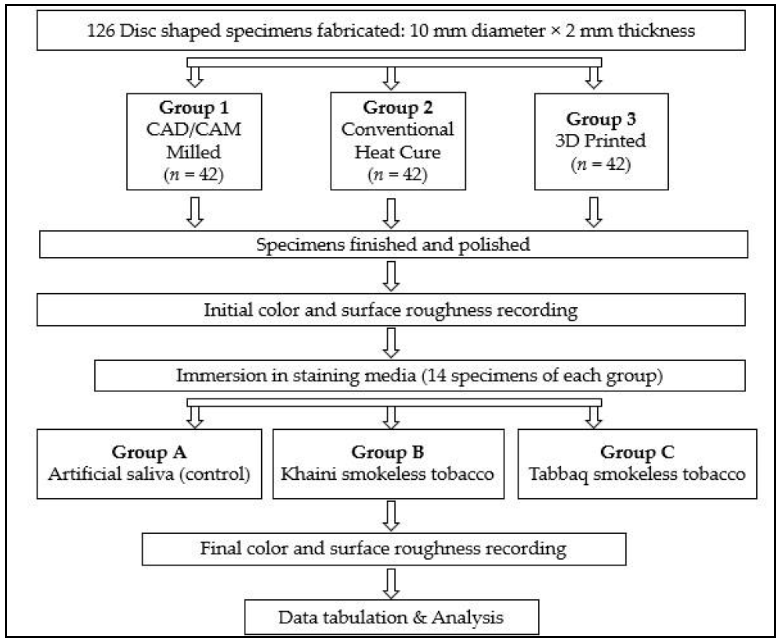
2.1. Materials
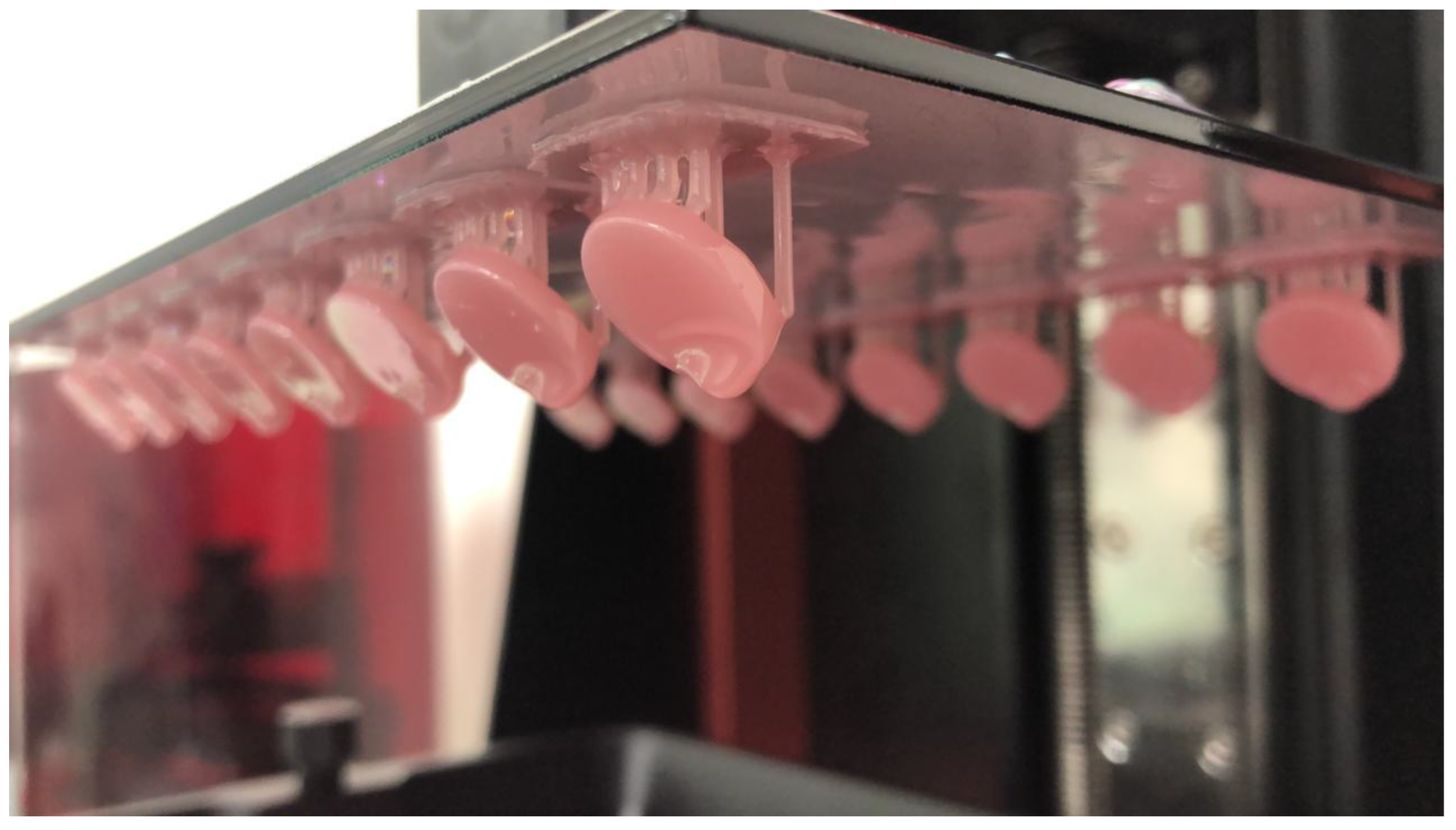
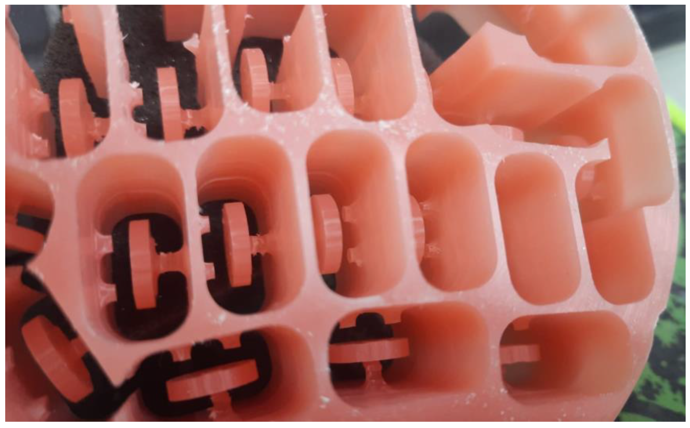
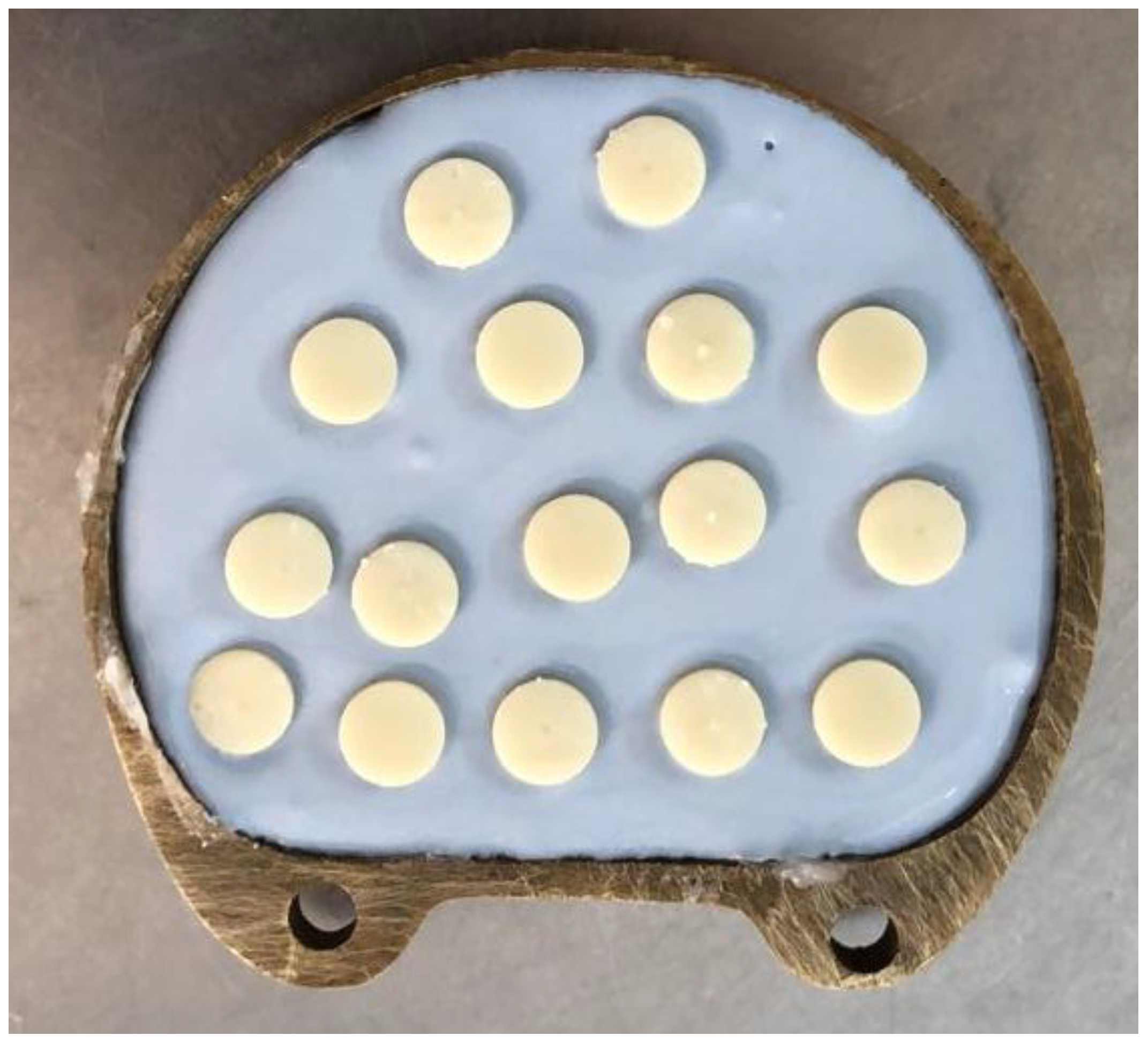
2.2. Specimen Preparation
2.3. Initial Color and Surface-Roughness Testing
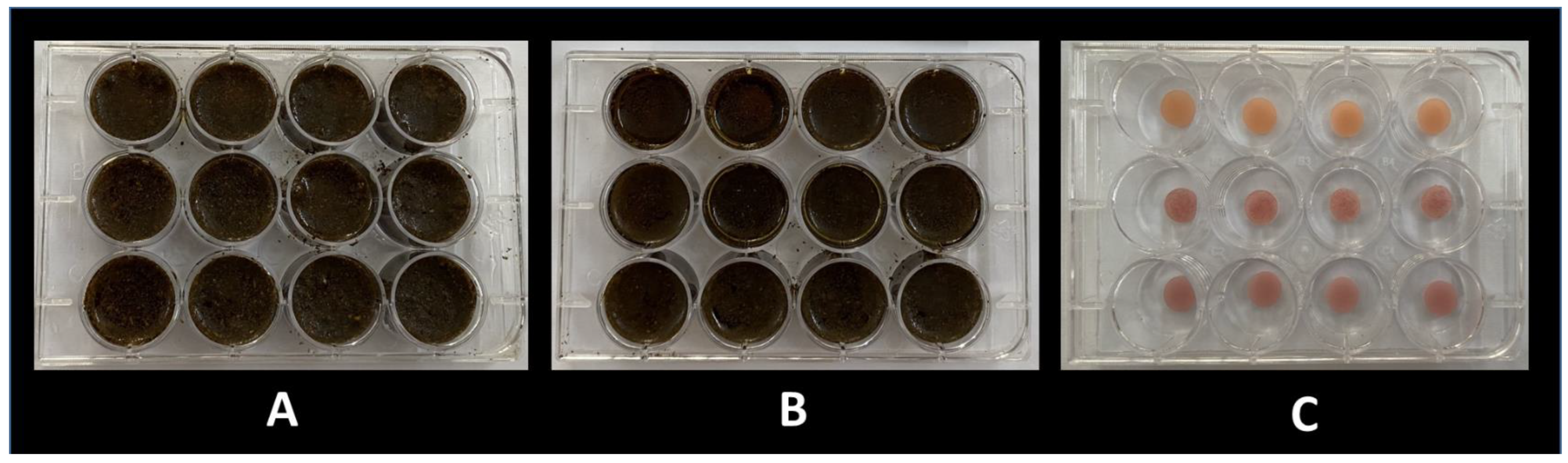
2.4. Immersion Protocol
2.5. Final Color and Surface-Roughness Testing
2.6. Data Analysis
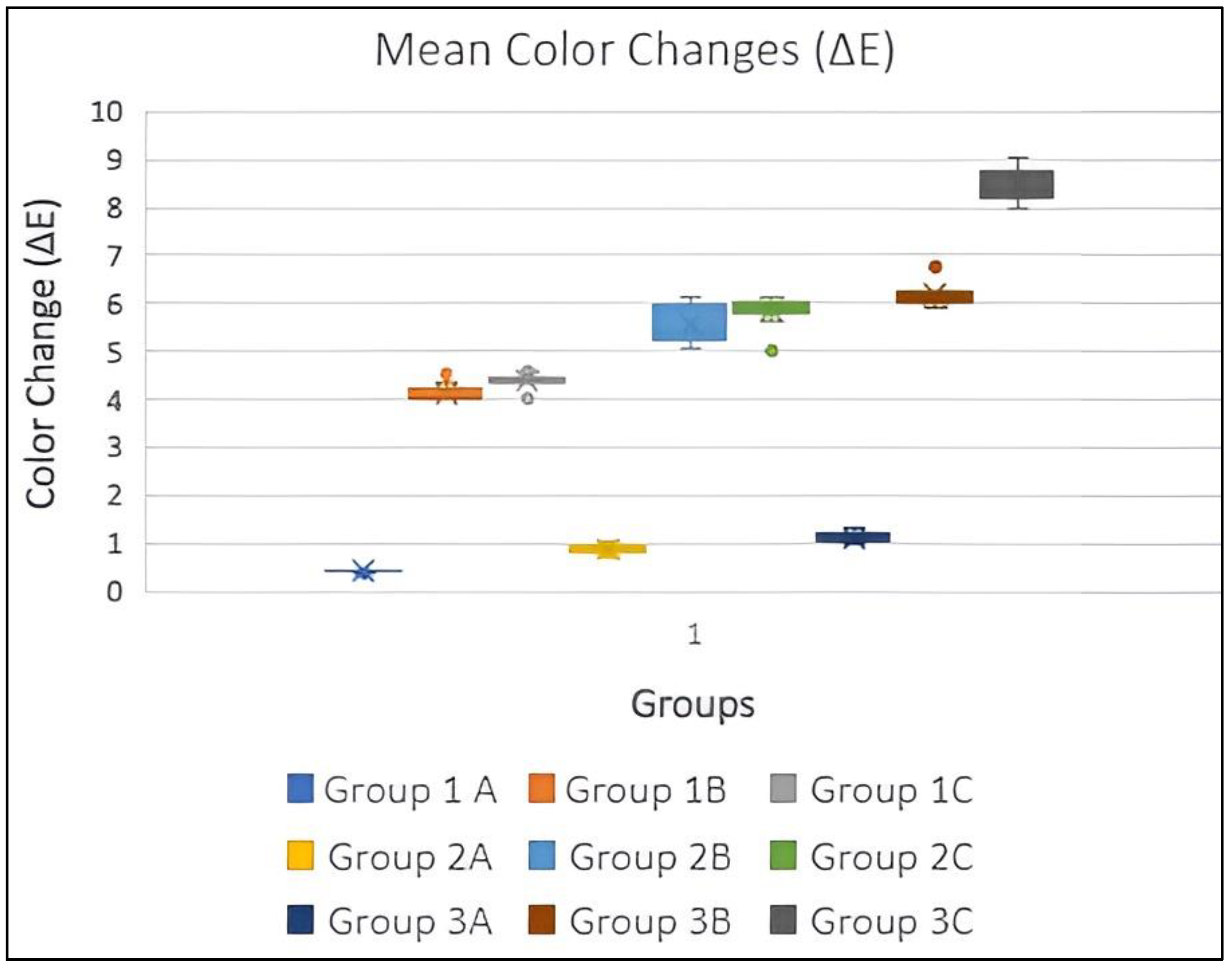
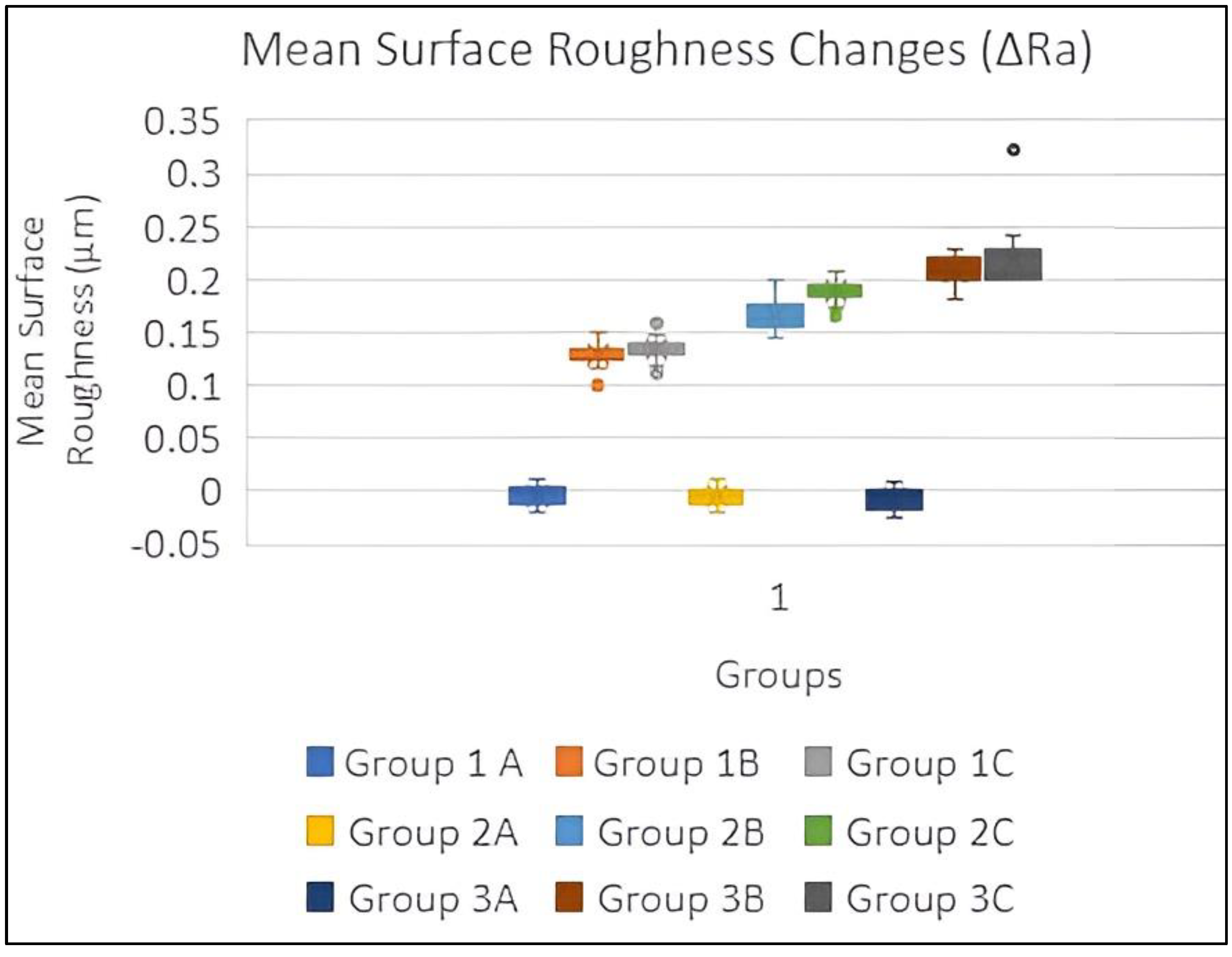
3. Results
4. Discussion
Limitations of the Study
- The dental materials in the oral cavity are exposed to various staining food components, temperature changes, pH changes, and occlusal loading. The effect of these additional factors could not be replicated in the current study. Future studies should be planned to evaluate the effect of smokeless tobacco on denture bases worn by patients.
- The effect of smokeless tobacco on denture base materials was evaluated only for 90 days. More studies with a longer duration should be conducted.
- Only one surface of the specimens was polished to imitate to denture intaglio surface, but an unpolished surface could influence the color measurements.
- Future research should evaluate the combined effects of denture cleansers, mechanical denture cleansing techniques and the different types of smokeless tobacco on denture base materials.
- The current study evaluated only two forms of smokeless tobacco. The effect of other forms of smokeless tobacco on different physical and mechanical properties of denture base materials should be evaluated in future studies.
5. Conclusions
Author Contributions
Funding
Institutional Review Board Statement
Informed Consent Statement
Data Availability Statement
Acknowledgments
Conflicts of Interest
References
- Çakmak, G.; Molinero-Mourelle, P.; De Paula, M.S.; Akay, C.; Cuellar, A.R.; Donmez, M.B.; Yilmaz, B. Surface Roughness and Color Stability of 3D-Printed Denture Base Materials after Simulated Brushing and Thermocycling. Materials 2022, 15, 6441. [Google Scholar] [CrossRef] [PubMed]
- Anadioti, E.; Musharbash, L.; Blatz, M.B.; Papavasiliou, G.; Kamposiora, P. 3D printed complete removable dental prostheses: A narrative review. BMC Oral Health 2020, 20, 343. [Google Scholar] [CrossRef] [PubMed]
- Alfouzan, A.F.; Alotiabi, H.M.; Labban, N.; Al-Otaibi, H.N.; Al Taweel, S.M.; AlShehri, H.A. Color stability of 3D-printed denture resins: Effect of aging, mechanical brushing and immersion in staining medium. J. Adv. Prosthodont. 2021, 13, 160–171. [Google Scholar] [CrossRef] [PubMed]
- Dayan, C.; Guven, M.C.; Gencel, B.; Bural, C. A comparison of the color stability of conventional and CAD/CAM polymethyl methacrylate denture base materials. Acta Stomatol. Croat. 2019, 53, 158–167. [Google Scholar] [CrossRef]
- Palaskar, J.N.; Mittal, S.; Singh, S. Comparative evaluation of surface porosities in conventional heat polymerized acrylic resin cured by water bath and microwave energy with microwavable acrylic resin cured by microwave energy. Contemp. Clin. Dent. 2013, 4, 147–151. [Google Scholar] [CrossRef]
- Anusavice, K.; Chiayi, S.; Ralph Rawls, H. Phillips’ Science of Dental Materials, 12th ed.; Saunders: Philadelphia, PA, USA, 2012. [Google Scholar]
- Hada, T.; Kanazawa, M.; Iwaki, M.; Arakida, T.; Soeda, Y.; Katheng, A.; Otake, R.; Minakuchi, S. Effect of printing direction on the accuracy of 3d-printed dentures using stereolithography technology. Materials 2020, 13, 3405. [Google Scholar] [CrossRef]
- Kattadiyil, M.T.; Goodacre, C.J.; Baba, N.Z. CAD/CAM complete dentures: A review of two commercial fabrication systems. J. Calif. Dent. Assoc. 2013, 41, 407–416. [Google Scholar]
- Scribante, A.; Gallo, S.; Pascadopoli, M.; Canzi, P.; Marconi, S.; Montasser, M.A.; Bressani, D.; Gandini, P.; Sfondrini, M.F. Properties of CAD/CAM 3D Printing Dental Materials and Their Clinical Applications in Orthodontics: Where Are We Now? Appl. Sci. 2022, 12, 551. [Google Scholar] [CrossRef]
- Jain, S.; Adawi, H. Effect of Effervescent Denture Cleansers on 3D Surface Roughness of Conventional Heat Polymerized, Subtractively, and Additively Manufactured Denture Base Resins: An In Vitro Study. J. Prosthodont. 2022; published online ahead of print. [Google Scholar] [CrossRef]
- Kraemer Fernandez, P.; Unkovskiy, A.; Benkendorff, V.; Klink, A.; Spintzyk, S. Surface Characteristics of Milled and 3D Printed Denture Base Materials Following Polishing and Coating: An In-Vitro Study. Materials 2020, 13, 3305. [Google Scholar] [CrossRef]
- Prpić, V.; Schauperl, Z.; Ćatić, A.; Dulčić, N.; Čimić, S. Comparison of Mechanical Properties of 3D-Printed, CAD/CAM, and Conventional Denture Base Materials. J. Prosthodont. 2020, 29, 524–528. [Google Scholar] [CrossRef]
- Zeidan, A.; El-Rahim, R.A.; Helal, M. Evaluation for Elastic Modulus of Cad-Cam Milled And 3d Printed Denture Base Resins. Al-Azhar J. Dent. Sci. 2022, 25, 241–246. [Google Scholar] [CrossRef]
- Jain, S.; Sayed, M.; Ahmed, W.M.; Halawi, A.H.A.; Najmi, N.M.A.; Aggarwal, A.; Bhandi, S.; Patil, S. An In-Vitro Study to Evaluate the Effect of Denture Cleansing Agents on Color Stability of Denture Bases Fabricated Using CAD/CAM Milling, 3D-Printing and Conventional Techniques. Coatings 2021, 11, 962. [Google Scholar] [CrossRef]
- Mann, R.S.; Ruse, N.D. Fracture toughness of conventional, milled and 3D printed denture bases. Dent. Mater. 2022, 38, 1443–1451. [Google Scholar] [CrossRef]
- Dyer, D.; Macdonald, E.; Newcombe, R.G.; Scratcher, C.; Ley, F.; Addy, M. Abrasion and stain removal by different manual toothbrushes and brush actions: Studies in vitro. J. Clin. Periodontol. 2001, 28, 121–127. [Google Scholar] [CrossRef]
- Ozyilmaz, O.Y.; Kara, O.; Akin, C. Evaluation of various denture cleansers on color stability and surface topography of polyetherketoneketone, polyamide, and polymethylmethacrylate. Microsc. Res. Tech. 2021, 84, 3–11. [Google Scholar] [CrossRef]
- Zidan, S.; Silikas, N.; Haider, J.; Yates, J. Effect of Cleansers on the Colour Stability of Zirconia Impregnated PMMA Bio-Nanocomposite. Nanomaterials 2020, 10, 1757. [Google Scholar] [CrossRef]
- Shotwell, J.L.; Razzoog, M.E.; Koran, A. Color stability of long-term soft denture liners. J. Prosthet. Dent. 1992, 68, 836–838. [Google Scholar] [CrossRef]
- Canay, S.; Hersek, N.; Tulunoğlu, I.; Uzun, G. Evaluation of colour and hardness changes of soft lining materials in food colorant solutions. J. Oral Rehabil. 1999, 26, 821–829. [Google Scholar] [CrossRef]
- Erdemir, U.; Yildiz, E.; Saygi, G.; Altay, N.I.; Eren, M.M.; Yucel, T. Effects of energy and sports drinks on tooth structures and restorative materials. World J. Stomatol. 2016, 5, 1–7. [Google Scholar] [CrossRef]
- Chuchulska, B.; Zlatev, S. Linear dimensional change and ultimate tensile strength of polyamide materials for denture bases. Polymers 2021, 13, 3446. [Google Scholar] [CrossRef] [PubMed]
- Chu, S.J.; Trushkowsky, R.D.; Paravina, R.D. Dental color matching instruments and systems. Review of clinical and research aspects. J. Dent. 2010, 38 (Suppl. 2), e2–e16. [Google Scholar] [CrossRef] [PubMed]
- Christen, A.G. The case against smokeless tobacco: Five facts for the health professional to consider. J. Am. Dent. Assoc. 1980, 101, 464–469. [Google Scholar] [CrossRef] [PubMed]
- Christen, A.G.; Swanson, B.Z.; Glover, E.D.; Henderson, A.H. Smokeless tobacco: The folklore and social history of snuffing, sneezing, dipping, and chewing. J. Am. Dent. Assoc. 1982, 105, 821–829. [Google Scholar] [CrossRef] [PubMed]
- Muthukrishnan, A.; Warnakulasuriya, S. Oral health consequences of smokeless tobacco use. Indian J. Med. Res. 2018, 148, 35–40. [Google Scholar]
- Thompson, S.; Griffin, G.; Meyer, N.; Pelaez, L.T.C. Effect of Smokeless Tobacco on Surface Roughness of Dental Restorations. US Army Med. Dep. J. 2017, 2, 80–87. [Google Scholar]
- Mahross, H.Z.; Mohamed, M.D.; Hassan, A.M.; Baroudi, K. Effect of Cigarette Smoke on Surface Roughness of Different Denture Base Materials. J. Clin. Diagn. Res. 2015, 9, ZC39–ZC42. [Google Scholar] [CrossRef]
- Al Moaleem, M.M.; Adawi, H.A.; Alsharif, K.F.; Alhazmi, H.A.; Alshahrani, F.A.; Abu Hadi, R.M.; Kara, R.; Muyidi, H.M.; Khalid, A.; Asiri, A.M.; et al. Impact of Smokeless Tobacco on the Color Stability of Zirconia, Zirconia-Reinforced Lithium Silicate and Feldspathic CAD/CAM Restorative Materials: An In Vitro Study. Coatings 2022, 12, 207. [Google Scholar] [CrossRef]
- Sayed, M.; Jain, S.; Jain, M.; Kubaisi, K.R.; Alhazmi, M.A.; Kamli, E.A.; Aggarwal, A. Effect of cigarette smoke on color stability and surface roughness of different soft denture lining materials. An in vitro study. Am. J. Dent. 2021, 34, 132–136. [Google Scholar]
- Wasilewski, M.S.; Takahashi, M.K.; Kirsten, G.A.; de Souza, E.M. Effect of cigarette smoke and whiskey on the color stability of dental composites. Am. J. Dent. 2010, 23, 4–8. [Google Scholar]
- Raptis, C.N.; Powers, J.M.; Fan, P.L.; Yu, R. Staining of composite resins by cigarette smoke. J. Oral Rehabil. 1982, 9, 367–371. [Google Scholar] [CrossRef]
- Brima, E.I. Determination of Metal Levels in Shamma (Smokeless Tobacco) with Inductively Coupled Plasma Mass Spectrometry (ICP-MS) in Najran, Saudi Arabia. Asian Pac. J. Cancer Prev. 2016, 17, 4761–4767. [Google Scholar]
- Uchida, H.; Vaidyanathan, J.; Viswanadhan, T.; Vaidyanathan, T.K. Color stability of dental composites as a function of shade. J. Prosthet. Dent. 1998, 79, 372–377. [Google Scholar] [CrossRef]
- Shetty, M.; Jain, S.; Wankhede, T.M.; Sayed, M.E.; Mohammed, Z.; Shetty, S.; Al Wadei, M.H.D.; Alqahtani, S.M.; Othman, A.A.A.; Alnijaiban, M.A.; et al. An In Vitro Study to Evaluate the Effect of Artificial Aging on Translucency, Contrast Ratio, and Color of Zirconia Dental Ceramic at Different Sintering Levels. Coatings 2022, 12, 642. [Google Scholar] [CrossRef]
- Al-Anesi, W.A.; Madfa, A.A.; Dubais, M.A.; Albahari, A.A. Effects of khat extract and other staining media on color change of composite resins subjected to various polishing methods. Mater. Sci. 2019, 7, 1. [Google Scholar] [CrossRef]
- Malek Afzali, B.; Ghasemi, A.; Mirani, A.; Abdolazimi, Z.; Akbarzade Baghban, A.; Kharazifard, M.J. Effect of Ingested Liquids on Color Change of Composite Resins. J. Dent. 2015, 12, 577–584. [Google Scholar]
- CIE Commission International de l’Eclairage (CIE). Colorimetry, 3rd ed.; CIE Publication No.15; Central Bureau of the CIE: Vienna, Austria, 2004. [Google Scholar]
- Gómez-Polo, C.; Montero, J.; Gómez-Polo, M.; Martin Casado, A. Comparison of the CIELab and CIEDE 2000 Color Difference Formulas on Gingival Color Space. J. Prosthodont. 2020, 29, 401–408. [Google Scholar] [CrossRef]
- Dimitrova, M.; Corsalini, M.; Kazakova, R.; Vlahova, A.; Barile, G.; Dell’Olio, F.; Tomova, Z.; Kazakov, S.; Capodiferro, S. Color Stability Determination of CAD/CAM Milled and 3D Printed Acrylic Resins for Denture Bases: A Narrative Review. J. Compos. Sci. 2022, 6, 201. [Google Scholar] [CrossRef]
- Pantea, M.; Totan, A.R.; Imre, M.; Petre, A.E.; Țâncu, A.M.; Tudos, C.; Farcașiu, A.T.; Butucescu, M.; Spînu, T.C. Biochemical Interaction between Materials Used for Interim Prosthetic Restorations and Saliva. Materials 2022, 15, 226. [Google Scholar] [CrossRef]
- Cho, K.J.; Park, S.J.; Cho, H.G.; Kim, D.J.; Hwang, Y.C.; Oh, W.M.; Hwang, I.N. Influence of the Surface roughness on translucency and surface color of the dental composite resins. J. Korean Acad. Conserv. Dent. 2006, 31, 312–322. [Google Scholar] [CrossRef]
- Alkhatib, M.N.; Holt, R.D.; Bedi, R. Smoking and tooth discolouration: Findings from a national cross-sectional study. BMC Public Health 2005, 5, 27. [Google Scholar] [CrossRef] [PubMed]
- Zanetti, F.; Zha, X.; Pan, J.; Peitsch, M.C.; Hoeng, J.; Ren, Y. Effects of cigarette smoke and tobacco heating aerosol on color stability of dental enamel, dentin, and composite resin restorations. Quintessence Int. 2019, 50, 156–166. [Google Scholar]
- Willers, S.; Gerhardsson, L.; Lundh, T. Environmental tobacco smoke (ETS) exposure in children with asthma-relation between lead and cadmium, and cotinine concentrations in urine. Respir. Med. 2005, 99, 1521–1527. [Google Scholar] [CrossRef] [PubMed]
- Prajapati, A.M.; Sanyal, K.; Guru, R.; Vaswani, P.; Tewary, S.; Sushma, R. Effect of Tobacco Extract, Cigarette Smoke, and Carbonated Beverage on Surface Roughness and Color Stability of Three different Restorative Materials: An in vitro Study. Int. J. Prev. Clin. Dent. Res. 2017, 4, 9–18. [Google Scholar] [CrossRef]
- Yao, Q.; Morton, D.; Eckert, G.J.; Lin, W.-S. The effect of surface treatments on the color stability of CAD-CAM interim fixed dental prostheses. J. Prosthet. Dent. 2020, 126, 248–253. [Google Scholar] [CrossRef]
- Taşın, S.; Ismatullaev, A.; Usumez, A. Comparison of Surface Roughness and Color Stainability of 3-Dimensionally Printed Interim Prosthodontic Material with Conventionally Fabricated and CAD-CAM Milled Materials. J. Prosthet. Dent. 2022, 128, 1094–1101. [Google Scholar] [CrossRef]
- Shin, J.W.; Kim, J.E.; Choi, Y.J.; Shin, S.H.; Nam, N.E.; Shim, J.S.; Lee, K.W. Evaluation of the color stability of 3D-printed crown and bridge materials against various sources of discoloration: An in vitro study. Materials 2020, 13, 5359. [Google Scholar] [CrossRef]
- Al-Qarni, F.D.; Goodacre, C.J.; Kattadiyil, M.T.; Baba, N.Z.; Paravina, R.D. Stainability of acrylic resin materials used in CAD-CAM and conventional complete dentures. J. Prosthet. Dent. 2020, 123, 880–887. [Google Scholar] [CrossRef]
- Abdulallah, A.Y.; Abdulsahib, A.J. Comparison of Denture Base Adaptation and Some Mechanical Properties among Conventional, CAD-CAM, and 3-D Printed Denture Base Materials. J. Res. Med. Dent. Sci. 2022, 10, 26–30. [Google Scholar]
- Alraheam, I.A.; Donovan, T.E.; Rodgers, B.; Boushell, L.; Sulaiman, T.A. Effect of masticatory simulation on the translucency of different types of dental zirconia. J. Prosthet. Dent. 2019, 122, 404–409. [Google Scholar] [CrossRef]
- Nogueira, A.D.; Della Bona, A. The effect of a coupling medium on color and translucency of CAD-CAM ceramics. J. Dent. 2013, 41 (Suppl. S3), e18–e23. [Google Scholar] [CrossRef]
- Alandia-Roman, C.C.; Cruvinel, D.R.; Sousa, A.B.S.; Pires-De-Souza, F.C.P.; Panzeri, H. Effect of Cigarette Smoke on Color Stability and Surface Roughness of Dental Composites. J. Dent. 2013, 41 (Suppl. 3), e73–e79. [Google Scholar] [CrossRef]
- Bollen, C.M.; Lambrechts, P.; Quirynen, M. Comparison of surface roughness of oral hard materials to the threshold surface roughness for bacterial plaque retention: A review of the literature. Dent. Mater. 1997, 13, 258–269. [Google Scholar] [CrossRef]
- Tahayeri, A.; Morgan, M.; Fugolin, A.P.; Bompolaki, D.; Athirasala, A.; Pfeifer, C.S.; Ferracane, J.L.; Bertassoni, L.E. 3D printed versus conventionally cured provisional crown and bridge dental materials. Dent. Mater. 2018, 34, 192–200. [Google Scholar] [CrossRef]
- Atria, P.J.; Lagos, I.; Sampaio, C.S. In vitro evaluation of surface roughness, color stability, and color masking of provisional restoration materials for veneers and crowns. Int. J. Comput. Dent. 2020, 23, 343–350. [Google Scholar]
- Duarte, S.; Sartori, N.; Phark, J.-H. Ceramic-reinforced polymers: CAD/CAM hybrid restorative materials. Curr. Oral Health Rep. 2016, 3, 198–202. [Google Scholar] [CrossRef]
- Engler, M.L.P.D.; Güth, J.F.; Keul, C.; Erdelt, K.; Edelhoff, D.; Liebermann, A. Residual monomer elution from different conventional and CAD/CAM dental polymers during artificial aging. Clin. Oral Investig. 2020, 24, 277–284. [Google Scholar] [CrossRef]
- Balkenhol, M.; Knapp, M.; Ferger, P.; Heun, U.; Wöstmann, B. Correlation between polymerization shrinkage and marginal fit of temporary crowns. Dent. Mater. 2008, 24, 1575–1584. [Google Scholar] [CrossRef]
- Köroğlu, A.; Sahin, O.; Dede, D.; Yilmaz, B. Effect of different surface treatment methods on the surface roughness and color stability of interim prosthodontic materials. J. Prosthet. Dent. 2016, 115, 447–455. [Google Scholar] [CrossRef]
- Sahin, O.; Dede, D.Ö.; Köroğlu, A.; Yılmaz, B. Influence of surface sealant agents on the surface roughness and color stability of artificial teeth. J. Prosthet. Dent. 2015, 114, 130–137. [Google Scholar] [CrossRef]
- Doray, P.G.; Eldiwany, M.S.; Powers, J.M. Effect of resin surface sealers on improvement of stain resistance for a composite provisional material. J. Esthet. Restor. Dent. 2003, 15, 244–250. [Google Scholar] [CrossRef]
- Song, S.-Y.; Shin, Y.-H.; Lee, J.-Y.; Shin, S.-W. Color stability of provisional restorative materials with different fabrication methods. J. Adv. Prosthodont. 2020, 12, 259–264. [Google Scholar] [CrossRef] [PubMed]
- Sarac, D.; Sarac, Y.S.; Kulunk, S.; Ural, C.; Kulunk, T. The effect of polishing techniques on the surface roughness and color change of composite resins. J. Prosthet. Dent. 2006, 96, 33–40. [Google Scholar] [CrossRef] [PubMed]
- Iazzetti, G.; Burgess, J.O.; Gardiner, D.; Ripps, A. Color stability of fluoride-containing restorative materials. Oper. Dent. 2000, 25, 520–525. [Google Scholar] [PubMed]
- Hersek, N.; Canay, S.; Uzun, G.; Yildiz, F. Color stability of denture base acrylic resins in three food colorants. J. Prosthet. Dent. 1999, 81, 375–379. [Google Scholar] [CrossRef]
- Arima, T.; Murata, H.; Hamada, T. Properties of highly cross-linked autopolymerizing reline acrylic resins. J. Prosthet. Dent. 1995, 73, 55–59. [Google Scholar] [CrossRef]
- Arima, T.; Murata, H.; Hamada, T. Analysis of composition and structure of hard autopolymerizing reline resins. J. Oral Rehabil. 1996, 23, 346–352. [Google Scholar] [CrossRef]
- Hollis, S.; Eisenbeisz, E.; Versluis, A. Color stability of denture resins after staining and exposure to cleansing agents. J. Prosthet. Dent. 2015, 114, 709–714. [Google Scholar] [CrossRef]
- Ferracane, J.L. Hygroscopic and hydrolytic effects in dental polymer networks. Dent. Mater. 2006, 22, 211–222. [Google Scholar] [CrossRef]


| Group | Manufacturing Technique | Commercial Product and Manufacturer | Main Ingredient |
|---|---|---|---|
| 1 | CAD/CAM milling | Wieland; Wieland Dental +, Technik GmbH and Co. KG, Pforzheim, Germany | PMMA resin |
| 2 | Heat polymerization using the conventional compression-molding technique | Meliodent; Kulzer-GmbH, Hanau, Germany | PMMA resin |
| 3 | 3D printing | NextDent; Denture 3D+, Vertex-Dental, Soesterberg, the Netherlands | Dimethacrylate-based resins |
| Sub Group | Immersion Solution | Trade Name | Composition | pH |
|---|---|---|---|---|
| A | Artificial salivary substitute (control) | - | Distilled water, NaCl, KCl, and CaCI2 | 7.4 |
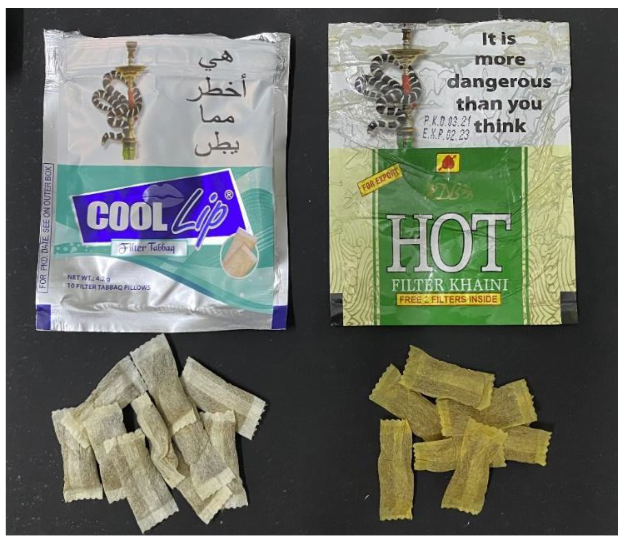
| B | Khaini smokeless tobacco | HOT Filter Khaini (SUN Products, Gujarat, India) | Tobacco, lime water, menthol, and spices | 9.3 |
| C | Tabbaq smokeless tobacco | Cool Lips Filter Tabbaq (Tej Ram Dharam Paul, Gujarat, India.) | Tobacco, mint, and spices (such as cinnamon, cardamom, and cloves) | 9.4 |
| Groups | Subgroups | n | Color Change (ΔE) | Surface Roughness Change (ΔRa) | ||
|---|---|---|---|---|---|---|
| Mean | SD | Mean | SD | |||
| Group 1 (CAD/CAM Milled) | GROUP 1A (Artificial Saliva) | 14 | 0.418 | 0.022 | −0.003 | 0.011 |
| GROUP 1B (Khaini) | 14 | 4.129 | 0.165 | 0.129 | 0.013 | |
| GROUP 1C (Tabbaq) | 14 | 4.383 | 0.144 | 0.135 | 0.012 | |
| Total | 42 | 2.977 | 1.838 | 0.087 | 0.066 | |
| Group 2 (Heat-Polymerized) | GROUP 2A (Artificial Saliva) | 14 | 0.888 | 0.087 | −0.005 | 0.010 |
| GROUP 2B (Khaini) | 14 | 5.544 | 0.412 | 0.166 | 0.015 | |
| GROUP 2C (Tabbaq) | 14 | 5.851 | 0.274 | 0.188 | 0.011 | |
| Total | 42 | 4.094 | 2.315 | 0.116 | 0.088 | |
| Group 3 (3D-Printed) | GROUP 3A (Artificial Saliva) | 14 | 1.118 | 0.122 | −0.007 | 0.011 |
| GROUP 3B (Khaini) | 14 | 6.204 | 0.274 | 0.208 | 0.015 | |
| GROUP 3C (Tabbaq) | 14 | 8.457 | 0.375 | 0.219 | 0.033 | |
| Total | 42 | 5.260 | 3.118 | 0.140 | 0.108 | |
| Total | Artificial Saliva | 42 | 0.808 | 0.307 | −0.005 | 0.011 |
| Khaini | 42 | 5.292 | 0.924 | 0.168 | 0.036 | |
| Tabbaq | 42 | 6.230 | 1.727 | 0.181 | 0.041 | |
| Total | 126 | 4.110 | 2.633 | 0.114 | 0.091 | |
| 2-Way ANOVA: Group Differences | p < 0.001 | p < 0.001 | ||||
| 2-Way ANOVA: Subgroup Differences | p < 0.001 | p < 0.001 | ||||
| 2-Way ANOVA: Group–Subgroup Interaction | p < 0.001 | p < 0.001 | ||||
| Multiple Comparisons (Group) | |||||||
|---|---|---|---|---|---|---|---|
| (I) Group | Mean Difference (I-J) | Std. Error | Sig. | 95% Confidence Interval | |||
| Lower Bound | Upper Bound | ||||||
| CAD/CAM Milled | Heat-Polymerized | −1.1177 * | 0.05304 | 0.000 | −1.2465 | −0.9889 | p < 0.001 |
| 3D-Printed | −2.2830 * | 0.05304 | 0.000 | −2.4118 | −2.1541 | p < 0.001 | |
| Heat-Polymerized | CAD/CAM Milled | 1.1177 * | 0.05304 | 0.000 | 0.9889 | 1.2465 | p < 0.001 |
| 3D-Printed | −1.1653 * | 0.05304 | 0.000 | −1.2941 | −1.0364 | p < 0.001 | |
| 3D-Printed | CAD/CAM Milled | 2.2830 * | 0.05304 | 0.000 | 2.1541 | 2.4118 | p < 0.001 |
| Heat-Polymerized | 1.1653 * | 0.05304 | 0.000 | 1.0364 | 1.2941 | p < 0.001 | |
| Multiple Comparisons (Subgroup) | |||||||
| Artificial Saliva | Khaini | −4.4840 * | 0.05304 | 0.000 | -4.6129 | −4.3552 | p < 0.001 |
| Tabbaq | −5.4219 * | 0.05304 | 0.000 | -5.5508 | −5.2931 | p < 0.001 | |
| Khaini | Artificial Saliva | 4.4840 * | 0.05304 | 0.000 | 4.3552 | 4.6129 | p < 0.001 |
| Tabbaq | −0.9379 * | 0.05304 | 0.000 | -1.0667 | −0.8091 | p < 0.001 | |
| Tabbaq | Artificial Saliva | 5.4219 * | 0.05304 | 0.000 | 5.2931 | 5.5508 | p < 0.001 |
| Khaini | −0.9379 * | 0.05304 | 0.000 | 0.8091 | 1.0667 | p < 0.001 | |
| Multiple Comparisons (Group) | |||||||
|---|---|---|---|---|---|---|---|
| (I) Group | Mean Difference (I-J) | Std. Error | Sig. | 95% Confidence Interval | |||
| Lower Bound | Upper Bound | ||||||
| CAD/CAM Milled | Heat-Polymerized | −0.0295 * | 0.00353 | 0.000 | −0.0381 | −0.0209 | p < 0.001 |
| 3D-Printed | −0.0532 * | 0.00353 | 0.000 | −0.0618 | −0.0447 | p < 0.001 | |
| Heat-Polymerized | CAD/CAM Milled | 0.0295 * | 0.00353 | 0.000 | 0.0209 | 0.0381 | p < 0.001 |
| 3D-Printed | −0.0237 * | 0.00353 | 0.000 | −0.0323 | −0.0152 | p < 0.001 | |
| 3D-Printed | CAD/CAM Milled | 0.0532 * | 0.00353 | 0.000 | 0.0447 | 0.0618 | p < 0.001 |
| Heat-Polymerized | 0.0237 * | 0.00353 | 0.000 | 0.0152 | 0.0323 | p < 0.001 | |
| Multiple Comparisons (Subgroup) | |||||||
| Artificial Saliva | Khaini | −0.1725 * | 0.00353 | 0.000 | −0.1810 | −0.1639 | p < 0.001 |
| Tabbaq | −0.1858 * | 0.00353 | 0.000 | −0.1943 | −0.1772 | p < 0.001 | |
| Khaini | Artificial Saliva | 0.1725 * | 0.00353 | 0.000 | 0.1639 | 0.1810 | p < 0.001 |
| Tabbaq | −0.0133 * | 0.00353 | 0.001 | −0.0218 | −0.0047 | p = 0.001 | |
| Tabbaq | Artificial Saliva | 0.1858 * | 0.00353 | 0.000 | 0.1772 | 0.1943 | p < 0.001 |
| Khaini | 0.0133 * | 0.00353 | 0.001 | 0.0047 | 0.0218 | p = 0.001 | |
Disclaimer/Publisher’s Note: The statements, opinions and data contained in all publications are solely those of the individual author(s) and contributor(s) and not of MDPI and/or the editor(s). MDPI and/or the editor(s) disclaim responsibility for any injury to people or property resulting from any ideas, methods, instructions or products referred to in the content. |
© 2023 by the authors. Licensee MDPI, Basel, Switzerland. This article is an open access article distributed under the terms and conditions of the Creative Commons Attribution (CC BY) license (https://creativecommons.org/licenses/by/4.0/).
Share and Cite
Mugri, M.H.; Jain, S.; Sayed, M.E.; Halawi, A.H.A.; Hamzi, S.A.I.; Aljohani, R.A.S.; Madkhali, Z.M.A.; Khalid, A.; Jokhadar, H.F.; Almarzouki, M.; et al. Effects of Smokeless Tobacco on Color Stability and Surface Roughness of 3D-Printed, CAD/CAM-Milled, and Conventional Denture Base Materials: An In Vitro Study. Biomedicines 2023, 11, 491. https://doi.org/10.3390/biomedicines11020491
Mugri MH, Jain S, Sayed ME, Halawi AHA, Hamzi SAI, Aljohani RAS, Madkhali ZMA, Khalid A, Jokhadar HF, Almarzouki M, et al. Effects of Smokeless Tobacco on Color Stability and Surface Roughness of 3D-Printed, CAD/CAM-Milled, and Conventional Denture Base Materials: An In Vitro Study. Biomedicines. 2023; 11(2):491. https://doi.org/10.3390/biomedicines11020491
Chicago/Turabian StyleMugri, Maryam H., Saurabh Jain, Mohammed E. Sayed, Amjad Hussain Asiri Halawi, Safa Ahmed Ibrahim Hamzi, Raniya Abdulaziz Saad Aljohani, Zainab Mousa Ali Madkhali, Asaad Khalid, Hossam F. Jokhadar, Mai Almarzouki, and et al. 2023. "Effects of Smokeless Tobacco on Color Stability and Surface Roughness of 3D-Printed, CAD/CAM-Milled, and Conventional Denture Base Materials: An In Vitro Study" Biomedicines 11, no. 2: 491. https://doi.org/10.3390/biomedicines11020491