Article

Testing the Resilience, Physiological Plasticity and Mechanisms Underlying Upper Temperature Limits of Antarctic Marine Ectotherms

1
British Antarctic Survey, Natural Environment Research Council, Cambridge CB3 0ET, UK
2
Department of Biology, University of Victoria, P.O. Box 1700, Victoria, BC V8W 2Y2, Canada
3
National Oceanography Centre, University of Southampton, Southampton SO14 3ZH, UK
*
Author to whom correspondence should be addressed.
Biology 2024, 13(4), 224; https://doi.org/10.3390/biology13040224
Submission received: 4 March 2024 / Revised: 22 March 2024 / Accepted: 25 March 2024 / Published: 29 March 2024

Abstract

:

Simple Summary

Antarctic marine invertebrates live in the constant of the Southern Ocean and are characterised by sensitivity to small increases in temperature. We conducted a series of aquarium experiments that tested this ability and found species-specific responses to warming. We found that some species were able to survive for many months at up to 10 °C, a temperature which is up to 4 °C warmer than previously recorded. We found that the survivors of three species had adjusted their biological systems (acclimated) and were better able to survive additional rapid warming, but one anemone species did not elevate its upper temperature limit, even though it survived for 270 days at 6 °C. There were also species-specific effects of increasing oxygen concentration on long-term survival to elevated temperatures, with extended, no change, or reduced survival duration all found in different species. Thermal sensitivity is clearly the product of multiple ecological and physiological capacities, and this diversity of response needs further investigation and interpretation to improve our ability to predict future patterns of biodiversity.

Abstract

Antarctic marine ectotherms live in the constant cold and are characterised by limited resilience to elevated temperature. Here we tested three of the central paradigms underlying this resilience. Firstly, we assessed the ability of eight species, from seven classes representing a range of functional groups, to survive, for 100 to 303 days, at temperatures 0 to 4 °C above previously calculated long-term temperature limits. Survivors were then tested for acclimation responses to acute warming and acclimatisation, in the field, was tested in the seastar Odontaster validus collected in different years, seasons and locations within Antarctica. Finally, we tested the importance of oxygen limitation in controlling upper thermal limits. We found that four of 11 species studied were able to survive for more than 245 days (245–303 days) at higher than previously recorded temperatures, between 6 and 10 °C. Only survivors of the anemone Urticinopsis antarctica did not acclimate CTmax and there was no evidence of acclimatisation in O. validus. We found species-specific effects of mild hyperoxia (30% oxygen) on survival duration, which was extended (two species), not changed (four species) or reduced (one species), re-enforcing that oxygen limitation is not universal in dictating thermal survival thresholds. Thermal sensitivity is clearly the product of multiple ecological and physiological capacities, and this diversity of response needs further investigation and interpretation to improve our ability to predict future patterns of biodiversity.

1. Introduction

Understanding the mechanisms underlying species’ resilience to environmental perturbations is key to predictions of future patterns of biodiversity. Physiological plasticity is a key element of species’ capacity to survive in fluctuating environments [1]. This plasticity consists of biochemical “resistance” pathways that can be switched on or off (e.g., anaerobic metabolism [2]; heat shock proteins [3]), but also the capacity to adjust biochemical pathways in response to altered environmental conditions [4]. Acclimation, the phenotypic response to a single stressor, often in response to laboratory-induced stress, and acclimatisation, the response to multiple stressors, often under field conditions [5], can occur over scales from hours to months. Physiological plasticity is an important component of species’ capacity, providing resilience to both short-term acute heating (e.g., heatwaves [6]) and long-term change (e.g., climate change [7]). For acclimation to be beneficial, it requires predictable “zeitgebers”, i.e., cues that are predictive of future conditions, ensuring that the cost of any physiological remodelling is outweighed by the benefit [8]. The benefit of physiological acclimation has been argued [9], as has its capacity to buffer against global warming [10,11].
Physiological responses vary over geographic and temporal scales, with many dimensions known to govern the evolution of thermal tolerance [1]. At one extreme for marine life, Antarctic marine ectotherms have evolved in the constant cold of the Southern Ocean for tens of millions of years and are predicted to be sensitive to warming, but also have limited physiological plasticity [12,13]. Antarctic marine ectotherms have atypical thermal responses, including the expression of heat shock proteins, an almost ubiquitous protection of proteins to acute warming (HSP [14]), failure to select preferred temperatures [15] and in some species, lack of escape response to dangerously high temperatures [15,16]. This combined evidence supports that universal protective responses to heat regimes experienced in the Antarctic could be missing in these taxa. Recent studies have shown that the responses of Antarctic marine ectotherms are much more complicated. For example, longer-term incubations can lead to acclimation of some Antarctic marine ectotherms [11] but not others [11,17], and cellular responses, including HSP expression, vary markedly between species and treatment regimens [18]. In the cold of the Southern Ocean all physiological processes take longer, and acclimation in Antarctic marine ectotherms can take many months and can occur more slowly in invertebrates than fish [11].
Mixed responses indicate that the mechanisms underlying thermal sensitivity are complex. Oxygen and capacity limitation of thermal tolerance (OCLTT [2]) is one of the mechanisms whose applicability has been questioned in the Antarctic [1], but also more broadly with studies finding mixed responses both inside and outside the Southern Ocean [19,20,21].
Here we synthesise a series of experiments conducted from 2006 to 2015 that investigated both the acclimation and acclimatisation capacity of selected Antarctic marine ectotherms. Previous estimates of upper thermal limits for the long-term survival of Antarctic marine ectotherms were extrapolated to temperatures between 1 and 6 °C for incubations of several months [12]. To test if 6 °C represents the long-term limit for survival, species from a range of phyla were selected to represent a range of functional groups, the ophiuroids Ophionotus victoriae (Bell, 1902) and Ophiura crassa (Mortensen, 1936), the echinoid Sterechinus neumayeri (Meissner, 1900), the asteroid Odontaster Validus (Koehler, 1906), the holothuroid Heterocucumis steineni (Ludwig, 1898), the anthozoa Urticinopsis antarctica (Verrill, 1922), and the molluscs Margarella antarctica (E. Lamy, 1906) and Laternula elliptica (P.P. King, 1832), were incubated at 6 and 8 °C for up to 10 months. Survivors were tested for whole body acclimation to see if acute thermal limits (CTmax—the critical thermal maximum) had been elevated through exposure to these elevated temperatures. Acclimitisation of O. validus was tested by comparing CTmax from field fresh individuals just after collection from the shallow sea around the British Antarctic Survey’s Rothera Research Station in spring, summer and winter seasons, and McMurdo Sound near Antarctica New Zealand’s Scott Base. The aim was to test for spatio-temporal variation in CTmax. To test if elevated oxygen increased survival times at elevated temperatures, species were incubated long-term under normoxic (21%) and mildly hyperoxic (30%) oxygen saturation. The arthropod Paraceradocus miersi (Pfeffer, 1888), the ophiuroids Ophionotus victoriae and O. crassa were incubated at 3 °C and S. neumayeri, O. Validus, and the holothuroids Cucumaria georgiana (Lampert, 1886) and H. steineni were reared at 8 °C rising to 10 °C, under both normoxia and mild hyperoxia. In this way, this study further investigated the paradigms of limited physiological resilience, plasticity and the importance of oxygen setting upper temperature limits in Antarctic marine ectotherms, searching for exceptions and potential new insights into the underlying mechanisms.

2. Materials and Methods

2.1. Collections and Animal Husbandry

Experiments were conducted between 2006 and 2015 with individuals hand collected by SCUBA divers in the austral summer, from 6–15 m depth, near Rothera Research Station, Adelaide Island (67°34′25″ S, 68°08′00″ W). Common marine ectotherms were selected from different phyla to represent a range of functional groups, for which aquarium husbandry is well established and long-term temperature limits have been estimated under normoxia [12]. All animals remained submerged throughout the transfer from the sea to the flow-through aquarium system at the station. To control for size-dependent effects on survival, we selected individuals of a similar size within each species group at the start of the experiment, and only studied fully reproductive adults.
The 3.0 °C temperature –oxygen experiment was conducted in flow through aquaria at Rothera Research Station. In Rothera, the tanks had a constant exchange of seawater that was balanced to allow temperature and oxygen treatments to be maintained while preventing any build-up of metabolic waste. In all experiments, seawater chemistry was monitored every 2–3 days using Nutrafin aquarium test kits. Ammonia, nitrite, and nitrates were maintained well below 0.4, 0.2 and 5 mg L−1 to prevent toxicity from metabolic by-products.
All other animals were transported back to the UK in purpose-built, recirculating, temperature-controlled transport aquaria maintained at 0.0 ± 0.3 °C with an average 12L:12D photoperiod for the six-week passage. Once in the UK, the animals were kept in a temperature-controlled aquaria at 0.1 ± 0.1 °C until the start of experiments. In the UK, holding and experiments were conducted in recirculating tanks that were fitted with biological filters (EHEIM GmbH & Co KG, Stuttgart, Germany), UV sterilisers, bubbled air, and plastic lids. Water quality was maintained with a combination of biological filtration, protein skimming, and partial seawater exchanges (approximately 5–15% every 2–3 days), which was confirmed as detailed above. Throughout transport and experimental incubations all animals were fed, to excess, on small pieces of fish or crustaceans except for the filter feeders C. georgiana, H. steineni, and L. elliptica which were fed on a mixture of instant concentrated Nanochloropsis spp. and Tetraselmis spp. (ZM Fish Food, Winchester, UK) and algae growing naturally in the water.
In all experiments, functional survival (CTmax) was monitored daily in both treatment and control animals through the following standard procedures cf [22]. For all but P. miersi, the first sign that individuals were approaching their limit was the inability to remain attached to the side of the tank. These individuals were then tested daily for the absence of movement of arms, spines, and tube feet. For P. miersi, we used the absence of movement of appendages (uropods or pleopods). If an individual was non-responsive it was returned to the treatment tank and checked again 24 h later. If, after 24 h, there was still no response to external stimuli then the number of days from the start of the experiment to the first day with no response was recorded as the time taken for CTmax to occur. To account for any effect of size on mortality, the size of each individual was recorded.

2.2. Identification of Anemone Species

A small piece of one tentacle was removed from each anemone and preserved in 96% ethanol. DNA was extracted from each tentacle using the DNeasy Blood and Tissue kit (Qiagen, Manchester, UK) according to manufacturer’s instructions. The cytochrome oxidase subunit I gene (COI) mitochondrial region was amplified using 1–2 µL extracted DNA and MyTaq DNA polymerase mix (30 µL reactions; Bioline UK (now Meridien Biosceince, London, UK), with 10 nmol each of the universal COI primers for invertebrates (LCO 1490 5′-GGTCAACAAATCATAAAGATATTGG-3′; HCO 2198 5′-TAAACTTCAGGGTGACCAAAAAATCA-3′) [23]. PCR conditions were 94 °C for 5 min, five cycles of 94 °C for 1 min, 45 °C for 1.5 min, 72 °C for 1.5 min, followed by 30 cycles of 94 °C for 1 min, 50 °C for 1 min, 72 °C for 1 min and a final elongation stage of 5 min at 72 °C. The COI fragments were bi-directionally sequenced by Source Bioscience (Cambridge, UK). The species identity of each individual was analysed using Blast sequence similarity searching of INSDC (International Nucleotide Sequence Database Collaboration) (https://www.insdc.org/, accessed on 12 March 2020).

2.3. Acclimation Experiments: Temperature Incubations

Temperatures were raised at the same rate in all experiments (0.3 ± 0.1 °C d−1) until the incubation temperature was reached, and subsequently monitored daily (Figure S1). O. victoriae, O. crassa, S. neumayeri, O. validus, H. steineni, U. antarctica, M. antarctica, and L. elliptica were held at temperatures of 6.0 and 8.0 °C, temperatures that were 0 to 4 °C above the long-term limits calculated from experiments with different rates of warming [12]. Control animals were kept at 0.0 °C in the main holding aquarium.
Acute thermal limits: individuals that survived beyond the duration of incubations at both 6.0 and 8.0 °C were then tested to see if acute thermal limits were elevated due to acclimation to these elevated incubation temperatures. These data were compared with acute thermal limits conducted on control individuals that had been kept in the aquarium at 0.0 °C. For the acute temperature ramping trials individuals were transferred to plastic jacketed tanks (Engineering Design and Plastics Ltd., Cambridge, UK), whose jackets were filled with 25% v/v ethanol in water solution, that was heated or cooled by LTD20G thermocirculators (Grant Instruments, Royston, UK). Temperatures were increased at 1.0 ± 0.1 °C d−1 until the last animal was no longer responding to the stimuli detailed above.

2.4. Field Comparisons

Acclimatisation in field animals was tested for in only one species, the common starfish O. validus. To test for field acclimatisation the assessment of CTmax of freshly collected O. validus was repeated between 2006 and 2015 in both summer and winter. There was also a test of individuals from Scott Base (77°50′57″ S, 166°46′06″ E) to allow for regional differences and comparison with a site with even less annual and seasonal variation than Rothera. As above, temperatures were increased at 1.0 ± 0.1 °C d−1 until the last animal was no longer responding to the stimuli. For analysis, field temperature from the Rothera Time Series [24] was measured by CTD (conductivity, temperature, depth) casts at 15 m depth and was averaged for the month of animal collection.

2.5. Temperature–Oxygen Incubations

In temperature–oxygen incubations, temperatures were raised from ambient at the same rate of warming as in previous trials (0.3 ± 0.1 °C d−1). In the 2010/11 experiments, individuals of P. miersi, and the ophiuroids O. victoriae and O. crassa, were incubated at 3.0 °C (Table 1), temperatures that were 1 °C above the calculated long-term limits for O. victoriae and that matched those for P. miersii, based on the results of the acclimation experiments and calculated long-term lethal limits [12]. S. neumayeri, O. validus, C. georgiana, and H. steineni were incubated long-term at 8.0 °C. After 148 days, survival of H. steineni, S. neumayeri, and O. validus was greater than 95%, and to increase the chance of testing for treatment effects, the incubation temperature was raised to 10.0 °C and maintained until the end of the experiment (351 days in total).
Oxygen levels were controlled by vigorously bubbling gas mixtures with different oxygen concentrations through the water to ensure oxygen did not fall below the required saturation and that the tanks were sufficiently mixed. In the Rothera experiments, premixed cylinders with 30% oxygen, 390 ppm of CO2 with the balance nitrogen, were used to control hyperoxia (BOC, London, UK). In the UK experiments, cylinders of 100% oxygen and air pumps were used, with the flow of gas mixtures into each tank controlled by needle valves and adjusted as necessary to ensure that required oxygen concentrations were maintained (Table 1). Stability of the oxygen concentration in all tanks was confirmed by daily monitoring of oxygen levels during set-up of the experiment using a pre-calibrated oxygen-sensitive foil and a Fibox-3 oxygen meter. We found that the stability of dissolved oxygen concentrations was such that after set-up, monitoring was only required when a gas cylinder was changed. In each experiment, controls were set up with animals held at normoxia (21% O2) and a temperature of 0.0 ± 0.1 °C in the respective aquaria for the duration of the experiment with no mortality.

2.6. Statistical Analysis

We used the Kaplan-Meier estimator to compare survival between treatments as it takes account of individuals that survived beyond the duration of the incubation (R package survival 3.5-8 [25]). Kaplan-Meier estimates median survival time including survivors as censored observations. Mantel-Haenszel tests were used to test for differences between temperature or oxygen treatments within each species.
For analysis of acute lethal limits after acclimation, trial (as a fixed factor) and replicate (as a random factor to account for non-independence of species) were added to account for any differences between the different experiments. To account for animal size, wet weight was added as a covariate. Lethal limits were Box-Cox transformed to normalise the residuals from ANOVA analysis (or in the case of U. antarctica, H. steineni, and O. Validus, acclimatisation to assess them as visually approximately normal). All data sets had equal variance (Levene’s test). To test for acclimatisation, CTmax of O. validus was regressed against average water temperature during the month of collection.

3. Results

3.1. Identification of Anemone Species

COI DNA sequences identified 27 out of 32 anemones as Urticinopsis antarctica. Five individuals were not identified with sufficient certainty and were removed from further analysis.

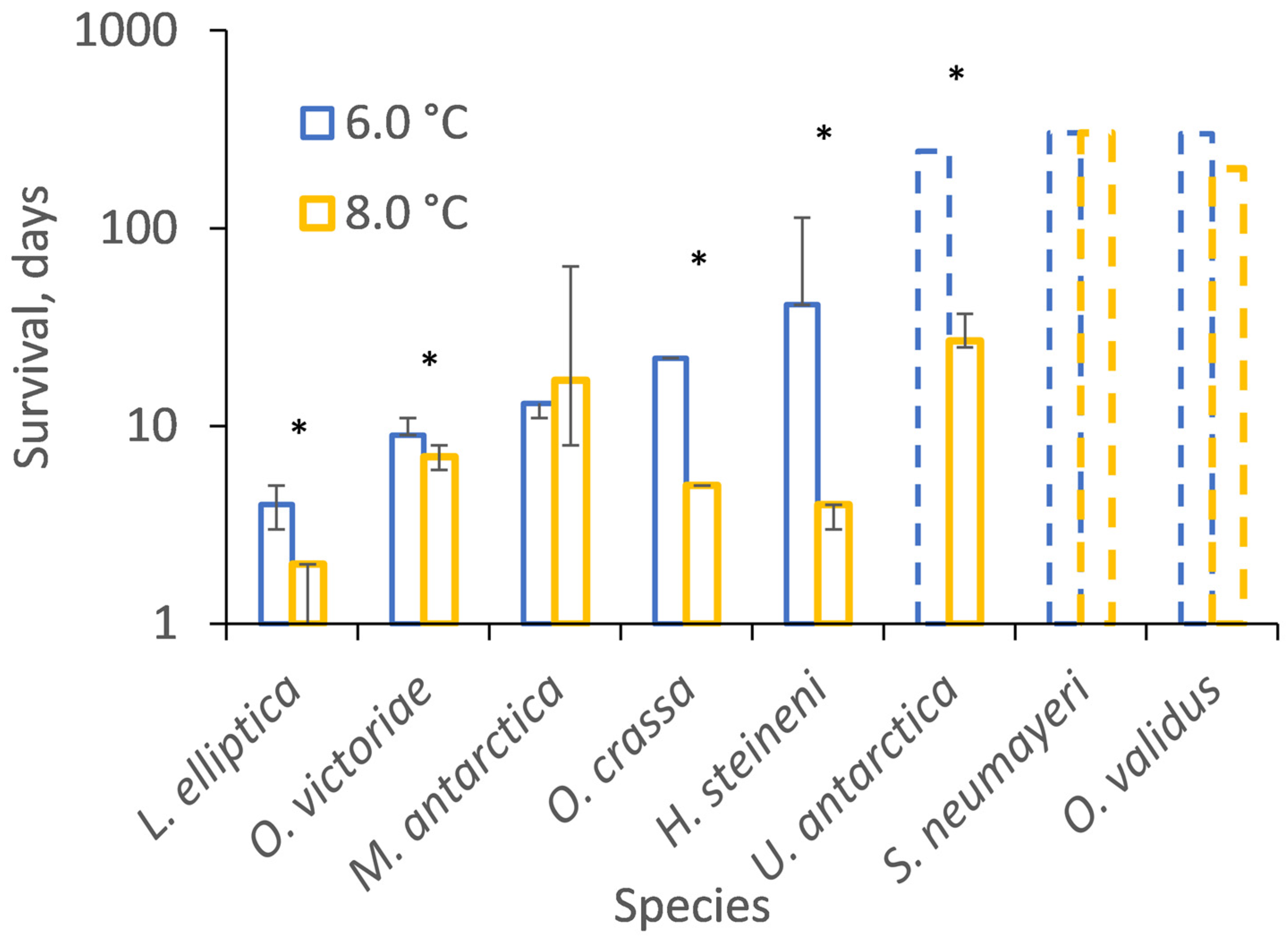
3.2. Temperature Incubations

In six out of eight trials, species survived for significantly longer when incubated at 6.0 than 8.0 °C (Figure 1, Table 2 and Table S1, Figure S2). The two exceptions were M. antarctica, whose median survival duration (13 and 17 days) was not significantly different at 6.0 and 8.0 °C (Chi2 = 2.4, p = 0.1; Table S1), and the trial with S. neumeyari, when the majority of individuals survived for the duration of incubations (303 days) at both 6.0 and 8.0 °C (Chi2 = 3.7, p = 0.06; Table S1). When considered across all trials, species for which median survival durations could be estimated at both 6.0 and 8.0 °C survived for significantly longer at the lower temperature (Chi2 = 24.5, p < 0.01). O. validus also survived more than 200 days at 8.0 °C until a technical failure of the tank.

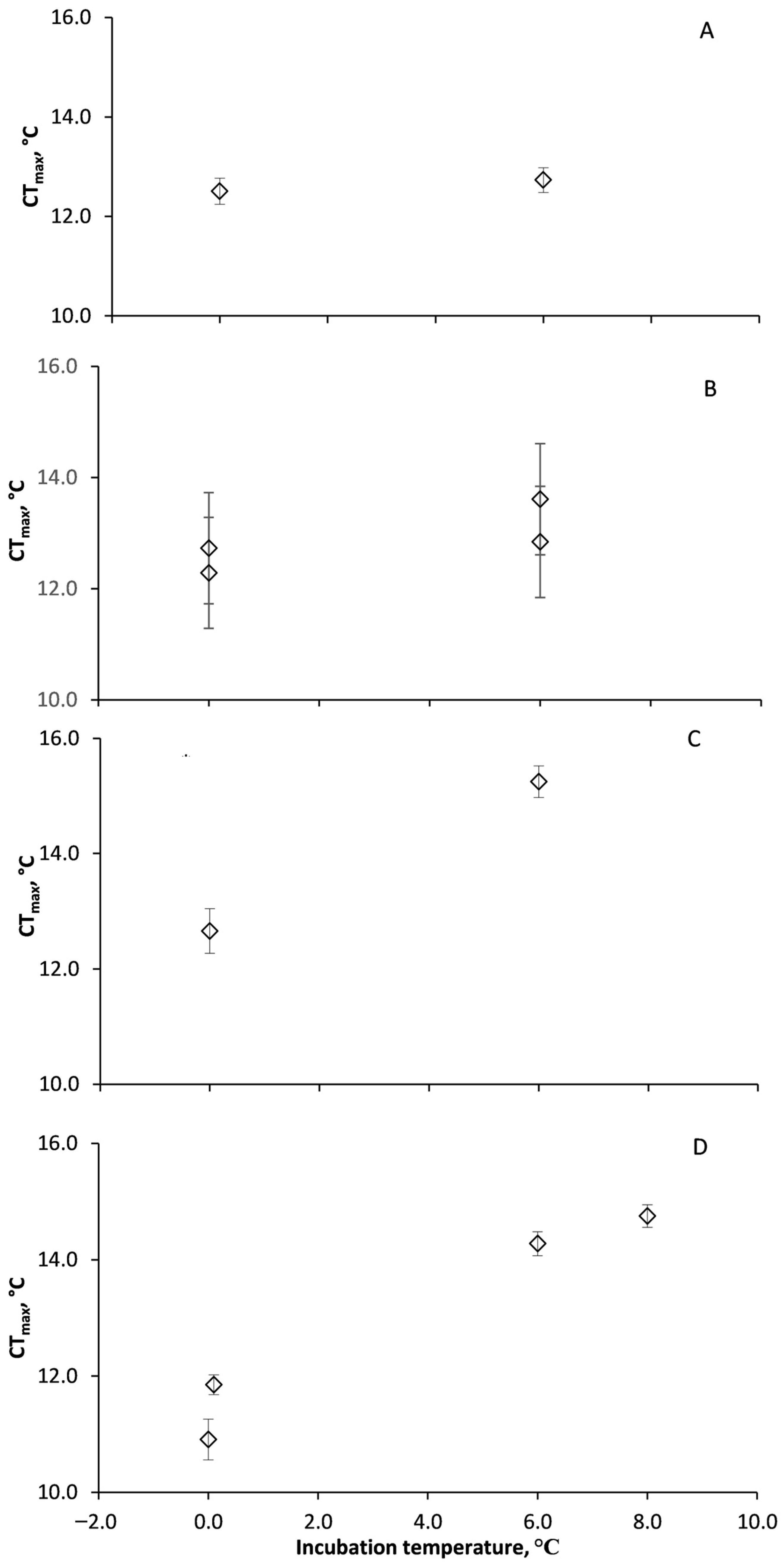
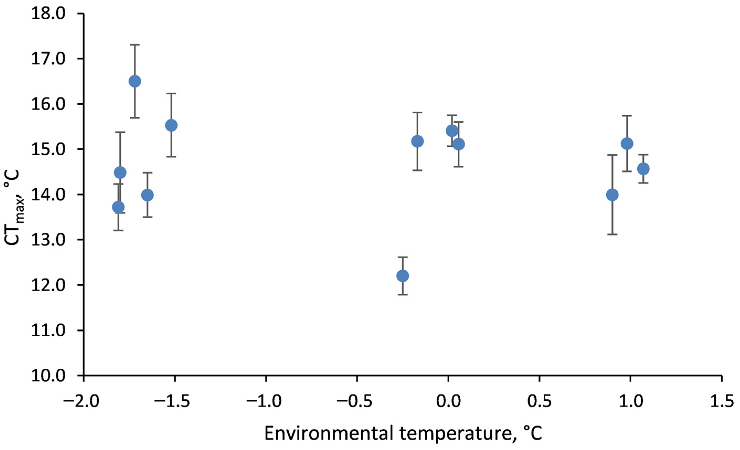
3.3. Acclimation of Acute CTmax

U. antarctica acute CTmax was not significantly different when incubated at either 0 or 6 °C (F(1,25) = 0.7, p = 0.69), with no effect of animal weight (F(1,25) = 0.6, p = 0.61) or trial (F(1.25) = 1.0, p = 0.33; Figure 2A; Table S2). H. steineni incubated at 6.0 °C had significantly higher CTmax (F(1,85) = 8.2, p < 0.01) than those incubated at 0.0 °C (Figure 2B) but with no effect of animal weight (F(1,85) = 0.2, p = 0.66; Table S2) or trial (F(1,85) = 0.8, p = 0.36). There was, however, a significant difference between replicates (F(1,85) = 5.9, p = 0.02), with the first incubation having significantly higher CTmax (12.7 ± 0.34 °C at 0.0 °C and 13.6 ± 0.29 °C at 6.0 °C) than the second (12.1 ± 0.31 °C at 0.0 °C and 12.8 ± 0.34 °C at 6.0 °C). O. validus incubated at 6.0 °C had a significantly higher CTmax (F(1,31) = 11.1, p < 0.01) than those incubated at 0.0 °C (Figure 2C), with no effect of animal weight (F(1,31) = 3.7, p = 0.07; Table S2), replicate (F(1,31) = 0.1, p = 0.72) or trial (F(1,31) = 2.2, p = 0.15). Acclimation of CTmax was also apparent for S. neumayeri (F(1,50) = 67.7, p < 0.01), with no effect of animal weight (F(1,50) = 1.0, p = 0.3) or replicate (F(1,50) = 1.5, p = 0.23; Table S2). Lethal limits were higher at 6.0 °C (T = 9.6, p < 0.01) and 8.0 °C (T = 9.4, p < 0.01) than 0.0 °C (Figure 2D).
There was no significant effect of average environmental (seawater) temperature during the month of collection on CTmax measured in O. validus (F(1,264) = 0.82, p = 0.37; Figure 3).

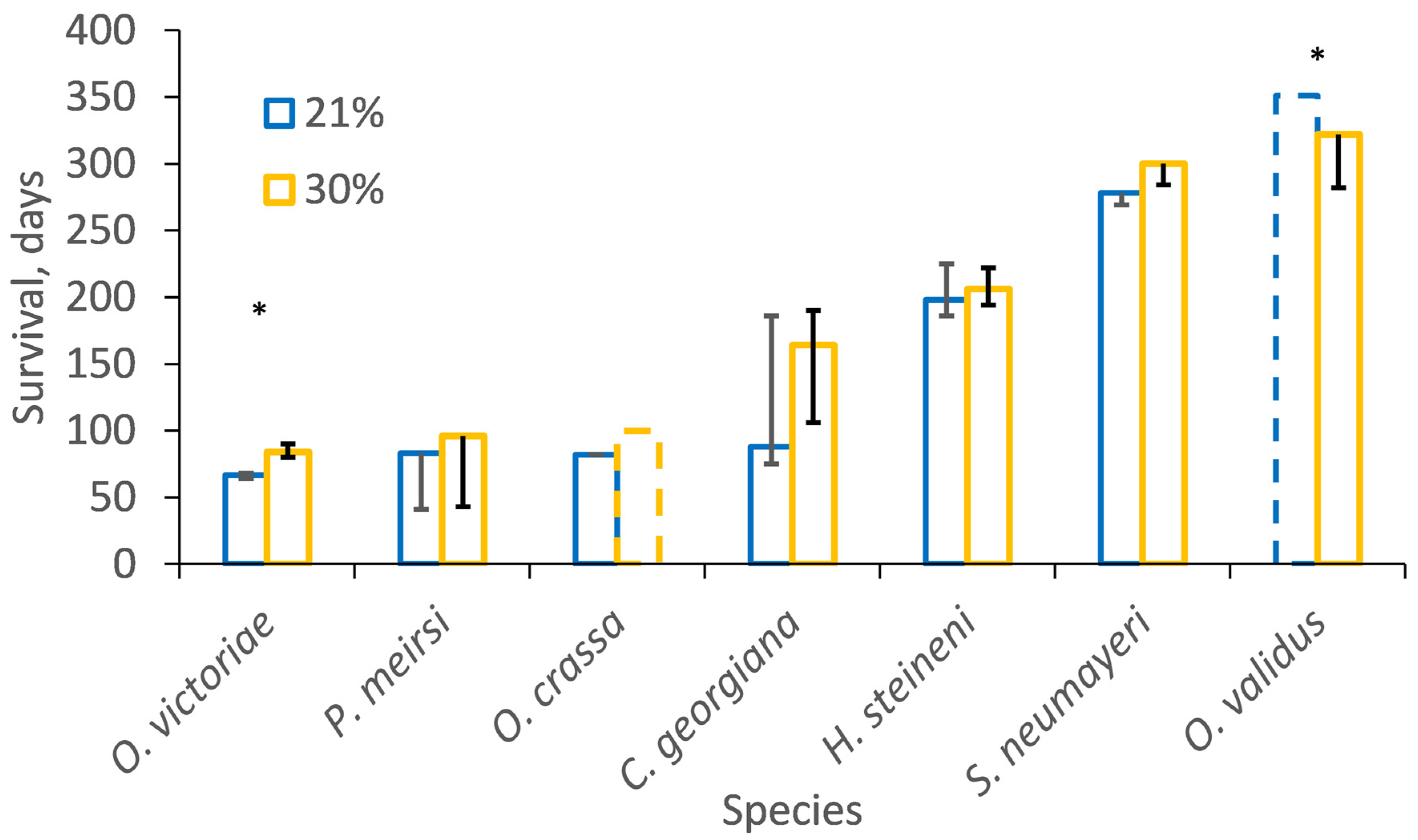
3.4. Temperature–Oxygen Incubations

In the first experiment all individuals of O. victoriae reached CTmax within the 99 days at 3.0 °C. The survival duration of both O. victoriae (median 84 versus 66.5; Chi2 = 47.8, p < 0.01) and O. crassa (>100 versus 82; Chi2 = 41.4, p < 0.01) was extended under hyperoxia (30%) compared to normoxia (21%; Figure 4 and Figure S2; Table S3). A total of 87% of O. crassa reached CTmax within 100 days under normoxia, whereas only 13% reached CTmax under hyperoxia. The other difference in survival duration was O. validus incubated at 8.0 increased to 10.0 °C, which showed the opposite pattern, and survived significantly longer under normoxia (median > 351 days) than hyperoxia (322 days). There was no significant effect of oxygen concentration on survival of the other species but when all species were considered together, survival was longer under hyperoxia (30%) than normoxia (21%; Likelihood ratio, Chi2 = 15.8, p < 0.01).

4. Discussion

Overall, we report mixed support for three central thermal physiology paradigms, with species-specific responses in: (1) physiological tolerance, (2) long-term acclimation, and (3) the effect of oxygen limitation on long-term survival. Previous estimates of long-term thermal tolerance of Antarctic marine ectotherms, extrapolated from CTmax assessments at multiple rates of warming, were between 1 and 6 °C if warming occurred over months, but dropped to a predicted average of 1.3 °C after about a year [12]. However, the current study extended known long-term incubation limits, with some individuals of U. antarcticus and H. steineni able to survive long-term at 6 °C, and O. validus and S. neumayeri at temperatures up to 10 °C (Table 1). Of all the Antarctic marine ectotherms tested to date, O. validus is one of the most tolerant, with the collapse of physiological processes (food processing and coelomic oxygen concentration) at 6 °C and loss of activity at 9 °C [12]. S. neumayeri also has a thermal tolerance towards the upper-end of the Antarctic ectotherm assemblage, with average functional thresholds in short-term warming around 8.3 ± 1.3 °C [26]. This study extended the long-term tolerance of O. validus and S. neumayeri to higher than previously reported temperatures of up to 10.0 °C, although it should be noted that individuals might have been resisting warming for long periods and might have impaired functions that would preclude, e.g., reproduction and recruitment. This is likely the case for U. antarcticus, which did not show evidence of acclimation, even after 245 days at 6 °C.
The current study, therefore, extends the known long-term experimental upper thermal limits of several Antarctic marine species to up to 8.2 °C, well above current maximum summer temperatures recorded in Marguerite Bay (1.8 °C [24]). The warming trend of air temperature along the Western Antarctic Peninsula since the 1970s has continued after a brief hiatus around the turn of the 21st century [27]. However, the warming signal in the Southern Ocean is less clear, with slower than average surface warming (0.02 °C per decade since 1950) [28]. The findings of the current study therefore suggest that the thermal limit of the most tolerant shallow water Antarctic marine ectotherms will not be breached for several centuries. However, thermal tolerance is species-specific, with limits for less tolerant species such as O. victoriae (this study and [19]), and the encrusting spirorbid worm Protolaeospira stalagmia [18], likely be reached in much less than 100 years, which will directly impact ecosystem biodiversity. Also, the limits for activity are often much lower than CTmax estimates, with species expected to suffer ecological impacts at temperatures well below CTmax. Species such as the scallop Adamussium colbecki, the limpet, Nacella concinna, and the bivalve mollusc, L. elliptica, have behavioural limits between 2 and 3 °C [28] which will clearly affect long-term survival in the wild. Understanding the variation in thermal tolerance between species, and between physiological functions, is crucial for determining the likely winners and losers in response to climate change and, therefore the impact of these changes on ecosystem function.
The responses of species to the effect of elevated oxygen on long-term survival arguing against the OCLTT being a universal mechanism controlling upper temperatures limits. While some species had clearly elevated CTmax under mild hyperoxia, indicating a positive effect of oxygen on whole animal CTmax, the response was not consistent across species and O. validus even showed a slight negative effect of hyperoxia on survival. As with the findings of the current study, Clark et al. [19] found multiple mechanisms underpinning species response when incubated at temperatures just below CTmax, with only two species showing the switch from aerobic to anaerobic metabolism [19]. Species-specific thermal tolerances have been linked to their functional traits, with the most active species having higher upper lethal limits [12]. Activity is correlated with aerobic scope, with more active species having an oxygen supply cascade that is capable of supplying more active tissues, resulting in a greater “total excess aerobic power budget” and therefore a higher capacity to resist thermal challenge [12,29]. However, where in the past Antarctic marine ectotherm species with higher activity levels have survived warming to higher temperatures [12], and this was taken as support for OCLTT, the species that acclimated to the higher temperatures in this study were all low-activity species, arguing against the importance of oxygen limitation. In the current study, the most resistant species came from a mixture of functional groups, from a mobile obligate predator through to a sessile primary consumer. It should be noted that the use of CTmax to test the OCLTT has been criticised and future research should include sub-lethal assays of thermal tolerance such as activity and the response of physiological pathways [30].
High concentrations of dissolved oxygen are expected to form free radicals, which could lead to tissue damage during long-term incubations [31]. If oxygen was not limiting to O. validus at the chosen incubation temperatures, then mild hyperoxia could have resulted in oxygen free-radical damage or additional costs for antioxidant defence mechanisms (e.g., superoxide dismutase [31]). This could potentially explain the negative effect of hyperoxia, but also provides a clear indication that oxygen was not limiting at these temperatures for this species. It is possible that a different pattern will be seen at higher temperatures.
At temperatures typical of polar oceans, the acclimation of physiological systems of marine invertebrates can take many months, with successful whole animal acclimation being recorded in three out of eight Antarctic marine invertebrate species within 2 and 9 months at 3–8 °C ([11] and current study). Acclimation in Antarctic fish was faster, with seven out of nine species acclimating within 5 to 36 days at 4 to 7 °C [32,33,34,35,36,37]. In the current study, the anemone U. antarcticus survived, with minimal mortality, for 270 days at 6 °C but with no whole animal adjustment of CTmax. Caution must be applied when interpreting the CTmax of survivors in thermal challenge experiments, as this will likely overestimate resilience, as only a subset of the more resilient survivors is being tested (5–95% survival). Also, the results under carefully controlled laboratory conditions may not reflect survival in the wild when sub-lethal effects will likely limit long-term population persistence, and how these reductions in sub-lethal performance will affect ecological interactions (e.g., reproduction, disease resistance, predation, and competition) is likely to be important. The slow rate of surface warming in the Southern Ocean [38] means that even a small amount of acclimation can provide species with a significant additional thermal buffer for many decades, long enough for generational plasticity and the possibility of adaptive change, even in Antarctic species [11].
It is therefore not surprising that O. validus did not show acclimatisation of acute CTmax in the field over the temperature range −1.8 to +1.1 °C. It is probable that the duration of the peak of summer temperatures (1–2 months [24]) or the maximum environmental temperature (+1.1 °C in the field) were not sufficient to trigger whole animal physiological adjustment of CTmax. The lack of predictable seasonal temperature change in the Southern Ocean could explain the lack of ecologically relevant acclimatisation, the observation that species do not select preferred temperatures, and why there are no observable escape response thresholds [15,16]. However, many other factors, such as tissue energy status, affect CTmax (e.g., [39]), and there might be greater seasonal differences in thermal tolerance in primary consumers with seasonally restricted food supplies, than in predators/scavengers which have a more constant food supply [40]. The complexity of the triggers for molecular thermal protective mechanisms has been illustrated for the HSP70 heat shock response in the Antarctic limpet, N. concinna. Acute laboratory thermal challenge did not elicit upregulation of HSP70 gene expression until temperatures of 15 °C, whereas limpets in the wild expressed HSP70 at foot temperatures of only 3.3 °C during a natural tidal cycle [41].
This study has highlighted species-specific variation in long-term survival at elevated temperatures, acclimation response and the influence of oxygen on CTmax of Antarctic marine ectotherms. The search for mechanisms underlying the evolution of physiological capacity [1,2,42] provides important frameworks against which exceptions can be identified and explanatory hypotheses constructed for future research. Thermal sensitivity is clearly the product of multiple ecological and physiological capacities, and this diversity of response needs further investigation and interpretation to improve our ability to predict future patterns of biodiversity.

5. Conclusions

Tests of three of the central paradigms underlying physiological response of Antarctic marine ectotherms to warming highlighted species-specific responses in regard to resilience, plasticity and the effect of oxygen limitation. This further highlights the complexity of thermal physiology and the need for further investigation and interpretation.

Supplementary Materials

The following supporting information can be downloaded at: https://www.mdpi.com/article/10.3390/biology13040224/s1, Figure S1. Example temperatures during ramping and incubation experiments at, A 6.0 °C and B 8.0 °C for 4 species Heterocucumis steineni, Odontaster validus, Sterechinus neumeyeri and Laternula elliptica, C for the 1st temperature-oxygen experiment. Figure S2. Kaplan Meier fits for survival of Ophiura crassa, Ophionotus victoriae, Margarella antarctica, Laternula elliptica, Sterechnius neumayeri, Odontaster Validus, Hetercucumis steineni and Urticinopsis antarctica. Figure S3. Kaplan-meier fits for survival of Odontaster validus, Sterechinus neumayeri, Heterocucumis steineni, Cucumaria georgiana, Ophionotus victoriae, Paraceradocus miersi and Ophiura crassa incubated at normoxia (21%) and hyperoxia (30%). See Table 1 and Figure 1 for incubation temperatures. Table S1. Median and interquartile range from Kaplan Meier fits in days (± Inter-Quartile Range). Nominal temperatures of 6.0 and 8.0 °C are listed that had a typical SE of ± 0.04 °C (Figure 1). Table S2. Wet mass of incubated individuals, mean, standard error and sample size. Within each species, there were no significant differences in wet mass between treatments. Table S3. Median and interquartile range from Kaplan Meier fits for Odontaster Validus, Sterechinus neumayeri, Heterocucumis steineni, Cucumaria georgiana, Ophionotus victoriae, Paraceradocus meirsi and Ophiura crassa incubated at either normoxia (21%) and hyperoxia (30%).

Author Contributions

Conceptualization, S.A.M., L.S.P., R.S. and M.S.C.; Methodology, S.A.M., L.S.P., E.F. and R.S.; Validation, S.A.M. and A.E.B.; Formal Analysis, S.A.M., R.E.S. and A.E.B.; Investigation, S.A.M., L.S.P., M.S.C., E.F. and R.S.; Resources, L.S.P., M.S.C. and R.S.; Data Curation, S.A.M.; Writing—Original Draft Preparation, S.A.M., R.E.S. and A.E.B.; Writing—Review and Editing, S.A.M., A.B, M.S.C., L.S.P., R.E.S. and E.F.; Visualization, S.A.M. and R.E.S.; Supervision, S.A.M. and A.E.B. All authors have read and agreed to the published version of the manuscript.

Funding

This study was funded by Natural Environment Research Council core funding to the British Antarctic Survey and Spitfire DTP funding to R.E.S.

Institutional Review Board Statement

All research was conducted under Antarctic Treaty permits issued by the UK Foreign, Commonwealth and Development Office to British Antarctic Survey. Experiments were conducted on marine invertebrate species not protected under the UK Animals (Scientific Procedures) Act 1986 and although ethical approval was not required the highest possible standards of animal welfare were applied.

Data Availability Statement

All data are either published with the manuscript or available on request from the lead author. Morley, S.A., Bates, A.E., Clark, M.S., Fitzcharles, E., Smith, R., Stainthorp, R.E., and Peck, L.S. (2024). Acclimation and acclimatisation of marine ectotherms collected at Rothera Research Station and Scott Base in Antarctica between 2004 and 2015 (Version 1.0) [Data set]. NERC EDS UK Polar Data Centre. https://doi.org/10.5285/60B777B4-0BD6-48C3-A301-C700854FBFA1.

Acknowledgments

The authors want to thank the marine teams and technical support at Rothera who enabled this research.

Conflicts of Interest

The authors declare no conflicts of interest.

References

  1. Gaston, K.J.; Chown, S.L.; Calosi, P.; Bernardo, J.; Bilton, D.T.; Clarke, A.; Clusella-Trullas, S.; Ghalambor, C.K.; Konarzewski, M.; Peck, L.S.; et al. Macrophysiology: A conceptual reunification. Am. Nat. 2009, 174, 595–612. [Google Scholar] [CrossRef]
  2. Pörtner, H.O. Climate variations and the physiological basis of temperature dependent biogeography: Systemic to molecular hierarchy of thermal tolerance in animals. Comp. Biochem. Physiol. A 2002, 132, 739–761. [Google Scholar] [CrossRef]
  3. Lindquist, S. The heat shock response. Annu. Rev. Biochem. 1986, 55, 1151–1159. [Google Scholar] [CrossRef]
  4. Tomanek, L. Environmental proteomics: Changes in the proteome of marine organisms in response to environmental stress, pollutants, infection, symbiosis, and development. Ann. Rev. Mar. Sci. 2011, 3, 373–399. [Google Scholar] [CrossRef]
  5. Collier, R.J.; Baumgard, L.H.; Zimbelman, R.B.; Xiao, Y. Heat stress: Physiology of acclimation and adaptation. Anim. Front. 2019, 9, 12–19. [Google Scholar] [CrossRef]
  6. Hobday, A.J.; Alexander, L.V.; Perkins, S.E.; Smale, D.A.; Straub, S.C.; Oliver, E.C.J.; Benthuysen, J.A.; Burrows, M.T.; Donat, M.G.; Feng, M.; et al. A hierarchical approach to defining marine heatwaves. Prog. Oceanogr. 2016, 141, 227–238. [Google Scholar] [CrossRef]
  7. Stillman, J. Acclimation underlies susceptibility to climate change. Science 2003, 301, 65. [Google Scholar] [CrossRef]
  8. Leroi, A.M.; Bennett, A.F.; Lenski, R.E. Temperature acclimation and competitive fitness: An experimental test of the Beneficial Acclimation Hypothesis. Proc. Natl. Acad. Sci. USA 1994, 91, 1917–1921. [Google Scholar] [CrossRef]
  9. Wilson, R.S.; Franklin, C.E. Testing the beneficial acclimation hypothesis. Trends Ecol. Evol. 2002, 17, 66–70. [Google Scholar] [CrossRef]
  10. Gunderson, A.R.; Stillman, J.H. Plasticity in thermal tolerance has limited potential to buffer ectotherms from global warming. Proc. R. Soc. B 2015, 282, 20150401. [Google Scholar] [CrossRef] [PubMed]
  11. Morley, S.A.; Peck, L.S.; Heiser, S.; Sunday, J.; Bates, A.E. Physiological acclimation extends predicted persistence of species across the globe. Glob. Ecol. Biodivers. 2019, 28, 1018–1037. [Google Scholar] [CrossRef]
  12. Peck, L.S.; Clark, M.S.; Morley, S.A.; Massey, A.; Rossetti, H. Animal temperature limits and ecological relevance: Effects of size, activity and rates of change. Funct. Ecol. 2009, 23, 248–256. [Google Scholar] [CrossRef]
  13. Molina, A.N.; Pulgar, J.M.; Rezende, E.L.; Carter, M.J. Heat tolerance of marine ectotherms in a warming Antarctica. Glob. Chang. Biol. 2023, 29, 179–188. [Google Scholar] [CrossRef]
  14. Clark, M.S.; Peck, L.S. Hsp70 heat shock proteins and environmental stress in Antarctic marine organisms: A mini-review. Mar. Genom. 2009, 2, 11–18. [Google Scholar] [CrossRef]
  15. Bates, A.E.; Lee, R.W.; Tunnicliffe, V.; Lamare, M.D. Deep-sea hydrothermal vent animals seek cool fluids in a highly variable thermal environment. Nat. Commun. 2010, 1, 14. [Google Scholar] [CrossRef]
  16. Morley, S.A.; Chu, J.W.F.; Peck, L.S.; Bates, A.E. Temperatures leading to heat escape responses in Antarctic marine ectotherms match acute thermal limits. Front. Physiol. 2022, 13, 1077376. [Google Scholar] [CrossRef]
  17. Clark, M.S.; Villota Nieva, L.; Hoffman, J.I.; Davies, A.J.; Trivedi, U.H.; Turner, F.; Ashton, G.; Peck, L.S. Lack of long-term acclimation in Antarctic encrusting species suggests vulnerability to warming. Nat. Commun. 2019, 10, 3383. [Google Scholar] [CrossRef]
  18. Collins, M.; Peck, L.S.; Clark, M.S. Large within, and between, species differences in marine cellular responses: Unpredictability in a changing environment. Sci. Total Environ. 2021, 794, 148594. [Google Scholar] [CrossRef]
  19. Clark, M.S.; Sommer, U.; Sihra, J.K.; Thorne, A.S.; Morley, S.A.; King, M.; Viant, M.R.; Peck, L.S. Biodiversity in marine invertebrate responses to acute warming revealed by a comparative multi-omics approach. Glob. Chang Biol. 2017, 23, 318–330. [Google Scholar] [CrossRef]
  20. Clark, T.D.; Sandblom, E.; Jutfelt, F. Aerobic scope measurements of fishes in an era of climate change: Respirometry, relevance and recommendations. J. Exp. Biol. 2013, 216, 2771–2782. [Google Scholar] [CrossRef]
  21. Verberk, W.C.; Overgaard, J.; Ern, R.; Bayley, M.; Wang, T.; Boardman, L.; Terblanche, J.S. Does oxygen limit thermal tolerance in arthropods? A critical review of current evidence. Comp. Biochem. Physiol. A Mol. Integr. Physiol. 2016, 192, 64–78. [Google Scholar] [CrossRef]
  22. Richard, J.; Morley, S.A.; Thorne, M.A.; Peck, L.S. Estimating long term survival temperatures at the assemblage level in the marine environment: Towards macrophysiology. PLoS ONE 2012, 7, e34655. [Google Scholar] [CrossRef]
  23. Folmer, O.; Black, M.; Hoeh, W.; Lutz, R.; Vrijenhoek, R. DNA primers for amplification of mitochondrial cytochrome c oxidase subunit I from diverse metazoan invertebrates. Mol. Mar. Biol. Biotech. 1994, 3, 294–299. [Google Scholar]
  24. Venables, H.; Meredith, M.P.; Hendry, K.R.; ten Hoopen, P.; Peat, H.; Chapman, A.; Beaumont, J.; Piper, R.; Miller, A.J.; Mann, P.; et al. Sustained, year-round oceanographic measurements from Rothera Research Station, Antarctica, 1997–2017. Sci. Data 2023, 10, 265. [Google Scholar] [CrossRef]
  25. Allignol, A.; Latouche, A. CRAN Task View: Survival Analysis, Version 2023-09-10. 2023. Available online: https://CRAN.R-project.org/view=Survival (accessed on 1 January 2023).
  26. De Leij, R.; Grange, L.J.; Peck, L.S. Functional thermal limits are determined by rate of warming during simulated marine heatwaves. Mar. Ecol. Prog. Ser. 2022, 685, 183–196. [Google Scholar] [CrossRef]
  27. Carrasco, J.F.; Bozkurt, D.; Cordero, R.R. A review of the observed air temperature in the Antarctic Peninsula. did the warming trend come back after the early 21st hiatus? Polar Sci. 2021, 28, 100653. [Google Scholar] [CrossRef]
  28. Peck, L.S.; Webb, K.E.; Bailey, D. Extreme sensitivity of biological function to temperature in Antarctic marine species. Funct. Ecol. 2004, 18, 625–630. [Google Scholar] [CrossRef]
  29. Pörtner, H.O.; Bock, C.; Mark, F.C. Oxygen- and capacity-limited thermal tolerance: Bridging ecology and physiology. J. Exp. Biol. 2017, 220, 2685–2696. [Google Scholar] [CrossRef]
  30. McArley, T.J.; Sandblom, E.; Herbert, N.A. Fish and hyperoxia—From cardiorespiratory and biochemical adjustments to aquaculture and ecophysiology implications. Fish Fish. 2020, 22, 324–355. [Google Scholar] [CrossRef]
  31. Abele, D.; Puntarulo, S. Formation of reactive species and induction of antioxidant defence systems in polar and temperate marine invertebrates and fish. Comp. Biochem. Physiol. A 2004, 138, 405–415. [Google Scholar] [CrossRef] [PubMed]
  32. Wilson, R.S.; Franklin, C.E.; Davison, W.; Kraft, P. Stenotherms at subzero temperatures: Thermal dependence of swimming performance in Antarctic fish. J. Comp. Physiol. 2001, 171, 263–269. [Google Scholar] [CrossRef] [PubMed]
  33. Seebacher, F.; Davison, W.; Lowe, C.J.; Franklin, C.E. A falsification of the thermal specialization paradigm: Compensation for elevated temperatures in Antarctic fishes. Biol. Lett. 2005, 1, 151–154. [Google Scholar] [CrossRef] [PubMed]
  34. Franklin, C.E.; Davison, W.; Seebacher, F. Antarctic fish can compensate for rising temperatures: Thermal acclimation of cardiac performance in Pagothenia borchgrevinki. J. Exp. Biol. 2007, 210, 3068–3074. [Google Scholar] [CrossRef] [PubMed]
  35. Robinson, E.; Davison, W. The Antarctic notothenioid fish Pagothenia borchgrevinki is thermally flexible: Acclimation changes oxygen consumption. Polar Biol. 2008, 31, 317–326. [Google Scholar] [CrossRef]
  36. Bilyk, K.T.; Devries, A.L. Heat tolerance and its plasticity in Antarctic fishes. Comp. Biochem. Physiol. A Mol. Integr. Physiol. 2011, 158, 382–390. [Google Scholar]
  37. Strobel, A.; Bennecke, S.; Leo, E.; Mintenbeck, K.; Pörtner, H.O.; Mark, F.C. Metabolic shifts in the Antarctic fish Notothenia rossii in response to rising temperature and PCO2. Front. Zool. 2012, 9, 28. [Google Scholar] [CrossRef] [PubMed]
  38. Cai, W.; Gao, L.; Luo, Y.; Li, X.; Zheng, X.; Zhang, X.; Cheng, X.; Jia, F.; Purich, A.; Santoso, A.; et al. Southern Ocean warming and its climatic impacts. Sci. Bull. 2023, 68, 946–960. [Google Scholar] [CrossRef] [PubMed]
  39. Brokordt, K.B.; Himmelman, J.H.; Guderley, H.E. Effect of reproduction on escape responses and muscle metabolic capacities in the scallop Chlamys islandica Müller. J. Exp. Mar. Biol. Ecol. 2000, 251, 205–225. [Google Scholar] [CrossRef] [PubMed]
  40. Obermüller, B.O.; Morley, S.A.; Clark, M.S.; Barnes, D.K.A.; Peck, L.S. Antarctic intertidal limpet ecophysiology: A winter-summer comparison. J. Exp. Mar. Biol. Ecol. 2011, 403, 39–45. [Google Scholar] [CrossRef]
  41. Clark, M.S.; Geissler, P.; Waller, C.; Fraser, K.P.P.; Barnes, D.K.A.; Peck, L.S. Low heat shock thresholds in wild Antarctic inter-tidal limpets (Nacella concinna). Cell Stress Chaperones 2008, 13, 51–58. [Google Scholar] [CrossRef]
  42. Brown, J.H.; Gilooly, J.F.; Allen, A.P.; Savage, V.M.; West, G.B. Towards a metabolic theory of ecology. Ecology 2004, 85, 1771–1789. [Google Scholar] [CrossRef]
Figure 1. The Kaplan-Meier estimate of median survival duration, in days, after incubation at 6.0 and 8.0 °C. Species ordered from lowest to highest survival. Median ± interquartile range. Hashed lines indicate the duration of experiments for species for which the Kaplan-Meier estimator could not be calculated as most individuals survived until the experiment ended (censored observations), except for O. validus at 8.0 °C when the experiment terminated after 200 days due to a heater failure. Missing interquartile ranges indicate that the Kaplan-Meier could not estimate that value (Table S1). An asterix (*) indicates significantly different survival of that species between incubation temperatures. See Figure S1 for the stability of incubation temperatures and Figure S2 for Kaplan-Meier estimation figures.
Biology 13 00224 g001
Figure 2. Lethal temperature limits in four Antarctic marine invertebrates incubated in the laboratory at 0.0, 6.0 and 8.0 °C. (A) Urticinopsis antarctica, (B) Heterocucumis steineni, (C) Odontaster validus, and (D) Sterechinus neumayeri. U. antarctica was the only species whose CTmax did not acclimate between control (0.0 °C) and elevated temperatures (6 and 8.0 °C). Where more than one data point is shown for a given temperature, more than one experiment was conducted at different times. Urticinopsis antarctica, 0 °C n = 14, 6 °C n = 13, Heterocucumis steineni, n = 19 to 24, Odontaster validus, 0 °C n = 14, 6 °C n = 19 and Sterechinus neumayeri, 0 °C n = 12 and 8, 6 °C n = 21, 8 °C n = 10.
Biology 13 00224 g002
Figure 3. Upper lethal limits of Odontaster validus tested directly after collection from the field in different years and seasons. Mean ± 2 SE.
Biology 13 00224 g003
Figure 4. The Kaplan-Meier estimated median survival duration, in days (±interquartile range) that each species survived when incubated under normoxia (21%) or hyperoxia (30%). Hashed lines indicate the duration of experiments for species for which the Kaplan-Meier estimator could not be calculated as most individuals survived until the experiment ended (censored observations). An asterix (*) indicates species with significantly different survivals under different oxygen treatments. For stability of incubation temperature see Table 1 and Figure S3 for the Kaplan-Meier estimation figures.
Biology 13 00224 g004
Table 1. Summary of the conditions (± SE) for temperature–oxygen incubations and a list of the species used in each experiment with sample size per tank in parentheses. A step-change in temperature in the 2014/15 experiment is indicated in parentheses. Species are (A) Paraceradocus miersii (30), Ophionotus victoriae (30), Ophiura crassa (30) and (B) Sterechinus neumayeri (10), Odontaster validus (10), Cucumaria georgiana (10), and Heterocucumis steineni (10).
YearDuration (d)TankTreatmentTemperature (°C)Oxygen (% O2)Oxygen (mg/L) *
(A)
2010/111001Control0.9 ± 0.0521.0 ± 0.113.9
2Normoxic3.0 ± 0.0121.0 ± 0.113.2
3Hyperoxic3.0 ± 0.0229.3 ± 0.518.3
(B)
2014/153511Control0.1 ± 0.0121.0 ± 0.114.2
2Normoxic8.6 ± 0.5 (10.0 ± 0.01)21.0 ± 0.311.4
3Normoxic8.1 ± 0.2 (9.9 ± 0.01)21.5 ± 0.511.8
4Hyperoxic8.1 ± 0.2 (10.0 ± 0.01)30.5 ± 0.616.8
5Hyperoxic8.0 ± 0.2 (10.0 ± 0.01)31.3 ± 0.717.2
* Mean oxygen concentration (% O2) was converted to mg L−1 using standard conversion tables (PreSens Precision Sensing GmbH, Regensburg, Germany).
Table 2. Survival in each incubation experiment.
SpeciesIncubationMortalitiesSurvivors% Survival
S. neumayeri6 °C102168
8 °C21083
O. validus6 °C11995
8 °CTechnical failure
O. victoriae6 °C2700
8 °C2300
O. crassa6 °C2400
8 °C2500
M. antarctica6 °C1300
8 °C1900
U. antarctica6 °C11393
8 °C1600
L. elliptica6 °C1900
8 °C1500
H. steineni6 °C21416
8 °C2500
O. validus21%81158
30%12840
O. crassa21%26413
30%42687
P. miersi21%151550
30%151550
O. victoriae21%3000
30%3000
C. georgiana21%2000
30%2000
S. neumayeri21%18314
30%1900
H. steineni21%2000
30%1915
Disclaimer/Publisher’s Note: The statements, opinions and data contained in all publications are solely those of the individual author(s) and contributor(s) and not of MDPI and/or the editor(s). MDPI and/or the editor(s) disclaim responsibility for any injury to people or property resulting from any ideas, methods, instructions or products referred to in the content.

