Article

Effect of Doxapram, a K2p Channel Blocker, and pH on Heart Rate: Larval Drosophila Model

by
Elizabeth R. Elliott
,
Alaina C. Taul
,
Maya O. Abul-Khoudoud
,
Nicole Hensley
and
Robin L. Cooper
*
Department of Biology, University of Kentucky, Lexington, KY 40506-0225, USA
*
Author to whom correspondence should be addressed.
Current address: College of Physicians & Surgeons, Columbia University, New York, NY 10032, USA.
Current address: School of Veterinary Medicine, University of California at Davis, Davis, CA 95616, USA.
Appl. Biosci. 2023, 2(3), 406-420; https://doi.org/10.3390/applbiosci2030026
Submission received: 2 June 2023 / Revised: 24 July 2023 / Accepted: 1 August 2023 / Published: 3 August 2023

Abstract

:

Simple Summary

This was a study into how doxapram (a therapeutic compound with the capacity to block K2p channel function), pH, and the interactions between the two affect cardiac function in larval Drosophila, with resulting insight into mammalian cardiac function and general physiology.

Abstract

Two-P-domain K+ (K2p) channels are responsible for maintaining the resting membrane potential. K2p channels have varied expression in healthy tissue, but they also change in cancerous or diseased states. The correlation and causation as regards the alteration of K2p channel expression are still being investigated. The compound doxapram seems to block K2p channels and depolarize cells. Using Drosophila, the increased expression of the ORK1 K2p channel in cardiac and skeletal muscle was investigated. The heart rate in larval Drosophila is very sensitive to pH, and since doxapram blocks a subset of the K2p channels that are known to be acid-sensitive, it was postulated that doxapram would affect heart rate. A pH change from 7.1 to 6.5 increased the rate, while that from 7.1 to 7.5 decreased the rate. An amount of 0.1 mM of doxapram had no effect, but 0.5 of mM depressed Drosophila heart rates within five minutes. Exposure to 5 mM of doxapram immediately decreased the rate. Lipopolysaccharides (LPSs) from Gram-negative bacteria acutely increased the rate. LPSs activate K2p channels in the skeletal muscle of larvae and are blocked by doxapram. LPSs slightly reduce depression in the rate induced by doxapram. The overexpression of K2p channels in the heart and skeletal muscle depressed the heart rate and heightened pH sensitivity. At larval neuromuscular junctions, the overexpression in skeletal muscle increases the frequency of spontaneous quantal events and produces a more negative resting membrane potential.

1. Introduction

The diversity in resting membrane potential across various cell types is broad, whether evaluated across organisms, within an organism, or even among cells within a tissue, such as myocytes in cardiac tissue. This diversity results from differing types of specific ionic channels, exchangers, and pumps. In beating cells such as cardiac myocytes, which are engaged in both pacing and contraction, the resting potential may not be at a set value, yet it is nonetheless driven by ionic driving gradients and proteins serving as ionic conductors. Generally, a cell’s resting membrane potential is driven toward the potassium equilibrium potential (EK) due to a large K+ permeability in a resting state, as well as toward pumps that help maintain the gradient. The K+ leak channels constitute one subclass of two-P-domain K+ channels (i.e., K2p) [1]. The various subtypes of K2p channels are diverse in both function and pharmacological profiling [2,3].
Alterations in the K2p channels under certain pathological conditions, such as in cancerous tissues, have been observed through gene-sequence profiling and the identification of the expression levels in different cells for various K2p channel subtypes [4]. Additionally, K2p expression profiles for mammalian cardiac tissues in differing regions of the heart have been compiled, and a recent review highlights how this profile changes in diseased states [5]. No full review of tissue-specific expression in invertebrate models (e.g., Drosophila melanogaster, Caenorhabditis elegans, planarians, crustaceans, nonmammalian vertebrate models such as zebrafish and amphibians, etc.) currently exists. These models have all been especially useful over the years, substantially contributing to the current understandings of animal physiology and enhancing the fundamental knowledge of mammalian physiology and human diseases. Thus, it would be beneficial to investigate both the expression and function for K2p channel subtypes in these models.
Drosophila melanogaster provides a good model for investigating cardiac function in relation to other organisms [6]. In mammalian hearts, both the resting membrane potential and the characteristic shapes of action potentials vary across their regions. These electrical distinctions stem from differences in both the types and densities of ion channels, pumps, and exchangers. The larval Drosophila myogenic heart tube shares these regional cardiac differences and is comparable to the mammalian heart in that different regions of the heart also have their own intrinsic rates while nonetheless being driven by master pacemaker cells [7,8]. The action of the pharmacological agents can vary depending on the cell type due to these electrical differences [9]. Additionally, the rate and rhythm of the mammalian heart rate is susceptible to pH alterations, and the larval Drosophila heart shares this sensitivity [7,10,11]. The channels responsible for maintaining the resting membrane potential in various cells are dependent on K2p channel subtypes [3,12,13]. Knowing more about these subtypes is of interest, as some are sensitive to pH and are blocked by protonation; this suggests that altered extracellular pH could affect the resting membrane potential and cellular physiology. Doxapram has been established for its capacity to block mammalian, pH-sensitive K2p channels and depolarize larval body wall muscles [3,14,15,16], so it was suspected to also increase the heart rate in larval Drosophila.
Currently, no reports fully address which mechanisms maintain the resting membrane potential, nor what drives the pacemaker cells in the larval heart. However, the rate is known to be dependent on extracellular calcium and the Drosophila’s intracellular-calcium regulatory processes: namely, the function of the plasmalemmal Na+/Ca2+ exchanger [NCX], the Ca2+-ATPase (PMCA), and the sarcoplasmic/endoplasmic reticulum Ca2+-ATPase (SERCA)’s effect on the endoplasmic reticulum. The rate is also dependent on at least one type of K2p channel for membrane potential maintenance. The pharmacological and physiological profiles of Drosophila K2p channel subtypes have not been fully investigated [2]. The Drosophila K2p channel known as the ORK1 channel has been identified within cardiac muscle, where the overexpression of the channel stops the heartbeat [2,12,17]. ORK1 channels that are overexpressed in the pupal heart leave the heart tube in a diastolic state [17]. Additionally, the selective knockdown of the ORK1 expression using RNAi resulted in an increased heartbeat, thereby suggesting that the cells were depolarized [17]. The heart was still able to beat upon electrical stimulation, even when the ORK1 was knocked down, which implies that other K2p channel subtypes must be present to maintain the membrane potential. Currently, there are eleven known genes for the K2p subtypes in the Drosophila genome [18,19].
By virtue of easy-to-obtain intracellular recordings in body wall muscles, gene expression in mesodermal cells, which comprise the heart and body wall muscles in larval Drosophila, allow for the ready conduction of electrophysiological assays; these elucidate the effects of ORK1 expression on the resting membrane potential. Thus, the body wall muscle could indicate how cardiac tissue may behave with altered gene expression in K2p channels. In this study, both a mesodermal strain (24B) and a heart-specific line (HAND) were utilized to overexpress the ORK1 and assess its effects on both the heart rate and resting membrane potential in body wall muscles. It is likely that one of the aforementioned acid-sensitive K2p channel subtypes (i.e., TASK) may be present on the larval heart. The TASK subtypes are present in mammalian cardiac tissue and are sensitive to certain volatile anesthetics, as well as in larval Drosophila, which expresses a decreased heartbeat upon chloroform exposure [5,20,21].
Recent studies also demonstrated that doxapram depolarized the body wall muscles in larval Drosophila and blocked hyperpolarization induced by lipopolysaccharides (LPSs) from Gram-negative bacteria [22]. It appears LPSs activate K2p channels on the body wall muscles, while doxapram dampens that effect [15,16,21]. Since doxapram targets mammalian, acid-sensitive K2p TASK channels, it was postulated that it may have similar effects on the cardiac tissue of larval Drosophila as on the body wall muscles. Thus, it was hypothesized that doxapram would also impact the larval heart, thereby resulting in an increased heart rate in a dose-dependent manner. However, if the effect is too robust in the depolarization, the heart might stop beating in a form of tetany. A range in the concentrations of doxapram was utilized to determine the compound’s effects in this model system. In rodent models, 100 mg/kg as an i.p. injection has been used in past studies [23]. The use of this compound is novel in its use in invertebrate systems.

2. Methods

2.1. Fly Lines and Culturing Conditions

Drosophila melanogaster Canton S (CS) flies were used in physiological assays. This strain has been isogenic in the laboratory for several years and was originally obtained from the Bloomington Drosophila Stock Center (BDSC). Earlier, third-instar Drosophila larvae were used (50–70 h) post-hatching for CS larvae. The CS larvae were maintained at room temperature, ~21 °C, in vials partially filled with a cornmeal–agar–dextrose–yeast medium. The ORK1 receptor overexpression in the heart and body wall muscles (i.e., mesoderm) was performed by crossing homozygous 24B-Gal4 (III) (BDSC stock # 1767) with female virgins of UAS-ORK1 (BDSC stock # 6586). Progeny carrying one copy each of GAL4 driver and UAS-ORK1, referred to as 24B > ORK1, were used for physiological analyses. UAS-ORK1 alone was used for control comparisons. The UAS-ORK1 is expressed as y [1] w[*]; P{w[+mC] = UAS-Ork1.Delta-C}2. These Drosophila strains were obtained from the Bloomington Drosophila Stock Center (BDSC). A heart-specific strain, Hand4.2-Gal4 (on II), was used for expression of ORK1. This strain was supplied by Dr. Anthony Cammarato. The F1 of the cross with ORK1 and Hand is referred to as HAND > ORK1.
Three different paradigms were used for raising Drosophila strains UAS-ORK1 and 24B > ORK1 for heart rate measures. One paradigm was to maintain the lines at 21 °C; another was to raise the lines at 21 °C for embryo to early 2nd instar and then place them at 18 °C for 48 h before examining the heart. The third paradigm was to pair males and females of the crosses or background controls and maintain the F1 from embryo to 2nd instar at 18 °C before examining them for cardiac function. This lower temperature was intended to decrease the expression of ORK1 compared to raising the 24B > ORK1 strain at 21 °C. The Gal4/UAS binary expression system expressed a lower level of ORK1 at 18 °C [24]. Second instars for the 24B > ORK1 were used to examine effects of doxapram and pH, as late 2nd instars died when raised at 21 °C and no 3rd instars were present. Second-instar UAS-ORK1 were used as controls raised in identical conditions to compare to 24B > ORK1. The Hand > ORK1 were able to be raised at 21 °C to assess the effect of pH on the rate of the heartbeat, since these larvae survived well into the 3rd instar, and many reached adulthood.

2.2. Dissection and Solutions

The general dissection technique used to directly assess heart rate in Drosophila larvae has been previously reported [25]. Dissections took three to six minutes. Larvae were dissected in modified HL3 saline, as described in de Castro et al. [26]. The saline contained (mM): 1.0 CaCl2·2H2O, 70 NaCl, 20 MgCl2, 5 KCl, 10 NaHCO3, 5 trehalose, 115 sucrose, and 25 5-N,N-bis(2-hydroxyethyl)-2-aminoethane sulfonic acid (BES). After aeration, the saline quantities were adjusted to pH 7.1 or 6.5 with HCl (1 M) or NaOH (1 M) as needed. The saline pH was monitored at room temperature (21C) using an Accumet model 10 pH meter (Fisher Scientific, Hampton, NH, USA) and Ag/AgCl glass electrode. Exchanges in bathing media are shown within the figures. The LPSs were from Serratia marcescens (Sigma-Aldrich, St. Louis, MO, USA). LPSs were dissolved in the physiological saline at a concentration of 500 µg/mL to match the concentration of the LPSs used in previous studies. To examine the effect of heart rate modulators, bath saline was exchanged with a saline containing compounds of interest. Modulator solutions (varying pH, doxapram, LPSs) were prepared fresh daily. All chemicals listed above were obtained from Sigma-Aldrich (St. Louis, MO, USA).
To investigate doxapram’s potential effects on heart rate, three different concentrations were each examined over a five-minute period. The rate was obtained once in saline, once immediately after exchanging the bath to doxapram, then again after five more minutes, and once upon switching back to saline.

2.3. Measures of Membrane Potential and Frequency of Spontaneous Quantal Responses in Body Wall Muscles

To monitor the transmembrane potentials of the larvae body wall muscle (m6), a sharp intracellular electrode (30 to 40 MΩ resistance) filled with 3 M KCl impaled the fiber. An Axoclamp 2B (Molecular Devices, Sunnyvale, CA, USA) amplifier and 1 × LU head stage were used. The bathing saline was 21 °C at pH of 7.1. Since the 24B > ORK1 were dying at 21 °C, the 2nd instar raised at 18 °C were used for examining the membrane potential at 21 °C. Controls, (i.e., the parental line UAS-ORK1) were also raised at 18 °C at the 2nd instar stage. The lower temperature likely decreased expression of ORK1 [27,28].
The spontaneous quantal events were monitored for two minutes in the same larva used to examine the resting membrane potential of m6 in segment four. The analysis proceeded in the following manner: once membrane potential had been observed at a steady state (i.e., approximately a minute after impaling the muscle fiber), the number of spontaneous quantal events were counted for the duration of one minute. All events with a sharp rise were counted by observation. In cases where multiple events stacked on top of one another, they were counted only if distinct events were observable by steep alterations. Some events appeared as extremely large, singular events. In these circumstances, the events were counted as a single event, although it was possible that multiple quantal synchronal events occurred.

2.4. Statistical Analysis

Some data are expressed as raw values. A sign pairwise test was used to analyze changes in heart rate or membrane potential after changing bath conditions. Because a few datasets were not normally distributed (largely thanks a number of zeroes in some groups as the heartbeat stopped), the nonparametric sign test was used. When appropriate, paired and unpaired t-tests were used. A significant difference was considered to be p < 0.05. Different symbols were used in the graphs to isolate individual preparations from one another. The percent change was determined for each larva using the mathematical process expressed by: [(experimental − control)/(control)] × 100. A mean (±SEM) of the percent changes are graphed and reported.

3. Results

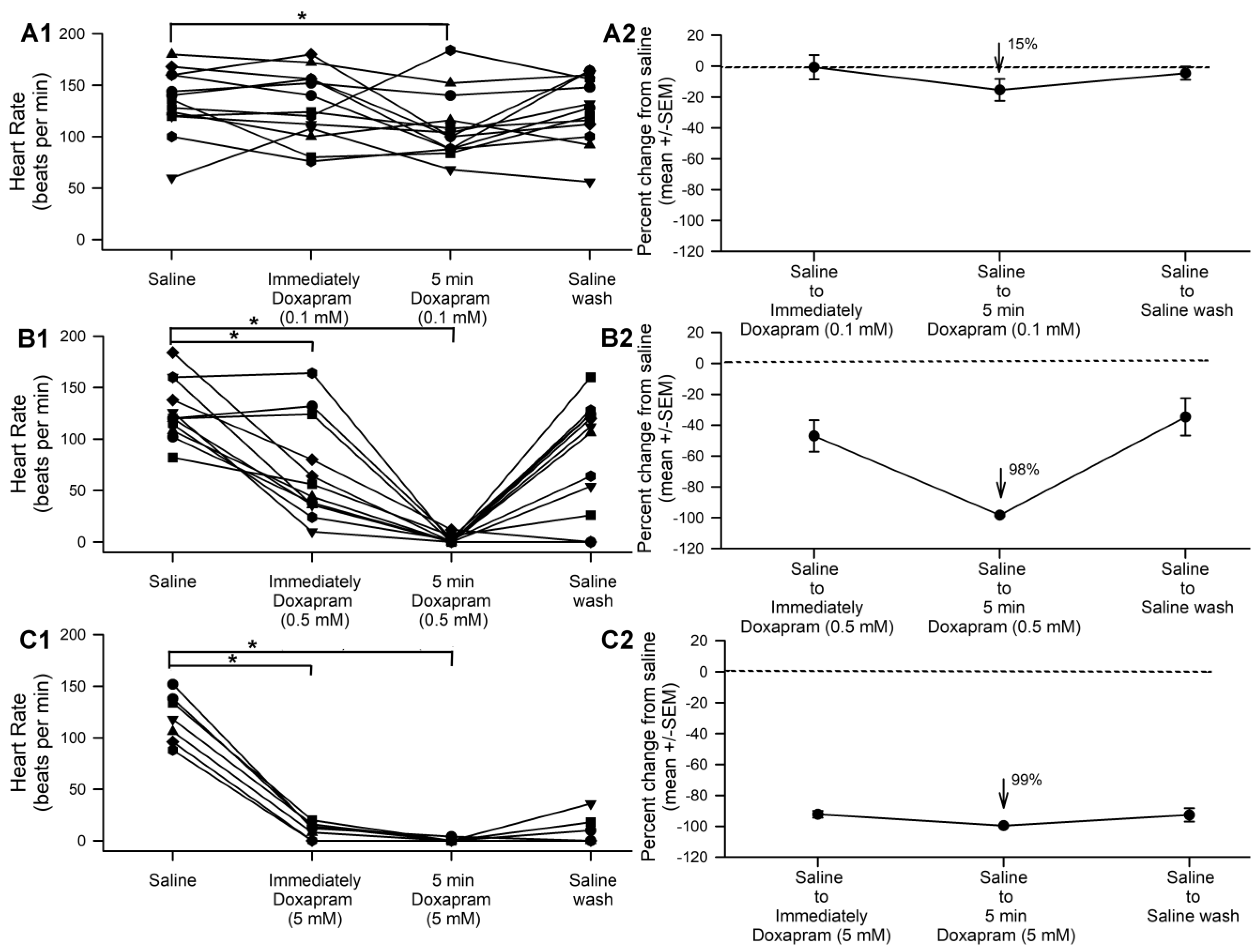
3.1. The Effect of Doxapram on Heart Rate

The exposure to 0.1 mM of doxapram solution produced a significant decrease in the heart rate after five minutes (representing a decrease by 15%); both the 0.5 and 5.0 mM solutions produced a rapid decrease in the rate immediately upon exposure (with both decreasing by nearly 100%), which lasted through the full five minutes of examination (Figure 1(A1,B1,C1); p < 0.05 two-way repeated ANOVA). The higher concentration (5.0 mM) did not exhibit the same washout success as the other two concentrations. The average percent changes from the initial saline are shown for each concentration (Figure 1(A2,B2,C2)).

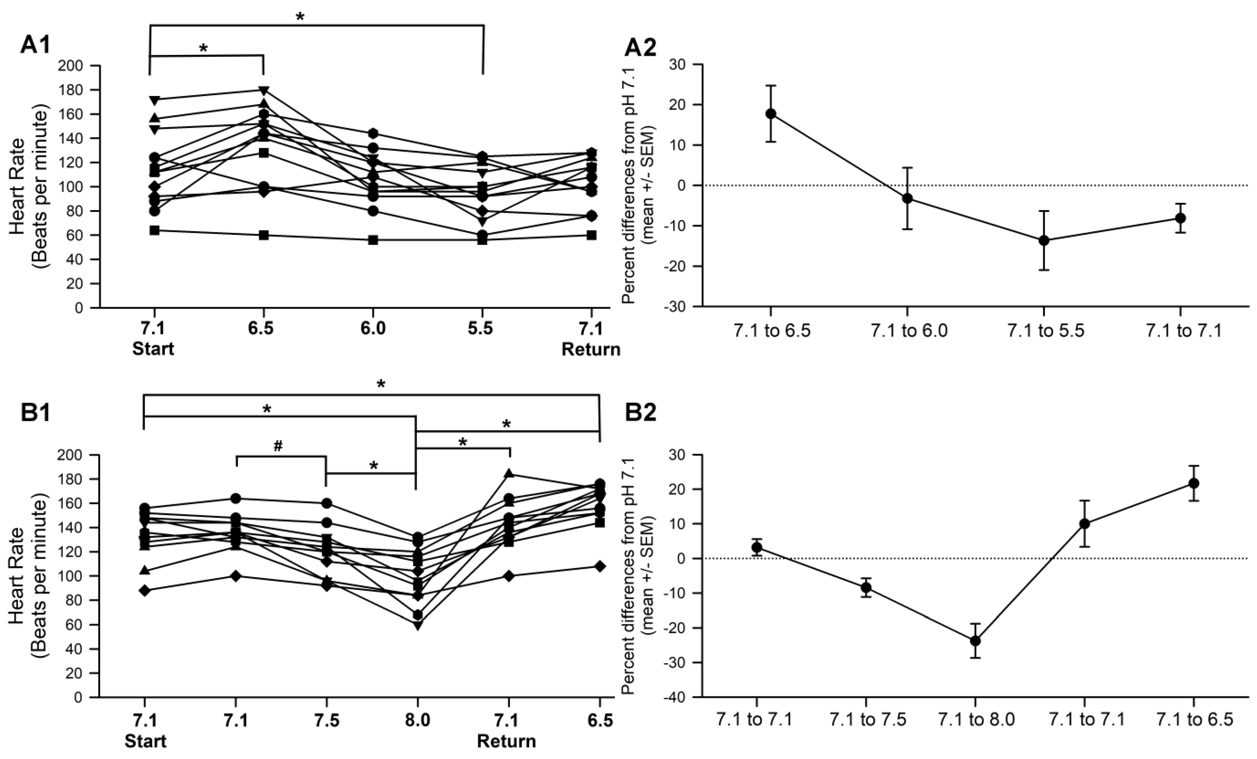
3.2. Effect of pH on HR

The effects of the pH on the heart rate were examined for in situ hearts on an individual larval basis, for both acidic and basic pHs. The decrease in the pH from 7.1 to 6.5 resulted in an increased rate, while the further lowering of the pH to 5.5 decreased the rate to a slower rate than that of pH 7.1 initially (Figure 2(A1)). Increasing the pH from 7.1 to 8.0 caused the rate to substantially decrease (Figure 2(B1)), while switching out the saline for a return to pH 6.5 (from a pH of 8.0) resulted in an increased rate (Figure 2(B1); Repeated measures were conducted two-way ANOVA p < 0.05). The rate did decrease upon raising the pH from 7.1 to 7.5, with the two being compared using a repeated measures test for these two distributions (p < 0.05). The percent changes of the saline at a pH 7.1 to each experimental value are shown for each condition (Figure 2(A2,B2)).

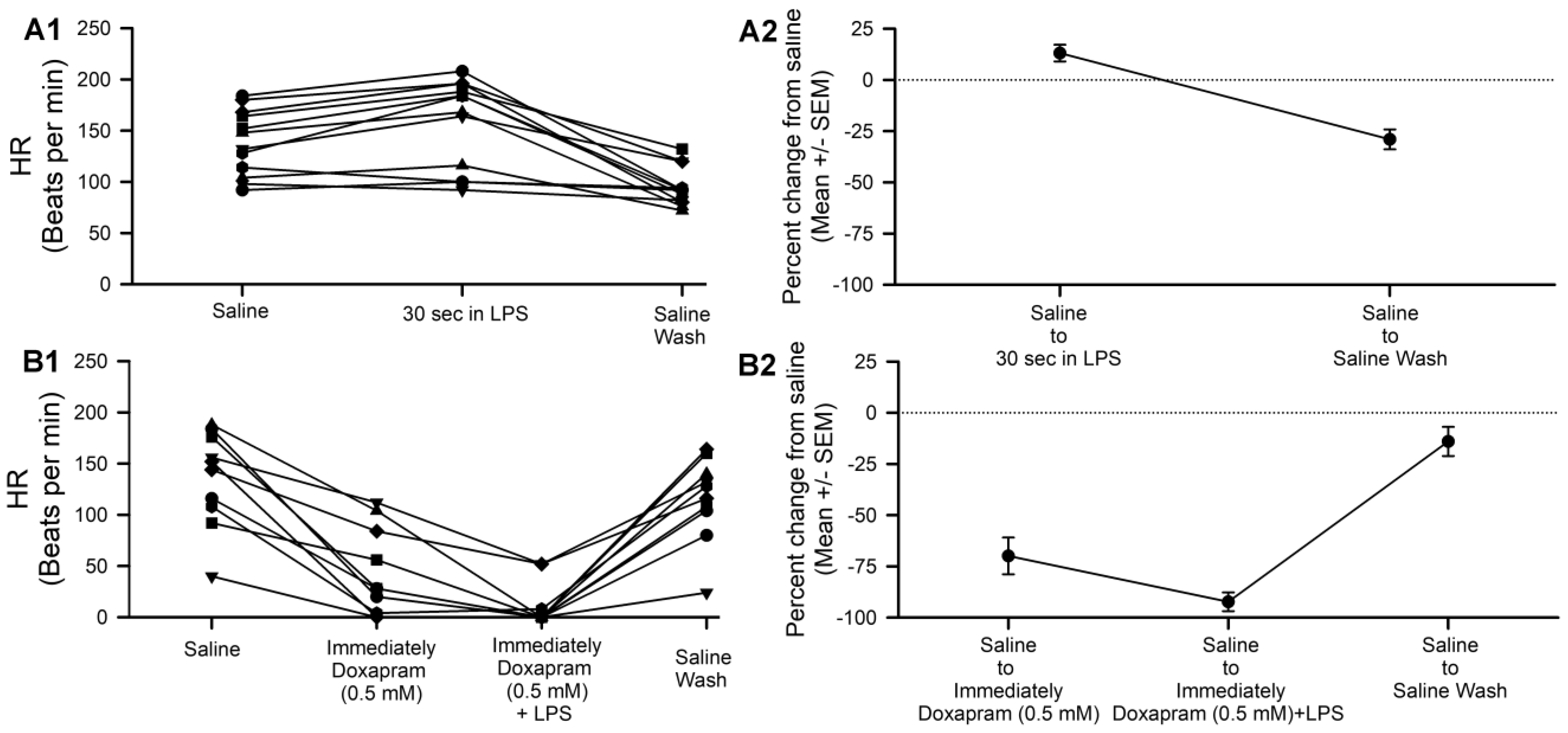
3.3. Effect of Doxapram on LPS Action

Exposure to that the LPSs (500 µg/mL; Serratia marcescens) increased the heart rate (Figure 3(A1); paired t-test, p < 0.05), while doxapram, both by itself and even in combination with LPSs, produced a substantial decrease in the rate (Figure 3(B1); Paired t-test, p< 0.05). The percent decreases with doxapram solutions (Figure 1(B1)) and doxapram–LPS combination solutions (Figure 3(B1)) were not significantly different (t-test, p > 0.5).
Since a pH of 6.5 sped up the rate compared to the pH of 7.1, it was of interest to examine whether doxapram would block this effect. The heart was exposed to a 0.5 mM doxapram solution, a solution of 0.5 of mM of doxapram in saline at pH 6.5, and then a pH 7.1 saline washout. Doxapram was not able to block the effect of the lowered pH, as the rate increased substantially (paired t-test, N = 12, p < 0.05; Figure 4).

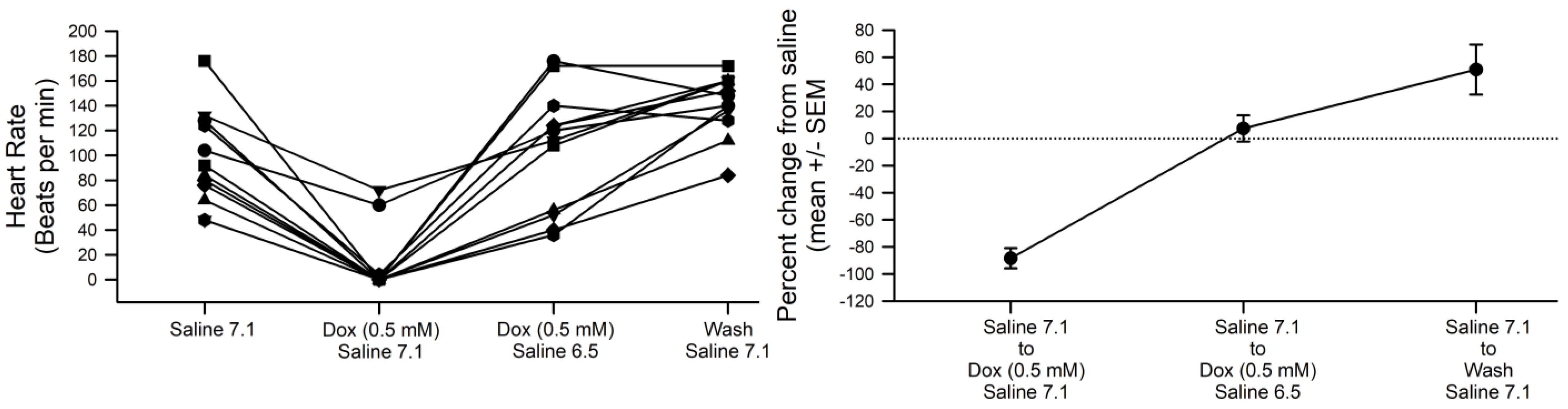
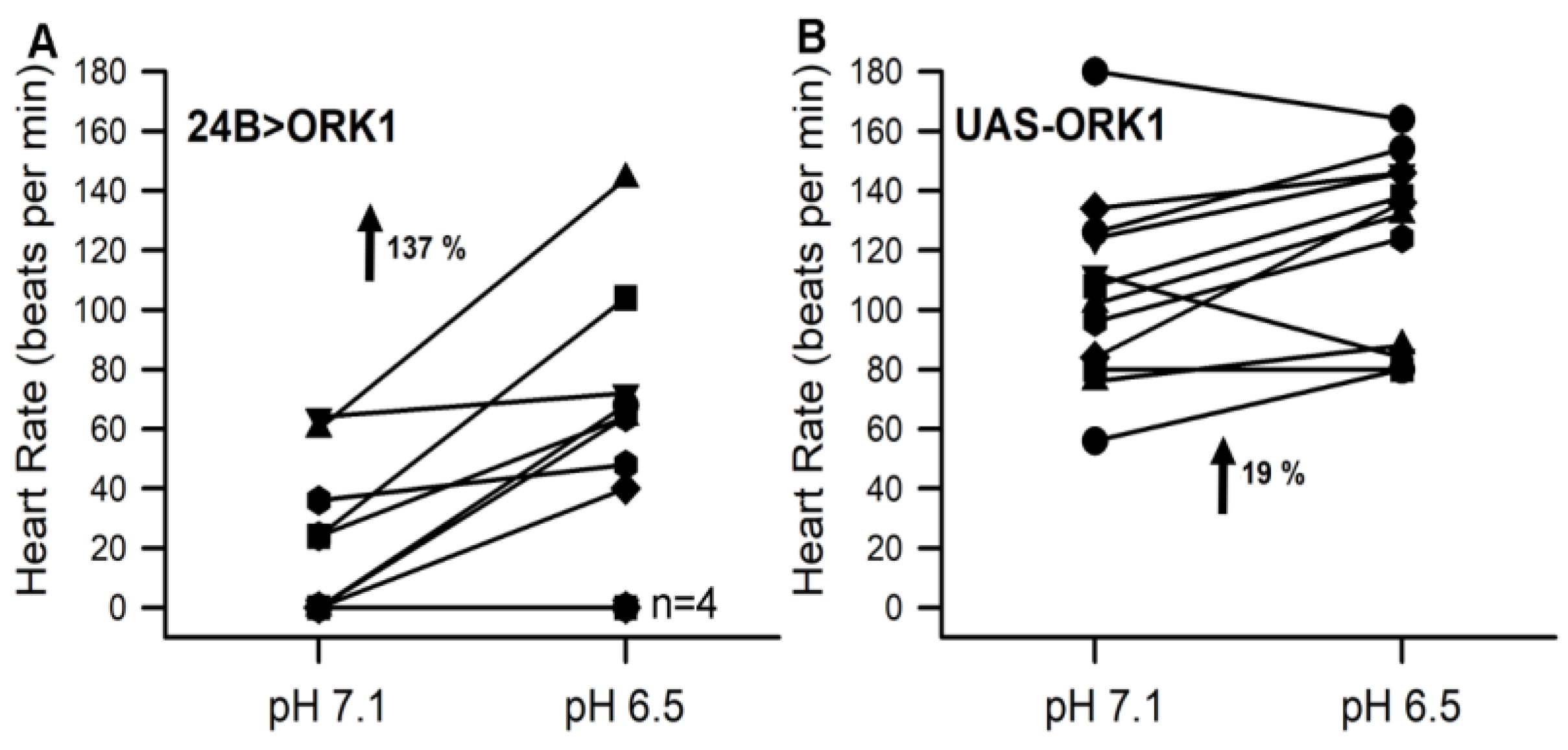
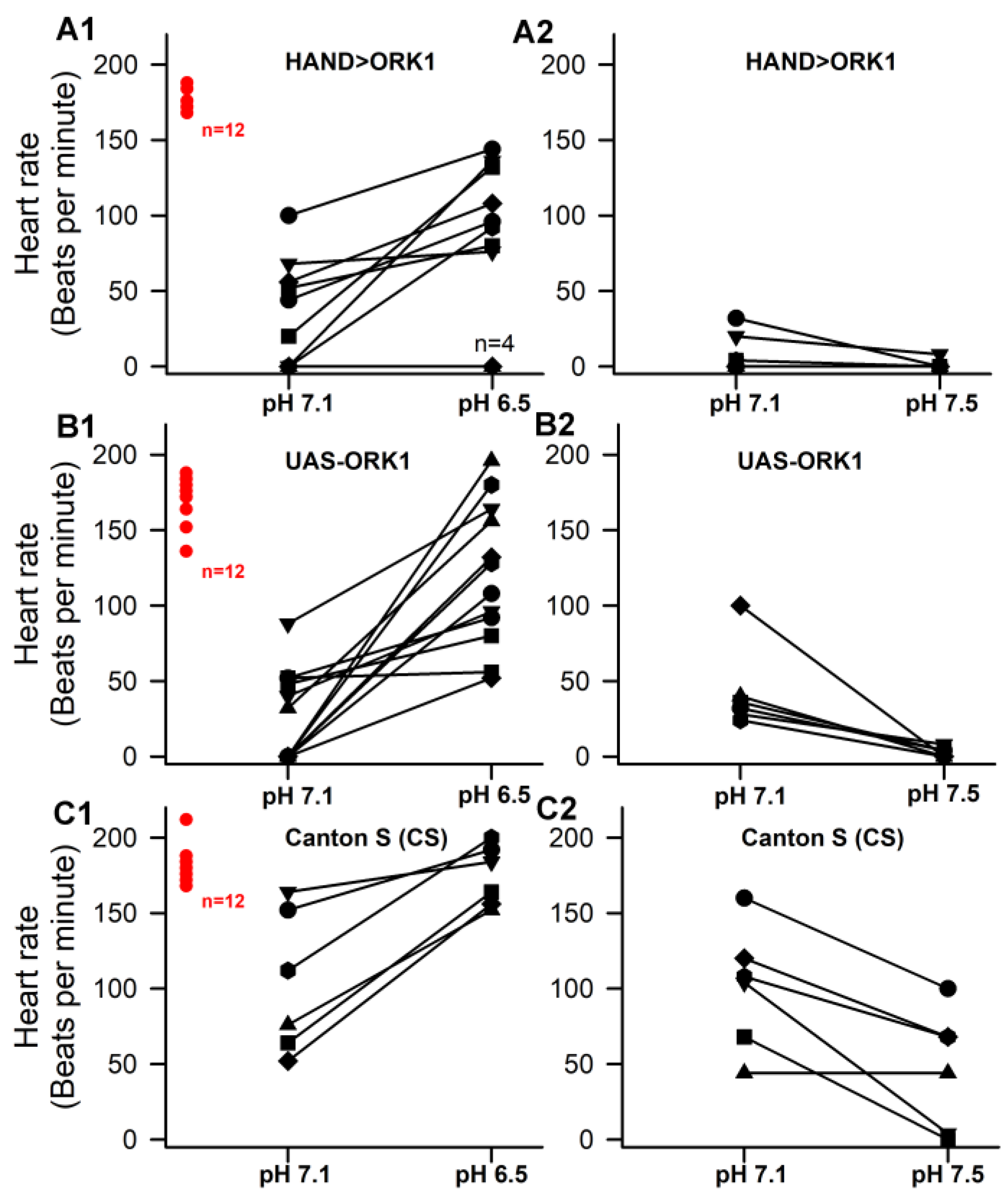
3.4. Sensitivity to pH in Hearts and Overexpressing ORK1 (a K2p Channel)

To investigate whether ORK1 overexpression resulted in an altered heart rate and pH response, the F1 generation of 24B > ORK1 was raised at room temperature (21 °C); however, the larvae all died at the end of the second-instar stage. This was also confirmed through examination of the larval mouth hook structural characteristics [29] in both the dead larvae and the largest larvae in the food.
All second-instar F1 larvae showed no heartbeat within the intact larvae (N = 15) and, upon dissection with HL3 saline, none of the larvae (N = 6) exhibited beating heart tubes. Upon switching the saline bath from a pH of 7.1 to a pH of 6.5, the heart tube still did not beat (N = 6). Neither the application of serotonin (100 nM) nor electrical stimulation resulted in a beating heart tube. Serotonin did result in an increased contractile rate in UAS-ORK1 of the 24B > ORK1 parental line, and electrical stimulation drove contractions in the UAS-ORK1 of the parental line, as has been shown for the CS lines in the past [25].
In an effort to reduce the ORK1 expression in the 24B > ORK1, the larvae were raised at room temperature (21 °C) and, upon reaching the early second-instar stage, they were placed in an incubator at 18 °C. Living larvae were able to crawl, with robust mouth hook movements when placed in a dilute yeast solution. The skeletal muscles were thus functioning well for those larvae used in assessing cardiac response to saline changes when dissected. Still, very few third instars formed in this paradigm, and most of the larvae died in the second-instar stage.
The heart tubes also did not beat upon changing the saline from a pH of 7.1 to 6.5. The 24B > ORK1 larval hearts could not be electrically driven, but the body wall muscles were able to be electrically excited by the focal electrode. In addition, the heart tubes did not show any altered response to serotonin (100 nM 5-HT). In some larvae, lateral movement of the heart tube was observed, which looked like the alary muscles [6,30] were contracting, thus pulling the heart to one side of the larvae.
An alternative approach was also used in which the embryos were raised by maintaining the UAS-ORK1 (virgin females) and Gal4-24B (non-stubble males) at 18 °C. The developmental time was slowed, but the survival rate of the third instars was still low. The mouth hook morphology helped in distinguishing between the smaller-than-average third instars and second instars. Many seemed to die during molting from the second- to third-instar stages, while most larvae died in the second-instar stage. However, those intact second-instar larvae that were capable of crawling had successfully beating heart tubes. Thus, this paradigm was used to investigate the effect of the pH changes from 7.1 to 6.5 in these dissected larvae.
The UAS-ORK1 instars were used for comparison and raised in the same conditions. Upon dissection of the UAS-ORK1 larvae, the heart tubes were beating in saline at pH 7.1 and continued to beat upon changing the pH to 6.5, although the heart rate was decreased in some of the 24B > ORK1 larvae (Figure 5). Other larvae had full contractions, and a few did not beat at all aside from lateral movements and slow, small, somewhat systolic contractions. Thus, considerable variability was observed in the initial contractile rates at a pH of 7.1. Among twelve dissected larvae, four did not beat at either the initial recording or recording at the pH of6.5. The other eight featured increased heart rates upon switching the saline (Figure 5).
The percent change was more pronounced in the 24B > ORK1 than the UAS-ORK1 larvae (t-test, p < 0.05), but only larvae showing beating hearts in the pH of 7.1 or 6.5 were used for analysis, since the percent changes from zero were not practical comparisons to make. When larvae without beating hearts were included in a nonparametric rank sum test, both the 24B > ORK1 and UAS-ORK1 larvae featured increased heart rates upon exchanging the pH to 6.5.
Since a number of larvae had very low initial heart rates for the dissected in situ study and a high third-instar larval death rate of the 24B > ORK1 strain, we used the heart specific strain HAND > ORK1 and the parental line UAS-ORK1. The 24B strain is a mesodermal tissue expression that includes both body wall muscles and the heart for Drosophila. The larvae of HAND > ORK1 live, pupate, and become adults at room temperature. Canton S larvae were also used as an independent measure, since values have been previously reported for intact Canton S larvae. The rates in the pH of 7.1 were significantly lower for both HAND > ORK1 and the parental line UAS-ORK1 compared to the Canton S (t-test, p < 0.05, N = 12; Figure 6), with no significant difference between the HAND > ORK1 and UAS-ORK1. The rates at the pH of 6.5 were not significantly different across all three strains. Changing the pH from 7.1 to 7.5 produced a significant decrease in the rate across all three types of larvae (Sign test p < 0.05; N = 6) These heart rates were calculated for one recording period for each set, with strains and saline solutions used on a rotating basis to reduce any experimental variability.
Since the rates for dissected in situ movements at the pH of 7.1 were extremely low for the HAND > ORK1 and the parental line UAS-ORK1, the rates were obtained for intact larvae across the three strains. The intact measures were obtained by pinning the larvae dorsal side up, placing one pin by the mouth hooks, and placing another adjacent to the lateral side from the spiracles (so as not to damage the dorsal heart tube). The recording dish was glass with a magnetic strip to hold the pins in place [25]. Light was shone from beneath the larvae to readily observe the beating of the heart tube. Measures were taken by a single individual and within a single recording period for all three strains for one larva at a time; thus, variability in the recording technique was minimized. The rates for intact larvae are shown (Figure 6). There were no significant differences across the three strains for the intact larvae.

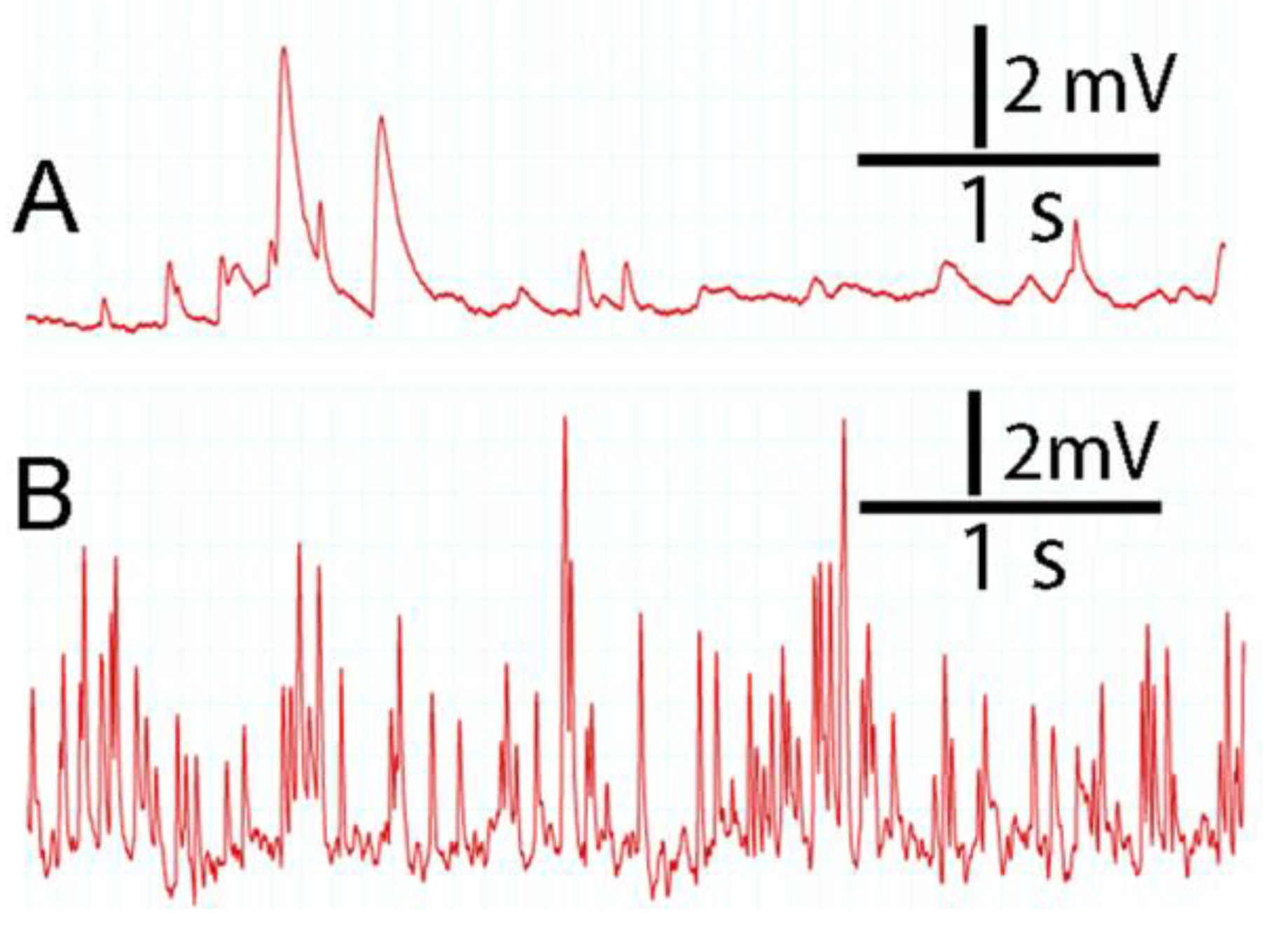
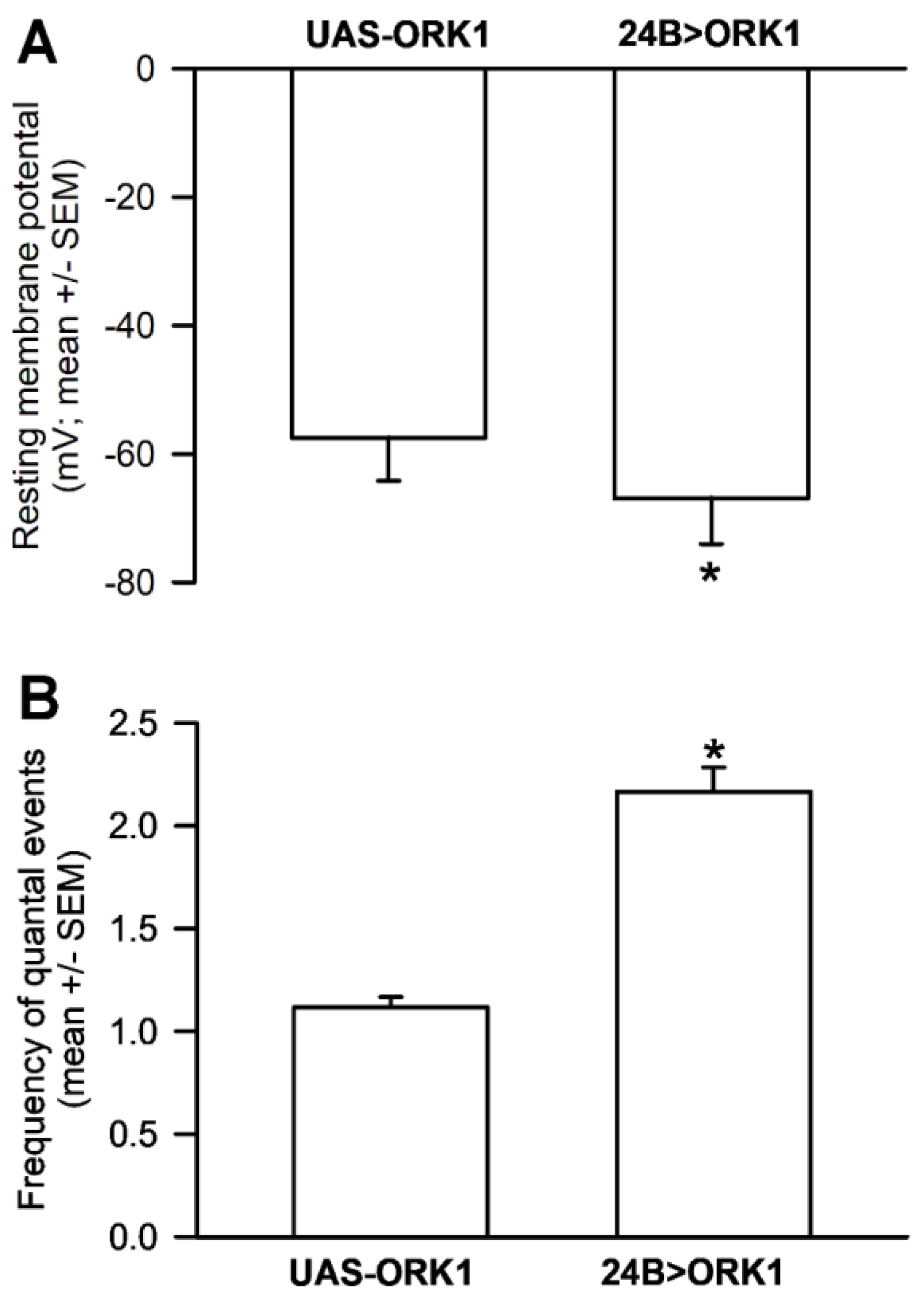
3.5. Resting Membrane Potential and Quantal Occurrences in Skeletal Muscle

While investigating whether the resting membrane potential of the body wall muscle M6 was different between the second-instar larvae of strains UAS-ORK1 and 24B > ORK1 (raised at 18 °C, but monitored at 21 °C), it was obvious that the frequency of spontaneous quantal events was very high for the 24B > ORK1 line (Figure 7), while the resting membrane potentials were slightly more negative. Thus, an attempt was made to quantify both the resting membrane potentials and the frequency of the spontaneous quantal events. The second-instar larvae of both strains were dissected as quickly as possible, and the segmental nerves were cut next to the central nervous system to avoid evoked excitatory junction potentials. Muscle M6 in segment 4 was the most easily assessable segment and, thus, was used for recording. All dissections and recordings were performed on the same morning and with the same saline for both the UAS-ORK1 and 24B > ORK1, and recordings alternated between the two strains; all of this served to reduce experimental variability. The mean (±SEM) of the resting membrane potential for the UAS-ORK1 was −57.4 mV (±6.8), and, for the 24B > ORK1, it was −66.8 mV (±7.3). The resting membrane potentials were significantly different (two-tailed t-Test, p = 0.00142). The frequency of the spontaneous quantal events for the UAS-ORK1 were, on average (±SEM) 1.13 (±0.04) and 2.17 (±0.11) for the 24B > ORK1. The frequency of events over a 1 minute period was significantly different (Figure 8; two-tailed t-Test, p = 0.0000108).

4. Discussion

In this study, the larval Drosophila heart model was used to demonstrate that doxapram decreases heart rate and dampens (though does not completely block) the increased heart rate produced by LPSs. Additionally, it was demonstrated that the rate increased with a lower extracellular pH of 6.5 but decreased both with pH values lower than that, as well as with more basic values. The overexpression of the K2p channel ORK1 in mesodermal tissue resulted in larval lethality by the third instar; however, selective overexpression in the heart allowed the larvae to survive. The ORK1 strain appeared to have an inherent lower heart rate compared to the CS strain. Intact larvae had a higher heart rate compared to the dissected in situ hearts, which is likely due to various cardiac modulators in the hemolymph. The overexpression of ORK1 in body wall muscles from the embryo to the second-instar stage resulted in a more negative resting membrane potential and a higher rate of spontaneous quantal occurrences compared to the parental strain UAS-ORK1.
Given that the larval Drosophila heart rate is sensitive to the pH of the external medium, the pH affects one or more ion channels that contribute to the pacing of the heart. The mechanism for this phenomenon is likely found in an acid-sensitive family of K2p channels that are responsible for maintaining the resting membrane potential. Since the larval body wall muscles depolarize under acidic conditions, it is likely the cardiac myocytes also depolarize in a slightly acidic bathing medium, which would lead to the increase in the heart rate. Surprisingly, doxapram showed a dose-response effect in decreasing the heart rate. Earlier studies suggested that the Gram-negative bacterial toxin LPSs may activate K2p channels to account for the acute hyperpolarization of the body wall muscles [15,16,21,31].
However, it was shown in past studies that the initial effect of LPS exposure increases the larval Drosophila heart rate. Thus, it does not appear that LPSs acutely activate the larval cardiac K2p channels, as the rate should rapidly decrease if a substantial hyperpolarization were to occur as in the body wall muscle, instead of increasing. It was also demonstrated that rapid exposure to the doxapram–LPS cocktail blocked the anticipated rise in the heart rate that would have been expected with solely LPS exposure. This suggests that the mechanisms of action by LPSs acting on the resting membrane potential of body wall muscle and cardiac pacing myocytes are different. Doxapram also did not perform as expected, as it did not increase the larval heart rate by blocking the K2p channels responsible for maintaining the resting membrane potential.
These unexpected findings may occur because the pacing heart tissue expresses multiple different subtypes of K2p channels, as well as ionic channels that are distinct from those in the body wall muscles, since the heart paces itself without neural innervation. The expression profiles of the eleven known K2p channel subtypes in Drosophila [12,18,19,32,33,34] have not been examined selectively for expression in the cardiac and skeletal myocytes of larval Drosophila. Of the general six subfamilies (TWIK, TREK, TASK, TALK, THIK, and TRESK) the Tandem-Pore Weak Inward rectifying K+ channel (TWIK-1) and the Tandem-pore Acid-Sensing K+ channels (TASK-1 and TASK-3) are sensitive to acidic conditions [14,35]. The TASK subtype is the putative type that the doxapram inhibits [14,36]. The K2p channel in Drosophila, termed the ORK1 channel, is known to be present in cardiac muscle, while others may also be present. The investigations described confirm earlier reports that ORK1 overexpression stops the heart from beating [2,12,17]. In addition, when expressed in all mesodermal tissues by using 24B-Gal4 (III) at 21 °C, the larvae died between the second- and third-instar stages. Also, upon examining the pH sensitivity with the overexpression in the heart-specific strain (HAND), it demonstrated a similar sensitivity to pH changes as the parental UAS-ORK1 strain. Considering that UAS-ORK1 larvae have a lower heart rate at pH 7.1 when compared to CS, it is likely that there are inherent differences in larval UAS-ORK1 responses in saline, as the rates measured for the intact larvae did not show differences. The K2P channels being expressed in the UAS-ORK1 and HAND > ORK1 might utilize a conformation in the membrane that is different from that of the CS strain, which would explain these strains’ more pronounced pH sensitivity, possibly due to the binding of protons to the histidine residues present in the K2P channels’ protein structure [37,38,39]. However, the rate measurements of the intact, restrained larvae may produce a stress response and/or a hormonal influence. Unless the hemolymph is removed from the dissected larvae, the variables in the hemolymph cannot be controlled. The heartbeat is sensitive to modulators such as serotonin (5-HT), dopamine, acetylcholine, octopamine, and peptides [8,40,41,42,43,44,45,46].
The fact that the ORK1 expression in the body wall muscles increase the frequency of quantal events is intriguing. There may potentially be a retrograde regulatory response on the motor nerve to increase transmission in the 24B > ORK1 from a more negative resting membrane potential, or perhaps other factors are involved from the genetic construction. Increased or decreased muscle activity in both Drosophila and mammals is known to impact the motor nerve terminal and influence synaptic transmission [47,48,49,50,51]. Thanks to the rapid growth of the Drosophila neuromuscular junction, the nerve-muscle matching may be more rapidly regulated than slower-developing mammalian models [52,53,54].
Future studies into the expression profiles and K2p channel subtypes as a method of regulating cardiac myocyte pacing are needed, as well as the exploration of the pharmacological effects on K2p channels. The specific mechanism for alteration of the resting membrane potential under acidic and basic conditions in both cardiac and body wall muscles is not fully understood, nor are the effects on cardiac pacing. Given that the overexpression of ORK1 in body wall muscles resulted in a more negative resting membrane potential, as well as a higher rate of spontaneous quantal fusion events, it would be of interesting to attempt a temporal expression of the ORK1 in early third-instar larvae, potentially with induction by the RU486 or designer receptors that are exclusively activated by designer drugs (DREADDs) [45,55]. The lethality observed in the larvae with an overexpression of ORK1 (K2P) channels in all mesoderm tissue with the 24B strain has yet to be addressed. Most studies focus on the effects of decreased expression (i.e., knockdown) of K2P channels, but some diseased states are known to cause an increase in the expression of various K2P channel subtypes [56]. The genetic and physiological examination of larval Drosophila will likely be of benefit in future studies.

Author Contributions

Conceptualization, E.R.E., A.C.T., M.O.A.-K., N.H. and R.L.C.; Methodology, E.R.E., A.C.T., M.O.A.-K. and R.L.C.; Software, A.C.T. and R.L.C.; Validation, E.R.E., A.C.T. and R.L.C.; Formal analysis, E.R.E., A.C.T., M.O.A.-K., N.H. and R.L.C.; Investigation, E.R.E., A.C.T., M.O.A.-K., N.H. and R.L.C.; Resources, R.L.C.; Data curation, E.R.E. and R.L.C.; Writing—original draft, E.R.E., A.C.T., M.O.A.-K., N.H. and R.L.C.; Writing—review and editing, E.R.E., A.C.T., M.O.A.-K., N.H. and R.L.C.; Visualization, E.R.E. and R.L.C.; Supervision, E.R.E. and R.L.C.; Project administration, E.R.E. and R.L.C.; Funding acquisition, E.R.E. and R.L.C. All authors have read and agreed to the published version of the manuscript.

Funding

This research was funded by alumni of the research group, personal funds (R.L.C.), and the Chellgren Endowed Funding (R.L.C.), as well as the University of Kentucky Office of Undergraduate Research (OUR) and Research Priority Areas (RPA) supported by the Office of the Vice President for Research (E.R.E.).

Institutional Review Board Statement

Ethical review and approval were waived for this study due to only use of insects in this study.

Informed Consent Statement

Not applicable.

Data Availability Statement

All data is presented in the manuscript.

Conflicts of Interest

The authors declare no conflict of interest.

References

  1. Enyedi, P.; Czirják, G. Molecular Background of Leak K+ Currents: Two-Pore Domain Potassium Channels. Physiol. Rev. 2010, 90, 559–605. [Google Scholar] [CrossRef] [PubMed]
  2. Buckingham, S.D.; Kidd, J.F.; Law, R.J.; Franks, C.J.; Sattelle, D.B. Structure and function of two-pore-domain K+ channels: Contributions from genetic model organisms. Trends Pharmacol. Sci. 2005, 26, 361–367. [Google Scholar] [CrossRef] [PubMed]
  3. Plant, L.D.; Goldstein, S.A.N. Two-Pore Domain Potassium Channels. In Handbook of Ion Channels, 1st ed.; Zheng, J., Trudeau, M.C., Eds.; CRC Press: Boca Raton, FL, USA, 2015; ISBN 9780429193965. [Google Scholar]
  4. Lee, L.M.; Müntefering, T.; Budde, T.; Meuth, S.G.; Ruck, T. Pathophysiological role of K2P channels in human diseases. Cell Physiol. Biochem. 2021, 55, 65–86. [Google Scholar] [PubMed]
  5. Wiedmann, F.; Frey, N.; Schmidt, C. Two-Pore-Domain Potassium (K2P) Channels: Cardiac Expression Patterns and Disease-Specific Remodelling Processes. Cells 2021, 10, 2914. [Google Scholar]
  6. Rotstein, B.; Paululat, A. On the Morphology of the Drosophila Heart. J. Cardiovasc. Dev. Dis. 2016, 3, 15. [Google Scholar] [CrossRef]
  7. Badre, N.H.; Martin, M.E.; Cooper, R.L. The physiological and behavioral effects of carbon dioxide on Drosophila melanogaster larvae. Comp. Biochem. Physiol. A. Mol. Integr. Physiol. 2005, 140, 363–376. [Google Scholar] [CrossRef]
  8. Majeed, Z.R.; Stacy, A.; Cooper, R.L. Pharmacological identification of serotonin receptor subtypes on the Dro-sophila larval heart. J. Comp. Physiol. B 2014, 184, 205–219. [Google Scholar] [CrossRef]
  9. Bartos, D.C.; Grandi, E.; Ripplinger, C.M. Ion Channels in the Heart. Compr. Physiol. 2015, 5, 1423–1464. [Google Scholar]
  10. Severi, S.; Cavalcanti, S.; Mancini, E.; Santoro, A. Effect of electrolyte and pH changes on the sinus node pacemaking in humans. J. Electrocardiol. 2002, 35, 115–124. [Google Scholar] [CrossRef]
  11. Achike, F.I.; Dai, S. Effects of blood gas/pH abnormalities on the cardiovascular actions of verapamil in rats. Clin. Exp. Pharmacol. Physiol. 1990, 17, 653–663. [Google Scholar] [CrossRef]
  12. Goldstein, S.A.; Price, L.A.; Rosenthal, D.N.; Pausch, M.H. ORK1, a potassium-selective leak channel with two pore domains cloned from Drosophila melanogaster by expression in Saccharomyces cerevisiae. Proc. Natl. Acad. Sci. USA 1996, 93, 13256–13261. [Google Scholar] [CrossRef] [PubMed]
  13. Goldstein, S.A.; Wang, K.W.; Ilan, N.; Pausch, M.H. Sequence and function of the two P domain potassium channels: Implications of an emerging superfamily. J. Mol. Med. 1998, 76, 13–20. [Google Scholar] [CrossRef] [PubMed]
  14. Cotten, J.F.; Keshavaprasad, B.; Laster, M.J.; Eger, E.I., II; Yost, C.S. The ventilatory stimulant doxapram inhibits TASK tandem pore (K2P) potassium channel function but does not affect minimum alveolar anesthetic concentration. Anesth. Analg. 2006, 102, 779–785. [Google Scholar] [CrossRef]
  15. Vacassenno, R.M.; Haddad, C.N.; Cooper, R.L. The effects on resting membrane potential and synaptic transmission by Doxapram (blocker of K2P channels) at the Drosophila neuromuscular junction. Comp. Biochem. Physiol. C 2023, 263, 109497. [Google Scholar] [CrossRef]
  16. Vacassenno, R.M.; Haddad, C.N.; Cooper, R.L. Bacterial lipopolysaccharide hyperpolarizes the membrane potential and is antagonized by the K2p channel blocker doxapram. Comp. Biochem. Physiol. C. Toxicol. Pharmacol. 2023, 266, 109571. [Google Scholar] [CrossRef]
  17. Lalevée, N.; Monier, B.; Sénatore, S.; Perrin, L.; Sémériva, M. Control of cardiac rhythm by ORK1, a Drosophila two-pore domain potassium channel. Curr. Biol. 2006, 16, 1502–1508. [Google Scholar] [CrossRef]
  18. Adams, M.D.; Celniker, S.E.; Holt, R.A.; Evans, C.A.; Gocayne, J.D.; Amanatides, P.G.; Scherer, S.E.; Li, P.; Hoskins, R.A.; Galle, R.; et al. The genome sequence of Drosophila melanogaster. Science 2000, 287, 2185–2195. [Google Scholar] [CrossRef] [PubMed]
  19. Littleton, J.T.; Ganetzky, B. Ion channels and synaptic organization: Analysis of the Drosophila genome. Neuron 2000, 26, 35–43. [Google Scholar] [CrossRef]
  20. Patel, A.J.; Honore, E.; Lesage, F.; Fink, M.; Romey, G.; Lazdunski, M. Inhalational anesthetics activate two-pore-domain background K channels. Nat. Neurosci. 1999, 2, 422–426. [Google Scholar] [CrossRef]
  21. Cooper, R.L.; Krall, R.M. Hyperpolarization Induced by Lipopolysaccharides but Not by Chloroform Is Inhibited by Doxapram, an Inhibitor of Two-P-Domain K+ Channel (K2P). Int. J. Mol. Sci. 2022, 23, 15787. [Google Scholar] [CrossRef] [PubMed]
  22. Brock, K.E.; Elliott, E.R.; Abul-Khoudoud, M.O.; Cooper, R.L. The effects of Gram-positive and Gram-negative bacterial toxins (LTA & LPS) on cardiac function in Drosophila melanogaster larvae. J. Insect Physiol. 2023, 147, 104518. [Google Scholar] [PubMed]
  23. Pleuvry, B.J. A study of the enhanced toxicity of doxapram in rodents treated with narcotic analgesics. Br. J. Anaesth. 1978, 50, 451–458. [Google Scholar] [CrossRef] [PubMed]
  24. Brand, A.H.; Perrimon, N. Targeted gene expression as a means of altering cell fates and generating dominant phenotypes. Devel. 1993, 118, 401–415. [Google Scholar] [CrossRef] [PubMed]
  25. Cooper, A.S.; Rymond, K.E.; Ward, M.A.; Bocook, E.L.; Cooper, R.L. Monitoring heart function in larval Drosophila melanogaster for physiological studies. J. Vis. Exp. 2009, 33, 1596. [Google Scholar] [CrossRef]
  26. De Castro, C.; Titlow, J.; Majeed, Z.R.; Cooper, R.L. Analysis of various physiological salines for heart rate, CNS function, and synaptic transmission at neuromuscular junctions in Drosophila melanogaster larvae. J. Comp. Physiol. A 2014, 200, 83–92. [Google Scholar] [CrossRef]
  27. Fast, I.; Rosenkranz, D. 2018. Temperature-dependent small RNA expression in Drosophila melanogaster. RNA Biol. 2018, 15, 308–313. [Google Scholar] [CrossRef]
  28. Haltenhof, T.; Kotte, A.; De Bortoli, F.; Schiefer, S.; Meinke, S.; Emmerichs, A.K.; Petermann, K.K.; Timmermann, B.; Imhof, P.; Franz, A.; et al. A conserved kinase-based body-temperature sensor globally controls alternative splicing and gene expression. Mol. Cell. 2020, 78, 57–69.e4. [Google Scholar] [CrossRef]
  29. Ashburner, M. Drosophila: A Laboratory Handbook; Cold Spring Harbor Laboratory Press: Cold Spring Harbor, NY, USA, 1989. [Google Scholar]
  30. Rizki, T.M. The circulatory system and associated cells and tissues. In The Genetics and Biology of Drosophila; Ashburner, M., Wright, T.R.F., Eds.; Academic Press: New York, NY, USA, 1978; Volume 2b, pp. 397–452. [Google Scholar]
  31. Cooper, R.L.; McNabb, M.; Nadolski, J. The effects of a bacterial endotoxin LPS on synaptic transmission at the neuromuscular junction. Heliyon 2019, 5, e01430. [Google Scholar] [CrossRef]
  32. Zilberberg, N.; Ilan, N.; Gonzalez-Colaso, R.; Goldstein, S.A. Opening and closing of KCNKO potassium leak channels is tightly regulated. J. Gen. Physiol. 2000, 116, 721–734. [Google Scholar] [CrossRef]
  33. Zilberberg, N.; Ilan, N.; Goldstein, S.A. KCNKØ: Opening and closing the 2-P-domain potassium leak channel entails “C-type” gating of the outer pore. Neuron 2001, 32, 635–648. [Google Scholar] [CrossRef]
  34. Ilan, N.; Goldstein, S.A. Kcnko: Single, cloned potassium leak channels are multi-ion pores. Biophys. J. 2001, 80, 241–253. [Google Scholar]
  35. Kim, D. Physiology and pharmacology of two-pore domain potassium channels. Curr. Pharm. Des. 2005, 11, 2717–2736. [Google Scholar] [CrossRef]
  36. Tian, F.; Qiu, Y.; Lan, X.; Li, M.; Yang, H.; Gao, Z. A small-molecule compound selectively activates K2P channel TASK-3 by acting at two distant clusters of residues. Mol. Pharmacol. 2019, 96, 26–35. [Google Scholar] [CrossRef] [PubMed]
  37. Lopes, C.M.; Zilberberg, N.; Goldstein, S.A. Block of Kcnk3 by protons. Evidence that 2-P-domain potassium channel subunits function as homodimers. J. Biol. Chem. 2001, 276, 24449–24452. [Google Scholar] [CrossRef] [PubMed]
  38. Rajan, S.; Wischmeyer, E.; Xin Liu, G.; Preisig-Müller, R.; Daut, J.; Karschin, A.; Derst, C. TASK-3, a novel tandem pore domain acid-sensitive K+ channel. An extracellular histiding as pH sensor. J. Biol. Chem. 2000, 275, 16650–16667. [Google Scholar] [CrossRef]
  39. O’Connell, A.D.; Morton, M.J.; Hunter, M. Two-pore domain K+ channels-molecular sensors. Biochim. Biophys. Acta 2002, 1566, 152–161. [Google Scholar] [CrossRef] [PubMed]
  40. Johnson, E.; Ringo, J.; Dowse, H. Modulation of Drosophila heartbeat by neurotransmitters. J. Comp. Physiol. B 1997, 167, 89–97. [Google Scholar] [CrossRef]
  41. Johnson, E.; Ringo, J.; Dowse, H. Native and heterologous neuropeptides are cardioactive in Drosophila melanogaster. J. Insect Physiol. 2000, 46, 1229–1236. [Google Scholar] [CrossRef]
  42. Nichols, R.; Kaminski, S.; Walling, E.; Zornik, E. Regulating the activity of a cardioacceleratory peptide. Peptides 1999, 20, 1153–1158. [Google Scholar] [CrossRef]
  43. Zornik, E.; Paisley, K.; Nichols, R. Neural transmitters and a peptide modulate Drosophila heart rate. Peptides 1999, 20, 45–51. [Google Scholar] [CrossRef]
  44. Titlow, J.S.; Rufer, J.; King, K.; Cooper, R.L. Pharmacological analysis of dopamine modulation in the Drosophila melanogaster larval heart. Physiol. Rep. 2013, 1, e00020. [Google Scholar] [CrossRef]
  45. Majeed, Z.R.; Nichols, C.D.; Cooper, R.L. 5-HT stimulation of heart rate in Drosophila does not act through cAMP as revealed by pharmacogenetics. J. Appl. Physiol. 2013, 115, 1656–1665. [Google Scholar] [CrossRef] [PubMed]
  46. Malloy, C.; Sifers, J.; Mikos, A.; Samadi, A.; Omar, A.; Hermanns, C.; Cooper, R.L. Using optogenetics to assess neuroendocrine modulation of heart rate in Drosophila melanogaster larvae. J. Comp. Physiol. A. Neuroethol. Sens. Neural Behav. Physiol. 2017, 203, 791–806. [Google Scholar] [CrossRef] [PubMed]
  47. Tsai, P.I.; Wang, M.; Kao, H.H.; Cheng, Y.J.; Lin, Y.J.; Chen, R.H.; Chien, C.T. Activity-dependent retrograde laminin A signaling regulates synapse growth at Drosophila neuromuscular junctions. Proc. Natl. Acad. Sci. USA 2012, 109, 17699–17704. [Google Scholar] [CrossRef] [PubMed]
  48. Berke, B.; Wittnam, J.; McNeill, E.; Van Vactor, D.L.; Keshishian, H. Retrograde BMP signaling at the synapse: A permissive signal for synapse maturation and activity-dependent plasticity. J. Neurosci. 2013, 33, 17937–17950. [Google Scholar] [CrossRef]
  49. Chakkalakal, J.V.; Nishimune, H.; Ruas, J.L.; Spiegelman, B.M.; Sanes, J.R. Retrograde influence of muscle fibers on their innervation revealed by a novel marker for slow motoneurons. Development 2010, 137, 3489–3499. [Google Scholar] [CrossRef] [PubMed]
  50. Marqués, G. Morphogens and synaptogenesis in Drosophila. J. Neurobiol. 2005, 64, 417–434. [Google Scholar] [CrossRef]
  51. Frank, C.A. Homeostatic plasticity at the Drosophila neuromuscular junction. Neuropharmacology 2014, 78, 63–74. [Google Scholar]
  52. Collins, C.A.; DiAntonio, A. Synaptic development: Insights from Drosophila. Curr. Opin. Neurobiol. 2007, 17, 35–42. [Google Scholar] [CrossRef]
  53. Li, H.; Peng, X.; Cooper, R.L. Development of Drosophila larval neuromuscular junctions: Maintaining synaptic strength. Neuroscience 2002, 115, 505–513. [Google Scholar]
  54. Xing, B.; Long, A.A.; Harrison, D.A.; Cooper, R.L. Developmental consequences of neuromuscular junctions with reduced presynaptic calcium channel function. Synapse 2005, 57, 132–147. [Google Scholar] [CrossRef] [PubMed]
  55. Nicholson, L.; Singh, G.K.; Osterwalder, T.; Roman, G.W.; Davis, R.L.; Keshishian, H. Spatial and temporal control of gene expression in Drosophila using the inducible GeneSwitch GAL4 system. I. Screen for larval nervous system drivers. Genetics 2008, 178, 215–234. [Google Scholar] [CrossRef] [PubMed]
  56. Feliciangeli, S.; Chatelain, F.C.; Bichet, D.; Lesage, F. The family of K2P channels: Salient structural and functional properties. J. Physiol. 2015, 593, 2587–2603. [Google Scholar] [CrossRef] [PubMed]
Figure 1. The effect of doxapram on the larval heart rate. (A1) The effect of doxapram (0.1 mM) on the heart rate. There was no significant acute effect, but after 5 min of incubation, the rate significantly decreased. (B1) The heart rate rapidly decreased upon application of doxapram at concentrations of 0.5 mM and (C1) 5 mM. After exposure to 5 mM doxapram, the effect was not easily recovered with a saline flush. (* Asterisk p < 0.05 two-way repeated ANOVA). Each line represents a single larva. The average percent changes from saline for each concentration are shown in (A2,B2,C2).
Applbiosci 02 00026 g001
Figure 2. The effects of pH on heart rate. (A1) As pH decreased from 7.1 to 6.5, the rate increased. Further reduction in pH to 6.0 decreased the rate as compared to values at pH 6.5. (B1) As pH increased from 7.1 to 7.5 and then to 8.0, the rate decreased. (* Repeated measures two-way ANOVA p < 0.05; # repeated measures t-test, p < 0.05). Each line represents a single larva. The percent changes from saline at a pH 7.1 to each experimental value are shown for each condition (A2,B2).
Applbiosci 02 00026 g002
Figure 3. The effects of LPSs and doxapram on the heart rate. (A1) The hearts were exposed to saline and, subsequently, to LPSs (500 µg/mL; Serratia marcescens), and then to a washout with fresh saline. The rate was measured in the 30 s after LPS application. The rate significantly increased (paired t-test, p < 0.05). (B1) After the initial exposure of doxapram (0.5 mM) and exchange of the bath to a cocktail of doxapram and LPSs (500 µg/mL), the rate decreased further (though not as much as is represented by the percent decrease from initial doxapram exposure). Each line represents a single larva. The percent changes are illustrated in the graph from saline at pH 7.1 to each experimental value (A2,B2).
Applbiosci 02 00026 g003
Figure 4. Doxapram and the effect of pH 6.5 on heart rate. Doxapram at 0.5 mM did not block the chronotropic effect of pH 6.5 in increasing the rate of the heartbeat. The change from doxapram in saline at pH 7.1 to one of doxapram in saline at pH 6.5 significantly increased the rate (paired t-test, N = 12, p < 0.05). Each line represents a single larva. The percent change from saline at pH 7.1 to each experimental value are shown for each condition.
Applbiosci 02 00026 g004
Figure 5. The effect of pH on the heart rate for larvae overexpressing ORK1. (A) The overexpressing strain of ORK1 (24B > ORK1) demonstrated a high sensitivity to the pH change from 7.1 to 6.5, as well as a lower initial rate of contractions in 7.1 as compared to the parental strain ((B), UAS-ORK1). These larvae were raised at 18 °C, dissected, and measured at 21 °C within an hour. Each line represents a single larva. The percent changes are illustrated in the graph; however, values of zero were not able to be used for the percent changes.
Applbiosci 02 00026 g005
Figure 6. Heart rates for in situ and intact larvae. (A1) The expression of ORK1 specifically targeted for the heart (HAND > ORK1) and the (B1) parental strain (UAS-ORK1), and an (C1) independent strain (Canton S) was used to examine the heart rate’s sensitivity to pH in dissected in situ heart tubes. All three strains exhibited a significant increase in rates when pH changed from 7.1 to 6.5. (A2,B2,C2) All three strains showed a decrease in rate when pH changed from 7.1 to 7.5. A significant difference was observed between the initial rates at pH 7.1 for Canton S larvae, as well as for the rates observed for HAND > ORK1 and UAS-ORK1. Each line represents a single larva. The heart rates for intact larvae are shown as individual points (red dots) on the left, with no significant differences among the three strains. These three strains were all raised at 21 °C.
Applbiosci 02 00026 g006
Figure 7. Representative occurrences of spontaneous quantal events: (A) UAS-ORK1 and (B) 24B > ORK1.
Applbiosci 02 00026 g007
Figure 8. Differences in (A) resting membrane potential and the (B) occurrences of spontaneous quantal events for larvae muscle (m6) for UAS-ORK1 and 24B > ORK1. There was a significant difference between UAS-ORK1 and 24B > ORK1 for both the resting membrane potential and the occurrences of spontaneous quantal events (t-test, * p < 0.01).
Applbiosci 02 00026 g008
Disclaimer/Publisher’s Note: The statements, opinions and data contained in all publications are solely those of the individual author(s) and contributor(s) and not of MDPI and/or the editor(s). MDPI and/or the editor(s) disclaim responsibility for any injury to people or property resulting from any ideas, methods, instructions or products referred to in the content.

