Review

Optical Coherence Tomography as a Biomarker in the Differential Diagnosis between Parkinson’s Disease and Atypical Parkinsonian Syndromes: A Narrative Review

by
Stella Karatzetzou
1,
Dimitrios Parisis
1,
Serafeim Ioannidis
2,
Theodora Afrantou
1 and
Panagiotis Ioannidis
1,*
1
2nd Department of Neurology, AHEPA University Hospital, Aristotle University of Thessaloniki, 546 36 Thessaloniki, Greece
2
School of Medicine, Aristotle University of Thessaloniki, 541 24 Thessaloniki, Greece
*
Author to whom correspondence should be addressed.
Appl. Sci. 2024, 14(6), 2491; https://doi.org/10.3390/app14062491
Submission received: 16 January 2024 / Revised: 1 March 2024 / Accepted: 13 March 2024 / Published: 15 March 2024
(This article belongs to the Collection Biomedical Optics: From Methods to Applications)

Abstract

:
Parkinsonism may be a clinical manifestation of a wide range of disease entities, and still poses a great diagnostic challenge. In an attempt to provide further insight into the differential diagnosis of PD versus progressive supranuclear palsy (PSP), multiple system atrophy (MSA), corticobasal degeneration (CBD), and Lewy body dementia (LBD), several biomarkers have been investigated, yielding inconclusive results, OCT being among them. The present review aims to explore the potential diagnostic value of evaluating retinal parameters through OCT implementation among patients presenting with a Parkinsonian syndrome, with an emphasis on effective differentiation between distinct syndromes. Having reviewed all the available literature published within the last decade, neurodegeneration seems to be paralleled with degeneration and alterations of the retina that may be quantified by OCT. Specific patterns of structural changes within the retina may provide valuable information on the underlying pathology, thus highlighting the role of OCT as a diagnostic tool within this group of patients. Although still not utilized in clinical practice, OCT, if further explored and validated, may significantly enhance overall Parkinsonism care.

1. Introduction

Parkinsonism represents a clinical syndrome that is characterized by a combination of bradykinesia, rigidity, resting tremor, as well as postural instability [1]. Around 80% of Parkinsonism cases are attributed to idiopathic Parkinson’s disease (PD), whereas non-PD disorders, the so-called Parkinson-plus syndromes, may also manifest with the aforementioned clinical features [2,3,4,5]. Progressive supranuclear palsy (PSP), multiple system atrophy (MSA), corticobasal degeneration (CBD), and Lewy body dementia (LBD) represent atypical Parkinsonian syndromes [1]. PD represents the second most common neurodegenerative disorder among elderly people [6,7], with a constantly increasing prevalence worldwide being documented. The differential diagnosis between the disease entities of Parkinsonism spectrum is mostly based on clinical findings and each patient’s response to treatment [8]. However, distinguishing between individuals presenting with PD and those suffering from atypical Parkinsonian syndromes remains a great challenge, especially within the early stage of the disease [9], with a high misdiagnosis rate between 10% and 20% being reported [1,10].
Taking into account the disease’s age-related character within an ageing global population in conjunction with the fact that the global burden of PD has more than doubled recently [11], a growing need has emerged for prompt and accurate differential diagnoses between PD and atypical Parkinsonian syndromes. Being able to identify the nature of each patient’s disease is of great importance in terms of overall management and decision making [1].
It is noteworthy that atypical Parkinsonian syndromes are accompanied by distinct clinical features: vertical supranuclear gaze palsy in PSP, autonomic nervous system dysfunction in MSA, alien limb phenomena in CBS, and fluctuating cognition in LBD being among them [1]. However, clinical information alone has not proved sufficient for valid differential diagnoses between PD and non-PD disorders. Thus, in a setting of Parkinsonism syndrome, biomarker-based approaches might be of added diagnostic value in an attempt to provide valuable insight into the further classification of Parkinsonism. To date, several biomarkers have been examined as adjunctive tools within the investigation process of Parkinsonism, yielding inconclusive results.
It is well established that movement disorders as a result of central nervous system (CNS) degeneration are usually coupled with a varying degree of retinal degeneration, as well as clinically evident abnormalities of visual system [12,13,14]. Specific patterns of retinal alterations may be identified through the implementation of optical coherence tomography (OCT) techniques within this group of patients. Introduced in medical imaging in 1991, OCT enables the imaging of internal structures in biological tissues through measures of optical reflections within a two-dimensional map [15]. Thus, the detection of subtle pathological alterations within biological tissues, including the retina, is made possible [16]. Representing a transparent multi-layer structure with distinct morphological features, the retina may be efficiently assessed by OCT [15,16]. OCT represents a non-invasive, readily available, reproducible, and cost-effective technique that enables in vivo visualization of the retina and consequently provides valuable information regarding subtle alterations of retinal morphology [17]. Similarly to basal ganglia, the retina is characterized by a high dopamine concentration, thus highlighting the potential role of OCT in detecting early dopamine dysfunction within the retina, and subsequently in the timely diagnosis and prognostication of Parkinsonism [18]. Revolutionizing retinal imaging since the 1990s [15], OCT may serve as a surrogate marker that potentially reflects the underlying brain degeneration process in a setting of Parkinsonism.
Taking into consideration the urgent need for the efficient differential diagnosis of a Parkinsonian syndrome and the potential utility of OCT on distinguishing between PD and Parkinson-plus syndromes, the objective of our study was to review all the available literature published within the last decade dealing with OCT as a tool within patients presenting with Parkinsonism.

2. Materials and Methods

The Preferred Reporting Items for Systematic Reviews and Meta-analyses (PRISMA) checklist was used to guide this study. Our study’s methods were designed a priori.

2.1. Search Strategy

A literature search of two databases (MEDLINE and Scopus) was conducted by one investigator in order to trace all relevant studies published between 13 April 2012 and 23 January 2023, using either “optical coherence tomography” as a keyword or the related term “OCT” as a search criterion. Moreover, the terms [“Parkinsonism” AND “atypical Parkinsonian syndromes”] OR [“Parkinson’s disease” AND (“progressive supranuclear palsy” OR “corticobasal degeneration” OR “multiple system atrophy” OR “Lewy body dementia”)] were used as second search criteria. The retrieved articles were also hand-searched for any further potential eligible articles. Any disagreement regarding the screening, or selection process, was solved by a second investigator until a consensus was reached.

2.2. Selection Criteria

Only full-text original articles published in the English language were included. Secondary analyses, reviews, guidelines, meeting summaries, comments, unpublished abstracts, or studies conducted in animals were excluded. There was no restriction on study design or sample characteristics.

2.3. Data Extraction

Data extraction was performed using a predefined data form created in Excel. We recorded the author, year of publication, type of study, number of participants, type of implemented OCT, assessed retinal parameters, cutoff values (specificity, sensitivity), and main results.

2.4. Data Analysis

No statistical analysis or meta-analysis was performed due to the high heterogeneity and relatively small number of the included studies. Thus, the data were only descriptively analyzed.

3. Results

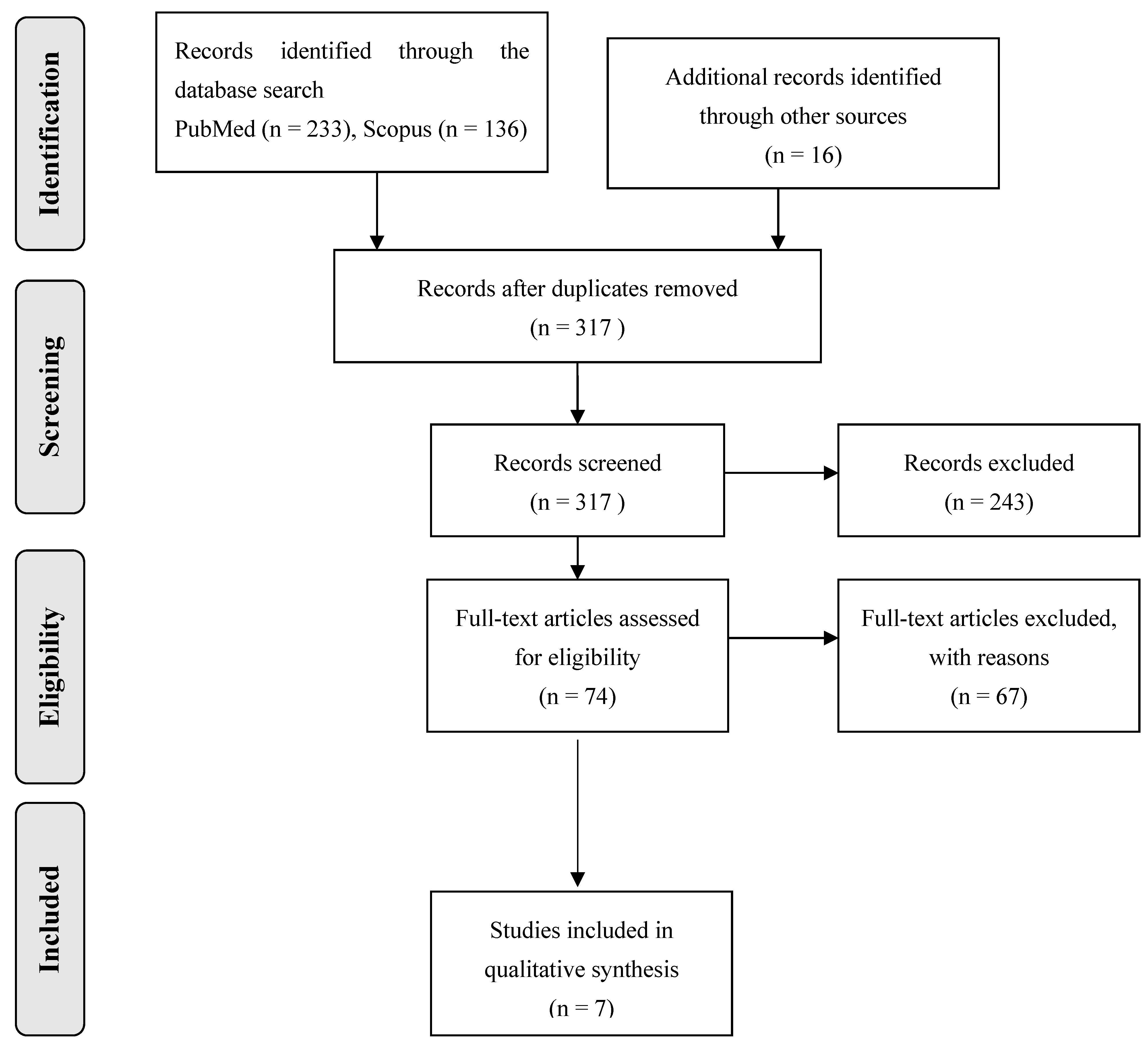
3.1. Database Searches

Overall, 385 records were retrieved from the database search. Duplicates and irrelevant studies were excluded; hence, a total of 74 articles were selected. After screening the full text of the articles, 7 studies were eligible for inclusion (Figure 1).

3.2. Study Characteristics

Seven publications fulfilled our inclusion criteria. They were classified into three groups, according to the nature of the compared disease entities. The first group consisted of three studies focusing on the differentiation between patients with typical (PD) and atypical Parkinsonism (PSP, MSA, and CBS) based on retinal parameters, as assessed with OCT [9,19,20]. The second group comprised three studies investigating the retinal morphology and its potential role in distinguishing patients with PD versus patients with PSP [21,22,23]. Finally, one study explored the value of retinal parameters through OCT in a setting of dementia associated with the presence of Lewy bodies or Parkinson’s disease [24] (Table 1).

4. Literature Review Sections

4.1. Exploring Retinal Findings in Patients with Typical (PD) and Atypical Parkinsonism (PSP, MSA, CBS)

In an attempt to provide insight into the linkage between retinal parameters evaluated through OCT implementation and the differential diagnosis of PD versus atypical Parkinsonian syndromes, Albrecht et al. examined a population of 84 patients presented both with PD and Parkinson-plus syndromes (PSP, MSA, and CBS) and recorded measures of peripapillary retinal nerve fiber layer (RNFL), paramacular thickness and volume, as well as the thickness of the different retinal layers. The researchers observed that the mean total macular thickness and volume of patients with PSP alone were significantly reduced when compared with patients with PD, in contrast to the decreased macular thickness and volume of patients with MSA and CBS versus PD, which did not reach statistical significance. As far as the mean peripapillary RNFL between groups is concerned, it was not proven to be significantly different. Regarding measurements of different retinal layers, it was demonstrated that not only the retinal ganglion cell and inner plexiform layer (RGC + IPL), but also the outer nuclear layer (ONL), was reduced in patients with PSP compared with PD. It was of great interest that the inner nuclear layer (INL) was reported to be thicker in individuals presenting with PD than those with MSA and PSP. Taking into account the reverse alterations of ONL and outer plexiform layer (OPL) within the group of patients diagnosed with PSP, the ratio between the aforementioned layers was calculated in order to enhance the differentiation accuracy between disease entities. Notably, the ONL/OPL ratio of patients with PSP combined with INL changes was found to be significantly different compared with those of patients with PD; thus, it seems able to distinguish between these groups of patients with both high sensitivity and specificity [9].
Similarly, Schneider et al., having enrolled 93 patients with clinically evident Parkinsonism attributed to pathophysiologically distinct diseases, investigated the utility of single retinal layer average perifoveal thickness measurement as a marker throughout the course of a Parkinsonian syndrome. In accordance with the aforementioned findings, the researchers concluded that ONL and OPL measures exhibited significant differences among patients with PSP and MSA, being able to discriminate between them with both a sensitivity and a specificity of around 90%. More specifically, the observed opposite changes in ONL/OPL ratios of PSP versus MSA with thinner ONL and thicker OPL within the group with PSP and the thicker ONL and thinner OPL within the group with MSA might be of additive value in an attempt to facilitate the differentiation of Parkinsonian syndromes. Interestingly, retinal parameters of patients with PD were not found to significantly differ when compared with those of healthy controls [19].
Additionally, Ma et al., having studied 52 individuals with Parkinsonism of varying etiology, provided further evidence regarding retinal involvement in the degenerative process of Parkinsonian syndromes. They reported higher peripapillary RNFL thickness among patients with PSP compared with groups with PD and MSA, especially within the temporal sector, a difference that reached statistical significance. In terms of macular thickness, patients with MSA were found to exhibit a shorter RNFL thickness, as well as a decreased macular volume in the foveal center circle sector, than patients with PD. It is also noteworthy that a significant correlation between disease duration and retinal measures was reported in patients with PD, PSP, and MSA. Given the unraveled specific patterns of retina changes with a significant marked peripapillary RNFL thickness and a remarkable thinning of macular thickness and volume among patients with PSP and MSA, respectively, in comparison to patients with PD, OCT may prove of great value in the differential diagnosis and disease progression assessment in a setting of Parkinsonism [20].

4.2. Investigating Retinal Parameters among Patients with PD versus PSP

With regard to the utility of OCT in differentiating between individuals presenting with PD and those being diagnosed with PSP, both with a disease duration of at least three years, Alkabie et al. evaluated retinal measurements of a total of 23 patients with PD and PSP and reported a significant reduction in mean RNFL thickness in eyes from patients with PSP as compared with those with PD, while the total macular volume was not found to markedly differ between the groups of Parkinsonian patients. Interestingly, an RNFL thickness threshold of 93 μm among patients with a long duration of disease (>3 years) seems able to distinguish between patients with PD and PSP, yielding a diagnostic sensitivity and specificity around 70%. It should be noted that the durations since symptom onset or since diagnosis were not correlated with either RNFL thickness or with total macular volume. On the contrary, the researchers concluded that the severity of the disease, as assessed by the Unified Parkinson Disease Rating scale (UPDRS), was positively correlated with the RNFL thickness within the group with PD, although negatively associated with total macular volume among all patients’ groups [21].
Exploring the role of OCT in delineating PSP from PD patients, Sevim et al., having enrolled 39 patients diagnosed with either PSP or PD with a mean disease duration over 3 years, examined the potential linkage between retinal parameters, included pRNFL value, macular volume and single retinal layers’ thickness, and the nature of Parkinsonian syndrome, duration of disease, and therapeutic use of levodopa. They observed a marked thinning of pRNFL among patients with PSP when compared with the group with PD in all sectors, a difference that reached statistical significance only as far as the superior quadrant is concerned. Moreover, in patients with PSP, the total macular volume was shown to be reduced, while GCL, IPL, and INL, as well as the inner retinal layer (IRL), were found to be thinner versus patients with PD. Regarding the impact of levodopa use among patients with PD on retinal morphology, a significantly decreased ONL/OPL ratio and an average pRNFL thinning in the inferotemporal sector were reported among levodopa non-users compared with levodopa users. It is of great interest that significant correlations were demonstrated between the duration of disease and retinal measures, both in groups of patients with PSP and PD [22].
Furthermore, in a study conducted by Rebolleda et al., the relationship between patterns of retinal changes and neurodegeneration in PD versus PSP was investigated, studying a total of 53 patients. The researchers exhibited significant differences between the two groups of patients, with a thicker mean average RNFL and a higher mean macular volume among patients with PD than a group with PSP. Notably, it was observed that a minimum GCIPL thickness cut-off value of 69 μm seems able to distinguish PSP from PD with high sensitivity and specificity values [23].

4.3. Examining Retinal Changes in a Setting of Dementia Associated with the Presence of Lewy Bodies or Parkinson’s Disease

In an attempt to provide further insight into the potential role of OCT as a candidate biomarker among patients with dementia other than Alzheimer’s disease, Moreno-Ramos et al. explored retinal morphology as well as subtle retinal changes in both settings of dementia with Lewy bodies and dementia coupled with PD. Having enrolled 20 patients presenting with the aforementioned types of dementia, the researchers observed a greater reduction in RNFL thickness in cases of patients with Lewy body dementia compared with a group with PD dementia, although this difference failed to reach statistical significance. Additionally, a positive correlation was found between disease severity, as evaluated by Mini-Mental State Examination (MMSE) and Mattis Dementia Rating Scale, and retinal measures as assessed by OCT. More specifically, the lower MMSE and Mattis Dementia Rating Scale scores reflected increased disease severity; the greater thinning of RNFL in both groups highlighted the fact that disease progression is accompanied by a degenerative process within the retina [24].

5. Discussion

A literature review over the last decade was conducted in order to elucidate the diagnostic value of OCT in a setting of clinically evident Parkinsonism. Original articles dealing with the potential utility of OCT implementation within a group of patients presenting with Parkinsonian features were identified and reviewed.
Despite the fact that Inzelberg et al. first applied OCT within a group of patients with PD almost twenty years ago [25], ongoing research regarding the potential role of OCT in patients presenting with Parkinsonism is still characterized by inconsistency, and has yielded conflicting results. To date, no reliable OCT biomarkers able to distinguish between PD and atypical Parkinsonian syndromes in clinical practice have been developed.
The assessment of RNFL and macular thickness, as well as the evaluation of single retinal layers, constitute OCT measures that are both readily accessible and reproducible, able to visualize subtle retinal changes as surrogate markers of brain abnormalities. There is growing evidence that different disease entities are coupled with distinct alterations of retinal morphology [26]. Being called a window to the brain [27,28], the eyes, and more specifically, the retinal microstructure, may reflect the underlying pathology of each patient’s disorder, thus providing unique insights into the neurodegenerative process [29,30,31]. Until now, specific diagnostic protocols utilizing OCT techniques have been developed in order to provide useful information regarding PD diagnosis [32].
Notably, various CNS disorders, multiple sclerosis and PD being among them, may have an impact on the optic nerve and the retina, which both embryologically originate from the diencephalon, with clinically evident visual disturbances [13,33,34,35,36]. Although reductions in RNFL thickness and the thickness of the macular layers have been linked to nigral dopaminergic depletion [37], indicating a similar pathophysiology mechanism to PD, retinal thinning was also demonstrated within the course of neurological diseases with a distinct etiological basis, including multiple sclerosis, neuromyelitis optica, and Alzheimer’s disease [38,39,40,41]. Thus, dopaminergic loss may not be solely responsible for RNFL and macular thickness reduction, as has been reported among patients presenting with neurological disorders without dopaminergic loss as well, making retinal thinning a rather non-specific degeneration marker [42,43,44].
There is growing evidence that OCT may serve as an early diagnostic tool among patients with PSP, independent of the disease’s clinical phenotypes [45]. Indeed, pRNFL reflecting the degree of retinal axonal degeneration has been shown to be significantly thinner within a group of patients with PSP than healthy controls [45,46,47,48]. Highlighting the positive correlation between retinal thinning and PSP severity and duration, non-motor symptoms, and cognitive performance, researchers have pointed out the potential role of OCT as a monitoring marker of disease progression [45,46,48], although contradictory results were yielded from another study [47].
Similarly, retinal microstructure alterations and, more specifically, a degree of retinal thinning, has been demonstrated within groups of patients with MSA when compared with healthy individuals [49,50,51,52,53]. Interestingly, retinal thickness reductions were found to be more marked among patients with the MSA-P subtype than those presenting with the MSA-C subtype, which is strongly linked to clinical severity of the disease [54]. According to a recent meta-analysis, a distinct pattern of retinal changes has been observed in patients with MSA when compared with patients with PD [55].
As far as retinal morphology in patients with PD is concerned, retinal thinning was demonstrated through OCT [56,57], located peripapillary [25], in the fovea [58], or in different retinal areas [59,60,61,62,63,64,65,66,67,68]. According to a recent meta-analysis, the thickness of the combined ganglion cell layer and inner plexiform layer was significantly reduced among patients with PD when compared with healthy controls, whereas similar differences regarding the thickness of INL, OPL, and ONL were not detected [69]. Furthermore, thinning of the macula was reported in a setting of PD [59,65,70,71,72]. It is of great interest that the observed macular thinning is coupled with both disease severity and progression, as opposed to RNFL thickness reduction, where similar correlations are controversial [73]. However, a linkage between PD and retinal structure changes, including RNFL thinning, could not be elicited by others [9,71,74,75,76]. Researchers have highlighted the fact that patients with PD are accompanied by less profound retinal changes when compared with patients with PSP and a distinct retinal damage pattern from that observed among patients with MSA [47,55], potentially facilitating differential diagnoses between PD and atypical Parkinsonian syndromes.
In an attempt to efficiently differentiate between PD and Parkinson-plus syndromes by OCT implementation, OCT measures were compared within patients’ groups. The most profound alterations were reported in cases of PSP and MSA, while the pattern of retinal involvement in PD remained rather heterogenous [9,19]. Notably, the ONL to OPL ratio was found to be of great discriminatory value between PSP and MSA due to an increased OPL thickness in PSP and an increased ONL thickness in MSA, whereas patients with PD were not accompanied by significant changes when compared with healthy controls [19]. The aforementioned findings seem to be in accordance with brain MRI morphometry studies, where specific patterns of atrophy are observed in patients with atypical Parkinsonism with more pronounced retinal alterations than PD cases that are reflected in MRI morphometry, with rather minor changes [77]. Combining the ONL to OPL ratio with INL measures may serve as an accurate indicator of PSP versus PD and other Parkinsonian syndromes’ patients (MSA, CBS), with a negative test almost certainly ruling out PSP [9].
With respect to the average pRNFL thickness and CMT, it was demonstrated that both measures were significantly thinner among patients with PSP and MSA than those with PD. Interestingly, MSA has been coupled with a distinct pattern of retinal thinning, capable of playing a crucial role in distinguishing between atypical Parkinsonism [26]. Reflecting the discriminatory value of specific OCT measures, pRNFL thinning was greater within a group of PSP patients than those with PD [21,22]. On the contrary, pRNFL thickness was reported as significantly thinner in PD and MSA compared with PSP, while macular thickness and volume thinning were more marked in MSA than in PD. Thus, the diagnostic potential of pRNFL thickness in differentiating between PSP and PD, and between PSP and MSA, was pointed out, highlighting the fact that macular thickness and volume evaluation may enhance the discrimination between MSA and PD [20]. Additionally, the mean RNFL was not found to be significantly different between groups of PSP and PD patients in a study conducted by Albrecht [9].
As far as the association between OCT parameters and duration of the disease is concerned, Albrecht et al. could not elicit a link between retinal measures and both disease duration and severity, suggesting that observed retinal alterations occur early in the course of the disease, thus implying a potential role of OCT in the early differential diagnosis of Parkinsonism [9]. However, Satue et al. supported the progression of retinal changes that are attributed to the progression of the disease itself, providing favorable relative evidence [78]. Similarly, Ma et al. and Sevim et al. demonstrated a significant correlation between retinal measures and disease duration among patients with PD and PSP, as well as PD, PSP, and MSA [20,22]. Especially in the group with PSP, the more the disease progressed, the more marked the reductions in pRNFL, ONL, IRL, and ORL thicknesses and macular volume were, indicating a rather faster neurodegeneration process in a setting of PSP than PD.
Regarding potential confounding factors among patients’ groups, it is noteworthy that patients with a history of ophthalmologic pathologies, including age-related macular degeneration, previous intraocular surgery, glaucomatous optic neuropathy, and diabetic retinopathy, among others, were excluded [9,20,21,22,24]. When participants’ age is concerned, the vast majority of the included studies reported no significant age difference between patients’ groups and matched healthy controls; thus, age-related decreases in OCT parameters were not observed [9,19,20,22,23,24]. In the study by Alkabie et al., it was demonstrated that patients with PSP were significantly older when compared with both the group with PD and control subjects [21]. Nevertheless, the impact of the aforementioned age difference between studied patients’ groups was mitigated by introducing age as a covariate in the linear mixed-effects model when assessing OCT measurements between groups.

6. Conclusions

The present review summarizes all the up-to-date evidence regarding the diagnostic value of OCT and its potential utility in effectively differentiating between PD and atypical Parkinsonian syndromes. Numerous studies yielded results that are in favor of a definite involvement of the retina in Parkinsonian disorders, indicating the valuable role of OCT implementation in accurately differentiating between PD and atypical Parkinsonian syndromes. Nevertheless, the lack of consistency, as well as the limited number of relevant studies aiming to support the potential of OCT as diagnostic biomarker, point to an emerging need for further exploration. In this direction, larger cohorts of patients in a longitudinal setting are required in order to confirm the current results and validate the utilization of the OCT technique as a marker of neurodegenerative processes in a setting of Parkinsonism, thus facilitating early diagnoses and enhancing effective therapeutic approaches. Under this prism, future studies involving both multivariate statistics and age- and acuity-matched controls, and taking into consideration demographic traits and visual clinical measures, may be of added clinical and research value.

Funding

This research received no external funding.

Conflicts of Interest

The authors declare no conflict of interest.

References

  1. Williams, D.R.; Litvan, I. Parkinsonian syndromes. Contin. Lifelong Learn. Neurol. 2013, 19, 1189–1212. [Google Scholar] [CrossRef]
  2. Keener, A.M.; Bordelon, Y.M. Parkinsonism. Semin. Neurol. 2016, 36, 330–334. [Google Scholar] [CrossRef]
  3. Shin, H.W.; Hong, S.W.; Youn, Y.C. Clinical Aspects of the Differential Diagnosis of Parkinson’s Disease and Parkinsonism. J. Clin. Neurol. 2022, 18, 259–270. [Google Scholar] [CrossRef]
  4. McFarland, N.R. Diagnostic Approach to Atypical Parkinsonian Syndromes. Continuum 2016, 22, 1117–1142. [Google Scholar] [CrossRef]
  5. Levin, J.; Kurz, A.; Arzberger, T.; Giese, A.; Hoglinger, G.U. The Differential Diagnosis and Treatment of Atypical Parkinsonism. Dtsch. Arztebl. Int. 2016, 113, 61–69. [Google Scholar] [CrossRef]
  6. Marino, B.L.B.; de Souza, L.R.; Sousa, K.P.A.; Ferreira, J.V.; Padilha, E.C.; da Silva, C.H.T.P.; Taft, C.A.; Hage-Melim, L.I.S. Parkinson’s Disease: A Review from Pathophysiology to Treatment. Mini Rev. Med. Chem. 2020, 20, 754–767. [Google Scholar] [CrossRef] [PubMed]
  7. Tysnes, O.B.; Storstein, A. Epidemiology of Parkinson’s disease. J. Neural Transm. 2017, 124, 901–905. [Google Scholar] [CrossRef]
  8. Vertes, A.C.; Beato, M.R.; Sonne, J.; Khan Suheb, M.Z. Parkinson-Plus Syndrome. In StatPearls; StatPearls Publishing: Treasure Island, FL, USA, 2023. Available online: https://www.ncbi.nlm.nih.gov/books/NBK585113/ (accessed on 4 February 2024).
  9. Albrecht, P.; Müller, A.K.; Südmeyer, M.; Ferrea, S. Optical coherence tomography in parkinsonian syndromes. PLoS ONE 2012, 7, e34891. [Google Scholar] [CrossRef]
  10. Hess, C.W.; Okun, M.S. Diagnosing parkinson disease. Continuum (Minneap. Minn.) 2016, 22, 1047–1063. [Google Scholar] [CrossRef]
  11. GBD 2016 Parkinson’s Disease Collaborators. Global, regional, and national burden of Parkinson’s disease, 1990–2016: A systematic analysis for the Global Burden of Disease Study 2016. Lancet Neurol. 2018, 17, 939–953. [Google Scholar] [CrossRef] [PubMed]
  12. Pula, J.H.; Kim, J.; Nichols, J. Visual aspects of neurologic protein misfolding disorders. Curr. Opin. Ophthalmol. 2009, 20, 482–489. [Google Scholar] [CrossRef]
  13. Bak, T.H.; Caine, D.; Hearn, V.C.; Hodges, J.R. Visuospatial functions in atypical parkinsonian syndromes. J. Neurol. Neurosurg. Psychiatry 2006, 77, 454–456. [Google Scholar] [CrossRef]
  14. Archibald, N.K.; Clarke, M.P.; Mosimann, U.P.; Burn, D.J. The retina in Parkinson’s disease. Brain 2009, 132, 1128–1145. [Google Scholar] [CrossRef]
  15. Huang, D.; Swanson, E.A.; Lin, C.P.; Schuman, J.S.; Stinson, W.G.; Chang, W.; Hee, M.R.; Flotte, T.; Gregory, K.; Puliafito, C.A.; et al. Optical coherence tomography. Science 1991, 254, 1178–1181. [Google Scholar] [CrossRef] [PubMed]
  16. Wang, Y.; Liu, S.; Lou, S.; Zhang, W.; Cai, H.; Chen, X. Application of optical coherence tomography in clinical diagnosis. J. Xray Sci. Technol. 2019, 27, 995–1006. [Google Scholar] [CrossRef] [PubMed]
  17. Galetta, K.M.; Calabresi, P.A.; Frohman, E.M.; Balcer, L.J. Optical coherence tomography (OCT): Imaging the visual pathway as a model for neurodegeneration. Neurother. J. Am. Soc. Exp. Neurother. 2011, 8, 117–132. [Google Scholar] [CrossRef] [PubMed]
  18. Harnois, C.; Di Paolo, T. Decreased dopamine in the retinas of patients with Parkinson’s disease. Investig. Ophthalmol. Vis. Sci. 1990, 31, 2473–2475. [Google Scholar]
  19. Schneider, M.; Müller, H.P.; Lauda, F.; Tumani, H.; Ludolph, A.C.; Kassubek, J.; Pinkhardt, E.H. Retinal single-layer analysis in Parkinsonian syndromes: An optical coherence tomography study. J. Neural Transm. 2014, 121, 41–47. [Google Scholar] [CrossRef] [PubMed]
  20. Ma, X.; Li, S.; Zheng, B.; Hu, L.; Liu, H.; Wang, Z.; Wang, Z.; Chen, H.; Su, W. Retinal Structure Abnormalities in Parkinson’s Disease and Atypical Parkinsonism. Biomolecules 2023, 13, 218. [Google Scholar] [CrossRef] [PubMed]
  21. Alkabie, S.; Lange, A.; Manogaran, P.; Stoessl, A.J.; Costello, F.; Barton, J.J.S. Optical coherence tomography of patients with Parkinson’s disease and progressive supranuclear palsy. Clin. Neurol. Neurosurg. 2020, 189, 105635. [Google Scholar] [CrossRef]
  22. Gulmez Sevim, D.; Unlu, M.; Gultekin, M.; Karaca, C.; Mirza, M.; Mirza, G.E. Evaluation of Retinal Changes in Progressive Supranuclear Palsy and Parkinson Disease. J. Neuroophthalmol. 2018, 38, 151–155. [Google Scholar] [CrossRef] [PubMed]
  23. Rebolleda, G.; Sanchez Sanchez, C.; Martinez Castrillo, J.C.; Gonzalez Lopez, J.J.; Oblanca Llamazares, N.; Muñoz-Negrete, F.J. Optical coherence tomography to distinguish Parkinson disease versus supranuclear progressive palsy. Acta Ophthalmol. 2016, 94, S256. [Google Scholar] [CrossRef]
  24. Moreno-Ramos, T.; Benito-León, J.; Villarejo, A.; Bermejo-Pareja, F. Retinal nerve fiber layer thinning in dementia associated with Parkinson’s disease, dementia with Lewy bodies, and Alzheimer’s disease. J. Alzheimers Dis. 2013, 34, 659–664. [Google Scholar] [CrossRef] [PubMed]
  25. Inzelberg, R.; Ramirez, J.A.; Nisipeanu, P.; Ophir, A. Retinal nerve fiber layer thinning in Parkinson disease. Vis. Res. 2004, 44, 2793–2797. [Google Scholar] [CrossRef] [PubMed]
  26. Ma, X.; Wang, Y.; Wang, N.; Zhang, R. Retina thickness in atypical parkinsonism: A systematic review and meta-analysis. J. Neurol. 2022, 269, 1272–1281. [Google Scholar] [CrossRef]
  27. Miller, N.; Drachman, D.A. The optic nerve: A window into diseases of the brain? Neurology. 2006, 67, 1742–1743. [Google Scholar] [CrossRef] [PubMed]
  28. London, A.; Benhar, I.; Schwartz, M. The retina as a window to the brain-from eye research to CNS disorders. Nat. Rev. Neurol. 2013, 9, 44–53. [Google Scholar] [CrossRef]
  29. Lim, J.K.; Li, Q.X.; He, Z.; Vingrys, A.J.; Wong, V.H.; Currier, N.; Mullen, J.; Bui, B.V.; Nguyen, C.T. The eye as a biomarker for Alzheimer’s disease. Front. Neurosci. 2016, 10, 536. [Google Scholar] [CrossRef]
  30. Martinez-Lapiscina, E.H.; Sanchez-Dalmau, B.; Fraga-Pumar, E.; Ortiz-Perez, S.; Tercero-Uribe, A.I.; Torres-Torres, R.; Villoslada, P. The visual pathway as a model to understand brain damage in multiple sclerosis. Mult. Scler. 2014, 20, 1678–1685. [Google Scholar] [CrossRef]
  31. Gupta, S.; Zivadinov, R.; Ramanathan, M.; Weinstock-Guttman, B. Optical coherence tomography and neurodegeneration: Are eyes the windows to the brain? Expert Rev. Neurother. 2016, 16, 765–775. [Google Scholar] [CrossRef]
  32. Garcia-Martin, E.; Satue, M.; Fuertes, I.; Otin, S.; Alarcia, R.; Herrero, R.; Bambo, M.P.; Fernandez, J.; Pablo, L.E. Ability and reproducibility of Fourier-domain optical coherence tomography to detect retinal nerve fiber layer atrophy in Parkinson’s disease. Ophthalmology 2012, 119, 2161–2167. [Google Scholar] [CrossRef] [PubMed]
  33. Wang, L.; Kuroiwa, Y.; Kamitani, T.; Li, M.; Takahashi, T.; Suzuki, Y.; Shimamura, M.; Hasegawa, O. Visual event-related potentials in progressive supranuclear palsy, corticobasal degeneration, striatonigral degeneration, and Parkinson’s disease. J. Neurol. 2000, 247, 356–363. [Google Scholar] [CrossRef] [PubMed]
  34. Oliveira, C.; Cestari, D.M.; Rizzo, J.F., 3rd. The use of fourth generation optical coherence tomography in multiple sclerosis: A review. Semin. Ophthalmol. 2012, 27, 187–191. [Google Scholar] [CrossRef] [PubMed]
  35. Bodis-Wollner, I.; Miri, S.; Glazman, S. Venturing into the no-man’s land of the retina in Parkinson’s disease. Mov. Disord. Off. J. Mov. Disord. Soc. 2014, 29, 15–22. [Google Scholar] [CrossRef] [PubMed]
  36. Lee, J.Y.; Ahn, J.; Kim, T.W.; Jeon, B.S. Optical coherence tomography in Parkinson’s disease: Is the retina a biomarker? J. Park. Dis. 2014, 4, 197–204. [Google Scholar] [CrossRef] [PubMed]
  37. Ahn, J.; Lee, J.Y.; Kim, T.W.; Yoon, E.J.; Oh, S.; Kim, Y.K.; Kim, J.M.; Woo, S.J.; Kim, K.W.; Jeon, B. Retinal thinning associates with nigral dopaminergic loss in de novo Parkinson disease. Neurology 2018, 91, e1003–e1012. [Google Scholar] [CrossRef] [PubMed]
  38. Oertel, F.C.; Zimmermann, H.; Paul, F.; Brandt, A.U. Optical coherence tomography in neuromyelitis optica spectrum disorders: Potential advantages for individualized monitoring of progression and therapy. EPMA J. 2017, 9, 21–33. [Google Scholar] [CrossRef]
  39. Balcer, L.J.; Balk, L.J.; Brandt, A.U.; Calabresi, P.A.; Martinez-Lapiscina, E.H.; Nolan, R.C.; Paul, F.; Petzold, A.; Saidha, S. The international multiple sclerosis visual system consortium: Advancing visual system research in multiple sclerosis. J. Neuroophthalmol. 2018, 38, 494–501. [Google Scholar] [CrossRef]
  40. Coppola, G.; Di Renzo, A.; Ziccardi, L.; Martelli, F.; Fadda, A.; Manni, G.; Barboni, P.; Pierelli, F.; Sadun, A.A.; Parisi, V. Optical coherence tomography in Alzheimer’s disease: A meta-analysis. PLoS ONE 2015, 10, e0134750. [Google Scholar] [CrossRef]
  41. Ratchford, J.N.; Quigg, M.E.; Conger, A.; Frohman, T.; Frohman, E.; Balcer, L.J.; Calabresi, P.A.; Kerr, D.A. Optical coherence tomography helps differentiate neuromyelitis optica and MS optic neuropathies. Neurology 2009, 73, 302–308. [Google Scholar] [CrossRef]
  42. Mailankody, P.; Lenka, A.; Pal, P.K. The role of Optical Coherence Tomography in Parkinsonism: A critical review. J. Neurol. Sci. 2019, 403, 67–74. [Google Scholar] [CrossRef]
  43. Cipollini, V.; Abdolrahimzadeh, S.; Troili, F.; De Carolis, A.; Calafiore, S.; Scuderi, L.; Giubilei, F.; Scuderi, G. Neurocognitive Assessment and Retinal Thickness Alterations in Alzheimer Disease: Is There a Correlation? J. Neuro-Ophthalmol. Off. J. N. Am. Neuro-Ophthalmol. Soc. 2020, 40, 370–377. [Google Scholar] [CrossRef] [PubMed]
  44. Di Pippo, M.; d’Agostino, S.; Ruggeri, F.; Carrozzi, C.; Fasciolo, D.; Abdolrahimzadeh, S. Parkinson’s Disease: What Can Retinal Imaging Tell Us? J. Integr. Neurosci. 2024, 23, 23. [Google Scholar] [CrossRef] [PubMed]
  45. Picillo, M.; Salerno, G.; Tepedino, M.F.; Abate, F.; Cuoco, S.; Gioia, M.; Coppola, A.; Erro, R.; Pellecchia, M.T.; Rosa, N.; et al. Retinal thinning in progressive supranuclear palsy: Differences with healthy controls and correlation with clinical variables. Neurol. Sci. 2022, 43, 4803–4809. [Google Scholar] [CrossRef] [PubMed]
  46. Woo, K.A.; Shin, J.Y.; Kim, H.; Ahn, J.; Jeon, B.; Lee, J.Y. Peripapillary retinal nerve fiber layer thinning in patients with progressive supranuclear palsy. J. Neurol. 2022, 269, 3216–3225. [Google Scholar] [CrossRef] [PubMed]
  47. Stemplewitz, B.; Kromer, R.; Vettorazzi, E.; Hidding, U.; Frings, A.; Buhmann, C. Retinal degeneration in progressive supranuclear palsy measured by optical coherence tomography and scanning laser polarimetry. Sci. Rep. 2017, 7, 5357. [Google Scholar] [CrossRef] [PubMed]
  48. Chen, Y.; Wang, H.; Wang, B.; Li, W.; Ye, P.; Xu, W.; Liu, P.; Chen, X.; Cen, Z.; Ouyang, Z.; et al. Retinal Thinning as a Marker of Disease Severity in Progressive Supranuclear Palsy. J. Mov. Disord. 2024, 17, 55–63. [Google Scholar] [CrossRef] [PubMed]
  49. Fischer, M.D.; Synofzik, M.; Heidlauf, R.; Schicks, J.; Srulijes, K.; Kernstock, C.; Berg, D.; Schols, L.; Schiefer, U. Retinal nerve fiber layer loss in multiple system atrophy. Mov. Disord. Off. J. Mov. Disord. Soc. 2011, 26, 914–916. [Google Scholar] [CrossRef]
  50. Pula, J.H.; Towle, V.L.; Staszak, V.M.; Cao, D.; Bernard, J.T.; Gomez, C.M. Retinal nerve fibre layer and macular thinning in spinocerebellar ataxia and cerebellar multisystem atrophy. Neuroophthalmology 2011, 35, 108–114. [Google Scholar] [CrossRef]
  51. Fischer, M.D.; Synofzik, M.; Kernstock, C.; Dietzsch, J.; Heidlauf, R.; Schicks, J.; Srulijes, K.; Wiethoff, S.; Menn, O.; Berg, D.; et al. Decreased retinal sensitivity and loss of retinal nerve fibers in multiple system atrophy. Graefes Arch. Clin. Exp. Ophthalmol. 2013, 251, 235–241. [Google Scholar] [CrossRef]
  52. Mendoza-Santiesteban, C.E.; Palma, J.A.; Martinez, J.; NorcliffeKaufmann, L.; Hedges, T.R., 3rd; Kaufmann, H. Progressive retinal structure abnormalities in multiple system atrophy. Mov. Disord. Off. J. Mov. Disord. Soc. 2015, 30, 1944–1953. [Google Scholar] [CrossRef] [PubMed]
  53. Mendoza-Santiesteban, C.; Palma, J.A.; Ortuño-Lizaran, I.; Cuenca, N.; Kaufmann, H. Pathologic confirmation of retinal ganglion cell loss in multiple system atrophy. Neurology 2017, 88, 2233–2235. [Google Scholar] [CrossRef]
  54. Ahn, J.; Lee, J.Y.; Kim, T.W. Retinal thinning correlates with clinical severity in multiple system atrophy. J. Neurol. 2016, 263, 2039–2047. [Google Scholar] [CrossRef] [PubMed]
  55. Mendoza-Santiesteban, C.E.; Gabilondo, I.; Palma, J.A.; Norcliffe-Kaufmann, L.; Kaufmann, H. The Retina in Multiple System Atrophy: Systematic Review and Meta-Analysis. Front. Neurol. 2017, 8, 206. [Google Scholar] [CrossRef]
  56. Cetin, E.N.; Bir, L.S.; Sarac, G.; Yaldızkaya, F.; Yaylalı, V. Optic Disc and Retinal Nerve Fibre Layer Changes in Parkinson’s Disease. Neuro-Ophthalmology 2013, 37, 20–23. [Google Scholar] [CrossRef]
  57. Stemplewitz, B.; Keserü, M.; Bittersohl, D.; Buhmann, C.; Skevas, C.; Richard, G.; Hassenstein, A. Scanning laser polarimetry and spectral domain optical coherence tomography for the detection of retinal changes in Parkinson’s disease. Acta Ophthalmol. 2015, 93, e672–e677. [Google Scholar] [CrossRef]
  58. Hajee, M.E.; March, W.F.; Lazzaro, D.R.; Wolintz, A.H.; Shrier, E.M.; Glazman, S.; Bodis-Wollner, I.G. Inner retinal layer thinning in Parkinson disease. Arch. Ophthalmol. 2009, 127, 737–741. [Google Scholar] [CrossRef]
  59. Altintaş, O.; Işeri, P.; Ozkan, B.; Cağlar, Y. Correlation between retinal morphological and functional findings and clinical severity in Parkinson’s disease. Doc. Ophthalmol. 2008, 116, 137–146. [Google Scholar] [CrossRef]
  60. Moschos, M.M.; Tagaris, G.; Markopoulos, I.; Margetis, I.; Tsapakis, S.; Kanakis, M.; Koutsandrea, C. Morphologic changes and functional retinal impairment in patients with Parkinson disease without visual loss. Eur. J. Ophthalmol. 2011, 21, 24–29. [Google Scholar] [CrossRef] [PubMed]
  61. Kirbas, S.; Turkyilmaz, K.; Tufekci, A.; Durmus, M. Retinal nerve fiber layer thickness in Parkinson disease. J. Neuroophthalmol. 2013, 33, 62–65. [Google Scholar] [CrossRef] [PubMed]
  62. La Morgia, C.; Barboni, P.; Rizzo, G.; Carbonelli, M.; Savini, G.; Scaglione, C.; Capellari, S.; Bonazza, S.; Giannoccaro, M.P.; Calandra-Buonaura, G.; et al. Loss of temporal retinal nerve fibers in Parkinson’s disease: A mitochondrial pattern? Eur. J. Neurol. 2013, 20, 198–201. [Google Scholar] [CrossRef] [PubMed]
  63. Rohani, M.; Langroodi, A.S.; Ghourchian, S.; Falavarjani, K.G.; SoUdi, R.; Shahidi, G. Retinal nerve changes in patients with tremor dominant and akinetic rigid Parkinson’s disease. Neurol. Sci. 2013, 34, 689–693. [Google Scholar] [CrossRef]
  64. Jimenez, B.; Ascaso, F.J.; Cristóbal, J.A.; López del Val, J. Development of a prediction formula of Parkinson disease severity by optical coherence tomography. Mov. Disord. 2014, 29, 68–74. [Google Scholar] [CrossRef]
  65. Garcia-Martin, E.; Satue, M.; Otin, S.; Fuertes, I.; Alarcia, R.; Larrosa, J.M.; Polo, V.; Pablo, L.E. Retina measurements for diagnosis of Parkinson disease. Retina 2014, 34, 971–980. [Google Scholar] [CrossRef] [PubMed]
  66. Satue, M.; Garcia-Martin, E.; Fuertes, I.; Otin, S.; Alarcia, R.; Herrero, R.; Bambo, M.P.; Pablo, L.E.; Fernandez, F.J. Use of Fourier-domain OCT to detect retinal nerve fiber layer degeneration in Parkinson’s disease patients. Eye 2013, 27, 507–514. [Google Scholar] [CrossRef]
  67. Yu, J.G.; Feng, Y.F.; Xiang, Y.; Huang, J.H.; Savini, G.; Parisi, V.; Yang, W.J.; Fu, X.A. Retinal nerve fiber layer thickness changes in Parkinson disease: A meta-analysis. PLoS ONE 2014, 9, e85718. [Google Scholar] [CrossRef]
  68. Aydin, T.S.; Umit, D.; Nur, O.M.; Fatih, U.; Asena, K.; Nefise, O.Y.; Serpil, Y. Optical coherence tomography findings in Parkinson’s disease. Kaohsiung J. Med. Sci. 2018, 34, 166–171. [Google Scholar] [CrossRef]
  69. Huang, L.; Zhang, D.; Ji, J.; Wang, Y.; Zhang, R. Central retina changes in Parkinson’s disease: A systematic review and meta-analysis. J. Neurol. 2021, 268, 4646–4654. [Google Scholar] [CrossRef]
  70. Schrier, E.M.; Adam, C.R.; Spund, B.; Glazman, S.; BodisWollner, I. Interocular asymmetry of foveal thickness in Parkinson’s disease. J. Ophthalmo. 2012, 2012, 728457. [Google Scholar] [CrossRef]
  71. Aaker, G.D.; Myung, J.S.; Ehrlich, J.R.; Mohammed, M.; Henchcliffe, C.; Kiss, S. Detection of retinal changes in Parkinson’s disease with spectral-domain optical coherence tomography. Clin. Ophthalmol. 2010, 4, 1427–1432. [Google Scholar] [CrossRef]
  72. Lee, Y.W.; Lim, M.N.; Lee, J.Y.; Yoo, Y. Central retina thickness measured with spectral-domain optical coherence tomography in Parkinson disease: A meta-analysis. Medicine 2023, 102, e35354. [Google Scholar] [CrossRef]
  73. Satue, M.; Obis, J.; Rodrigo, M.J.; Otin, S.; Fuertes, M.I.; Vilades, E.; Gracia, H.; Ara, J.R.; Alarcia, R.; Polo, V.; et al. Optical Coherence Tomography as a Biomarker for Diagnosis, Progression, and Prognosis of Neurodegenerative Diseases. J. Ophthalmol. 2016, 2016, 8503859. [Google Scholar] [CrossRef] [PubMed]
  74. Tsironi, E.E.; Dastiridou, A.; Katsanos, A.; Dardiotis, E.; Veliki, S.; Patramani, G.; Zacharaki, F.; Ralli, S.; Hadjigeorgiou, G.M. Perimetric and retinal nerve fiber layer findings in patients with Parkinson’s disease. BMC Ophthalmol. 2012, 12, 54. [Google Scholar] [CrossRef] [PubMed]
  75. Cubo, E.; López Peña, M.J.; Diez-Feijo Varela, E.; Pérez Gil, O.; Garcia Gutierrez, P.; Araus González, E.; Prieto Tedejo, R.; Mariscal Pérez, N.; Armesto, D. Lack of association of morphologic and functional retinal changes with motor and non-motor symptoms severity in Parkinson’s disease. J. Neural Transm. 2014, 121, 139–145. [Google Scholar] [CrossRef] [PubMed]
  76. Archibald, N.K.; Clarke, M.P.; Mosimann, U.P.; Burn, D.J. Retinal thickness in Parkinson’s disease. Park. Relat. Disord. 2011, 17, 431–436. [Google Scholar] [CrossRef]
  77. Messina, D.; Cerasa, A.; Condino, F.; Arabia, G.; Novellino, F.; Nicoletti, G.; Salsone, M.; Morelli, M.; Lanza, P.L.; Quattrone, A. Patterns of brain atrophy in Parkinson’s disease, progressive supranuclear palsy and multiple system atrophy. Park. Relat. Disord. 2011, 17, 172–176. [Google Scholar] [CrossRef]
  78. Satue, M.; Rodrigo, M.J.; Obis, J.; Vilades, E.; Gracia, H.; Otin, S.; Fuertes, M.I.; Alarcia, R.; Crespo, J.A.; Polo, V.; et al. Evaluation of progressive visual dysfunction and retinal degeneration in patients with Parkinson’s disease. Investig. Ophthalmol. Vis. Sci. 2017, 58, 1151–1157. [Google Scholar] [CrossRef]
Figure 1. Study flow chart (PRISMA diagram).
Applsci 14 02491 g001
Table 1. Characteristics of the 7 included studies.
Authors, Year of PublicationNumber of ParticipantsType of Implemented OCTAssessed Retinal ParametersCutoff Values; (Specificity); [Sensitivity]Main Results
Albrecht et al., 2012 [9]35 healthy controls
84 patients
- 40 PD
- 19 MSA
- 10 CBS
- 15 PSP
Spectral domain optical coherence tomography with manual segmentationpRNFL, paramacular thickness and volume, thickness of all retinal layersONL/OPL ratio cutoff of 3.1 for differentiation between PSP and PD (96%); [59%], combined ONL/OPL ratio with INL cutoff of 46 mm (96%); [70%]The mean total macular thickness and volume of patients with PSP were found to be significantly reduced compared with patients with PD, while the ONL/OPL ratio combined with INL changes within group with PSP may serve as a diagnostic marker
Schneider et al., 2013 [19]41 healthy controls
93 patients
- 65 PD
- 16 PSP
- 12 MSA
Spectral domain OCT with a semiautomatic algorithmRNFL, GCL + IPL, INL, OPL, ONL, WRTONL/OPL ratio cutoff of 5.03 for discrimination between PSP and MSA (88%); [91%]Retinal parameters of patients with PD were not significantly different that those of healthy individuals, while opposite changes of ONL/OPL ratios were reported between patients’ groups with PSP and MSA
Ma et al., 2023 [20]14 healthy controls
52 patients
- 24 PD
- 19 MSA
- 9 PSP
Spectral domain OCTpRNFL thickness, macular thickness and volumeN/ASpecific patterns of retina alterations were reported among patients with PD and atypical Parkinsonism with a macular thinning in patients with PD and MSA and a higher peripapillary RNFL thickness in patients with PSP
Alkabie et al., 2019 [21]12 healthy controls
23 patients
- 12 PD
- 11 PSP
Spectral domain OCTRNFL thickness, macular volumeRNFL thickness cutoff of 93 μm in patients with disease duration of ≥3 years for distinguishing between patients with PD and PSP (~70%), [~70%]A significant reduction in mean RNFL thickness was observed in eyes from patients with PSP as compared with those of patients with PD with a disease duration of at least 3 years
Sevim et al., 2018 [22]33 healthy controls
39 patients
- 29 PD
- 10 PSP
Spectral domain OCT with automatic segmentationpRNFL thickness, thickness and volume of retinal layers at the macula (mRNFL, GCL, IPL, INL, OPL, ONL, PRs, RPE)N/AA significant thinning of both pRNFL and GCL, IPL, INL was reported among patients with PSP when compared with a group with PD. Regarding levodopa use, a decreased ONL/OPL ratio and pRNFL thinning were observed among levodopa non-users compared with levodopa users
Rebolleda et al., 2016 [23]53 patients
- 38 PD
- 15 PSP
Spectral domain OCTpRNFL, macular thickness, GCAMinimum GCIPL thickness cut-off value of 69 μm for differentiating PSP from PD (91.7%), [72.7%]A thicker mean average RNFL and a higher mean macular volume was found among patients with PD than those patients of group with PSP
Moreno-Ramos et al., 2013 [24]10 healthy controls
30 patients
- 10 AD
- 10 LBD
- 10 PD
OCTRNFL thicknessN/AA greater reduction in RNFL thickness was observed in patients with Lewy body dementia compared with the group with PD dementia, with a significant positive correlation between disease severity and retinal parameters
Abbreviations: peripapillary retinal nerve fiber layer (pRNFL), ganglion cell layer plus inner plexiform layer (GCL + IPL), inner nuclear layer (INL), outer nuclear layer (ONL), outer plexiform layer (OPL), whole retinal thickness (WRT), macular retinal nerve fiber layer (mRNFL), ganglion cell layer (GCL), inner plexiform layer (IPL), inner and outer segments of the photoreceptors (PRs), retinal pigment epithelium (RPE), ganglion cell layer inner plexiform analysis (GCA).
Disclaimer/Publisher’s Note: The statements, opinions and data contained in all publications are solely those of the individual author(s) and contributor(s) and not of MDPI and/or the editor(s). MDPI and/or the editor(s) disclaim responsibility for any injury to people or property resulting from any ideas, methods, instructions or products referred to in the content.

