Systematic Review

Plant-Mediated Inorganic Nanoparticles for Anti-Tumor Therapy in Colorectal Cancer: A Systematic Review

by
Cristina Mesas
1,2,†,
Francisco Quiñonero
1,2,†,
Francisco Revueltas
3,†,
Laura Cabeza
1,2,4,
Gloria Perazzoli
1,2,4,
Consolación Melguizo
1,2,4,* and
Jose Prados
1,2,4
1
Institute of Biopathology and Regenerative Medicine (IBIMER), Center of Biomedical Research (CIBM), University of Granada, 18100 Granada, Spain
2
Instituto Biosanitario de Granada (ibs.GRANADA), 18014 Granada, Spain
3
School of Medicine, University of Granada, 18016 Granada, Spain
4
Department of Anatomy and Embryology, Faculty of Medicine, University of Granada, 18071 Granada, Spain
*
Author to whom correspondence should be addressed.
These authors contributed equally to this work.
Appl. Sci. 2023, 13(18), 10156; https://doi.org/10.3390/app131810156
Submission received: 28 July 2023 / Revised: 22 August 2023 / Accepted: 6 September 2023 / Published: 9 September 2023
(This article belongs to the Special Issue Bioactive Compounds from Natural Products - Volume II)

Abstract

:
Colon cancer is the third most frequent neoplasm and the second most lethal worldwide. Despite progress in its treatment, new therapies are still needed to improve the prognosis of this type of tumor and, in this context, the use of plant compounds with anti-tumor properties has been increasing in recent years. The aim of this systematic review was to analyze the potential benefits of encapsulation of compounds derived from plant extracts in nanoparticles and their cytotoxic effect under in vitro conditions. Once the search strategy was defined based on the selected MESH terms, 147 publications published since 2012 were identified from three different databases (PubMed, SCOPUS and WOS). After eliminating duplicates and applying the inclusion and exclusion criteria, 17 studies were finally included. The results showed that the use of natural extracts encapsulated in nanoparticles offered significant cytotoxic activity against colon neoplastic cells by increasing the therapeutic effect of free plant extracts through their encapsulation and without producing toxicity on healthy cells. In addition, most studies (14) involved metal-derived nanoparticles (zinc, iron and gold). Despite the possible efficacy of these nanodrugs, more in vivo studies are needed to elucidate their potential future therapeutic application and their biocompatibility.

1. Introduction

Colorectal cancer (CRC) is a disease that accounts for 10% of diagnosed (1.9 million) tumors and cancer-related deaths (935,000) worldwide [1]. Its incidence is lower in women (32 out of every 100,000) compared to men (42 per 100,000), being mainly diagnosed in developed regions such as Europe, Australia, and North America, and with Hungary and Norway being the two countries with the highest incidence [1]. In a gradual process, it is estimated that the incidence of this type of cancer will increase over the next few years, reaching a diagnosis of 2.5 million new cases in the year 2035 (600,000 more than currently) [2]. Family history of cancer is linked to 10–20% of diagnosed patients, with 5–7% of all patients being correctly defined as having a hereditary syndrome. In addition, there are risk factors that predispose to this disease, such as smoking, alcohol consumption, overweight and intake of red meat and processed food [3]. This disease initially presents as a malignant polyp that will progressively evolve into cancer within 10–15 years through a chromosomal instability resulting from a mutation in the APC gene, an activation of the RAS proto-oncogene and a loss of function of the TP53 tumor suppressor [4]. It is due to this progressive process that the risk of incidence of this disease is linked to age, increasing from 2.1 cases/100,000 inhabitants in the 25–29-year age group to 234.7 cases in the over 85-year age group in the United States [5].
Due to the screening programs of international health systems, the incidence of this type of tumor has increased, as well as its early diagnosis. Thus, most European countries have a nationally organized screening system since 2003, as do North America and Eastern countries such as Japan, South Korea, Taiwan, Thailand, and Australia. Meanwhile, many countries in Central and South America do not have organized programs due to the limited resources available to them [6]. In early-stage localized tumors or large polyps, endoscopic resection may be the correct therapy for patients [7]. Surgery is also a curative therapy in colon cancer, with laparoscopy being the standard technique in several countries worldwide [8]. Meanwhile, the treatment of rectal cancer is more complicated, with mesorectal excision being the gold standard of treatment [9]. Meanwhile, in metastatic patients, the long-term curative options are based on the total elimination of metastatic nodes by surgery. Thus, resection of these secondary tumors allows the cure of 20% of patients, although it remains incurable for the vast majority. Current treatment is based on the use of pyrimidine analogues (5-fluorouracil, known as 5-FU, and capecitabine), alkylating agents (oxaliplatin) and topoisomerase I inhibitors (irinotecan), alone or in combination with others in regimens such as FOLFOX (folinic acid, 5-FU and oxaliplatin), FOLFIRI (folinic acid, 5-FU and irinotecan) or FOLFOXIRI (folinic acid, 5-FU, oxaliplatin and irinotecan) [10,11]. Additionally, there are new treatments based on monoclonal antibodies or immunotherapy that could be useful [12]. One of the greatest anticancer drugs at present are PD-1 inhibitors such as pembrolizumab, which is approved by the European Medicines Agency (EMA) for use in microsatellite instability in colorectal cancer (MSI-H/dMMR) resistant to standard chemotherapy. An increase in disease progression-free survival during the duration of response of 24 months versus chemotherapy has been demonstrated, in addition to a better safety profile [13].
Despite the latest advances in treatments applied in clinics, the results when the disease is advanced are still limited, and the search for new, more selective, and more effective antitumor agents are necessary. In recent years, the use of natural products as a possible antitumor treatment has been proposed as a possible tool. From products of plant origin, the extraction of elements of interest is achieved so that they can be applied against neoplastic cells, either in isolation or when looking for a possible improvement of their antitumor activity when they are transported in nanoparticles (NPs). For this reason, numerous preclinical studies are being carried out to verify the possible chemopreventive and therapeutic effect of these elements, with the aim of incorporating them as adjuvants to treatments with current scientific evidence to increase the beneficial effect on patients [14].
Among the various groups of natural products with anticarcinogenic action are polyphenols, terpenoids, cardiac glycosides, saponins, flavonoids and others [15]. One of the most studied groups that stand out for their bioactivity are polyphenols, which are chemical substances found in fruits, cereals and vegetables that have anti-inflammatory and anticarcinogenic activities (Figure 1) [16]. Within them, we can distinguish flavonoids, flavanes, flavonols and flavanones [15,17]. Cardiac glycosides have also been shown to be useful as anti-arrhythmic agents and are beneficial for heart failure, increasing cardiac contraction by inhibiting the sodium–potassium ATPase pump [15,18]. Furthermore, terpenoids are compounds derived from mevalonic acid, which have anti-inflammatory, antineoplastic and antibacterial activities. One of the most widely used is paclitaxel, which is used in many centers as an antitumor agent for treating different types of cancer, such as ovarian, breast and lung cancers [19]. Finally, saponins show interesting analgesic, anti-inflammatory and anti-tumor effects [15].
Nanotechnology is defined as the design of structures that have a size set on the nanometric scale (1–100 nm), and these structures are applicable in various sectors such as nutrition [20,21]. In addition, related to health, nanomedicine can be a good tool for cancer diagnosis, tumor cell treatment (development or controlled release of drugs) or tissue regeneration [22]. Additionally, these nanoformulations have been used for the encapsulation of small bioactive plant compounds, such as curcumin or quercetin, with proven efficacy [20]. Therefore, nanotechnology is shown as a strategy to effectively encapsulate bioactive compounds derived from plant extracts, allowing their entry into cells and producing an improved cytotoxic effect compared to the use of free extracts. In addition, natural extracts are a great alternative to traditional drugs because, through their reducing power, they can offer stability to nanoformulations synthesized from them, generating additional cytotoxicity to tumor cells without any damage to non-tumor cells [23]. Previously, metallic nanoformulations (silver and gold) have been presented that are able to encapsulate these compounds correctly based on green synthesis with few steps. This process is based on the reductive capacity of plant extracts, which allows the conversion of metal salts into nanoformulations without requiring other compounds during the synthesis process. In addition, many of these nanoparticles are able to exert a better therapeutic effect compared to the extract from which they are derived since they specifically encapsulate bioactive compounds with the highest cytotoxic activity [24,25]. On the other hand, nanoformulations of other metals (such as nickel) have been synthesized using root extracts and possess clear antioxidant, antibacterial and antitumor activities [26].
The discovery of new plant extracts that possess high cytotoxic activity on tumor cells is of great interest for the possible improvement of current treatments available for CRC. In addition to this, their nanoencapsulation could lead to more effective entry of bioactive compounds into cells, thereby increasing their efficacy. Therefore, the aim of this systematic review is based on the study of the possibility of using natural extracts in NPs as individual therapy or as adjuvant therapy for the treatment of colorectal cancer, providing a new avenue with possible alternatives to enhance the effect of current therapies and to achieve a possible reduction in the lethality of this neoplasm.

2. Materials and Methods

The systematic search was carried out for the selection and collection of relevant information from publications. The search protocol was previously registered on 13 June 2023 in the OSF database (https://doi.org/10.17605/OSF.IO/E9FTY, accessed on 13 June 2023).

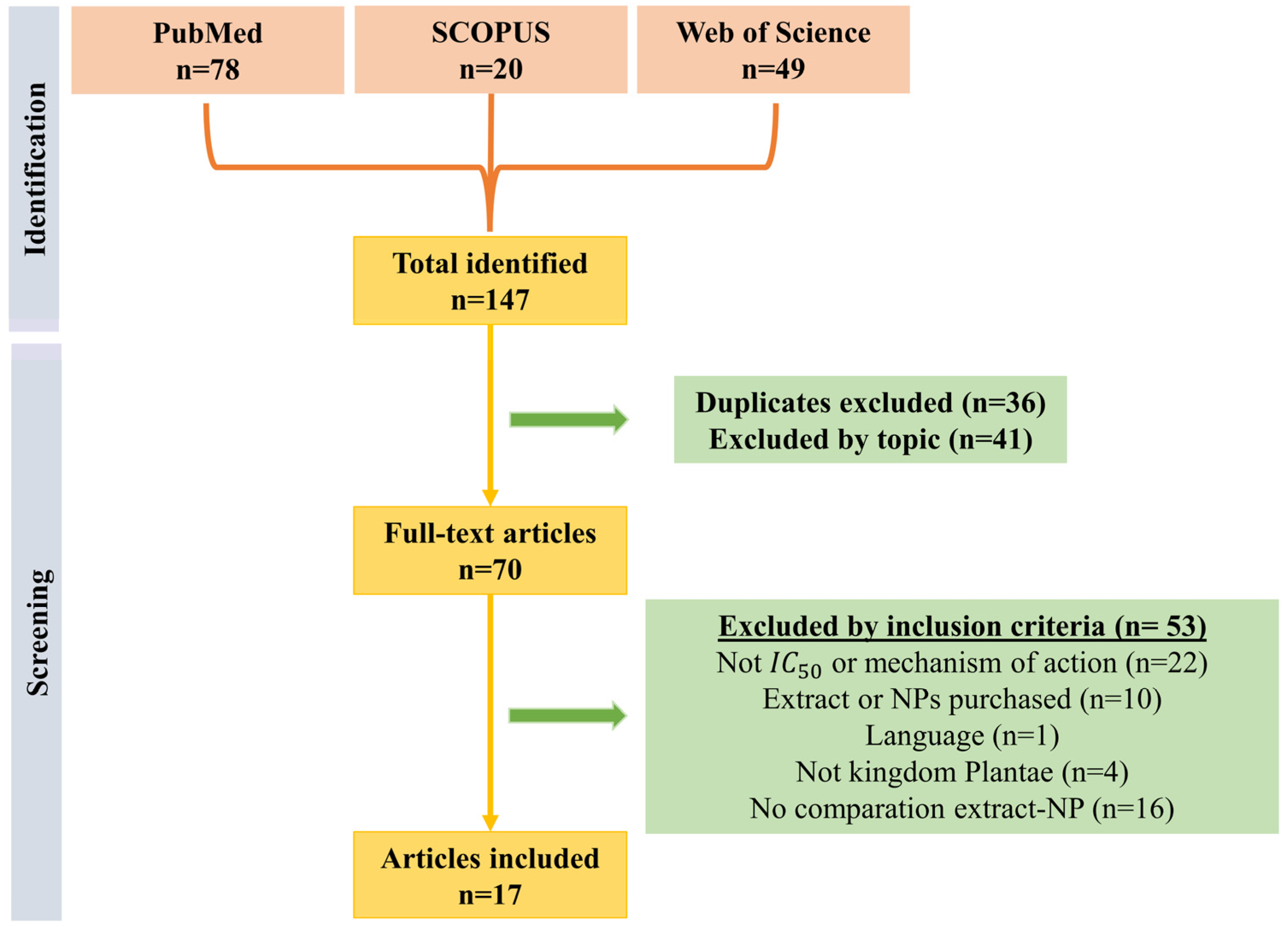
2.1. Study Eligibility

The purpose of the present systematic review was to review information from studies carried out to date on NPs loaded with plant biomolecules against colon cancer, with the aim of analyzing any possible increase in anti-tumor effectiveness compared to the use of isolated compounds. This review was carried out according to the criteria set out in the PRISMA guidelines [27].

2.2. Data Sources

For the search, three different databases (PubMed, SCOPUS and Web of Science) were consulted. The formula used in the PubMed database was based on the use of MeSH terms, as follows: (“plant extracts”[MeSH Terms] OR (“plant”[All Fields] AND “extracts”[All Fields]) OR “plant extracts”[All Fields] OR (“plant”[All Fields] AND “extract”[All Fields]) OR “plant extract”[All Fields]) AND (“nanoparticles”[All Fields] OR “nanoparticles”[MeSH Terms] OR “nanoparticles”[All Fields] OR “nanoparticle”[All Fields]) AND (“colonic neoplasms”[MeSH Terms] OR (“colonic”[All Fields] AND “neoplasms”[All Fields]) OR “colonic neoplasms”[All Fields] OR (“colon”[All Fields] AND “cancer”[All Fields]) OR “colon cancer”[All Fields]). This formula was adapted for the search engines included in the other databases, with the last bibliographic search carried out in these databases on 19 May 2023.

2.3. Inclusion Criteria

We included articles in which functional extracts of plant origin whose bioactive compounds were encapsulated in nanoparticles were studied, with the aim of evaluating their action against colon tumor cells. In addition, we included publications where these extracts were generated from wild plant species and where the extraction method and the solvent used were specified. Furthermore, the articles included had to specify the IC50 values obtained to determine the cytotoxic effect of the nanoformulations used, as well as the molecular mechanisms by which cell death is triggered.

2.4. Exclusion Criteria

We excluded all studies that were not performed with in vitro or in vivo models derived from colon cancer tumor lines, as well as those in which biomolecules or extracts were commercially acquired and not obtained from nature. Furthermore, all publications where the extraction method was not explained or where the mechanism of antitumor action was described without providing data on IC50 values were excluded. In addition, articles published in languages other than Spanish and English were excluded to facilitate the authors’ understanding. Finally, non-original articles, such as reviews, meta-analyses or epidemiological studies, were also excluded. It should be noted that there were no exclusion criteria by year of publication.

2.5. Study Selection

The authors (Cristina Mesas and Francisco Quiñonero) performed the literature search based on the previously established criteria, analyzing the title and abstract of all the articles located and selecting those that met the inclusion criteria. Subsequently, the full text of all the articles was reviewed, considering their inclusion based on the previously described inclusion and exclusion criteria.

2.6. Data Extraction

Following the selection process, the two authors indicated above analyzed the articles for data extraction. The Cohen’s Kappa statistical test exceeded 0.8, indicating a good agreement between the two authors [28]. All discrepancies were resolved by consensus between C.M and F.Q following discussion, and when necessary, a third author was involved. A specific questionnaire was used to establish the quality of the selected articles. Papers scoring less than 6 points were excluded from the systematic review.

3. Results

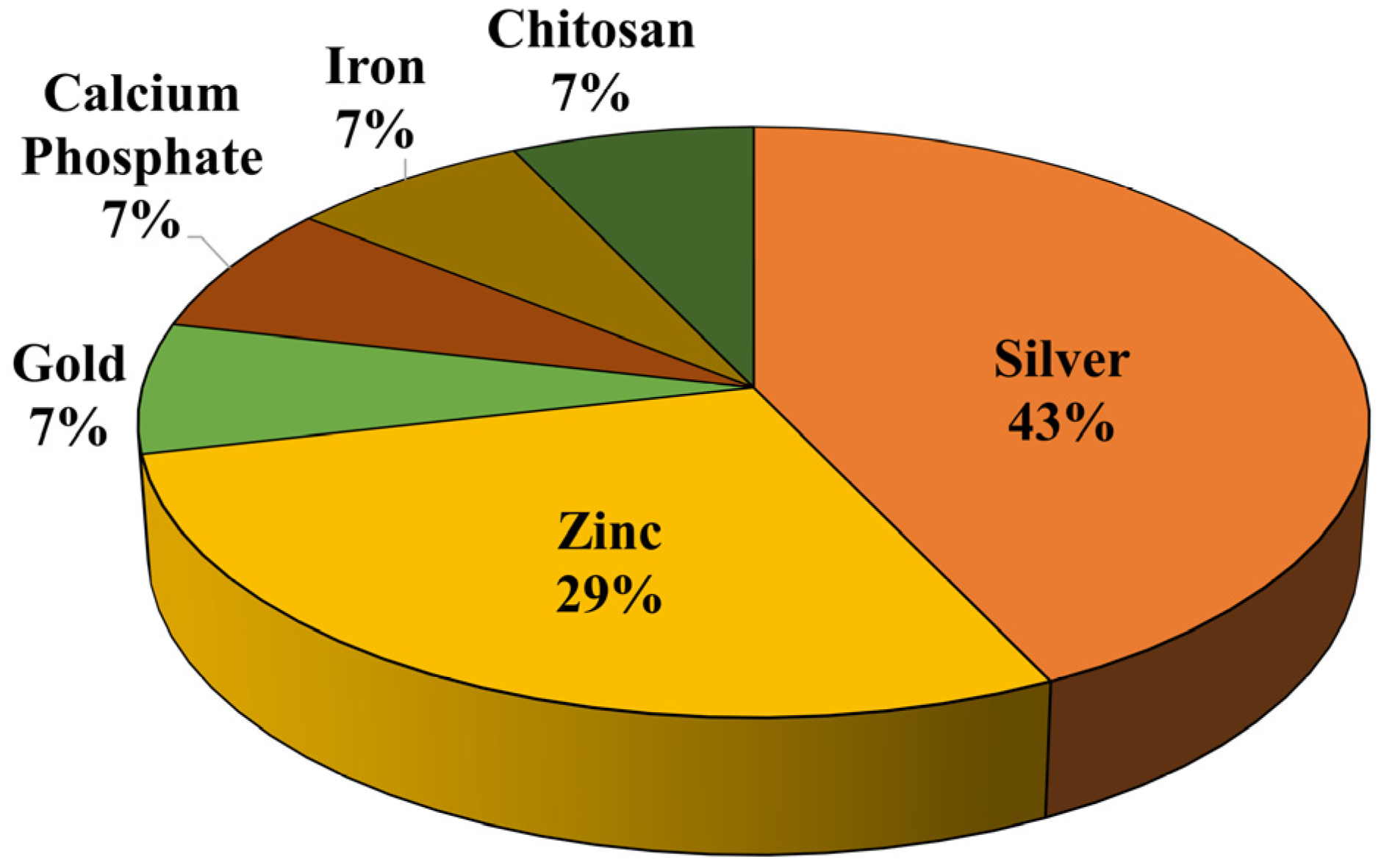
3.1. Study Description

As shown in Figure 2, after the initial search, a total of 147 articles were obtained: 78 from PubMed, 20 from SCOPUS and 49 from Web of Science. After the first analysis, 36 duplicates were discarded, leaving 111. Subsequently, a reading of the title and abstract of these articles was carried out, in which 41 more articles were eliminated as they were not relevant to the subject matter, leaving 70 articles to which the inclusion and exclusion criteria were applied. A review of these 70 articles was then carried out, discarding those articles that did not include IC50 or the mechanism of anticancer action, resulting in a total of 48 articles. Those in which the nanoparticle or extract under study was purchased, or its origin was unknown, those that were not written in English or Spanish, and those in which the extract under study did not belong to the Plantae kingdom were eliminated, leaving 33 articles, i.e., those that did not meet the inclusion criteria. Finally, we excluded articles that did not evaluate the binding activity between the extract and the nanoparticle, as compared to the binding activity between the isolated extract or nanoparticle, to assess whether the results of the combination were superior. This left 17 articles on which this review was based.
Given the diversity in the composition of nanoparticles, the results obtained in this systematic review are presented based on the different types of nanoparticles used. Of the articles included, eight used silver nanoparticles, with this material being the most employed, followed by zinc nanoparticles (four articles), gold nanoparticles (two articles), and calcium phosphate, and iron and lipid nanoparticles coated with chitosan, respectively (Figure 3). Regarding plant-derived compounds with antitumor activity against CRC, it should be noted that the plant part most frequently used to obtain these compounds was leaves, followed by seeds, skin, fruits, petals, aerated parts, bark and even the whole plant. On the other hand, the solvent most often used to obtain the extract was distilled water, followed by ethanol and methanol.

3.2. Silver Nanoparticles

Of the eight articles that used silver nanoparticles loaded with antitumor biomolecules, seven were in vitro cell culture studies [29,30,31,32,33,34,35] and one was an in vivo study with Wistar rats [23]. In each of these studies, the action of the extract alone was studied in comparison with the compound loaded in NPs. As can be seen from the results in Table 1, the IC50 values obtained were lower in the nanoparticle treatments containing encapsulated extracts compared to the use of free extracts. Additionally, the studies by Aboulthana et al. (2021) and González-Pedroza et al. (2021) showed that the cytotoxicity effect exerted by these silver nanoformulations only occurred in tumor lines, while healthy cells were resistant to the treatment as these silver nanoformulations did not induce any type of death process such as apoptosis [23,31]. Thus, in the different tests that were carried out, NPs were loaded with a wide range of compounds extracted from different plant species, such as Moringa oleifera, Rosa indica and Vitex negundo.
Aboulthana et al. (2021) examined the antitumor activity of Moringa oleifera by obtaining an aqueous extract with 10% methanol from its leaves. This test was carried out in vivo on Wistar rats after promoting the development of dysplasia in their intestinal epithelium using azoxymethane (AOM). The encapsulation of the extract in silver nanoformulations obtained IC50 values similar to the free extract, although those rats treated with these silver nanoformulations showed a decrease in the levels of carcinoembryonic antigen (CEA), Ca19.9 and inflammation markers. This effect is believed to be due to the presence of antioxidant polyphenols in the nano-extract, which could confer protection from oxidative damage by ROS. It also led to an increase in the levels of ROS detoxification enzymes, which were similar in the mice treated with the nano-extract compared to those of the control group without tumor. In addition, APC and TP53 levels decreased to normal values [23]. González-Pedroza et al. (2021) created extracts from the leaves and skin of the fruits of Annona muricata. These extracts were tested on several CRC lines, such as MCF-7, MDA-MB-468 and HCT-116, showing a decrease in IC50 values from 2004 to 404.8 µg/mL in the extract from leaves, and a reduction from 1285 to 309.3 µg/mL in the fruit skin extracts when encapsulated in iron nanoparticles. The enhanced cytotoxic effect could be due to the release of ions from nanoparticles, which are capable of activating signaling cascades, releasing calcium ions, and producing ROS damage at the mitochondrial level, thus increasing the cytotoxic effect generated by the extract itself [31]. On the other hand, Balkrishna et al. (2020) created extracts of Putranjiva roxburghii (PJ) seeds that were subsequently encapsulated in iron NPs (PJ-SNPs). The results showed that the IC50 of these nanoparticles (0.54 µg/mL) was lower when compared to the free extract in this cell line (6 µg/mL), which showed a better effect through an increase in apoptosis induction [29]. Deepika et al. (2020) evaluated the antitumor activity of Caesalpinia pulcherrima and five types of extracts were obtained, one aqueous extract and four extracts from solvents (methanol, ethyl acetate, petroleum ether and chloroform). When comparing these extracts, it was observed that the aqueous extract presented the best antitumor activity, with an IC50 of 18.7 µg/mL, compared to the extracts obtained from the solvents, whose IC50 was 51, 72, 69.3 and 77 µg/mL, respectively. Moreover, the encapsulation of the aqueous extract in a nanoformulation led to a decrease in IC50 up to 3.8 µg/mL, with encapsulation improving the effect of all the extracts tested on the HCT-116 cell line [30].
Manikandan et al. (2015) obtained an extract of Rosa indica using 70% ethanol. By using the isolated extract, they showed a decrease in cell viability up to a concentration of 250 mg/mL without being able to reach the IC50 value. Meanwhile, the binding of the extract to silver nanoparticles led to an increase in its cytotoxic effect, obtaining an IC50 of 300 µg/mL. This improvement was due to the induction of apoptosis by this nanoformulation through increased oxidative stress after the treatment of cells, as demonstrated by an overexpression of caspases 3 and 9 and an overexpression of the pro-apoptotic protein BAX. Additionally, these nanoparticles possessed anti-inflammatory effect through attenuation of nitric oxide and superoxide anion production in macrophages [33]. Lastly, regarding the study of silver NPs, Prabhu et al. (2013) studied the possible cytotoxic effect with the use of methanol extract of Vitex negundo leaves. In the CRC HCT-15 line, an IC50 of 150 µg/mL was obtained for the extract alone, whereas when the extract was encapsulated in a silver nanoformulation, its value decreased to 20 µg/mL. In this case, extract encapsulation allowed a greater induction of cell apoptosis and genotoxicity, which was demonstrated through increased tails in a comet assay [34].
In contrast to the data obtained in the previously mentioned studies, in the assay performed by Yassin et al. (2017), the binding between NPs and the natural extract could not be demonstrated to be superior to the isolated extract. In both cases, the IC50 obtained was similar, at around 0.625 µg/mL. But this study highlights the fact that several apoptosis pathways were activated, thereby elevating free radicals (ROS) and leading to cell death. These data reveal a basic benefit obtained from the use of Balanites aegyptiaca extract, opening the possibility of greater antitumor activity in the case of finding a NP that reinforces its activity [35].

3.3. Zinc Nanoparticles

Four in vitro assays were studied on colon tumor cells. In two of them, the action of the extract alone was examined in comparison with NPs loaded with the extract [36,38], while in the other two, unloaded NPs were compared with NPs encapsulating the extract [37,39]. Overall, the results shown in Table 1 indicate that encapsulation of these extracts in nanoformulations is able to improve the therapeutic efficacy of the extracts alone. Among these studies, Aboulthana et al. (2022) obtained three extracts from Croton tiglium seeds: one ethanolic extract, one aqueous extract and one petroleum ether extract. Once obtained, a comparative study was carried out between these three extracts in isolation to compare them after being bound to zinc NPs. Based on the IC50 values obtained for the isolated extracts, the best results were obtained for the aqueous one, with a concentration of 176.90 µg/mL, followed by ethanol (309.70 µg/mL) and petroleum ether (692.10 µg/mL). Next, analyzing the values for the binding of the extracts and nanoparticles, the data yielded results in a similar order, but with a higher efficiency, being 100.20 µg/mL for ZnO–aqueous extract, 252.50 µg/mL for ZnO–ethanol extract and, finally, 353.30 µg/mL for ZnO–petroleum ether extract. Thus, we can observe the superiority in terms of antitumor activity that the nano-extracts offer, compared to the isolated extracts, with the aqueous extract being the best. This increased cytotoxic effect is due to the rupture of the nanoformulations, generating Zn+2 ions which are released, thereby inducing ROS production and cell apoptosis [36]. Selim et al. (2020) generated an aqueous extract of Deverra tortuosa leaves and compared the isolated extract with the extract encapsulated in zinc NPs and a chemotherapy traditionally used in CRC, Doxorubicin. The highest antitumor activity was obtained with the extract-bound NPs, with an IC50 value of 50.81 µg/mL. Meanwhile, the isolated aqueous extract yielded a value of 136.12 µg/mL, with the IC50 of Doxorubicin being higher than both (145.26 µg/mL) [38].
Ahlam et al. (2021) created an aqueous extract of Spondias pinnata leaves and this was encapsulated in Zinc nanoparticles. After their test on the CRC cell line HCT-116, the results showed that after 72 h, the IC50 obtained was 53 µg/mL for the nanoformulations, while the extract alone did not exert any toxic effect. This encapsulation of the extract by NPs led to a decrease in the clonogenic and migration capacity of the CRC cell line. In addition, the treatment induced intracellular calcium release, thereby inducing damage through oxidative stress, genotoxicity and induction of apoptosis, as demonstrated by the RT-qPCR results through an overexpression of genes such as PARP and caspases 3, 8 and 9 [37]. Finally, Aziz et al. (2019) obtained an aqueous extract from soursop fruit. Subsequently, they performed nanoencapsulation of this extract in Zinc nanoparticles and compared its activity against uncharged nanoparticles. Despite the interesting results of the previously mentioned formulations, in this case, these nanoparticles did not show a great cytotoxic effect, having a very similar IC50 without the extract and with its encapsulation (60 µg/mL) [39].

3.4. Gold Nanoparticles

Of the included articles, two were carried out using gold NPs. The first of the assays studied was carried out by Lee et al. (2022), where nanoparticles were synthesized from a crude extract of Garcinia mangostana. These nanoformulations possessed a gold–silver double core in which they encapsulated a natural bioactive compound called protocatechuic acid (PCA). An interesting tumoricidal effect was shown in the CRC cell line HCT-116 with the use of these nanoparticles encapsulating PCA (10.78 µg/mL) versus the use of free compounds (148.09 µg/mL) and nanoparticles without any encapsulated compound (15.63 µg/mL). In addition, the cytotoxic effect demonstrated selectivity on tumor cells since the non-tumorigenic cell line CCD112 did not show as much toxicity with the pharmacological doses used [41]. The second study, carried out by Kuppusamy et al. (2016), used aqueous extracts of Commelina nudiflora L. for the synthesis of gold and silver nanoparticles. The silver nanoformulations presented an IC50 of 100 µg/mL in the HCT-116 cell line, being lower than the value of 200 µg/mL of the gold nanoformulations. In addition, it was shown that treatment with these nanoformulations induced the expression of proapoptotic genes, such as caspase 3, caspase 8 and caspase 9, which were overexpressed when tumor cells were treated with a traditional chemotherapeutic such as cisplatin. Therefore, these nanoformulations exerted their cytotoxic effect through the induction of apoptosis [40].

3.5. Other Types of Nanoparticles

Finally, three other types of nanoparticles were studied that also based their cytotoxic effect on the encapsulation of plant extracts. Initially, Mesas et al. (2022) synthesized calcium phosphate nanoparticles loaded with a functional extract from Euphorbia lathyris seeds. It was shown that this nanoformulation was able to encapsulate in its interior two bioactive compounds derived from this seed, esculetin and euphorbetin, thus eliminating elements without bioactive capacities contained in the seeds during synthesis. It was shown that the encapsulation of euphorbetin and esculetin in nanoparticles was able to increase the cytotoxic effect of these bioactive agents compared to their isolated use. In addition, these nanoformulations showed in vivo hemocompatibility and absence of toxicity in non-tumor cell lines. On the other hand, the authors performed an in vivo study using a model of colorectal cancer induced from AOM/DSS, demonstrating a reduction in the number and size of malignant polyps after treatment with these nanoparticles [42]. A second study was based on the production of functional extracts from Garcinia mangostana fruit peel and the synthesis of iron nanoformulations from these extracts. While the free extract had an IC50 of 133 µg/mL in the HCT-116 cell line, its encapsulation in nanoparticles led to a decrease of this value to 99.8 µg/mL. In addition, unlike the other nanoformulations mentioned above, by possessing a magnetic iron core, hyperthermia experiments could be performed. This application was used to increase the cytotoxicity of these nanoformulations in the tumor cell line [43].
The last nanoformulation studied was synthesized by Kamel et al. (2017), consisting of two solid lipid nanoparticles (SLNs) coated with chitosan and encapsulating 70% ethanolic extracts of Cinnamomum cassia bark and Origanum vulgare leaves. In this case, after studying the cytotoxic capacity of the free and encapsulated extracts in the nanoformulations in the CRC cell line HCT-116, IC50 values of 19.90 µg/mL were obtained for the cinnamon extract and of 26.48 µg/mL for the oregano extract. Meanwhile, the values were 29.80 µg/mL for SLNs encapsulating the cinnamon extract and 37.22 µg/mL for NPs encapsulating the oregano extract. The cytotoxic effect exerted by nanoencapsulation of these extracts was generated by inducing cell apoptosis. Finally, this study showed the synergistic potential of these extracts with the drug 5-FU, which could be a possible therapeutic option to reduce the concentration of the traditional chemotherapeutic agent used, thereby reducing its side effects [44].

4. Discussion

Natural products remain one of the main bases for the discovery of new anticancer compounds. Certain plant species are gaining great interest for treatment of colorectal cancer therapy, as current therapies for advanced-stage tumors do not achieve the expected results. In this context, the discovery of new bioactive compounds derived from nature and their encapsulation in nanoformulations would increase the antitumor capacity of the former. It is worth noting that interest in nanoencapsulation of plant compounds for the treatment of CRC has increased significantly over the last 3 years, with 11 of the 17 articles included in the review published between 2020 and 2022. We must emphasize that, except in some specific cases [32,36,44], nanoencapsulation of plant-derived extracts demonstrates antitumor efficacy against colorectal cancer tumor cells. On the other hand, it should be noted that most nanoparticles analyzed in the articles included are of metallic origin, mainly silver. This is because part of their generation is based on the reduction of metal ions, which can be carried out through biological materials such as plant-derived extracts [45]. In addition, these formulations base their cytotoxic effect on different mechanisms, including the release of the drug, possible application of hyperthermia, pharmacological release through pH variations, or possible radiation-mediated release [46,47]. In one of the studies included in this review, Yusefi et al. (2021) developed iron oxide nanoparticles that possessed magnetic capabilities, which allowed the application of in vitro hyperthermia, and obtained interesting results that could be generalized to other types of formulations through the encapsulation of small magnetite nanoparticles, a ferric mineral with great superparamagnetic capabilities [43].
Although the studied extracts possess antitumor activity, the vehiculation of these extracts using NPs generally increases their therapeutic effect. This not only occurs with these plant compounds, where effect was shown with extracts obtained from different parts of plants (leaves, fruits, and bark), but also with traditional chemotherapeutic drugs. The vehiculation of paclitaxel in NPs of lactic and glycolic acid increased the therapeutic efficacy of this drug in lung cancer by increasing the concentration of the drug in the tumor tissue and decreasing it in the ganglia of the spinal cord, thus reducing the neuropathy associated with the drug [48]. The vast majority of nanoformulations generated cell death through the induction of apoptosis, showing an increased cytotoxic effect compared to the use of free extracts due to increased intracellular oxidative stress mediated by nanoparticle rupture, thus releasing ions inside the cell that generated dysregulation at the oxidative level [23,31,33,35,36,37,49]. Despite this interesting cytotoxic effect on CRC cells, it should be noted that in those studies in which the toxicity of NPs in non-tumor cells was analyzed, it was observed that they did not have any relevant toxicity; thus, they seem to be safe platforms for the administration of bioactive compounds derived from natural extracts [41,42]. Although this in vitro toxicity has not been observed, it is important that all these results can be further tested in in vivo models to verify their biocompatibility since it has previously been shown that the administration of methanolic or ethanolic extracts derived from plants can be toxic in murine models at high concentrations (2 g/kg), which may limit their use in the clinic [50,51]. In addition to their antitumor activity, they are also used for their reducing capacity when synthesizing NPs. In this way, it is possible to create them in a simpler, more economical, and environmentally friendly way by reducing or eliminating the number of chemical compounds used in other ways of producing NPs [52].
The use of these extracts and their encapsulation in NPs should not be studied as a possible isolated treatment route; their possible combination with chemotherapeutics such as 5-FU has also been observed to obtain a greater cytotoxic effect by reducing the amount of the drug used and, therefore, the possible side effects generated [44]. This possible synergy between plant extracts and chemotherapy drugs used in CRC is justified by the presence of different types of bioactive compounds in these extracts, such as curcumin, resveratrol and epigallocatechin gallate (EGCG), which have shown, in previous studies, their synergistic capacity with cytotoxic drugs such as 5-FU, doxorubicin, oxaliplatin, carboplatin or irinotecan [53,54]. One of the major problems of the NPs studied is the absence of molecules that reduce the immunogenicity they generate in the blood. Thus, other authors such as Lei et al. (2021) synthesized and characterized NPs based on hyaluronic acid (HA) and albumin capable of demonstrating high solubility, stability, biocompatibility, and absence of immunogenicity. In addition, functionalization with HA would grant them some selectivity against CD44 receptor, a characteristic lacking in the nanoformulations presented in this review [55]. This type of functionalization against CD44 is quite promising in the synthesis of new NPs, given that it is a receptor associated with a worse prognosis in CRC and that it is overexpressed in tumor stem cells in this type of tumor [56]. Other types of functionalization have been studied, which attempted to use antibodies, nucleic acids, vitamins or peptides to increase the interaction between nanoformulations and cells, allowing greater entry of the encapsulated pharmaceuticals and improving their therapeutic effect [57,58]. In addition, the nanoformulations included in this review were also tested in other types of tumors; González-Pedroza et al. (2021) demonstrated the performance of their nanoformulations on breast cancer and melanoma cells, while Balkrishna et al. (2020) additionally studied their nanoparticles on pancreatic and breast adenocarcinoma cells [29,31].
Of the 17 articles finally selected in this review, in vivo experiments were performed in only two of them [23,42]. Among them, we highlight the study carried out by Mesas et al. (2022), where the in vitro benefit of the encapsulation of the extract in nanoparticles was demonstrated and, additionally, a lower number and a smaller size of colon cancer precursor lesions were observed in a model generated in situ using AOM/DSS [42]. Thus, the loading of extracts in NPs produced an increase in antitumor efficacy in those assays in which in vivo models were used, since it favors the entry and deposition of NPs in the tumor tissue due to the increased retention and permeabilization effect (EPR), which, in addition to a defective lymphatic drainage, reduces the amount of concentration necessary to achieve the same tumor damage [59,60]. Despite the interesting results obtained both in vitro and in vivo in the included studies, it should be noted that any nanoparticles based on plant extracts or natural compounds are currently in clinical trials. Thus, the nanoformulations currently in different phases are based on conventional chemotherapeutics, such as capecitabine, irinotecan, doxorubicin and 5-FU, or drug combinations such as FOLFOX [61]. The big problem with these formulations is the large number of side effects they generate in patients due to their lack of specificity; thus research is already underway to examine the ability of different plant compounds (such as flavonoids) to have a palliative effect on symptoms in combination with conventional chemotherapy [62].
Therefore, the nanoformulations synthesized and characterized in the studies included in this review would be potentially useful for the treatment of CRC, avoiding cellular resistance mechanisms such as efflux pumps [63]. In addition to their proven antitumor activity, their economic and environmental benefits due to the use of natural extracts stand out, as well as their safety profile against non-tumor cells. Nevertheless, more studies are needed to demonstrate the antitumor activity of different plant-derived extracts, as well as their encapsulation in functionalized NPs, that improve the results obtained in in vivo experiments and, subsequently, in future clinical trials.

Author Contributions

Conceptualization, J.P. and C.M. (Consolación Melguizo); methodology, C.M. (Cristina Mesas), F.Q. and F.R.; software, L.C. and G.P.; validation, L.C. and G.P.; investigation, C.M. (Cristina Mesas), F.Q. and F.R.; resources, L.C.; writing—original draft preparation, C.M. (Cristina Mesas), F.Q. and F.R.; writing—review and editing, J.P. and C.M. (Consolación Melguizo); visualization, L.C. and G.P.; supervision, J.P.; funding acquisition, J.P. and C.M. (Consolación Melguizo). All authors have read and agreed to the published version of the manuscript.

Funding

This work was supported by the Instituto de Salud Carlos III (PI19/01478-FEDER and PMPTA22/00136), partially by the Junta de Andalucía (A-CTS-666-UGR20, P20_00540, B-CTS-122-UGR20), the Ministerio de Ciencia e Innovación (RTC2019-006870-1) FEDER program and from group CTS-107 (Andalusian Government).

Institutional Review Board Statement

Not applicable.

Informed Consent Statement

Not applicable.

Data Availability Statement

The search protocol is registered on in the OSF database, and can be found here: https://doi.org/10.17605/OSF.IO/E9FTY.

Acknowledgments

The authors thank the personnel at the Center of Scientific Instrumentation of the University of Granada for technical assistance.

Conflicts of Interest

The authors declare no conflict of interest.

References

  1. Sung, H.; Ferlay, J.; Siegel, R.L.; Laversanne, M.; Soerjomataram, I.; Jemal, A.; Bray, F. Global Cancer Statistics 2020: GLOBOCAN Estimates of Incidence and Mortality Worldwide for 36 Cancers in 185 Countries. CA Cancer J. Clin. 2021, 71, 209–249. [Google Scholar] [CrossRef]
  2. Dekker, E.; Tanis, P.J.; Vleugels, J.L.A.; Kasi, P.M.; Wallace, M.B. Colorectal Cancer. Lancet 2019, 394, 1467–1480. [Google Scholar] [CrossRef] [PubMed]
  3. Binefa, G.; Rodríguez-Moranta, F.; Teule, À.; Medina-Hayas, M. Colorectal Cancer: From Prevention to Personalized Medicine. World J. Gastroenterol. 2014, 20, 6786–6808. [Google Scholar] [CrossRef]
  4. Muzny, D.M.; Bainbridge, M.N.; Chang, K.; Dinh, H.H.; Drummond, J.A.; Fowler, G.; Kovar, C.L.; Lewis, L.R.; Morgan, M.B.; Newsham, I.F.; et al. Comprehensive Molecular Characterization of Human Colon and Rectal Cancer. Nature 2012, 487, 330–337. [Google Scholar] [CrossRef]
  5. Siegel, R.L.; Wagle, N.S.; Cercek, A.; Smith, R.A.; Jemal, A. Colorectal Cancer Statistics, 2023. CA Cancer J. Clin. 2023, 73, 233–254. [Google Scholar] [CrossRef]
  6. Navarro, M.; Nicolas, A.; Ferrandez, A.; Lanas, A. Colorectal Cancer Population Screening Programs Worldwide in 2016: An Update. World J. Gastroenterol. 2017, 23, 3632. [Google Scholar] [CrossRef]
  7. Ferlitsch, M.; Moss, A.; Hassan, C.; Bhandari, P.; Dumonceau, J.M.; Paspatis, G.; Jover, R.; Langner, C.; Bronzwaer, M.; Nalankilli, K.; et al. Colorectal Polypectomy and Endoscopic Mucosal Resection (EMR): European Society of Gastrointestinal Endoscopy (ESGE) Clinical Guideline. Endoscopy 2017, 49, 270–297. [Google Scholar] [CrossRef]
  8. Emmanuel, A.; Haji, A. Complete Mesocolic Excision and Extended (D3) Lymphadenectomy for Colonic Cancer: Is It Worth That Extra Effort? A Review of the Literature. Int. J. Color. Dis. 2016, 31, 797–804. [Google Scholar] [CrossRef]
  9. Ma, B.; Gao, P.; Song, Y.; Zhang, C.; Zhang, C.; Wang, L.; Liu, H.; Wang, Z. Transanal Total Mesorectal Excision (TaTME) for Rectal Cancer: A Systematic Review and Meta-Analysis of Oncological and Perioperative Outcomes Compared with Laparoscopic Total Mesorectal Excision. BMC Cancer 2016, 16, 380. [Google Scholar] [CrossRef]
  10. Kuipers, E.J.; Grady, W.M.; Lieberman, D.; Seufferlein, T.; Sung, J.J.; Boelens, P.G.; van de Velde, C.J.H.; Watanabe, T. Colorectal Cancer. Nat. Rev. Dis. Prim. 2015, 1, 15065. [Google Scholar] [CrossRef]
  11. Biller, L.H.; Schrag, D. Diagnosis and Treatment of Metastatic Colorectal Cancer: A Review. JAMA 2021, 325, 669–685. [Google Scholar] [CrossRef] [PubMed]
  12. Fan, A.; Wang, B.; Wang, X.; Nie, Y.; Fan, D.; Zhao, X.; Lu, Y. Immunotherapy in Colorectal Cancer: Current Achievements and Future Perspective. Int. J. Biol. Sci. 2021, 17, 3837–3849. [Google Scholar] [CrossRef] [PubMed]
  13. André, T.; Shiu, K.-K.; Kim, T.W.; Jensen, B.V.; Jensen, L.H.; Punt, C.; Smith, D.; Garcia-Carbonero, R.; Benavides, M.; Gibbs, P.; et al. Pembrolizumab in Microsatellite-Instability–High Advanced Colorectal Cancer. N. Engl. J. Med. 2020, 383, 2207–2218. [Google Scholar] [CrossRef]
  14. Gustin, D.M.; Brenner, D.E. Chemoprevention of Colon Cancer: Current Status and Future Prospects. Cancer Metastasis Rev. 2002, 21, 323–348. [Google Scholar] [CrossRef]
  15. Deng, L.-J.; Qi, M.; Li, N.; Lei, Y.-H.; Zhang, D.-M.; Chen, J.-X. Natural Products and Their Derivatives: Promising Modulators of Tumor Immunotherapy. J. Leukoc. Biol. 2020, 108, 493–508. [Google Scholar] [CrossRef]
  16. Yahfoufi, N.; Alsadi, N.; Jambi, M.; Matar, C. The Immunomodulatory and Anti-Inflammatory Role of Polyphenols. Nutrients 2018, 10, 1618. [Google Scholar] [CrossRef] [PubMed]
  17. Bhosale, P.B.; Ha, S.E.; Vetrivel, P.; Kim, H.H.; Kim, S.M.; Kim, G.S. Functions of Polyphenols and Its Anticancer Properties in Biomedical Research: A Narrative Review. Transl. Cancer Res. 2020, 9, 7619. [Google Scholar] [CrossRef]
  18. Menger, L.; Vacchelli, E.; Kepp, O.; Eggermont, A.; Tartour, E.; Zitvogel, L.; Kroemer, G.; Galluzzi, L. Trial Watch: Cardiac Glycosides and Cancer Therapy. Oncoimmunology 2013, 2, e23082. [Google Scholar] [CrossRef]
  19. Kamran, S.; Sinniah, A.; Abdulghani, M.A.M.; Alshawsh, M.A. Therapeutic Potential of Certain Terpenoids as Anticancer Agents: A Scoping Review. Cancers 2022, 14, 1100. [Google Scholar] [CrossRef]
  20. Bazana, M.T.; Codevilla, C.F.; de Menezes, C.R. Nanoencapsulation of Bioactive Compounds: Challenges and Perspectives. Curr. Opin. Food Sci. 2019, 26, 47–56. [Google Scholar] [CrossRef]
  21. Mundekkad, D.; Cho, W.C. Nanoparticles in Clinical Translation for Cancer Therapy. Int. J. Mol. Sci. 2022, 23, 1685. [Google Scholar] [CrossRef]
  22. Haleem, A.; Javaid, M.; Singh, R.P.; Rab, S.; Suman, R. Applications of Nanotechnology in Medical Field: A Brief Review. Glob. Health J. 2023, 7, 70–77. [Google Scholar] [CrossRef]
  23. Aboulthana, W.M.; Shousha, W.G.; Essawy, E.A.-R.; Saleh, M.H.; Salama, A.H. Assessment of the Anti-Cancer Efficiency of Silver Moringa Oleifera Leaves Nano-Extract against Colon Cancer Induced Chemically in Rats. Asian Pac. J. Cancer Prev. 2021, 22, 3267–3286. [Google Scholar] [CrossRef] [PubMed]
  24. Mittal, A.K.; Chisti, Y.; Banerjee, U.C. Synthesis of Metallic Nanoparticles Using Plant Extracts. Biotechnol. Adv. 2013, 31, 346–356. [Google Scholar] [CrossRef]
  25. Jadoun, S.; Arif, R.; Jangid, N.K.; Meena, R.K. Green Synthesis of Nanoparticles Using Plant Extracts: A Review. Environ. Chem. Lett. 2021, 19, 355–374. [Google Scholar] [CrossRef]
  26. Khan, S.A.; Shahid, S.; Ayaz, A.; Alkahtani, J.; Elshikh, M.S.; Riaz, T. Phytomolecules-Coated NiO Nanoparticles Synthesis Using Abutilon Indicum Leaf Extract: Antioxidant, Antibacterial, and Anticancer Activities. Int. J. Nanomed. 2021, 16, 1757–1773. [Google Scholar] [CrossRef] [PubMed]
  27. Page, M.J.; McKenzie, J.E.; Bossuyt, P.M.; Boutron, I.; Hoffmann, T.C.; Mulrow, C.D.; Shamseer, L.; Tetzlaff, J.M.; Akl, E.A.; Brennan, S.E.; et al. The PRISMA 2020 Statement: An Updated Guideline for Reporting Systematic Reviews. BMJ 2021, 372, n71. [Google Scholar] [CrossRef]
  28. Wanden-Berghe, C.; Sanz-Valero, J. Systematic Reviews in Nutrition: Standardized Methodology. Br. J. Nutr. 2012, 107, S3–S7. [Google Scholar] [CrossRef] [PubMed]
  29. Balkrishna, A.; Sharma, V.K.; Das, S.K.; Mishra, N.; Bisht, L.; Joshi, A.; Sharma, N. Characterization and Anti-Cancerous Effect of Putranjiva Roxburghii Seed Extract Mediated Silver Nanoparticles on Human Colon (HCT-116), Pancreatic (PANC-1) and Breast (MDA-MB 231) Cancer Cell Lines: A Comparative Study. Int. J. Nanomed. 2020, 15, 573–585. [Google Scholar] [CrossRef]
  30. Deepika, S.; Selvaraj, C.I.; Roopan, S.M. Screening Bioactivities of Caesalpinia pulcherrima L. Swartz and Cytotoxicity of Extract Synthesized Silver Nanoparticles on HCT116 cell Line. Mater. Sci. Eng. C Mater. Biol. Appl. 2020, 106, 110279. [Google Scholar] [CrossRef]
  31. González-Pedroza, M.G.; Argueta-Figueroa, L.; García-Contreras, R.; Jiménez-Martínez, Y.; Martínez-Martínez, E.; Navarro-Marchal, S.A.; Marchal, J.A.; Morales-Luckie, R.A.; Boulaiz, H. Silver Nanoparticles from Annona Muricata Peel and Leaf Extracts as a Potential Potent, Biocompatible and Low Cost Antitumor Tool. Nanomaterials 2021, 11, 1273. [Google Scholar] [CrossRef] [PubMed]
  32. Javed, B.; Mashwani, Z.-U.-R.; Sarwer, A.; Raja, N.I.; Nadhman, A. Synergistic Response of Physicochemical Reaction Parameters on Biogenesis of Silver Nanoparticles and Their Action against Colon Cancer and Leishmanial Cells. Artif. Cells Nanomed. Biotechnol. 2020, 48, 1340–1353. [Google Scholar] [CrossRef]
  33. Manikandan, R.; Manikandan, B.; Raman, T.; Arunagirinathan, K.; Prabhu, N.M.; Jothi Basu, M.; Perumal, M.; Palanisamy, S.; Munusamy, A. Biosynthesis of Silver Nanoparticles Using Ethanolic Petals Extract of Rosa Indica and Characterization of Its Antibacterial, Anticancer and Anti-Inflammatory Activities. Spectrochim. Acta A Mol. Biomol. Spectrosc. 2015, 138, 120–129. [Google Scholar] [CrossRef] [PubMed]
  34. Prabhu, D.; Arulvasu, C.; Babu, G.; Manikandan, R.; Srinivasan, P. Biologically Synthesized Green Silver Nanoparticles from Leaf Extract of Vitex negundo L. Induce Growth-Inhibitory Effect on Human Colon Cancer Cell Line HCT15. Process. Biochem. 2013, 48, 317–324. [Google Scholar] [CrossRef]
  35. Yassin, A.M.; El-Deeb, N.M.; Metwaly, A.M.; El Fawal, G.F.; Radwan, M.M.; Hafez, E.E. Induction of Apoptosis in Human Cancer Cells Through Extrinsic and Intrinsic Pathways by Balanites Aegyptiaca Furostanol Saponins and Saponin-Coated SilverNanoparticles. Appl. Biochem. Biotechnol. 2017, 182, 1675–1693. [Google Scholar] [CrossRef]
  36. Aboulthana, W.M.; Omar, N.I.; El-Feky, A.M.; Hasan, E.A.; Ibrahim, N.E.-S.; Youssef, A.M. In Vitro Study on Effect of Zinc Oxide Nanoparticles on the Biological Activities of Croton tiglium L. Seeds Extracts. Asian Pac. J. Cancer Prev. 2022, 23, 2671–2686. [Google Scholar] [CrossRef]
  37. Ahlam, A.A.; Shaniba, V.S.; Jayasree, P.R.; Manish Kumar, P.R. Spondias Pinnata (L.f.) Kurz Leaf Extract Derived Zinc Oxide Nanoparticles Induce Dual Modes of Apoptotic-Necrotic Death in HCT 116 and K562 Cells. Biol. Trace Elem. Res. 2021, 199, 1778–1801. [Google Scholar] [CrossRef]
  38. Selim, Y.A.; Azb, M.A.; Ragab, I.; Abd El-Azim, M.H.M. Green Synthesis of Zinc Oxide Nanoparticles Using Aqueous Extract of Deverra Tortuosa and Their Cytotoxic Activities. Sci. Rep. 2020, 10, 3445. [Google Scholar] [CrossRef]
  39. Aziz, A.A.; Shaniba, V.S.; Jayasree, P.R.; Manish Kumar, P.R. Physico-Chemical, Photocatalytic and Cytotoxicity Evaluation of Annona muricata L. Fruit Extract Derived Zinc Oxide Nanoparticles in Comparison to the Commercial Chemical Version. Curr. Sci. 2019, 117, 1492. [Google Scholar] [CrossRef]
  40. Kuppusamy, P.; Ichwan, S.J.A.; Al-Zikri, P.N.H.; Suriyah, W.H.; Soundharrajan, I.; Govindan, N.; Maniam, G.P.; Yusoff, M.M. In Vitro Anticancer Activity of Au, Ag Nanoparticles Synthesized Using Commelina nudiflora L. Aqueous Extract Against HCT-116 Colon Cancer Cells. Biol. Trace Elem. Res. 2016, 173, 297–305. [Google Scholar] [CrossRef]
  41. Lee, K.X.; Shameli, K.; Nagao, Y.; Yew, Y.P.; Teow, S.-Y.; Moeini, H. Potential Use of Gold-Silver Core-Shell Nanoparticles Derived from Garcinia Mangostana Peel for Anticancer Compound, Protocatechuic Acid Delivery. Front. Mol. Biosci. 2022, 9, 997471. [Google Scholar] [CrossRef] [PubMed]
  42. Mesas, C.; Garcés, V.; Martínez, R.; Ortiz, R.; Doello, K.; Dominguez-Vera, J.M.; Bermúdez, F.; Porres, J.M.; López-Jurado, M.; Melguizo, C.; et al. Colon Cancer Therapy with Calcium Phosphate Nanoparticles Loading Bioactive Compounds from Euphorbia Lathyris: In Vitro and in Vivo Assay. Biomed. Pharmacother. 2022, 155, 113723. [Google Scholar] [CrossRef] [PubMed]
  43. Yusefi, M.; Shameli, K.; Su Yee, O.; Teow, S.-Y.; Hedayatnasab, Z.; Jahangirian, H.; Webster, T.J.; Kuča, K. Green Synthesis of Fe3O4 Nanoparticles Stabilized by a Garcinia Mangostana Fruit Peel Extract for Hyperthermia and Anticancer Activities. Int. J. Nanomed. 2021, 16, 2515–2532. [Google Scholar] [CrossRef]
  44. Kamel, K.M.; Khalil, I.A.; Rateb, M.E.; Elgendy, H.; Elhawary, S. Chitosan-Coated Cinnamon/Oregano-Loaded Solid Lipid Nanoparticles to Augment 5-Fluorouracil Cytotoxicity for Colorectal Cancer: Extract Standardization, Nanoparticle Optimization, and Cytotoxicity Evaluation. J. Agric. Food Chem. 2017, 65, 7966–7981. [Google Scholar] [CrossRef]
  45. Alphandéry, E. Natural Metallic Nanoparticles for Application in Nano-Oncology. Int. J. Mol. Sci. 2020, 21, 4412. [Google Scholar] [CrossRef]
  46. Gago, L.; Quiñonero, F.; Perazzoli, G.; Melguizo, C.; Prados, J.; Ortiz, R.; Cabeza, L. Nanomedicine and Hyperthermia for the Treatment of Gastrointestinal Cancer: A Systematic Review. Pharmaceutics 2023, 15, 1958. [Google Scholar] [CrossRef]
  47. Shinn, J.; Kwon, N.; Lee, S.A.; Lee, Y. Smart PH-Responsive Nanomedicines for Disease Therapy. J. Pharm. Investig. 2022, 52, 441. [Google Scholar] [CrossRef] [PubMed]
  48. Jiménez-López, J.; El-Hammadi, M.M.; Ortiz, R.; Cayero-Otero, M.D.; Cabeza, L.; Perazzoli, G.; Martin-Banderas, L.; Baeyens, J.M.; Prados, J.; Melguizo, C. A Novel Nanoformulation of PLGA with High Non-Ionic Surfactant Content Improves in Vitro and in Vivo PTX Activity against Lung Cancer. Pharm. Pharmacol. Res. 2019, 141, 451–465. [Google Scholar] [CrossRef]
  49. Barabadi, H.; Vahidi, H.; Damavandi Kamali, K.; Rashedi, M.; Hosseini, O.; Saravanan, M. Emerging Theranostic Gold Nanomaterials to Combat Colorectal Cancer: A Systematic Review. J. Clust. Sci. 2020, 31, 651–658. [Google Scholar] [CrossRef]
  50. Mlozi, S.H.; Mmongoyo, J.A.; Chacha, M. The in Vivo Toxicity Evaluation of Leaf and Root Methanolic Extracts of Tephrosia Vogelii Hook.f Using Animal Model. Clin. Phytosci. 2020, 6, 73. [Google Scholar] [CrossRef]
  51. Kifayatullah, M.; Mustafa, M.S.; Sengupta, P.; Sarker, M.M.R.; Das, A.; Das, S.K. Evaluation of the Acute and Sub-Acute Toxicity of the Ethanolic Extract of Pericampylus glaucus (Lam.) Merr. in BALB/c Mice. J. Acute Dis. 2015, 4, 309–315. [Google Scholar] [CrossRef]
  52. Ying, S.; Guan, Z.; Ofoegbu, P.C.; Clubb, P.; Rico, C.; He, F.; Hong, J. Green Synthesis of Nanoparticles: Current Developments and Limitations. Environ. Technol. Innov. 2022, 26, 102336. [Google Scholar] [CrossRef]
  53. Quiñonero, F.; Mesas, C.; Peña, M.; Cabeza, L.; Perazzoli, G.; Melguizo, C.; Ortiz, R.; Prados, J. Vegetal-Derived Bioactive Compounds as Multidrug Resistance Modulators in Colorectal Cancer. Appl. Sci. 2023, 13, 2667. [Google Scholar] [CrossRef]
  54. Gavrilas, L.I.; Cruceriu, D.; Mocan, A.; Loghin, F.; Miere, D.; Balacescu, O. Plant-Derived Bioactive Compounds in Colorectal Cancer: Insights from Combined Regimens with Conventional Chemotherapy to Overcome Drug-Resistance. Biomedicines 2022, 10, 1948. [Google Scholar] [CrossRef] [PubMed]
  55. Bhattacharya, D.S.; Svechkarev, D.; Souchek, J.J.; Hill, T.K.; Taylor, M.A.; Natarajan, A.; Mohs, A.M. Impact of Structurally Modifying Hyaluronic Acid on CD44 Interaction. J. Mater. Chem. B 2017, 5, 8183. [Google Scholar] [CrossRef]
  56. Wang, Z.; Tang, Y.; Xie, L.; Huang, A.; Xue, C.; Gu, Z.; Wang, K.; Zong, S. The Prognostic and Clinical Value of CD44 in Colorectal Cancer: A Meta-Analysis. Front. Oncol. 2019, 9, 309. [Google Scholar] [CrossRef] [PubMed]
  57. Abd Ellah, N.H.; Abouelmagd, S.A. Surface Functionalization of Polymeric Nanoparticles for Tumor Drug Delivery: Approaches and Challenges. Expert. Opin. Drug. Deliv. 2017, 14, 201–214. [Google Scholar] [CrossRef] [PubMed]
  58. Subbiah, R.; Veerapandian, M.; Yun, K.S. Nanoparticles: Functionalization and Multifunctional Applications in Biomedical Sciences. Curr. Med. Chem. 2010, 17, 4559–4577. [Google Scholar] [CrossRef]
  59. Shi, Y.; van der Meel, R.; Chen, X.; Lammers, T. The EPR Effect and beyond: Strategies to Improve Tumor Targeting and Cancer Nanomedicine Treatment Efficacy. Theranostics 2020, 10, 7921–7924. [Google Scholar] [CrossRef]
  60. Shinde, V.R.; Revi, N.; Murugappan, S.; Singh, S.P.; Rengan, A.K. Enhanced Permeability and Retention Effect: A Key Facilitator for Solid Tumor Targeting by Nanoparticles. Photodiagnos. Photodyn. Ther. 2022, 39, 102915. [Google Scholar] [CrossRef]
  61. Cabeza, L.; Perazzoli, G.; Mesas, C.; Jiménez-Luna, C.; Prados, J.; Rama, A.R.; Melguizo, C. Nanoparticles in Colorectal Cancer Therapy: Latest In Vivo Assays, Clinical Trials, and Patents. AAPS Pharm. Sci. Tech. 2020, 21, 178. [Google Scholar] [CrossRef] [PubMed]
  62. Fernández, J.; Silván, B.; Entrialgo-Cadierno, R.; Villar, C.J.; Capasso, R.; Uranga, J.A.; Lombó, F.; Abalo, R. Antiproliferative and Palliative Activity of Flavonoids in Colorectal Cancer. Biomed. Pharmacother. 2021, 143, 112241. [Google Scholar] [CrossRef] [PubMed]
  63. Yao, Y.; Zhou, Y.; Liu, L.; Xu, Y.; Chen, Q.; Wang, Y.; Wu, S.; Deng, Y.; Zhang, J.; Shao, A. Nanoparticle-Based Drug Delivery in Cancer Therapy and Its Role in Overcoming Drug Resistance. Front. Mol. Biosci. 2020, 7, 558493. [Google Scholar] [CrossRef] [PubMed]
Figure 1. Chemical structure of plant compounds that are present in the extracts studied in this review and possess antitumor properties.
Applsci 13 10156 g001
Figure 2. Schematic diagram representative of the systematic search process carried out for this review.
Applsci 13 10156 g002
Figure 3. Representative schematic of the synthesis material of the nanoformulations included in this systematic review.
Applsci 13 10156 g003
Table 1. Most relevant information obtained from the studies included in the systematic review.
MaterialExtract (Reference)Cell Line/Animal ModelIC50/LD50Results
SilverME (10%) of Moringa oleifera leaves [23]36 Wistar rats: chemical induction by AOM-
  • Decreased CEA.Ca19.9, APC and TP53 expression in nano-extract treatment.
  • Absence of toxicity in non-tumor cells and protection against oxidative damage.
AE of leaves (LE) and peel (PE) of Annona muricata [31]HCT-116AgNPs-PE: 1.285 µg/mL
AgNPs-LE: 2.004 µg/mL
PE: 309.3 µg/mL
LE: 404.8 µg/mL
  • Induction of apoptosis by ROS.
  • AgNPs-PE are less toxic compared to AgNPs-LE against macrophages.
AE of leaves of Mentha lonqifolia [32]HCT-116-Encapsulation of phytochemicals by nanoparticles is low, so there is no cytotoxic activity.
AE of seeds of Putranjiva roxburghii [29]HCT-116PJSNPs: 0.54 µg/mL
PJ: 6.0 µg/mL
The nanoparticles encapsulating the extract induce genotoxicity, triggering cell apoptosis to a greater extent than the free extract.
AE, ME, EAE, PEE and CE of leaves of Caesalpinia pulcherrima [30]HCT-116AE: 18,7 µg/mL
ME: 51 µg/mL
EAE: 72 µg/mL
PEE: 69.3 µg/mL
CE: 77 µg/mL
  • The increased antitumor activity by NPs is attributed to an increased apoptotic induction.
  • Greater antitumor activity was observed with the aqueous extract, compared to the extracts obtained through solvents.
AE, EEAE, NBE of fruits of Balanites aegyptiaca [35]Caco-2Extracts: 0.625–1.25 µg/mL
AgNPs: 0.625 µg/mL
  • Apoptosis induction via ROS damage.
  • No increased antitumor activity was observed with NPs compared to the functional extracts.
EA (70%) of petals of Rose indica [33]HCT-15Extract: >250 mg/mL
AgNPs: 300 μg/mL
  • Increased antitumor activity with NPs compared to functional extracts.
  • Apoptosis induction via ROS damage.
ME of leaves of Vitex negundo [34]HCT-15ME: 150 μg/mL
AgNPs: 20 µg/mL
  • Increased antitumor activity with NPs compared to the functional extracts.
  • Cell death is attributed to apoptosis due to increased genotoxicity.
ZincAE, EE and PEE of seeds of Croton tiglium [36]Caco-2EE: 309.70 μg/mL
AE: 176.90 μg/mL
PEE: 692.10 μg/mL
ZnO-EE: 252.50 μg/mL
ZnO-AE: 100.20 μg/mL
ZnO-PEE: 353.30 μg/mL
  • Apoptosis induction via ROS damage and decreased EGFR expression
  • Greater antitumor activity with AE and ZnO-A compared to other extracts and NPs. The highest activity was achieved by ZnO-AE compared to the functional aqueous extract.
AE of leaves of Spondias pinnata (SpL) [37]HCT-116SpL-ZnNPs: 53 µg/mL
ZnNPs: 60 µg/mL
  • Cell death is attributed to apoptosis due to increased ROS and DNA fragmentation.
  • Increased antitumor activity with SpLZnONPs compared to non-extract NPs.
AE of aerated parts of Deverra tortuosa [38]Caco-2ZnO-NPs: 50.81 μg/mL
AE: 136.12 μg/mL
  • Increased antitumor activity with NPs compared to the functional extracts.
  • Cell death is attributed to apoptosis due to increased ROS and intracellular release of Zn ions.
AE of fruits of Annona muricata [39]HCT-116ZnNPs: 60 μg/mL
Am-ZnNPs: 60 μg/mL
The encapsulation of Annona muricata extract did not exert any additional cytotoxic effect compared to the administration of ZnNPs alone.
GoldEE of Commelina nudiflora [40]HCT-116AuNPs: 200 μg/mL
AgNPs: 100 μg/mL
  • Cell death is attributed to apoptosis due to DNA fragmentation.
  • The cytotoxic effect of AgNPs is better than AuNPs.
AE of skin of Garcinia mangostana [41]HCT-116GM extract: 35.74 µg/mL
AuNPs: 82.99 µg/mL
Au-AgNPs: 24.36 µg/mL
  • Cell death is attributed to a mechanism of apoptosis, probably caused by increased ROS, impaired mitochondrial function, and DNA damage.
  • It was observed that greater antitumor activity is associated with the combination of gold–silver core–shell NPs, compared to gold-only NPs.
Calcium phosphateAE, EE and CHA of seeds of Euphrobia lathyrism [42]T84BC-ACP (0.25/0.02): 50% of cell growth inhibition
Esculetin/Euphorbetin (0.25/0.02): 10% inhibition
  • BC-ACP NPs improve the antitumor activity of isolated molecules.
  • Reduced number and size of polyps in MC38 in vivo model after the NP treatment.
IronAE of skin of Garcinia mangostana [43]HCT-116Fe3O4 NPs: 99.8 µg/mL (after hyperthermia treatment)
  • The extract is used for the stabilization of the nanoparticle structure.
  • The increasing concentration of nanoparticles allows a greater effect through hyperthermia.
Chitosan-coatedEE (70%) of bark of Cinnamomum cassia (CI) and leaves of Origanum vulgare (OR) [44]HCT-116CI: 19.90 µg/mL
OR: 26.48 µg/mL
CI-NPs: 29.80 µg/mL
OR-NPs: 37.22 µg/mL
  • Greater antitumor activity of the free extracts compared to NPs due to incomplete release of NPs at 24 h.
  • The combination of both extracts with 5-FU induced synergism, while the greatest inhibition was achieved with the combination of 5FU + CI + OR.
  • Cell death is attributed to both necrosis and apoptosis.
AE (aqueous extract); AOM (azoxymethane); CAE (chlorohydric acid extract); CE (chloroform extract); EE (ethanolic extract); EAE (ethyl acetate extract); EEAE (ethyl ethanoate extract); ME (methanolic extract); NBE (n-butanol extract); PEE (petroleum ether extract); ROS (reactive oxygen species).
Disclaimer/Publisher’s Note: The statements, opinions and data contained in all publications are solely those of the individual author(s) and contributor(s) and not of MDPI and/or the editor(s). MDPI and/or the editor(s) disclaim responsibility for any injury to people or property resulting from any ideas, methods, instructions or products referred to in the content.

