
Biodiversity Dynamics of Campylobacter Species in Chicken Tissues in Rural Households in Region Epirus, Greece
 ,
,
Abstract
:1. Introduction
2. Materials and Methods
2.1. Samples Collection
Classification
- (A)
- The size of the flock: up to 15 birds (Gallus domesticus), 15–40 birds, and more than 40 birds to 60;
- (B)
- The presence or not in the same household of other poultry species like turkeys, ducks, etc;
- (C)
- The presence or not in the same household of small ruminants (sheep and goats) and pigs;
- (D)
- The administration of households’ leftovers of plant origin (potatoes, tomatoes etc.) or the administration of industrial-grade concentrated feeds (corn, barley, etc.).
2.2. Recovery of Presumptive Campylobacter spp. Isolates
2.2.1. Quantitative Analysis
2.2.2. Qualitative Analysis
2.2.3. Species Identification
2.3. Statistical Analysis
3. Results
4. Discussion
5. Conclusions
- Eighteen species of the Campylobacter genus have been isolated from the free grazing chicken in the rural environment, an impressive abundance that suggests that this genus can survive in more environmental niches than has been thought.
- The isolation of Campylobacter strains raises public health issues as well as animal health issues, concerning rural nonindustrial environments.
- The multitude of birds in the flock of chickens was the most decisive factor which affected the prevalence of most Campylobacter species.
- The presence of small ruminants in the same household significantly affected the prevalence of certain species such as C. fetus.
- The presence of other species of poultry and the feeding practice (leftovers or concentrates) did not affect the prevalence of Campylobacter strains.
- Although the danger to human health is possible, the anthropogenic contamination of the birds cannot be excluded, particularly for some Campylobacter species involved in some human oral cavity ailments such as periodontitis.
- The qualitative methods were more proliferative in isolating the Campylobacter species, especially at 42 °C.
- Rural populations must be educated on the necessity to cook chicken meat well enough and maintain good personal hygiene practices.
Author Contributions
Funding
Institutional Review Board Statement
Informed Consent Statement
Data Availability Statement
Conflicts of Interest
References
- Yulistiani, R.; Praseptiangga, D.; Supyani; Sudibya. Occurrences of Salmonella spp. and Escherichia coli in chicken meat, intestinal contents and rinse water at slaughtering place from traditional market in Surabaya, Indonesia. IOP Conf. Ser. Mater. Sci. Eng. 2019, 633, 012007. [Google Scholar] [CrossRef]
- Hessel, C.T.; de Oliveira Elias, S.; Pessoa, J.P.; Zanin, L.M.; Stedefeldt, E.; Tondo, E.C. Food safety behavior and handling practices during purchase, preparation, storage and consumption of chicken meat and eggs. Food Res. Int. 2019, 125, 108631. [Google Scholar] [CrossRef]
- Sequeira, M.; Signorini, L.; Frizzo, L.S. Occurrence of Thermotolerant Campylobacter spp. at Different Stages of the Poultry Meat Supply Chain in Argentina. N. Z. Vet. J. 2013, 61, 337–343. [Google Scholar]
- Goddard, A.D.; Arnold, M.E.; Allen, V.M.; Snary, E.L. Estimating the time at which commercial broiler flocks in Great Britain become infected with Campylobacter: A Bayesian approach. Epidemiol. Infect. 2014, 142, 1884–1892. [Google Scholar] [CrossRef]
- Mughini-Gras, L.; Penny, C.; Ragimbeau, C.; Schets, F.M.; Blaak, H.; Duim, B.; Wagenaar, J.A.; de Boer, A.; Cauchie, H.M.; Mossong, J.; et al. Quantifying potential sources of surface water contamination with Campylobacter jejuni and Campylobacter coli. Water Res. 2016, 101, 36–45. [Google Scholar] [CrossRef]
- Kaakoush, N.O.; Castaño-Rodríguez, N.; Mitchell, H.M.; Man, S.M. Global Epidemiology of Campylobacter Infection. Clin. Microbiol. Rev. 2015, 28, 687–720. [Google Scholar] [CrossRef]
- Igwaran, A.; Okoh, A.I. Human campylobacteriosis: A public health concern of global importance. Heliyon 2019, 5, e02814. [Google Scholar] [CrossRef]
- Facciolà, A.; Riso, R.; Avventuroso, E.; Visalli, G.; Delia, S.A.; Laganà, P. Campylobacter: From microbiology to prevention. J. Prev. Med. Hyg. 2017, 58, E79–E92. [Google Scholar]
- Finsterer, J. Triggers of Guillain–Barré Syndrome: Campylobacter jejuni Predominates. Int. J. Mol. Sci. 2022, 23, 14222. [Google Scholar] [CrossRef]
- Ang, C.W.; De Klerk, M.A.; Endtz, H.P.; Jacobs, B.C.; Laman, J.D.; Van Der Meché, F.G.A.; Van Doorn, P.A. Guillain-Barré Syndrome- and Miller Fisher Syndrome-Associated Campylobacter jejuni Lipopolysaccharides Induce Anti-GM 1 and Anti-GQ 1b Antibodies in Rab. Infect. Immun. 2001, 69, 2462–2469. [Google Scholar] [CrossRef]
- Campylobacter. Available online: https://www.efsa.europa.eu/en/topics/topic/campylobacter (accessed on 30 March 2023).
- Hadiyan, M.; Momtaz, H.; Shakerian, A. Prevalence, antimicrobial resistance, virulence gene profile and molecular typing of Campylobacter species isolated from poultry meat samples. Vet. Med. Sci. 2022, 8, 2482–2493. [Google Scholar] [CrossRef] [PubMed]
- Hakeem, M.J.; Lu, X. Survival and Control of Campylobacter in Poultry Production Environment. Front. Cell. Infect. Microbiol. 2021, 10, 615049. [Google Scholar] [CrossRef]
- Chicken and Food Poisoning. Available online: https://www.cdc.gov/foodsafety/chicken.html (accessed on 30 March 2023).
- Outbreaks Involving Campylobacter. Available online: https://www.cdc.gov/campylobacter/outbreaks/outbreaks.html (accessed on 25 March 2023).
- Campylobacteriosis Annual Epidemiological Report for 2021. Available online: https://www.ecdc.europa.eu/sites/default/files/documents/campylobacteriosis-annual-epidemiological-report-2021.pdf (accessed on 25 March 2023).
- Miljković-Selimović, B.; Babić, T.; Kocić, B.; Aleksić, E.; Malešević, A.; Tambur, Z. Campylobacter concisus. J. Infect. Dev. Ctries. 2021, 15, 1216–1221. [Google Scholar] [CrossRef]
- Awada, B.; Hindy, J.R.; Chalfoun, M.; Kanj, S.S. Cervical osteomyelitis potentially caused by Campylobacter fetus. J. Infect. Public Health 2021, 14, 1233–1236. [Google Scholar] [CrossRef]
- Dobrović, K.; Fila, B.; Janeš, A.; Civljak, R. Campylobacter fetus Bacteremia Related to Vascular Prosthesis and Pseudoaneurysm Infection: A Case Report and Review. Pathogens 2022, 11, 1536. [Google Scholar] [CrossRef]
- Bullman, S.; Corcoran, D.; O’Leary, J.; Lucey, B.; Byrne, D.; Sleator, R.D. Campylobacter ureolyticus: An emerging gastrointestinal pathogen? FEMS Immunol. Med. Microbiol. 2011, 61, 228–230. [Google Scholar] [CrossRef]
- Habib, I.; Sampers, I.; Uyttendaele, M.; Berkvens, D.; De Zutter, L. Baseline Data from a Belgium-Wide Survey of Campylobacter Species Contamination in Chicken Meat Preparations and Considerations for a Reliable Monitoring Program. Appl. Environ. Microbiol. 2008, 74, 5483–5489. [Google Scholar] [CrossRef]
- ISO 10272-1; Microbiology of Food and Animal Feeding Stuffs—Horizontal Method for Detection and Enumeration of Campylobacter spp.—Part 1: Detection Method. International Organization for Standardization: Geneva, Switzerland, 2006.
- EN ISO 10272–2:2006; Microbiology of Food and Animal Feeding Stuffs—Horizontal Method for Detection and Enumeration of Campylobacter spp.—Part 2: Colony-Count Technique. International Organization for Standardization: Geneva, Switzerland, 2007.
- EN ISO 10272-2:2017; Microbiology of the Food Chain—Horizontal Method for Detection and Enumeration of Campylobacter spp.—Part 2: Colony-Count Technique. International Organization for Standardization: Geneva, Switzerland, 2007.
- Tzora, A.; Nelli, A.; Voidarou, C.; Fthenakis, G.; Rozos, G.; Theodorides, G.; Bonos, E.; Skoufos, I. Microbiota “Fingerprint” of Greek Feta Cheese through Ripening. Appl. Sci. 2021, 11, 5631. [Google Scholar] [CrossRef]
- Tzora, A.; Nelli, A.; Voidarou, C.; Fotou, K.; Bonos, E.; Rozos, G.; Grigoriadou, K.; Papadopoulos, P.; Basdagianni, Z.; Giannenas, I.; et al. Impact of an Omega-3-Enriched Sheep Diet on the Microbiota and Chemical Composition of Kefalograviera Cheese. Foods 2022, 11, 843. [Google Scholar] [CrossRef]
- Levin, R.E. Campylobacter jejuni: A Review of its Characteristics, Pathogenicity, Ecology, Distribution, Subspecies Characterization and Molecular Methods of Detection. Food Biotechnol. 2007, 21, 271–347. [Google Scholar] [CrossRef]
- Silva, J.; Leite, D.; Fernandes, M.; Mena, C.; Gibbs, P.A.; Teixeira, P. Campylobacter spp. as a Foodborne Pathogen: A Review. Front. Microbiol. 2011, 2, 200. [Google Scholar] [CrossRef]
- Lazou, T.P.; Gelasakis, A.I.; Chaintoutis, S.C.; Iossifidou, E.G.; Dovas, C.I. Method-Dependent Implications in Foodborne Pathogen Quantification: The Case of Campylobacter coli Survival on Meat as Comparatively Assessed by Colony Count and Viability PCR. Front. Microbiol. 2021, 12, 604933. [Google Scholar] [CrossRef] [PubMed]
- Jacobs-Reitsma, W.; Lyhs, U.; Wagenaar, J. Campylobacter in the food supply. In Campylobacter; Nachamkin, I., Szymanski, C., Blaser, J., Eds.; ASM Press: Washington, DC, USA, 2008; pp. 627–644. [Google Scholar]
- Fitzgerald, C.; Whichard, J.; Fields, P.I. The Genus Campylobacter. In Practical Handbook of Microbiology, 2nd ed.; CRC Press: Boca Raton, FL, USA, 2008; pp. 563–574. [Google Scholar]
- Fitzgerald, C.; Whichard, J.; Nachamkin, I. Diagnosis and antimicrobial susceptibility of Campylobacter species. In Campylobacter, 3rd ed.; ASM Press: Washington, DC, USA, 2008; pp. 227–243. [Google Scholar]
- Debruyne, L.; Broman, T.; Bergström, S.; Olsen, B.; On, S.L.W.; Vandamme, P. Campylobacter volucris sp. nov., isolated from black-headed gulls (Larus ridibundus). Int. J. Syst. Evol. Microbiol. 2010, 60 Pt 8, 1870–1875. [Google Scholar] [CrossRef]
- Lastovica, A.J.; On, S.L.W.; Zhang, L. The Family Campylobacteraceae. In The Prokaryotes; Springer: Heidelberg/Berlin, Germany, 2014; pp. 307–335. [Google Scholar] [CrossRef]
- Epps, S.; Harvey, R.; Hume, M.; Phillips, T.; Anderson, R.; Nisbet, D. Foodborne Campylobacter: Infections, Metabolism, Pathogenesis and Reservoirs. Int. J. Environ. Res. Public Health 2013, 10, 6292–6304. [Google Scholar] [CrossRef]
- Schets, F.M.; Jacobs-Reitsma, W.F.; Van Der Plaats, R.Q.J.; Heer, L.K.-D.; Van Hoek, A.H.A.M.; Hamidjaja, R.A.; Husman, A.M.D.R.; Blaak, H. Prevalence and types of Campylobacter on poultry farms and in their direct environment. J. Water Health 2017, 15, 849–862. [Google Scholar] [CrossRef] [PubMed]
- Sibanda, N.; McKenna, A.; Richmond, A.; Ricke, S.C.; Callaway, T.; Stratakos, A.C.; Gundogdu, O.; Corcionivoschi, N. A Review of the Effect of Management Practices on Campylobacter Prevalence in Poultry Farms. Front. Microbiol. 2018, 9, 2002. [Google Scholar] [CrossRef]
- Thames, H.T.; Fancher, C.A.; Colvin, M.G.; McAnally, M.; Tucker, E.; Zhang, L.; Kiess, A.S.; Dinh, T.T.N.; Sukumaran, A.T. The Prevalence of Salmonella and Campylobacter on Broiler Meat at Different Stages of Commercial Poultry Processing. Animals 2022, 12, 2460. [Google Scholar] [CrossRef] [PubMed]
- Thames, H.T.; Sukumaran, A.T. A Review of Salmonella and Campylobacter in Broiler Meat: Emerging Challenges and Food Safety Measures. Foods 2020, 9, 776. [Google Scholar] [CrossRef]
- Tang, Y.; Sahin, O.; Pavlovic, N.; LeJeune, J.; Carlson, J.; Wu, Z.; Dai, L.; Zhang, Q. Rising fluoroquinolone resistance in Campylobacter isolated from feedlot cattle in the United States. Sci. Rep. 2017, 7, 494. [Google Scholar] [CrossRef]
- Gahamanyi, N.; Mboera, L.E.G.; Matee, M.I.; Mutangana, D.; Komba, E.V.G. Prevalence, Risk Factors, and Antimicrobial Resistance Profiles of Thermophilic Campylobacter Species in Humans and Animals in Sub-Saharan Africa: A Systematic Review. Int. J. Microbiol. 2020, 2020, 2092478. [Google Scholar] [CrossRef]
- Skarp, C.P.A.; Hänninen, M.-L.; Rautelin, H.I.K. Campylobacteriosis: The role of poultry meat. Clin. Microbiol. Infect. 2016, 22, 103–109. [Google Scholar] [CrossRef] [PubMed]
- Al Hakeem, W.G.; Fathima, S.; Shanmugasundaram, R.; Selvaraj, R.K. Campylobacter jejuni in Poultry: Pathogenesis and Control Strategies. Microorganisms 2022, 10, 2134. [Google Scholar] [CrossRef] [PubMed]
- Corry, J.E.; Atabay, H.I. Poultry as a source of Campylobacter and related organisms. Symp. Ser. Soc. Appl. Microbiol. 2001, 90, 96S–114S. [Google Scholar] [CrossRef]
- Shange, N.; Gouws, P.; Hoffman, L.C. Campylobacter and Arcobacter species in food-producing animals: Prevalence at primary production and during slaughter. World J. Microbiol. Biotechnol. 2019, 35, 146. [Google Scholar] [CrossRef] [PubMed]
- Thépault, A.; Rose, V.; Quesne, S.; Poezevara, T.; Béven, V.; Hirchaud, E.; Touzain, F.; Lucas, P.; Méric, G.; Mageiros, L.; et al. Ruminant and chicken: Important sources of campylobacteriosis in France despite a variation of source attribution in 2009 and 2015. Sci. Rep. 2018, 8, 9305. [Google Scholar] [CrossRef]
- Ridley, A.M.; Morris, V.K.; Cawthraw, S.A.; Ellis-Iversen, J.; Harris, J.A.; Kennedy, E.M.; Newell, D.G.; Allen, V.M. Longitudinal Molecular Epidemiological Study of Thermophilic Campylobacters on One Conventional Broiler Chicken Farm. Appl. Environ. Microbiol. 2011, 77, 98–107. [Google Scholar] [CrossRef]
- Awada, R.; Ghssein, G.; El Roz, A.; Farhat, M.; Nehme, N.; Hassan, H.F. Prevalence of Campylobacter spp. in broilers in North Lebanon. Vet. World 2023, 16, 322–328. [Google Scholar] [CrossRef]
- Silva, M.F.; Kienesberger, S.; Pereira, G.; Mateus, L.; Lopes-da-Costa, L.; Silva, E. Molecular diagnosis of bovine genital campylobacteriosis using high-resolution melting analysis. Front. Microbiol. 2022, 13, 969825. [Google Scholar] [CrossRef]
- Yde Aagaard, M.E.; Frahm Kirk, K.; Linde Nielsen, H.; Steffensen, R.; Nielsen, H. Campylobacter concisus from chronic inflammatory bowel diseases stimulates IL-8 production in HT-29 cells. Gut Pathog. 2023, 15, 5. [Google Scholar] [CrossRef]
- Costa, D.; Iraola, G. Pathogenomics of Emerging Campylobacter Species. Clin. Microbiol. Rev. 2019, 32, e00072-18. [Google Scholar] [CrossRef]
- Horrocks, S.M.; Anderson, R.C.; Nisbet, D.J.; Ricke, S.C. Incidence and ecology of Campylobacter jejuni and coli in animals. Anaerobe 2009, 15, 18–25. [Google Scholar] [CrossRef]
- Suzuki, H.; Yamamoto, S. Campylobacter Contamination in Retail Poultry Meats and By-Products in the World: A Literature Survey. J. Vet. Med. Sci. 2009, 71, 255–261. [Google Scholar] [CrossRef]
- Wayou, B.A.; Kassa, G.M.; Sori, T.; Mondin, A.; Tucciarone, C.M.; Cecchinato, M.; Pasotto, D. Molecular Survey and Identification of Campylobacter spp. in Layer Farms in Central Ethiopia. Trop. Med. Infect. Dis. 2022, 7, 31. [Google Scholar] [CrossRef]
- Miller, S.; Amadi, V.; Bekele, A.Z.; Zieger, U.; Hariharan, H.; Stone, D. Identification of Human and Poultry Campylobacter Sequence Types in Small Indian Mongooses (Herpestesauropunctatus) in Grenada, West Indies. Int. J. Vet. Med. Res. Rep. 2014, 2014, 676408. [Google Scholar] [CrossRef]
- Phung, C.; Vezina, B.; Anwar, A.; Wilson, T.; Scott, P.C.; Moore, R.J.; Van, T.T.H. Campylobacter hepaticus, the Cause of Spotty Liver Disease in Chickens: Transmission and Routes of Infection. Front. Vet. Sci. 2020, 6, 505. [Google Scholar] [CrossRef]
- Crawshaw, T.R.; Chanter, J.I.; Young, S.C.; Cawthraw, S.; Whatmore, A.M.; Koylass, M.S.; Vidal, A.B.; Salguero, F.J.; Irvine, R.M. Isolation of a novel thermophilic Campylobacter from cases of spotty liver disease in laying hens and experimental reproduction of infection and microscopic pathology. Vet. Microbiol. 2015, 179, 315–321. [Google Scholar] [CrossRef]
- Petrovska, L.; Tang, Y.; Jansen van Rensburg, M.J.; Cawthraw, S.; Nunez, J.; Sheppard, S.K.; Ellis, R.J.; Whatmore, A.M.; Crawshaw, T.R.; Irvine, R.M. Genome Reduction for Niche Association in Campylobacter Hepaticus, A Cause of Spotty Liver Disease in Poultry. Front. Cell. Infect. Microbiol. 2017, 7, 354. [Google Scholar] [CrossRef] [PubMed]
- Zanoni, R.G.; Debruyne, L.; Rossi, M.; Revez, J.; Vandamme, P. Campylobacter cuniculorum sp. nov., from rabbits. Int. J. Syst. Evol. Microbiol. 2009, 59, 1666–1671. [Google Scholar] [CrossRef] [PubMed]
- Lawson, A.J.; On, S.L.; Logan, J.M.; Stanley, J. Campylobacter hominis sp. nov., from the human gastrointestinal tract. Int. J. Syst. Evol. Microbiol. 2001, 51, 651–660. [Google Scholar] [CrossRef] [PubMed]
- Macuch, P.J.; Tanner, A.C. Campylobacter species in health, gingivitis, and periodontitis. J. Dent. Res. 2000, 79, 785–792. [Google Scholar] [CrossRef]
- Liu, M.M.; Boinett, C.J.; Chan, A.C.K.; Parkhill, J.; Murphy, M.E.P.; Gaynor, E.C. Investigating the Campylobacter jejuni Transcriptional Response to Host Intestinal Extracts Reveals the Involvement of a Widely Conserved Iron Uptake System. mBio 2018, 9, e01347-18. [Google Scholar] [CrossRef] [PubMed]
- Miller, W.G.; Yee, E. Complete Genome Sequence of Campylobacter gracilis ATCC 33236T. Genome Announc. 2015, 3, e01087-15. [Google Scholar] [CrossRef] [PubMed]
- Omara, S.T.; El Fadaly, H.A.; Barakat, A.M.A. Public Health Hazard of Zoonotic Campylobacter jejuni Reference to Egyptian Regional and Seasonal Variations. Res. J. Microbiol. 2015, 10, 343–354. [Google Scholar] [CrossRef]
- Couturier, B.A.; Hale, D.C.; Couturier, M.R. Association of Campylobacter upsaliensis with Persistent Bloody Diarrhea. J. Clin. Microbiol. 2012, 50, 3792–3794. [Google Scholar] [CrossRef]
- Chaban, B.; Ngeleka, M.; Hill, J.E. Detection and quantification of 14 Campylobacter species in pet dogs reveals an increase in species richness in feces of diarrheic animals. BMC Microbiol. 2010, 10, 73. [Google Scholar] [CrossRef]
- Miller, W.G.; Yee, E.; Chapman, M.H.; Bono, J.L. Comparative Genomics of All Three Campylobacter sputorum Biovars and a Novel Cattle-Associated C. sputorum Clade. Genome Biol. Evol. 2017, 9, 1513–1518. [Google Scholar] [CrossRef]
- Wilkinson, D.A.; O’donnell, A.J.; Akhter, R.N.; Fayaz, A.; Mack, H.J.; Rogers, L.E.; Biggs, P.J.; French, N.P.; Midwinter, A.C. Updating the genomic taxonomy and epidemiology of Campylobacter hyointestinalis. Sci. Rep. 2018, 8, 2393. [Google Scholar] [CrossRef] [PubMed]
- Costa, D.; Lévesque, S.; Kumar, N.; Fresia, P.; Ferrés, I.; Lawley, T.D.; Iraola, G. Pangenome analysis reveals genetic isolation inCampylobacter hyointestinalissubspecies adapted to different mammalian hosts. Sci. Rep. 2021, 3431. [Google Scholar] [CrossRef]
- Vandamme, P.; Debruyne, L.; De Brandt, E.; Falsen, E. Reclassification of Bacteroides ureolyticus as Campylobacter ureolyticus comb. nov., and emended description of the genus Campylobacter. Int. J. Syst. Evol. Microbiol. 2010, 60 Pt 9, 2016–2022. [Google Scholar] [CrossRef]


| Study Groups | Households (n) | Number of sampled birds per household (Includes 3 sub-samples: pectoral muscle; neck skin, and; liver with one swab of visceral cavity) |
| (1) A1, B(no), C(no), D(a) | 20 | 1 |
| (2) A1, B(yes), C(no), D(a) | 20 | 1 |
| (3) A1, B(no), C(yes), D(a) | 20 | 1 |
| (4) A1, B(yes), C(yes), D(a) | 20 | 1 |
| (5) A1, B(yes), C(yes), D(b) | 20 | 1 |
| Total | 100 | 100 |
| (6) A2, B(no), C(no), D(a) | 20 | 2 |
| (7) A2, B(yes), C(no), D(a) | 20 | 2 |
| (8) A2, B(no), C(yes), D(a) | 20 | 2 |
| (9) A2, B(yes), C(yes), D(a) | 20 | 2 |
| (10) A2, B(yes), C(yes), D(b) | 20 | 2 |
| Total | 100 | 200 |
| (11) A3, B(no), C(no), D(a) | 20 | 3 |
| (12) A3, B(yes), C(no), D(a) | 20 | 3 |
| (13) A3, B(no), C(yes), D(a) | 20 | 3 |
| (14) A3, B(yes), C(yes), D(a) | 20 | 3 |
| (15) A3, B(yes), C(yes), D(b) | 20 | 3 |
| Total | 100 | 300 |
| Total | 300 | 600 birds |
| Species | Method | ||||
|---|---|---|---|---|---|
| Quantitative 42 °C | Qualitative 42 °C | Quantitative 37 °C | Qualitative 37 °C | ||
| 1 | C. coli | 64 | 64 | 63 | 63 |
| 2 | C. rectus | 1 | 1 | - | - |
| 3 | C. hominis | - | - | - | 1 |
| 4 | C. helveticus | 1 | 1 | 1 | 1 |
| 5 | C. upsaliensis | 2 | 2 | 2 | 2 |
| 6 | C. jejeuni | 97 | 98 | 96 | 97 |
| 7 | C. avium | 17 | 21 | 18 | 21 |
| 8 | C. fetus | 19 | 19 | 19 | 19 |
| 9 | C. hepaticus | 10 | 11 | 10 | 11 |
| 10 | C. lari | 23 | 24 | 24 | 24 |
| 11 | C. sputorum | 4 | 4 | 4 | 4 |
| 12 | C. mucosalis | 1 | 1 | 1 | 1 |
| 13 | C. gracilis | 2 | 2 | 2 | 2 |
| 14 | C. showae | 1 | 1 | 1 | 1 |
| 15 | C. hyointestinalis | 4 | 4 | 4 | 4 |
| 16 | C. concisus | 2 | 2 | 2 | 2 |
| 17 | C. cuniculorum | 1 | 1 | 1 | 1 |
| 18 | C. ureolyticus | 5 | 6 | 6 | 6 |
| Total | 256 | 264 | 256 | 262 | |
| Species | Tissue | ||||
|---|---|---|---|---|---|
| Skin | Pectoral Muscle | Visceral Cavity/Liver | Total | ||
| 1 | C. coli | 57 | 21 | - | 78 |
| 2 | C. rectus | 1 | 1 | 1 | 3 |
| 3 | C. helveticus | 1 | - | 2 | 3 |
| 4 | C. upsaliensis | 3 | 1 | - | 4 |
| 5 | C. jejeuni | 92 | 49 | 2 | 143 |
| 6 | C. avium | 14 | 4 | 2 | 20 |
| 7 | C. fetus | 18 | 9 | - | 27 |
| 8 | C. hepaticus | 5 | 3 | 8 | 16 |
| 9 | C. lari | 18 | 9 | 1 | 28 |
| 10 | C. sputorum | 2 | 2 | - | 4 |
| 11 | C. mucosalis | - | 1 | 1 | 2 |
| 12 | C. gracilis | 2 | - | - | 2 |
| 13 | C. showae | 1 | - | - | 1 |
| 14 | C. hyointestinalis | 2 | 3 | 2 | 7 |
| 15 | C. concisus | 1 | 1 | - | 2 |
| 16 | C. cuniculorum | - | 1 | 1 | 2 |
| 17 | C. ureolyticus | 5 | 1 | 3 | 9 |
| Total | 222 | 106 | 23 | 351 | |
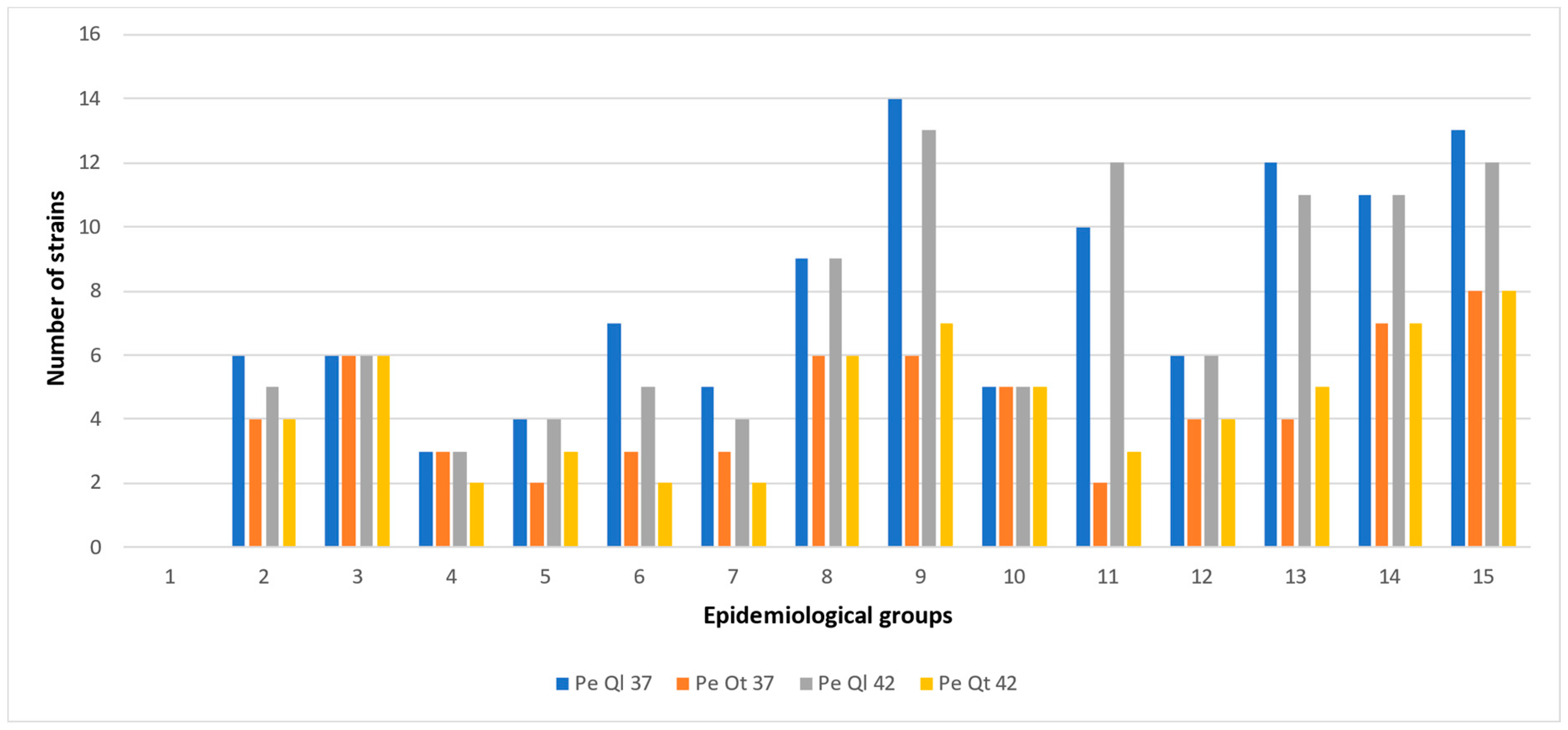
| Group | Method | ||||
|---|---|---|---|---|---|
| Qt 42 °C | Ql 42 °C | Qt 37 °C | Ql 37 °C | ||
| 1 | A (1), B(no), C(no), D (a) | 5 | 5 | 3 | 2 |
| 2 | A (1), B (yes), C (no), D (a) | 9 | 9 | 9 | 8 |
| 3 | A (1), B (no), C (yes), D (a) | 9 | 9 | 9 | 9 |
| 4 | A (1), B (yes), C (yes), D (a) | 9 | 9 | 9 | 9 |
| 5 | A (1), B (yes), C (yes), D (b) | 7 | 7 | 7 | 7 |
| 6 | A (2), B (no), C (no), D (a) | 13 | 13 | 15 | 12 |
| 7 | A (2), B (yes), C (no), D (a) | 12 | 12 | 12 | 12 |
| 8 | A (2), B (no), C (yes), D (a) | 14 | 16 | 14 | 17 |
| 9 | A (2), B (yes), C (yes), D (a) | 22 | 22 | 22 | 22 |
| 10 | A (2), B (yes), C (yes), D (b) | 18 | 18 | 18 | 18 |
| 11 | A (3), B (no), C (no), D (a) | 18 | 18 | 18 | 18 |
| 12 | A (3), B (yes), C (no), D (a) | 21 | 27 | 27 | 27 |
| 13 | A (3), B (no), C (yes), D (a) | 26 | 25 | 26 | 26 |
| 14 | A (3), B (yes), C (yes), D (a) | 30 | 30 | 30 | 30 |
| 15 | A (3), B (yes), C (yes), D (b) | 29 | 29 | 29 | 29 |
| Total | 242 | 249 | 248 | 246 | |
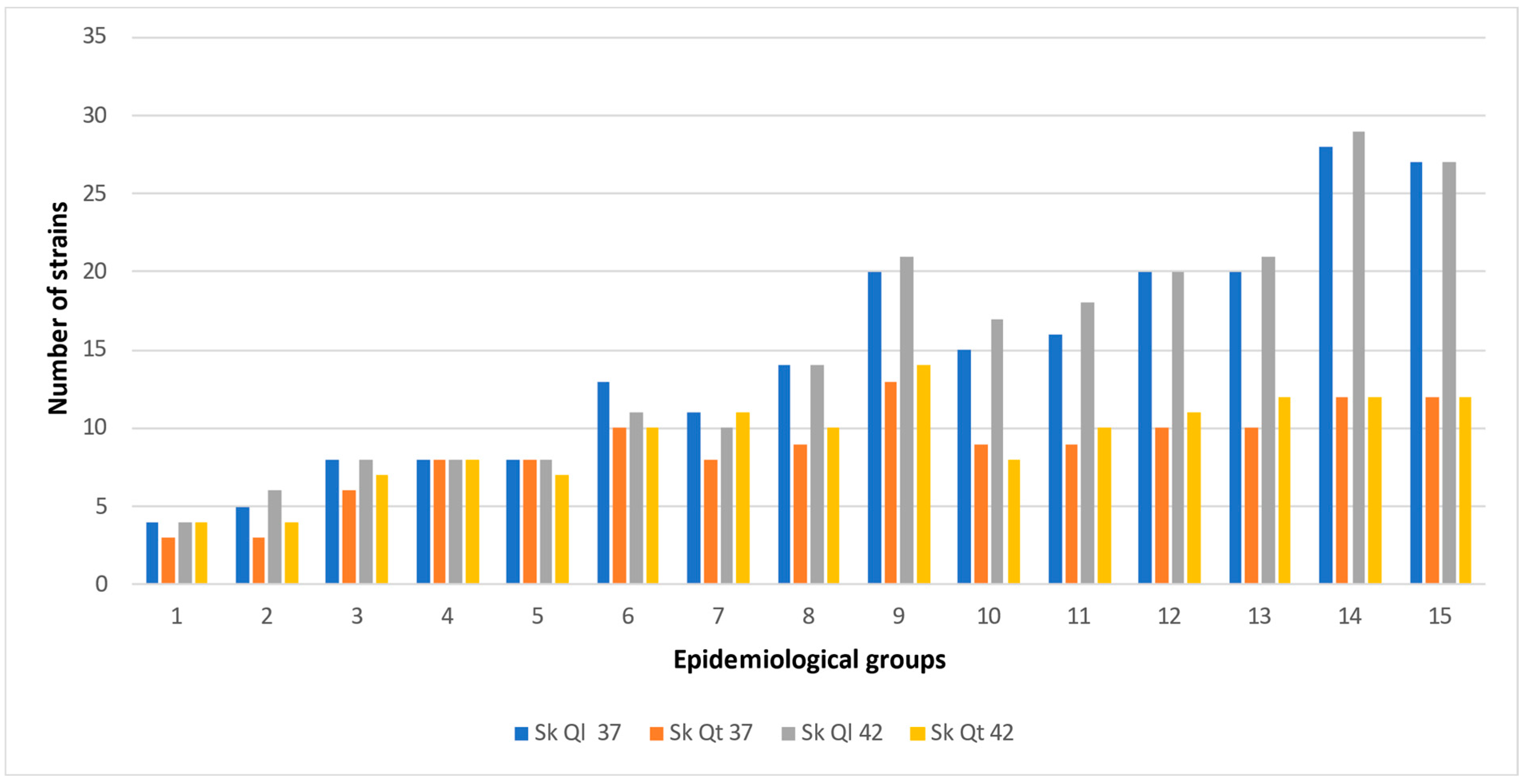
| Group | Method | ||||
|---|---|---|---|---|---|
| Quantitative 42 °C | Qualitative 42 °C | Quantitative 37 °C | Qualitative 37 °C | ||
| 1 | A (1), B(no), C(no), D (a) | 6 | 5 | 4 | 4 |
| 2 | A (1), B (yes), C (no), D (a) | 9 | 10 | 8 | 8 |
| 3 | A (1), B (no), C (yes), D (a) | 9 | 9 | 9 | 9 |
| 4 | A (1), B (yes), C (yes), D (a) | 7 | 8 | 7 | 8 |
| 5 | A (1), B (yes), C (yes), D (b) | 7 | 8 | 7 | 8 |
| 6 | A (2), B (no), C (no), D (a) | 13 | 15 | 15 | 15 |
| 7 | A (2), B (yes), C (no), D (a) | 11 | 12 | 12 | 12 |
| 8 | A (2), B (no), C (yes), D (a) | 14 | 16 | 14 | 17 |
| 9 | A (2), B (yes), C (yes), D (a) | 22 | 22 | 22 | 22 |
| 10 | A (2), B (yes), C (yes), D (b) | 18 | 18 | 18 | 18 |
| 11 | A (3), B (no), C (no), D (a) | 27 | 28 | 27 | 28 |
| 12 | A (3), B (yes), C (no), D (a) | 27 | 27 | 27 | 27 |
| 13 | A (3), B (no), C (yes), D (a) | 26 | 26 | 26 | 26 |
| 14 | A (3), B (yes), C (yes), D (a) | 30 | 30 | 30 | 30 |
| 15 | A (3), B (yes), C (yes), D (b) | 30 | 30 | 30 | 30 |
| Total | 256 | 264 | 256 | 262 | |
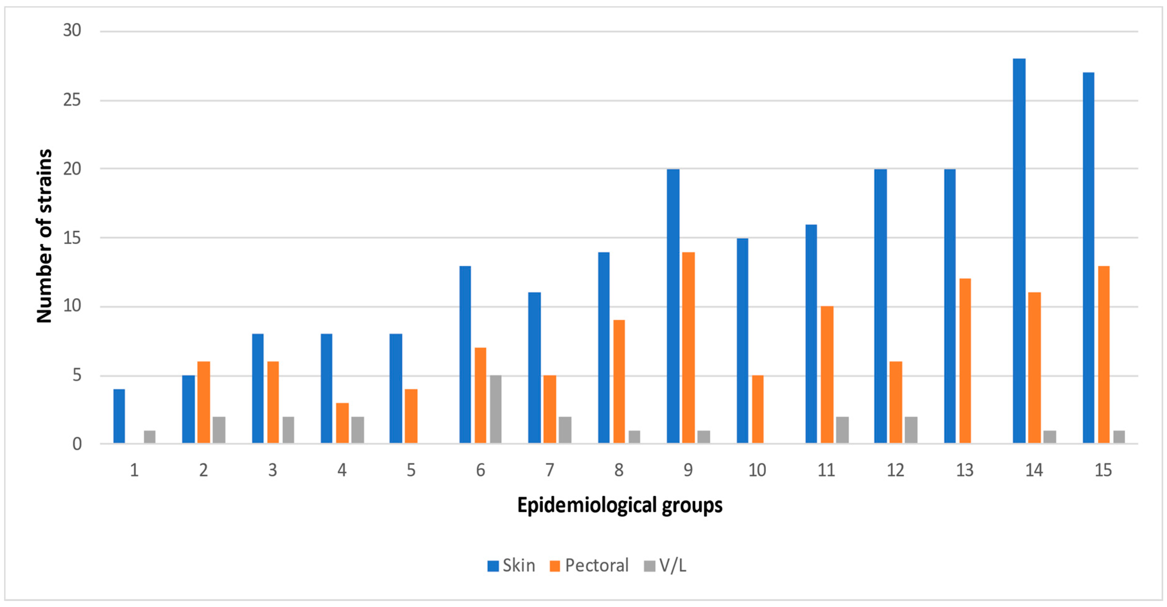
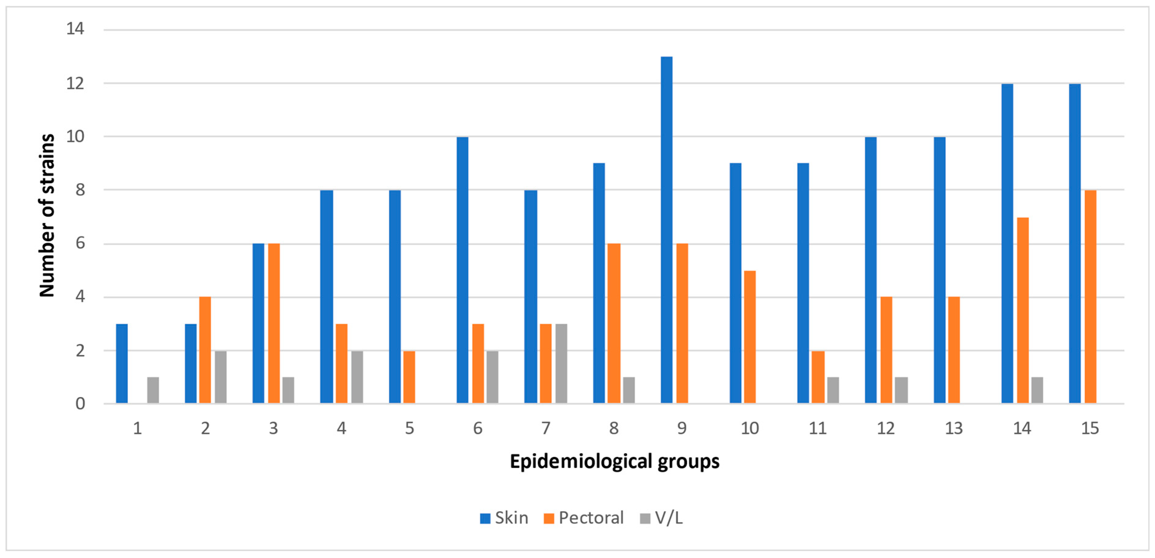
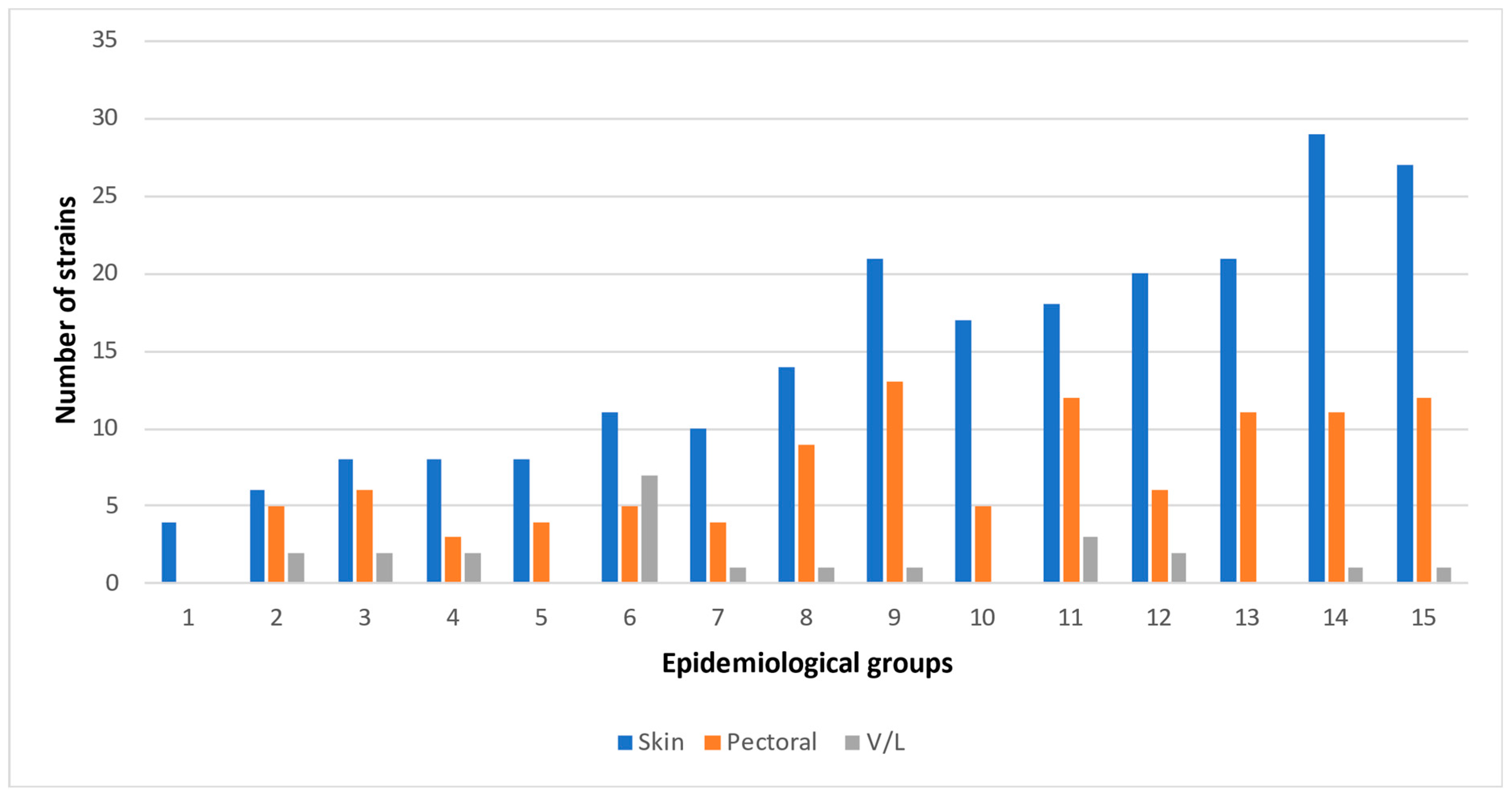
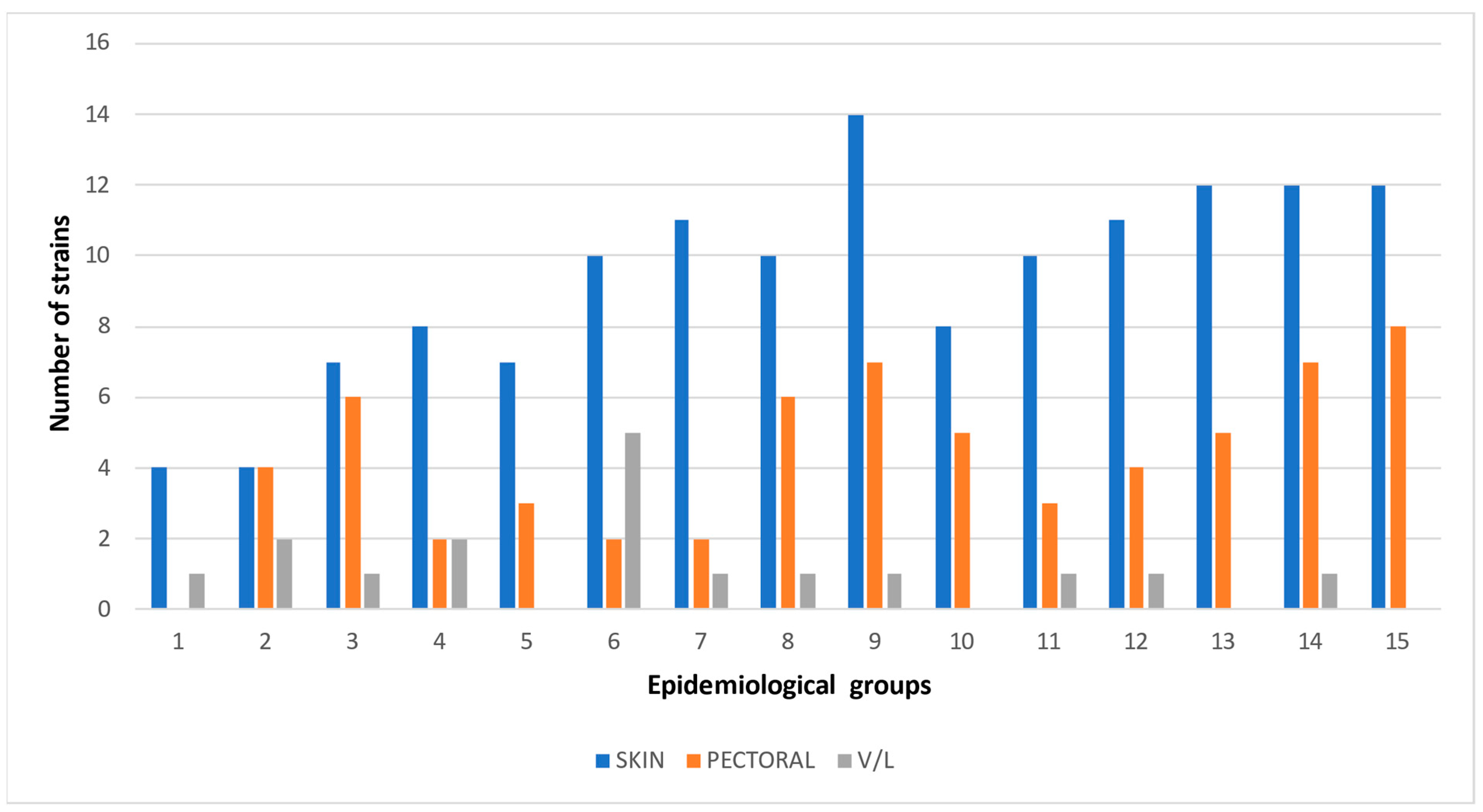
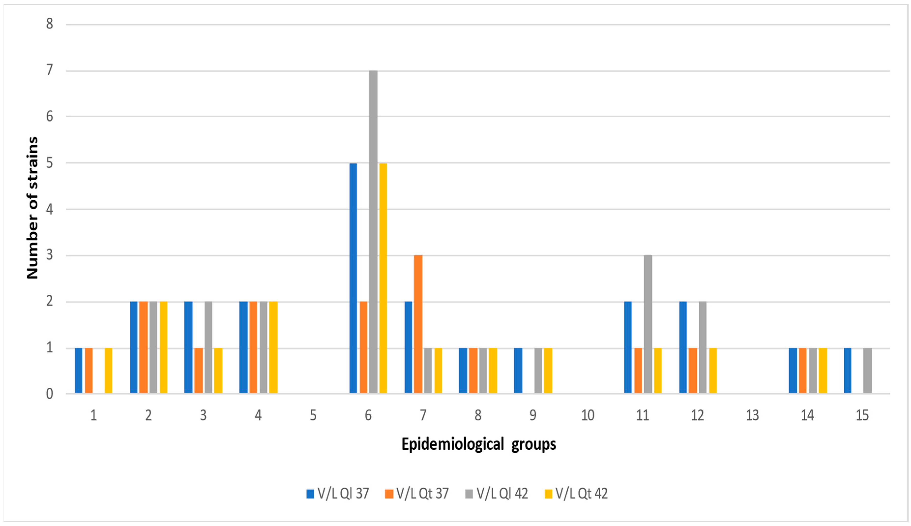
| Method | Quantitative 42 °C | Qualitative 42 °C | Quantitative 37 °C | Qualitative 37 °C | ||||||||
|---|---|---|---|---|---|---|---|---|---|---|---|---|
| Tissue | S a | P b | V c | S | P | V | S | P | V | S | P | V |
| Group | Campylobacter strains (n) | |||||||||||
| 1 * | 4 | 0 | 1 | 4 | 0 | 0 | 3 | 0 | 1 | 4 | 0 | 1 |
| 2 | 4 | 4 | 2 | 6 | 5 | 2 | 3 | 4 | 2 | 5 | 6 | 2 |
| 3 | 7 | 6 | 1 | 8 | 6 | 2 | 6 | 6 | 1 | 8 | 6 | 2 |
| 4 | 8 | 2 | 2 | 8 | 3 | 2 | 8 | 3 | 2 | 8 | 3 | 2 |
| 5 | 7 | 3 | 0 | 8 | 4 | 0 | 8 | 2 | 0 | 8 | 4 | 0 |
| 6 | 10 | 2 | 5 | 11 | 5 | 7 | 10 | 3 | 2 | 13 | 7 | 5 |
| 7 | 11 | 2 | 1 | 10 | 4 | 1 | 8 | 3 | 3 | 11 | 5 | 2 |
| 8 | 10 | 6 | 1 | 14 | 9 | 1 | 9 | 6 | 1 | 14 | 9 | 1 |
| 9 | 14 | 7 | 1 | 21 | 13 | 1 | 13 | 6 | 0 | 20 | 14 | 1 |
| 10 | 8 | 5 | 0 | 17 | 5 | 0 | 9 | 5 | 0 | 15 | 5 | 0 |
| 11 | 10 | 3 | 1 | 18 | 12 | 3 | 9 | 2 | 1 | 16 | 10 | 2 |
| 12 | 11 | 4 | 1 | 20 | 6 | 2 | 10 | 4 | 1 | 20 | 6 | 2 |
| 13 | 12 | 5 | 0 | 21 | 11 | 0 | 10 | 4 | 0 | 20 | 12 | 0 |
| 14 | 12 | 7 | 1 | 29 | 11 | 1 | 12 | 7 | 1 | 28 | 11 | 1 |
| 15 | 12 | 8 | 0 | 27 | 12 | 1 | 12 | 8 | 0 | 27 | 13 | 1 |
| Total | 140 | 64 | 17 | 222 | 106 | 23 | 130 | 63 | 15 | 217 | 111 | 22 |
| Group | c/sa | j/sb | a/sc | f/sd | l/se | c/pf | j/pg | |
|---|---|---|---|---|---|---|---|---|
| 1 | A (1), B(no), C(no), D (a) | 3 | 0 | 0 | 0 | 0 | 0 | 1 |
| 2 | A (1), B (yes), C (no), D (a) | 2 | 2 | 2 | 0 | 0 | 3 | 1 |
| 3 | A (1), B (no), C (yes), D (a) | 1 | 5 | 0 | 0 | 0 | 2 | 3 |
| 4 | A (1), B (yes), C (yes), D (a) | 1 | 3 | 0 | 2 | 0 | 2 | 1 |
| 5 | A (1), B (yes), C (yes), D (b) | 1 | 3 | 1 | 1 | 2 | 1 | 1 |
| 6 | A (2), B (no), C (no), D (a) | 2 | 3 | 3 | 0 | 0 | 1 | 1 |
| 7 | A (2), B (yes), C (no), D (a) | 4 | 2 | 2 | 0 | 1 | 2 | 0 |
| 8 | A (2), B (no), C (yes), D (a) | 3 | 5 | 0 | 4 | 0 | 1 | 5 |
| 9 | A (2), B (yes), C (yes), D (a) | 4 | 9 | 0 | 5 | 3 | 1 | 8 |
| 10 | A (2), B (yes), C (yes), D (b) | 5 | 7 | 0 | 2 | 1 | 1 | 2 |
| 11 | A (3), B (no), C (no), D (a) | 8 | 0 | 3 | 0 | 0 | 3 | 0 |
| 12 | A (3), B (yes), C (no), D (a) | 8 | 9 | 1 | 0 | 4 | 0 | 6 |
| 13 | A (3), B (no), C (yes), D (a) | 5 | 10 | 0 | 2 | 2 | 1 | 6 |
| 14 | A (3), B (yes), C (yes), D (a) | 6 | 17 | 0 | 2 | 2 | 2 | 7 |
| 15 | A (3), B (yes), C (yes), D (b) | 4 | 17 | 2 | 0 | 3 | 1 | 8 |
| Total | 57 | 92 | 14 | 18 | 18 | 21 | 49 | |
Disclaimer/Publisher’s Note: The statements, opinions and data contained in all publications are solely those of the individual author(s) and contributor(s) and not of MDPI and/or the editor(s). MDPI and/or the editor(s) disclaim responsibility for any injury to people or property resulting from any ideas, methods, instructions or products referred to in the content. |
© 2023 by the authors. Licensee MDPI, Basel, Switzerland. This article is an open access article distributed under the terms and conditions of the Creative Commons Attribution (CC BY) license (https://creativecommons.org/licenses/by/4.0/).
Share and Cite
Dermatas, A.; Rozos, G.; Voidarou, C.; Akrida-Demertzi, K.; Demertzis, P. Biodiversity Dynamics of Campylobacter Species in Chicken Tissues in Rural Households in Region Epirus, Greece. Appl. Sci. 2023, 13, 6073. https://doi.org/10.3390/app13106073
Dermatas A, Rozos G, Voidarou C, Akrida-Demertzi K, Demertzis P. Biodiversity Dynamics of Campylobacter Species in Chicken Tissues in Rural Households in Region Epirus, Greece. Applied Sciences. 2023; 13(10):6073. https://doi.org/10.3390/app13106073
Chicago/Turabian StyleDermatas, Argyrios, Georgios Rozos, Chrysoula (Chrysa) Voidarou, Konstantoula Akrida-Demertzi, and Panagiotis Demertzis. 2023. "Biodiversity Dynamics of Campylobacter Species in Chicken Tissues in Rural Households in Region Epirus, Greece" Applied Sciences 13, no. 10: 6073. https://doi.org/10.3390/app13106073