Article

Motor Imagery and Paired Associative Stimulation in Poststroke Rehabilitation: Dissociating Motor and Electrophysiological Effects

1
Toulouse NeuroImaging Center (ToNIC), Toulouse University, Inserm, UPS, 31024 Toulouse, France
2
Department of Rehabilitation and Physical Medicine, Toulouse University Hospital, 31059 Toulouse, France
3
Department of Epidemiology, Toulouse University Hospital, 31073 Toulouse, France
4
Department of Physiological Explorations, Toulouse University Hospital, 31059 Toulouse, France
*
Authors to whom correspondence should be addressed.
Appl. Sci. 2023, 13(10), 6063; https://doi.org/10.3390/app13106063
Submission received: 5 April 2023 / Revised: 8 May 2023 / Accepted: 10 May 2023 / Published: 15 May 2023

Abstract

:

Featured Application

PAS and MI are easy-to-implement rehabilitation interventions whose potential appears to be confirmed for hand motor recovery after stroke. However, there does not seem to be a specific advantage in combining PAS and MI interventions, as designed in MIPAS.

Abstract

Paired associative stimulation (PAS) is an intervention that modulates cortical plasticity. Motor imagery (MI) is used in the rehabilitation of stroke patients. We aimed to evaluate the possible synergistic effect of associating both interventions for potentiating motor recovery poststroke. MIPAS is a single-center, randomized controlled trial that enrolled 24 hemiparetic poststroke participants. Three single-session interventions were tested in a crossover design: PAS/MI, PAS, and ShamPAS/MI during which the affected Extensor Carpi Radialis (ECR) muscle was targeted. During MI, the participants were instructed to imagine extending their paretic wrist. We used Sham, subthreshold stimulation during ShamPAS. Changes in ECR Motor-Evoked Potential (MEP) areas and paretic wrist Range of Motion (aROM) during active extension were compared between the interventions. We observed no significant superior effect of any intervention, neither on MEP nor on wrist aROM. A time of assessment effect was highlighted for both outcome measures, with MEP- and aROM-measured post-interventions significantly higher than those measured pre-intervention. Despite the beneficial effect of each intervention on participant paretic wrist motor function, not always associated with MEP change, our results do not highlight a specific advantage in combining PAS and MI interventions in post-stroke motor rehabilitation.

1. Introduction

Stroke remains a leading cause of impairment and functional disability, which affects millions of people worldwide. Among patients who have severe motor deficits after a stroke, approximately 80% have grip and hand extensor muscle deficits [1,2]. Therefore, the motor recovery of wrist and finger extensions in poststroke patients remains a major rehabilitation challenge [3].
Noninvasive brain stimulation (NIBS) interventions have been suggested to modulate brain plasticity and improve upper extremity (UE) motor recovery. However, the results of three decades of research using NIBS targeting UE motor cortex representations poststroke remain inconclusive, although many meta-analyses are positive, their effect sizes remain small and their results often conflicting [4,5].
Facilitatory corticomuscular paired associative stimulation (PAS), targeting the motor cortex and administered with an interstimulus interval (ISI) of 25 ms, is one of these NIBS interventions that relies on long-term potentiation (LTP)-like mechanisms and reproduces the process of associative motor learning by strengthening and modulating specifically the corticospinal excitability (CSE) of the targeted muscle [6]. Therefore, PAS can be considered as a NIBS intervention that is “physiologically compatible” with the current knowledge about the mechanisms underlying poststroke motor recovery and/or rehabilitation. Durable, reversible, and topographically specific facilitatory effects of PAS protocols have been demonstrated in healthy subjects on flexor and abductor intrinsic finger muscles [6,7]. In a way to be more suitable for stroke rehabilitation, the possibility to elicit a PAS effect has also been experimented on wrist extensor muscles in healthy subjects [8] and in subacute subcortical ischemic [9] and in chronic [10] stroke patients. Although the number of randomized controlled trials (RCTs) using PAS is very limited, the results obtained with PAS trials seem similar to those obtained with other NIBS techniques. Mainly, the modulation of CSE was observed after PAS intervention in healthy and stroke individuals, without a significant effect on motor performance improvement or function recovery [10,11]. In spinal cord injury (SCI), repeated PAS has demonstrated therapeutic efficacy, with observed improvements in motor function [12,13,14,15]. Limited and mostly preclinical evidence of efficacy exists in stroke patients [16,17]. In a previous double-blinded Sham-controlled RCT, we tested and compared against Sham, a daily PAS protocol repeated over 5 days. The PAS protocol targeted the Extensor Carpi Radialis (ECR) muscle in a heterogeneous population of patients with cortical and/or subcortical strokes, mixing hemorrhagic or ischemic etiologies. We observed no significant difference in terms of CSE or motor scores, as assessed with the UE Fugl–Meyer Assessment scale (UEFMS) [18]. The absence of a difference could have been explained by the weak and variable CSE and motor effects induced by the repeated PAS intervention, also described previously [9,10,19], and the lack of sensitivity of the clinical scale used for motor function assessment [18].
Motor imagery (MI) is an easy-to-use and easy-to-administer intervention suitable for all stroke patients regardless of their motor deficit [20]. It is defined as the imagination of movements without their concomitant execution. It has been shown that MI training improves motor output by targeting cortical, subcortical, and cerebral networks similar to those involved in motor execution, and thus facilitates brain plasticity and cortical reorganization [21,22]. CSE facilitation occurs mainly during the imagination periods [23] and is increased and more persistent in time when MI is combined with muscle functional electrical stimulation (ES) [24] or with noninvasive brain stimulation protocols [25]. In the later study, the effects were cancelled when both interventions were applied asynchronously, showing the synergetic potential of combining MI with NIBS interventions.
Given the cited evidence, one can imagine that the concomitant MI of movements specific to the muscle targeted by a PAS intervention might decrease the variability in response to the latter and potentiate its effect. The potential beneficial synergistic effect of combining the two interventions that modulate cerebral plasticity led us to design the MIPAS (Motor Imagery and Paired Associative Stimulation) protocol. The objective was to study the electrophysiological and motor effects of a single intervention wherein MI exercises of paretic wrist extension movements were combined with a PAS protocol targeting the ECR, as compared to controlled interventions, in a population of hemiparetic stroke participants, with the hypothesis of greater changes after the combined intervention. The results would therefore demonstrate the potential therapeutic interest of such a combination and the usefulness of MI as an adjunct therapy to enhance the effects of PAS. The goal would be to create a model-based intervention aiming at potentiating their synergistic effects and being more readily transferrable in clinical sittings for effective motor rehabilitation after a stroke.

2. Materials and Methods

2.1. Study Design

MIPAS is a prospective, randomized, controlled, crossover (3 interventions, 3 visits), single-blind clinical trial that tests and compares the effect of 15 min single interventions of ‘PAS’, ‘ShamPAS/MI’, and ‘PAS/MI’, targeting the affected ECR muscle in hemiparetic stroke participants (Figure 1). All the stroke patients participated in all three interventional sessions. The overall protocol lasted 3 weeks with 1-week wash-out periods between each intervention. At the inclusion visit, UE motor deficits and MI ability were assessed (details, see Section 2.4.2), and each participant had an MRI examination during which weighted T1 images were acquired. The results associated with the participants’ neuroimaging data were published previously [26]. Three interventional visits were then scheduled over 3 consecutive weeks during which 1 of the interventions was randomly administered to the participants. The interventions were administrated the same day of the week, in the early afternoon. Before and after each intervention, electrophysiological (transcranial magnetic stimulation (TMS) motor evoked potential (MEP)) measurements and assessments of paretic wrist motor function (active range of motion of the affected wrist—aROM) were performed.

2.2. Participants

Participants were recruited in the Physical Medicine and Rehabilitation department of Toulouse University Hospital. The study was approved by the local Ethics Committee (Comité de Protection des Personnes Sud-Ouest et Outre-Mer II) in January 2013 (RCB 2012-A 01419-34) and registered in the ClinicalTrials.gov database (NCT02779218). All the participants gave their written informed consent in accordance with the Declaration of Helsinki.
Inclusion criteria were a first stroke, a motor deficit of the contralateral UE with an Upper-Extremity Fugl–Meyer subscore (UE-FMS) < 50/66, an ability to perform MI as assessed with the Time-Dependent Motor Imagery (TDMI) screening test, and the presence of an MEP for the ECR longus muscle. Participants were excluded if they had a history of epilepsy or contraindication to the use of TMS [27] and/or MRI, such as the presence of metal in the body, claustrophobia, or pregnant and/or breastfeeding women.

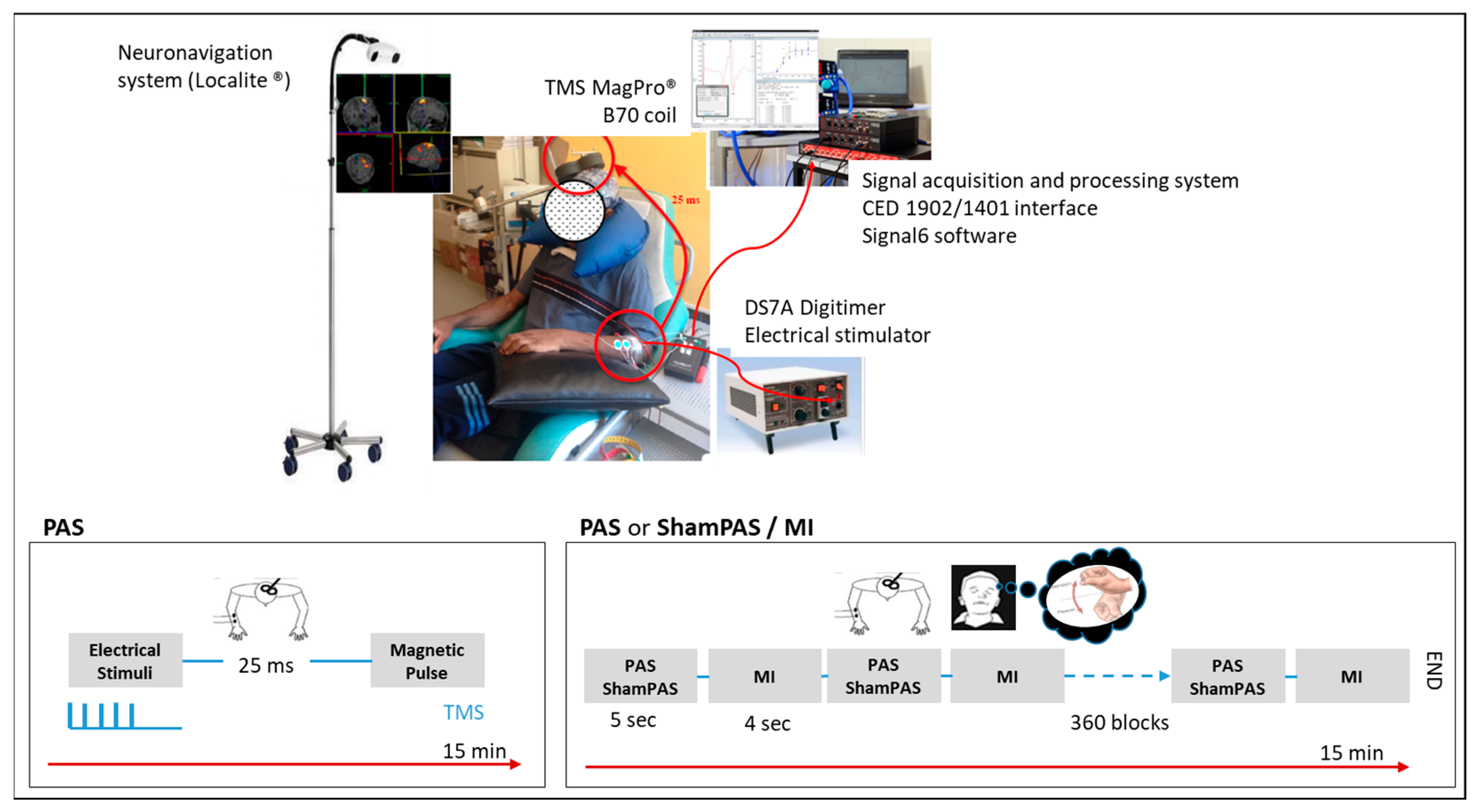
2.3. Experimental Interventions (Figure 1)

2.3.1. TMS Stimulation

Participants were comfortably seated in a reclining chair in a quiet room. A neuronavigation system (TMS navigator, Localite®, Bonn, Germany) with the anatomical wT1 of each participant was used to precisely record the position of the stimulation site (i.e., the hotspot (HS), see below) and to ensure the accurate repositioning of the TMS coil throughout and between the visits.
Brain stimulation was produced using a figure-of-eight-shaped Cool-B70 coil connected to a MagProX100 magnetic stimulator (MagVenture, Farum, Denmark). The coil was held tangentially to the scalp, pointing backward and approximatively 45° away from the midline over the lesioned primary motor cortex (M1), at the optimal position (HS) to elicit MEPs in the contralateral ECR. To determine the HS, we first placed the TMS coil over the theoretical cortical representation (TCR) of the ECR muscle and stimulated at suprathreshold TMS intensity until a distinguishable ECR MEP was elicited. We then moved the coil slightly around the TCR until reliable MEPs were consistently recorded. The cortical position was then considered as the HS, and the corresponding coil position was recorded using Localite®. The participant-specific suprathreshold TMS intensity used to record the MEPs was defined as the reference stimulation intensity (rSI) and was used during the PAS interventions as well as to evaluate MEP changes induced by the interventions. The rSI was chosen to ensure the absence of MEP ceiling or floor effect in response to the interventions.

2.3.2. Peripheral Electrical Stimulation (ES)

The surface electrodes used for the electrical stimulation were placed on the motor point of the ECR muscle of the participant’s paretic limb. We employed motor point stimulation instead of nerve stimulation [6], in an attempt to focus the induced changes on the targeted ECR [8,9,18]. The electrodes were connected to a DS7A constant-current stimulator (Digitimer, Welwyn Garden City, UK) that delivered 1 ms square-wave pulse trains at 5 Hz for 500 ms. The intensity of the ES was set at 1.5× the ECR motor threshold, specific to each participant, a tolerable intensity that caused only visible muscle twitches without overt movement.

2.3.3. PAS Intervention

During this intervention, participants received peripheral ES to the ECR motor point, as described above, followed 25 ms later by a TMS cortical pulse over the ECR HS at the rSI, specific to each participant [8] (see details, Section 2.3.1). A PAS with an interstimulus interval (ISI) set at 25 ms is believed to produce facilitatory LTP-like effects [6]. The pairing of the electrical and magnetic stimuli was applied every 5 s (frequency of 0.2 Hz) for 15 min. Therefore, 180 pairs of stimuli were delivered during a PAS intervention.

2.3.4. PAS/MI Intervention

During MI, participants were asked to have their eyes closed and to imagine extending their paretic wrist from a first-person perspective using visual and kinesthetic representations, as they were familiarized and trained to do so during the TDMI test, between each pair of PAS stimuli. The imagined movement had to be as large as possible and last 4 s (imagined movement initiation (1 s), maximum voluntary contraction (2 s), and imagined relaxation (1 s)). The participants were thus required to imagine performing a wrist extension movement every 5 s. Therefore, 180 extension movements were imagined by the participant during a 15 min intervention. The absence of muscle contraction during MI exercises was controlled by visual feedback. Additional questions were asked at the end of the intervention to review MI ability and performance and potential fatigue during stimulation.

2.3.5. ShamPAS/MI Intervention

ShamPAS stimulation was administered at a subthreshold and minimal cortical SI, only sufficient for producing a sound by the TMS coil (≈20% of the Maximum Stimulator Output, MSO). Indeed, given the overall low CSE observed with the participants included, the ShamPAS intervention design is believed to be not sufficient to produce CSE changes. The peripheral ES was the same as used during PAS. The participants had to perform MI between each pair of ShamPAS stimuli, i.e., every 5 s for 15 min, so that peripheral stimulation and MI were interleaved.
In these conditions, the assessment operator was blinded to the type of intervention being delivered during a specific session, and the participants were unable to distinguish PAS from ShamPAS but knew if they had to perform MI exercises and were thus considered partially blinded to the study.

2.4. Study Outcomes

The participants’ arms were comfortably positioned on the chair armrests. CSE (MEPs) was evaluated using the EMG activity recorded from the pairs of silver Ag–AgCl surface electrodes, placed on the paretic ECR muscle belly. The EMG signals were amplified (×3000), filtered (bandpass filter of 10–1000 Hz) with a CED 1902, and digitized at a sampling frequency of 4 kHz for 70 ms. The data were recorded using the CED1401 interface (Cambridge Electronic Design, Cambridge, UK) controlled by Signal6 software and stored offline for subsequent analysis.

2.4.1. Motor Imagery Outcomes

At the inclusion visit, MI ability was evaluated with the TDMI screening test [28], a chronometric test in which the number of imagined movements is recorded over 3 time periods (15, 25, and 45 s). The kinesthetic and visual imagery questionnaire (KVIQ) [29] was also administered to assess MI ability and performance. It scores the clarity of images and the intensity of sensations during MI. The maximal total KVIQ score is 170.

2.4.2. Electrophysiological Outcomes

At the rSI, MEP was measured before (MEPPre) and at 4 time points (i.e., 5, 15, 25, and 35 min, respectively, corresponding to MEP5, MEP15, MEP25, and MEP35) after each intervention. For each measure, 10 consecutive MEPs were recorded. Given the polyphasic nature of the ECR muscle [30], the areas (mV.ms) of the redressed and centered MEP were calculated and averaged across the ten measurements. For each intervention, CSE change was expressed as the difference (∆MEPx) between Post MEP values (MEPx) and the corresponding Pre MEP values (MEPPre) measured before the intervention:
∆MEPx (mV·ms) = MEPx − MEPPre
The calculation of the difference between post- and prestimulation values was preferred to the calculation of a percentage change from baseline, which is less recommended for the statistical analyses used [31].

2.4.3. Hand Motor Function Outcomes

We assessed each participant’s UE motor function using instrumental measurements of the active wrist extension amplitudes (in degrees) obtained with electronical goniometers (Biometrics®, Ltd., Newport, UK). Goniometric measurements of active wrist extension amplitudes (active range of motion—aROM) of the paretic wrist were obtained before (aROMPre) and at 0, 15, and 30 min after each intervention (aROM0, aROM15, and aROM30). The maximum aROM from three consecutive trials was considered. For each intervention, motor function changes (∆aROMx) were expressed as differences between the aROM measurement of interest, at a specific time point after the intervention, and the aROM prevalue:
∆aROMx (°) = aROMx − aROMPre

2.5. Randomization

The random allocation sequence was generated using the “Williams” SAS macro program, for Williams crossover designs [32]. The Williams is a type of crossover design useful for randomly assigning treatment sequences to the participants in a case of “3 treatments, 3 periods” protocol. The participants are randomized in equal numbers to six possible “treatment” sequences of the interventions to be studied [32]. The physician in charge of the protocol was responsible for enrolling the participants and assigning them to a specific treatment sequence.

2.6. Sample Size

According to Stefan et al. [6] and Talelli et al. [33], a PAS protocol could increase the MEP amplitude from 1.1 (0.3) mV to 1.7 (0.8) mV. To detect differences of 0.6 mV among the 3 interventions, assuming a standard deviation of 0.8 mV, the calculated sample size was 22 for paired comparisons with an 80% power and a 1.7% significance level (Bonferroni correction for 3 comparisons). The Williams designs require sample sizes that are multiples of 6. Therefore, the chosen sample size was set at 24 participants.

2.7. Statistical Analysis

The statistical analyses were performed with SPSS (IBM, Chicago, IL, USA. ver. 10) and Stata SE (StataCorp LLC, College Station, TX, USA. ver. 14.2). Normal distributions were checked for each variable with the Shapiro–Wilk test and by visual inspection using scattered plots. For repeated measures comparison, the Mauchly test was used to test the sphericity of the data and the Greenhouse–Geisser correction was used, when appropriate, to correct for nonsphericity. The corrected statistical parameters were then reported.
Concerning MI ability assessment with the TDMI and KVIQ, a 2-way repeated measures analysis of variance, rmANOVA (2 × 2: TDMI: “time allocated for imagination” and “hand assessed”; KVIQ: “MI strategy: visual vs. kinesthetic” and “hand assessed”) and paired t-tests, respectively, were performed. Pearson correlation analysis was used to investigate the relation between the two outcomes.
To ensure baseline outcome stability between visits and interventional sessions, repeated measures analysis of variance (rmANOVA) with the factors visit (S1, S2, and S3) or intervention (PAS/MI, PAS, and ShamPAS/MI) was applied on the outcome measured pre-intervention (MEPPre, aROMPre). Repeated measures analysis of covariance (rmANCOVA) analysis was applied on the repeated outcome measures (ΔMEPx) to estimate and compare the effects of the three interventions (PAS/MI, PAS, and ShamPAS/MI) on CSE at different times of assessment (TOA: Pre, Post5,15,25,35), while including the baseline measure (MEP measured pre-intervention at the first visit: MEPPre_S1) as covariate. rmANOVA analysis was applied on the repeated outcome measures (ΔaROMx) to estimate and compare the effects of the three interventions (PAS/MI, PAS, and ShamPAS/MI) on hand motor function at different TOA (Pre, Post0,15,30).
Models considered intervention, TOA, and intervention × TOA interaction terms to obtain the point estimates at the different TOA. We also report partial eta-squared ηp2 to judge the size of the effect observed. Additionally, the impact of each single intervention is indicated in the Supplementary Tables according to the 95% Confidence Interval (CI95) and p-values. For all the statistical analyses, a threshold of p = 0.05 was set. In the case of a significant result, a Fisher’s LSD correction post hoc test was performed.

3. Results

3.1. Participants

Between December 2013 and December 2017, twenty-five participants were included in the study. Patients #20 dropped out because of the occurrence of an epileptic seizure not imputable to the TMS protocol. Here, we report the results from 24 patients (mean age (SD) = 53 (13.2) years old, 8 women, 1 left-handed, and 12 lesioned in the right hemisphere) with a mean poststroke time of 9.7 months (1–105 months) were included. The mean UE-FMS was 27.4 (13.3)/66, and the mean KVIQ was 118.8 (22.7)/170. A detailed description of the group characteristics is provided in Table 1, and a lesion overlap map of the population included was shown and described in Brihmat et al. 2020 [26]. The available data from the inclusion visit were, however, included in the appropriate analyses.

3.2. Checking for Preliminary Assumptions

To compare the electrophysiological and motor results among the three randomized interventions, the stability of the baseline measurements from one visit to another had to be ensured. We found no significant differences between the prestimulation MEP values (MEPPre) measured at the beginning of each of the three visits (MEPPre_S1 = 3.4 (4.4), MEPPre_S2 = 3.3 (5.5), and MEPPre_S3 = 3.0 (4.0), unit = ×10−3 mV·ms, p = 0.92) and the three interventions (MEPPre_PAS/MI = 3.5 (4.5), MEPPre_PAS = 2.6 (3.7), and MEPPre_ShamPAS/MI = 3.5 (5.6), unit = ×10−3 mV·ms, p = 0.51). Similarly, for the aROMPre of the paretic wrist, we found no significant differences between the baseline measurements obtained during the three visits (aROMPre_S1 = 42.8 (22.4)°, aROMPre_S2 = 44,1 (22.9)°, and aROMPre_S3 = 44.6 (23.2)°, p = 0.26) and those of the three interventions (aROMPre_PAS/MI = 43 (22.6)°, aROMPre_PAS = 44 (23.7)°, and aROMPreShamPAS/MI = 45.0 (22.2)°, p = 0.10).

3.3. Motor Imagery Outcomes

For MI ability as assessed with the TDMI screening test, the individual and the rmANOVA group analyses (Supplementary Figure S1) reveal a significant increase in the number of imagined movements with the time allowed (F2,42 = 95.4, p < 0.001, partial η2 = 0.82) with a side effect (F1,21 = 5.0, p = 0.04, partial η2 = 0.19) and a side × time interaction (F2,42 = 3.4, p = 0.04, partial η2 = 0.14). Tukey’s post hoc analysis revealed that the increase in imagined movement between 15 and 45 s was lower for the affected, paretic hand than for the unaffected hand (F2,42 = 3.3, p = 0.04).
Regarding the KVIQ results, all the patients had relatively good scores on the questionnaire (Table 1, min–max: 80–170). We observed significantly higher visual subscores compared to kinesthetic subscores (t. test, t = 4.1, df = 22, p < 0.001).
A significant positive correlation was observed between the kinesthetic subscores (KIQ) obtained by the participants and the number of imagined movements with the paretic hand during the 25 and 45 s time periods of the TDMI (Supplementary Figure S2, Pearson correlation, 25 s period: r = 0.48, r2 = 0.23; p = 0.03; 45 s period: r = 0.54, p = 0.02, r2 = 0.54).

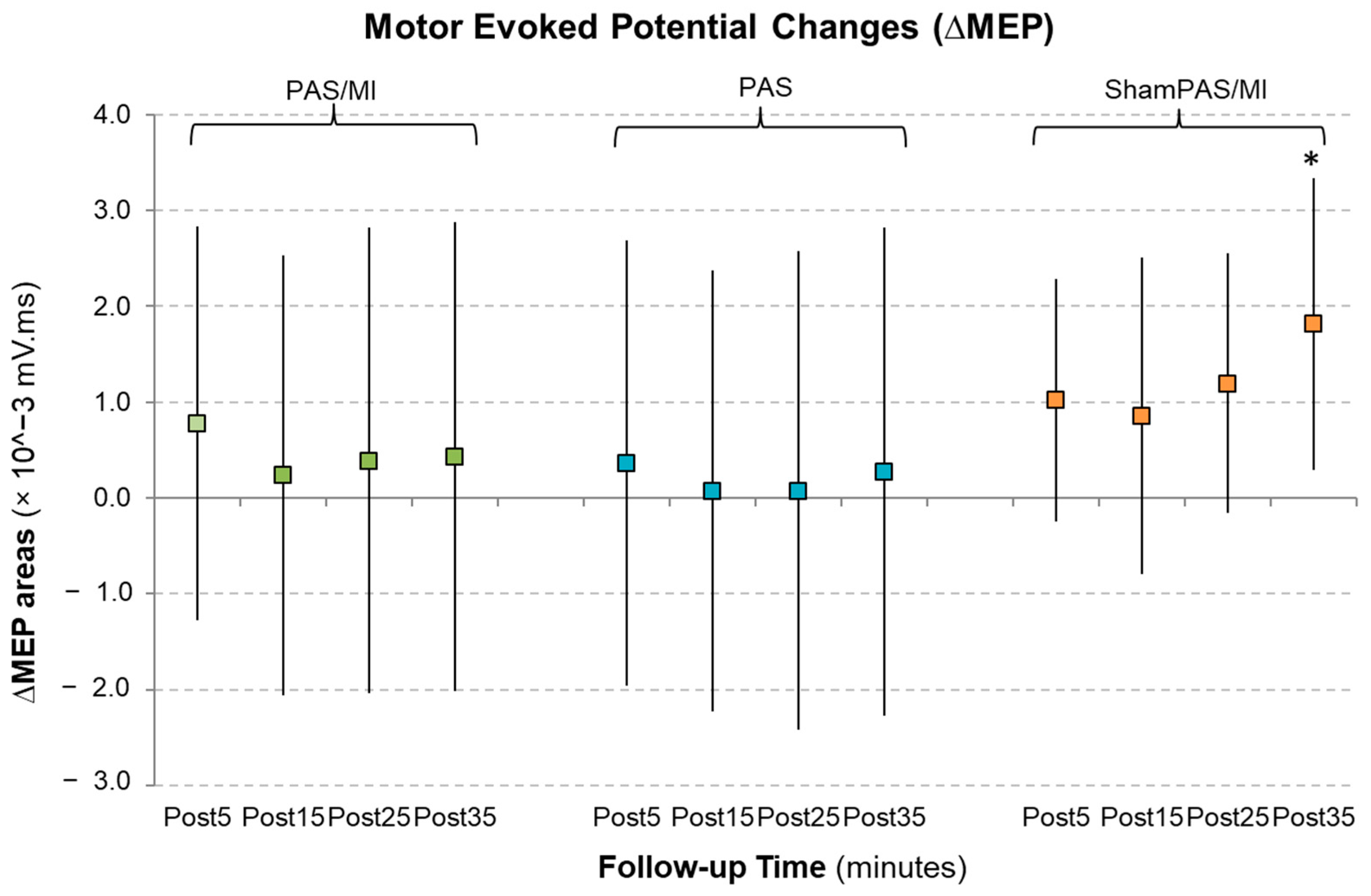
3.4. Corticospinal Excitability Outcomes

Regarding CSE changes (Figure 2, Supplementary Table S1), the rmANCOVA results revealed no intervention effect (intervention × MEPPre_S1: F1.24,13.66 = 1.64, p = 0.226, ηp = 0.13) and no intervention × TOA effect (intervention × TOA × MEPPre_S1:F2.17,23.87 = 0.54, p = 0.0.603, ηp = 0.05). The results, however, reveal a significant TOA effect (TOA × MEPPre_S1: F1.63, 17.95 = 4.33, p = 0.036, ηp = 0.28), with the MEP measured at Post5 and Post35 being higher than that measured at the pre-intervention (p = 0.032 and p = 0.05, respectively). Figure 2 shows the absolute values of the mean ΔMEP areas adjusted to the pre-intervention values at the different TOA. The ShamPAS/MI intervention appears to induce less variable, gradual, and larger MEP area increases postintervention (significant at 35 min postintervention, mean change: +1.813 × 10⁻³ mV·ms; CI95 not including the null value: 0.296–3.331 × 10⁻³ mV·ms; p = 0.02, Supplementary Table S1).

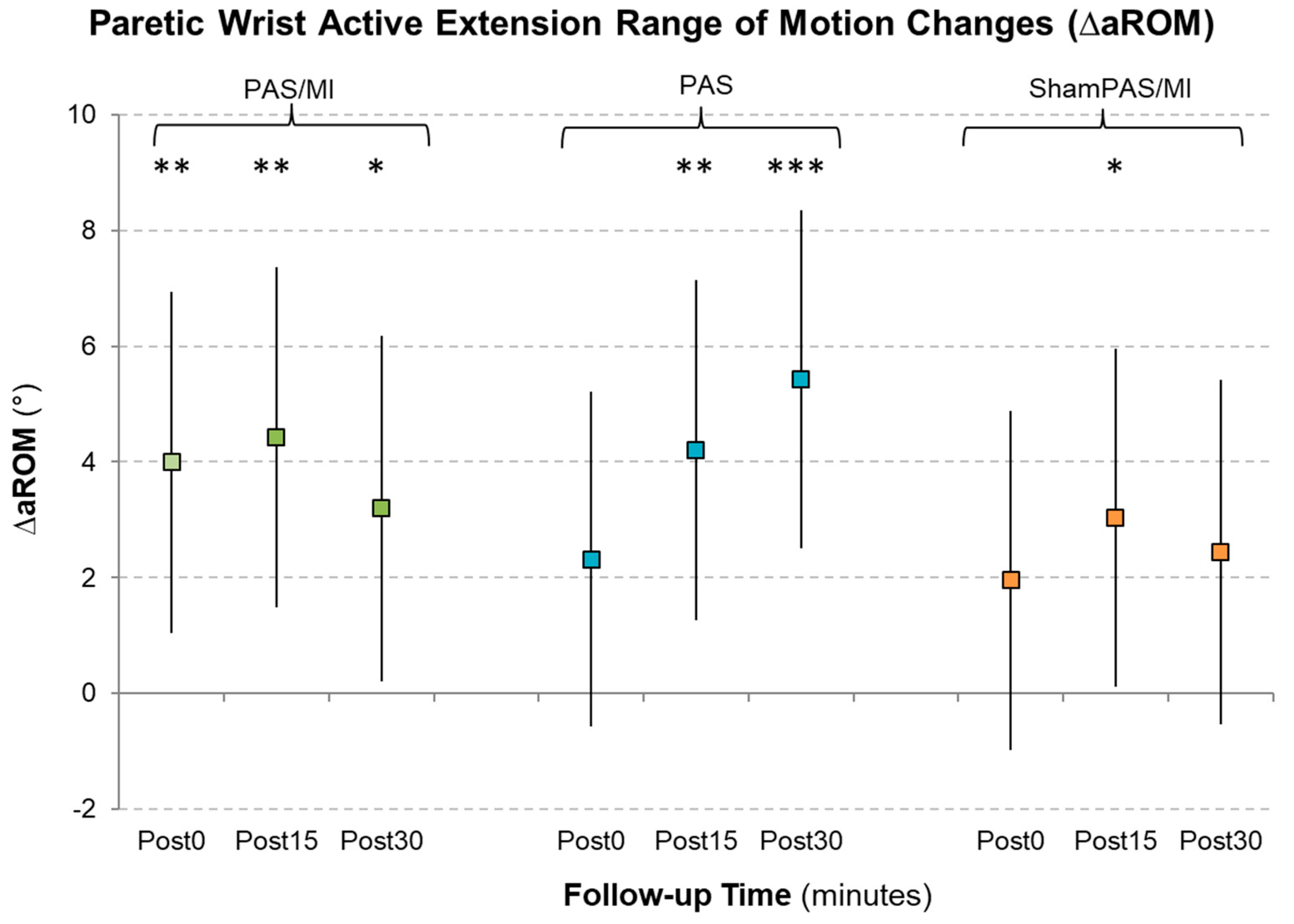
3.5. Hand Motor Function Outcomes

Like paretic hand motor function (Figure 3, Supplementary Table S2), the rmANOVA revealed no significant intervention (F2,34 = 0.49, p = 0.619, ηp2 = 0.03) and no intervention × TOA effect (F3.49,59.28 = 1.26, p = 0.297, ηp2 = 0.07) but a TOA effect on the aROM measure changes (F1.43,24.3 = 4.65, p = 0.029, ηp2 = 0.22). Fisher’s LSD revealed that aROM values measured at 15 min (p = 0.016) and 30 min (p = 0.03) postintervention were higher than the pre-intervention aROM values. Figure 3 shows different aROM change profiles across TOA and interventions. The values are indicated in Supplementary Table S2. All three interventions seem to have induced paretic wrist extension aROM increases. ShamPAS/MI induced aROM increases, significant at 15 min poststimulation (∆aROM15: p = 0.04); PAS induced a continuous increase in aROM that became significant at 15 min (∆aROM15: p = 0.005; ∆aROM30: p < 0.001); and PAS/MI induced a significant gradual increase in aROM from 0 to 30 min poststimulation (∆aROM0: p = 0.008; ∆aROM15: p = 0.003; ∆aROM30: p = 0.04).

4. Discussion

MIPAS investigated the relation between PAS and MI in a sample of poststroke participants. Corticomuscular PAS is a model of associative plasticity. It associates a peripheral muscle electrical stimulation to a cortical TMS stimulation administered repetitively at a constant interval of time. In its initial description [6], PAS was designed as a single ES to the median nerve followed by a TMS pulse over the abductor pollicis brevis (APB) muscle cortical representation. Long-term potentiation or depression phenomenon (LTP or LTD, depending on the ISI used) in relation to a mechanism of spatial summation has been proposed to explain the PAS mechanism of action [34]. To optimize its efficiency, some authors have associated a temporal facilitation component to PAS, by additionally administering a train of stimulation to the median nerve [7]. However, the functional prognostic of poststroke UE is mainly determined by the recovery of wrist and finger active extension movements [35]. In an effort to be more suitable for stroke rehabilitation, we thus developed and tested a PAS intervention targeting the ECR muscle [8,9]. Despite encouraging results obtained with single PAS intervention [8,9], the result of an RCT trial published by our group [18], using daily PAS stimulation over five consecutive days during stroke rehabilitation against Sham, was negative. The MIPAS protocol was therefore designed to investigate the potential of combining PAS and MI interventions to increase the effect size of each intervention administered separately. Indeed, more recently, it was shown that a short training combining overt movements, MI, and TMS increased post-training MEP amplitude in healthy individuals [36]. The observed results and the associated limitations are discussed in the appropriate sections below.

4.1. Preliminary Assumptions about MI Ability and Performance

At the beginning of MIPAS, each participant was assessed for MI ability and performance with the TDMI screening test and the KVIQ. These tests provided complementary information and showed that all the participants were able to imagine the movement regardless of their lesion location or type, poststroke time, and level of motor deficit. Indeed, this ability seems to be recovered during the first weeks after a stroke [37]. As suggested by [28], TDMI also helped the patients to become familiarized with the movement they had to imagine during the MI-related interventions. The participants’ ability to perform MI was also supported by the observation of characteristic cerebral activation patterns during the fMRI examination performed at the inclusion in MIPAS, where they were asked to imagine wrist extension movements, similar to those performed during MI interventions (results published previously in Brihmat et al. 2020 [26]). This confirms the relevance of using a multidimensional approach for MI ability assessment [38] and supports the preliminary assumption that all the stroke participants included in MIPAS were able to perform MI when asked to do so during the interventions. Additional questions were asked at the end of the stimulation intervention to ensure participant ability to perform MI when asked to during the appropriate sessions.

4.2. General Results

No significant superior electrophysiological and/or motor effect was observed after a 15 min single intervention in which PAS was combined with MI exercises as compared to sessions where each intervention was administered separately in the stroke participants tested. Although attractive, the idea of a synergistic effect of different interventions with initially limited size effects, such as PAS and MI, might be inconsistent with the model of homeostatic plasticity proposed by Siebner and collaborators [39]. M1 plasticity could have been modulated differentially according to the first administered intervention, therefore not demonstrating the expected higher plasticity in response to the second applied intervention [40]. This might be due to the processes underlying the interaction between both interventions. For example, in healthy subjects, the PAS effect was reversed from LTP- to LTD-like plasticity following MI practice [41], and the ability of PAS to induce associative plasticity was abolished after a dynamic motor training [40]. Additionally, the CSE facilitation observed after a combined MI/paired-pulse rTMS intervention was cancelled when functional ES was added [25]. The expected synergistic effects on excitability facilitation, as observed in few publications in healthy subjects [24], do not seem to be transferable to our stroke population. The observed results suggest more complicated processes underlying this combination of interventions in stroke. In fact, in addition to these general negative results, a dissociation of the electrophysiological and motor results of each single intervention could be observed; the very weak effects of each intervention on MEP changes contrast with their systematic effects on paretic wrist aROM.

4.3. Corticospinal Excitability Effects

We did not observe significant CSE changes among the three interventions at any time of the assessment. However, a time of assessment effect was highlighted, and changes were observed when individual interventions were then considered. Indeed, higher MEP values were measured postintervention, revealing the potential of a single 15 min stimulation intervention to induce significant CSE changes poststroke. Additionally, larger, gradual, and less variable CSE increases were observed after ShamPAS/MI (Figure 2). Only few studies have investigated CSE changes related to mental training [42]. A CSE increase has been described in the literature after MI training, alone or in combination with other rehabilitation interventions, in healthy [25,36] and in stroke participants [43]. Better recovery of hand function and higher amplitude of the APB MEP were observed in stroke patients who underwent mental training associated with traditional rehabilitation compared to patient groups with traditional rehabilitation alone [43]. In a recent study, CSE changes persisted beyond the imagination period and were still measurable 30 min postintervention [44], which is in keeping with our findings in a stroke population. The observed CSE increase (in ≈ 60% of the participants) might be due to the progressive decrease in the corticospinal threshold and/or the increase in motor neuron recruitment during the course of MI training [45]. This CSE modulation induced by the ShamPAS/MI seems less subject to variability, as compared to the sessions where real PAS was also administered. The observed effects might, however, not be due to the effects of MI alone, given the design of the ShamPAS intervention in MIPAS and the presence of the muscle ES.
The absence of a significant effect of PAS alone on CSE may be explained by several factors. Firstly, a recent paper [46] showed that the ability to induce plastic changes in motor cortical output after stimulation requires the integration of sensory input, as in the PAS paradigm, which follows a certain gradient in the human brain. Potentially, the participants included in MIPAS had additional sensory impairments, preventing the PAS-induced LTP-like plasticity. Additionally, intrinsic hand flexor muscles seem to have a higher potential to show plasticity than extensor muscles in response to PAS, and the induced plastic changes in extensors are among the most difficult to highlight [46]. Moreover, the observed results confirm once again that the intra- and interindividual variability in response to PAS is significant and is extensively described in the literature [47]. This variability is thought to be due to numerous factors among which are age, gender, level of attention, baseline CSE, and stimulation frequency [9,10,18,19,48]. Even though we tried to control most of these factors, we observed a variability in CSE changes in our population ranging from −57.1 to +258.8%. Only 45% of the patients could be considered as responders, and only 4% had facilitation greater than 30%, which is a low rate compared to similar studies [10]. This might be due to the stimulation frequency used during PAS, which was higher in MIPAS (0.2 Hz vs. 0.1 Hz in other protocols [40,49]) in an effort to reduce the experimental time and participants’ mental fatigue.

4.4. Hand Motor Function Effects

In MIPAS, we highlight the fact that all three interventions induced improvements in paretic wrist aROM. PAS/MI induced an immediate and large increase in paretic wrist aROM that lasted 30 min postintervention. Similar motor effects were observed in healthy subjects when MI was combined with anodal transcranial direct current stimulation (tDCS) [50]. However, the effect in MIPAS has neither a higher effect size nor a longer-lasting effect than PAS alone. This result again supports the idea that both interventions are not synergistic, presumably through a mechanism of regulatory homeostatic plasticity [39,40,41].
PAS induced a gradual and significant increase in active paretic wrist aROM, lasting from 15 to 30 min postintervention, compared to pre-intervention values. To our knowledge, this is the first evidence of a beneficial effect of PAS on the UE motor function in stroke participants. As compared to MI, PAS is supposed to induce a more specific strengthening of the corticospinal connections of the targeted muscle [6,9]. A beneficial effect of PAS on motor performance has been observed in SCI individuals [12,13,51] but not in stroke patients [10,18]. These differences might be explained by the difference in the lesion location/size between SCI and stroke and is in line with the fact that PAS might not be suitable for individuals with large cortical lesions. Additionally, the assessment method is critical and especially in single-session designs. Indeed, the use of kinematic aROM measurements is more specific to the tested muscle and more sensitive to single-intervention effects than the standardized clinical scales, such as the FMA [18].
ShamPAS/MI induced less clear improvement of paretic aROM compared to PAS and PAS/MI, although still significant at 15 min postintervention. This effect is of interest given the easy use and implementation of MI-based exercise in stroke neurorehabilitation [15]. The repetition of MI-based exercises over a greater number of sessions could induce greater functional improvements.

4.5. Dissociation of Electrophysiological and Motor Effects

Surprisingly, the beneficial effects of PAS and PAS/MI interventions on motor function were not associated with significant CSE changes. Furthermore, ShamPAS/MI induced a CSE increase later than the observed motor facilitation. A dissociation of the mechanisms underlying such effects, which could rely on different cerebral substrates, seems to exist and has already been suggested [52]. In the Pascual-Leone study [52], greater improvement in motor performance in the ‘physical training’ group compared to the ‘MI training’ group was not found to be associated with a greater difference in CSE. This dissociation could be explained by the limitations of TMS in identifying subtle neural changes that may cause differences in motor performance [52], the variability of the MEP responses and that of the responses to NIBS [53,54], and/or by the fact that CSE inferred from MEP measurements could primarily reflect fast monosynaptic CSE changes. In humans, the fast monosynaptic pathways of the corticospinal tract (CST) are the most-developed fiber pathways [55]. Phylogenetically, these pathways are fundamental for fine and fast independent finger movements [56]. Numerous studies have pointed out that motor recovery after stroke was only possible with sufficient preservation of the CST [57], especially for fine independent finger movements [58]. However, it represents a limited portion of all the corticospinal motor tract fibers [55], and some recent studies have suggested that other tracts might have a role in poststroke motor recovery although their role remains limited compared to that of the CST [59,60,61]. Moreover, the degree of preservation of the corticospinal pathways required for good motor recovery also depends on the residual possible interactions between M1, connected to motoneurons through fast corticospinal pathways, and the premotor cortex, which is not connected monosynaptically to the motoneurons. This interaction indirectly suggests an action on motoneurons through alternate motor tracts [62]. Therefore, the dissociation that we observed between the improvement of motor abilities and unchanged MEPs at the group level is in favor of the idea that MEP could be a tool to assess fast monosynaptic corticospinal projections but does not allow the identification of specific changes in relation to nonmonosynaptic pathways. On the contrary, the improvement in active extension movements observed particularly with the PAS intervention indirectly suggests that the mechanism of PAS might also involve alternate motor tracts, at least in poststroke patients. The absence of synergistic effects between PAS and MI emphasizes the fact that the combination of different rehabilitation interventions cannot be reduced to a simple summation of their effects, and further work is necessary to effectively combine interventions in neurorehabilitation [63].

4.6. Limitations and Future Perspectives

The main limitation of the study was the very large data variability potentially due to different factors. The heterogeneity of the patient population, in terms of the time since stroke, stroke location, stroke etiology, MI ability, and UE motor impairment, is a factor to consider in the variability observed with the PAS interventions [47,64]. Even if MI can be applied regardless of these characteristics [37], PAS may be more suitable for patients with small, subcortical lesions [8,9] and with better motor performance [10]. Furthermore, functional benefits were observed when PAS was repeated over multiple sessions [17] and mainly when the ISI was individually defined, as performed in SCI individuals [12,13,15]. This needs to be further investigated in stroke participants.
In addition, the lesions to the corticospinal tract (CST) due to the stroke make the MEP measurements difficult and unreliable. Many of the patients included in MIPAS had a high motor threshold near or sometimes higher than the maximum TMS stimulator output, making it impossible to measure input–output (I/O) curves. Additionally, even if we verified the ability for MEP to increase, it was sometimes difficult to ensure that the selected intensity was in the linear portion of the I/O curve, which introduces greater variability in the sensitivity of MEP response.
Significant CSE facilitation was observed 30 min after ShamPAS/MI, but not after the other two interventions. Even if we previously demonstrated that muscle ES alone for 30 min did not significantly affect CSE in stroke [8], we cannot rule out that it is the ES, present in ShamPAS, combined with MI that led to the significant and less variable CSE facilitation observed.
All three interventions demonstrated beneficial effects on paretic hand motor function, but the results might not be related to the only effect of the interventions. The repetition of active wrist extension induces a generic learning effect and/or an increase in unspecific neurovegetative excitation. However, given that ShamPAS/MI induced the least important and lasting changes in aROM suggests that other mechanisms may be involved in the motor changes observed. Unlike the CSE results, the motor behavioral results are not in favor of MI size effects overshadowing the influence of PAS, but suggest different mechanisms of action between PAS and MI.
Finally, the large variability of the data, associated also with the small sample size is responsible for the low study power, which limits the generalization of the results. After a second negative trial, we can say that even if the PAS mechanism is attractive, it might not be suitable for all stroke individuals and is not ready to be implemented in poststroke rehabilitation. Similarly, the difficulties in ensuring the methodological expectations of MEP measures in poststroke participants and the different results observed between the CSE and motor behavioral outcomes emphasize the need to not directly associate both outcomes. All the motor behavioral outcome changes in poststroke participants cannot be explained by changes in CSE alone.

5. Conclusions

In this study, we were unable to demonstrate a superior electrophysiological and/or motor effect of a single intervention where PAS was combined with MI exercises compared to interventions where each intervention was administered separately in hemiparetic stroke patients. However, each intervention demonstrated a beneficial effect on stroke participants’ hand motor function as shown with the increases in paretic wrist aROM. The MIPAS results also suggest a dissociation between MEP area measurements and wrist motor function, which once again calls into question the relevance of using MEP measurements to explain poststroke motor recovery and the mechanisms underlying improvements in motor performance in poststroke patients. The results of MIPAS need to be confirmed in less heterogeneous subgroups of stroke patients to further investigate questions related to the stratification of patients for PAS interventions, based on individual and specific characteristics.

Supplementary Materials

The following supporting information can be downloaded at: https://www.mdpi.com/article/10.3390/app13106063/s1, Table S1: Mean MEP changes relative to prestimulation values across time for each intervention session (PAS/MI, PAS, and ShamPAS/MI).; Table S2: Mean paretic wrist active extension range of motion changes (ΔaROM, in degrees °) relative to prestimulation values across time for each session (PAS/MI, PAS, and ShamPAS/MI); Figure S1: Number (mean ± SD) of imagined movements with the affected and unaffected hands during the three time periods of the TDMI screening test. Figure S2: Pearson correlation results between the number of movements imagined with the paretic hand during the 25 and 45 s periods of the TDMI screening test and the kinesthetic scores obtained during the KVIQ test.

Author Contributions

Conceptualization, E.C.-L., M.T. and P.M.; Formal analysis, N.B., E.C.-L., B.L. and I.L.; Funding acquisition, E.C.-L., M.T. and P.M.; Investigation, N.B., E.C.-L. and M.T.; Methodology, N.B., E.C.-L., M.T., I.L. and P.M.; Resources, E.C.-L., E.M., C.C., X.d.B. and D.G.; Writing—original draft, N.B., E.C.-L. and P.M.; Writing—review and editing, N.B., E.C.-L., M.T., B.L., E.M., C.C., X.d.B., D.G., I.L. and P.M. All authors have read and agreed to the published version of the manuscript.

Funding

This work was supported by a grant from the ‘Fondation de l’Avenir’ (trial n°ET2-669) and from the Toulouse University Hospital, local grant 2012, N° RC31/12/38902.

Institutional Review Board Statement

The study was approved by the local Ethics Committee (Comité de Protection des Personnes Sud-Ouest et Outre-Mer II) in January 2013 (RCB 2012-A 01419-34) and registered in the ClinicalTrials.gov database (NCT02779218).

Informed Consent Statement

All the participants gave their written informed consent in accordance with the Declaration of Helsinki.

Data Availability Statement

The dataset used and analyzed during the current study may be available upon reasonable request to the corresponding author.

Acknowledgments

We would like to thank the Inserm/UPS UMR1214 Technical Platform for their help in setting up the fMRI experiment and for the acquisitions of the MRI sequences and the patients who agreed to participate in the study. The author N.B. is now affiliated with the Tim and Caroline Reynolds Center for Spinal Stimulation, Kessler Foundation and the Department of Physical Medicine and Rehabilitation, Rutgers University—New Jersey Medical School; NJ, United States.

Conflicts of Interest

The authors declare no conflict of interest.

References

  1. Hendricks, H.T.; van Limbeek, J.; Geurts, A.C.; Zwarts, M.J. Motor recovery after stroke: A systematic review of the literature. Arch. Phys. Med. Rehabil. 2002, 83, 1629–1637. [Google Scholar] [CrossRef] [PubMed]
  2. Jørgensen, H.S.; Nakayama, H.; Raaschou, H.O.; Vive-Larsen, J.; Støier, M.; Olsen, T.S. Outcome and time course of recovery in stroke. Part II: Time course of recovery. The Copenhagen Stroke Study. Arch. Phys. Med. Rehabil. 1995, 76, 406–412. [Google Scholar] [CrossRef] [PubMed]
  3. Kamper, D.; Harvey, R.; Suresh, S.; Rymer, W. Relative contributions of neural mechanisms versus muscle mechanics in promoting finger extension deficits following stroke. Muscle Nerve 2003, 28, 309–318. [Google Scholar] [CrossRef] [PubMed]
  4. Hatem, S.M.; Saussez, G.; della Faille, M.; Prist, V.; Zhang, X.; Dispa, D.; Bleyenheuft, Y. Rehabilitation of motor function after stroke: A multiple systematic review focused on techniques to stimulate upper extremity recovery. Front. Hum. Neurosci. 2016, 10, 442. [Google Scholar] [CrossRef]
  5. Kubis, N. Non-invasive brain stimulation to enhance post-stroke recovery. Front. Neural Circuits 2016, 10, 56. [Google Scholar] [CrossRef]
  6. Stefan, K.; Kunesch, E.; Cohen, L.G.; Benecke, R.; Classen, J. Induction of plasticity in the human motor cortex by paired associative stimulation. Brain 2000, 123 Pt 3, 572–584. [Google Scholar] [CrossRef] [PubMed]
  7. Ridding, M.C.; Taylor, J.L. Mechanisms of motor-evoked potential facilitation following prolonged dual peripheral and central stimulation in humans. J. Physiol. 2001, 537, 623–631. [Google Scholar] [CrossRef]
  8. Castel-Lacanal, E.; Gerdelat-Mas, A.; Marque, P.; Loubinoux, I.; Simonetta-Moreau, M. Induction of cortical plastic changes in wrist muscles by paired associative stimulation in healthy subjects and post-stroke patients. Exp. Brain Res. 2007, 180, 113–122. [Google Scholar] [CrossRef]
  9. Castel-Lacanal, E.; Marque, P.; Tardy, J.; De Boissezon, X.; Guiraud, V.; Chollet, F.; Loubinoux, I.; Simonetta-Moreau, M. Induction of Cortical Plastic Changes in Wrist Muscles by Paired Associative Stimulation in the Recovery Phase of Stroke Patients. Neurorehabil. Neural Repair 2009, 23, 366–372. [Google Scholar] [CrossRef]
  10. Palmer, J.A.; Wolf, S.L.; Borich, M.R. Paired associative stimulation modulates corticomotor excitability in chronic stroke: A preliminary investigation. Restor. Neurol. Neurosci. 2018, 36, 183–194. [Google Scholar] [CrossRef]
  11. Versace, V.; Langthaler, P.B.; Höller, Y.; Frey, V.N.; Brigo, F.; Sebastianelli, L.; Saltuari, L.; Nardone, R. Abnormal cortical neuroplasticity induced by paired associative stimulation after traumatic spinal cord injury: A preliminary study. Neurosci. Lett. 2017, 664, 167–171. [Google Scholar] [CrossRef] [PubMed]
  12. Bunday, K.L.; Urbin, M.; Perez, M.A. Potentiating paired corticospinal-motoneuronal plasticity after spinal cord injury. Brain Stimul. 2018, 11, 1083–1092. [Google Scholar] [CrossRef] [PubMed]
  13. Bunday, K.L.; Perez, M.A. Motor recovery after spinal cord injury enhanced by strengthening corticospinal synaptic transmission. Curr. Biol. 2012, 22, 2355–2361. [Google Scholar] [CrossRef] [PubMed]
  14. Tolmacheva, A.; Savolainen, S.; Kirveskari, E.; Brandstack, N.; Mäkelä, J.P.; Shulga, A. Paired associative stimulation improves hand function after non-traumatic spinal cord injury: A case series. Clin. Neurophysiol. Pract. 2019, 4, 178–183. [Google Scholar] [CrossRef] [PubMed]
  15. Jo, H.J.; Perez, M.A. Corticospinal-motor neuronal plasticity promotes exercise-mediated recovery in humans with spinal cord injury. Brain 2020, 143, 1368–1382. [Google Scholar] [CrossRef]
  16. Guo, T.-C.; Hu, Y.; Zhang, X.-Y.; Tian, J.; Lu, Y.-S. Paired associative stimulation improves synaptic plasticity and functional outcomes after cerebral ischemia. Neural Regen. Res. 2019, 14, 1968–1976. [Google Scholar] [CrossRef]
  17. Sui, Y.-F.; Tong, L.-Q.; Zhang, X.-Y.; Song, Z.-H.; Guo, T.-C. Effects of paired associated stimulation with different stimulation position on motor cortex excitability and upper limb motor function in patients with cerebral infarction. J. Clin. Neurosci. 2021, 90, 363–369. [Google Scholar] [CrossRef]
  18. Tarri, M.; Brihmat, N.; Gasq, D.; Lepage, B.; Loubinoux, I.; De Boissezon, X.; Marque, P.; Castel-Lacanal, E. Five-day course of paired associative stimulation fails to improve motor function in stroke patients. Ann. Phys. Rehabil. Med. 2018, 61, 78–84. [Google Scholar] [CrossRef]
  19. Fathi, D.; Ueki, Y.; Mima, T.; Koganemaru, S.; Nagamine, T.; Tawfik, A.; Fukuyama, H. Effects of aging on the human motor cortical plasticity studied by paired associative stimulation. Clin. Neurophysiol. 2010, 121, 90–93. [Google Scholar] [CrossRef]
  20. Malouin, F.; Jackson, P.L.; Richards, C.L. Towards the integration of mental practice in rehabilitation programs. A critical review. Front. Hum. Neurosci. 2013, 7, 576. [Google Scholar] [CrossRef]
  21. Kim, Y.K.; Park, E.; Lee, A.; Im, C.-H.; Kim, Y.-H. Changes in network connectivity during motor imagery and execution. PLoS ONE 2018, 13, e0190715. [Google Scholar] [CrossRef] [PubMed]
  22. Szameitat, A.J.; Shen, S.; Conforto, A.; Sterr, A. Cortical activation during executed, imagined, observed, and passive wrist movements in healthy volunteers and stroke patients. Neuroimage 2012, 62, 266–280. [Google Scholar] [CrossRef] [PubMed]
  23. Stinear, C.M.; Byblow, W.; Steyvers, M.; Levin, O.; Swinnen, S.P. Kinesthetic, but not visual, motor imagery modulates corticomotor excitability. Exp. Brain Res. 2006, 168, 157–164. [Google Scholar] [CrossRef]
  24. Kaneko, F.; Hayami, T.; Aoyama, T.; Kizuka, T. Motor imagery and electrical stimulation reproduce corticospinal excitability at levels similar to voluntary muscle contraction. J. Neuroeng. Rehabil. 2014, 11, 94. [Google Scholar] [CrossRef]
  25. Ziegler, L.; Schulte, R.; Gharabaghi, A. Combined endogenous and exogenous disinhibition of intracortical circuits augments plasticity induction in the human motor cortex. Brain Stimul. 2019, 12, 1027–1040. [Google Scholar] [CrossRef] [PubMed]
  26. Brihmat, N.; Tarri, M.; Gasq, D.; Marque, P.; Castel-Lacanal, E.; Loubinoux, I. Cross-modal Functional Connectivity of the Premotor Cortex reflects Residual Motor Output after Stroke. Brain Connect. 2020, 10, 236–249. [Google Scholar] [CrossRef]
  27. Wasserman, E.M. Risk and safety of repetitive transcranial magnetic stimulation: Report and suggested guidelines from the International Workshop on the Safety of Repetitive Transcranial Magnetic Stimulation, June 5–7, 1996. Electroenchaphalogr. Clin. Neurophysiol. 1998, 108, 1–16. [Google Scholar] [CrossRef]
  28. Malouin, F.; Richards, C.L.; Durand, A.; Doyon, J. Reliability of Mental Chronometry for Assessing Motor Imagery Ability After Stroke. Arch. Phys. Med. Rehabil. 2008, 89, 311–319. [Google Scholar] [CrossRef]
  29. Malouin, F.; Richards, C.L.; Jackson, P.; LaFleur, M.F.; Durand, A.; Doyon, J. The Kinesthetic and Visual Imagery Questionnaire (KVIQ) for assessing motor imagery in persons with physical disabilities: A reliability and construct validity study. J. Neurol. Phys. Ther. 2007, 31, 20–29. [Google Scholar] [CrossRef]
  30. Sattler, V.; Dickler, M.; Michaud, M.; Simonetta-Moreau, M. Interhemispheric inhibition in human wrist muscles. Exp. Brain Res. 2012, 221, 449–458. [Google Scholar] [CrossRef]
  31. Vickers, A.J. The use of percentage change from baseline as an outcome in a controlled trial is statistically inefficient: A simulation study. BMC Med. Res. Methodol. 2001, 1, 6. [Google Scholar] [CrossRef] [PubMed]
  32. Wang, B.-S.; Wang, X.-J.; Gong, L.-K. The Construction of a Williams Design and Randomization in Cross-Over Clinical Trials Using SAS. J. Stat. Softw. 2009, 29, 1–10. [Google Scholar] [CrossRef]
  33. Talelli, P.; Greenwood, R.; Rothwell, J. Exploring Theta Burst Stimulation as an intervention to improve motor recovery in chronic stroke. Clin. Neurophysiol. 2007, 118, 333–342. [Google Scholar] [CrossRef] [PubMed]
  34. Stefan, K.; Kunesch, E.; Benecke, R.; Cohen, L.G.; Classen, J. Mechanisms of enhancement of human motor cortex excitability induced by interventional paired associative stimulation. J. Physiol. 2002, 543, 699–708. [Google Scholar] [CrossRef] [PubMed]
  35. Stinear, C.; Byblow, W.; Ward, S. An update on predicting motor recovery after stroke. Ann. Phys. Rehabil. Med. 2014, 57, 489–498. [Google Scholar] [CrossRef] [PubMed]
  36. Foysal, K.M.R.; Baker, S.N. Induction of plasticity in the human motor system by motor imagery and transcranial magnetic stimulation. J. Physiol. 2020, 598, 2385–2396. [Google Scholar] [CrossRef]
  37. de Vries, S.; Tepper, M.; Otten, B.; Mulder, T. Recovery of motor imagery ability in stroke patients. Rehabil. Res. Pract. 2011, 2011, 283840. [Google Scholar] [CrossRef]
  38. Collet, C.; Guillot, A.; Lebon, F.; MacIntyre, T.; Moran, A. Measuring motor imagery using psychometric, behavioral, and psychophysiological tools. Exerc. Sport Sci. Rev. 2011, 39, 85–92. [Google Scholar] [CrossRef]
  39. Siebner, H.R.; Lang, N.; Rizzo, V.; Nitsche, M.A.; Paulus, W.; Lemon, R.N.; Rothwell, J.C. Preconditioning of Low-Frequency Repetitive Transcranial Magnetic Stimulation with Transcranial Direct Current Stimulation: Evidence for Homeostatic Plasticity in the Human Motor Cortex. J. Neurosci. 2004, 24, 3379–3385. [Google Scholar] [CrossRef]
  40. Stefan, K.; Wycislo, M.; Gentner, R.; Schramm, A.; Naumann, M.; Reiners, K.; Classen, J. Temporary Occlusion of Associative Motor Cortical Plasticity by Prior Dynamic Motor Training. Cereb. Cortex 2006, 16, 376–385. [Google Scholar] [CrossRef]
  41. Avanzino, L.; Gueugneau, N.; Bisio, A.; Ruggeri, P.; Papaxanthis, C.; Bove, M. Motor cortical plasticity induced by motor learning through mental practice. Front. Behav. Neurosci. 2015, 9, 105. [Google Scholar] [CrossRef] [PubMed]
  42. Ruffino, C.; Papaxanthis, C.; Lebon, F. Neural plasticity during motor learning with motor imagery practice: Review and perspectives. Neuroscience 2017, 341, 61–78. [Google Scholar] [CrossRef]
  43. Li, F.; Zhang, T.; Li, B.-J.; Zhang, W.; Zhao, J.; Song, L.-P. Motor imagery training induces changes in brain neural networks in stroke patients. Neural Regen. Res. 2018, 13, 1771–1781. [Google Scholar] [CrossRef]
  44. Mrachacz-Kersting, N.; Voigt, M.; Stevenson, A.; Aliakbaryhosseinabadi, S.; Jiang, N.; Dremstrup, K.; Farina, D. The effect of type of afferent feedback timed with motor imagery on the induction of cortical plasticity. Brain Res. 2017, 1674, 91–100. [Google Scholar] [CrossRef] [PubMed]
  45. Kasai, T.; Kawai, S.; Kawanishi, M.; Yahagi, S. Evidence for facilitation of motor evoked potentials (MEPs) induced by motor imagery. Brain Res. 1997, 744, 147–150. [Google Scholar] [CrossRef] [PubMed]
  46. Foysal, K.M.R.; Baker, S.N. A hierarchy of corticospinal plasticity in human hand and forearm muscles. J. Physiol. 2019, 597, 2729–2739. [Google Scholar] [CrossRef]
  47. Guerra, A.; Lopez-Alonso, V.; Cheeran, B.; Suppa, A. Variability in non-invasive brain stimulation studies: Reasons and results. Neurosci. Lett. 2017, 719, 133330. [Google Scholar] [CrossRef]
  48. Sale, M.V.; Ridding, M.C.; Nordstrom, M.A. Factors influencing the magnitude and reproducibility of corticomotor excitability changes induced by paired associative stimulation. Exp. Brain Res. 2007, 181, 615–626. [Google Scholar] [CrossRef]
  49. Fratello, F.; Veniero, D.; Curcio, G.; Ferrara, M.; Marzano, C.; Moroni, F.; Pellicciari, M.C.; Bertini, M.; Rossini, P.; De Gennaro, L. Modulation of corticospinal excitability by paired associative stimulation: Reproducibility of effects and intraindividual reliability. Clin. Neurophysiol. 2006, 117, 2667–2674. [Google Scholar] [CrossRef]
  50. Saimpont, A.; Mercier, C.; Malouin, F.; Guillot, A.; Collet, C.; Doyon, J.; Jackson, P.L. Anodal transcranial direct current stimulation enhances the effects of motor imagery training in a finger tapping task. Eur. J. Neurosci. 2016, 43, 113–119. [Google Scholar] [CrossRef]
  51. Jo, H.J.; Perez, M.A. Changes in motor-evoked potential latency during grasping after tetraplegia. J. Neurophysiol. 2019, 122, 1675–1684. [Google Scholar] [CrossRef]
  52. Pascual-Leone, A.; Nguyet, D.; Cohen, L.G.; Brasil-Neto, J.P.; Cammarota, A.; Hallett, M. Modulation of muscle responses evoked by transcranial magnetic stimulation during the acquisition of new fine motor skills. J. Neurophysiol. 1995, 74, 1037–1045. [Google Scholar] [CrossRef]
  53. Goldsworthy, M.R.; Hordacre, B.; Rothwell, J.C.; Ridding, M.C. Effects of rTMS on the brain: Is there value in variability? Cortex 2021, 139, 43–59. [Google Scholar] [CrossRef] [PubMed]
  54. Pitcher, J.B.; Ogston, K.M.; Miles, T.S. Age and sex differences in human motor cortex input–output characteristics. J. Physiol. 2003, 546 Pt 2, 605–613. [Google Scholar] [CrossRef]
  55. Lemon, R.N. Descending Pathways in Motor Control. Annu. Rev. Neurosci. 2008, 31, 195–218. [Google Scholar] [CrossRef]
  56. Welniarz, Q.; Dusart, I.; Roze, E. The corticospinal tract: Evolution, development, and human disorders. Dev. Neurobiol. 2016, 77, 810–829. [Google Scholar] [CrossRef] [PubMed]
  57. Stinear, C.M.; Byblow, W.D.; Ackerley, S.J.; Smith, M.C.; Borges, V.M.; Barber, P.A. PREP2: A biomarker-based algorithm for predicting upper limb function after stroke. Ann. Clin. Transl. Neurol. 2017, 4, 811–820. [Google Scholar] [CrossRef] [PubMed]
  58. Birchenall, J.; Térémetz, M.; Roca, P.; Lamy, J.-C.; Oppenheim, C.; Maier, M.A.; Mas, J.-L.; Lamy, C.; Baron, J.-C.; Lindberg, P.G. Individual recovery profiles of manual dexterity, and relation to corticospinal lesion load and excitability after stroke—A longitudinal pilot study. Neurophysiol. Clin. 2018, 49, 149–164. [Google Scholar] [CrossRef]
  59. Choudhury, S.; Shobhana, A.; Singh, R.; Sen, D.; Anand, S.S.; Shubham, S.; Baker, M.; Kumar, H.; Baker, S.N. The Relationship Between Enhanced Reticulospinal Outflow and Upper Limb Function in Chronic Stroke Patients. Neurorehabil. Neural Repair 2019, 33, 375–383. [Google Scholar] [CrossRef]
  60. Lindenberg, R.; Renga, V.; Zhu, L.L.; Betzler, F.; Alsop, D.; Schlaug, G. Structural integrity of corticospinal motor fibers predicts motor impairment in chronic stroke. Neurology 2010, 74, 280–287. [Google Scholar] [CrossRef]
  61. Schulz, R.; Park, C.-H.; Boudrias, M.-H.; Gerloff, C.; Hummel, F.C.; Ward, N. Assessing the Integrity of Corticospinal Pathways From Primary and Secondary Cortical Motor Areas After Stroke. Stroke 2012, 43, 2248–2251. [Google Scholar] [CrossRef] [PubMed]
  62. Schulz, R.; Park, E.; Lee, J.; Chang, W.H.; Lee, A.; Kim, Y.-H.; Hummel, F.C. Synergistic but independent: The role of corticospinal and alternate motor fibers for residual motor output after stroke. NeuroImage Clin. 2017, 15, 118–124. [Google Scholar] [CrossRef] [PubMed]
  63. Bestmann, S.; Krakauer, J.W. The uses and interpretations of the motor-evoked potential for understanding behaviour. Exp. Brain Res. 2015, 233, 679–689. [Google Scholar] [CrossRef] [PubMed]
  64. López-Alonso, V.; Cheeran, B.; Río-Rodríguez, D.; Fernández-Del-Olmo, M. Inter-individual variability in response to non-invasive brain stimulation paradigms. Brain Stimul. 2014, 7, 372–380. [Google Scholar] [CrossRef]
Figure 1. MIPAS experimental protocol design. Upper panel: picture of a participant comfortably seated in the reclining chair during a stimulation session. The enlargements represent the TMS coil placed on the ECR cortical hotspot and tracked using the Localite® neuronavigation system, the electrical stimulator connected to the surface electrodes placed on the motor point of the ECR muscle, and the acquisition system and software (CED1902/Signal6) to visualize and record the electrophysiological signals. Lower panel: schematic representations of the stimulation interventions (PAS, and PAS or ShamPAS/MI; see the ‘experimental interventions’ section for details).
Applsci 13 06063 g001
Figure 2. Mean MEP area changes (ΔMEP) over time for the three interventions. ΔMEP area values, in microvolt × milliseconds (mV·ms), adjusted to the prestimulation values and across different time points of assessment (TOA at 5, 15, 25, and 35 min postintervention) for the three interventions (PAS/MI (green squares), PAS (blue squares), and ShamPAS/MI (orange squares). * Only the ShamPAS/MI intervention induced a significant change in the MEP area at 35 min poststimulation compared to the prestimulation value (mean change: +1.813 × 10⁻³ mV·ms; CI95 not including the null value: 0.296–3.331 × 10⁻³ mV·ms; p = 0.02).
Applsci 13 06063 g002
Figure 3. Mean active range of motion changes (∆aROM) over time for the three interventions. ΔaROM values, in degrees (°), adjusted to prestimulation values across different times of assessment (TOA at 5, 15, and 30 min postintervention) for the three types of interventions PAS/MI (green squares), PAS (blue squares), and ShamPAS/MI (orange squares). The three interventions induced increases in participants’ active paretic wrist extension amplitudes at different TOA. Intraintervention significance is indicated by asterisks (* p < 0.05; ** p < 0.01; *** p < 0.001).
Applsci 13 06063 g003
Table 1. Participants’ clinical data.
Characteristics MeanSD (Range)
Age (years)53.113.2 [27–74]
Time since stroke (months)9.720.9 [1–105]
UEFMS
UEFMSA (/36)176.9 [4–28]
UEFMSB (/10)32.8 [0–7]
Total (/66)27.413.3 [7–49]
KVIQ
VIQ (/85)65.112 [40–85]
KIQ (/85)53.614.1 [23–85]
Total (/170)118.822.7 [80–170]
Paretic wrist aROM (°)42.822.1 [0–78]
Number (n)Percentage (%)
Lesion Locations
Cortical 1145.8
Subcortical1041.7
Brainstem312.5
Stroke Types
Ischemic1979.2
Hemorrhagic520.8
Abbreviations: UEFMS (Upper Extremity Fugl–Meyer Subscore, total score); UEFMSA (part A of the FM scale specific to the assessment of the shoulder/elbow function); UEFMSB (part B of the FM scale specific to the assessment of the wrist); KVIQ (kinesthetic and visual imagery questionnaire score, total); VIQ (visual imagery questionnaire score); KIQ (kinesthetic imagery questionnaire score); aROM (active range of motion for wrist extension).
Disclaimer/Publisher’s Note: The statements, opinions and data contained in all publications are solely those of the individual author(s) and contributor(s) and not of MDPI and/or the editor(s). MDPI and/or the editor(s) disclaim responsibility for any injury to people or property resulting from any ideas, methods, instructions or products referred to in the content.

