Article

Salivary Interleukin-6, Interleukin-1β, and C-Reactive Protein as a Diagnostic Tool for Plaque-Induced Gingivitis in Children

1
Department of Biochemistry, Molecular Medicine and Nutrigenomics, Faculty of Pharmacy, Medical University of Varna, 9000 Varna, Bulgaria
2
Department of Pediatric Dentistry, Faculty of Dental Medicine, Medical University of Varna, 9000 Varna, Bulgaria
3
Department of Periodontology and Dental Implantology, Faculty of Dental Medicine, Medical University of Varna, 9000 Varna, Bulgaria
*
Author to whom correspondence should be addressed.
Appl. Sci. 2023, 13(8), 5046; https://doi.org/10.3390/app13085046
Submission received: 22 March 2023 / Revised: 11 April 2023 / Accepted: 13 April 2023 / Published: 18 April 2023
(This article belongs to the Special Issue New Techniques, Materials and Technologies in Dentistry)

Abstract

:
Plaque-induced gingivitis (PIG) is one of the most widely distributed oral disorders in children. We aimed to identify the diagnostic value of interleukin-6 (IL-6), interleukin-1β (IL-1β), and c-reactive protein (CRP) in the unstimulated whole saliva of children with different degrees of PIG. The study included 45 healthy children (aged between 4–14 years). The participants were divided into four groups according to their Silness–Löe plaque index and Löe–Silness gingival index. ELISA methods for the quantification of salivary IL-6, IL-1β, and CRP were used. The highest levels of IL-6, IL-1β, and CRP were recorded in the group with severe gingivitis—14.96 pg/mL, 28.94 pg/mL, and 490.0 pg/mL, respectively—significantly exceeding those in the control group (9.506 pg/mL, 16.93 pg/mL and 254.4 pg/mL, respectively). Based on receiver operating characteristic (ROC) curve analysis, salivary IL-1β and CRP showed good diagnostic accuracy (0.8 ≤ AUC < 0.9) and IL-6 showed fair diagnostic accuracy (0.7 ≤ AUC < 0.8) with statistical significance to distinguish between children with a moderate degree of PIG and those with a severe degree of PIG. Sensitivity for IL-6, CRP, and IL-1β was 87.5% (p < 0.05), 87.5% (p < 0.01), and 75% (p < 0.01), respectively, and specificity was 63.16% (p < 0.05), 78.95% (p < 0.01), 83.33% (p < 0.01), respectively. Based on our results, we suggest salivary IL-1β and CRP as potential diagnostic tools that can be used to differentiate between moderate and severe PIG.

1. Introduction

Contemporary diagnostic methods increasingly rely on the potential of saliva as a promising diagnostic medium. The advantages of saliva sampling are the high abundance of different biomolecules and the non-invasive painless collection procedure. Saliva is rich in biomolecules of local and systemic origin which can serve as promising diagnostic markers, especially for oral-dental diseases [1,2,3]. Various studies report potential salivary markers for the diagnosis of several oral diseases—dental caries, diseases of the dental apparatus of an inflammatory and destructive nature, periodontitis, and gingivitis [4,5].
One of the most widely occurring oral disorders among children and adults is plaque-induced gingivitis (PIG) [6,7,8,9,10,11]. Frequent consumption of sugar-containing foods and beverages, especially among children, is associated with intensive plaque formation and accumulation. Another factor that increases plaque accumulation is poor oral hygiene. PIG is characterized by gingival inflammation provoked by plaque accumulation and the pathogenicity of microflora within the dental plaque. Hallmarks of gingival inflammation include increased vascular permeability and cell migration of mainly neutrophils, monocytes, and macrophages from the peripheral blood to gingival crevicular fluid. Gingival crevicular fluid is in constant and intensive interaction with saliva [12]. In parallel, adaptive immunity is mobilized through the activated influx of T and B into the inflammatory site. These cells trigger a specific inflammatory response with the local production of a significant amount of pro-inflammatory factors, namely interleukin 1β (IL-1β) and interleukin 6 (IL-6) [13,14]. The levels of these cytokines reflect the degree of gingival inflammation [15,16]. The C-reactive protein (CRP) is established as a reliable marker for the diagnosis of periodontal disorders [17]. To date, some of the most studied markers in saliva, with regard to gingivitis and periodontitis, are certain pro-inflammatory factors, due to their role in the pathogenic mechanisms of disease progression [18,19,20]. Kim et al. (2021) confirmed the prognostic value of salivary IL-1β for the diagnosis of periodontal disease and its use as a reliable salivary biomarker [20].
These findings reveal the potential use of IL-6, IL-1β, and CRP as local prognostic and diagnostic markers for gingival inflammation disorders in children, such as PIG. The application of these salivary biomarkers in children will be useful for disease monitoring and control, together with other parameters used in the clinical practice— Silness–Löe plaque index (PLI) and Löe–Silness gingival index (GI) [21]. Depending on the severity of gingival inflammation, PIG is characterized as mild, moderate, or severe [21]. The individual immune response and reactivity differ in each of the stages of this oral-dental disorder [21]. We hypothesized that the salivary levels of the examined pro-inflammatory factors IL-6, IL-1β, and CRP would correspond to the degree of clinical manifestation of PIG in children and that their diagnostic and prognostic significance would be high.
The identification and implementation of distinct inflammatory biomarkers such as IL-6, IL-1β, and CRP in saliva potentially provide novel prognostic, diagnostic, and preventive approaches for children suffering from, or at risk of developing, this periodontal disease. The use of saliva as a non-invasive diagnostic medium for the quantification of these inflammatory markers is important, especially for the youngest patients. The aim of our study was to assess the diagnostic and prognostic value of IL-6, IL-1β, and CRP levels in the unstimulated whole saliva of children with different degrees of PIG.

2. Materials and Methods
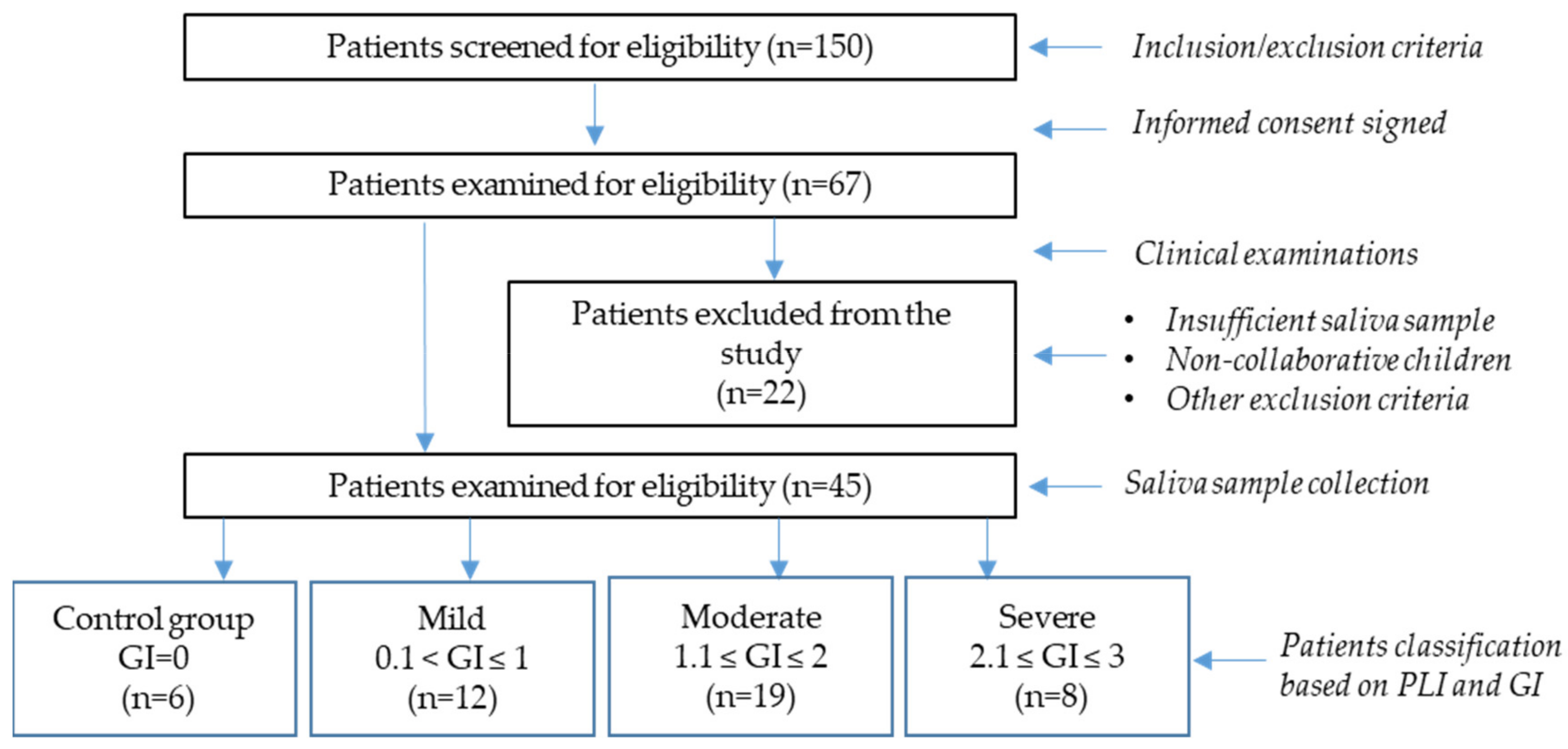
2.1. Study Design

This is a pilot prospective study. Declarations of informed consent were signed by the parents of the children or their legal guardians. The study was approved by the Ethics Committee of the Medical University of Varna (protocol No. 82/28.03.2019).
Participants in the study were recruited during their primary visit to the University Dental Medicine Center of the Medical University of Varna based on the inclusion and exclusion criteria.
Inclusion criteria:
  • Informed consent signed by the parent or guardian of each participant;
  • Age between 4–14 years;
  • No acute or chronic disease reported;
  • No medicine intake, including homeopathic remedies;
  • No known allergic reactions.
Exclusion criteria:
  • Children who are not collaborative during the examination and/or sample collection procedures;
  • Children with reported acute or chronic disease at the time of examination;
  • Children taking any treatment (anti-inflammatory, antibiotics, anti-allergy drugs, or homeopathic remedies);
  • Children with established allergic reactions;
  • Children displaying an open bite and mouth breathing;
This pilot prospective study included 45 children (26 girls and 19 boys) with no acute or chronic disease reported (aged between 4–14 years). For all participants, the clinical indices of PLI and GI were recorded and applied for the evaluation of the accumulation of dental plaque (Silness–Löe plaque index—PLI), assessment of gingival conditions, and registration of the qualitative changes in gingival tissues (Löe–Silness gingival index—GI).
The plaque index (PLI) is applied for the evaluation of dental plaque accumulation. This index is registered for Ramfjörd teeth 16, 22, 24, 36, 42 and 44. In children with primary and mixed dentition, in the case of missing representative teeth, the PLI is registered for mesially situated teeth. The recording of the PLI index is conducted by an atraumatic periodontal probe on the vestibular, oral, mesial, and distal surfaces along the site of contact with the marginal gingiva. The degree of dental plaque accumulation is recorded by the scores 0, 1, 2, and 3. The criteria are as follows: 0—no dental plaque on the examined tooth surface; 1—low levels of dental plaque on the peak of the probe; 2—moderate levels of dental plaque scratched by the probe; 3—a considerable, macroscopically visible accumulation of dental plaque on the tooth surface. The sum of all of the registered figures is divided by the total number of examined surfaces. The values of PLI in the interval from 0.1 to 1.1 indicate a condition that is “excellent with a tendency to have very good and good oral hygiene”. PLI values between 1.2 and 2.0 indicate a condition that is “good, with a tendency to have satisfactory oral hygiene”. PLI values from 2.1 to 3.0 are descriptive of a condition that is “satisfactory, with a tendency to have poor oral hygiene”.
The GI is utilized to evaluate the severity of gingivitis. This index records the marginal and interproximal tissues separately on the bases of 0 to 3. The same Ramfjörd teeth applied for the assessment for PLI are applied for the recording of GI, with the following criteria: 0—normal gingiva; 1—mild-inflammation hyperemia and slight edema, without bleeding on probing; 2—moderate-inflammation hyperemia, edema, and provoked bleeding; 3—severe-inflammation hyperemia, considerable edema, and spontaneous bleeding. The assessment of bleeding was applied by atraumatic sampling along the site of the soft tissue of the gingival sulcus. To calculate the GI, the values of the four walls of the gingival tissues encircling the tooth were summed up and divided by four. The sum of these values was divided by the total number of examined sites. By the application of the GI, the diagnostic scores were determined as follows: GI = 0—normal gingiva; GI levels from 0.1 to 1.0—mild degree of gingivitis; GI values from 1.1 to 2.0—moderate degree of gingivitis; GI values from 2.1 to 3.0—severe degree of gingivitis [21].
The participants were divided into four groups according to their PLI and GI [21]: a control group without gingival inflammation (GI = 0; n = 6); a group with a mild degree of PIG (0.1 < GI ≤ 1; n = 12); a group with a moderate degree of PIG (1.1 ≤ GI ≤ 2; n = 19); a group with a severe degree of PIG (2.1 ≤ GI ≤ 3; n = 8).
Clinical index determination and saliva sample collection were carried out on the day of dental examination and recruitment during the primary visit.
In summary, the study design is presented in Figure 1.

2.2. Sample Size Calculation

The calculation of the sample size was carried out according to El-Patal et al. (2022) [22]. The calculated sample size was equal to 64 children with a power of 80% and α = 0.05 using the SD (σ) with a margin of error of (E) = 0.8. In reality, the number of participants in our study was 44, because this is a pilot study and future investigations may proceed.

2.3. Salivary Sample Collection and Analysis

The collection of unstimulated whole saliva was conducted in sterile DNase- and RNase-free collection tubes, and the samples were frozen immediately on dry ice and stored at −80 °C until analysis. Saliva specimen collection was performed between 9.00 a.m. and 11.30 a.m. All the participants in the study were instructed to brush their teeth just before the sample collection. Enzyme-linked immunosorbent assays (ELISA) with monoclonal antibodies were used to determine the levels of IL-6, IL-1β, and CRP in saliva (Shanghai Sunred Biological Technology Co., Ltd., Shanghai, China). The respective absorptions were measured at 450 nm using a microtiter plate reader, Synergy 2, BioTek, Santa Clara, CA, USA.

2.4. Statistical Analysis

Statistical methods of descriptive statistics, an unpaired t-test, Spearman’s correlation and receiver operating characteristic (ROC) analysis were applied. The significance level was established at p < 0.05. GraphPad prism version 5 (Boston, MA, USA) was used to perform the statistical analysis.

3. Results

3.1. Clinical Indicators for Plaque-Induced Gingivitis

All the examined clinical indicators were characterized by considerably higher values in the groups with gingivitis, compared to the control group (p < 0.05) (Table 1).
The control group was represented by 4 girls and 2 boys. Two of the children had stable primary dentition. In four subjects, a state of mixed dentition was recorded. The mean age of the representatives in the group equaled 7 years and 5 months.
The group with a mild degree of gingivitis was represented by 5 girls and 7 boys. Two of the children had stable primary dentition. In ten subjects, a state of mixed dentition was recorded. The mean age of the representatives in the group equaled 7 years and 6 months.
The group with a moderate degree of gingivitis was represented by 11 girls and 8 boys. There were no children with stable primary dentition. In thirteen subjects, a state of mixed dentition was recorded. Five children had permanent dentition. The mean age of the representatives in the group equaled 7 years and 8 months.
The group with a severe degree of gingivitis was represented by 6 girls and 2 boys. There was one child with stable primary dentition. In seven subjects, a state of mixed dentition was recorded. The mean age of the representatives in the group equaled 8 years and 4 months.

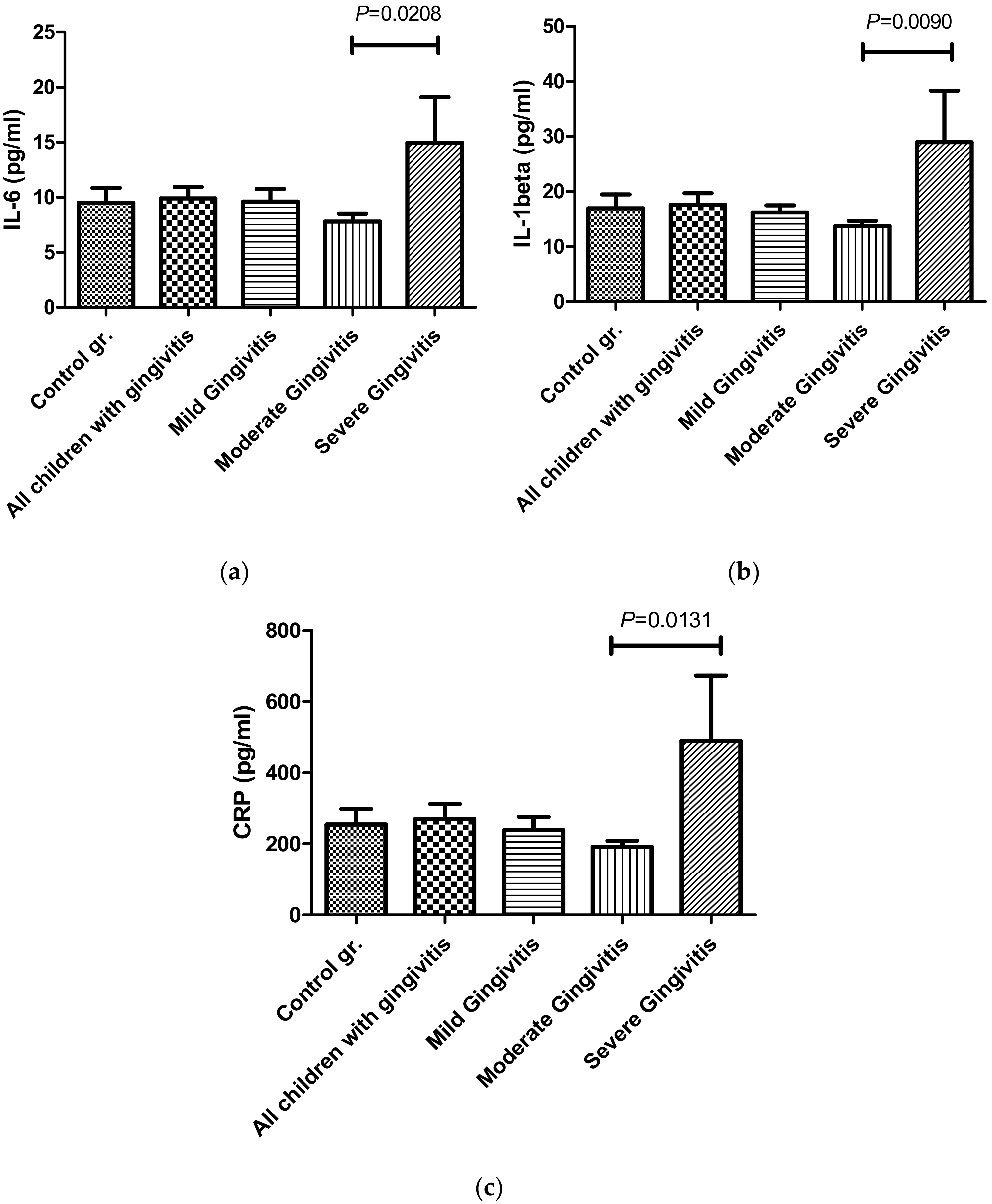
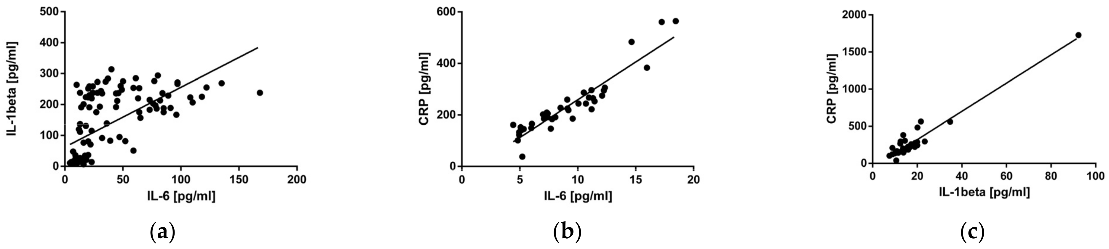
3.2. Salivary Levels of IL-6, IL-1β, and CRP in Children with Different Degrees of Plaque-Induced Gingivitis

In the present study, we examined the concentrations of IL-6, IL-1β, and CRP in unstimulated whole saliva from children with different degrees of PIG. The mean levels of IL-6, IL-1β, and CRP were higher in children with gingivitis compared to those of children in the control group (ns). The highest mean concentrations of IL-6, IL-1β, and CRP were recorded in the group with a severe degree of PIG, which were 14.96 pg/mL, 28.94 pg/mL, and 490.0 pg/mL, respectively. These values are considerably higher compared to those in the control group (9.506 pg/mL; 16.93 pg/mL; 254.4 pg/mL) (ns) (Figure 2). The mean concentration of IL-6 was elevated 1.57-fold, that of IL-1β was elevated 1.71-fold, and that of CRP was elevated 1.93-fold among the children with a severe degree of PIG compared to the control group. A statistically significant difference was established only between the groups with a moderate and severe degree of gingivitis for all three investigated markers (p < 0.05) (Figure 2). We assume that the relatively small number of representatives did not allow the establishment of a statistical significance between the other groups. The levels of these pro-inflammatory biomarkers had the lowest mean values among the children with a moderate degree of gingivitis compared to those in the other groups and the controls (ns) (Figure 2). The statistically significant differences in the mean concentrations of IL-6 (p = 0.0208), IL-1β (p = 0.009), and CRP (p = 0.0131) between the moderate and severe degrees of PIG are presented in Figure 2.
A positive significant correlation was observed between the investigated pro-inflammatory salivary biomarkers among all the participants with gingivitis. The Spearman’s rank correlation coefficients between IL-6 and IL-1β equaled 0.7576 (p < 0.0001); those between IL-6 and CRP equaled 0.9272 (p < 0.0001); and those between IL-1β and CRP equaled 0.7546 (p < 0.0001) (Figure 3).

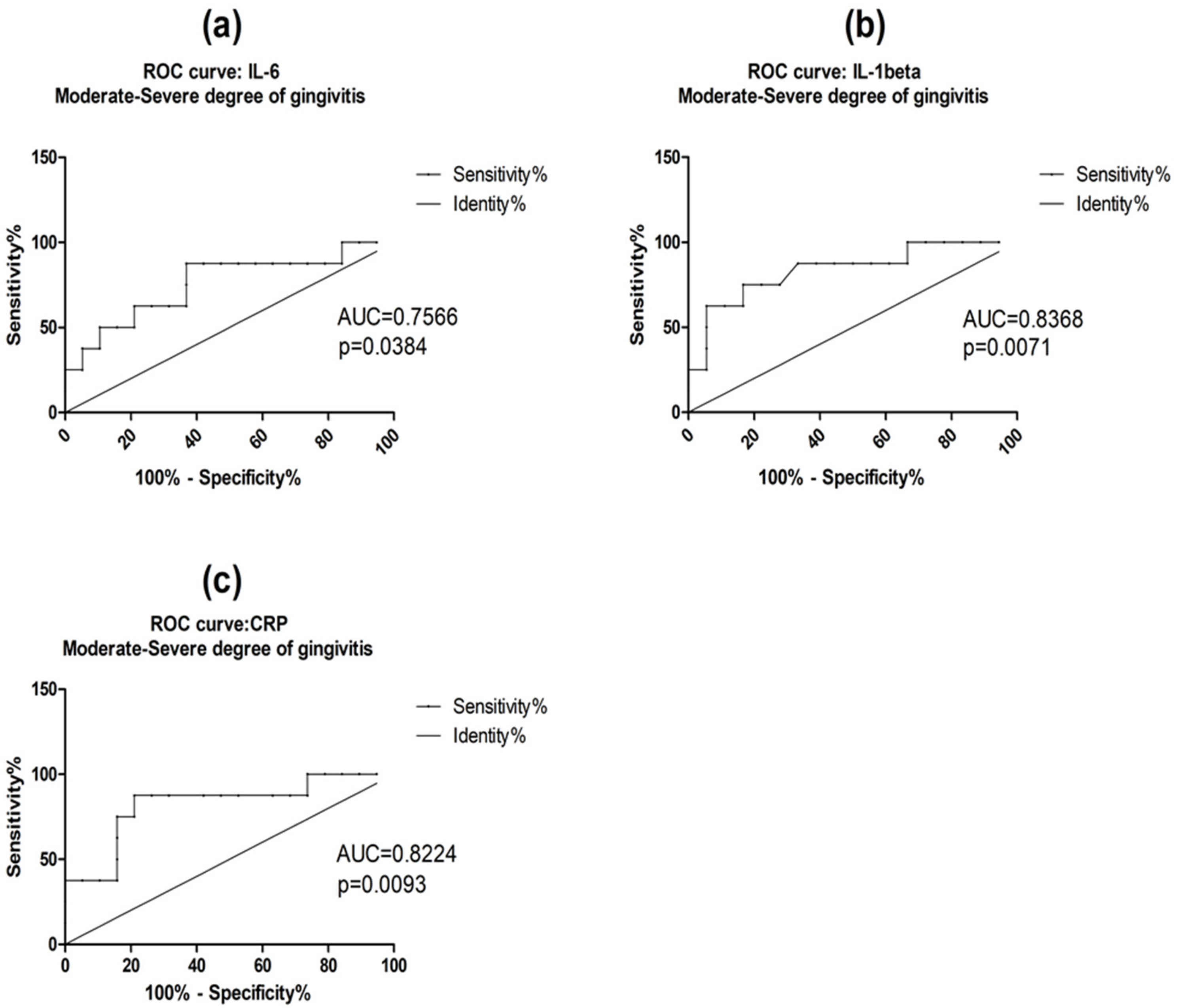
3.3. Diagnostic Accuracy of Salivary IL-6, IL-1β, and CRP

Based on the ROC curve analysis, salivary IL-1β and CRP showed good diagnostic accuracy (0.8 ≤ AUC < 0.9) and IL-6 showed fair diagnostic accuracy (0.7 ≤ AUC < 0.8), as categorized by Nahm et al. (2022) [23]. Statistical significance was found to distinguish children with moderate PIG from children with a severe degree of PIG (Table 2, Figure 4). At the optimal cut-off values, based on the largest Youden’s index, IL-6, IL-1β, and CRP demonstrated sensitivity values ranging from 75% to 87.5% and specificity values ranging from 63.16% to 83.33% (Table 3). Sensitivity for IL-6, CRP, and IL-1β was 87.5% (p = 0.0384), 87.5% (p = 0.0093), and 75% (p = 0.0071), respectively, and specificity was 63.16% (p = 0.0384), 78.95% (p = 0.0093), and 83.33% (p = 0.0071), respectively. IL-6 and CRP showed the highest sensitivity (87.5%) and IL-1β showed the highest specificity (83.33%), distinguishing between children with moderate PIG and children with a severe degree of PIG (p < 0.05) (Table 3).
We estimated the fair diagnostic accuracy (0.7 ≤ AUC < 0.8) of the investigated salivary pro-inflammatory biomarkers in distinguishing children without PIG from those with a severe degree of PIG (ns) (Table 4) [23]. IL-6 and CRP showed the highest sensitivity (87.5%) and IL-1β showed the highest specificity (83.33%), with p = 0.1968 (Table 5). Salivary IL-6, IL-1β, and CRP showed poor diagnostic capacity (0.6 ≤ AUC < 0.7) to discriminate between children without PIG and those with a mild or moderate degree of PIG (ns) (Table 4) [23]. Our future research has to include a larger number of participants in all of the studied groups.

4. Discussion

Non-invasive and routine laboratory examination of potential salivary biomarkers such as IL-1β, IL-6, and CRP can be applied as a novel tool for the assessment of PIG severity in children, and could contribute towards practitioners’ efforts in developing methods for the precise diagnosis, prognosis, treatment, and even prevention of PIG [1]. The precise diagnosis of the state of gingival inflammation in children is important for disease monitoring and control. In this study, we estimated that salivary IL-1β and CRP have good diagnostic accuracy and that IL-6 showed fair diagnostic accuracy in differentiating between children with moderate and children with a severe degree of PIG. These salivary markers may be implemented in the practice of precision dental medicine. The application of these markers in children with different degrees of PIG is important for the accurate diagnosis of the state of the disease, especially of a severe degree of PIG, and for adequate therapy and treatment to prevent the progression into periodontitis.
Etiologically, PIG is related to the accumulation of dental plaque, which has a tendency to increase the severity of pathological traits. The growth and maturation of dental plaque provoke inflammatory host tissue reactions. The conventional clinical parameters of the PLI and GI are applied for the evaluation of the degree of clinically manifested PIG [21]. The contemporary concept of personalized dental medicine is focused on the individual’s specifics, including the progression of periodontal diseases. The sensitivity and severity of the host’s immune response determine the course of inflammation on the systemic level and on the local level in the oral cavity.
The processes of the immune system that occur in response to the accumulation of bacterial biofilm are influenced by a great variety of pro- and anti-inflammatory cytokines and enzymes. Different studies have ascertained that immune cells in patients with diagnosed periodontal disorders release a greater amount of pro-inflammatory cytokines compared to cells in periodontally healthy individuals [21]. This corresponds to the higher salivary levels of the pro-inflammatory biomarkers of IL-6, IL-1β, and CRP among children suffering from PIG in comparison to controls, as established in our study. Other authors also report that the salivary concentration of IL-6 is increased among individuals with gingival inflammation [24,25].
We established a statistically significant difference in the mean levels of IL-6, IL-1β, and CRP between children with a moderate degree of PIG and those with a severe degree of PIG. The salivary levels of these pro-inflammatory biomarkers were significantly higher in the group with a severe degree of PIG compared to the group with a moderate degree of gingivitis. This is associated with the development of the inflammatory process that occurs in parallel to the progression of gingivitis. IL-1β and IL-6 are characterized as basic innate cytokines and serve as key inflammatory mediators under conditions of periodontal disorder [26,27]. IL-1β is secreted by monocytes, macrophages, and neutrophils. This pro-inflammatory cytokine is associated with the secretion of other mediators that provoke not only inflammatory alterations but also tissue damage [27,28]. Its function is related to the specifics of the clinical manifestation of PIG with the progression from moderate to severe degrees of PIG. The possible physiological feature connected to the decreased levels of IL-6, IL-1β, and CRP in the group with a moderate degree of PIG could be the shift in specific pathogenic microflora in the children with a severe degree of gingivitis, which would have led to an increase in the severity of the inflammatory response, increased gingival inflammation, and increased production of inflammatory mediators, such as IL-6 and IL-1β [21]. The qualitative and quantitative content of plaque is different as a result of plaque maturation [29,30]. The significant difference between moderate and severe PIG only seems reasonable because in patients with a low degree of PIG, host protective mechanisms (e.g., cell immunity) could still be not active enough. In patients with a moderate degree of PIG, the immunological mechanism differs from that of the severe form. The transition from a moderate to severe degree of gingivitis has been reported to be characterized by a shift from the innate immune response to the acquired immune response [31]. Macrophages, which predominate in the innate phase, have a reduced capacity for pro-inflammatory cytokine production compared to B-cells, which represent the acquired immune response [31].
We did not establish a statistical significance between IL-6, IL-1β, and CRP levels in the control group and the group with a mild degree of PIG because the PIG in the latter group was characterized as an initial phase of plaque-induced gingivitis. This initial phase of PIG is characterized by the formation of so-called initial lesions, where the appearance of gingival inflammation is low and characterized by hyperemia and slight edema, without bleeding upon probing [32]. The observed cytokine levels in the saliva in our study were also low, and not statistically different compared to those of the control group. The innate immune mechanisms are active during this stage and cytokine production is very low. The rate of collagen destruction also is low [32].
The moderate degree of gingivitis is characterized by hyperemia, edema, provoked bleeding, and the formation of early lesions. The inflammatory cells which are involved in this stage are lymphocytes and macrophages. The collagen degradation rate is higher than that in mild-degree PIG but lower than in severe-degree PIG [32]. The observed lowest levels of IL-6, IL-1β, and CRP in moderate-degree PIG could be due to the resolution of macrophages from M1 (classical) into M2 (alternative) phenotypes [33]. The M1 phenotype is the pro-inflammatory phenotype which is characterized by increased IL-6 production [34,35]. The M2 phenotype is involved in tissue repair and is characterized by decreased IL-6 production [36,37]. Probably, the anti-inflammatory M2 phenotype involved in active tissue repair is the distinctive immune response, a compensatory mechanism in moderate-degree PIG [36,37]. This explains the lowest cytokine levels that were found in moderate-degree PIG. Garaicoa-Pazmino et al. (2019) discuss in their study the transition of macrophages from M1 to M2 phenotypes in patients with gingivitis and periodontitis. Interestingly, they also did not find statistically significant differences between the M1 and M2 polarization of macrophages in healthy tissues and tissues with periodontitis [33]. The increased production of IL-6 and IL-1β demonstrated by the results of our study may have been related to the infiltration of T and B cells, which is typical for severe-degree PIG only. In severe gingivitis, the highest inflammatory response occurs through the increased local secretion of pro-inflammatory cytokines and other inflammatory molecules [32].
The established significant positive correlation between salivary levels of IL-6, IL-1β, and CRP among the participants with PIG is confirmed by several studies exploring the mechanism of interaction between these pro-inflammatory biomarkers. Bacterial lipopolysaccharide and IL-1β significantly increase IL-6 production in human gingival fibroblasts [27]. It was shown that the combined activity of IL-1β and IL-6 enhanced gingival inflammation and the risk of progression of periodontitis [27,38]. The established strong positive correlation between the pro-inflammatory cytokines IL-6 and IL-1β and the acute phase protein CRP described in our study has been widely explored and corroborated in the scientific literature [28,38]. Inflammatory stimuli such as IL-1β and IL-6 induce the synthesis of CRP from the liver. IL-6 and IL-1β act synergistically for the induction of CRP gene expression, as IL-1β alone has no effect [39]. These observations confirm the combined action of these molecules and their role in the development of inflammation [28,38,39].
The good diagnostic accuracy of these salivary biomarkers was confirmed by ROC curve analyses and the established AUC values in our study. IL-1β and CRP showed good diagnostic accuracy (0.8 ≤ AUC < 0.9) and IL-6 showed fair diagnostic accuracy (0.7 ≤ AUC < 0.8) with statistical significance in distinguishing between children with moderate PIG and children with a severe degree of PIG. the t-test analysis established a statistically significant difference between the levels of IL-1β, IL-6, and CRP in children with moderate and severe degrees of gingivitis. The trend of the considerable elevation of these inflammatory biomarkers parallel to the progression of PIG from a moderate to a severe degree could have been due to the action of the acquired immune system in the oral cavity. The rapid increase in the levels of these inflammatory parameters may be associated with the activation of enzymes involved in tissue destruction [40]. The results of our study are in line with the findings of other investigations in the field. The adoption of these inflammatory biomarkers in non-stimulated mixed saliva has the potential to serve as a prognostic tool for the progression of gingivitis from moderate to severe degrees before the clinical manifestation of the advanced inflammation of gingival tissues. Namely, the combined application of IL-6, IL-1β, and CRP, interpreted in the context of recording clinical indices and the evaluation of clinical findings and symptoms, can serve as prognostic and diagnostic indicators of the risk of PIG progression. This is key to the development of adequate, personalized prophylactic measures and the efficient control and minimization of the risk of the progression of PIG in later periods. The poor control and inefficient, sporadic treatment of PIG in children can be a predisposing factor for the initiation of periodontitis in adulthood. Many authors accentuate the late impact of gingivitis in children in terms of its effect on the gingival sulcus, which facilitates the establishment of the disease and the colonization of the site by periodontal pathogenic microflora [1]. On an epidemiological basis, strong evidence exists that shows the progression of gingivitis to periodontitis in adults [1]. In our study, we accentuated the relationship between clinical parameters for the wide-spread oral disorder of PIG and for inflammatory biomarkers in non-stimulated saliva. In light of the trending advancement of non-invasive and simple methods of assessing inflammation status, our results concerning the diagnostic potential of IL-6, IL-1β, and CRP as biomarkers for differentiating between moderate and severe degrees of clinical manifestation of PIG, can be useful for the development of gingivitis prevention strategies. Pediatric and dental practitioners should take into account the fact that the clinical manifestation of the severe forms of gingivitis in primary, mixed, and permanent dentition correlates with the degree of inflammatory cell infiltration into gingival tissues. It has been established that the chronic inflammation of periodontal tissues in childhood can provoke local tissue destruction, which could later develop into periodontitis [1]. Our findings are also relevant to the prophylaxis and limitations of favorable conditions that predispose gingivitis to periodontitis progression in later years. Non-invasive and routine laboratory biomarkers such as IL-1β and CRP with new applications in the diagnosis of the degree of PIG in children may add to the efforts of practitioners in the precise diagnosis, prognosis, treatment, and even prevention of the progression of PIG [1].
The main limitation of this study is the relatively small number of representatives in the examined groups, which will be taken into account in future work. Based on scientific sources, we do assume that under conditions in which there is a larger number of participants, we would obtain statistically significant results when comparing all the study groups, including the control.
This is a pilot study and future investigations may proceed. The search for non-invasive informative biomarkers is expected to provide fast and reliable diagnostics, especially in children. In this aspect, saliva appears to be a suitable matrix for further studies in dental medicine and oral biology.

5. Conclusions

Based on our results, we suggest salivary IL-1β and CRP as potential diagnostic tools to differentiate between moderate and severe degrees of plaque-induced gingivitis. Given the therapeutic approaches that differ depending on the severity of the disease, these markers could be implemented in personalized dental clinical practice.

Author Contributions

Conceptualization, S.A., Y.K.-K., D.I., S.P., A.S., B.R. and T.S.; methodology, B.R., A.S. and S.A.; validation, S.A., A.S. and B.R.; formal analysis, A.S., S.A., B.R. and T.S.; resources, S.A.; data curation, A.S., S.A. and B.R.; writing—original draft preparation, S.A., A.S., B.R. and T.S.; writing—review and editing, D.I., Y.K.-K. and S.P.; visualization, A.S., S.A., B.R. and T.S.; supervision, D.I., S.P. and Y.K.-K.; All authors have read and agreed to the published version of the manuscript.

Funding

This research was funded by Fund “Science” MU-Varna, Bulgaria, project number 18036, FN-12/11.02.2019.

Institutional Review Board Statement

The study was conducted in accordance with the Declaration of Helsinki, and approved by the Ethics Committee of Medical University Varna (protocol no. 82/28.03.2019).

Informed Consent Statement

Informed consent was given and a declaration of informed consent was signed by parents or legal guardians of children accompanying each child participating in the research.

Data Availability Statement

Not applicable.

Conflicts of Interest

The authors declare no conflict of interest.

References

  1. Bimstein, E.; Huja, P.E.; Ebersole, J.L. The potential lifespan impact of gingivitis and periodontitis in children. J. Clin. Pediatr. Dent. 2013, 38, 95–99. [Google Scholar] [CrossRef] [PubMed]
  2. Modéer, T.; Wondimu, B. Periodontal diseases in children and adolescents. Rev. Dent. Clin. N. Am. 2000, 44, 633–658. [Google Scholar] [CrossRef]
  3. Murakami, S.; Mealey, B.L.; Mariotti, A.; Chapple, I.L.C. Dental plaque-induced gingival conditions. J. Periodontol. 2018, 89, 17–27. [Google Scholar] [CrossRef]
  4. Ahmadi-Motamayel, F.; Goodarzi, M.T.; Mahdavinezhad, A.; Jamshidi, Z.; Darvishi, M. Salivary and serum antioxidant and oxidative stress markers in dental caries. Caries Res. 2018, 52, 565–569. [Google Scholar] [CrossRef]
  5. Kaczor-Urbanowicz, K.E.; Trivedi, H.M.; Lima, P.O.; Camargo, P.M.; Giannobile, W.V.; Grogan, T.R.; Gleber-Netto, F.O.; Whiteman, Y.; Li, F.; Lee, H.J.; et al. Salivary exRNA biomarkers to detect gingivitis and monitor disease regression. J. Clin. Periodontol. 2018, 45, 806–817. [Google Scholar] [CrossRef]
  6. Jenkins, W.M.; Papapanou, P.N. Epidemiology of periodontal disease in children and adolescents. Periodontol. 2000 2001, 26, 16–32. [Google Scholar] [CrossRef]
  7. Singh, A.K. Prevalence of gingivitis and periodontitis among schools children in Lucknow region of Uttar Pradesh, India. IOSR J. Dent. Med. Sci. 2014, 13, 21–23. [Google Scholar] [CrossRef]
  8. Bossnjak, A.; Curilovic, Z.; Vuccicevic-Boras, V.; Plancak, D.; Jorgic-Srdjak, K.; Relja, T.; Bozic, D.; Varnica, H. Prevalence of gingivitis in 6- to 11-year-old Croatian children. Eur. J. Med. Res. 2003, 8, 313–317. [Google Scholar]
  9. Leous, P.; Palianskaya, L.; Leous, L. Oral hygiene and gingival inflammation in 6–8-year-olds from a junior school in Minsk who participated in a supervised oral hygiene programme. Oral Health Dent. Manag. 2009, 7, 27–30. [Google Scholar]
  10. Burt, B. Research, Science and Therapy Committee of the American Academy of Periodontology. Position paper: Epidemiology of periodontal diseases. J. Periodontol. 2005, 76, 1406–1419. [Google Scholar] [CrossRef]
  11. Dye, B.A. Global periodontal disease epidemiology. Periodontol. 2000 2012, 58, 10–25. [Google Scholar] [CrossRef] [PubMed]
  12. Yaghobee, S.; Khorsand, A.; Rasouli Ghohroudi, A.A.; Sanjari, K.; Kadkhodazadeh, M. Assessment of interleukin-1beta and interleukin-6 in the crevicular fluid around healthy implants, implants with peri-implantitis, and healthy teeth: A cross-sectional study. J. Korean Assoc. Oral Maxillofac. Surg. 2014, 40, 220–224. [Google Scholar] [CrossRef] [PubMed]
  13. Amar, S.; Oyaisu, K.; Li, L.; Van Dyke, T. Moesin: A potential LPS receptor on human monocytes. J. Endotoxin. Res. 2001, 7, 281–286. [Google Scholar] [CrossRef] [PubMed]
  14. Isola, G.; Polizzi, A.; Santonocito, S.; Alibrandi, A.; Williams, R.C. Periodontitis activates the NLRP3 inflammasome in serum and saliva. J. Periodontol. 2022, 93, 135–145. [Google Scholar] [CrossRef] [PubMed]
  15. Gorska, R.; Gregorek, H.; Kowalski, J.; Laskus-Perendyk, A.; Syczewska, M.; Madaliński, K. Relationship between clinical parameters and cytokine profiles in inflamed gingival tissue and serum samples from patients with chronic periodontitis. J. Clin. Periodontol. 2003, 30, 1046–1052. [Google Scholar] [CrossRef]
  16. Aurer, A.; Jorgić-Srdjak, K.; Plancak, D.; Stavljenić-Rukavina, A.; Aurer-Kozelj, J. Proinflammatory factors in saliva as possible markers for periodontal disease. Coll. Antropol. 2005, 29, 435–439. [Google Scholar]
  17. Shojaee, M.; Fereydooni Golpasha, M.; Maliji, G.; Bijani, A.; Aghajanpour Mir, S.M.; Mousavi Kani, S.N. Creactive protein levels in patients with periodontal disease and normal subjects. Int. J. Mol. Cell Med. 2013, 2, 151–155. [Google Scholar]
  18. Belstrøm, D.; Damgaard, C.; Könönen, E.; Gürsoy, M.; Holmstrup, P.; Gürsoy, U.K. Salivary cytokine levels in early gingival inflammation. J. Oral Microbiol. 2017, 9, 1364101. [Google Scholar] [CrossRef]
  19. Boronat-Catalá, M.; Catalá-Pizarro, M.; Bagán Sebastián, J.V. Salivary and crevicular fluid interleukins in gingivitis. J. Clin. Exp. Dent. 2014, 2, e175–e179. [Google Scholar] [CrossRef]
  20. Kim, J.Y.; Kim, K.R.; Kim, H.N. The potential impact of salivary IL-1 on the diagnosis of periodontal disease: A pilot study. Healthcare 2021, 9, 729. [Google Scholar] [CrossRef]
  21. Rebelo, M.A.; Queiroz, A.C. Gingival Indices: State of Art. In Gingival Diseases—Their Aetiology, Prevention and Treatment; Panagakos, F., Davies, R., Eds.; IntechOpen: Rijeka, Croatia, 2011; pp. 41–54. [Google Scholar] [CrossRef]
  22. El-Patal, M.A.; Khalil, M.A.; Shipl, W.; Barakat, I.; Youssef, E.M.I.; El Attar, S.; Fathi, A.; Abdallah, A.A. Detection of soluble urokinase type plasminogen activator receptors in children with gingivitis and normal subjects. BMC Oral Health. 2022, 22, 436. [Google Scholar] [CrossRef] [PubMed]
  23. Nahm, F.S. Receiver operating characteristic curve: Overview and practical use for clinicians. Korean J. Anesthesiol. 2022, 75, 25–36. [Google Scholar] [CrossRef] [PubMed]
  24. Taylor, J.J.; Preshaw, P.M.; Donaldson, P.T. Cytokine gene polymorphism and immunoregulation in periodontal disease. Periodontol. 2000 2004, 35, 158–182. [Google Scholar] [CrossRef] [PubMed]
  25. Lee, A.; Ghaname, C.B.; Braun, T.M.; Sugai, J.V.; Teles, R.P.; Loesche, W.J.; Kornman, K.S.; Giannobile, W.V.; Kinney, J.S. Bacterial and salivary biomarkers predict the gingival inflammatory profile. J. Periodontol. 2012, 83, 79–89. [Google Scholar] [CrossRef]
  26. Syndergaard, B.; Al-Sabbagh, M.; Kryscio, R.J.; Xi, J.; Ding, X.; Ebersole, J.L.; Miller, C.S. Salivary biomarkers associated with gingivitis and response to therapy. J. Periodontol. 2014, 85, e295–e303. [Google Scholar] [CrossRef]
  27. Naruishi, K.; Nagata, T. Biological effects of interleukin-6 on Gingival Fibroblasts: Cytokine regulation in periodontitis. J. Cell Physiol. 2018, 233, 6393–6400. [Google Scholar] [CrossRef]
  28. Volanakis, J.E. Human C-reactive protein: Expression, structure, and function. Mol. Immunol. 2001, 38, 189–197. [Google Scholar] [CrossRef]
  29. Marsh, P.D.; Moter, A.; Devine, D.A. Dental plaque biofilms: Communities, conflict and control. Periodontol. 2000 2011, 55, 16–35. [Google Scholar] [CrossRef]
  30. Marsh, P.D. Are dental diseases examples of ecological catastrophes? Microbiology 2003, 149, 279–294. [Google Scholar] [CrossRef]
  31. Cekici, A.; Kantarci, A.; Hasturk, H.; Van Dyke, T.E. Inflammatory and immune pathways in the pathogenesis of periodontal disease. Periodontol. 2000 2014, 64, 57–80. [Google Scholar] [CrossRef]
  32. Rathee, M.; Jain, P. Gingivitis; StatPearls Publishing: Treasure Island, FL, USA, 2022. [Google Scholar]
  33. Garaicoa-Pazmino, C.; Fretwurst, T.; Squarize, C.H.; Berglundh, T.; Giannobile, W.V.; Larsson, L.; Castilho, R.M. Characterization of macrophage polarization in periodontal disease. J. Clin. Periodontol. 2019, 46, 830–839. [Google Scholar] [CrossRef] [PubMed]
  34. Yu, T.; Zhao, L.; Huang, X.; Ma, C.; Wang, Y.; Zhang, J.; Xuan, D. Enhanced activity of the macrophage m1/m2 phenotypes and phenotypic switch to m1 in periodontal infection. J. Periodontol. 2016, 87, 1092–1102. [Google Scholar] [CrossRef] [PubMed]
  35. Martinez, F.O.; Gordon, S. The M1 and M2 paradigm of macrophage activation: Time for reassessment. F1000Prime Rep. 2014, 6, 13. [Google Scholar] [CrossRef] [PubMed]
  36. Das, A.; Sinha, M.; Datta, S.; Abas, M.; Chaffee, S.; Sen, C.K.; Roy, S. Monocyte and macrophage plasticity in tissue repair and regeneration. Am. J. Pathol. 2015, 185, 2596–2606. [Google Scholar] [CrossRef] [PubMed]
  37. Garlet, G.P.; Giannobile, W.V. Macrophages: The bridge between inflammation resolution and tissue repair? J. Dent. Res. 2018, 97, 1079–1081. [Google Scholar] [CrossRef] [PubMed]
  38. Young, D.P.; Kushner, I.; Samols, D. Binding of C/EBPbeta to the C-reactive protein (CRP) promoter in Hep3B cells is associated with transcription of CRP mRNA. J. Immunol. 2008, 181, 2420–2427. [Google Scholar] [CrossRef]
  39. Kida, Y.; Kobayashi, M.; Suzuki, T.; Takeshita, A.; Okamatsu, Y.; Hanazawa, S.; Hasegawa, K. Interleukin-1 stimulates cytokines, prostaglandin E2 and matrix metalloproteinase-1 production via activation of MAPK/AP-1 and NF-kappaB in human gingival fibroblasts. Cytokine 2005, 29, 159–168. [Google Scholar] [CrossRef]
  40. Sawada, S.; Chosa, N.; Ishisaki, A.; Naruishi, K. Enhancement of gingival inflammation induced by synergism of IL-1β and IL-6. Biomed. Res. 2013, 34, 31–40. [Google Scholar] [CrossRef]
Figure 1. Study design.
Applsci 13 05046 g001
Figure 2. Mean concentrations of (a) IL-6, (b) IL-1β, and (c) CRP in the saliva of children with different degrees of PIG, the group with all of the children with plaque-induced gingivitis and healthy controls. Data expressed as mean ± SD. Significance level established at p < 0.05. Abbreviations: IL-6—interleukin-6, IL-1β—interleukin-1β, and CRP—C-reactive protein.
Applsci 13 05046 g002
Figure 3. Spearman′s correlations between (a) IL-6/IL-1β, (b) IL-6/CRP and (c) IL-1 β/CRP in the saliva of children with PIG. Abbreviations: IL-6—interleukin-6, IL-1β—interleukin-1β, and CRP—C-reactive protein.
Applsci 13 05046 g003
Figure 4. ROC curves of (a) IL-6, (b) IL-1β, and (c) CRP as potential diagnostic biomarkers for differentiating children with a moderate degree of PIG from children with a severe degree of PIG. Significance level established at p < 0.05. Abbreviations: ROC—receiver operating characteristic; AUC—area under the curve; IL-6—interleukin-6; IL-1β—interleukin-1β; CRP—C-reactive protein.
Applsci 13 05046 g004
Table 1. Clinical indicators of PIG.
Clinical IndicatorsControl Group
n = 6
Mild-Degree
n = 12
Moderate-Degree
n = 18
Severe-Degree
n = 8
p Value
t-Test
PLI0.74 ± 0.251.2 ± 0.121.64 ± 0.291.98 ± 0.21p < 0.05
GI0 ± 00.75 ± 0.241.38 ± 0.172.19 ± 0.09p < 0.05
Data are expressed as mean ± SD; abbreviations: PLI (Silness–Löe plaque index), and GI (Löe–Silness gingival index); significance level established at p < 0.05.
Table 2. ROC logistic regression analysis of salivary IL-6, IL-1β, and CRP levels comparing the groups of children with different degrees of plaque-induced gingivitis.
Mild–Moderate Degree of Gingivitis
AUC (95% CI)
p-ValueMild–Severe
Degree of Gingivitis
AUC (95% CI)
p-ValueModerate–Severe Degree of Gingivitis
AUC (95% CI)
p-Value
IL-60.62500.24780.63540.31590.75660.0384
IL-1β0.66200.13850.69790.14270.83680.0071
CRP0.62720.23960.67190.20310.82240.0093
The significance level was established at p < 0.05. Abbreviations: ROC—Receiver Operating Characteristic; AUC—Area Under the Curve; CI—Confidence Interval.
Table 3. ROC logistic regression analysis of salivary IL-6, IL-1β, and CRP as potential diagnostic biomarkers to differentiate children with a moderate degree of PIG from children with a severe degree of PIG.
Cut-OffAUC
(95% CI)
Youden IndexSensitivity%Specificity%p-Value
IL-68.385
pg/mL
0.75660.506687.5063.160.0384
IL-1β16.99
pg/mL
0.83680.583375.0083.330.0071
CRP223.8
pg/mL
0.82240.664587.5078.950.0093
Significance level established at p < 0.05. Abbreviations: AUC—Area Under the Curve; CI—Confidence Interval.
Table 4. ROC logistic regression analysis of salivary IL-6, IL-1β, and CRP levels comparing the control group with the groups with mild, moderate, and severe degrees of plaque-induced gingivitis.
Control Group—Mild Degree of Gingivitis
AUC (95% CI)
p-ValueControl Group—Moderate Degree of Gingivitis
AUC (95% CI)
p-ValueControl Group—Severe Degree of Gingivitis
AUC (95% CI)
p-Value
IL-60.51390.92540.67110.21480.70830.1968
IL-1β0.51390.92540.64350.30150.70830.1968
CRP0.51390.92540.66670.22670.70830.1968
Significance level established at p < 0.05. Abbreviations: ROC—receiver operating characteristic; AUC—area under the curve; CI—confidence interval; IL-6—interleukin-6; IL-1β—interleukin-1β; CRP—C-reactive protein.
Table 5. ROC logistic regression analysis of salivary IL-6, IL-1β, and CRP as potential diagnostic biomarkers for differentiating between children with a severe degree of PIG and those without plaque-induced gingival inflammation.
Cut-OffAUC
(95% CI)
Youden IndexSensitivity%Specificity%p-Value
IL-68.576
pg/mL
0.70830.541787.5066.670.1968
IL-1β16.62
pg/ml
0.70830.583375.0083.330.1968
CRP218.4
pg/ml
0.70830.541787.5066.670.1968
Significance level established at p < 0.05. Abbreviations: ROC—receiver operating characteristic; AUC—area under the Curve; CI—confidence interval; IL-6—interleukin-6; IL-1β—interleukin-1β; CRP—C-reactive protein.
Disclaimer/Publisher’s Note: The statements, opinions and data contained in all publications are solely those of the individual author(s) and contributor(s) and not of MDPI and/or the editor(s). MDPI and/or the editor(s) disclaim responsibility for any injury to people or property resulting from any ideas, methods, instructions or products referred to in the content.

