
Color Stability of Nanoparticles-Modified Dental Resin-Based Composites
by
 , , and
Yousif A. Al-Dulaijan
1,* ,
,
Maram A. AlGhamdi
1,
Emad Azmy
2,
Mohamed Reda Zaki Al-Kholy
3,
Khalid S. Almulhim
4 and
Mohamed A. Helal
3,* 1
Department of Substitutive Dental Sciences, College of Dentistry, Imam Abdulrahman Bin Faisal University, P.O. Box 1982, Dammam 31441, Saudi Arabia
2
Department of Prosthodontics, Faculty of Dental Medicine, Sinai University, Sinai 16020, Egypt
3
Department of Prosthodontics, Faculty of Dental Medicine, Al-Azhar University, Cairo 11751, Egypt
4
Department of Restorative Dental Sciences, College of Dentistry, Imam Abdulrahman Bin Faisal University, P.O. Box 1982, Dammam 31441, Saudi Arabia
*
Authors to whom correspondence should be addressed.
Appl. Sci. 2023, 13(6), 3870; https://doi.org/10.3390/app13063870
Submission received: 25 January 2023
/
Revised: 14 March 2023
/
Accepted: 16 March 2023
/
Published: 18 March 2023
(This article belongs to the Special Issue Developments and Applications of Dental Materials II)
Abstract
:To evaluate the effect of beverages (coffee, tea, Cola-Cola, and mineral water) on the color stability of resin-based composite (RBC) materials modified with different nanoparticles (NPs). The specimens (70/beverage) were fabricated from light-cured RBCs and divided according to NPs into four groups: one control, unmodified (N0); and three experimental—ZrO2 (Zr), TiO2 (Ti), and SiO2 (Si) groups. Each experimental group was further subdivided into two subgroups according to NP concentrations: 3 wt.% and 7 wt.% (n = 10). A spectrophotometer was used to assess the color change (∆E) before and after six months of immersion. Data were analyzed and compared using one-way-ANOVA followed by Bonferroni’s post-hoc test (α = 0.05) and subsequently ∆E value conversion to National Bureau of Standards (NBS) units. The modified light-cured RBCs with ZrO2, TiO2, and SiO2 demonstrated smaller color changes after immersion in the beverages than the unmodified group (p < 0.001). Zr groups showed the lowest ΔE, followed by Ti and Si groups; a 3% concentration resulted in a lower mean ΔE than the 7% concentration. NBS findings showed that coffee and tea produced marked unacceptable color changes (NBS units were >3), and Coca-Cola resulted in noticeable color changes (NBS units between 1.5 and 3), while water produced slight color changes (NBS ≤ 1). Modification of RBCs with both concentrations of ZrO2, as well as 3% of TiO2 and SiO2, may improve its color stability. Based on NBS results, RBCs immersed in mineral water and Coca-Cola showed clinically acceptable color changes, while those immersed in coffee and tea were clinically unacceptable.
1. Introduction
Resin-based composites (RBCs) have been among the most common choices for restoring anterior and posterior teeth, mainly due to their high aesthetic properties, improved durability and success rate, and high patient acceptance. However, inadequate mechanical properties, polymerization shrinkage, plaque accumulation, and stainability are the major drawbacks of dental restorations [1,2,3].
The staining susceptibility of restorative materials is influenced by several factors. Intrinsic resin matrix degradation can occur due to oxidation or hydrolysis in the matrix of filling materials [4,5,6]. Moreover, the structural properties of resin matrix monomers, filler size, and distribution in the matrix play essential roles in the discoloration effect. The incorporation of inorganic nanoparticles (NPs) was found to decrease the hygroscopic absorption of water in the resin matrix, leading to less discoloration [7,8]. On the other hand, extrinsic discoloration may also occur, due to the absorption of staining products found in food and drinks [9].
Many previous studies have shown that immersion of light-cured composite restorative material in different beverages may cause varying degrees of staining. This variation depends mainly on their composition and properties, the concentration of staining agents, and the amount of exposure time [10,11]. Several advancements were introduced to improve the properties of composite filling, including the incorporation of nanofillers, which enhance their mechanical, physical, and optical properties [12,13].
The use of NPs can offer many advantages when they are incorporated into dental restorations; due to their decreased size, they provide a larger specific surface area that leads to unique characterization compared to bulk-size particles [3,14]. However, NPs show an aggregation tendency that may interfere with the chemical interaction between these particles and the organic matrix. To overcome this drawback, a silane-coupling agent was used for the treatment of inorganic filler, leading to improved bond strength of the NPs and resin matrix and thus enhancing the properties of nanocomposite [15].
This enhancement encouraged the researchers to incorporate different NPs into dental biomaterials to improve their properties, including zirconium dioxide (ZrO2), titanium dioxide (TiO2), and silicon dioxide (SiO2) [16,17,18]. TiO2 is favored in the dental field due to its superior properties, including corrosion resistance, high microhardness, adequate antimicrobial properties, and low cost. Moreover, the incorporation of TiO2 into polymeric materials can enhance the mechanical, physical, and optical properties of the nanocomposite [19]. ZrO2 is a white crystalline metal oxide that has widespread application in dentistry due to its biocompatibility, great strength, high wear resistance, and aesthetic acceptability; it has been used in many previous studies for the reinforcement of acrylic resin denture base materials and RBCs [12,19]. Similarly, the tested SiO2-incorporated nanocomposites showed adequate resistance to abrasiveness and appropriate thermal stability. Prosthetically, the addition of SiO2 nanoparticles to the acrylic resin denture base improved the wear resistance; additionally, it showed greater color stability after immersion in different beverage solutions [20,21]. Azmy et al. and Liu et al. found that the addition of NPs to dental restorations could improve their different properties, such as flexural strength, hardness, and wear resistance [12,22].
The color stability of a restorative material greatly determines the success of dental fillings and patient acceptance [23]. NPs incorporating restorative materials presented improved optical properties when tested in previous studies. Because the NP dimension is less than the wavelength of visible light, the results showed an improved color appearance and enhanced light transmittance [24,25].
Color measurement devices have been heavily used when testing dental materials to accurately assess their color changes. A spectrophotometer is considered an accurate and flexible measurement tool [26]. Compared to human eye observation, it can provide 33% increased accuracy and allow for higher objective matches in most of the cases tested [27]. A three-dimensional color space-based system was developed by the International Commission on Illumination (Commission Internationale de l’Eclairage; CIE) in 1976, and it consists of three axes. The L axis is a grayscale that represents the lightness, the a axis represents the red-green coordinates, and the b axis describes the blue-yellow coordinates. This CIE L*, a*, b* scale covers all colors visible to the human eye, and using these coordinates, color changes (∆E) are usually calculated using the changes in the color parameters of the CIE L*, a*, b* system [28].
Several studies have assessed the color stability of regular dental restorations when immersed in different solutions; however, little is known regarding the effect of this immersion on the color stability of dental restorations modified with different nanoparticles. Accordingly, the purpose of this study was to assess the color stability of light-cured composite filling reinforced with two concentrations of NPs (ZrO2, TiO2, and SiO2) after immersion into four different solutions (coffee, tea, Coca-Cola, and mineral water). The null hypothesis stated that NP (ZrO2, TiO2, or SiO2) incorporation at two concentrations (3 wt.% and 7 wt.%) into RBCs would have no significant effects on color stability.
2. Materials and Methods
Table 1 summarizes all material details used to conduct the current study. Disc-shaped specimens were prepared according to ISO 4049:2000 that were 7 × 2 ± 0.03 mm in diameter and thickness, respectively [29]. To reduce variability, all specimens were fabricated by the same examiner.
Two hundred RBC specimens (n = 10, 70/beverage) were fabricated based on a sample size calculated using the necessary values from previous studies [20,21,30]. According to NP types, specimens were divided into four groups: three modified groups—ZrO2 (Zr), TiO2 (Ti), and SiO2 (Si); and one that remained unmodified as a control group (N0). According to NP concentrations (3 wt.% and 7 wt.%), these three groups were further subdivided into two subgroups (Zr3, Zr7, Ti3, Ti7, Si3, and Si7) for a total of six tested experimental groups.
The selected nanoparticles (ZrO2, TiO2, and SiO2) were treated separately using a silane coupling agent [3-trimethoxysilyl-propyl-methacrylate (TMSPM)] (Shanghai Richem International Co., Ltd., Shanghai, China) to generate reactive groups on their surfaces for better adhesion between the NPs and resin matrix. The amount of silane coupling agent (X) required for efficient and uniform coverage of the nanoparticles was calculated by the following equation [15]:
where A is the surface area of the nanoparticles (m2/g), ω is the surface coverage per gram of silane MPS (ω = 2525 m2/g), and ƒ is the number of nanoparticles (g).
X = (A/ω) ƒ
The TMSPM was dissolved in acetone to ensure that it would evenly coat the surface of the NPs; then, the NPs were collected in the TMSPM-acetone solution and stirred with a magnetic stirrer for 60 min. A rotary evaporator (Rotavapor® R-300, Buchi AG, Flawil, Switzerland) was used to eliminate the solvent under a vacuum for 30 min at 60 °C and 150 rpm. As the mixture dried, it was heated at 120 °C for 2 h and then bench cooled to room temperature to obtain surface-treated nanoparticles [31,32,33]. An electronic scale of 0.001 g in accuracy was used to weigh and incorporate two concentrations (3 wt.% and 7 wt.%) of RBCs.
Regarding the nanocomposite formulation, a glass beaker with a 7 mm-diameter glass rod was used to mix the NP powder into a composite resin material for 5 min at room temperature to form a homogeneous mix expressed as a constant color. Subsequently, the homogenous mix was packed into a Teflon split mold with the exact dimensions (7 × 2 mm) and then covered by Mylar strips prior to light curing [14,34].
To ensure complete polymerization, each specimen was light cured with a light-curing machine (Mega Physio Dental, Rastatt, Germany) for 20 sec on each side, and the curing probe was placed vertically to the specimens’ surface at zero distance. Furthermore, Mylar strips were detached, and finishing burs (SofLex, 3M ESPE, Dental Products, Saint Paul, MN, USA) were used to remove any excess. For the purpose of surface standardization, each specimen was polished with grit-600 and grit-800 silicon sandpaper (3M ESPE, Saint Paul, MN, USA) attached to a polishing machine (MetaServ 250 Grinder-Polisher, Buehler, Lake Bluff, IL, USA) to standardize the amount and direction of pressure applied during polishing and to avoid excessive heat generation, which may lead to specimen distortion, as described in the previous study [9]. Subsequently, a visual examination of all specimens was performed, and any specimen with surface defects, warpage, porosities, or broken edges was excluded from the study. The rest of the samples were stored in distilled water for 48 h at 37 °C before starting the experimental tests [3,35].
Before immersion, color measurements (L*, a*, and b* values) were performed for all specimens using a calibrated reflective spectrophotometer (X-Rite, model RM200QC, Neu-Isenburg, Germany) at baseline, recording color parameters L1, a1, and b1 following the methods detailed previously [27,36].
Regarding immersion solutions, Table 2 summarizes each solution preparation and immersion time/temperature. The pH of each freshly made immersion solution was measured by a pH meter (Orion Dual Star, pH/ISE meter, Thermo Scientific Inc., Waltham, MA, USA) (Table 2). Subsequently, each specimen was attached to a thread and then placed into the assigned jar that was filled with the immersion solution. According to the manufacturer of the coffee, the average consumption time for one cup of coffee is 15 min, and among coffee drinkers, the average consumption quantity is 3.2 cups per day. Therefore, a 24-h storage time simulates about 1 month of coffee drinking. Specimens were immersed for 6 days, simulating beverage consumption of 6 months [4,37], and the jars were kept in an incubator at 37 °C for the whole duration. The immersion solutions were prepared and replaced daily by an investigator to reduce variances/errors in methodology [4,38].
After immersion, all specimens were removed, gently rinsed with running distilled water for 5 min, and then left to dry. Then, the second color measurements were performed as formerly mentioned (L2, a2, and b2). The difference in color (∆E) was calculated based on the difference between recorded coordinate parameters before and after immersion using the following formula [39]:
Additionally, ∆E was converted to NBS units to transmit the data and stimulate a clinical scenario using the following equation [40]:
NBS units = ∆E* × 0.92
(∆E) mean values and standard deviation (SD) were calculated, followed by statistical analysis using SPSS software (version 23; IBM Corp., Armonk, NY, USA). The Shapiro–Wilk and Kolmogorov–Smirnov tests were used to evaluate the normality. One-way analysis of variance (ANOVA) was used to compare all the groups. For pair-wise comparisons, Bonferroni’s post-hoc test was used. The significance level was set at p ≤ 0.05. For color change effects in relation to NBS values, the following criteria were used: trace changes—0.0–0.5 NBS; slight changes—0.5–1.5 NBS; noticeable changes—1.5–3 NBS; marked changes—3–6 NBS; extremely marked changes—6–12 NBS; and change to a different color—12 or more [40].
3. Results
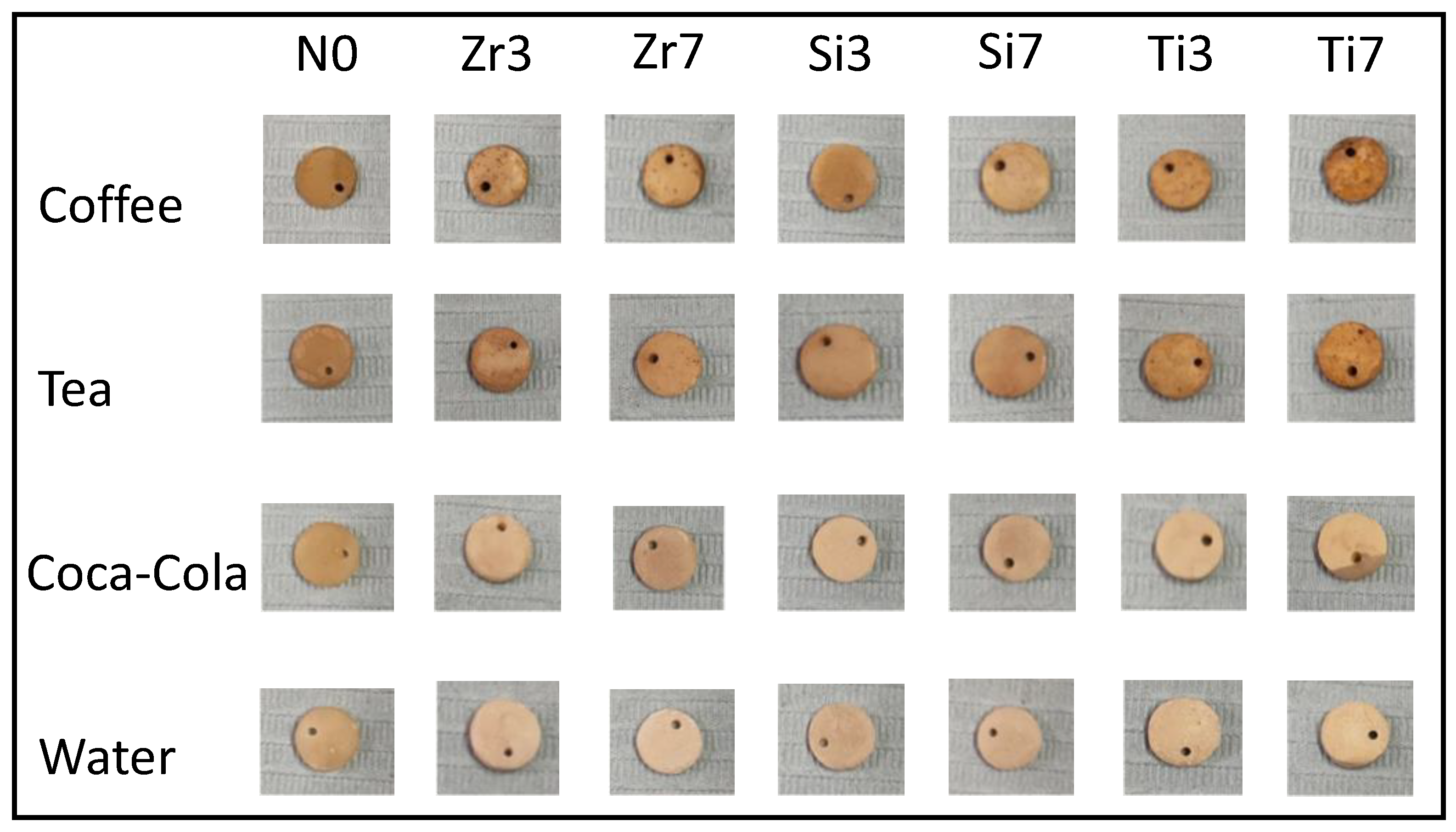
Table 3 presents the comparisons of mean values, SDs, and significances of ΔE. A statistically significant difference was found after using one-way-ANOVA between ΔE of the different RBC groups (p < 0.001) during immersion in different beverages. Color changes of different subgroups after exposure to different beverages are shown in Figure 1.
After immersion in the coffee solution, the highest ΔE value was recorded with N0, and the lowest ΔE value recorded with N0 had the uppermost Zr3 group. There was a statistically significant difference among N0, Zr3, and Zr7 (p = 0.002). Zr3 presented a statistically significant difference with Si3 (p = 0.006) and Si7 (p < 0.001). Additionally, Zr7 showed a significant difference with Si3 (p = 0.044) and Si7 (p = 0.022).
After immersion in the tea solution, the highest mean ΔE was recorded with N0, and the lowest ΔE was recorded with the Zr3 group. N0 presented a significant increase in ΔE compared with Zr3 (p < 0.001), Zr7 (p < 0.001), Ti3 (p = 0.005), and Si3 (p = 0.031). Zr3 showed a significant decrease in ΔE compared with Ti7 (p = 0.005) and Si7 (p < 0. 001). Additionally, a significant difference was found between Zr7 and Si7 (p = 0.013).
After immersion in the Coca-Cola solution, N0 displayed the highest ΔE value, and Zr3 recorded the lowest ΔE value. N0 showed a significant increase in ΔE with all modified groups p < 0.001) except Ti7 and Si7 (p > 0.05). Among the modified groups, Zr3 showed a significant decrease in ΔE compared with Ti3 (p = 0.015), Ti7 (p < 0.001), Si3 (p = 0.001), and Si7 (p < 0.001). Zr7 showed a statistically significant difference with Ti7 (p = 0.002) and Si7 (p < 0.001). Significant differences were found between Ti3 and. Si7 (p = 0.002) and between Si3 and Si7 (p = 0.040).
After immersion in mineral water, the control group (N0) showed the highest mean ΔE with a statistically significant difference from Zr3 (p < 0.001), Zr7 (p < 0.001), Ti3 (p < 0.001), and Si3 (p = 0.007), while Zr3 showed the lowest mean ΔE with a statistically significant difference from Ti7 (p < 0.001), Si3 (p = 0.014), and Si7 (p < 0.001). There was a statistically significant difference of Zr7 with Ti7 (p < 0.001) and Si7 (p < 0.001). Additionally, there was a statistically significant difference of Ti3 with Ti7 (p = 0.012) and Si7 (p = 0.019).
Based on NBS findings, markedly unacceptable color changes (NBS > 3) were detected after immersion of RBCs in coffee and tea, while in Coca-Cola immersion, the values of NBS lay in the 1.5–3 range, indicating noticeable color changes. Slight color changes were seen after immersion in water (NBS ≤ 1). Overall, the highest NBS values were recorded with the control group (N0), while the lowest values were recorded with Zr3.
4. Discussion
RBCs are widely used in the field of dentistry as an aesthetic restorative material and for the fabrication of artificial teeth, in addition to recontouring of abutment teeth [41,42,43].
The prognosis of any aesthetic restorative material depends on its ability for shade matching and color stability; it was demonstrated that light-cured RBCs can be discolored over time by either extrinsic or intrinsic discoloration, resulting in patient frustration and extra replacement expenses [11]. Extrinsic factors include surface roughness, staining agent, and contact time in coloring solutions; frequent consumption of coloring and acidic drinks, such as Coca-Cola and coffee, may affect the color stability of RBCs through absorption and adsorption of colorant agents into the resin matrix [7,9,44]. Additionally, the yellowing effect can develop because of the teeth’s and the restorative material’s permeability allowing the penetration of pigments and chromogenic substances into the daily dietary intake [45].
The selection of beverages was made based on the daily consumption frequency, and they have often been used in previous in-vitro studies [4,8,9]. Ingredients such as sugar or white creamer were not added to beverages since they might interfere with the discoloring effect of the beverages. Simulation of long-term exposure to beverage solutions was conducted by storing the specimens in these solutions throughout the study period [4,46].
Color changes of dental materials are usually evaluated by colorimeters and spectrophotometers since they prevent subjective interpretations and permit the detection of minor color differences. The CIE L*, a*, b* color evaluation method is considered a proper tool to assess alterations in color in dental materials. The color change values (∆E) were converted into NBS values to assess whether the color change was in the range of clinically acceptable levels [27,47].
Our results showed significant differences between the ΔE values of several groups (p ≤ 0.05) after immersion of specimens in different beverages, causing us to conclude that the color stability of light-cured RBCs was significantly improved by NP addition, so the null hypothesis of the current study was rejected.
In the current study, the spectrophotometer results revealed that great color changes occurred in specimens immersed in coffee, followed by tea and then Coca-Cola, while mineral water recorded the lowest ΔE values. The observed NBS values were out of the clinically accepted limits with coffee and tea with marked color changes; this finding is in accordance with previous studies demonstrating that more discoloration of RBC specimens was associated with coffee solution [30,48,49]. Additionally, it agrees with Guler et al.’s and Ertas et al.’s studies since they reported that coffee and tea produced discoloration in RBCs more than Coca-Cola [4,37].
This outcome may be attributed to the different polarities of yellow colorants contained in tea and coffee; the polarity degree determines the penetration extent into the RBCs. Less polar dyes, such as coffee, can easily enter into the polymer matrix by the processes of absorption and adsorption, leading to more staining. On the other hand, more polar dyes, such as tea, only impregnate the material’s surface and can be removed easily, thus causing less staining [37]. In disagreement with these findings, many studies have reported that tea is the most staining solution and can produce discoloration more than coffee [50,51].
Compared to coffee and tea (pH 5.0 and 6.1, respectively), the Coca-Cola solution did not cause greater discoloration, although it had a low pH (pH 2.7), which may affect the surface roughness of RBCs. The minor degree of color change in Coca-Cola could be attributed to the absence of yellow colorants in its ingredients and the presence of carbonated water, which buffers the acidity present, thus reducing surface integrity damage [46]. Several articles are in agreement with our results [37,52,53]. On the other hand, Ghiorghe et al. showed that Coca-Cola can cause color changes in composite resin more than coffee and tea [54].
A slight color alteration was detected in specimens soaked in mineral water (NBS ≤ 1), which can be attributed to the water’s absence of colorant and neutral pH. This outcome is in accordance with many previous studies [8,9,38,47]. On the other hand, the slight detected color changes may be due to softening of the polymer since water causes network swelling and loosening of the frictional forces between polymer chains [55].
Color changes are considered acceptable in in-vitro studies if (ΔE) ≤ 3.7, while they become visually detected and unacceptable when ∆E > 3.7 [11,23]. Regarding the present outcomes, composite specimens immersed in mineral water and Coca-Cola after 6 days presented clinically acceptable color changes, while those immersed in coffee and tea were clinically unacceptable, in agreement with the results of Guler et al. [4].
According to the findings of the current study, addition of NPs decreases color changes. This outcome is in agreement with previous studies that reported that the optical properties of light-cured RBCs improved with NP addition [56,57,58]. Irrespective of NP type, low concentrations (3 wt.%) presented less ΔE compared with high concentrations (7 wt.%). This finding may be attributed to increased density and reduced porosity due to the presence of inorganic nanofillers within the material, resulting in less stain absorption [59].
In agreement with the present findings, Soichiro et al. showed better discoloration resistance in RBC artificial teeth with silanized SiO2 filler soaked in different beverages, and this finding may be explained by SiO2 potentially being able to improve the hydrolytic stability and decreased water sorption, leading to fewer color changes [43]. Furthermore, Azmy et al. showed the same results concerning the anti-staining effects of ZrO2, TiO2, and SiO2 on heat-polymerized PMMA [19].
Light refraction and reflection at the interface between the matrix and filler are affected by the differences between the inorganic nanoparticles and organic matrix refractive indices. Thus, the greater the refractive index difference, the greater the nanocomposite opacity [25,26].
In contrast to the present study findings, Rodrigues et al. reported high surface roughness of nanocomposites due to the irregular arrangement of inorganic NPs, resulting in easier staining effects over time [44]. The discrepancies between the results of this study and other articles [44,50,51,54] may be explained by differences in experimental methodology, and types and concentrations of the tested NPs, and the types and concentrations of the tested beverages, as well as the usage of different commercial RBCs and the duration and storage technique.
From the clinical view, Azmy et al. [19] concluded that the addition of ZrO2, TiO2, and SiO2 to light-cured RBCs could improve their mechanical and surface properties, but one of the most common issues seen regarding the reinforcement of light-cured RBCs via NPs is the color instability. Furthermore, this study added that modification of light-cured RBCs with ZrO2, TiO2, or SiO2 can reduce their stainability, which is beneficial in prolonging restorative materials’ lifetime and improving patient acceptance. Long-term color stability of RBCs can be achieved by reducing the consumption of beverages (tea, coffee, and Coca-Cola); these beverages can greatly increase the stainability of RBCs and are thought to indicate deterioration and aging of the material.
One of the most important determining elements in the selection of ideal NPs is cytotoxicity [60]. Generally, these NPs have shown a variation in the cytotoxic effects when they were incorporated into different dental materials [1,61,62,63,64]. Raj et al. evaluated the cytotoxic effects of 3% TiO2 NP-modified denture base resin at 1 and 7 days [62]. The results indicating biocompatibility due to lesser toxicity were observed after the 7-day assessment [62]. Furthermore, the addition of ZrO2 to 3D-printed resin showed significant antibacterial properties without cellular side effects [63]. Additionally, a study by Ahangaran and Navarchian showed no cytotoxicity effects of SiO2 NPs when they were added to self-healing dental composites [64]. Nevertheless, the long-term assessment of the cytotoxicity of the proposed modified nanocomposites is mandatory before clinical applicability.
The oral environment exposes restorative materials to various challenges, including cyclic fatigue, thermal cycling, and pH cycling, which were not simulated in this study. Therefore, further studies are needed to investigate the influence of these challenges on the surface properties and quality of the tested materials. Furthermore, using one type of RBC with a simple disc-shaped specimen, the hand mixing procedures, and the lack of clinical polishing techniques and oral conditions were considered the major limitations of this study. In addition, thermo-cycling was not performed, so while the presented findings are encouraging, further investigations are needed. Accordingly, future work should involve the effect of the polymerization technique and other types of RBCs and NPs. Additionally, the polishing effect should be investigated. Future directions of this study are to evaluate the long-term effects of impeding nanoparticles into RBCs on the mechanical properties, shear bonding strength, surface properties, including roughness and water absorption, and biocompatibility.
5. Conclusions
Concerning the results of the present study and according to its limitations, it was concluded that:
- Modification of RBCs with 3% and 7% of ZrO2, as well as 3% of TiO2 and SiO2, could improve its color stability;
- The chromatic effect on modified and unmodified RBCs varies between immersion solutions, in which the high range effect was reported with coffee, followed by tea and then Coca-Cola;
- Careful selection of both the type and concentration of reinforcing filler to achieve a balance between mechanical properties and aesthetic concerns of the restorative materials was recommended; and
- Thorough selection of restorative materials and techniques is essential in achieving the optimal aesthetic properties and minimizing discolorations.
Author Contributions
Conceptualization, E.A. and Y.A.A.-D.; methodology, E.A. and M.R.Z.A.-K.; validation, M.A.H. and Y.A.A.-D.; formal analysis, M.A.H.; investigation, E.A. and M.R.Z.A.-K.; resources, K.S.A. and M.A.A.; writing—original draft preparation, E.A. and M.R.Z.A.-K.; writing—review and editing, M.A.A., K.S.A. and Y.A.A.-D.; supervision, M.A.H.; project administration, M.A.H.; funding acquisition, Y.A.A.-D., K.S.A. and M.A.A. All authors have read and agreed to the published version of the manuscript.
Funding
This research received no external funding.
Institutional Review Board Statement
Not applicable.
Informed Consent Statement
Not applicable.
Data Availability Statement
Not applicable.
Conflicts of Interest
The authors declare no conflict of interest.
Abbreviations
| Abbreviations | Description |
| ADA | American Dental Association |
| ANOVA | Analysis of variance |
| °C | Centigrade |
| CIE | Commission Internationale de l’Eclairage |
| G | Gram |
| H | Hour |
| ISO | International Organization for Standardization |
| Mm | Millimeter |
| Min | Minute |
| NPs | Nanoparticles |
| NBS | National Bureau of Standards |
| RBCs | Resin-based composites |
| Rpm | Round per minute |
| Sec | Second |
| SiO2 | Silica dioxide |
| SD | Standard deviation |
| SPSS | Statistical Package for Social Sciences |
| TiO2 | Titanium dioxide |
| TMSPM | Trimethoxysilyl propyl methacrylate |
| ZrO2 | Zirconium oxide |
References
- Chen, M.-H. Update on Dental Nanocomposites. J. Dent. Res. 2010, 89, 549–560. [Google Scholar] [CrossRef] [PubMed]
- Al-Nabulsi, M.; Daud, A.; Yiu, C.; Omar, H.; Sauro, S.; Fawzy, A.; Daood, U. Co-Blend Application Mode of Bulk Fill Composite Resin. Materials 2019, 12, 2504. [Google Scholar] [CrossRef] [PubMed] [Green Version]
- Al-Dulaijan, Y.A.; Cheng, L.; Weir, M.D.; Melo, M.A.S.; Liu, H.; Oates, T.W.; Wang, L.; Xu, H.H.K. Novel Rechargeable Calcium Phosphate Nanocomposite with Antibacterial Activity to Suppress Biofilm Acids and Dental Caries. J. Dent. 2018, 72, 44–52. [Google Scholar] [CrossRef]
- Guler, A.U.; Yilmaz, F.; Kulunk, T.; Guler, E.; Kurt, S. Effects of Different Drinks on Stainability of Resin Composite Provisional Restorative Materials. J. Prosthet. Dent. 2005, 94, 118–124. [Google Scholar] [CrossRef] [PubMed]
- Paolone, G.; Formiga, S.; De Palma, F.; Abbruzzese, L.; Chirico, L.; Scolavino, S.; Goracci, C.; Cantatore, G.; Vichi, A. Color Stability of Resin-Based Composites: Staining Procedures with Liquids—A Narrative Review. J. Esthet. Restor. Dent. 2022, 34, 865–887. [Google Scholar] [CrossRef]
- Karanjkar, R.R.; Preshaw, P.M.; Ellis, J.S.; Holliday, R. Effect of Tobacco and Nicotine in Causing Staining of Dental Hard Tissues and Dental Materials: A Systematic Review and Meta-Analysis. Clin. Exp. Dent. Res. 2023, 9, 150–164. [Google Scholar] [CrossRef]
- Mansouri, S.A.; Zidan, A.Z. Effect of Water Sorption and Solubility on Color Stability of Bulk-Fill Resin Composite. J. Contemp. Dent. Pract. 2018, 19, 1129–1134. [Google Scholar]
- Al Kheraif, A.A.A.; Qasim, S.S.B.; Ramakrishnaiah, R.; ur Rehman, I. Effect of Different Beverages on the Color Stability and Degree of Conversion of Nano and Microhybrid Composites. Dent. Mater. J. 2013, 32, 326–331. [Google Scholar] [CrossRef] [Green Version]
- Barutcigil, Ç.; Yıldız, M. Intrinsic and Extrinsic Discoloration of Dimethacrylate and Silorane Based Composites. J. Dent. 2012, 40, e57–e63. [Google Scholar] [CrossRef]
- Park, J.-K.; Kim, T.-H.; Ko, C.-C.; Garcia-Godoy, F.; Kim, H.-I.; Kwon, Y.H. Effect of Staining Solutions on Discoloration of Resin Nanocomposites. Am. J. Dent. 2010, 23, 39. [Google Scholar]
- Poggio, C.; Ceci, M.; Beltrami, R.; Mirando, M.; Wassim, J.; Colombo, M. Color Stability of Esthetic Restorative Materials: A Spectrophotometric Analysis. Acta Biomater. Odontol. Scand. 2016, 2, 95–101. [Google Scholar] [CrossRef] [PubMed]
- Azmy, E.; Al-Kholy, M.R.Z.; Fattouh, M.; Kenawi, L.M.M.; Helal, M.A. Impact of Nanoparticles Additions on the Strength of Dental Composite Resin. Int. J. Biomater. 2022, 2022, 1165431. [Google Scholar] [CrossRef]
- Bapat, R.A.; Joshi, C.P.; Bapat, P.; Chaubal, T.V.; Pandurangappa, R.; Jnanendrappa, N.; Gorain, B.; Khurana, S.; Kesharwani, P. The Use of Nanoparticles as Biomaterials in Dentistry. Drug Discov. Today 2019, 24, 85–98. [Google Scholar] [CrossRef] [PubMed]
- Vagkopoulou, T.; Koutayas, S.O.; Koidis, P.; Strub, J.R. Zirconia in Dentistry: Part 1. Discovering the Nature of an Upcoming Bioceramic. Eur. J. Esthet. Dent. 2009, 4, 130–151. [Google Scholar] [PubMed]
- Karabela, M.M.; Sideridou, I.D. Synthesis and Study of Properties of Dental Resin Composites with Different Nanosilica Particles Size. Dent. Mater. 2011, 27, 825–835. [Google Scholar] [CrossRef] [PubMed]
- Xia, Y.; Zhang, F.; Xie, H.; Gu, N. Nanoparticle-Reinforced Resin-Based Dental Composites. J. Dent. 2008, 36, 450–455. [Google Scholar] [CrossRef]
- Furman, B.; Rawls, H.R.; Wellinghoff, S.; Dixon, H.; Lankford, J.; Nicolella, D. Metal-Oxide Nanoparticles for the Reinforcement of Dental Restorative Resins. Crit. Rev. Biomed. Eng. 2000, 28, 439–443. [Google Scholar] [CrossRef]
- Al-Dulaijan, Y.A.; Balhaddad, A.A. Prospects on Tuning Bioactive and Antimicrobial Denture Base Resin Materials: A Narrative Review. Polymers 2023, 15, 54. [Google Scholar] [CrossRef]
- Azmy, E.; Al-Kholy, M.R.Z.; Al-Thobity, A.M.; Gad, M.M.; Helal, M.A. Comparative Effect of Incorporation of ZrO2, TiO2, and SiO2 Nanoparticles on the Strength and Surface Properties of PMMA Denture Base Material: An In Vitro Study. Int. J. Biomater. 2022, 2022, 5856545. [Google Scholar] [CrossRef]
- Azmy, E.; Al-Kholy, M.R.Z.; Gad, M.M.; Al-Thobity, A.M.; Emam, A.-N.M.; Helal, M.A. Influence of Different Beverages on the Color Stability of Nanocomposite Denture Base Materials. Int. J. Dent. 2021, 2021, 5861848. [Google Scholar] [CrossRef]
- Helal, M.A.; Yang, B.; Saad, E.; Abas, M.; Al-Kholy, M.R.; Imam, A.Y.; Gad, M.M. Effect of SiO2 and Al2O3 Nanoparticles on Wear Resistance of PMMA Acrylic Denture Teeth. Braz. Dent. Sci. 2020, 23, 12. [Google Scholar] [CrossRef]
- Liu, Y.; Sun, Y.; Zeng, F.; Xie, W.; Liu, Y.; Geng, L. Effect of Nano SiO2 Particles on the Morphology and Mechanical Properties of POSS Nanocomposite Dental Resins. J. Nanopart. Res. 2014, 16, 2736. [Google Scholar] [CrossRef]
- Wang, H.; Zhu, M.; Li, Y.; Zhang, Q.; Wang, H. Mechanical Properties of Dental Resin Composites by Co-Filling Diatomite and Nanosized Silica Particles. Mater. Sci. Eng. C 2011, 31, 600–605. [Google Scholar] [CrossRef]
- Sahin, O.; Koroglu, A.; Dede, D.Ö.; Yilmaz, B. Effect of Surface Sealant Agents on the Surface Roughness and Color Stability of Denture Base Materials. J. Prosthet. Dent. 2016, 116, 610–616. [Google Scholar] [CrossRef] [PubMed]
- Yu, B.; Ahn, J.-S.; Lim, J.I.; Lee, Y.-K. Influence of TiO2 Nanoparticles on the Optical Properties of Resin Composites. Dent. Mater. 2009, 25, 1142–1147. [Google Scholar] [CrossRef] [PubMed]
- Arikawa, H.; Kanie, T.; Fujii, K.; Takahashi, H.; Ban, S. Effect of Filler Properties in Composite Resins on Light Transmittance Characteristics and Color. Dent. Mater. J. 2007, 26, 38–44. [Google Scholar] [CrossRef] [Green Version]
- Paul, S.; Peter, A.; Pietrobon, N.; Hämmerle, C. Visual and Spectrophotometric Shade Analysis of Human Teeth. J. Dent. Res. 2002, 81, 578–582. [Google Scholar] [CrossRef]
- Commission Internationale de l’ Éclairage—CIE, Recommendations on Uniform Color Spaces, Color-Difference Equations, Psychometric Color Terms, Bureau Central de la CIE, Paris, UK, 2004, Supplement no.2 of Publication CIE No. 15 (E-1.3.1).
- ISO 4049:2000; Dentistry-Polymer-Based Filling, Restorative and Luting Materials. ISO: Geneva, Switzerland, 2000; pp. 1–22.
- Gupta, R.; Bhatheja, A.; John, A.G.; Ramchandran, M.; Raina, A.A.; Behera, A.; Mittal, N. Effect of Beverages on Color Stability of Resin Composites: An in Vitro Study. J. Appl. Dent. Sci. 2019, 5, 92–95. [Google Scholar]
- Nguyen, T.-C.; Nguyen, T.-D.; Vu, D.-T.; Dinh, D.-P.; Nguyen, A.-H.; Ly, T.-N.-L.; Dao, P.-H.; Nguyen, T.-L.; Bach, L.-G.; Thai, H. Modification of Titanium Dioxide Nanoparticles with 3-(Trimethoxysilyl)Propyl Methacrylate Silane Coupling Agent. J. Chem. 2020, 2020, e1381407. [Google Scholar] [CrossRef]
- Hayichelaeh, C.; Reuvekamp, L.a.E.M.; Dierkes, W.K.; Blume, A.; Noordermeer, J.W.M.; Sahakaro, K. Enhancing the Silanization Reaction of the Silica-Silane System by Different Amines in Model and Practical Silica-Filled Natural Rubber Compounds. Polymers 2018, 10, 584. [Google Scholar] [CrossRef] [Green Version]
- He, X.; Mahtabani, A.; Rytöluoto, I.; Saarimäki, E.; Lahti, K.; Paajanen, M.; Anyszka, R.; Dierkes, W.; Blume, A. Surface Modification of Fumed Silica by Dry Silanization for PP-Based Dielectric Nanocomposites. In Proceedings of the 2019 2nd International Conference on Electrical Materials and Power Equipment (ICEMPE), Guangzhou, China, 7–10 April 2019; pp. 254–259. [Google Scholar]
- Aminoroaya, A.; Esmaeely Neisiany, R.; Nouri Khorasani, S.; Panahi, P.; Das, O.; Ramakrishna, S. A Review of Dental Composites: Methods of Characterizations. ACS Biomater. Sci. Eng. 2020, 6, 3713–3744. [Google Scholar] [CrossRef] [PubMed]
- Al-Dulaijan, Y.A.; Weir, M.D.; Melo, M.A.S.; Sun, J.; Oates, T.W.; Zhang, K.; Xu, H.H.K. Protein-Repellent Nanocomposite with Rechargeable Calcium and Phosphate for Long-Term Ion Release. Dent. Mater. 2018, 34, 1735–1747. [Google Scholar] [CrossRef] [PubMed]
- Da Silva, J.D.; Park, S.E.; Weber, H.-P.; Ishikawa-Nagai, S. Clinical Performance of a Newly Developed Spectrophotometric System on Tooth Color Reproduction. J. Prosthet. Dent. 2008, 99, 361–368. [Google Scholar] [CrossRef]
- Ertas, E.; Güler, A.U.; Yücel, A.Ç.; Köprülü, H.; Güler, E. Color Stability of Resin Composites after Immersion in Different Drinks. Dent. Mater. J. 2006, 25, 371–376. [Google Scholar] [CrossRef] [Green Version]
- Sepúlveda-Navarro, W.F.; Arana-Correa, B.E.; Ferreira Borges, C.P.; Habib Jorge, J.; Urban, V.M.; Campanha, N.H. Color Stability of Resins and Nylon as Denture Base Material in Beverages. J. Prosthodont. Implant Esthet. Reconstr. Dent. 2011, 20, 632–638. [Google Scholar] [CrossRef]
- Lee, Y.-K.; Lim, B.-S.; Rhee, S.-H.; Yang, H.-C.; Powers, J.M. Changes of Optical Properties of Dental Nano-Filled Resin Composites after Curing and Thermocycling. J. Biomed. Mater. Res. Part B Appl. Biomater. Off. J. Soc. Biomater. Jpn. Soc. Biomater. Aust. Soc. Biomater. Korean Soc. Biomater. 2004, 71, 16–21. [Google Scholar] [CrossRef]
- Tuncdemir, A.R.; Aykent, F. Effects of Fibers on the Color Change and Stability of Resin Composites after Accelerated Aging. Dent. Mater. J. 2012, 31, 872–878. [Google Scholar] [CrossRef] [PubMed] [Green Version]
- Helal, M.A.; Baraka, O.A.; Sanad, M.E.; Ludwig, K.; Kern, M. Effects of Long-Term Simulated RPD Clasp Attachment/Detachment on Retention Loss and Wear for Two Clasp Types and Three Abutment Material Surfaces. J. Prosthodont. Implant Esthet. Reconstr. Dent. 2012, 21, 370–377. [Google Scholar] [CrossRef]
- Helal, M.A.; Al-Gazzar, A.E.; Abas, M.; Akhtar, S.; Gad, M.M.; Al-Thobity, A.M. Comparative Effect of Different Surface Treatments on the Shear Bond Strength of Two Types of Artificial Teeth Bonded to Two Types of Denture Base Resins. J. Prosthodont. 2022, 31, 427–433. [Google Scholar] [CrossRef]
- Imamura, S.; Takahashi, H.; Hayakawa, I.; Loyaga-Rendon, P.G.; Minakuchi, S. Effect of Filler Type and Polishing on the Discoloration of Composite Resin Artificial Teeth. Dent. Mater. J. 2008, 27, 802–808. [Google Scholar] [CrossRef] [Green Version]
- Rodrigues, S.A.; Scherrer, S.S.; Ferracane, J.L.; Bona, Á.D. Microstructural Characterization and Fracture Behavior of a Microhybrid and a Nanofill Composite. Dent. Mater. 2008, 24, 1281–1288. [Google Scholar] [CrossRef] [PubMed]
- Fiorillo, L.; Laino, L.; De Stefano, R.; D’Amico, C.; Bocchieri, S.; Amoroso, G.; Isola, G.; Cervino, G. Dental Whitening Gels: Strengths and Weaknesses of an Increasingly Used Method. Gels 2019, 5, 35. [Google Scholar] [CrossRef] [Green Version]
- Bahbishi, N.; Mzain, W.; Badeeb, B.; Nassar, H.M. Color Stability and Micro-Hardness of Bulk-Fill Composite Materials after Exposure to Common Beverages. Materials 2020, 13, 787. [Google Scholar] [CrossRef] [PubMed] [Green Version]
- Erdemir, U.; Yıldız, E.; Eren, M.M. Effects of Sports Drinks on Color Stability of Nanofilled and Microhybrid Composites after Long-Term Immersion. J. Dent. 2012, 40, e55–e63. [Google Scholar] [CrossRef] [PubMed]
- Mundim, F.M.; Garcia, L.d.F.R.; Pires-de-Souza, F.d.C.P. Effect of Staining Solutions and Repolishing on Color Stability of Direct Composites. J. Appl. Oral Sci. 2010, 18, 249–254. [Google Scholar] [CrossRef] [PubMed] [Green Version]
- Macedo, M.G.F.P.; Volpato, C.A.M.; Henriques, B.A.P.C.; Vaz, P.C.S.; Silva, F.S.; Silva, C.F.C.L. Color Stability of a Bis-Acryl Composite Resin Subjected to Polishing, Thermocycling, Intercalated Baths, and Immersion in Different Beverages. J. Esthet. Restor. Dent. 2018, 30, 449–456. [Google Scholar] [CrossRef] [PubMed]
- Tekçe, N.; Tuncer, S.; Demirci, M.; Serim, M.E.; Baydemir, C. The Effect of Different Drinks on the Color Stability of Different Restorative Materials after One Month. Restor. Dent. Endod. 2015, 40, 255–261. [Google Scholar] [CrossRef]
- Karaman, E.; Tuncer, D.; Firat, E.; Ozdemir, O.S.; Karahan, S. Influence of Different Staining Beverages on Color Stability, Surface Roughness and Microhardness of Silorane and Methacrylate-Based Composite Resins. J. Contemp. Dent. Pract. 2014, 15, 319–325. [Google Scholar] [CrossRef]
- Al-kheraif, A.A. Effects of Curing Units and Staining Solutions on the Color Susceptibility of a Microhybrid Composite Resin. J. Dent. Sci. 2011, 6, 33–40. [Google Scholar] [CrossRef] [Green Version]
- Villalta, P.; Lu, H.; Okte, Z.; Garcia-Godoy, F.; Powers, J.M. Effects of Staining and Bleaching on Color Change of Dental Composite Resins. J. Prosthet. Dent. 2006, 95, 137–142. [Google Scholar] [CrossRef]
- Ghiorghe, C.; Iovan, G.; Topoliceanu, C.; Sandu, A.V.; Andrian, S. Comparative Study Regarding the Colorimetric Changes of Two Composite Resins after Immersion in Several Beverages and One Antibacterial Mouthwash. Rev. De Chim. Buchar. Orig. Ed. 2013, 64, 1436. [Google Scholar]
- Ie, R. Analysis and Characterization of Dental Polymers. Crit. Rev. Biocompat. 1988, 4, 247–279. [Google Scholar]
- Mitra, S.B.; Wu, D.; Holmes, B.N. An Application of Nanotechnology in Advanced Dental Materials. J. Am. Dent. Assoc. 2003, 134, 1382–1390. [Google Scholar] [CrossRef] [PubMed] [Green Version]
- Jain, N. Effect of Nanofiller Technology on Surface Properties of Nanofilled and Nanohybrid Composites. Int. J. Dent. Oral Health 2015, 1, 1–5. [Google Scholar] [CrossRef] [Green Version]
- Nasim, I.; Neelakantan, P.; Sujeer, R.; Subbarao, C.V. Color Stability of Microfilled, Microhybrid and Nanocomposite Resins—An in Vitro Study. J. Dent. 2010, 38, e137–e142. [Google Scholar] [CrossRef]
- Keller, J.C.; Lautenschalger, E.P. Porosity Reduction and Its Associated Effect on the Diametral Tensile Strength of Activated Acrylic Resins. J. Prosthet. Dent. 1985, 53, 374–379. [Google Scholar] [CrossRef] [PubMed]
- Song, W.; Ge, S. Application of Antimicrobial Nanoparticles in Dentistry. Molecules 2019, 24, 1033. [Google Scholar] [CrossRef] [PubMed] [Green Version]
- Mansoor, A.; Khurshid, Z.; Khan, M.T.; Mansoor, E.; Butt, F.A.; Jamal, A.; Palma, P.J. Medical and Dental Applications of Titania Nanoparticles: An Overview. Nanomaterials 2022, 12, 3670. [Google Scholar] [CrossRef]
- Raj, V.; Bhat, V.; John, N.; Shetty, A.; Joseph, S.; Kuriakose, R.; Hameed, S. Assessment of Flexural Strength and Cytotoxicity of Heat Cure Denture Base Resin Modified with Titanium Dioxide Nanoparticles: An in Vitro Study. J. Contemp. Dent. Pract. 2021, 22, 1025–1029. [Google Scholar] [PubMed]
- Aati, S.; Shrestha, B.; Fawzy, A. Cytotoxicity and Antimicrobial Efficiency of ZrO2 Nanoparticles Reinforced 3D Printed Resins. Dent. Mater. 2022, 38, 1432–1442. [Google Scholar] [CrossRef]
- Ahangaran, F.; Navarchian, A.H. Towards the Development of Self-Healing and Antibacterial Dental Nanocomposites via Incorporation of Novel Acrylic Microcapsules. Dent. Mater. 2022, 38, 858–873. [Google Scholar] [CrossRef] [PubMed]
Figure 1.
Color changes of different specimens after immersion in different beverages (N0: control; Zr3: 3 wt.% of ZrO2; Zr7: 7 wt.% of ZrO2; Ti3: 3 wt.% of TiO2; Ti7: 7 wt.% of TiO2; Si3; 3 wt.% of SiO2; and Si7; 7 wt.% of SiO2).
Figure 1.
Color changes of different specimens after immersion in different beverages (N0: control; Zr3: 3 wt.% of ZrO2; Zr7: 7 wt.% of ZrO2; Ti3: 3 wt.% of TiO2; Ti7: 7 wt.% of TiO2; Si3; 3 wt.% of SiO2; and Si7; 7 wt.% of SiO2).


Table 1.
List of materials used in the current study.
| Brand Name/Manufacturer | Company Name | Specifications |
|---|---|---|
| Nexcomp | META BIOMED, Cheongju-si, Republic of Korea | Resin: Bis-GMA, Bis-EMA, UDMA, TEGDMA. Fillers: 0.04–0.7 µm barium aluminum boro-silicate. Light cured, A2 |
| ZrO2 nanoparticles | NanoGATE, 6 October, Giza, Egypt | Spherical, white, and tetragonal nanoparticles (12 ± 3 nm; purity > 99%) |
| TiO2 nanoparticles | Spherical, white, and anatase nanoparticles (15 ± 3 nm; purity > 99%) | |
| SiO2 nanoparticles | Spherical, white, and amorphous nanoparticles (21 ± 3 nm; purity > 99%) | |
| Silane-coupling-agent | Sigma/Aldrich Chemie GmbH, Taufkirchen, Germany | 3-trimethoxysilyl propyl methacrylate (TMSPM). Purity 98%, ethanol 99.7%., lot No. 440159 |
ZrO2: zirconium dioxide; TiO2: titanium dioxide; SiO2: silicon dioxide; Bis-GMA: bisphenol A glycidyl methacrylate; Bis-EMA: bisphenol A ethoxylated dimethacrylate; UDMA: urethane dimethacrylate; and TEGDMA: triethylene glycol dimethacrylate.
Table 2.
Protocol of beverage preparation.
| Beverage Solution | Company Name | Preparation of Beverage Solution | Soaking Time | Temperature | pH |
|---|---|---|---|---|---|
| Coffee | Abu Auf, New Cairo, Egypt | 200 mL distilled water boiled for 2 min then used to made 2 g of coffee, the mix then was filtered before usage. | 6 days | 37 °C | 5.0 |
| Tea | Lipton, London, UK | 200 mL distilled water boiled for 2 min then used to made 2 g of tea, the mix then was filtered before usage. | 6.1 | ||
| Cola-Cola | Coca-Cola Co., Atlanta, GA, USA | Ready made | 2.7 | ||
| Water | Dasani, Atlanta, GA, USA | Ready made | 7.1 |
Table 3.
(ΔE) mean (M) values, standard deviations (±SDs), and significance between tested groups in terms of immersion solution effect.
Table 3.
(ΔE) mean (M) values, standard deviations (±SDs), and significance between tested groups in terms of immersion solution effect.
| Group | Coffee | Tea | Coca-Cola | Mineral Water | ||||
|---|---|---|---|---|---|---|---|---|
| Mean ± SD | NBS | Mean ± SD | NBS | Mean ± SD | NBS | Mean ± SD | NBS | |
| N0 | 4.46 ± 0.07 A | 4.1 | 3.42 ± 0.05 A | 3.1 | 2.49 ± 0.04 A | 2.3 | 1.12 ± 0.02 A | 1 |
| Zr3 | 4.31 ± 0.03 BC | 3.96 | 3.23 ± 0.10 D | 3 | 2.24 ± 0.04 D | 2.1 | 0.99 ± 0.05 D | 0.9 |
| Zr7 | 4.36 ± 0.05 B | 4 | 3.28 ± 0.09 CD | 3 | 2.29 ± 0.11 CD | 2.1 | 1.0 ± 0.03 CD | 0.9 |
| Ti3 | 4.39 ± 0.04 AB | 4 | 3.3 ± 0.06 BCD | 3 | 2.34 ± 0.08 BC | 2.2 | 1.02 ± 0.05 CD | 0.9 |
| Ti7 | 4.42 ± 0.07 AB | 4.1 | 3.35 ± 0.09 ABC | 3.1 | 2.41 ± 0.06 AB | 2.2 | 1.09 ± 0.02 AB | 1 |
| Si3 | 4.4 ± 0.04 A | 4 | 3.32 ± 0.05 BCD | 3.1 | 2.37 ± 0.03 BC | 2.2 | 1.05 ± 0.04 BC | 1 |
| Si7 | 4.44 ± 0.06 A | 4.1 | 3.39 ± 0.04 AB | 3.1 | 2.46 ± 0.05 A | 2.3 | 1.08 ± 0.06 AB | 1 |
| p-value | <0.001 * | <0.001 * | <0.001 * | <0.001 * | ||||
| Effect size | 0.461 | 0.457 | 0.650 | 0.580 | ||||
The different superscripts within columns denote statistically non-significant differences (*: Significant at p ≤ 0.05; SD: standard deviation; NBS: National Bureau of Standards; N0: control; Zr3: 3 wt.% of ZrO2; Zr7: 7 wt.% of ZrO2; Ti3: 3 wt.% of TiO2; Ti7: 7 wt.% of TiO2; Si3; 3 wt.% of SiO2; and Si7; 7 wt.% of SiO2).
Disclaimer/Publisher’s Note: The statements, opinions and data contained in all publications are solely those of the individual author(s) and contributor(s) and not of MDPI and/or the editor(s). MDPI and/or the editor(s) disclaim responsibility for any injury to people or property resulting from any ideas, methods, instructions or products referred to in the content. |
© 2023 by the authors. Licensee MDPI, Basel, Switzerland. This article is an open access article distributed under the terms and conditions of the Creative Commons Attribution (CC BY) license (https://creativecommons.org/licenses/by/4.0/).
Share and Cite
MDPI and ACS Style
Al-Dulaijan, Y.A.; AlGhamdi, M.A.; Azmy, E.; Al-Kholy, M.R.Z.; Almulhim, K.S.; Helal, M.A. Color Stability of Nanoparticles-Modified Dental Resin-Based Composites. Appl. Sci. 2023, 13, 3870. https://doi.org/10.3390/app13063870
AMA Style
Al-Dulaijan YA, AlGhamdi MA, Azmy E, Al-Kholy MRZ, Almulhim KS, Helal MA. Color Stability of Nanoparticles-Modified Dental Resin-Based Composites. Applied Sciences. 2023; 13(6):3870. https://doi.org/10.3390/app13063870
Chicago/Turabian StyleAl-Dulaijan, Yousif A., Maram A. AlGhamdi, Emad Azmy, Mohamed Reda Zaki Al-Kholy, Khalid S. Almulhim, and Mohamed A. Helal. 2023. "Color Stability of Nanoparticles-Modified Dental Resin-Based Composites" Applied Sciences 13, no. 6: 3870. https://doi.org/10.3390/app13063870
Note that from the first issue of 2016, this journal uses article numbers instead of page numbers. See further details here.