Article

The Effect of LEDs on Biomass and Phycobiliproteins Production in Thermotolerant Oscillatoria sp.

by
Jefferson E. Contreras-Ropero
1,
Valentina S. Lidueñez-Ballesteros
1,
Angie D. Rodríguez-Bohórquez
1,
Janet B. García-Martínez
1,
Néstor A. Urbina-Suarez
1,
Germán L. López-Barrera
1,
Andrés F. Barajas-Solano
1,
Samantha J. Bryan
2 and
Antonio Zuorro
3,*
1
Department of Environmental Sciences, Universidad Francisco de Paula Santander, Av. Gran Colombia No. 12E-96, Cucuta 540003, Colombia
2
Department of Chemical and Environmental Engineering, University of Nottingham, Nottingham NG7 2RD, UK
3
Department of Chemical Engineering, Materials and Environment, Sapienza University, Via Eudossiana 18, 00184 Roma, Italy
*
Author to whom correspondence should be addressed.
Appl. Sci. 2022, 12(22), 11664; https://doi.org/10.3390/app122211664
Submission received: 22 March 2022 / Revised: 23 August 2022 / Accepted: 13 November 2022 / Published: 17 November 2022
(This article belongs to the Special Issue New Insights into Microalgal Biorefinery for Bioenergy Production)

Abstract

:

Featured Application

The selection of LEDs wavelength, intensity, and light: Dark cycle positively enhances the biomass production and phycocyanin synthesis in Oscillatoria sp.

Abstract

This study evaluates the role of different LED lights (white, blue/red), intensity (µmol m−2 s−1), and photoperiod in the production of biomass and phycocyanin-C, allophycocyanin and phycoerythrin (C-PC, APC, and PE respectively) from a novel thermotolerant strain of Oscillatoria sp. Results show that a mixture of white with blue/red LEDs can effectively double the biomass concentration up to 1.3 g/L, while the concentration of the selected phycobiliproteins increased proportionally to biomass. Results also indicate that high light intensities (>120 µmol m−2 s−1) can diminish the final concentration of C-PC, APC, and PE, significantly reducing the overall biomass produced. Finally, the photoperiod analysis showed that longer light exposure times (18:6 h) improved both biomass and phycobiliproteins concentration. These results demonstrate that the application of LEDs to produce a novel strain of Oscillatoria sp can double the biomass concentration, and the photoperiod regulation can eventually enhance the final concentration of specific phycobiliproteins such as APC and PE.

1. Introduction

Cyanobacteria are potential producers of value-added bioactive compounds such as chlorophyll a, b and c; β-carotene; astaxanthin; xanthophyll; and phycobiliprotein [1]. Most of the bioactive compounds isolated from cyanobacteria consist of amino acids and fatty acids and antibacterial, antifungal, anti-algal, antiprotozoal, and antiviral agents [2,3,4,5,6]. Phycobiliproteins (PBPs) are a group of brilliant water-soluble pigment proteins found in cyanobacteria and red algae [7]. These proteins are divided according to their color into blue (phycocyanin or C-PC), blue-green (allophycocyanin, or APC), and pink-purple (phycoerythrin, or PE) [8]. This group of proteins is exploited as colorants for the food industry (desserts, gums, gelatins, ice cream), pharmaceuticals (eyeliners, lipsticks, and makeup), and even in the development of anticancer agents [9,10]—with a market price of up to 1500 USD per mg (highly purified phycobiliprotein) [11]. The number of strains that are industrially produced is limited to a handful of genera (such as Anabaena sp., Nostoc sp., Phormidium valderianum, Porphyridium cruentum, Spirulina platensis, and Galdieria sulphuraria) [12,13,14], with only one strain (G. sulphuraria) isolated from a thermophilic environment [15,16,17]. Thermal environments are the new frontier for isolating novel cyanobacterial strains with unique characteristics [18]; however, several culture parameters must be defined before exploiting novel strains [19,20,21,22].
As photosynthetic microorganisms, light is one of the most critical factors during microalgae and cyanobacteria production [23]. Light wavelength and light intensity affect cell growth and pigment composition [24,25,26,27,28]. In the case of cyanobacteria, they are known for arranging their pigmentation to a specific light source to optimize light harvest [29]. Therefore, the understanding of light on the growth and deposition of metabolites is crucial for improving their production [30].
Most microalgal and cyanobacterial cultures employ sunlight; however, to produce specific metabolites (astaxanthin, lutein, and phycobiliproteins), most companies prefer controlled environments to maximize the synthesis of those metabolites [23,31]. Typically, those specialized environments employ fluorescent lamps, but the companies have switched to light-emitting diodes (LEDs) [32]. LEDs are a sustainable alternative since they consume less energy, have a higher energy conversion efficiency, and last longer than traditional fluorescent lamps [33,34,35].
Unlike fluorescent lamps, LEDs can produce a specific wavelength with better quality, favoring the synthesis of specific metabolites such as photosynthetic colorants (carotenoids, chlorophylls, and phycobiliproteins) [30]. Worldwide, most algal production facilities use sunlight as the main light source; however, to improve production efficiency and avoid negative environmental conditions (such as winter), several companies are using LEDs as a more viable light source. Over the years, evaluating LEDs in phycocyanin production has gained momentum as an interesting tool for improving their synthesis [36,37,38,39,40,41,42,43,44,45,46]. Several wavelengths (colors) such as white [26,47,48,49,50,51,52,53,54,55,56,57,58,59,60,61,62,63,64], red [23,25,26,30,51,52,53,54,57,58,59,63], and blue [49,50,51,52,53,57,58,59] have been studied.
In the last ten years several strains have been studied using LEDs, including Arthorspira sp [23,47,48], A. maxima [30,49,50], A. platensis [26,50,51,52,53,54,55,56,57,58,59,60,61], Chlorogloeopsis fritschii [62], Cyanobium sp. [45], Gracilaria tikvahiae [63], Porphyridium purpureum [64], and Synechococcus PCC 6715 [25]. White LEDs [23,26,45,47,48,49,50,53,55,58,60,61,62,64] is the most common light wavelength used on the production of several cyanobacterial strains; However, “white” light is a mixture of different wavelengths trying to simulate natural daylight, which can increase the overall biomass production. Still, it cannot increase the synthesis of specific photosensitive molecules such as phycobiliproteins.
According to Yim et al. [53], green (λmax = 525 nm) and red (λmax = 660 nm) improved both biomass and C-phycocyanin concentration (green color). Other researchers such as Prates et al. [23], Park et al. [30], and Bachchhav et al. [54] have found similar results where red LEDs with a maximum wavelength of 660 nm improved the concentration of C-phycocyanin (C-PC). However, not every study evaluates the effect of wavelength on the synthesis of different phycobiliproteins present in the chromophore; other phycobiliproteins, such as Allophycocyanin (APC) and phycoerythrin (PE), can be found in lower concentrations in comparison to C-PC in most of the species studied [65]. On the other hand, most studies used single wavelengths, and the effect of multiple specific wavelengths (blue:red, or others) is highly underrepresented in scientific literature as an effective tool to improve both biomass and phycobiliproteins in cyanobacterial strains. The present study aimed to evaluate the effects of light intensity and wavelength using LEDs on the growth rate and phycobiliprotein composition in a thermotolerant Oscillatoria sp.

2. Materials and Methods

2.1. Strain

Oscillatoria sp. OSCI_UFPS001 was isolated from a thermal spring in Cucuta (Colombia) and kept at the INNOValgae collection (Universidad Francisco de Paula Santander, Colombia). The strain was cultured in a 2 L tubular glass flask with 1.3 L of BG-11 media [66]. The strain was mixed through the injection of filtered air with 1% (v/v) CO2 at a flow rate of 0.78 L min−1, with a photoperiod of 12:12 h at 100 µmol m−2 s−1 for 15 days.

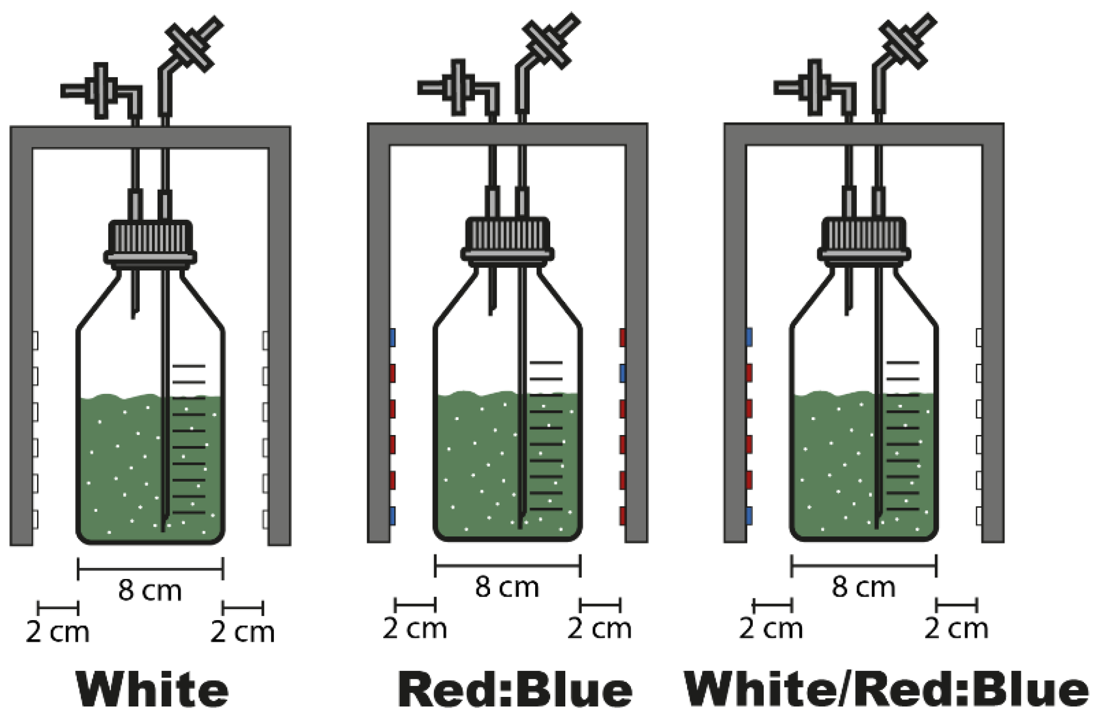
2.2. Experimental Design

Three configurations of LEDs: Cool white (60 LEDs/m, 400–700 nm, 12 V, 8 W/m) (Sinowell, Shanghai, China), Red:Blue (4:1, chips ratio, 60 LEDs/m, Blue: 660 nm, Red 450 nm, 12 V, 8 W/m) (Sinowell, Shanghai, China), and a mixture of the lights mentioned above (white/Red:Blue) were initially evaluated. For each experiment, Oscillatoria sp. was cultured (in triplicate) in 500 mL GL45 flasks (Schott Duran) with 250 mL of BG-11 culture media of working volume. Each flask was enclosed in a box (Figure 1) with 1 m of LEDs strip (2 cm from the surface of the flask).
Each flask was mixed using filtered air at a flow rate of 0.15 Lair min−1 and a photoperiod of 12:12 h at 100 µmol m−2 s−1 for 15 days. The air was enriched with 1 % (v/v) CO2 to accelerate the cyanobacterial growth. Since fluorescent lamps are the most widely available light source for producing algal and cyanobacterial biomass, this lamp was used as a control (Control FL) in all experiments.
The configuration that maximizes biomass and phycobiliproteins was further analyzed to identify the effect of light intensity (50, 80, 120, 150, and 180 µmol m−2 s−1) and photoperiod (12:12, 18:6, and 24:0 light:dark). The light intensity was monitored using a PAR (Photosynthetically Active Radiation) sensor (MQ-510, Apogee Instruments, Inc., North Logan, UT, USA).
The results were analyzed using a one-way ANOVA in GraphPad Prism version 9.3.1. The significant differences obtained in the analysis were represented in each figure.

2.3. Biomass and PBPs Quantification
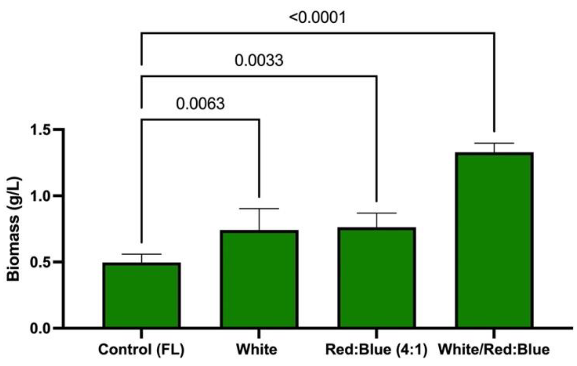
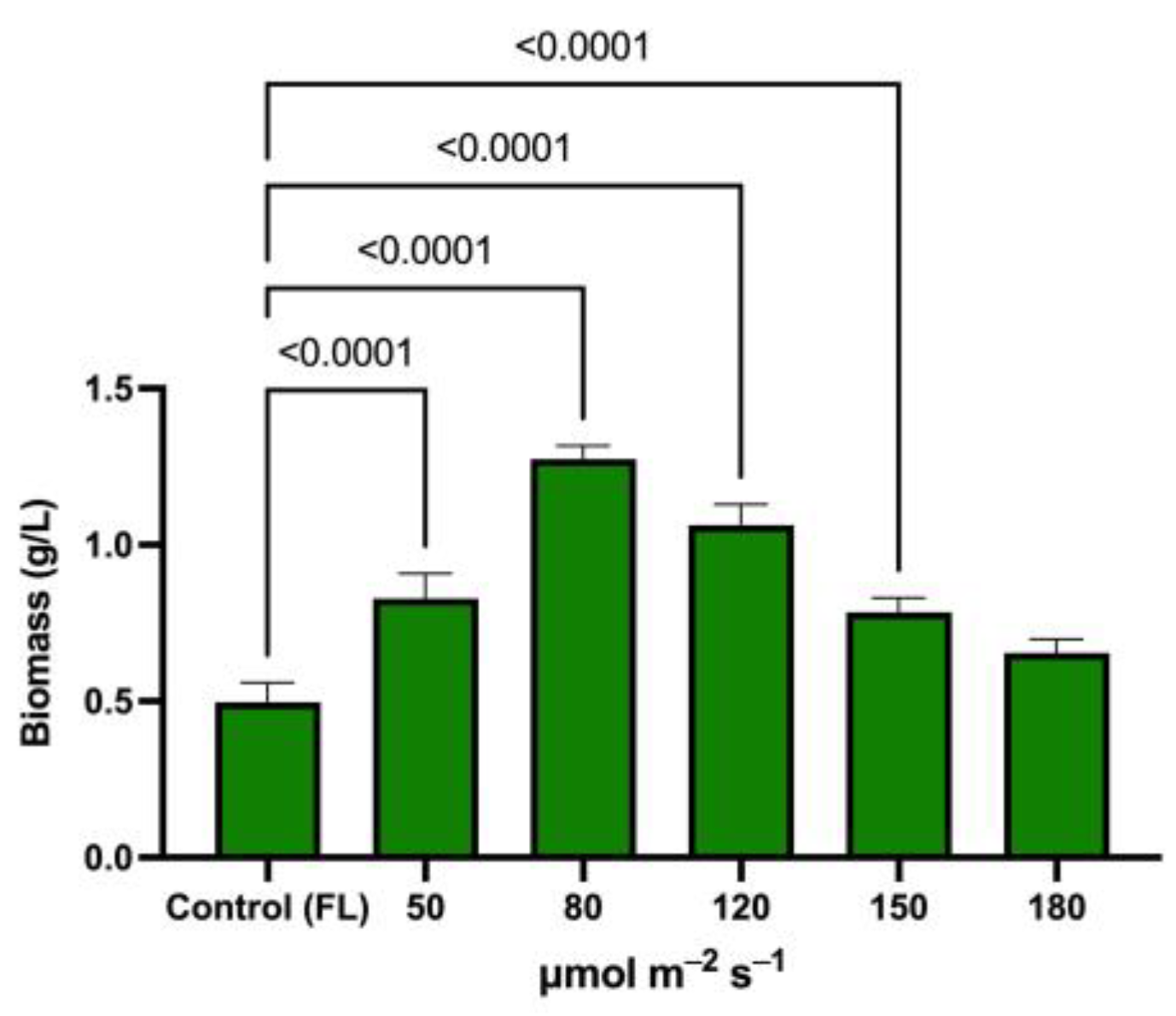
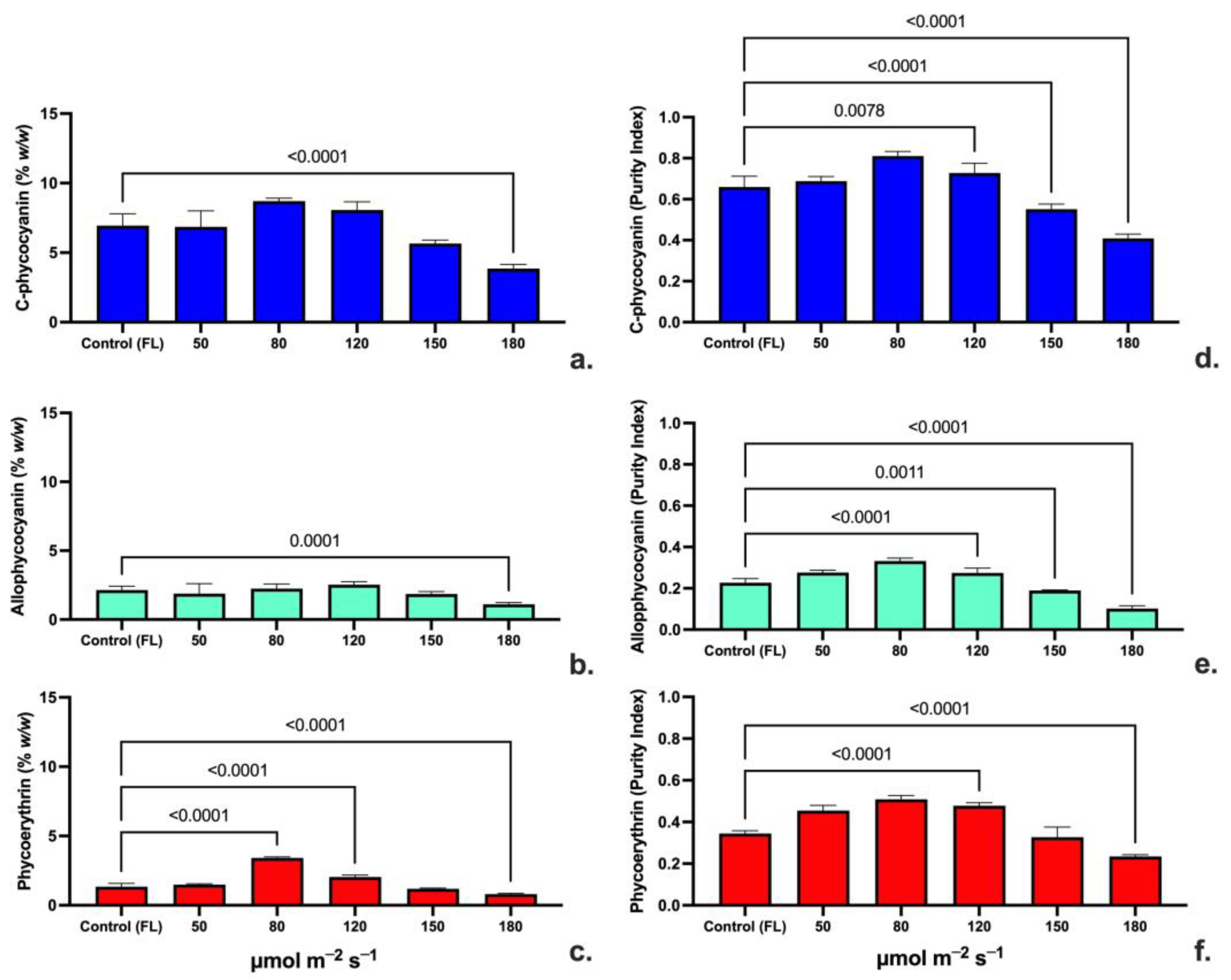
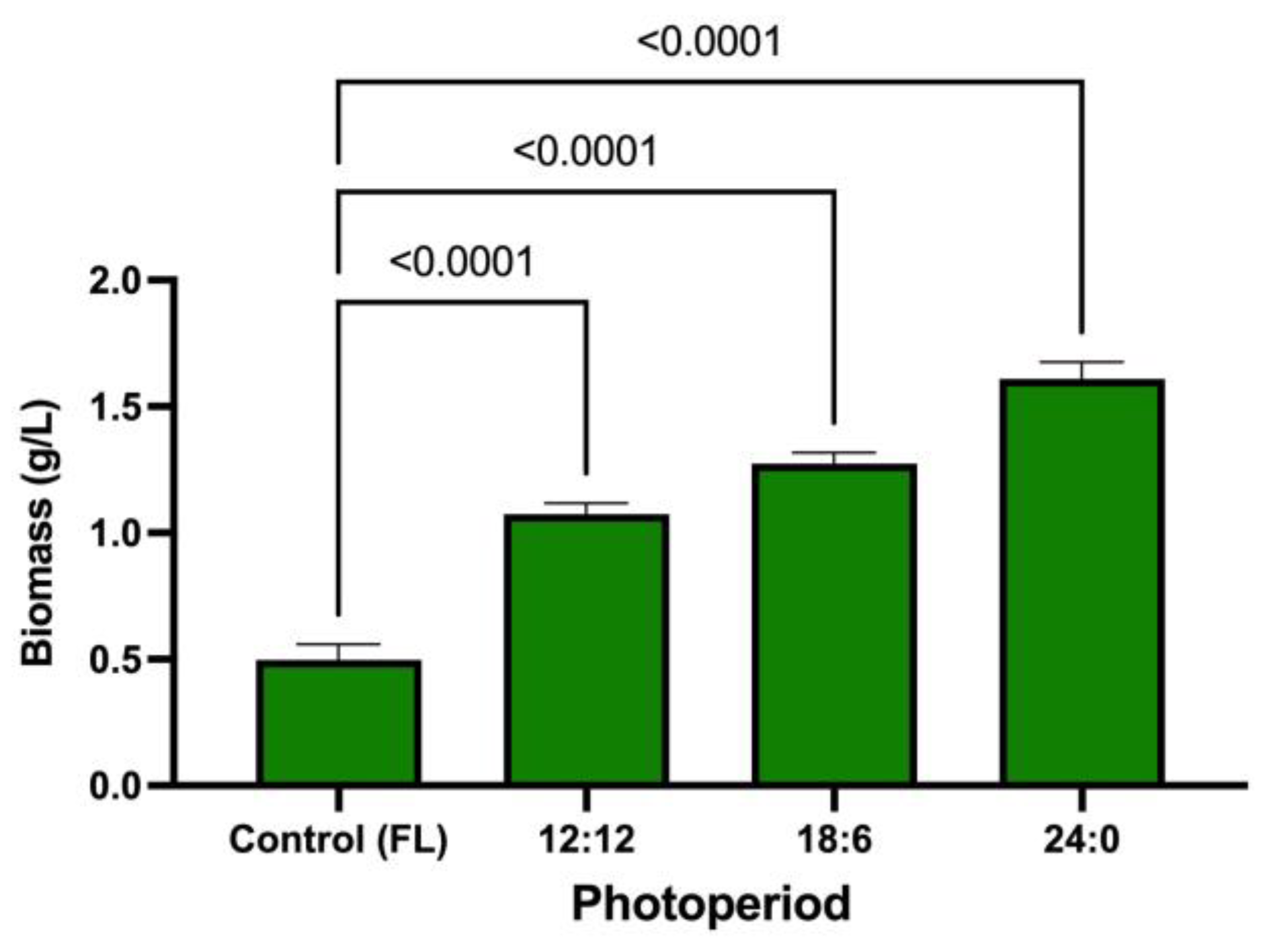
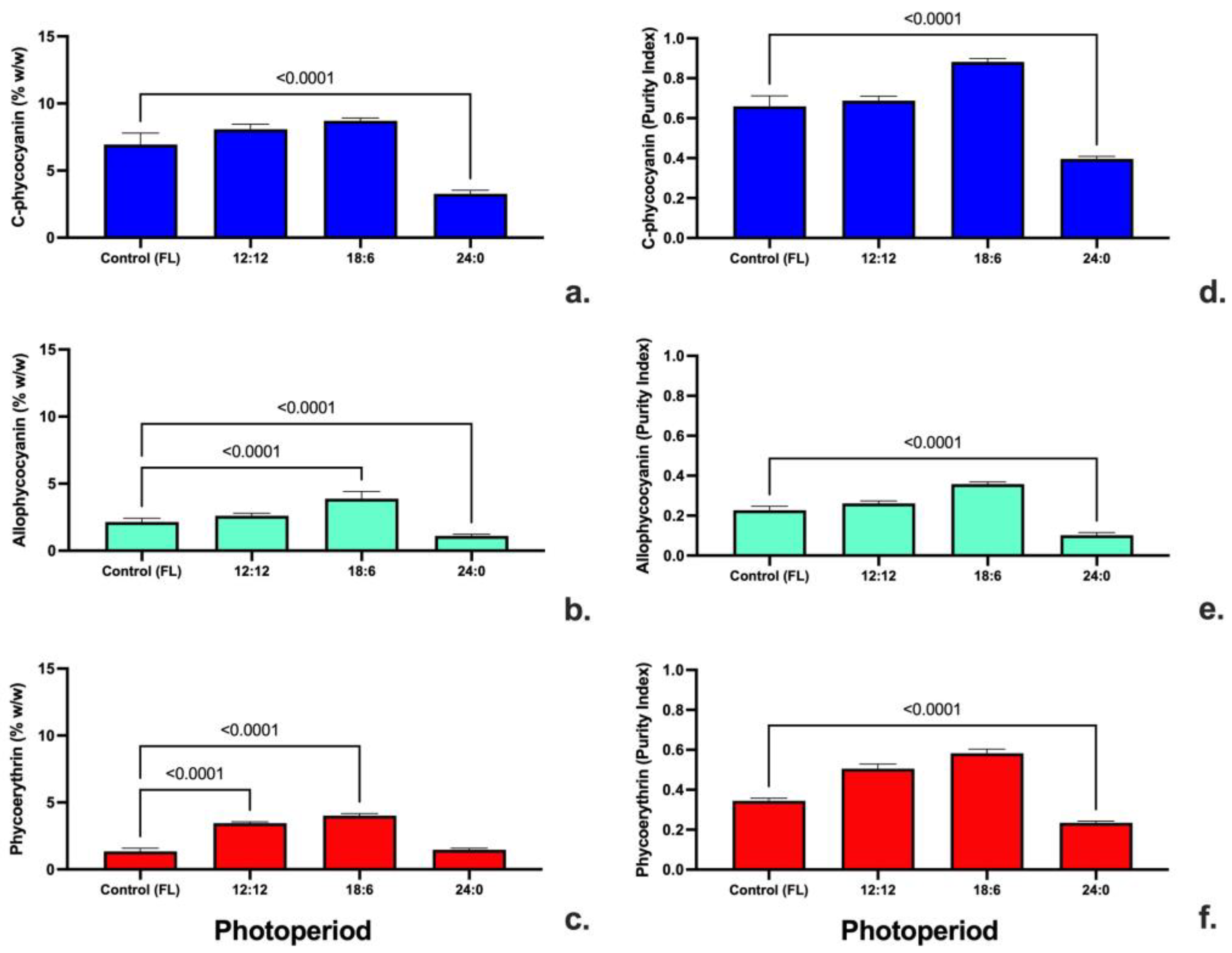
The biomass was concentrated by centrifugation at 3500 rpm (20 °C, 20 min) and dried using a food-grade dehydrator (30 h, 40 °C) [65], and stored in a desiccator until a constant weight [4]. The phycobiliproteins were extracted from the dried biomass using the method described by Zuorro et al. [2]; briefly, a known amount of dried biomass was mixed with a volume (0.26 % w/v) of cold phosphate buffer solution (0.05 M, pH 6.8) and a known amount of glass beads (0.5 mm diameter) (15 % w/w). The solution was mixed using an automatic vortex (Multi Reax, Heidolph, Germany) and stored in a refrigerator to promote the solubilization of the phycobiliproteins (4 °C, 24 h). PBPs were separated from cell debris by centrifugation (3400 rpm, 30 min, 20 °C). The deep blue supernatant was collected and measured in a spectrophotometer at specific wavelengths for C-PC (620 nm), APC (652 nm), and PE (562 nm). The concentration of phycocyanin (C-PC), allophycocyanin (APC), and phycoerythrin (PE) were calculated using Equations (1)–(3), which were described by Bennett and Bogorad [67].
The purity of C-PC, APC, and PE was determined using Equations (4)–(6) proposed by Patil [68] and Antello et al. [69], where each of the phycobiliprotein is divided by the total content of proteins measured at 280 nm
PC   [ g / L ] = OD 620 0.474 ( OD 652 ) 5.34
APC   [ g / L ] = OD 652 0.208 ( OD 620 ) 5 . 09
PE   [ g / L ] = ( OD 562 2.41 ( P PC ) 0.849 ( APC ) ) 9 . 62
PC   [ purity ] = OD 620 OD 280
APC   [ purity ] = OD 652 OD 280
PE   [ purity ] = OD 562 OD 280

3. Results

The results for biomass production using different LEDs are shown in Figure 2. The strain grew in all the LED configurations, with better results than in fluorescent lamps (0.49 g/L). Cool white and red:blue (4:1) LEDs increased the final concentration by up to 0.7 g/L; however, according to the ANOVA analysis, a higher difference was observed (<0.0001) in the biomass produced using the mixture of white/red:blue, with up to three times the concentration of the control (1.3 g/L).
The effect of the different LEDs on the concentration and purity of the different phycobiliproteins (C-PC, APC, and PE) can be found in Figure 2. Unlike biomass, C-PC (Figure 3a) (%w/w) in Oscillatoria sp. UFPS_001 shows no difference between the cool white and red:blue LEDs and the fluorescent lamps—with values between 7.5 and 7.8% (w/w); however, the white/red:blue mixture significantly increased the final concentration (10% w/w). In the case of APC (Figure 3b), the ANOVA analysis shows that white LEDs and the mixture of white/red:blue are significantly different (<0.0001) compared to the control, with values higher than 3.5% (w/w). The same behavior occurs with the PE (Figure 3c), where the same LED configuration increases the final content of PE. In the case of purity, the ANOVA analysis found that the mixture of white/red:blue increased the purity for C-PC, APC, and PE compared to the fluorescent lamps that were used as controls; however, no statistical differences were obtained when comparing the tested LEDs configurations.
According to the previous results, the white/red:blue LEDs configuration was used to determine the effect of intensity and the light:dark cycle. In the case of biomass concentration (Figure 4), it was found that Oscillatoria sp. grows better at low light intensities. An ANOVA analysis showed that intensities up to 80 µmol m−2 s−1 significantly (<0.0001) improved the biomass concentration compared to the control (up to 1.4 g/L). On the other hand, higher intensities significantly reduced the final biomass concentration.
The effect of the LEDs’ intensity on the concentration and purity of C-PC, APC, and PE can be found in Figure 4. The concentration of C-PC (Figure 5a) of Oscillatoria sp. behaves like the biomass, where 80 µmol m−2 s−1 significantly improves the C-PC concentration by up to 8% (w/w) in comparison with other light intensities. In the case of APC (Figure 5b), the ANOVA analysis shows no significant difference between the intensities evaluated, except at 180 µmol m−2 s−1; in this case, at higher light intensities, the concentration of APC is significantly reduced. On the other hand, the concentration of PE is substantially increased at 80 µmol m−2 s−1 compared to the control and the different intensities tested. In the case of purity, the ANOVA analysis found that the mixture of white/red:blue increased the purity for C-PC, APC, and PE compared to the fluorescent lamps that were used as controls; however, no statistical differences were obtained when comparing the tested LEDs configurations.
The previous results highlighted the intensity of 80 µmol m−2s−1 for evaluating the light:dark cycle. According to the results from the ANOVA analysis shown in Figure 6, when Oscillatoria sp. OSCI_UFPS001 is exposed to more extended light regimes at 80 µmol m−2 s−1, the biomass increased significantly (<0.0001) by up to 1.6 g/L (24:0 photoperiod) in comparison with the control, which used fluorescent lamps at 100 µmol m−2 s−1 (0.49 g/L, 12:12 photoperiod).
The effect of the LEDs’ photoperiod on the concentration and purity of phycobiliproteins can be found in Figure 7. In the case of C-PC, APC, and PE, more extended light periods substantially increased these proteins’ final concentrations and purities, with higher values using 18 h light and 6 h dark of photoperiod; however, when the flask was exposed to continuous light (24 h), the concentration and purity in all the evaluated phycobiliproteins significantly diminished compared to the control and the other photoperiods.

4. Discussion

Light quality is one of the most critical variables in microalgal and cyanobacterial industrial biomass production and specific metabolites [30]. In the case of biomass production using cyanobacterial strains, the application of LEDs with specific wavelengths has proved an interesting alternative (Table 1). While it is important from a research point of view to identify the best wavelength using LEDs to produce phycocyanin, it is also essential for determining the possibility of scaling up this type of technology to industrial production systems. Worldwide, most microalgae and cyanobacteria production plants use sunlight for their operations; however, to maintain high productivity, it is necessary to avoid the uncertainty generated by adverse environmental conditions that can substantially reduce the photosynthetic efficiency, and therefore reduce the company’s total profits. Thus, the search for ways to improve photosynthetic efficiency throughout the day is critical for sustainability in producing microalgae and cyanobacteria of commercial interest.
Globally, there are two industrial examples worth mentioning. The first is the company, Algalif (https://algalif.is; accessed on 12 November 2022), located in Iceland. Algalif is recognized worldwide as the most sustainable microalgae producer. They use ultra-pure glacier water to produce Haematococcus pluvialis and use geothermal energy for its operation. This company only makes astaxanthin indoors using LEDs tuned for cell growth and carotenogenesis. This allows Algalif to create high-quality astaxanthin year-round without relying on environmental changes. Other companies such as Algamo (https://www.algamo.cz/index.php/en/homepageen/; accessed on 12 November 2022) in the Czech Republic and Yemoja (https://yemojaltd.com; accessed on 12 November 2022) in Israel apply indoor cultivation systems with LEDs to maximize the production of metabolites. In the case of phycocyanin production, specifically in S. platensis, to the best of the author’s knowledge, there is only one indoor production site that uses LEDs to produce phycocyanin, which is in Sardinia, Italy (https://www.c-led.it/magazine/en/inaugurati-due-nuovi-impianti-di-coltivazione-di-spirulina-in-sardegna-con-lampade-c-led/; accessed on 12 November 2022).
The most studied wavelengths in the production of biomass and phycobiliproteins are white [26,47,48,49,50,51,52,53,54,55,56,57,58,59,60,61,62,63,64], red [23,25,26,30,51,52,53,54,57,58,59,63], and blue [49,50,51,52,53,57,58,59]. Other less studied wavelengths are orange [50], green [53,54], yellow [51,54,58], and far-red [62]. In the case of A. maxima, different LEDs wavelengths can modulate the final concentration of biomass, with values up to 3.9 g/L (white LEDs) [50] or as low as 0.78 g/L (red LEDs) [30]. In A. platensis, white LEDs can increase the final concentration of biomass by up to 6.5 g/L [55], and even yellow and red LEDs (6.6 and 6.2 g/L, respectively) have shown a significant increase in the biomass produced; however, the results obtained from different researchers lack homogeneity among strains of the same species. It is impossible to highlight any trend in the effect of a specific wavelength. These differences may be due to culture conditions used in each strain, such as the culture medium and organic carbon sources (mixotrophic culture).
In this work, the mixture of white and red/blue lights (4:1) enhanced the production and biomass and C-PC synthesis in Oscillatoria sp. A possible explanation for the synergistic effect in this strain can be explained by the fact that white LEDs will provide a wide spectrum of light, which will favor biomass production, while the red/blue LEDs will enhance the synthesis of PBPs (CPC-APC and PE) in this strain. Most of the literature focuses on the last two variables, but there is no evidence on the evaluation of the purity of PBPs. Purity is measured by the concentration of each PBPs divided by the absorbance at 280 nm (PBPs/Abs280 nm), which corresponds to the wavelength used to quantify total proteins in the Lowrey and Bradford methods [70]. This PBPs-to-protein ratio helps us understand how well the strain grows since PBPs are also a storage for nitrogen in cyanobacteria [71]. Therefore, lower purities will imply that more protein is synthesized; consequently, less nitrogen will be available to produce PBPs. In this case, the purity for C-PC, APC, and PE is statistically higher than the control (fluorescent lamps), indicating a balance between biomass building up and synthesis of PBPs; moreover, there are few cases reported in the literature that can support these results. Lee et al. [59] found that biomass and C-PC content can be improved when A. platensis is grown in a two-stage process. In this case, they tested red/blue LEDs (1:1) for biomass production, followed by a second stage using blue LEDs to increase the synthesis of C-PC; however, there was no evidence of the effect of mixing white light with other LEDs.
Other factors such as light intensity and photoperiod (also known as light:dark cycle) are as important as the quality of light [53]. Schipper et al. [72] found out in their preliminary experiments that a thermotolerant Leptolyngbya sp. strain could not grow normally at high radiations (up to 2800 µmol m−2 s−1) due to their lack of adaptation. Therefore, the low biomass concentration of Oscillatoria sp. at elevated radiation requires acclimatization since this strain was isolated from a hot spring in Colombia with high radiation.
For the case of the photoperiod, longer light times favor photosynthesis, and by adjusting the intensity and wavelength, it is possible to increase biomass concentration; however, this interaction between the light cycle and intensity may depend on the strain evaluated. In the case of A. maxima, when growing under medium intensities (80 µmol m−2 s−1) with white LED lights and complete exposure to light (24 h), it is possible to obtain more biomass compared to blue or orange LED lights [50]; for the case of A. platensis, blue light with the same conditions mentioned above are the ones that favor its growth. In another work, Xie et al. [55] demonstrated yellow LEDs lights with an intensity of 250 µmol m−2 s−1. A cycle of 12 h light and 12 h dark can maximize the biomass of A. platensis by up to 6.6 g/L, which is much higher than that found by Milia et al. [39] (0.59 g/L) or Bachchhav et al. [54] (8.95 g/L).
Another example of this is the results achieved by Klepacz-Smółka et al. [25] by using Synechococcus PCC 6715 (red LEDs, 100 µmol m−2 s−1, and 16 h of light and 8 h of dark), which achieved the highest concentration reported for research of this type (8.6 g/L) and is even more interesting since no inorganic carbon sources were used in the BG11 medium. According to different articles published internationally in recent years, the effect of the parameters mentioned above on the synthesis of phycobiliproteins does not follow a fixed pattern [73,74,75]. Therefore, strains of the same species may have different light requirements. This can be explained by the unique composition of PBPs of each cyanobacterial strain. This would explain the unusual behavior of Oscillatoria sp., which not only has high concentrations of C-PC but, under certain conditions, the concentration of PE increased significantly compared to the control. It is worth mentioning that the short distance between the light source and the small diameter of the flask is a variable that must be considered in the selection of the culture method (raceways, column, or tubular PBR) for a scaling process.

5. Conclusions

This research shows the capability of LEDs with a specific color to improve biomass and phycobiliproteins (PBPs). In this case, a mixture of white/Red:Blue LEDs at 80 µmol m−2 s−1 and a photoperiod of 24 h of light significantly increases the final biomass concentration up to 3 times compared to the same strain cultured on fluorescent lamps. However, total exposure negatively affects the synthesis of the different PBPs (C-PC, APC, and PE). A better cycle of 18 h of light and 6 h of darkness allows a better synthesis of these proteins with a slight reduction in biomass concentration. Further studies should focus on the possible interaction between specific nutrients (N, P, Mg, etc.) and LEDs that can enhance different metabolites.

Author Contributions

Conceptualization, S.J.B., A.F.B.-S. and A.Z.; methodology, J.B.G.-M., V.S.L.-B. and A.D.R.-B.; software, A.F.B.-S. and A.Z.; validation, J.E.C.-R., N.A.U.-S. and G.L.L.-B.; formal analysis, J.E.C.-R., J.B.G.-M. and A.Z.; investigation, V.S.L.-B. and A.D.R.-B.; resources, A.F.B.-S. and S.J.B.; data curation, A.Z.; writing—original draft preparation, J.B.G.-M. and J.E.C.-R.; writing—review and editing, N.A.U.-S. and G.L.L.-B.; visualization, G.L.L.-B.; supervision, N.A.U.-S., J.B.G.-M. and A.Z; Project administration, A.F.B.-S. and S.J.B.; funding acquisition, A.F.B.-S. and S.J.B. All authors have read and agreed to the published version of the manuscript.

Funding

This paper was supported by Newton-Caldas Fund Institutional Links, with the project “ALGALCOLOR: BIO-PLATFORM FOR THE SUSTAINABLE PRODUCTION OF CYANOBACTERIAL-BASED COLOURS AND FINE CHEMICALS” ID 527624805. It was also funded by Sapienza for Academic Mid Projects 2021 n. RM12117A8B58023A.

Institutional Review Board Statement

Not applicable.

Informed Consent Statement

Not applicable.

Data Availability Statement

Not applicable.

Acknowledgments

We would like to express our sincere gratitude to Sapienza University of Rome (Italy) and Universidad Francisco de Paula Santander (Colombia) for providing the equipment for this research. We also thank the Colombian Ministry of Science, Technology and Innovation MINCIENCIAS for supporting national Ph.D. Doctorates through the Francisco José de Caldas scholarship program.

Conflicts of Interest

The authors declare no conflict of interest.

References

  1. Zuorro, A.; Leal-Jerez, A.G.; Morales-Rivas, L.K.; Mogollón-Londoño, S.O.; Sanchez-Galvis, E.M.; García-Martínez, J.B.; Barajas-Solano, A.F. Enhancement of Phycobiliprotein Accumulation in Thermotolerant Oscillatoria Sp. through Media Optimization. ACS Omega 2021, 6, 10527–10536. [Google Scholar] [CrossRef]
  2. Zuorro, A.; Lavecchia, R.; Maffei, G.; Marra, F.; Miglietta, S.; Petrangeli, A.; Familiari, G.; Valente, T. Enhanced lipid extraction from unbroken microalgal cells using enzymes. Chem. Eng. Trans. 2015, 43, 211–216. [Google Scholar] [CrossRef]
  3. Barajas-Solano, A.F.; Guzmán-Monsalve, A.; Kafarov, V. Effect of Carbon-Nitrogen Ratio for the Biomass Production, Hydrocarbons and Lipids on Botryoccus braunii UIS 003. Chem. Eng. Trans. 2016, 49, 247–252. [Google Scholar] [CrossRef]
  4. García-Martínez, J.B.; Ayala-Torres, E.; Reyes-Gómez, O.; Zuorro, A.; Andrés, F.; Barajas-Solano, B.; Crisóstomo, C.; Barajas-Ferreira, B. Evaluation of a Two-Phase Extraction System of Carbohydrates and Proteins from Chlorella vulgaris Utex 1803. Chem. Eng. Trans. 2016, 49, 355–360. [Google Scholar] [CrossRef]
  5. Zuorro, A.; Maffei, G.; Lavecchia, R. Kinetic Modeling of Azo Dye Adsorption on Non-Living Cells of Nannochloropsis oceanica. J. Environ. Chem. Eng. 2017, 5, 4121–4127. [Google Scholar] [CrossRef]
  6. Zuorro, A.; Malavasi, V.; Cao, G.; Lavecchia, R. Use of Cell Wall Degrading Enzymes to Improve the Recovery of Lipids from Chlorella sorokiniana. Chem. Eng. J. 2019, 377, 120325. [Google Scholar] [CrossRef]
  7. Chittapun, S.; Jonjaroen, V.; Khumrangsee, K.; Charoenrat, T. C-Phycocyanin Extraction from Two Freshwater Cyanobacteria by Freeze Thaw and Pulsed Electric Field Techniques to Improve Extraction Efficiency and Purity. Algal Res. 2020, 46, 101789. [Google Scholar] [CrossRef]
  8. Vega-Gálvez, A.; Miranda, M.; Clavería, R.; Quispe, I.; Vergara, J.; Uribe, E.; Paez, H.; Di Scala, K. Effect of Air Temperature on Drying Kinetics and Quality Characteristics of Osmo-Treated Jumbo Squid (Dosidicus gigas). LWT-Food Sci. Technol. 2011, 44, 16–23. [Google Scholar] [CrossRef]
  9. Falkeborg, M.F.; Roda-Serrat, M.C.; Burnæs, K.L.; Nielsen, A.L.D. Stabilising Phycocyanin by Anionic Micelles. Food Chem. 2018, 239, 771–780. [Google Scholar] [CrossRef]
  10. Fratelli, C.; Burck, M.; Amarante, M.C.A.; Braga, A.R.C. Antioxidant Potential of Nature’s “Something Blue”: Something New in the Marriage of Biological Activity and Extraction Methods Applied to C-Phycocyanin. Trends Food Sci. Technol. 2021, 107, 309–323. [Google Scholar] [CrossRef]
  11. Ojit, S.K.; Indrama, T.; Gunapati, O.; Avijeet, S.O.; Subhalaxmi, S.A.; Silvia, C.; Indira, D.W.; Romi, K.; Thadoi, D.A.; Tiwari, O.N.; et al. The Response of Phycobiliproteins to Light Qualities in Anabaena circinalis. J. Appl. Biol. Biotechnol. 2015, 3, 1–6. [Google Scholar] [CrossRef] [Green Version]
  12. Pez Jaeschke, D.; Rocha Teixeira, I.; Damasceno Ferreira Marczak, L.; Domeneghini Mercali, G. Phycocyanin from Spirulina: A Review of Extraction Methods and Stability. Food Res. Int. 2021, 143, 110314. [Google Scholar] [CrossRef] [PubMed]
  13. Seghiri, R.; Legrand, J.; Hsissou, R.; Essamri, A. Comparative Study of the Impact of Conventional and Unconventional Drying Processes on Phycobiliproteins from Arthrospira platensis. Algal Res. 2021, 53, 102165. [Google Scholar] [CrossRef]
  14. Tavanandi, H.A.; Raghavarao, K.S.M.S. Ultrasound-Assisted Enzymatic Extraction of Natural Food Colorant C-Phycocyanin from Dry Biomass of Arthrospira platensis. LWT 2020, 118, 108802. [Google Scholar] [CrossRef]
  15. Lee, E.; Pruvost, J.; He, X.; Munipalli, R.; Pilon, L. Design Tool and Guidelines for Outdoor Photobioreactors. Chem. Eng. Sci. 2014, 106, 18–29. [Google Scholar] [CrossRef] [Green Version]
  16. Chaneva, G.; Furnadzhieva, S.; Minkova, K.; Lukavsky, J. Effect of Light and Temperature on the Cyanobacterium Arthronema africanum—A Prospective Phycobiliprotein-Producing Strain. J. Appl. Phycol. 2007, 19, 537–544. [Google Scholar] [CrossRef]
  17. Sloth, J.K.; Wiebe, M.G.; Eriksen, N.T. Accumulation of Phycocyanin in Heterotrophic and Mixotrophic Cultures of the Acidophilic Red Alga Galdieria sulphuraria. Enzyme Microb. Technol. 2006, 38, 168–175. [Google Scholar] [CrossRef]
  18. Tang, J.; Jiang, D.; Luo, Y.; Liang, Y.; Li, L.; Shah, M.M.R.; Daroch, M. Potential New Genera of Cyanobacterial Strains Isolated from Thermal Springs of Western Sichuan, China. Algal Res. 2018, 31, 14–20. [Google Scholar] [CrossRef]
  19. Mehariya, S.; Fratini, F.; Lavecchia, R.; Zuorro, A. Green Extraction of Value-Added Compounds Form Microalgae: A Short Review on Natural Deep Eutectic Solvents (NaDES) and Related Pre-Treatments. J. Environ. Chem. Eng. 2021, 9, 105989. [Google Scholar] [CrossRef]
  20. Zuorro, A.; García-Martínez, J.B.; Barajas-Solano, A.F. The Application of Catalytic Processes on the Production of Algae-Based Biofuels: A Review. Catalysts 2021, 11, 22. [Google Scholar] [CrossRef]
  21. Cuellar García, D.J.; Rangel-Basto, Y.A.; Barajas-Solano, A.F.; Muñoz-Peñalosa, Y.A.; Urbina-Suarez, N.A. Towards the Production of Microalgae Biofuels: The Effect of the Culture Medium on Lipid Deposition. Biotechnologia 2019, 100, 273–278. [Google Scholar] [CrossRef]
  22. Quintero-Dallos, V.; García-Martínez, J.B.; Contreras-Ropero, J.E.; Barajas-Solano, A.F.; Barajas-Ferrerira, C.; Lavecchia, R.; Zuorro, A. Vinasse as a Sustainable Medium for the Production of Chlorella vulgaris UTEX 1803. Water 2019, 11, 1526. [Google Scholar] [CrossRef] [Green Version]
  23. Prates, D.d.F.; Radmann, E.M.; Duarte, J.H.; Morais, M.G.d.; Costa, J.A.V. Spirulina Cultivated under Different Light Emitting Diodes: Enhanced Cell Growth and Phycocyanin Production. Bioresour. Technol. 2018, 256, 38–43. [Google Scholar] [CrossRef] [PubMed]
  24. Hoi, S.K.; Winayu, B.N.R.; Hsueh, H.T.; Chu, H. Light Factors and Nitrogen Availability to Enhance Biomass and C-Phycocyanin Productivity of Thermosynechococcus Sp. CL-1. Biochem. Eng. J. 2021, 167, 107899. [Google Scholar] [CrossRef]
  25. Klepacz-Smółka, A.; Pietrzyk, D.; Szeląg, R.; Głuszcz, P.; Daroch, M.; Tang, J.; Ledakowicz, S. Effect of Light Colour and Photoperiod on Biomass Growth and Phycocyanin Production by Synechococcus PCC 6715. Bioresour. Technol. 2020, 313, 123700. [Google Scholar] [CrossRef] [PubMed]
  26. Ho, S.H.; Liao, J.F.; Chen, C.Y.; Chang, J.S. Combining Light Strategies with Recycled Medium to Enhance the Economic Feasibility of Phycocyanin Production with Spirulina platensis. Bioresour. Technol. 2018, 247, 669–675. [Google Scholar] [CrossRef] [PubMed]
  27. Bergmann, P.; Trösch, W. Repeated Fed-Batch Cultivation of Thermosynechococcus elongatus BP-1 in Flat-Panel Airlift Photobioreactors with Static Mixers for Improved Light Utilization: Influence of Nitrate, Carbon Supply and Photobioreactor Design. Algal Res. 2016, 17, 79–86. [Google Scholar] [CrossRef]
  28. Wu, H.-L.; Wang, G.-H.; Xiang, W.-Z.; Li, T.; He, H. Stability and Antioxidant Activity of Food-Grade Phycocyanin Isolated from Spirulina Platensis. Int. J. Food Prop. 2016, 19, 2349–2362. [Google Scholar] [CrossRef] [Green Version]
  29. Khatoon, H.; Kok Leong, L.; Abdu Rahman, N.; Mian, S.; Begum, H.; Banerjee, S.; Endut, A. Effects of Different Light Source and Media on Growth and Production of Phycobiliprotein from Freshwater Cyanobacteria. Bioresour. Technol. 2018, 249, 652–658. [Google Scholar] [CrossRef]
  30. Park, J.; Dinh, T.B. Contrasting Effects of Monochromatic LED Lighting on Growth, Pigments and Photosynthesis in the Commercially Important Cyanobacterium Arthrospira maxima. Bioresour. Technol. 2019, 291, 121846. [Google Scholar] [CrossRef]
  31. Luimstra, V.M.; Schuurmans, J.M.; Verschoor, A.M.; Hellingwerf, K.J.; Huisman, J.; Matthijs, H.C.P. Blue Light Reduces Photosynthetic Efficiency of Cyanobacteria through an Imbalance between Photosystems I and II. Photosynth. Res. 2018, 138, 177–189. [Google Scholar] [CrossRef] [PubMed] [Green Version]
  32. Blanken, W.; Cuaresma, M.; Wijffels, R.H.; Janssen, M. Cultivation of Microalgae on Artificial Light Comes at a Cost. Algal Res. 2013, 2, 333–340. [Google Scholar] [CrossRef]
  33. Schulze, P.S.C.; Barreira, L.A.; Pereira, H.G.C.; Perales, J.A.; Varela, J.C.S. Light Emitting Diodes (LEDs) Applied to Microalgal Production. Trends Biotechnol. 2014, 32, 422–430. [Google Scholar] [CrossRef]
  34. Zhao, Y.; Wang, J.; Zhang, H.; Yan, C.; Zhang, Y. Effects of Various LED Light Wavelengths and Intensities on Microalgae-Based Simultaneous Biogas Upgrading and Digestate Nutrient Reduction Process. Bioresour. Technol. 2013, 136, 461–468. [Google Scholar] [CrossRef] [PubMed]
  35. Teo, C.L.; Atta, M.; Bukhari, A.; Taisir, M.; Yusuf, A.M.; Idris, A. Enhancing Growth and Lipid Production of Marine Microalgae for Biodiesel Production via the Use of Different LED Wavelengths. Bioresour. Technol. 2014, 162, 38–44. [Google Scholar] [CrossRef] [PubMed]
  36. Assunção, J.; Pagels, F.; Tavares, T.; Malcata, F.X.; Guedes, A.C. Light Modulation for Bioactive Pigment Production in Synechocystis Salina. Bioengineering 2022, 9, 331. [Google Scholar] [CrossRef]
  37. Okamoto, A.; Imamura, M.; Tani, K.; Matsumoto, T. The Effect of Using Light Emitting Diodes and Fluorescent Lamps as Different Light Sources in Growth Inhibition Tests of Green Alga, Diatom, and Cyanobacteria. PLoS ONE 2021, 16, e0247426. [Google Scholar] [CrossRef]
  38. Mehariya, S.; Goswami, R.K.; Verma, P.; Lavecchia, R.; Zuorro, A. Integrated Approach for Wastewater Treatment and Biofuel Production in Microalgae Biorefineries. Energies 2021, 14, 2282. [Google Scholar] [CrossRef]
  39. Banayan, S.; Jahadi, M.; Khosravi-Darani, K. Pigment Productions by Spirulina Platensis as a Renewable Resource. J. Appl. Biotechnol. Rep. 2022, 9, 614–621. [Google Scholar] [CrossRef]
  40. Van Hieu, H.; Quang-Tuong, L.; Cong Doan, B. The Production of High Phycocyanin by Applications of Red Light-Emitting Diodes (LEDs) In Vitro Algae Growth on Spirulina platensis. J. Nano-Electron. Phys. 2021, 13, 3034-1–3034-4. [Google Scholar] [CrossRef]
  41. Rangel-Basto, Y.A.; García-Ochoa, I.E.; Suarez-Gelvez, J.H.; Zuorro, A.; Barajas-Solano, A.F.; Urbina-Suarez, N.A. The Effect of Temperature and Enzyme Concentration in the Transesterification Process of Synthetic Microalgae Oil. Chem. Eng. Trans. 2018, 64, 331–336. [Google Scholar] [CrossRef]
  42. Mao, R.; Guo, S. Performance of the Mixed LED Light Quality on the Growth and Energy Efficiency of Arthrospira platensis. Appl. Microbiol. Biotechnol. 2018, 102, 5245–5254. [Google Scholar] [CrossRef] [PubMed]
  43. Borlongan, I.A.; Suzuki, S.; Nishihara, G.N.; Kozono, J.; Terada, R. Effects of Light Quality and Temperature on the Photosynthesis and Pigment Content of a Subtidal Edible Red Alga Meristotheca papulosa (Solieriaceae, Gigartinales) from Japan. J. Appl. Phycol. 2020, 32, 1329–1340. [Google Scholar] [CrossRef]
  44. Barajas-Solano, A.F.; Gonzalez-Delgado, A.D.; Kafarov, V. Effect Of Thermal Pre-Treatment On Fermentable Sugar Production Of Chlorella vulgaris. Chem. Eng. Trans. 2014, 37, 655–660. [Google Scholar] [CrossRef]
  45. Pagels, F.; Lopes, G.; Vasconcelos, V.; Guedes, A.C. White and Red LEDs as Two-Phase Batch for Cyanobacterial Pigments Production. Bioresour. Technol. 2020, 307, 123105. [Google Scholar] [CrossRef]
  46. Urbina-Suarez, N.A.; Ayala-González, D.D.; Rivera-Amaya, J.D.; Barajas-Solano, A.F.; Machuca-Martínez, F. Evaluation of the Light/Dark Cycle and Concentration of Tannery Wastewater in the Production of Biomass and Metabolites of Industrial Interest from Microalgae and Cyanobacteria. Water 2022, 14, 346. [Google Scholar] [CrossRef]
  47. Mehar, J.; Shekh, A.; Nethravathy, M.U.; Sarada, R.; Chauhan, V.S.; Mudliar, S. Automation of Pilot-Scale Open Raceway Pond: A Case Study of CO2-Fed PH Control on Spirulina Biomass, Protein and Phycocyanin Production. J. CO2 Util. 2019, 33, 384–393. [Google Scholar] [CrossRef]
  48. Kovač, D.; Babić, O.; Milovanović, I.; Mišan, A.; Simeunović, J. The Production of Biomass and Phycobiliprotein Pigments in Filamentous Cyanobacteria: The Impact of Light and Carbon Sources. Appl. Biochem. Microbiol. 2017, 53, 539–545. [Google Scholar] [CrossRef]
  49. García-López, D.A.; Olguín, E.J.; González-Portela, R.E.; Sánchez-Galván, G.; De Philippis, R.; Lovitt, R.W.; Llewellyn, C.A.; Fuentes-Grünewald, C.; Parra Saldívar, R. A Novel Two-Phase Bioprocess for the Production of Arthrospira (Spirulina) maxima LJGR1 at Pilot Plant Scale during Different Seasons and for Phycocyanin Induction under Controlled Conditions. Bioresour. Technol. 2020, 298, 122548. [Google Scholar] [CrossRef] [PubMed]
  50. Milia, M.; Corrias, F.; Addis, P.; Zitelli, G.C.; Cicchi, B.; Torzillo, G.; Andreotti, V.; Angioni, A. Influence of Different Light Sources on the Biochemical Composition of Arthrospira Spp. Grown in Model Systems. Foods 2022, 11, 399. [Google Scholar] [CrossRef]
  51. Tayebati, H.; Pajoum Shariati, F.; Soltani, N.; Sepasi Tehrani, H. Effect of Various Light Spectra on Amino Acids and Pigment Production of Arthrospira platensis Using Flat-Plate Photobioreactor. Prep. Biochem. Biotechnol. 2021, 51, 1–12. [Google Scholar] [CrossRef] [PubMed]
  52. Szwarc, D.; Zieliński, M. Effect of Lighting on the Intensification of Phycocyanin Production in a Culture of Arthrospira platensis. Proceedings 2018, 2, 1305. [Google Scholar] [CrossRef] [Green Version]
  53. Yim, S.K.; Ki, D.W.; Doo, H.S.; Kim, H.; Kwon, T.H. Internally Illuminated Photobioreactor Using a Novel Type of Light-Emitting Diode (LED) Bar for Cultivation of Arthrospira platensis. Biotechnol. Bioprocess Eng. 2016, 21, 767–776. [Google Scholar] [CrossRef]
  54. Bachchhav, M.B.; Kulkarni, M.V.; Ingale, A.G. Enhanced Phycocyanin Production from Spirulina platensis Using Light Emitting Diode. J. Inst. Eng. Ser. E 2017, 98, 41–45. [Google Scholar] [CrossRef]
  55. Xie, Y.; Jin, Y.; Zeng, X.; Chen, J.; Lu, Y.; Jing, K. Fed-Batch Strategy for Enhancing Cell Growth and C-Phycocyanin Production of Arthrospira (Spirulina) platensis under Phototrophic Cultivation. Bioresour. Technol. 2015, 180, 281–287. [Google Scholar] [CrossRef]
  56. Walter, A.; Carvalho, J.C.D.; Soccol, V.T.; Bisinella, A.B.; Faria, D.; Ghiggi, V.; Soccol, C.R. Study of Phycocyanin Production from Spirulina platensis Under Different Light Spectra. Braz. Arch. Biol. Technol. 2011, 54, 675–682. [Google Scholar] [CrossRef]
  57. Markou, G. Effect of Various Colors of Light-Emitting Diodes (LEDs) on the Biomass Composition of Arthrospira platensis Cultivated in Semi-Continuous Mode. Appl. Biochem. Biotechnol. 2014, 172, 2758–2768. [Google Scholar] [CrossRef]
  58. Chen, H.B.; Wu, J.Y.; Wang, C.F.; Fu, C.C.; Shieh, C.J.; Chen, C.I.; Wang, C.Y.; Liu, Y.C. Modeling on Chlorophyll a and Phycocyanin Production by Spirulina platensis under Various Light-Emitting Diodes. Biochem. Eng. J. 2010, 53, 52–56. [Google Scholar] [CrossRef]
  59. Lee, S.H.; Lee, J.E.; Kim, Y.; Lee, S.Y. The Production of High Purity Phycocyanin by Spirulina platensis Using Light-Emitting Diodes Based Two-Stage Cultivation. Appl. Biochem. Biotechnol. 2016, 178, 382–395. [Google Scholar] [CrossRef]
  60. Sivasankari, S.; Vinoth, M.; Ravindran, D.; Baskar, K.; Alqarawi, A.A.; Abd_Allah, E.F. Efficacy of Red Light for Enhanced Cell Disruption and Fluorescence Intensity of Phycocyanin. Bioprocess Biosyst. Eng. 2021, 44, 141–150. [Google Scholar] [CrossRef]
  61. Niangoran, N.U.F.; Buso, D.; Zissis, G.; Prudhomme, T. Influence of Light Intensity and Photoperiod on Energy Efficiency of Biomass and Pigment Production of Spirulina (Arthrospira platensis). OCL-Oilseeds Fats Crop. Lipids 2021, 28, 37. [Google Scholar] [CrossRef]
  62. Silkina, A.; Kultschar, B.; Llewellyn, C.A. Far-Red Light Acclimation for Improved Mass Cultivation of Cyanobacteria. Metabolites 2019, 9, 170. [Google Scholar] [CrossRef] [PubMed] [Green Version]
  63. Kim, J.K.; Mao, Y.; Kraemer, G.; Yarish, C. Growth and Pigment Content of Gracilaria tikvahiae McLachlan under Fluorescent and LED Lighting. Aquaculture 2015, 436, 52–57. [Google Scholar] [CrossRef]
  64. Velea, S.; Ilie, L.; Filipescu, L. Optimization of Porphyridium purpureum Culture Growth Using Two Variables Experimental Design: Light and Sodium Bicarbonate. UPB Sci. Bull. Ser. B Chem. Mater. Sci. 2011, 73, 81–94. [Google Scholar]
  65. Barajas-Solano, A.F. Optimization of Phycobiliprotein Solubilization from a Thermotolerant Oscillatoria Sp. Processes 2022, 10, 836. [Google Scholar] [CrossRef]
  66. Andersen, R.A.; Berges, J.A.; Harrison, P.J.; Watanabe, M.M. Appendix A—Recipes for Freshwater and Seawater Media. In Algal Culturing Techniques; Andersen, R.A., Ed.; Elsevier Academic Press: Burlington, MA, USA, 2005; pp. 429–538. [Google Scholar]
  67. Bennett, A.; Bogorad, L. Complementary Chromatic Adaptation in a Filamentous Blue-Green Alga. J. Cell Biol. 1973, 58, 419–435. [Google Scholar] [CrossRef]
  68. Patil, G.; Chethana, S.; Sridevi, A.S.; Raghavarao, K.S.M.S. Method to Obtain C-Phycocyanin of High Purity. J. Chromatogr. A 2006, 1127, 76–81. [Google Scholar] [CrossRef]
  69. Antelo, F.; Anschau, A.; Costa, J.; Kalil, S. Extraction and Purification of C-Phycocyanin from Spirulina platensis in Conventional and Integrated Aqueous Two-Phase Systems. J. Braz. Chem. Soc. 2010, 21, 921–926. [Google Scholar] [CrossRef] [Green Version]
  70. Moheimani, N.R.; Borowitzka, M.A.; Isdepsky, A.; Sing, S.F. Standard Methods for Measuring Growth of Algae and Their Composition BT—Algae for Biofuels and Energy; Borowitzka, M.A., Moheimani, N.R., Eds.; Springer Netherlands: Dordrecht, The Netherlands, 2013; pp. 265–284. [Google Scholar] [CrossRef]
  71. Lamela, T.; Marquez-Rocha, F.J. Phycocyanin Production in Seawater Culture of Arthrospira maxima. Ciencias Mar. 2000, 26, 607–619. [Google Scholar] [CrossRef] [Green Version]
  72. Schipper, K.; Das, P.; Al Muraikhi, M.; AbdulQuadir, M.; Thaher, M.I.; Al Jabri, H.M.S.J.; Wijffels, R.H.; Barbosa, M.J. Outdoor Scale-up of Leptolyngbya Sp.: Effect of Light Intensity and Inoculum Volume on Photoinhibition and -Oxidation. Biotechnol. Bioeng. 2021, 118, 2368–2379. [Google Scholar] [CrossRef] [PubMed]
  73. Jung, C.H.G.; Waldeck, P.; Sykora, S.; Braune, S.; Petrick, I.; Küpper, J.-H.; Jung, F. Influence of Different Light-Emitting Diode Colors on Growth and Phycobiliprotein Generation of Arthrospira platensis. Life 2022, 12, 895. [Google Scholar] [CrossRef] [PubMed]
  74. Hotos, G.N.; Antoniadis, T.I. The Effect of Colored and White Light on Growth and Phycobiliproteins, Chlorophyll and Carotenoids Content of the Marine Cyanobacteria Phormidium Sp. and Cyanothece Sp. in Batch Cultures. Life 2022, 12, 837. [Google Scholar] [CrossRef] [PubMed]
  75. Zanolla, V.; Biondi, N.; Niccolai, A.; Abiusi, F.; Adessi, A.; Rodolfi, L.; Tredici, M.R. Protein, Phycocyanin, and Polysaccharide Production by Arthrospira platensis Grown with LED Light in Annular Photobioreactors. J. Appl. Phycol. 2022, 34, 1189–1199. [Google Scholar] [CrossRef]
Figure 1. Cultivation system diagram.
Applsci 12 11664 g001
Figure 2. Biomass is produced under different LEDs configurations.
Applsci 12 11664 g002
Figure 3. The concentration of phycocyanin (C-PC) (a), allophycocyanin (APC) (b), and phycoerythrin (PE) (c) and their purity (df) under different LEDs configurations.
Applsci 12 11664 g003
Figure 4. Biomass concentration under different intensities of white/red:blue LEDs.
Applsci 12 11664 g004
Figure 5. The concentration of phycocyanin (C-PC) (a), allophycocyanin (APC) (b), and phycoerythrin (PE) (c) and their purity (df) under different intensities of white/red:blue LEDs.
Applsci 12 11664 g005
Figure 6. Biomass concentration under different photoperiods of white/red:blue LEDs.
Applsci 12 11664 g006
Figure 7. The concentration of phycocyanin (C-PC) (a), allophycocyanin (APC) (b), and phycoerythrin (PE) (c) and their purity (df) different photoperiods of white/red:blue LEDs.
Applsci 12 11664 g007
Table 1. Strains of cyanobacteria produced under different LEDs colors for PBPs production.
StrainLEDBiomass (g/L)PBPsReference
LEDs Radiation Colorμmol m−2 s−1PhotoperiodConcentration (mg/L)Type
Arthrospira sp.White *50N/A 10.791C-PC[47]
7012:123.21.1A-PC[48]
0.75C-PC
320024:01.77103[23]
Red500
A. maximaWhite35012:120.78120[49]
Blue
Red1012:120.782.3[30]
0.4PE
1.57A-PC
White8024:03.997[50]
351C-PC
Blue3.7481
111A-PC
Orange1.224
84C-PC
A. platensisOrange1.7119
40A-PC
White3.4135
340C-PC
Blue3.6288
90A-PC
Blue15012:120.440C-PC[51]
Red0.670
White0.650
Yellow0.530
Red2500 2N/A 13.917.6% w/wC-PC[52]
Blue3.62.9% w/w
White *2.815.7% w/w
White10000.8112C-PC[53]
Blue0.230
Green0.9126
Red1140
Yellow25012:126.61300[54]
Red6.2800
Red:Blue (3:1)35016:85700
White300N/A 16.71072[55]
Natural light with Filtered Red6012:120.7198[56]
Natural light with Filtered Blue1000.5144
Red700N/A 10.660[57]
Blue10500.45
Red300012:120.3654[58]
White0.2130
Yellow0.114
Green0.1219
Blue0.056
75N/A 13.1209C-PC[59]
Red
5000.7534[60]
WhiteN/A 10.8738
White *4006.2806[26]
White7.51200
Red3.9234
Blue1.456
White16020:40.4540[61]
Red15012:120.4958[51]
Yellow0.546
Blue0.4157
White0.5846
Chlorogloeopsis fritschiiWhiteN/A 116:80.147.8[62]
0.39
Far-red
Cyanobium sp.White2002.8357PBP[45]
Gracilaria tikvahiaeRed10012:122.226A-PC[63]
10PE
Porphyridium purpureumWhite120N/A 14400C-PC[64]
114A-PC
480PE
Synechococcus PCC 6715Red10016:88.670C-PC[25]
20APC
Oscillatoria sp. UFPS001White/Blue:red (4:1)8018:61.38.7% w/wC-PCThis paper
3.8% w/wAPC
4.1% w/wPE
* Fluorescent; 1 N/A: Non-Available data; 2 Lux.
Publisher’s Note: MDPI stays neutral with regard to jurisdictional claims in published maps and institutional affiliations.

