Article

Damage to Oral Mucosae Induced by Weekend Alcohol Consumption: The Role of Gender and Alcohol Concentration

by
Laura Ligia González-López
1,†,
Ángel Morales-González
2,†,
Alejandra Sosa-Gómez
1,
Eduardo Osiris Madrigal-Santillán
3,
Liliana Anguiano-Robledo
4,
Eduardo Madrigal-Bujaidar
5,
Isela Álvarez-González
5,
Luis Delgado-Olivares
6,
Carmen Valadez-Vega
7,
César Esquivel-Chirino
8,
Nancy Vargas-Mendoza
3,* and
José A. Morales-González
3,*
1
Centro Interdisciplinario de Ciencias de la Salud Unidad Santo Tomas, Instituto Politécnico Nacional, Del. Miguel Hidalgo, Ciudad de México 11340, Mexico
2
Escuela Superior de Cómputo, Instituto Politécnico Nacional, Av. Juan de Dios Bátiz s/n Esquina Miguel Othón de Mendizabal, Unidad Profesional Adolfo López Mateos, Ciudad de México 07738, Mexico
3
Laboratorio de Medicina de Conservación, Escuela Superior de Medicina, Instituto Politécnico Nacional, México, Plan de San Luis y Díaz Mirón, Col. Casco de Santo Tomás, Del. Miguel Hidalgo, Ciudad de México 11340, Mexico
4
Escuela Superior de Medicina, Laboratorio de Farmacología Molecular, Instituto Politécnico Nacional, Del. Miguel Hidalgo, Ciudad de México 11340, Mexico
5
Escuela Nacional de Ciencias Biológicas, Instituto Politécnico Nacional, Unidad Profesional A. López Mateos, Av. Wilfrido Massieu. Col., Zacatenco, Ciudad de México 07738, Mexico
6
Centro de Investigación Interdisciplinario, Área Académica de Nutrición, Instituto de Ciencias de la Salud, Universidad Autónoma del Estado de Hidalgo, Circuito Actopan-Tilcuauttla, s/n, Ex Hacienda la Concepción, San Agustín Tlaxiaca, Hidalgo 42160, Mexico
7
Área Académica de Medicina, Instituto de Ciencias de la Salud, Universidad Autónoma del Estado de Hidalgo, Ex-Hacienda de la Concepción, Ticuautla 42160, Mexico
8
Área de Básicas Médicas, División de Estudios Profesionales, Facultad de Odontología, Universidad Nacional Autónoma de México, Ciudad de México 04510, Mexico
*
Authors to whom correspondence should be addressed.
These authors equally contributed to the work.
Appl. Sci. 2022, 12(7), 3464; https://doi.org/10.3390/app12073464
Submission received: 2 February 2022 / Revised: 21 March 2022 / Accepted: 23 March 2022 / Published: 29 March 2022
(This article belongs to the Special Issue Oral Medicine, Theory, Methods and Applications)

Abstract

:
The damage caused by chronic alcohol consumption is frequently reported, but the effects caused by weekend recreational consumption, which is much more frequent than even daily consumption, have not, to our knowledge, been reported. The metabolism of ethanol, once consumed, starts from the mouth, and the biotransformation process follows different routes. In this study, the effect of weekend alcohol consumption on the oral cavity was observed. Methods: Thirty male and female rats were divided into six groups (n = 5), with control groups (male/female) and groups administered with 5% and 40% ethanol solution ad libitum consumption 2 days a week for 3 months. After treatment, the animals were sacrificed, an incisional slice of the cheek and back of the tongue was obtained, and the tissues were processed according to the histological technique and routine staining (hematoxylin-eosin, H&E). Samples were observed using light microscopy. Results: Histological changes were observed in samples of tongue and cheek mucosa including different levels of keratinization of the surface layer. Epithelial dysplasia, acanthosis, and chronic inflammation were also observed. The vascularization level also increased because of the ethanol-induced damage. The results were very similar between female and male groups. Conclusion: Weekend alcohol consumption for a period of 3 months causes oral-cavity tissue alterations that could contribute to tumor growth and the development of cancer in the oral cavity.

1. Introduction

Alcohol consumption damages health in many different ways and is one of the main causes of disability [1]. Many people worldwide die due to acute or chronic alcohol consumption. In recent decades, it has been proven that the ingestion of alcoholic beverages has been increasing alarmingly and indiscriminately among young people and adolescents. Alcohol consumption is high in the student population, with a pattern of sporadic and intensive consumption outside the home [2,3]. Consequently, it is becoming one of the most important sociomedical issues. The elevated tolerance and permissiveness concerning its use may be the result of the current lifestyle, emotional stress, anxiety, low self-esteem, feelings of depression, susceptibility to school pressure, and family problems [4,5].
Excessive alcohol consumption has a negative impact on health, damaging nearly every organ in the human body. Approximately 60 different diseases and conditions are related to alcohol consumption [1,6,7]. In 2016, the World Health Organization (W.H.O.) estimated that 3 million deaths worldwide were attributable to alcohol consumption [1,8]. In addition, it is considered the seventh leading risk factor for death and disability worldwide [1].
There is high and excessive alcohol consumption in the population of legal age, but also in a large number of young people between 12 and 17 years of age, who reported an increase from 4.3% to 8.3% in 2011 and 2016, respectively. Especially in women, the numbers grew 3.5 times (2.2% in 2011 to 7.7% in 2016), with the greatest proportion in adolescent women, which was very similar to the prevalence reported in men between 12 and 17 years of age (40.7% men, and 37.2% women). Alcohol consumption in the entire population between 18 and 65 years of age increased from 13.9% to 22.1% [9]. According to the data, beer is the most preferred drink in the population and doubles the consumption of any other alcoholic drink. However, the consumption of canned alcoholic beverages, which also have elevated levels of sugar, showed significant growth and they appear to be becoming popular among youth [9].
Alcohol consumption might affect oral health, increasing the risk of developing cavities, periodontal diseases, and malignancies [10].

1.1. Oral Cavity

Once the beverage is drunk, alcohol makes its first contact with the oral cavity. At that moment, the components of the drink are concentrated maximally for their subsequent subjection to different biotransformation processes through the body’s enzymatic systems.
It has been reported that alcohol acts as an independent risk factor for oral cancer [11,12,13]. Some studies have shown the relationship between the dose and/or duration of alcohol intake [12,14,15], but the mechanism of action remains unknown [11,16,17]. Moreover, there is evidence of increased epithelial proliferation and greater permeability to tobacco carcinogens in the oral mucosa after exposure to alcohol. These findings support the hypothesis that alcohol induces alterations in the oral mucosa, which could be related to oral cancer because increased cell proliferation is one of the first stages of carcinogenesis [15].

1.2. Alcohol Effects on Oral Cavity

In chronic drinkers, the salivary glands, especially the parotid glands, can swell [18,19]; this is associated with ethanol-induced peripheral neuropathy [20,21]. This situation results in alterations in the metabolism and the excretion of saliva [21]. The decrease in salivary secretion affects the buffering capacity because it reduces the secretion of the epidermal growth factor, which protects the oral mucosa from lesions, promoting an acidic environment that increases the risk of ulcerations in the mucosa. In addition, this context tends to be paid less attention in oral hygiene, leading to an increased risk of dental caries and periodontal disease.
Alcoholics generally have a high incidence of decayed teeth; in consequence, these individuals undergo tooth extraction and experience tooth loss that is three times higher than that of the average person [22]. Another study by Marc Niquille et al. in alcoholic and non-alcoholic subjects revealed a positive association between alcoholism and dental caries [23].
Prolonged alcohol consumption is associated with multiple systemic effects plus a high probability of altering the host-mediated response and the consequences of such an alteration [24]. It can cause periodontal disease for several reasons, including the following: (1) Irritation to the gum tissue, (2) poor immune response to harmful chemicals, and (3) dehydration caused by alcohol consumption induces the accumulation of bacteria and biofilm; thus, they are not eliminated by saliva. These latter reasons result in more severe conditions and the progression of periodontal diseases.
Moreover, the taste sensation may have also changed, most commonly rendering a metallic taste. Consumers of alcohol undergo a series of indirect effects that are related to the lack of adequate nutrition [25]. The most common of these include inflammation of the tongue (glossitis), inflammation of the gingiva (gingivitis), and sometimes, inflammation in the corner of the mouth (angular cheilitis) [26]. In the early stages of glossitis, the tongue is painful but occasionally exhibits swollen fungiform papillae. Subsequently, a burning sensation occurs, and the tongue exhibits an intense red color, followed by atrophy of the filiform and fungiform papillae. Angular cheilitis develops painful cracks in the corners of the mouth.
Many studies have demonstrated that ethanol also plays a key role in the progression of cancerous lesions of the oral mucosa; however, the specific pathological process remains unclear. Nonetheless, it has been attributed to the fact that ethanol can increase the permeability of the oral mucosa, resulting in atrophy of the epithelial tissue [27]. Furthermore, it can break down the lipid composition of the outer epithelial membrane of the mucosal tissue, which increases the “susceptibility” of the oral mucosa to other carcinogens. These also act on the secretion of the salivary glands in the oral cavity [16]. The dehydrating effect of alcohol on cell walls increases the permeability of the mucosa to other toxins and carcinogens [28].
Some other alterations include a change in mucosal morphology with a reduction in the thickness of the epithelium. In addition, the metabolism of ethanol produces acetaldehyde, which damages the DNA of oral epithelial cells and enhances the oncogenic expression of oral keratinocytes [29]. On the other hand, nutritional deficiencies associated with excessive alcohol consumption can decrease the body’s natural ability to utilize antioxidants for the prevention of the formation of cancers [19]. Thus, in this study, we observed histologic changes in the oral cavity from rats administered with alcohol at different concentrations for a period of 12 weeks. We attempted to determine the effect of exposure to alcohol in tissues of the oral cavity for only 2 days a week, considering that there is a more frequent pattern of drinking on weekends among the population compared to a daily alcohol-consumption pattern.

2. Materials and Methods

2.1. Experimental Design

A total of 30 eight-week-old rats of the Wistar strain (Rattus norvegicus), 15 males and 15 females, 200 g body weight (bw), were employed in the study. The rodents were maintained at room temperature with water and food ad libitum (LabDiet Formulab diet A-8003-037). All procedures were approved by the Institutional Animal Care and Use Committee of the Autonomous University of Hidalgo State, Mexico, with the approval number CICUAL/F011/2021. In addition, all procedures were performed according to the Official Mexican Guidelines for Laboratory Animal Use and Care (NOM-062-ZOO-1999). The rats were divided into six experimental groups (n = 5) according to the ethanol treatment, which was as follows:
(a)
Control female rats to which only water and food ad libitum were administered without ethanol.
(b)
Female rats to which 40% ethanol with ad libitum consumption was administered to the drink container, 2 days a week for 3 months.
(c)
Female rats to which 5% ethanol with ad libitum consumption was administered to the drink container, 2 days a week for 3 months.
(d)
Control male rats to which only water and food ad libitum were administered without ethanol.
(e)
Male rats to which 40% ethanol was administered with ad libitum consumption, 2 days a week for 3 months.
(f)
Male rats to which 5% ethanol was administered with ad libitum consumption, 2 days a week for 3 months.
After 3 months of treatment 8 days after the last alcohol intake, the experimental subjects were sacrificed by decapitation after being previously anesthetized with sodium pentobarbital (40 mg/kg of bw).

2.2. Microscopic Analysis

An incisional slice from the cheek and back of the tongue was obtained (according to the macroscopic changes, such as red, white, or both plates). To avoid modifications in tissue structure, these were fixed in 10% formalin. Subsequently, the tissues were processed according to the histological technique and routine staining (hematoxylin–eosin, H&E). Biopsy specimens were coded and read blindly without knowledge of the other data by independent observers at two different laboratories (AS-G and JB-R).
The samples were identified and read without knowledge of the data. Tongue and cheek samples were observed using the Nikon eclipse 200 LED MV light microscope. The analyzed tissues were evaluated according to the epithelium of the surface layer (non-keratinized, parakeratinized, keratinized, or hyperorthokeratinized) and characteristics of the epithelium (epithelial hyperplasia, acanthosis, chronic inflammatory infiltrate (monouclear inflammatory cells with a predominance of lymphocytes), and vascularization), all of these according to the following criteria: (0) no presence; (+) minimal presence; (++) moderate presence, and (+++) high presence. The symbology indicated on the tables is the result of the average of the samples observed when all the fields in each of the groups are traversed. Each tissue was analyzed according to its characteristics. Evaluation of both the tongue (back) and the cheek mucosa included the following: (1) Surface layer (parakeratinized, keratinized, orthokeratinized, and hyperorthokeratinized); (2) stratum granulosa; (3) stratum spinosum; (4) stratum basalis; (5) epithelial hyperplasia; (6) acanthosis; (7) the presence of mitosis in the different strata (both normal and aberrant); (8) hyperchromasia; (9) connective tissue; (10) inflammatory infiltrate; and (11) vascularization.

3. Results

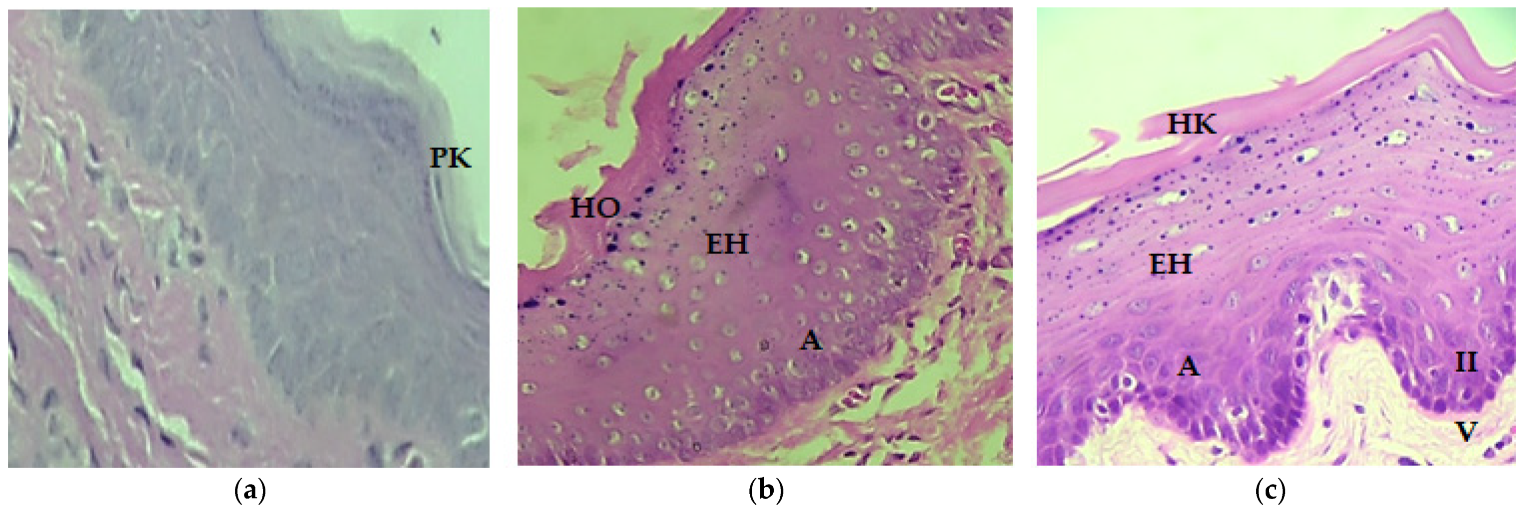
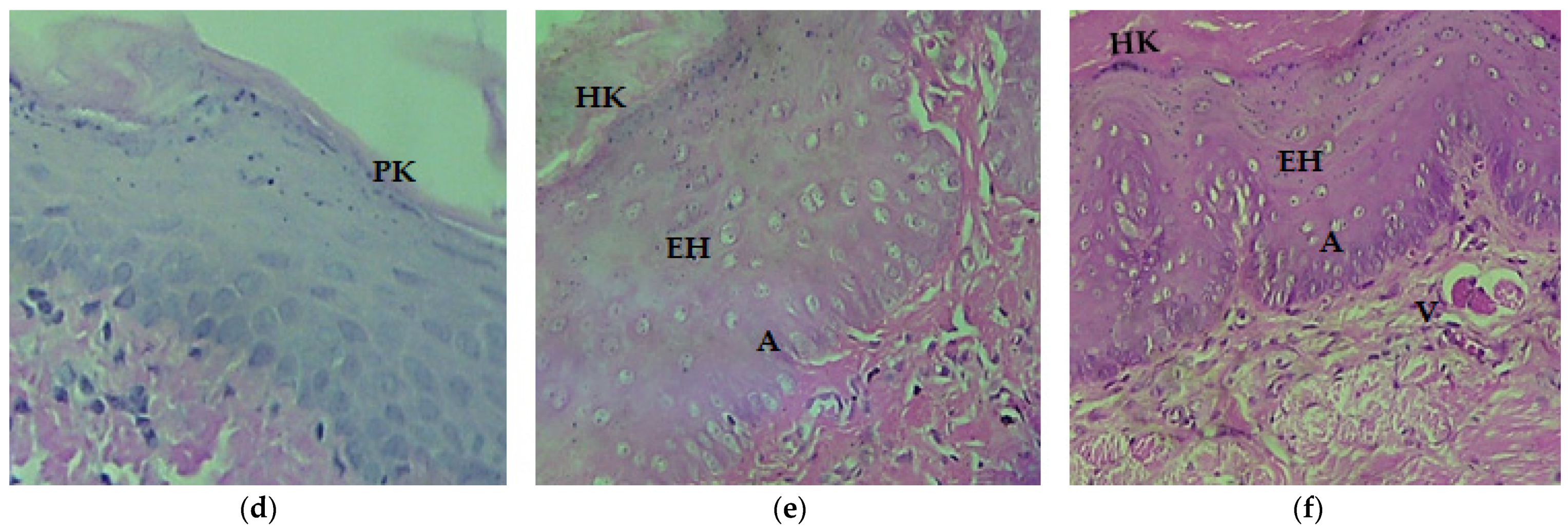
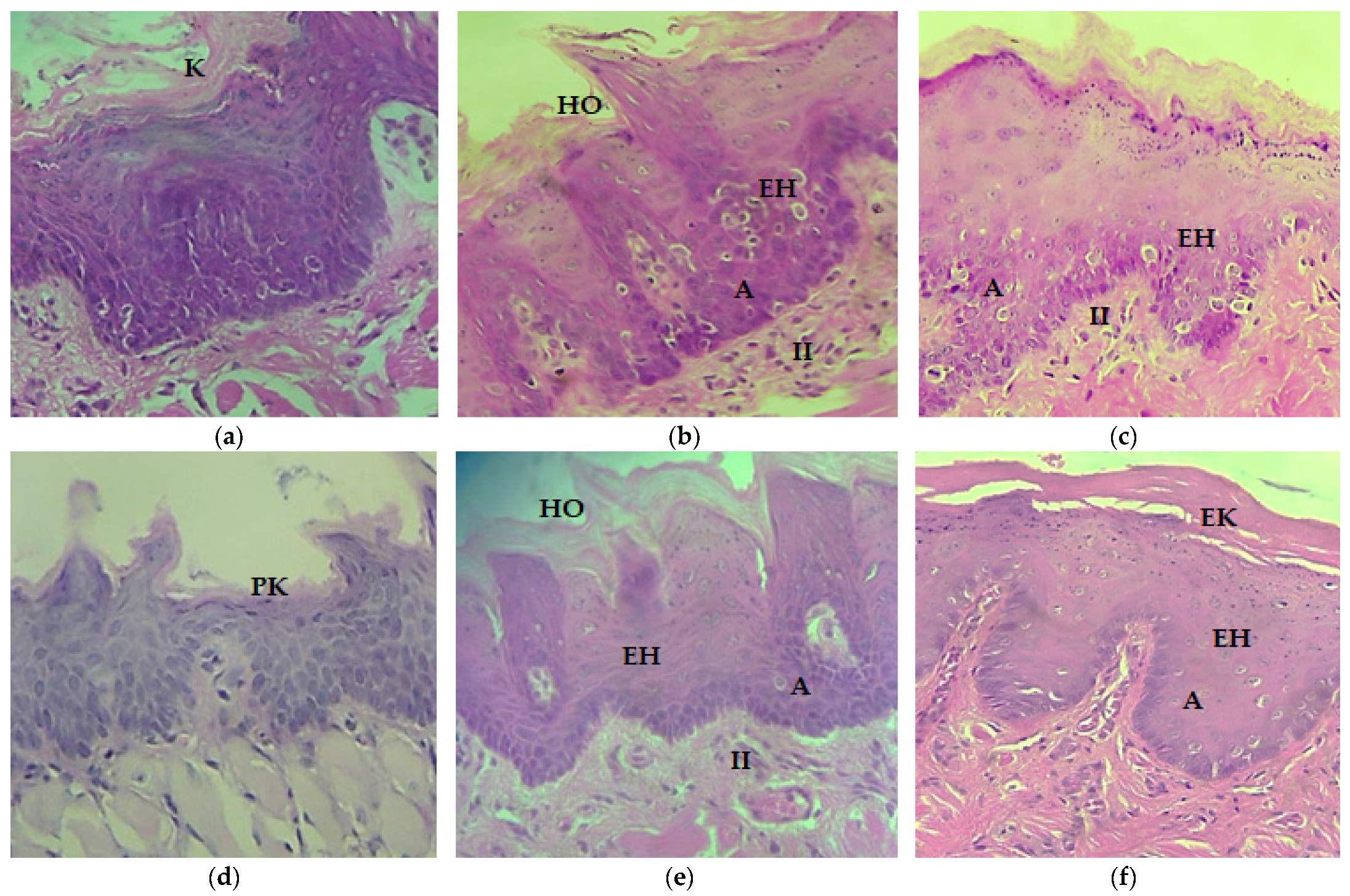
Histological Analysis

The histological changes in the mucosa of the cheeks corresponding to the surface layer in the groups were reported in Table 1. It can be observed that the control group did not present significant changes. The presence of keratinized epitheliums was high, and only in the male 5% group was it moderate. The parakeratinized and hyperorthokeratinized variants had minimal presence in the female 40% group, while in the other groups, they were not present. The male and female control groups were observed with parakeratinized epitheliums (Figure 1a,d) in contrast to the three remaining groups (Figure 1b,c,e,f), where it was hyperorthokeratinized and hyperkeratinized (Figure 1b,c,e,f).
Histological changes in the cheek mucosa are reported in Table 2. In all alcohol-administered groups, we observed epithelial hyperplasia and acanthosis, with a high and moderate presence (in that order) in the female 5% group for both epithelial hyperplasia and acanthosis. It should be noted that the male 5% group demonstrated nearly the same results in hyperplasia and acanthosis (high-to-moderate presence). On the other hand, epithelial hyperplasia and acanthosis had variations, although they were present in the four alcohol groups. The female 40% group presented moderate-to-high epithelial hyperplasia and acanthosis, the male 40% group was low to moderate, while the female and male 5% groups were moderate to high. Regarding the inflammatory infiltrate, in the male 5% group, this was absent, and it was minimal to none in the remaining groups. Vascularization was constant in the four alcohol-administered groups, where it was moderate to minimal.
Epithelial hyperplasia is observed from the basal strata and toward the superior strata, and it is also manifested in the presence of acanthosis (Figure 1b,c,e,f). The inflammatory infiltrate is minimal (Figure 1c) and null in the male 5% group (Figure 1f).
Histological changes of the tongue regarding the type of surface layer are presented in Table 3. In all alcohol-administered groups, the presence of a keratinized epithelium was observed, and it was high to moderate. In the female and male 40% groups, it was similar, with a high presence; in the female and male 5% groups, it was high to moderate. On the other hand, the female 40% and 5% groups presented moderate hyperorthokeratinized epitheliums, compared to the male 40 and 5% groups, which did not.
Histological changes in the tongue are reported in Table 4. In all alcohol-administered groups, epithelial hyperplasia and acanthosis were observed. Vascularization was constant, from moderate to minimal. It should be noted that the presence of epithelial hyperplasia was identical in the female and male 40% groups (moderate, high, and minimal, in this order), while the female and male 5% groups were similar with a high presence. Regarding acanthosis, the female and male 5% groups had similar results, with a moderate presence (Figure 2c,f). The inflammatory infiltrate remained constant the in female 40% and 5% groups along with the male 40% group, with a minimal and null presence, while in the male 5% group, it was not observed (Figure 2b,c,e,f).
In the female control group, the lingual epithelium was keratinized, while in the female 40% and 5% groups, it was hyperorthokeratinized and hyperkeratinized, exhibited as thicker on the surface (Figure 2b,c). These changes are also distinguished in comparison with the male control group (Figure 2d), where the lingual epithelium was parakeratinized, while the male 40% and 5% groups presented greater thickness in the surface layer, where it was hyperorthokeratinized and hyperkeratinized (Figure 2e,f).

4. Discussion

Alcohol consumption is currently increasing dramatically among the young population, and the damage and consequences involved seriously affect numerous aspects of an individual’s health and well-being. There is vast evidence on the effects that alcohol consumption generates on different organs and tissues of the human body; however, many of these studies are based on the evaluation of models that consider chronic daily alcohol consumption, and it is rarely considered that weekend consumption is much more frequent among the young population. In this study, we evaluated the effect of weekend alcohol consumption on oral tissue (tongue and cheek) for a period of 12 weeks in a female and male rodent model. The results clearly show damage-causing histological changes in tongue and cheek mucosae in nearly all indicators. Notable changes in the level of keratinization on the surface layer of the cheek mucosa could be observed in all groups that consumed alcohol twice a week. Previously, it was known that alcohol in contact with the oral mucosa is capable of producing an alteration in its morphology, characterized by epithelial atrophy [14,16,30,31], which implies an increase in susceptibility to other chemical carcinogens. Several studies mentioned anomalies in oral tissues due to chronic alcohol consumption, including epithelial atrophy attributable to a reduction in basal cellular size [32]. Oral mucosal atrophy is associated with hyper-regeneration, which is observed as an enlargement in the size of the basal-cell nuclei of the oral mucosa from the floor of the mouth and the base of the tongue. In addition, there was an increased size of the basal-cell layer and altered cell stratification; taken together, all of the latter increase susceptibility to carcinogens [33,34], and similar results were described in the glottic mucosal epithelium [35]. In terms of the effects of the concentration of the alcohol, Müller et al. reported that the direct toxic action of the alcohol in short-term experiments is proportional to the degree of concentration of the alcohol (20%, 40%, and 96%) and that it leads to local damage of the mucous membrane. Chronic exposure to alcohol (12 months) induces leukoplakia-like epithelial dysplasia (keratosis, dyskeratosis), increased density of the basal layer, and a small increase in the number of mitotic figures [36].
The mucosa tends to be affected by other chemicals, as mentioned previously, due to an increase in their solubility in the presence of alcohol [16], as well as to an increase in the permeability of the mucosa [27]. The reported increase of permeability is explained by the dissolving effect of alcohol, which is capable of eliminating the membrane-lipid content that surrounds the granules of the epithelium stratum spinosum [28]. However, other authors, such as Howie [37], point out that the increase in permeability is due to a rearrangement of the constituent elements of the cell membrane, as observed in samples of lingual tissues from recent human cadavers, in which ethanol has the ability to promote the penetration of high-molecular-weight molecules without producing any type of variation in their lipid component. Rearrangement of the cell membrane in oral mucosa is also related to the altered lipid-containing permeability barrier of stratified squamous epithelium observed after 120 days of alcohol ingestion in rats [38]. Alcohol is able to alter structural epithelial lipid molecules and destroy lipid composition, covering the acanthosis granules and resulting in the susceptibility of epithelial cells, augmenting oral mucosal permeability. Thus, soft tissues become an easy attack target for carcinogens.
Alcohol can destroy the lipid composition of the protective layer of oral mucosa covering the acanthosis granules, and disrupt the normal order of epithelial lipid molecules, resulting in a gap between epithelial cells and increasing oral mucosal permeability. In other words, alcohol opens a pathway to deep soft tissues [39]. In this study, we could observe that weekend alcohol consumption for 12 weeks induced remarkable changes, such as epithelial hyperplasia, acanthosis, chronic inflammatory infiltrate, and vascularization. The female group at 40% presented the most severe alteration in mucosa cheek and tongue, while the other alcohol groups presented very similar results. In agreement with what we found, Feng and Wang [39] analyzed, under light microscopy, oral-mucosa samples from 60 female and male subjects who died of chronic alcoholism and alcoholic cirrhosis. They reported that 10% of the tissue sections presented epithelial hyperplasia points with acanthosis and hyperkeratosis. Additionally, 90% of the sections had epithelial atrophy points and several degrees of damage. At the same time, visible moderate infiltration of lymphocytes–macrophages in the basal oral mucosa was identified. In deep mucosa, muscle-fiber atrophy could be observed with the appearance of injury in the neurological regulatory system. In individuals who died from cardiovascular disease with a history of alcohol abuse, approximately 50% of the sections presented extensive necrotic points in different parts of the oral mucosa, together with secondary infection. Approximately 15% comprised dense and necrotic tissue with microbial colonization and a small number of neutrophils and macrophages, and 5% of the necrotic points were located in epithelial tissue. These results suggest that prolonged alcohol consumption enhances carcinogenic alterations in the oral mucosa. The study conducted by Srinivasamurthy et al. [40] revealed that cytomorphometric analysis of the oral mucosa in individuals above 25 years of age who consumed a minimum of 45 mL of alcohol per day for at least 10 years presented alterations in buccal mucosal cells, manifested as an increase in the mean cytoplasmic area, mean nuclear area, and cell-to-nuclear parameter ratio prior the onset of oral carcinoma.
Subsequently, the acetaldehyde will undergo second oxidation via aldehyde dehydrogenase (ALDH), which will transform it into acetate, preventing the toxic activity of the first metabolite [41]. Acetaldehyde has a potent carcinogenic effect and is highly toxic and mutagenic. The role of oral microflora in the oxidation of ethanol has been studied by Homann [42,43], who demonstrated the production of considerable amounts of acetaldehyde during social alcohol consumption. Tobacco and alcohol can disrupt the oral microbiome. The oral-mucosa microbiome participates in the production of the genotoxic metabolite acetaldehyde by means of oxidation of ethanol, which causes nucleic-acid damage by forming DNA adducts in the epithelial cells of the mouth. The oral microbiome can function as synergistic risk factors alongside primary risk factors, such as alcohol and tobacco use. In this same line of research, an association has been found between the low levels of oral hygiene present in alcoholic subjects and bacterial overgrowth, which would have repercussions in a higher concentration of salivary acetaldehyde through this pathway [42]. This would explain the increased risk of oral cancer in alcoholic patients with poor oral health [30]. In the study by Homann [42], oral-mucosa biopsies were analyzed to identify changes caused by acetaldehyde, which was applied to a group of rats during a period of 8 months, where epithelial hyperproliferation was noted, considering this the first step in the genesis of cancer, prior to dysplastic changes. In parallel, the higher acetaldehyde production in smoking and heavy-drinking individuals is associated with Gram-positive aerobic bacteria and yeast in bacterial analysis, which could be one of the reasons for the synergistic carcinogenic effect of alcohol and smoking on upper-gastrointestinal-tract cancer [44]. Meanwhile, in addition to oral atypical changes, alcohol and smoking cigarettes promote oral bacterial infection, essentially of cocci and Actinomyces species, and the degree of alterations is related to the duration of drinking and smoking [45]. For instance, population-attributable risks for oral-cavity cancer increased up to 80.7% for tobacco and/or alcohol consumption in a French population [46].
Cell proliferation is a phenomenon that can be affected, it is a process of maintenance of homeostasis since proliferation replaces desquamated cells. Between 6 and 12 months, there is an increase in proliferation as a response to aggression or to eliminate the harmful agent. Canard et al. [17] demonstrated the mitogenic effect of alcohol on the mucosa, similar to findings reported by Homann [42]. In the study, the author shows that alcohol with topical and intermittent application causes only an increase in cell desquamation, while continuous alcohol intake is associated with an increase in cell proliferation. The research group explains that intermediate cells are responsible for cell proliferation when there is an over-demand for cell replacement.
On the other hand, Sanfelice et al. observed an increase in the thickness of the epithelium (acanthosis) in the control group; this cannot be related to cell proliferation, but rather results from the decrease in desquamation. Increased epithelium thickness may be a physiological characteristic of the mouse species employed in the study. A decrease has been shown in epithelial thickness with age, and gender also exerts an impact on epithelium thickness [47]. Reis et al. evaluated the buccal mucosa from the lateral border of the tongue in 36 non-smoker alcoholics (ethanol group) and 18 non-smokers and non-drinkers (control group). These authors found pyknosis, karyorrhexis, and karyolysis in increased numbers of tongue and buccal mucosa cells observed as cytologic changes, likely as induction of keratinization in an attempt to protect non-keratinized oral mucosa from damage caused by ethanol exposure. These researchers suggest that chronic exposure to ethanol is related to carcinogenic cytologic alteration in oral mucosa, essentially on the lateral border of the tongue, and they found a relation of this to hepatobiliary injury [48]. Such results are similar to those that our research group reported regarding liver damage caused by weekend alcohol consumption. Our data demonstrated that the biochemical parameters of ALT, AST, and bilirubin were significantly elevated, as well as fatty acids and inflammation, which comprised liver histological alterations after 12 weeks of alcohol consumption. These results provide solid evidence that weekend alcohol consumption can cause irreversible liver damage [49].
It is noteworthy that it is highly possible that the alterations in oral-cavity tissues found in this study are also related to an increase in oxidative stress (OS). The latter has been associated with elevated oral peroxidase activity in alcohol-dependent persons, leading to chronic OS and poor periodontal status [50].

5. Conclusions

In this study, we present solid evidence with respect to the damage induced by weekend alcohol consumption at two different alcohol concentrations, which are the most common drinks among alcohol consumers. It appears that alcohol affects both females and males indistinctly. The relevance of this work lies in the fact that there is vast evidence of damage due to the day-to-day consumption of alcohol; however, few reports regarding weekend drinking are available. The weekend intake of alcoholic beverages is a very frequent drinking pattern among the population, and it is not considered as risky as daily consumption. This social pattern of drinking is regularly observed as natural in human behavior as a form of coexistence and relationships among individuals, and alcohol very often falls into abuse in consumption without taking into account the health risks that this entails. We attempt to demonstrate that the weekend use of alcohol is a powerful factor in developing alterations in oral tissue that could contribute to the development of oral carcinoma. The limitations of the study were likely related to the duration of the study since it is pertinent to carry out studies of more than 3 months to evaluate more significant structural modifications that give rise to malignant cellular changes. In addition, it would also be interesting to evaluate different doses and concentrations of alcohol in future research, as well as to compare the effects of daily vs. weekend consumption. This type of research should certainly be carried out in humans. This would likely help to better understand the changes reported.
As a proposal for future lines of research, we consider the observation of the nucleolar organizing region (AgNOR, the silver-staining nucleolar organizing region). Proteins located in the nucleolus are identified as black dots with yellow nuclei and appear in cell proliferation. On determining the expression of Ki67, the stimulus of cell proliferation is acetaldehyde, which can derive from the liver, the oral mucosa, or through the action of microorganisms in the oral cavity.

Author Contributions

L.L.G.-L., A.S.-G. and J.A.M.-G., Conceived designed the study; Á.M.-G., E.O.M.-S., L.A.-R., E.M.-B., I.Á.-G., L.D.-O., C.V.-V. and C.E.-C. edited and wrote some portion of the paper, compiled the references, and analyzed the data; L.L.G.-L., J.A.M.-G. and N.V.-M. wrote the manuscript. All authors have read and agreed to the published version of the manuscript.

Funding

Secretaría de Investigación y Posgrado, Instituto Politécnico Nacional: SIP-IPN:20220185.

Institutional Review Board Statement

The study was conducted according to the Official Mexican Guidelines for Laboratory Animal Use and Care (NOM-062-ZOO-1999) and was approved by the Institutional Animal Care and Use Committee of the Autonomous University of Hidalgo State, Mexico, approval number CICUAL/F011/2021.

Conflicts of Interest

The authors declare no conflict of interest.

References

  1. WHO. Global Status Report on Alcohol and Health 2018; License: CC BY-NC-SA 3.0 IGO; World Health Organization: Geneva, Switzerland, 2018; p. 450.
  2. De Souza, D.P.; Areco, K.N.; da Silveira Filho, D.X. Alcohol and alcoholism among Brazilian adolescent public-school students. Rev. Saúde Pública 2005, 39, 585–592. [Google Scholar] [CrossRef] [PubMed] [Green Version]
  3. Benjet, C.; Borges, G.; Méndez, E.; Casanova, L.; Medina-Mora, M.E. Adolescent alcohol use and alcohol use disorders in Mexico City. Drug Alcohol Depend. 2014, 136, 43–50. [Google Scholar] [CrossRef] [PubMed]
  4. Aguirre, A.A.; Alonso Castillo, M.M.; Zanetti, A.C. Alcohol consumption and self esteem in adolescents. Rev. Lat.-Am. Enferm. 2010, 18, 634–640. [Google Scholar] [CrossRef] [Green Version]
  5. González-Bautista, E.; Zavala-Arciniega, L.; Rivera-Rivera, L.; Leyva-López, A.; Natera-Rey, G.; Reynales-Shigematsu, L.M. Social factors associated with tobacco and alcohol consumption in Mexican adolescents from populations under 100,000 inhabitants. Salud Publica Mex. 2019, 61, 764–774. [Google Scholar] [CrossRef] [PubMed] [Green Version]
  6. Hoes, L.; Dok, R.; Verstrepen, K.J.; Nuyts, S. Ethanol-Induced Cell Damage Can Result in the Development of Oral Tumors. Cancers 2021, 13, 3846. [Google Scholar] [CrossRef]
  7. Room, R.; Babor, T.; Rehm, J. Alcohol and public health. Lancet 2005, 365, 519–530. [Google Scholar] [CrossRef]
  8. Griswold, M.G.; Fullman, N.; Hawley, C.; Arian, N.; Zimsen, S.R.M.; Tymeson, H.D.; Venkateswaran, V.; Tapp, A.D.; Forouzanfar, M.H.; Salama, J.S.; et al. Alcohol use and burden for 195 countries and territories, 1990–2016: A systematic analysis for the Global Burden of Disease Study 2016. Lancet 2018, 392, 1015–1035. [Google Scholar] [CrossRef] [Green Version]
  9. INPRFM. Encuesta Nacional de Consumo de Drogas, Alcohol y Tabaco 2016–2017: Reporte de Alcohol; Instituto Nacional de Psiquiatría Ramón de la Fuente Muñíz, Instituto Nacional de Salud Pública, Comisión Contra las Adicciones, Secretaría de Salud: Ciudad de México, Mexico, 2017. [Google Scholar]
  10. Jovanovic, A.; Schulten, E.A.; Kostense, P.J.; Snow, G.B.; van der Waal, I. Tobacco and alcohol related to the anatomical site of oral squamous cell carcinoma. J. Oral Pathol. Med. Off. Publ. Int. Assoc. Oral Pathol. Am. Acad. Oral Pathol. 1993, 22, 459–462. [Google Scholar] [CrossRef]
  11. Carrard, V.C.; Pires, A.S.; Mendez, M.; Pasquali, M.A.; Badauy, C.M.; Lauxen, I.S.; Moreira, J.C.; Sant’Ana Filho, M. Exploring the mechanisms of alcohol-related damage in oral mucosa—Is oxidative stress associated with the increase in cell proliferation in rat tongue epithelium? Pharm. Biol. 2013, 51, 160–169. [Google Scholar] [CrossRef]
  12. Bagnardi, V.; Blangiardo, M.; La Vecchia, C.; Corrao, G. A meta-analysis of alcohol drinking and cancer risk. Br. J. Cancer 2001, 85, 1700–1705. [Google Scholar] [CrossRef]
  13. Moreno-López, L.A.; Esparza-Gómez, G.C.; González-Navarro, A.; Cerero-Lapiedra, R.; González-Hernández, M.J.; Domínguez-Rojas, V. Risk of oral cancer associated with tobacco smoking, alcohol consumption and oral hygiene: A case-control study in Madrid, Spain. Oral Oncol. 2000, 36, 170–174. [Google Scholar] [CrossRef]
  14. Fioretti, F.; Bosetti, C.; Tavani, A.; Franceschi, S.; La Vecchia, C. Risk factors for oral and pharyngeal cancer in never smokers. Oral Oncol. 1999, 35, 375–378. [Google Scholar] [CrossRef]
  15. Goldstein, B.Y.; Chang, S.C.; Hashibe, M.; La Vecchia, C.; Zhang, Z.F. Alcohol consumption and cancers of the oral cavity and pharynx from 1988 to 2009: An update. Eur. J. Cancer Prev. Off. J. Eur. Cancer Prev. Organ. (ECP) 2010, 19, 431–465. [Google Scholar] [CrossRef] [PubMed] [Green Version]
  16. Wight, A.J.; Ogden, G.R. Possible mechanisms by which alcohol may influence the development of oral cancer—A review. Oral Oncol. 1998, 34, 441–447. [Google Scholar] [CrossRef]
  17. Carrard, V.C.; Filho, M.S.; Rados, P.V.; Chaves, A.C.; Lauxen Ida, S. Quantification of silver-staining nucleolar organizer region in epithelial cells of tongue of mice after exposure to, or intake of, alcohol. Alcohol 2004, 34, 233–238. [Google Scholar] [CrossRef]
  18. Mandel, L.; Hamele-Bena, D. Alcoholic parotid sialadenosis. J. Am. Dent. Assoc. (1939) 1997, 128, 1411–1415. [Google Scholar] [CrossRef]
  19. Khairnar, M.R.; Wadgave, U.; Khairnar, S.M. Effect of Alcoholism on Oral Health: A Review. J. Alcohol Drug Depend. 2017, 5, 1–4. [Google Scholar] [CrossRef]
  20. Guggenheimer, J.; Close, J.M.; Eghtesad, B. Sialadenosis in patients with advanced liver disease. Head Neck Pathol. 2009, 3, 100–105. [Google Scholar] [CrossRef] [Green Version]
  21. Chilla, R. Sialadenosis of the salivary glands of the head. Studies on the physiology and pathophysiology of parotid secretion. Adv. Oto-Rhino-Laryngol. 1981, 26, 1–38. [Google Scholar]
  22. Dunkley, R.P.; Carson, R.M. Dental requirements of the hospitalized alcoholic patient. J. Am. Dent. Assoc. (1939) 1968, 76, 800–803. [Google Scholar] [CrossRef]
  23. Niquille, M.; Burnand, B.; Magnenat, P.; Paccaud, F.; Yersin, B. Dental disease among alcoholic individuals: A comparative study of hospitalized patients. J. Gen. Intern. Med. 1993, 8, 470–475. [Google Scholar] [CrossRef]
  24. Shepherd, S. Alcohol consumption a risk factor for periodontal disease. Evid.-Based Dent. 2011, 12, 76. [Google Scholar] [CrossRef] [Green Version]
  25. Himmerich, H.; Anghelescu, I.; Klawe, C.; Szegedi, A. Vitamin B12 and hepatic enzyme serum levels correlate in male alcohol-dependent patients. Alcohol Alcohol. 2001, 36, 26–28. [Google Scholar] [CrossRef] [PubMed]
  26. Stornetta, A.; Guidolin, V.; Balbo, S. Alcohol-Derived Acetaldehyde Exposure in the Oral Cavity. Cancers 2018, 10, 20. [Google Scholar] [CrossRef] [PubMed] [Green Version]
  27. Figuero Ruiz, E.; Carretero Peláez, M.A.; Cerero Lapiedra, R.; Esparza Gómez, G.; Moreno López, L.A. Effects of the consumption of alcohol in the oral cavity: Relationship with oral cancer. Med. Oral Organo Of. Soc. Esp. Med. Oral Acad. Iberoam. Patol. Med. Bucal 2004, 9, 14–23. [Google Scholar]
  28. Ogden, G.R.; Wight, A.J. Aetiology of oral cancer: Alcohol. Br. J. Oral Maxillofac. Surg. 1998, 36, 247–251. [Google Scholar] [CrossRef]
  29. Reidy, J.; McHugh, E.; Stassen, L.F. A review of the relationship between alcohol and oral cancer. Surg. J. R. Coll. Surg. Edinb. Irel. 2011, 9, 278–283. [Google Scholar] [CrossRef] [PubMed]
  30. Seitz, H.K.; Matsuzaki, S.; Yokoyama, A.; Homann, N.; Väkeväinen, S.; Wang, X.D. Alcohol and cancer. Alcohol. Clin. Exp. Res. 2001, 25, 137S–143S. [Google Scholar] [CrossRef]
  31. Valentine, J.A.; Scott, J.; West, C.R.; St Hill, C.A. A histological analysis of the early effects of alcohol and tobacco usage on human lingual epithelium. J. Oral Pathol. 1985, 14, 654–665. [Google Scholar] [CrossRef]
  32. Mascrès, C.; Ming-Wen, F.; Joly, J.G. Morphologic changes of the esophageal mucosa in the rat after chronic alcohol ingestion. Exp. Pathol. 1984, 25, 147–153. [Google Scholar] [CrossRef]
  33. Maier, H.; Weidauer, H.; Zöller, J.; Seitz, H.K.; Flentje, M.; Mall, G.; Born, I.A. Effect of chronic alcohol consumption on the morphology of the oral mucosa. Alcohol. Clin. Exp. Res. 1994, 18, 387–391. [Google Scholar] [CrossRef] [PubMed]
  34. Born, I.A.; Zöller, J.; Weidauer, H.; Maier, H. Effects of chronic alcohol drinking on mouth mucosa. A morphometric study. Laryngo-Rhino-Otol. 1996, 75, 754–758. [Google Scholar] [CrossRef] [PubMed]
  35. Maier, H.; Tisch, M.; Schneeberg, E.; Born, A. An association of chronic alcohol consumption with morphological alterations of the laryngeal mucosa in rats. Eur. Arch. Oto-Rhino-Laryngol. Off. J. Eur. Fed. Oto-Rhino-Laryngol. Soc. (EUFOS) Affil. Ger. Soc. Oto-Rhino-Laryngol.-Head Neck Surg. 1999, 256, 247–249. [Google Scholar] [CrossRef] [PubMed]
  36. Müller, P.; Hepke, B.; Meldau, U.; Raabe, G. Tissue damage in the rabbit oral mucosa by acute and chronic direct toxic action of different alcohol concentrations. Exp. Pathol. 1983, 24, 171–181. [Google Scholar] [CrossRef]
  37. Howie, N.M.; Trigkas, T.K.; Cruchley, A.T.; Wertz, P.W.; Squier, C.A.; Williams, D.M. Short-term exposure to alcohol increases the permeability of human oral mucosa. Oral Dis. 2001, 7, 349–354. [Google Scholar] [CrossRef]
  38. Squier, C.A.; Kremer, M.J.; Wertz, P.W. Effect of ethanol on lipid metabolism and epithelial permeability barrier of skin and oral mucosa in the rat. J. Oral Pathol. Med. Off. Publ. Int. Assoc. Oral Pathol. Am. Acad. Oral Pathol. 2003, 32, 595–599. [Google Scholar] [CrossRef]
  39. Feng, L.; Wang, L. Effects of alcohol on the morphological and structural changes in oral mucosa. Pak. J. Med. Sci. 2013, 29, 1046–1049. [Google Scholar] [CrossRef]
  40. Srinivasamurthy, B.C.; Balamurugesan, K.; Sathishkumar, N.; Prakash, M.; Bhat, R.V. Cytomorphometric Study of Changes in Buccal Mucosal Cells in Alcoholics. Adv. Biomed. Res. 2020, 9, 48. [Google Scholar] [CrossRef]
  41. Paquot, N. The metabolism of alcohol. Rev. Med. Liege 2019, 74, 265–267. [Google Scholar]
  42. Homann, N.; Jousimies-Somer, H.; Jokelainen, K.; Heine, R.; Salaspuro, M. High acetaldehyde levels in saliva after ethanol consumption: Methodological aspects and pathogenetic implications. Carcinogenesis 1997, 18, 1739–1743. [Google Scholar] [CrossRef] [PubMed]
  43. Homann, N.; Tillonen, J.; Rintamäki, H.; Salaspuro, M.; Lindqvist, C.; Meurman, J.H. Poor dental status increases acetaldehyde production from ethanol in saliva: A possible link to increased oral cancer risk among heavy drinkers. Oral Oncol. 2001, 37, 153–158. [Google Scholar] [CrossRef]
  44. Homann, N.; Tillonen, J.; Meurman, J.H.; Rintamäki, H.; Lindqvist, C.; Rautio, M.; Jousimies-Somer, H.; Salaspuro, M. Increased salivary acetaldehyde levels in heavy drinkers and smokers: A microbiological approach to oral cavity cancer. Carcinogenesis 2000, 21, 663–668. [Google Scholar] [CrossRef] [PubMed]
  45. Abdelaziz, M.S.; Osman, T.E. Detection of Cytomorphological Changes in Oral Mucosa among Alcoholics and Cigarette Smokers. Oman Med. J. 2011, 26, 349–352. [Google Scholar] [CrossRef] [PubMed]
  46. Radoï, L.; Paget-Bailly, S.; Cyr, D.; Papadopoulos, A.; Guida, F.; Schmaus, A.; Cénée, S.; Menvielle, G.; Carton, M.; Lapôtre-Ledoux, B.; et al. Tobacco smoking, alcohol drinking and risk of oral cavity cancer by subsite: Results of a French population-based case-control study, the ICARE study. Eur. J. Cancer Prev. Off. J. Eur. Cancer Prev. Organ. (ECP) 2013, 22, 268–276. [Google Scholar] [CrossRef]
  47. Scott, J.; Valentine, J.A.; St Hill, C.A.; Balasooriya, B.A. A quantitative histological analysis of the effects of age and sex on human lingual epithelium. J. Biol. Buccale 1983, 11, 303–315. [Google Scholar]
  48. Reis, S.R.; do Espírito Santo, A.R.; Andrade, M.G.; Sadigursky, M. Cytologic alterations in the oral mucosa after chronic exposure to ethanol. Braz. Oral Res. 2006, 20, 97–102. [Google Scholar] [CrossRef] [Green Version]
  49. Morales-González, J.A.; Sernas-Morales, M.L.; Morales-González, Á.; González-López, L.L.; Madrigal-Santillán, E.O.; Vargas-Mendoza, N.; Fregoso-Aguilar, T.A.; Anguiano-Robledo, L.; Madrigal-Bujaidar, E.; Álvarez-González, I.; et al. Morphological and biochemical effects of weekend alcohol consumption in rats: Role of concentration and gender. World J. Hepatol. 2018, 10, 297–307. [Google Scholar] [CrossRef]
  50. Waszkiewicz, N.; Zalewska, A.; Szajda, S.D.; Szulc, A.; Kępka, A.; Minarowska, A.; Wojewódzka-Żelezniakowicz, M.; Konarzewska, B.; Chojnowska, S.; Supronowicz, Z.B.; et al. The effect of chronic alcohol intoxication and smoking on the activity of oral peroxidase. Folia Histochem. Cytobiol. 2012, 50, 450–455. [Google Scholar] [CrossRef]
Figure 1. Histological modifications of the cheek mucosa tissue with hematoxylin–eosin (H&E) staining (40×). (a) Female control group, (b) female group 40% alcohol consumption, (c) female group 5% alcohol consumption, (d) male control group, (e) male group 40% alcohol consumption, and (f) group of males consuming 5% alcohol. PK, parakeratinized; K, keratinized; HO, hyperorthokeratinized; HK, hyperkeratinized; EH, epithelial hyperplasia; A, acanthosis; II, inflammatory infiltrate; V, blood vessels.
Applsci 12 03464 g001aApplsci 12 03464 g001b
Figure 2. Histological modifications of the lingual tissue with hematoxylin–eosin (H&E) staining (40×). (a) Female control group, (b) female group 40% alcohol consumption, (c) female group 5% alcohol consumption, (d) male control group, (e) male group 40% alcohol consumption, and (f) group of males consuming 5% alcohol. PK, parakeratinized; K, keratinized; HO, hyperorthokeratinized; HK, hyperkeratinized; EH, epithelial hyperplasia; A, acanthosis; II, inflammatory infiltrate; V, blood vessels.
Applsci 12 03464 g002
Table 1. Type of surface layer in the cheek mucosa after the consumption of 3 months of weekend alcohol consumption at 5% and 40% in both females and males.
GroupParakeratinizedKeratinizedOrtokeratinizedHyperorthokeratinized
Female control+000
Female 40%++++0+
Female 5%0++++++0
Male control+000
Male 40%0++++0
Male 5%0++0+
Criteria: (0) no presence, (+) minimal presence, (++) moderate presence, (+++) high presence. The results are the average of the observed samples (n = 5).
Table 2. Histological changes in the cheek mucosa in rats after weekend alcohol consumption for 3 months.
GroupEpithelial HyperplasiaAcanthosisChronic Inflammatory Infiltrate
(Monocuclear Inflammatory Cells)
Vascularization
female control0000
female 40%+/++/+++++/+++/+0/+++/+
female 5%+++/+++++/+++/++++/+
male control0000
male 40%++/+++/++/0+/++
male 5%++++++/++0++/+
Criteria: (0) no presence, (+) minimal presence, (++) moderate presence, (+++) high presence. The results are the average of the observed samples (n = 5).
Table 3. Type of surface layer on the tongue after 3 months of weekend alcohol at 5% and 40% in both females and males.
GroupParakeratinizedKeratinizedOrtokeratinizedHyperortokeratinized
Female control0+00
Female 40%0++++++
Female 5%++++0++
Male control+000
Male 40%+++++0
Male, 5%0++++0
Criteria: (0) no presence, (+) minimal presence, (++) moderate presence, (+++) high presence. The results are the average of the observed samples (n = 5).
Table 4. Histological changes in the tongue of rats after weekend alcohol consumption for 3 months.
GroupEpithelial HyperplasiaAcanthosisChronic Inflammatory Infiltrate (Mononuclear Inflammatory Cells)Vascularization
Female control000+
Female 40%++/+++/+++/+++/++/0++/+++
Female 5%+++++/++++/0++/+
Male control000+
Male 40%++/+++/++/+++/0++/+
Male 5%+++/++++0++/+++
Criteria: (0) no presence, (+) minimal presence, (++) moderate presence, (+++) high presence. The results are the average of the observed samples (n = 5).
Publisher’s Note: MDPI stays neutral with regard to jurisdictional claims in published maps and institutional affiliations.

