
Mineralogy of the Mudeungsan Tuff (Republic of Korea) Using Synchrotron X-ray Powder Diffraction and Rietveld Quantitative Analysis

 ,
,  and
and 
Abstract
:1. Introduction
2. Materials and Methods
3. Results
4. Discussion
5. Conclusions
Supplementary Materials
Author Contributions
Funding
Data Availability Statement
Acknowledgments
Conflicts of Interest
References
- Kim, S.W.; Kwon, S.; Park, S.-I.; Lee, C.; Cho, D.-L.; Lee, H.-J.; Ko, K.; Kim, S.J. SHRIMP U–Pb dating and geochemistry of the Cretaceous plutonic rocks in the Korean Peninsula: A new tectonic model of the Cretaceous Korean Peninsula. Lithos 2016, 262, 88–106. [Google Scholar] [CrossRef]
- Ahn, K.S. Geology and landscape of Mt. Mudeung Province Park, Korea. J. Petrol. Soc. Korea 2010, 19, 109–121. [Google Scholar]
- Kim, Y.J.; Park, J.B.; Park, B.K. Petrochemistry on igneous rocks in the Mt. Mudeung area. J. Petrol. Soc. Korea 2002, 11, 214–233. [Google Scholar]
- Jung, W.; Kil, Y.; Huh, M. A petrological study of the Mudeungsan Tuff focused on Cheonwangbong and Anyangsan. J. Petrol. Soc. Korea 2014, 23, 325–336. [Google Scholar] [CrossRef] [Green Version]
- Lim, C.; Huh, M.; Yi, K.; Lee, C. Genesis of the columnar joints from welded tuff in Mount Mudeung National Geopark, Republic of Korea. Earth Planets Space 2015, 67, 1–19. [Google Scholar] [CrossRef] [Green Version]
- Rietveld, H.M. A profile refinement method for nuclear and magnetic structures. J. Appl. Crystallogr. 1969, 2, 65–71. [Google Scholar] [CrossRef]
- Bish, D.L.; Howard, S.A. Quantitative phase analysis using the Rietveld method. J. Appl. Crystallogr. 1988, 21, 86–91. [Google Scholar] [CrossRef]
- Schreiner, W.T.; Jenkins, R. A second derivative algorithm for identification of peaks in powder diffraction patterns. Adv. X-Ray Anal. 1979, 23, 287–293. [Google Scholar] [CrossRef]
- Tompson, P.; Cox, D.E.; Hastings, J.B. Rietveld Refinement of Debye-Scherrer Synchrotron X-ray Data from Al2O3. J. Appl. Crystallogr. 1987, 20, 79–83. [Google Scholar] [CrossRef] [Green Version]
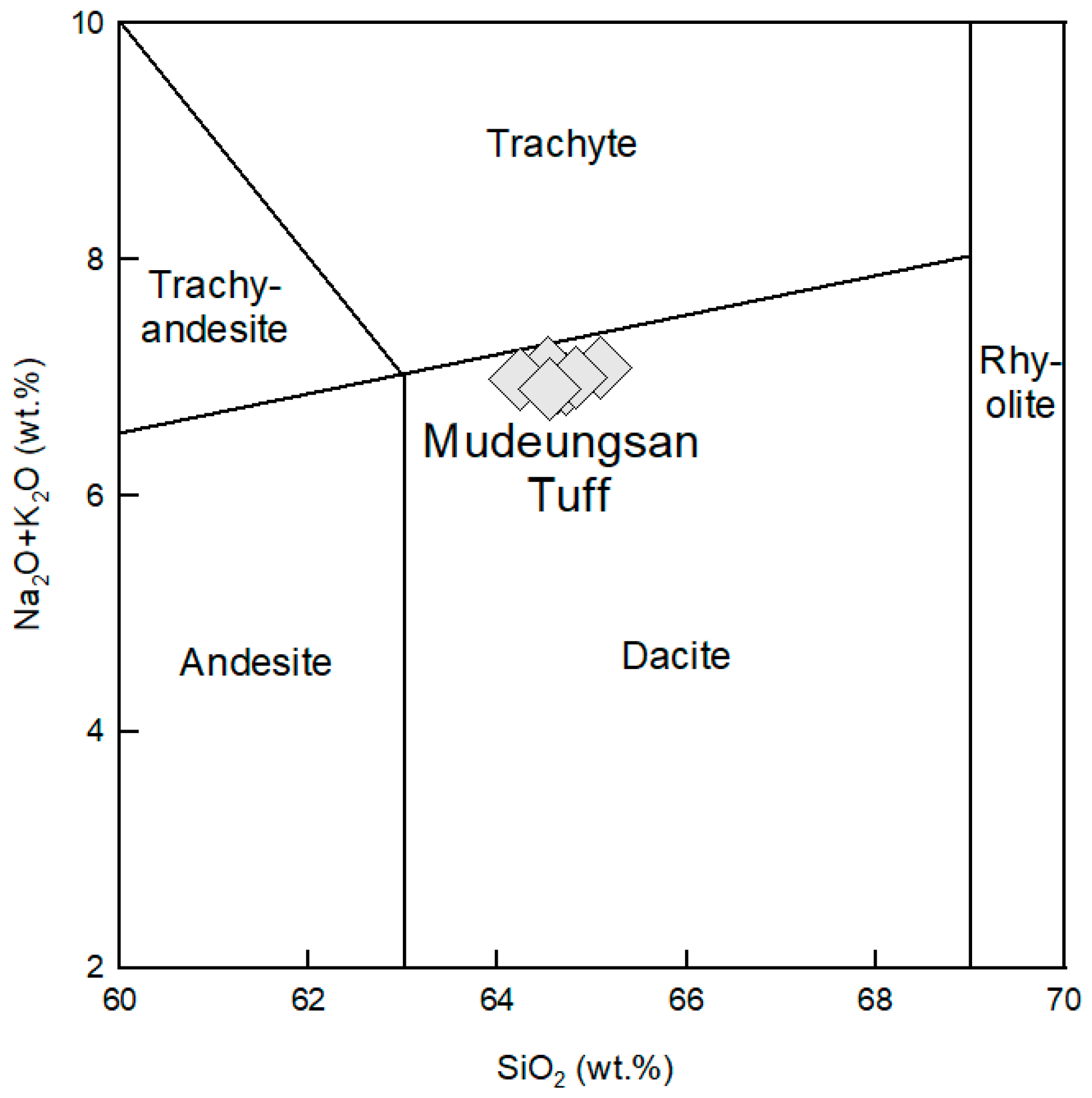
- Bas, M.J.L.; Maitre, R.W.L.; Streckeisen, A.; Zanettin, B.; IUGS Subcommission on the Systematics of Igneous Rocks. A Chemical Classification of Volcanic Rocks Based on the Total Alkali-Silica Diagram. J. Petrol. 1986, 27, 745–750. [Google Scholar] [CrossRef]
- Schipper, C.I.; Castro, J.M.; Tuffen, H.; Wadsworth, F.B.; Chappell, D.; Pantoja, A.E.; Simpson, M.P.; Le Ru, E.C. Cristobalite in the 2011–2012 Cordón Caulle eruption (Chile). Bull. Volcanol. 2015, 77, 34. [Google Scholar] [CrossRef]
- Baxter, P.J.; Bonadonna, C.; Dupree, R.; Hards, V.L.; Kohn, S.C.; Murphy, M.D.; Nichols, A.; Nicholson, R.A.; Norton, G.; Searl, A.; et al. Cristobalite in volcanic ash of the soufriere hills volcano, montserrat, british west indies. Science 1999, 283, 1142–1145. [Google Scholar] [CrossRef] [PubMed]
- Damby, D.E.; Llewellin, E.W.; Horwell, C.J.; Williamson, B.J.; Najorka, J.; Cressey, G.; Carpenter, M. The [alpha]-[beta] phase transition in volcanic cristobalite. J. Appl. Crystallogr. 2014, 47, 1205–1215. [Google Scholar] [CrossRef] [PubMed] [Green Version]
- Reich, M.; Zúñiga, A.; Amigo, Á.; Vargas, G.; Morata, D.; Palacios, C.; Parada, M.Á.; Garreaud, R.D. Formation of cristobalite nanofibers during explosive volcanic eruptions. Geology 2009, 37, 435–438. [Google Scholar] [CrossRef]
- Horwell, C.J.; Williamson, B.J.; Llewellin, E.W.; Damby, D.E.; Le Blond, J.S. The nature and formation of cristobalite at the Soufrière Hills volcano, Montserrat: Implications for the petrology and stability of silicic lava domes. Bull. Volcanol. 2013, 75, 696. [Google Scholar] [CrossRef] [Green Version]
- Duffy, C.J. Kinetics of Silica-Phase Transitions; Report Number LA-12564-MS; Los Alamos National Lab. (LANL): Los Alamos, NM, USA, 1993; p. 22. [Google Scholar]
- Ernst, W.G.; CALVERT, S.E. An experimental study of the reerystallization of porcelanite and its bearing on the origin of some bedded cherts. Am. J. Sci. 1969, 267, 114–133. [Google Scholar]
- Boudon, G.; Balcone-Boissard, H.; Villemant, B.; Morgan, D.J. What factors control superficial lava dome explosivity? Sci. Rep. 2015, 5, 14551. [Google Scholar] [CrossRef] [PubMed] [Green Version]
- Andrade, F.R.D.d.; Polo, L.A.; Janasi, V.d.A.; Carvalho, F.M.d.S. Volcanic glass in Cretaceous dacites and rhyolites of the Paraná Magmatic Province, southern Brazil: Characterization and quantification by XRD-Rietveld. J. Volcanol. Geotherm. Res. 2018, 355, 219–231. [Google Scholar] [CrossRef]


| SiO2 | Al2O3 | TiO2 | Fe2O3 | MgO | CaO | Na2O | K2O | MnO | P2O5 | LOI | |
|---|---|---|---|---|---|---|---|---|---|---|---|
| ISD-1 | 64.64 | 16.16 | 0.58 | 4.61 | 1.55 | 4.11 | 3.79 | 3.17 | 0.09 | 0.19 | 1.08 |
| ISD-2 | 64.53 | 15.91 | 0.56 | 4.41 | 1.51 | 4.09 | 3.83 | 3.22 | 0.09 | 0.19 | 1.61 |
| IR-1 | 64.73 | 15.75 | 0.57 | 4.52 | 1.60 | 4.21 | 3.83 | 3.11 | 0.10 | 0.19 | 1.34 |
| IR-2 | 64.53 | 15.56 | 0.58 | 4.59 | 1.58 | 4.00 | 3.78 | 3.31 | 0.10 | 0.19 | 1.75 |
| BN-1 | 65.09 | 16.07 | 0.58 | 4.44 | 1.44 | 3.86 | 3.75 | 3.34 | 0.08 | 0.19 | 1.06 |
| BN-2 | 64.83 | 15.57 | 0.57 | 4.45 | 1.45 | 3.84 | 3.77 | 3.23 | 0.09 | 0.19 | 1.98 |
| NT-1 | 64.24 | 15.93 | 0.59 | 4.60 | 1.56 | 4.08 | 3.79 | 3.20 | 0.09 | 0.19 | 1.70 |
| Anyang | 64.55 | 15.54 | 0.59 | 4.52 | 1.48 | 3.67 | 3.57 | 3.34 | 0.09 | 0.19 | 2.39 |
| Average | 64.64 | 15.81 | 0.58 | 4.52 | 1.52 | 3.98 | 3.76 | 3.24 | 0.09 | 0.19 | 1.61 |
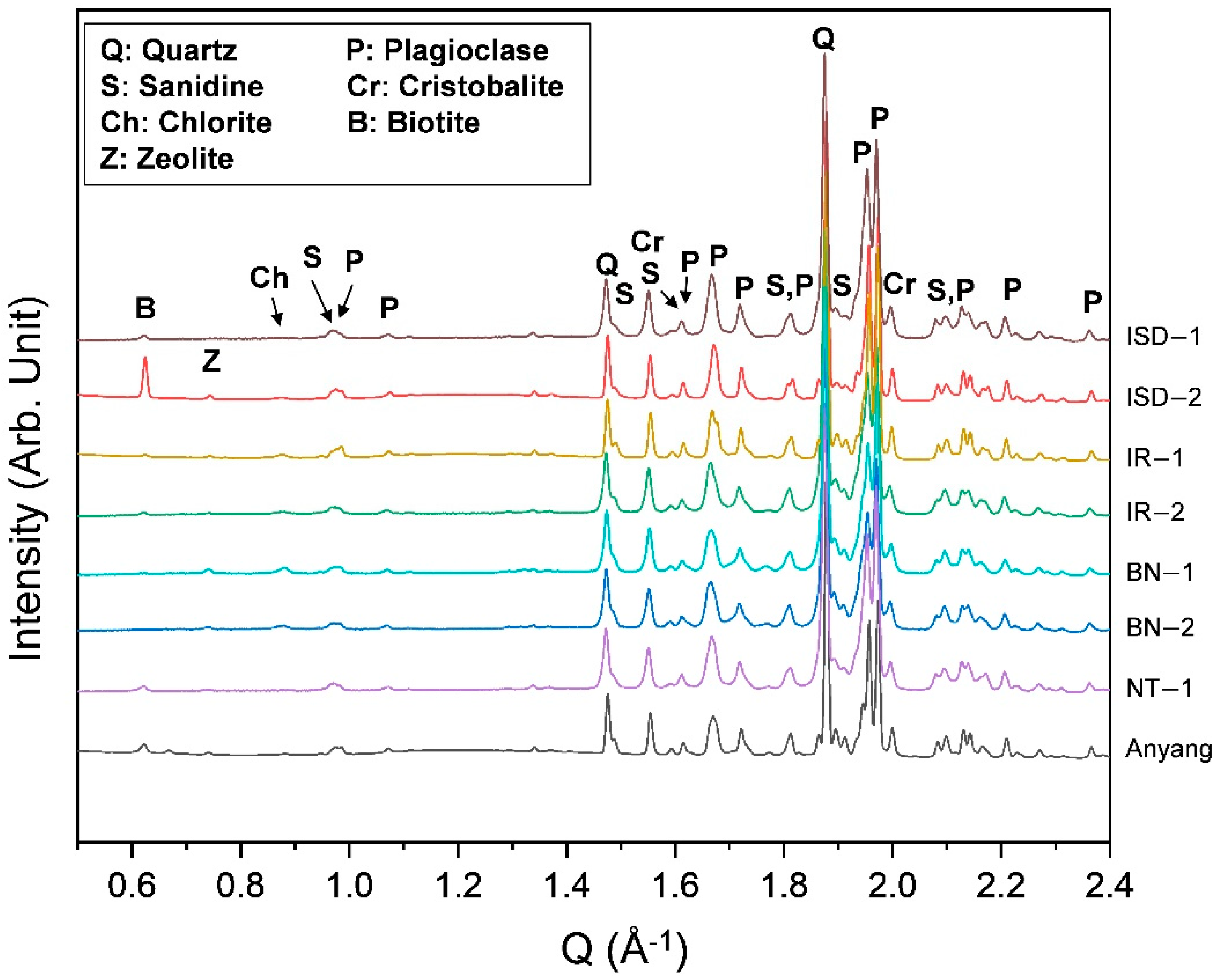
| wRp | Quartz | Plagioclase | Sanidine | α-Cristobalite | Chlorite | Biotite | Zeolite | Sum | |
|---|---|---|---|---|---|---|---|---|---|
| ISD-1 | 5.8(1) | 26.8(1) | 62.7(1) | 9.2(1) | 0.6(1) | 0.6(1) | 99.9(1) | ||
| ISD-2 | 11.7(1) | 29.0(1) | 58.2(1) | 9.9(1) | 0.7(1) | 0.9(1) | 1.1(1) | 0.2(1) | 100.0(1) |
| IR-1 | 5.7(1) | 25.1(1) | 56.9(1) | 15.8(1) | 1.1(1) | 0.5(1) | 0.5(1) | 99.9(1) | |
| IR-2 | 5.4(1) | 33.6(1) | 49.3(1) | 14.2(1) | 2.7(1) | 0.1(1) | 99.9(1) | ||
| BN-1 | 5.7(1) | 22.3(1) | 61.5(1) | 11.6(1) | 2.6(1) | 2.0(1) | 100(1) | ||
| BN-2 | 4.3(1) | 32.5(1) | 47.7(1) | 19.3(1) | 0.4(1) | 0.1(1) | 100(1) | ||
| NT-1 | 4.3(1) | 21.9(1) | 64.4(1) | 13.0(1) | 0.6(1) | 0.1(1) | 100(1) | ||
| Anyang | 4.8(1) | 29.9(1) | 55.7(1) | 11.7(1) | 0.1(1) | 1.1(1) | 0.4(1) | 1.1(1) | 100(1) |
Publisher’s Note: MDPI stays neutral with regard to jurisdictional claims in published maps and institutional affiliations. |
© 2021 by the authors. Licensee MDPI, Basel, Switzerland. This article is an open access article distributed under the terms and conditions of the Creative Commons Attribution (CC BY) license (https://creativecommons.org/licenses/by/4.0/).
Share and Cite
Seoung, D.; Kim, P.; Kim, H.; Lee, H.; Huh, M.; Lee, H.; Lee, Y. Mineralogy of the Mudeungsan Tuff (Republic of Korea) Using Synchrotron X-ray Powder Diffraction and Rietveld Quantitative Analysis. Appl. Sci. 2021, 11, 10796. https://doi.org/10.3390/app112210796
Seoung D, Kim P, Kim H, Lee H, Huh M, Lee H, Lee Y. Mineralogy of the Mudeungsan Tuff (Republic of Korea) Using Synchrotron X-ray Powder Diffraction and Rietveld Quantitative Analysis. Applied Sciences. 2021; 11(22):10796. https://doi.org/10.3390/app112210796
Chicago/Turabian StyleSeoung, Donghoon, Pyosang Kim, Hyeonsu Kim, Hyunseung Lee, Min Huh, Hyunwoo Lee, and Yongmoon Lee. 2021. "Mineralogy of the Mudeungsan Tuff (Republic of Korea) Using Synchrotron X-ray Powder Diffraction and Rietveld Quantitative Analysis" Applied Sciences 11, no. 22: 10796. https://doi.org/10.3390/app112210796