Article

The Relationship between Skinfold and Ultrasound Measures of Subcutaneous Fat in Untrained Healthy Males

Faculty of Health and Wellbeing, University of Winchester, Winchester SO22 4NR, UK
*
Author to whom correspondence should be addressed.
Appl. Sci. 2021, 11(22), 10561; https://doi.org/10.3390/app112210561
Submission received: 30 September 2021 / Revised: 27 October 2021 / Accepted: 29 October 2021 / Published: 10 November 2021

Abstract

:
Ultrasound measurement of adipose tissue offers an alternative measure of body composition with less technical skill requirement than skinfolds. However, the relationship between skinfold and ultrasound measurements of adipose tissue is uncertain. The aim of this study was to compare these measures in a healthy untrained male population. One hundred male participants (aged 18–40 years) of varying body compositions had skinfold measures taken at the biceps, triceps and front thigh sites. Ultrasound measures were also taken at the same sites using B-wave ultrasound with a linear probe in the transverse plane. Strong, significant (p < 0.01), positive correlations were observed between skinfold and ultrasound measures at the biceps (r = 0.828), triceps (r = 0.813), and front thigh (r = 0.888) sites. However, there was significant (p < 0.01) variance between the techniques at all measurement sites. Whilst skinfold and ultrasound measures of adipose tissue have good linear agreement, skinfolds are consistently higher at all sites indicating a difference in the nature of the tissue measured via each technique. The exact nature of the relationship should be established on a population-specific basis.

1. Introduction

The measurement of body composition is commonly used in sport and clinical physiology to monitor aspects of performance and the health of populations [1,2]. Research has examined both the accuracy and accessibility of body composition measurement in various settings [3,4,5]. Anthropometric measurements are one of the most widely used methods of measuring body composition [6]. However, the technical skill required in anthropometry can lead to large errors, so research seeks to find less erroneous methods. A more recently used method of measuring body composition is via the use of ultrasound imaging [7].
Ultrasound uses wave reflections of varying frequencies to form images that differ according to the densities of the tissues [8]. Adipose tissue is made up of fat cells as well as the accompanying supporting structures such as protein and water [9], and thus each structure reflects the waves back at different speeds [7,10]. Thickness of adipose tissue, muscle, and bone can be seen and measured in a cross-sectional image [11,12,13]. Ultrasound is a low cost, rapid measurement and a noninvasive procedure; the operator, however, needs awareness that excessive pressure on the skin when taking the measurements can produce false data [14]. There is no standardized procedure for measurement of adipose tissue layers via ultrasound [14,15].
Skinfold anthropometric measurements require the use of skinfold callipers to measure the thickness of subcutaneous adipose tissue and to estimate body fat percentage [16]. The International Society for the Advancement of Kinanthropometry (ISAK), defines eight skinfold sites in its Level One qualification [17]; these are universal, standardized skinfold locations used in research [18]. The technique, while requiring practice and being subject to human error, is quick and convenient, and the equipment needed can be easily transported [14]. However, the upper limit of skinfold callipers (~9 cm) and the increasing error rate associated with larger skinfolds [19,20,21] results in a restriction of their application in particularly overweight populations.
Research comparing ultrasound imaging and skinfold site measurement to assess body composition is limited. Previous research has demonstrated a strong relationship between ultrasound imaging and skinfold measurements as methods of measuring body composition [7,21,22]. However, the studies involved either solely female participant groups or a mixed sex cohort. Male and female adipose tissue appears to possess structural differences [23], and so should be assessed for measurement comparison separately. Muller et al. [22] also indicated that there was greater agreement between measurement techniques at specific skinfold sites, with images in locations such as the subscapular and trunk being difficult to interpret. More research is required that compares the two techniques for various populations at selected sites, so the use of ultrasound as an alternative measure of adipose tissue can be ascertained.
This study will determine the relationship between skinfold measures of subcutaneous adipose tissue and ultrasound imaging techniques at three skinfold sites (biceps, triceps and front thigh). It is posited that the findings of this study will assist in obtaining accurate and accessible measures of body composition in a clinical, health or elite athlete setting.

2. Materials and Methods

One hundred untrained but physically active males (mean [SD] 25.4 [6.0] y; 79.5 [17.2] kg; 1.8 [0.1] m) were recruited to the study from the local community. Participants provided informed consent prior to participating in the study. The study received ethical approval from the Institutional Human Ethics Committee.
Participants were tested on one occasion. Participants were instructed to attend a testing session fully hydrated and having refrained from intense physical activity for the 24 h preceding testing. Participants’ biceps, triceps and front thigh skinfolds on the right side of the body were measured using skinfold callipers (Harpenden Skinfold Calliper F0120, Baty International, Burgess Hill, UK) by a Level 3 ISAK anthropometrist using ISAK protocols [17], with a minimum of two measures taken at each site. If a difference existed between the first two measures of <5% for skinfolds, a third measure was taken [17]. Ultrasound images were taken in triplicate at each site for each participant and the average calculated.
Participants underwent ultrasound assessment of biceps, triceps and front thigh skinfold sites using B-Wave ultrasound (u smart 3300, Terason, Burlington, MA, USA) with a multifrequency linear transducer (5–12 MHz wave frequency). Images were taken in the transverse plane with the participants standing with weight evenly distributed on both legs [24]. The ultrasound probe was placed lightly on top of the skin with no pressure applied to avoid compression of the skin and adipose tissue; inadvertent pressure has produced inaccurate measures in past research [25].
Images were analysed for subcutaneous adipose thickness using the in-built callipers. Adipose thickness was assessed as the distance between the skin–adipose-tissue interface and adipose-tissue–muscle interface at the middle of the image [26].
All data points were converted into the same units, and SPSS (IBM SPSS statistics version 26) was used to compare the measurements of ultrasound and skinfold sites. The data were tested for normal distribution and three separate Pearson’s correlation coefficient tests were presented for each skinfold site. Significance was set at p < 0.05 and the resulting r value was compared to Cohen’s [27] guidelines on the strength of a correlation (small = 0.1–0.29; medium = 0.3–0.49; large ≥ 0.5).
The difference between the two measurement techniques was calculated for each site. A one-sample t-test was used to test the variance of the mean difference from zero with significance set at p < 0.05.

3. Results

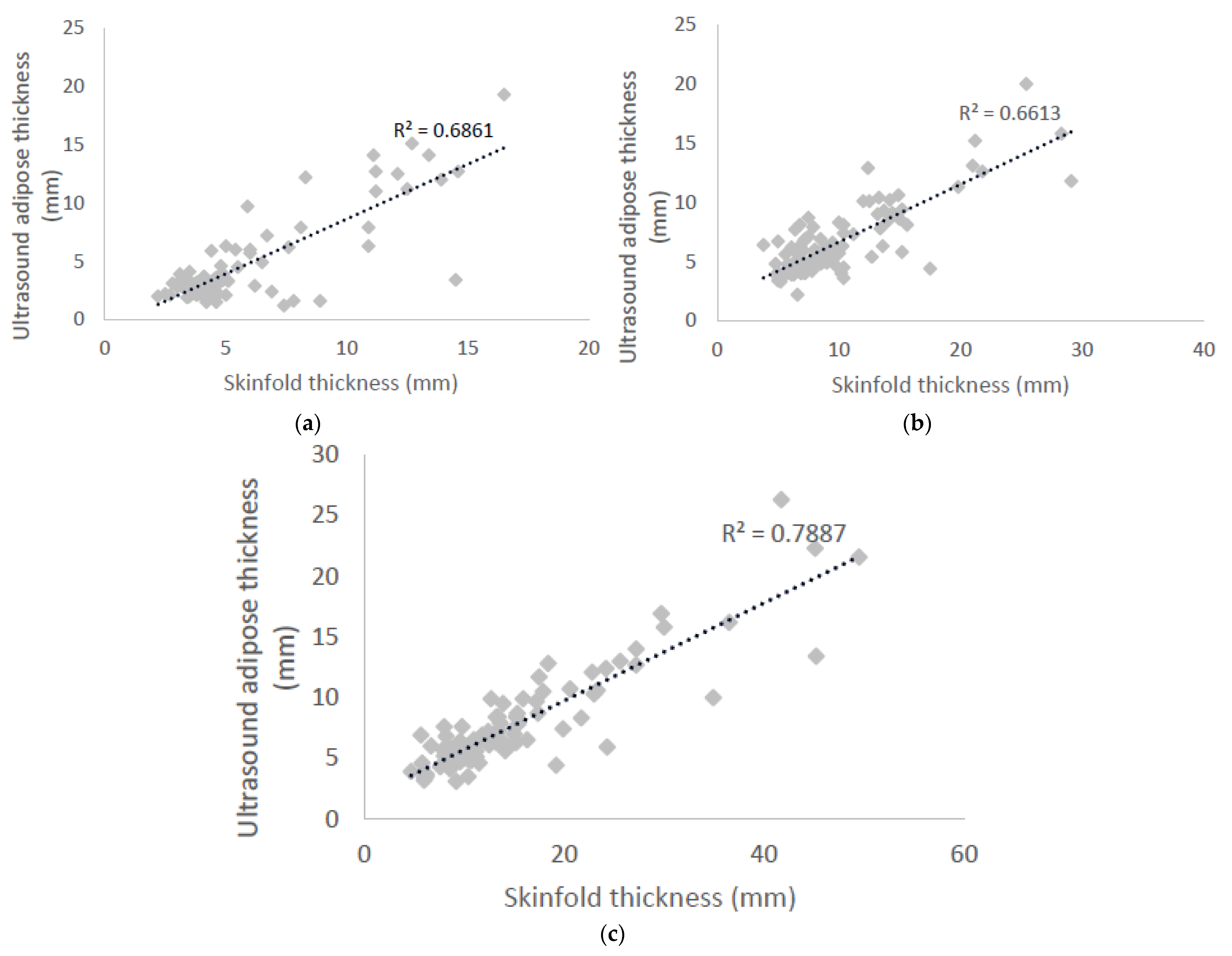
3.1. Pearson’s Correlation

Strong positive correlations were shown between ultrasound and skinfold measurement at the biceps (r = 0.83; 95% CI = 0.72–0.94), triceps (r = 0.81; 95% CI = 0.70–0.93) and front thigh (r = 0.89; 95% CI = 0.80–0.98) site (all p < 0.001; see Figure 1). R2 values were 68.6, 66.1 and 78.9%, respectively.

3.2. Variance of Techniques

There was a significant difference between the two measurement techniques for the biceps (M = 1.1, SD = 2.0 mm), triceps (M = 3.3, SD = 3.1 mm), and front thigh sites (M = 7.1, SD = 5.7 mm) (all p < 0.001; Table 1).

4. Discussion

The aim of this present study was to determine the relationship between skinfold measures of subcutaneous adipose tissue and ultrasound imaging techniques at three skinfold sites (biceps, triceps and front thigh). At all three skinfold sites, a significant, strong, positive correlation was observed. However, the mean difference between the two techniques differed significantly from zero for all three sites, with the skinfold technique producing higher values than the ultrasound images, and so caution needs to be exercised in directly comparing skinfold measures to ultrasound measures of adipose tissue.
The significant, strong, positive correlations for all sites found in the current study are slightly lower than those found in various locations on the anterior thigh (r = 0.90–0.93) in mixed-sex healthy adults [21], and are similar to that for the front thigh (r = 0.86) in mixed-sex junior rowers [28], both with the knee flexed to 90°. The addition of females into the population group and the use of non-ISAK specific measurement sites may account for the differences to the results from the current study. In fact, it has previously been suggested that ISAK measurement sites do not fulfil the criteria for noncompressed ultrasound measurements and so may result in inaccuracy in the ultrasound measurements in the current data set [26]. Appropriate technique is important to acknowledge, since previous technique comparison that failed to indicate standardization of technique has demonstrated inconsistent linear relationships on various sites in normal and overweight military personnel [29]. Nonetheless, the current research demonstrates a significant linear relationship between the two measurement techniques in a healthy, untrained, male population.
Figure 1 indicates that as values of adipose tissue get larger, the relationship between skinfold and ultrasound measurement becomes less obvious (more points further away from the linear trendline). The occurrence of more outliers being associated with greater adipose tissue values was also observed in skinfold–ultrasound comparison studies looking at the triceps skinfold site (r = 0.72) by Ng et al. [30] and various anterior thigh sites in mixed-sex healthy adults [21]. When measuring larger adipose tissue thicknesses, results may be limited by the skinfold exceeding the width of the calliper [31]. The calliper may just fit around the skinfold but with difficulty, altering the method of measurement. On larger skinfold measurements, the callipers may slip off the skinfold slightly, recording an inaccurate measurement [32]. When adipose tissue is thicker, the ultrasound waves lose more energy through absorption, reflection and scattering by attenuation [33]. In ultrasound studies involving adipose tissue (both visceral and subcutaneous), ultrasound wave beams were attenuated more in the thicker tissue due to the beam having to travel further [34]. Due to potential errors in measurement via both techniques, care should be taken when interpreting larger adipose tissue thickness from skinfolds and from ultrasound measures.
Despite the significant, strong, positive correlations demonstrated in the current study, there was significant variance between skinfold and ultrasound measurement at all three sites. Specifically, the skinfold measurements were consistently larger than ultrasound measures at all three sites. The discrepancy between the two techniques appears to be further exacerbated when adipose tissue is larger, as shown by the bigger difference between skinfolds and ultrasound measures as the measures get larger in Table 1. This observation is consistent with previous research [21]. Comparison of both ultrasound and skinfold techniques to regional measures from dual-energy X-ray absorptiometry (DEXA) has indicated that ultrasound demonstrates a stronger relationship with DEXA than skinfolds [35]. Although some of the error in the skinfold technique can be due to poor technical skill, this was not the case in the current data set, as the lead researcher was ISAK Level 3 with over 15 years of experience in completing skinfold measures. The larger measurements achieved with the skinfold technique are likely a result of skinfold measuring a slightly different aspect of surface anatomy to ultrasound and DEXA. When a skinfold is raised, it includes a double layer of the dermis, and may also include more than a single layer of adipose. Furthermore, using skinfold callipers to assess adipose tissue can present difficulties when examining the separation of muscle and fat [21]. Ultrasound, when used correctly, may give a more valid measure of the adipose tissue depth alone. Muller et al. [22] outlined the importance of the adipose being uncompressed, with a significantly less-accurate measurement recorded when the ultrasound probe exerted any pressure on the skin. The previous authors also noted that due to the nature of the skinfold calliper measurement, compression of the adipose tissue cannot be avoided.
The larger difference between the two techniques on the front thigh location may also be a result of slightly different body positions during the measurement. ISAK [17] stipulates that when a skinfold measurement is taken at the front thigh site, the participant should be seated with their back upright and leg straight. The quadricep muscles are relaxed to allow for easier pick up of the skinfold. In the present study, ultrasound images were taken with participant stood up with their feet shoulder-width apart. Although participants were instructed to relax, there is likely to be an element of error included in this comparison from the different body positions.
Previous comparisons of skinfold and ultrasound techniques have used mixed-sex or female-only populations [21,22,27], and despite often demonstrating a strong linear relationship between the techniques have consistently shown skinfolds to be higher. However, acknowledgement of the difference in compressibility between male and female adipose tissue [23] indicates that the exact nature of the relationship between skinfolds and ultrasound measures needs to be established in a sex-specific manner. This study provides indication of this relationship for the untrained male population.
In conclusion, skinfold and ultrasound measurement techniques demonstrate significant, strong correlations that indicate a linear relationship between the two measures. Caution should be exercised when assessing those with higher adipose deposits due to some discrepancy from the linear trend when measures are high. Despite this, skinfolds measure consistently higher than ultrasound and so the exact nature of the relationship should be established on a population basis before using either technique as a surrogate for the other.

Author Contributions

Conceptualization, H.R.-S., S.J., A.O.; methodology, H.R.-S., A.O.; software, H.R.-S., A.O.; validation, H.R.-S., J.F., A.O., S.J.; formal analysis, A.O., H.R.-S., E.P., J.F., S.J.; investigation, H.R.-S., E.P.; resources, H.R.-S., E.P.; data curation, H.R.-S., A.O.; writing—original draft preparation, H.R.-S. and A.O.; writing—review and editing, J.F., E.P. and S.J.; visualization, H.R.-S.; supervision, J.F. and S.J.; project administration, H.R.-S. and E.P. All authors have read and agreed to the published version of the manuscript.

Funding

This research received no external funding.

Institutional Review Board Statement

The study was conducted according to the guidelines of the Declaration of Helsinki, and was approved by the Institutional Ethics Committee of University of Winchester (protocol code BLS/11/15 and 06/11/2015).

Informed Consent Statement

Informed consent was obtained from all subjects involved in the study.

Data Availability Statement

The data presented in this study are available on request from the corresponding author. The data are not publicly available due to original ethical approval and con-sent not allowing for the sharing of data on a public repository.

Conflicts of Interest

The authors declare no conflict of interest.

References

  1. Norton, K.; Olds, T. Anthropometrica: A Textbook of Body Measurement for Sports and Health Courses; UNSW Press: Kennington, Australia, 1996. [Google Scholar]
  2. Fosbøl, M.Ø.; Zerahn, B. Contemporary methods of body composition measurement. Clin. Physiol. Funct. Imaging 2015, 35, 81–97. [Google Scholar] [CrossRef] [PubMed]
  3. Ackland, T.R.; Lohman, T.G.; Sundgot-Borgen, J.; Maughan, R.J.; Meyer, N.L.; Stewart, A.D.; Müller, W. Current status of body composition assessment in sport. Sports Med. 2012, 42, 227–249. [Google Scholar] [CrossRef] [PubMed]
  4. Lemos, T.; Gallagher, D. Current body composition measurement techniques. Curr. Opin. Endocrinol. Diabetes Obes. 2017, 24, 310. [Google Scholar] [CrossRef] [PubMed]
  5. Kuriyan, R. Body composition techniques. Indian J. Med. Res. 2018, 148, 648. [Google Scholar] [CrossRef]
  6. Buchheit, M.; Mendez-Villanueva, A. Reliability and stability of anthropometric and performance measures in highly-trained young soccer players: Effect of age and maturation. J. Sports Sci. 2013, 31, 1332–1343. [Google Scholar] [CrossRef] [PubMed]
  7. Wagner, D.R. Ultrasound as a tool to assess body fat. J. Obes. 2013, 2013, 280713. [Google Scholar] [CrossRef]
  8. Duck, F.A.; Baker, A.C.; Starritt, H.C. Ultrasound in Medicine; CRC Press: Boca Raton, FL, USA, 2020. [Google Scholar]
  9. McArdle, W.D.; Katch, F.I.; Katch, V.L. Exercise Physiology: Nutrition, Energy and Human Performance; Wolters Kluwer Health: Baltimore, MD, USA, 2015. [Google Scholar]
  10. Pineau, J.C.; Lalys, L.; Bocquet, M.; Guihard-Costa, A.M.; Polak, M.; Frelut, M.L.; Peres, G.; Dabbas-Tyan, M. Ultrasound measurement of total body fat in obese adolescents. Ann. Nutr. Metab. 2010, 56, 36–44. [Google Scholar] [CrossRef]
  11. Ismail, C.; Zabal, J.; Hernandez, H.J.; Woletz, P.; Manning, H.; Teixeira, C.; DiPietro, L.; Blackman, M.R.; Harris-Love, M.O. Diagnostic ultrasound estimates of muscle mass and muscle quality discriminate between women with and without sarcopenia. Front. Physiol. 2015, 6, 302. [Google Scholar] [CrossRef] [Green Version]
  12. Takai, Y.; Ohta, M.; Akagi, R.; Kato, E.; Wakahara, T.; Kawakami, Y.; Fukunaga, T.; Kanehisa, H. Applicability of ultrasound muscle thickness measurements for predicting fat-free mass in elderly population. J. Nutr. Health Aging 2014, 18, 579–585. [Google Scholar] [CrossRef]
  13. Pereira, A.Z.; Marchini, J.S.; Carneiro, G.; Arasaki, C.H.; Zanella, M.T. Lean and fat mass loss in obese patients before and after Roux-en-Y gastric bypass: A new application for ultrasound technique. Obes. Surg. 2012, 22, 597–601. [Google Scholar] [CrossRef]
  14. Andreoli, A.; Garaci, F.; Cafarelli, F.P.; Guglielmi, G. Body composition in clinical practice. Eur. J. Radiol. 2016, 85, 1461–1468. [Google Scholar] [CrossRef] [Green Version]
  15. Bazzocchi, A.; Filonzi, G.; Ponti, F.; Albisinni, U.; Guglielmi, G.; Battista, G. Ultrasound: Which role in body composition? Eur. J. Radiol. 2016, 85, 1469–1480. [Google Scholar] [CrossRef]
  16. Madden, A.M.; Smith, S. Body composition and morphological assessment of nutritional status in adults: A review of anthropometric variables. J. Hum. Nutr. Diet. 2016, 29, 7–25. [Google Scholar] [CrossRef]
  17. Esparza-Ros, F.; Vaquero-Cristobal, R.; Marfell-Jones, M. International Standards for Anthropometric Assessment; The International Society for the Advancement of Kinanthropomety: Murcia, Spain, 2019. [Google Scholar]
  18. Hume, P.; Marfell-Jones, M. The importance of accurate site location for skinfold measurement. J. Sports Sci. 2008, 26, 1333–1340. [Google Scholar] [CrossRef] [PubMed]
  19. Bray, G.A.; Greenway, F.L.; Molitch, M.E.; Dahms, W.T.; Atkinson, R.L.; Hamilton, K. Use of anthropometric measures to assess weight loss. Am. J. Clin. Nutr. 1978, 31, 769–773. [Google Scholar] [CrossRef] [PubMed] [Green Version]
  20. Himes, J.H.; Roche, A.F.; Siervogel, R.M. Compressibility of skinfolds and the measurement of subcutaneous fatness. Am. J. Clin. Nutr. 1979, 32, 1734–1740. [Google Scholar] [CrossRef] [Green Version]
  21. Selkow, N.M.; Pietrosimone, B.G.; Saliba, S.A. Subcutaneous thigh fat assessment: A comparison of skinfold calipers and ultrasound imaging. J. Athl. Train. 2011, 46, 50–54. [Google Scholar] [CrossRef] [Green Version]
  22. Müller, W.; Horn, M.; Fürhapter-Rieger, A.; Kainz, P.; Kröpfl, J.M.; Maughan, R.J.; Ahammer, H. Body composition in sport: A comparison of a novel ultrasound imaging technique to measure subcutaneous fat tissue compared with skinfold measurement. Br. J. Sports Med. 2013, 47, 1028–1035. [Google Scholar] [CrossRef]
  23. McRae, M.P. Male and female differences in variability with estimating body fat composition using skinfold calipers. J. Chiropr. Med. 2010, 9, 157–161. [Google Scholar] [CrossRef] [PubMed] [Green Version]
  24. Thoirs, K.; English, C. Ultrasound measures of muscle thickness: Intra-examiner reliability and influence of body position. Clin. Physiol. Funct. Imaging 2009, 29, 440–446. [Google Scholar] [CrossRef]
  25. Müller, W.; Maughan, R.J. The need for a novel approach to measure body composition: Is ultrasound an answer? Br. J. Sports Med. 2013, 47, 1001–1002. [Google Scholar] [CrossRef] [PubMed]
  26. Müller, W.; Lohman, T.G.; Stewart, A.D.; Maughan, R.J.; Meyer, N.L.; Sardinha, L.B.; Kirihennedige, N.; Reguant-Closa, A.; Risoul-Salas, V.; Sundgot-Borgen, J.; et al. Subcutaneous fat patterning in athletes: Selection of appropriate sites and standardisation of a novel ultrasound measurement technique: Ad hoc working group on body composition, health and performance, under the auspices of the IOC Medical Commission. Br. J. Sports Med. 2016, 50, 45–54. [Google Scholar] [CrossRef] [PubMed] [Green Version]
  27. Cohen, J. Set correlation and contingency tables. Appl. Psychol. Meas. 1988, 12, 425–434. [Google Scholar] [CrossRef]
  28. Kelso, A.; Trájer, E.; Machus, K.; Treff, G.; Müller, W.; Steinacker, J.M. Assessment of subcutaneous adipose tissue using ultrasound in highly trained junior rowers. Eur. J. Sport Sci. 2017, 17, 576–585. [Google Scholar] [CrossRef] [PubMed]
  29. Ulbricht, L.; Neves, E.B.; Ripka, W.L.; Romaneli, E.F. Comparison between body fat measurements obtained by portable ultrasound and caliper in young adults. In Proceedings of the 2012 Annual International Conference of the IEEE Engineering in Medicine and Biology Society, San Diego, CA, USA, 28 August–1 September 2012; pp. 1952–1955. [Google Scholar]
  30. Ng, J.; Rohling, R.; Lawrence, P.D. Automatic measurement of human subcutaneous fat with ultrasound. IEEE Trans. Ultrason. Ferroelectr. Freq. Control. 2009, 56, 1642–1653. [Google Scholar] [CrossRef] [Green Version]
  31. Vatier, C.; Poitou, C.; Clément, K. Evaluation of Visceral Fat. In Nutrition in the Prevention and Treatment of Abdominal Obesity; Watson, R.R., Ed.; Elseiver: London, UK, 2014; pp. 68–73. [Google Scholar]
  32. Kuczmarski, R.J.; Fanelli, M.T.; Koch, G.G. Ultrasonic assessment of body composition in obese adults: Overcoming the limitations of the skinfold caliper. Am. J. Clin. Nutr. 1987, 45, 717–724. [Google Scholar] [CrossRef] [Green Version]
  33. Spinner, D.A.; Kirschner, J.S.; Herrera, J.E. Atlas of Ultrasound Guided Musculoskeletal Injections; Springer: New York, NY, USA, 2014. [Google Scholar]
  34. Saranteas, T. Limitations in ultrasound imaging techniques in anesthesia: Obesity and muscle atrophy? Anesth. Analg. 2009, 109, 993–994. [Google Scholar] [CrossRef]
  35. Gomes, A.C.; Landers, G.J.; Binnie, M.J.; Goods, P.S.; Fulton, S.K.; Ackland, T.R. Body composition assessment in athletes: Comparison of a novel ultrasound technique to traditional skinfold measures and criterion DXA measure. J. Sci. Med. Sport 2020, 23, 1006–1010. [Google Scholar] [CrossRef]
Figure 1. Pearson’s correlation of ultrasound adipose thickness against skinfold thickness for: (a) biceps measurement site; (b) triceps measurement site; (c) front thigh measurement site.
Applsci 11 10561 g001
Table 1. Mean (±standard deviation) measures at each site and the difference between the measures.
Measurement SiteSkinfold (mm)Ultrasound (mm)Difference (mm)
Biceps5.5 ± 3.14.4 ± 3.61.1 ± 2.0 *
Triceps9.9 ± 5.06.6 ± 3.03.3 ± 3.1 *
Front Thigh14.8 ± 9.07.6 ± 4.17.1 ± 5.7 *
* p < 0.01 when compared to zero difference.
Publisher’s Note: MDPI stays neutral with regard to jurisdictional claims in published maps and institutional affiliations.

