Article

Evaluation of Depth of Invasion in Oral Squamous Cell Carcinoma with Ultra-High Frequency Ultrasound: A Preliminary Study

1
Unit of Dentistry and Oral Surgery, Department of Surgical, Medical and Molecular Pathology and Critical Care Medicine, University of Pisa, 56126 Pisa, Italy
2
Unit of Dermatology, Department of Clinical and Experimental Medicine, University of Pisa, 56126 Pisa, Italy
3
Diagnostic and Interventional Radiology, Department of Translational Research and of New Technologies in Medicine and Surgery, University of Pisa, 56126 Pisa, Italy
*
Authors to whom correspondence should be addressed.
Appl. Sci. 2021, 11(16), 7647; https://doi.org/10.3390/app11167647
Submission received: 6 July 2021 / Revised: 7 August 2021 / Accepted: 16 August 2021 / Published: 20 August 2021
(This article belongs to the Special Issue Materials and Technologies in Oral Research)

Abstract

:

Featured Application

In the present work, the application of ultra-high frequency ultrasonography in the assessment of oral squamous cell carcinoma is explored, providing insights into the potential role of the technique in the diagnosis, management, and follow-up of malignant lesions of the oral cavity.

Abstract

Intraoral ultrasonography has been widely employed for the preoperative assessment of tumor margins due to its capability to evaluate depth of invasion (DOI) and tumor thickness (TT). Recently, a novel ultrasonographic technique, ultra-high frequency ultrasound (UHFUS) has been increasingly applied to the study of oral lesions. This study evaluates the potential application of intraoral UHFUS to assess DOI and TT parameters of oral squamous cell carcinoma (OSCC) lesions. Patients clinically suspected of OSCC lesions were enrolled and underwent an intraoral UHFUS examination preoperatively. The parameters of TT, DOI, echogenicity, and vascularization were assessed. The parameters of TT and DOI as evaluated by means of UHFUS were compared to histology, which was set as the benchmark. Ten patients in total were enrolled. UHFUS-based DOI and TT measurements were found to positively correlate with histology (p < 0.05), although UHFUS provided a slight overestimation of DOI. No differences were found in terms of echogenicity or vascularization depending on the site of the lesion. According to these preliminary results, UHFUS could support the preoperative assessment of TT and DOI, potentially enhancing the clinical evaluation of OSCC.

1. Introduction

Oral squamous cell carcinoma (OSCC) is the most frequently encountered neoplasm of the oral cavity, accounting for 98% of all oral cancers [1]. OSCC predominantly affects males in the 6th decade (male/female ratio: 2.22:1) and is most frequently localized in the tongue [1]. Although some environmental factors, such as smoking and alcohol consumption, have been claimed to have a role in OSCC development, at present the pathogenesis is unclear [2]. The prognosis is poor, with a survival rate of 57% at 5 years in patients >40 years of age, mostly due to late diagnosis when metastases have already developed [2].
Surgery is the treatment of choice, and obtaining clear resection margins (≥5 mm for all T stages) is fundamental to achieve the locoregional control of the disease [3,4]; however, the assessment of the deep margins of the tumor is often difficult due to the infiltrative nature of OSCC [5]. The application of diagnostic imaging techniques, in particular intraoral ultrasonography (US), computed tomography (CT) and magnetic resonance (MR), may support the assessment of tumor dimensions preoperatively. Intraoral US has been reported to show good correlation with histopathology and good accuracy compared to other diagnostic imaging techniques [4,6,7]. Specifically, intraoral US has been employed for the evaluation of the parameters tumor thickness (TT) and depth of invasion (DOI). TT is defined as the distance from the surface of the tumor to the point of deepest invasion, while DOI is the distance from the adjacent epithelial surface to the deepest point of invasion of the tumor [5].
Interestingly, DOI evaluation has been introduced in the 8th American Joint Committee on Cancer- Union for International Cancer Control (AJCC-UICC) TNM staging system in stages 1, 2, and 3 [8]. DOI seems to be a good predictor in terms of prognosis compared to TT, as it may help to discriminate between superficial and deeply invasive tumors [8]. From this perspective, DOI may also be predictive of the presence of regional nodal metastases [9,10]. Apart from the prognostic usefulness of DOI estimation, the possibility to obtain information on the deeper invasion of the tumor may improve the surgical planning of the resection and give an indication of prophylactic neck dissection in cases of DOI ≥ 4 mm [6,11].
Recently, a novel ultrasonographic technique, ultra-high frequency ultrasound (UHFUS), has been gaining increasing importance in several medical fields, due to the high resolution provided by the use of frequencies >30 MHz, which makes it particularly suitable for the investigation of superficial anatomy [12,13,14]. UHFUS was initially developed in the mid-1990s for preclinical evaluation of small animal models, in particular for the assessment of tumor behavior and pharmacology [12]. In the early 2000s, the technique entered the clinical setting, especially for vascular and dermatological applications [13].
In oral medicine, the intraoral evaluation of oral mucosal lesions has become increasingly applied, with promising results [15,16,17,18,19]. Moreover, UHFUS appears to show high correspondence with histology [20,21,22,23,24]. At present, intraoral UHFUS has been predominantly employed for the characterization of oral lesions, including oral cancer [17], although in the absence of a focused assessment of DOI and TT.
The aim of the present study was to evaluate the utility of UHFUS in the assessment of OSCC and to compare DOI and TT values with histology.

2. Materials and Methods

2.1. Patients Flow

Patients referred to the Unit of Dentistry and Oral Surgery, University Hospital of Pisa (Pisa, Italy) with a suspect of OSCC lesion were enrolled in the study between January and November 2020. Inclusion criteria were (a) adults ≥ 18 years of age, (b) clinical diagnosis of OSCC, (c) a lesion eligible for surgical treatment, and (d) acceptance to be included in the study. Exclusion criteria were (a) any acute or chronic condition that would limit the ability of the patient to participate in the study, (b) refusal to give informed consent, and (c) histological diagnosis different from OSCC. The study protocol was approved by the local ethics committee (CEAVNO, protocol no. 32822/18), and all subjects gave written consent to undergo UHFUS examination. The study was conducted according to the principles outlined in the Declaration of Helsinki on experimentation involving human subjects.

2.2. UHFUS Acquisition Protocol and Image Postprocessing

Intraoral UHFUS scans were performed with a standardized acquisition protocol by two examiners using Vevo MD (VisualSonics, Toronto, ON, Canada) equipped with a 70 MHz probe. Training and calibration between the examiners were performed on patients who were not part of the study to standardize UHFUS scan performance, image acquisition, and image interpretation prior to study beginning. Image analysis was conducted on DICOM format images processed with Horos software (Version 3.3.6, Annapolis, MD, USA). Data collection started when Cohen’s kappa value between examiners was >0.90.
A disposable sterile probe cover was employed for the management of cross-infections in order to avoid the direct contact between the oral mucosa and the probe. Sterile ultrasound gel was employed as a couplant material to improve ultrasound beam transmission.
The standardized UHFUS acquisition was conducted by placing the probe perpendicular to the mucosal surface with minimal pressure on the tumor.
B-mode axial and longitudinal acquisitions were performed using a standardized preset. Scan depth and focus position were adjusted to optimize the scan, while other parameters (gain, time gain compensation, dynamic range, mechanical index, and thermal index) were kept constant throughout the UHFUS examination.
The parameters of TT, DOI, and echogenicity were measured on B-mode axial images. Histology was set as benchmark for TT and DOI evaluation.
TT (expressed in mm) was measured as the distance between the lesion’s surface and the deepest margin in the tissue. DOI (expressed in mm) was measured as the distance between the normal mucosal surface and the deepest margin of the lesion in the tissue.
Echogenicity was evaluated through grey scale analysis of a selected region of interest (ROI). The average grey level distribution value, minimum value, maximum value, and standard deviation were retrieved and compared.
Color Doppler was employed for the assessment of vascularization, using a setting scale of ±1.9 cm/s to detect blood vessels with a low flow rate. A qualitative analysis of the degree of vascularization was performed by assigning each lesion a score from 1 to 3. Score 1 corresponded to vascularization present in <30% of the lesion, with perilesional pattern; score 2 indicated vascularization between 30% and 60% of the lesion, with predominantly intralesional pattern; finally, score 3 indicated intense perilesional and intralesional vascularization in >60% of the lesion, with the presence of aliasing phenomena.

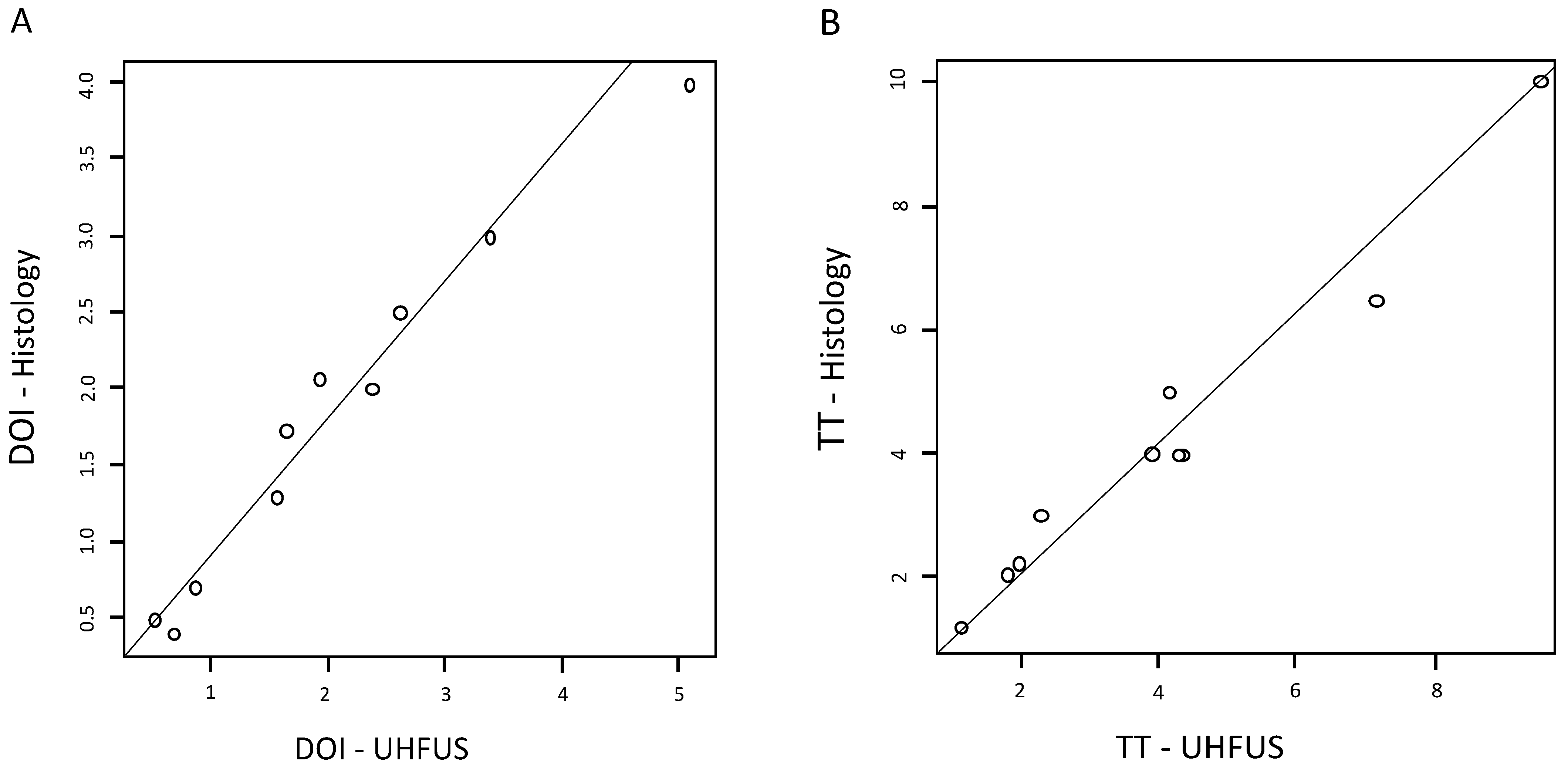
2.3. Statistical Analysis

Data were analyzed using SPSS software (IBM) (Version 26.0, IBM Corp.; Armonk, NY, USA). Pearson’s correlation and scatter plots were employed to assess the relationship between UHFUS measurements and histology. The comparison between groups was performed using the Mann–Whitney test. p-value significance was set at p < 0.05.

3. Results

3.1. Sample Characteristics

Ten patients in total were enrolled (6 females, 4 males, mean age 68.7, standard deviation 10.2). Six patients were smokers, who smoked a mean of 18.3 cigarettes/day. Three patients presented comorbidities, in particular two had a history of breast cancer and one had HCV-related liver damage. In 6 patients, OSCC was localized in the tongue, while in 2 patients the lesions occurred in the mandibular gingival mucosa, in 1 patient in the buccal mucosa, and in 1 patient on the mouth floor. Four lesions presented with endophytic growth, and six were exophytic lesions.
For endophytic lesions, the mean time for lesion development ranged between 3 and 8 months, with an average of 5.25 months (SD 2.22 months). Exophytic lesions development ranged between 3 and 12 months, with an average of 7.33 months (SD 3.45 months).
In four patients the lesion was associated with an absence of pain or mild pain (<4 as assessed by means of Visual Analogue Scale—VAS), while in 6 patients values >6 were registered. Overall, the mean VAS value was 5.1. VAS values were significantly lower (p < 0.05) in patients who developed endophytic lesions (3.75 ± 3.10) compared to exophytic lesions (6.33 ± 3.09).
In Table 1, the sample characteristics are summarized. In Figure 1, Figure 2 and Figure 3, some of the included cases are presented.

3.2. Histology

The OSCC lesions were well-differentiated (G1) in 4 patients, moderately differentiated (G2) in 5 patients, and poorly differentiated (G3) in 1 patient. According to the pathological TNM (pTNM), the pathological T categories included 2 Tis (20%), 3 T1 N0 M0 (30%), 3 T2 N0 M0 (30%), and 1 T2 N1 M0 (10%). Histological tumor sizes ranged from 0.5 to 25 mm and DOI ranged from 0.4 to 6 mm (mean 2.46 mm, SD 1.91 mm). TT ranged from 3 to 20 mm (mean 9.15 mm, SD 5.89 mm). The average TT of exophytic lesions was 5.28 ± 2.47 mm, while the TT of endophytic lesions was 2.55 ± 1.22 mm. The mean DOI for exophytic lesions was 2.07 ± 1.29 mm, while for endophytic lesions the value was 2.05 ± 1.73 mm. In 8 cases, the tumor was found to infiltrate the lamina propria. No evidence of distant metastases was found in any of the included patients.

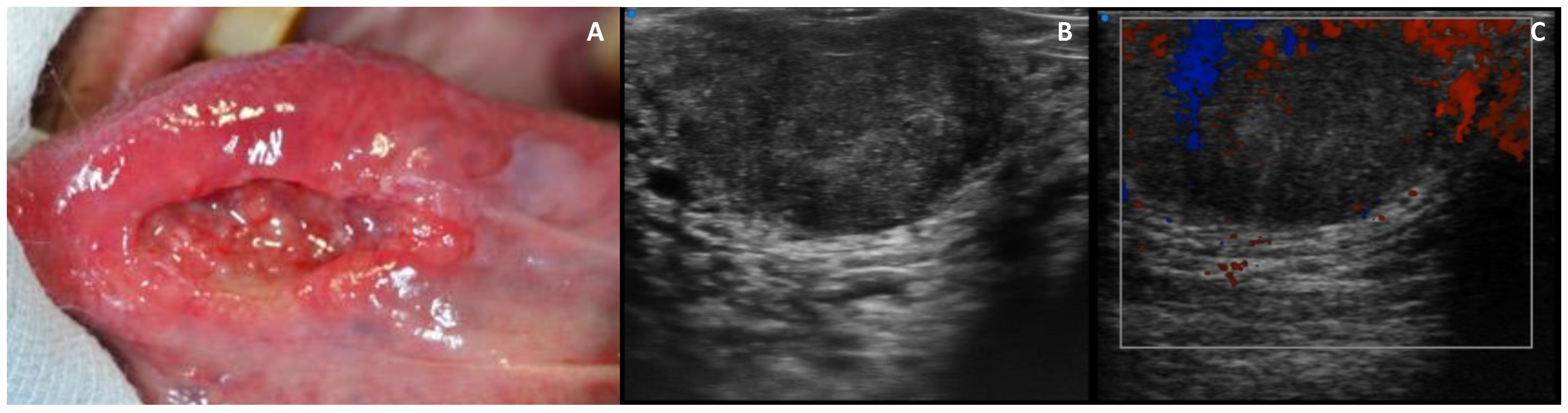
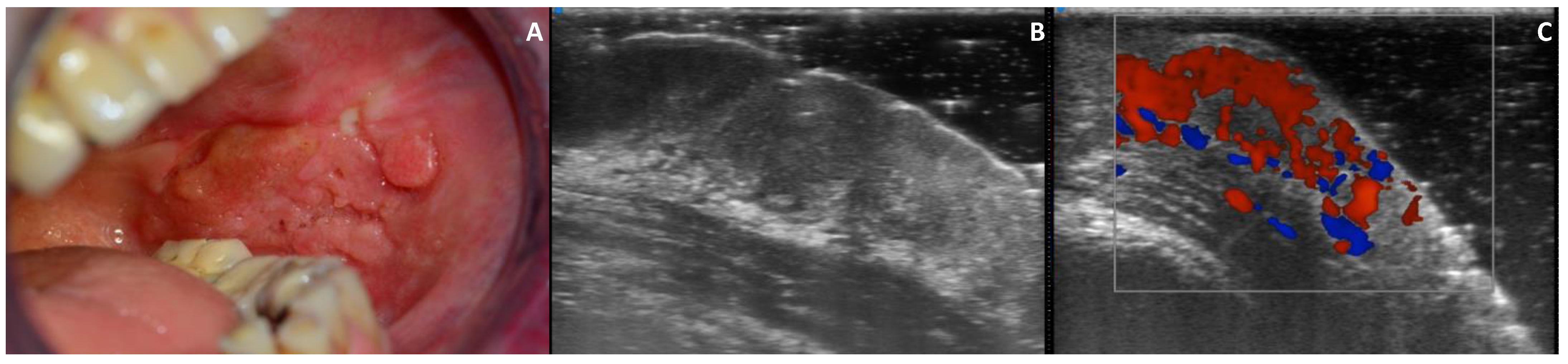
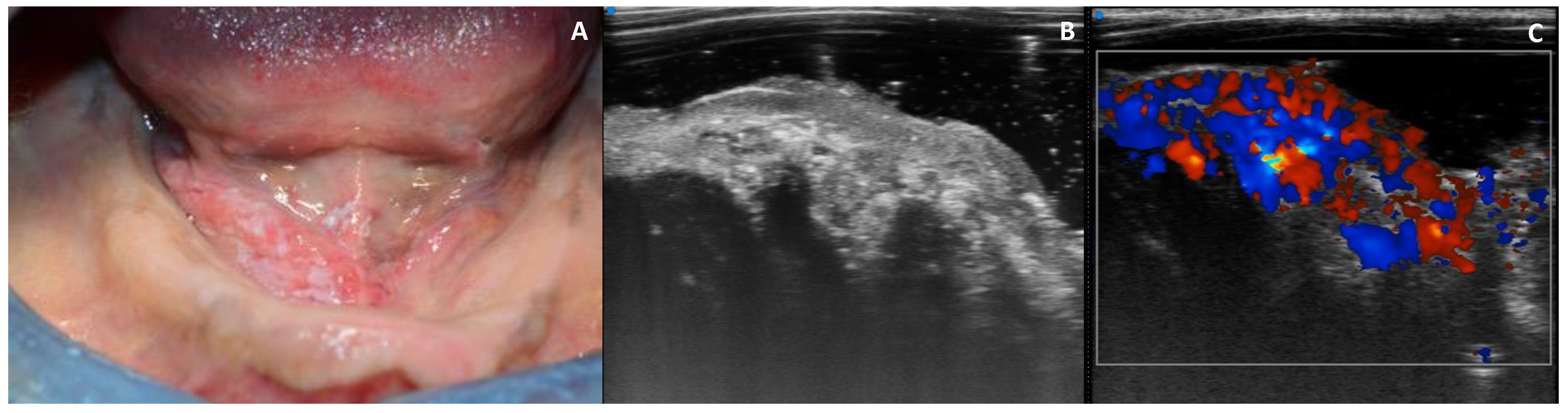
3.3. UHFUS Examination

On UHFUS, the average TT of exophytic lesions was 5.17 ± 2.48 mm, while the TT of endophytic lesions was 2.40 ± 1.40 mm. The mean DOI for exophytic lesions was 2.44 ± 1.57 mm, while for endophytic lesions the value was 1.92 ± 1.65 mm. TT and DOI assessments by means of UHFUS were found to correlate with histology (R = 0.9835, p < 0.05 for TT, R = 0.9631, p < 0.05 for DOI) (Figure 4). TT was slightly underestimated on UHFUS (mean difference 0.10 mm), while DOI was overestimated (mean difference 0.14 mm).

3.4. Echogenicity

Mean values of echogenicity were slightly higher for exophytic lesions (69.71 ± 11.78) compared to endophytic lesions (61.76 ± 19.97) in the absence of statistically significant differences. No differences were found in terms of echogenicity depending on the site of the lesion.

3.5. Vascularization

Vascularization scores significantly differed between exophytic and endophytic growth pattern, with score 3 being more frequently assigned to the endophytic lesions (p < 0.05). No differences were registered between exophytic and endophytic lesions depending on the site.

4. Discussion

According to these preliminary results, preoperative evaluation of the TT and DOI parameters by means of UHFUS appeared to significantly correlate with histology. A minimal and clinically acceptable overestimation was found for UHFUS DOI measurements, while TT was underestimated by 0.1 mm. We might speculate that higher frequencies allowed better visualization of tumor margins, providing reliable measurements of the lesions’ dimensions; however, it should be noted that histological TT measurements appeared to be higher than the UHFUS measurements in the absence of statistically significant differences. Although the UHFUS scan was performed avoiding the compression of the lesion, it cannot be excluded that minimal pressure was applied, resulting in reduced TT UHFUS dimensions compared to histology.
Intraoral US is a well-established technique that has been previously described in the literature, and US-based parameters are prognosticators in terms of oncological outcomes during and after the management of oral cancer [4]. TT was the most frequently employed US parameter for the assessment of the longitudinal extension in terms of the depths of oral cavity tumors prior to the introduction of DOI as a T-category modifier [9,25]. Although both TT and DOI appear strongly correlated with the risk of presence of nodal metastases, albeit with different predictive cut-off limits, at present DOI is the parameter of choice due to its higher reliability [7]. Nevertheless, combined TT and DOI evaluations can be useful in terms of staging and prognosis, especially in small tumors where the issue of occult metastases is extremely relevant.
Shintani et al. reported that US TT measurements showed strong correlations with histology, especially in the assessment of tumors >10.0 mm [11]. Songra et al. [26] employed US both preoperatively and intraoperatively. While preoperative TT assessments appeared to be reliable compared to histology, the intraoperative assessment of deep resection margins had moderate predictive value due to tissue compression. Moreover, US measurements have been reported to have a predictive value for the development of nodal metastases, potentially influencing the therapeutic approach [27,28]. Natori et al. [29] evaluated TT and tumor margins to grade the malignancy of tongue tumors and set a thickness of 8 mm as the cut-off point to predict the risk of developing neck metastases. Brockoff et al. [30] reported that DOI was a relevant variable, especially in clinical T1 N0 disease, which could give indication of the performance of neck dissection. Finally, Rocchetti et al. [31] found a significant correlation between DOI and histology, suggesting a potential role in the assessment of the aggressiveness of the tumor, as well as supporting the choice of the optimal treatment.
Overall, US was reported to be superior to CT and MR, especially in cases of thin tumors with TT < 5 mm [5]; however, over- or under-estimation can occur due to the presence of inflammation or to the pressure applied by the US probe [4]. Other factors accountable for the discrepancy between US and histology measurements can be the shrinkage of the resection specimen during formalin fixation and slicing errors from the cutting plane during histological processing, which have been reported to range between 10 and 20% [32]. Nevertheless, the decrease in tumor size between post-excision and post-formalin fixation was reported to be around 4.7% in the absence of significance [32]. In our sample, a minimal discrepancy was observed between UHFUS and histological measurements, which did not reach statistical significance. It could be hypothesized that the increased conspicuity obtained by means of the higher frequencies employed may have improved the preoperative assessment of OSCC lesions. Interestingly, UHFUS measurements were relatively smaller than histology measurements when evaluating the TT parameter. This fact could be related to the application of a slight pressure on the tumor, which can occur during the UHFUS scan. The compression of the lesion may cause an underestimation of the lesion dimensions, especially when evaluating TT; however, we recognize that further evaluation is mandatory to further assess the reliability of UHFUS measurements.
An initial characterization of OSCC lesions involved the evaluation of the UHFUS parameters of echogenicity and vascularization. Previous research evaluating the echogenicity of both normal mucosa and oral lesions highlighted that the grey scale distribution varied depending on the anatomical site and lesion typology [16,17]. In our sample, the mean grey values of endophytic and exophytic lesions did not show significant difference. However, a trend was observed for exophytic lesions to present higher mean grey values. Interestingly, the localization did not influence the echogenicity of the lesion. The semiquantitative score employed on the color Doppler UHFUS images highlighted the presence of more intense vascularization in correspondence with endophytic lesions, suggesting that this type of growth pattern could be associated with increased angiogenesis and inflammation. However, further evaluation is needed to better correlate vascularization evaluated by means of color Doppler and histology.
To the best of our knowledge, this is the first study to report the application of UHFUS to the evaluation of OSCC, and in particular for TT and DOI estimation. Although a good correlation between UHFUS measurements and histology was observed, the present study has some limitations. First of all, the study was performed on a limited number of subjects and mostly involved early-stage tumors of reduced dimensions. Secondly, no comparison with conventional US was provided, as histology was set as benchmark. Therefore, we recognize that the superiority of UHFUS compared to conventional US is still to be demonstrated. Finally, the evaluation of echogenicity and vascularization needs further correlation with histology and clinical parameters. Nevertheless, these preliminary results suggest that UHFUS has the potential to become a valuable technique for the evaluation of OSCC lesions, and give insight into its application in OSCC diagnostic work-up and surgical management.

Author Contributions

Conceptualization, R.I., M.N.; methodology, R.I.; software, R.I., S.G.; validation, L.C., D.C., F.G.; formal analysis, R.I., M.N., S.G.; investigation, R.I.; resources, T.O.; data curation, R.I., M.N., S.G.; writing—original draft preparation, R.I.; writing—review and editing, R.I., M.N.; visualization, S.G., T.O.; supervision, D.C., F.G.; project administration, R.I. All authors have read and agreed to the published version of the manuscript.

Funding

This research received no external funding.

Institutional Review Board Statement

The study was conducted according to the guidelines of the Declaration of Helsinki and approved by the Institutional Review Board of the University Hospital of Pisa, protocol code 32822-19/06/2018.

Informed Consent Statement

Informed consent was obtained from all subjects involved in the study.

Data Availability Statement

The data presented in this study are contained within the article.

Conflicts of Interest

The authors declare no conflict of interest.

References

  1. Dhanuthai, K.; Rojanawatsirivej, S.; Thosaporn, W.; Kintarak, S.; Subarnbhesaj, A.; Darling, M.; Kryshtalskyj, E.; Chiang, C.P.; Shin, H.I.; Choi, S.Y.; et al. Oral cancer: A multicenter study. Med. Oral Patol. Oral Cir. Bucal. 2018, 23, e23–e29. [Google Scholar] [CrossRef] [PubMed]
  2. Chen, S.; Lin, Z.; Chen, J.; Yang, A.; Zhang, Q.; Xie, C.; Zhang, X.; Yang, Z.; Chen, W.; Song, M. Older age is a risk factor associated with poor prognosis of patients with squamous cell carcinoma of the oral cavity. Eur. Arch. Otorhinolaryngol. 2020, 277, 2573–2580. [Google Scholar] [CrossRef] [Green Version]
  3. Dik, E.A.; Willems, S.M.; Ipenburg, N.A.; Adriaansens, S.O.; Rosenberg, A.J.; van Es, R.J. Resection of early oral squamous cell carcinoma with positive or close margins: Relevance of adjuvant treatment in relation to local recurrence: Margins of 3 mm as safe as 5 mm. Oral Oncol. 2014, 50, 611–615. [Google Scholar] [CrossRef] [PubMed]
  4. Nulent, T.J.K.; Noorlag, R.; Van Cann, E.M.; Pameijer, F.A.; Willems, S.M.; Yesuratnam, A.; Rosenberg, A.; de Bree, R.; van Es, R.J. Intraoral ultrasonography to measure tumor thickness of oral cancer: A systematic review and meta-analysis. Oral Oncol. 2018, 77, 29–36. [Google Scholar] [CrossRef] [PubMed]
  5. Tarabichi, O.; Bulbul, M.G.; Kanumuri, V.V.; Faquin, W.C.; Juliano, A.F.; Cunnane, M.E.; Varvares, M.A. Utility of intraoral ultrasound in managing oral tongue squamous cell carcinoma: Systematic review. Laryngoscope 2019, 129, 662–670. [Google Scholar] [CrossRef] [PubMed]
  6. Marchi, F.; Filauro, M.; Iandelli, A.; Carobbio, A.; Mazzola, F.; Santori, G.; Parrinello, G.; Canevari, F.; Piazza, C.; Peretti, G. Magnetic resonance vs. intraoral ultrasonography in the preoperative assessment of oral squamous cell carcinoma: A systematic review and meta-analysis. Front. Oncol. 2020, 9, 1571. [Google Scholar] [CrossRef] [Green Version]
  7. Shintani, S.; Yoshihama, Y.; Ueyama, Y.; Terakado, N.; Kamei, S.; Fijimoto, Y.; Hasegawa, Y.; Matsuura, H.; Matsumura, T. The usefulness of intraoral ultrasonography in the evaluation of oral cancer. Int. J. Oral Maxillofac. Surg. 2001, 30, 139–143. [Google Scholar] [CrossRef]
  8. Pollaers, K.; Hinton-Bayre, A.; Friedland, P.L.; Farah, C.S. AJCC 8th Edition oral cavity squamous cell carcinoma staging—Is it an improvement on the AJCC 7th Edition? Oral Oncol. 2018, 82, 23–28. [Google Scholar] [CrossRef]
  9. Lydiatt, W.M.; Patel, S.G.; O’Sullivan, B.; Brandwein, M.S.; Ridge, J.A.; Migliacci, J.C.; Loomis, A.M.; Shah, J.P. Head and neck cancers—Major changes in the American Joint Committee on cancer eighth edition cancer staging manual. CA Cancer J. Clin. 2017, 67, 122–137. [Google Scholar] [CrossRef]
  10. Piazza, C.; Montalto, N.; Paderno, A.; Taglietti, V.; Nicolai, P. Is it time to incorporate ‘depth of infiltration’ in the T staging of oral tongue and floor of mouth cancer? Curr. Opin. Otolaryngol. Head Neck Surg. 2014, 22, 81–89. [Google Scholar] [CrossRef] [Green Version]
  11. van Lanschot, C.; Klazen, Y.P.; de Ridder, M.; Mast, H.; Ten Hove, I.; Hardillo, J.A.; Monserez, D.A.; Sewnaik, A.; Meeuwis, C.A.; Keereweer, S.; et al. Depth of invasion in early stage oral cavity squamous cell carcinoma: The optimal cut-off value for elective neck dissection. Oral Oncol. 2020, 111, 104940. [Google Scholar] [CrossRef]
  12. Izzetti, R.; Oranges, T.; Janowska, A.; Gabriele, M.; Graziani, F.; Romanelli, M. The application of ultra-high-frequency ultrasound in dermatology and wound management. Int. J. Low. Extrem. Wounds 2020, 19, 334–340. [Google Scholar] [CrossRef]
  13. Izzetti, R.; Vitali, S.; Aringhieri, G.; Nisi, M.; Oranges, T.; Dini, V.; Ferro, F.; Baldini, C.; Romanelli, M.; Caramella, D.; et al. Ultra-high frequency ultrasound, a promising diagnostic technique: Review of the literature and single-center experience. Can. Assoc. Radiol. J. 2021, 72, 418–431. [Google Scholar] [CrossRef]
  14. Aringhieri, G.; Izzetti, R.; Vitali, S.; Ferro, F.; Gabriele, M.; Baldini, C.; Caramella, D. Ultra-high frequency ultrasound (UHFUS) applications in Sjogren syndrome: Narrative review and current concepts. Gland Surg. 2020, 9, 2248–2259. [Google Scholar] [CrossRef] [PubMed]
  15. Izzetti, R.; Fantoni, G.; Gelli, F.; Faggioni, L.; Vitali, S.; Gabriele, M.; Caramella, D. Feasibility of intraoral ultrasonography in the diagnosis of oral soft tissue lesions: A preclinical assessment on an ex vivo specimen. Radiol. Med. 2018, 123, 135–142. [Google Scholar] [CrossRef]
  16. Izzetti, R.; Vitali, S.; Aringhieri, G.; Oranges, T.; Dini, V.; Nisi, M.; Graziani, F.; Gabriele, M.; Caramella, D. Discovering a new anatomy: Exploration of oral mucosa with ultra-high frequency ultrasound. Dentomaxillofac. Radiol. 2020, 49, 20190318. [Google Scholar] [CrossRef]
  17. Izzetti, R.; Vitali, S.; Aringhieri, G.; Caramella, D.; Nisi, M.; Oranges, T.; Dini, V.; Graziani, F.; Gabriele, M. The efficacy of ultra-high frequency ultrasonography in the diagnosis of intraoral lesions. Oral Surg. Oral Med. Oral Pathol. Oral Radiol. 2020, 129, 401–410. [Google Scholar] [CrossRef]
  18. Izzetti, R.; Vitali, S.; Oranges, T.; Dini, V.; Romanelli, M.; Caramella, D.; Gabriele, M. Intraoral ultra-high frequency ultrasound study of oral lichen planus: A pictorial review. Skin Res. Technol. 2020, 26, 200–204. [Google Scholar] [CrossRef] [PubMed]
  19. Izzetti, R.; Vitali, S.; Gabriele, M.; Caramella, D. Feasibility of a combination of intraoral UHFUS and CBCT in the study of peri-implantitis. Oral Surg. Oral Med. Oral Pathol. Oral Radiol. 2019, 127, e89–e94. [Google Scholar] [CrossRef]
  20. Oranges, T.; Vitali, S.; Benincasa, B.; Izzetti, R.; Lencioni, R.; Caramella, D.; Romanelli, M.; Dini, V. Advanced evaluation of hidradenitis suppurativa with ultra-high frequency ultrasound: A promising tool for the diagnosis and monitoring of disease progression. Skin Res. Technol. 2020, 26, 513–519. [Google Scholar] [CrossRef]
  21. Oranges, T.; Janowska, A.; Vitali, S.; Loggini, B.; Izzetti, R.; Romanelli, M.; Dini, V. Dermatoscopic and ultra-high frequency ultrasound evaluation in cutaneous postradiation angiosarcoma. J. Eur. Acad. Dermatol. Venereol. 2020, 34, e741. [Google Scholar] [CrossRef] [PubMed]
  22. Ferro, F.; Izzetti, R.; Vitali, S.; Aringhieri, G.; Fonzetti, S.; Donati, V.; Dini, V.; Mosca, M.; Gabriele, M.; Caramella, D.; et al. Ultra-high frequency ultrasonography of labial glands is a highly sensitive tool for the diagnosis of Sjögren’s syndrome: A preliminary study. Clin. Exp. Rheumatol. 2020, 38, 210–215. [Google Scholar]
  23. Izzetti, R.; Ferro, F.; Vitali, S.; Nisi, M.; Fonzetti, S.; Oranges, T.; Donati, V.; Caramella, D.; Baldini, C.; Gabriele, M. Ultra-high frequency ultrasonography (UHFUS)-guided minor salivary gland biopsy: A promising procedure to optimize labial salivary gland biopsy in Sjögren’s syndrome. J. Oral Pathol. Med. 2021, 50, 485–491. [Google Scholar] [CrossRef]
  24. Izzetti, R.; Nisi, M.; Aringhieri, G.; Vitali, S.; Oranges, T.; Romanelli, M.; Caramella, D.; Graziani, F.; Gabriele, M. Ultra-high frequency ultrasound in the differential diagnosis of oral pemphigus and pemphigoid: An explorative study. Skin Res. Technol. 2021. [Google Scholar] [CrossRef]
  25. Moeckelmann, N.; Ebrahimi, A.; Tou, Y.K.; Gupta, R.; Low, T.H.; Ashford, B.; Ch’ng, S.; Palme, C.E.; Clark, J.R. Prognostic implications of the 8th edition American Joint Committee on Cancer (AJCC) staging system in oral cavity squamous cell carcinoma. Oral Oncol. 2018, 85, 82–86. [Google Scholar] [CrossRef]
  26. Songra, A.K.; Ng, S.Y.; Farthing, P.; Hutchison, I.L.; Bradley, P.F. Observation of tumour thickness and resection margin at surgical excision of primary oral squamous cell carcinoma—Assessment by ultrasound. Int. J. Oral Maxillofac. Surg. 2006, 35, 324–331. [Google Scholar] [CrossRef] [PubMed]
  27. Yesuratnam, A.; Wiesenfeld, D.; Tsui, A.; Iseli, T.A.; Hoorn, S.V.; Ang, M.T.; Guiney, A.; Phal, P.M. Preoperative evaluation of oral tongue squamous cell carcinoma with intraoral ultrasound and magnetic resonance imaging-comparison with histopathological tumour thickness and accuracy in guiding patient management. Int. J. Oral Maxillofac. Surg. 2014, 43, 787–794. [Google Scholar] [CrossRef] [PubMed]
  28. Angelelli, G.; Moschetta, M.; Limongelli, L.; Albergo, A.; Lacalendola, E.; Brindicci, F.; Favia, G.; Maiorano, E. Endocavitary sonography of early oral cavity malignant tumors. Head Neck 2017, 39, 1349–1356. [Google Scholar] [CrossRef] [PubMed]
  29. Natori, T.; Koga, M.; Anegawa, E.; Nakashima, Y.; Tetsuka, M.; Yoh, J.; Kusukawa, J. Usefulness of intra-oral ultrasonography to predict neck metastasis in patients with tongue carcinoma. Oral Dis. 2008, 14, 591–599. [Google Scholar] [CrossRef]
  30. Brockhoff, H.C., 2nd; Kim, R.Y.; Braun, T.M.; Skouteris, C.; Helman, J.I.; Ward, B.B. Correlating the depth of invasion at specific anatomic locations with the risk for regional metastatic disease to lymph nodes in the neck for oral squamous cell carcinoma. Head Neck 2017, 39, 974–979. [Google Scholar] [CrossRef]
  31. Rocchetti, F.; Tenore, G.; Montori, A.; Cassoni, A.; Cantisani, V.; Di Segni, M.; Di Gioia, C.; Carletti, R.; Valentini, V.; Polimeni, A.; et al. Preoperative evaluation of tumor depth of invasion in oral squamous cell carcinoma with intraoral ultrasonography: A retrospective study. Oral Surg. Oral Med. Oral Pathol. Oral Radiol. 2021, 131, 130–138. [Google Scholar] [CrossRef] [PubMed]
  32. Umstattd, L.A.; Mills, J.C.; Critchlow, W.A.; Renner, G.J.; Zitsch, R.P., 3rd. Shrinkage in oral squamous cell carcinoma: An analysis of tumor and margin measurements in vivo, post-resection, and post-formalin fixation. Am. J. Otolaryngol. 2017, 38, 660–662. [Google Scholar] [CrossRef] [PubMed]
Figure 1. OSCC (oral squamous cell carcinoma) of the tongue margin in a 59-year-old male: (A) clinical aspect of the lesion, which appears to be ulcerated; (B) B-mode UHFUS (ultra-high frequency ultrasound) examination, showing the presence of an endophytic lesion, with unclear lateral margins and a mixed echogenic pattern; (C) color Doppler image, showing mild (score 1) perilesional vascularization.
Applsci 11 07647 g001
Figure 2. OSCC of the buccal mucosa in a 58-year-old female: (A) clinical aspect of the lesion; (B) B-mode UHFUS (ultra-high frequency ultrasound) examination, showing a hypoechoic exophytic lesion with irregular margins; the underlying submucosa appears to be thinned; (C) color Doppler image, showing moderate (score 2) intralesional vascularization.
Applsci 11 07647 g002
Figure 3. OSCC of the mouth floor in a 56-year-old female: (A) clinical aspects of the lesion; (B) B-mode UHFUS (ultra-high frequency ultrasound) examination, showing a lobulated exophytic lesion with characteristics of hypoechogenicity compared to the surrounding tissue; the lower margins appear blurred and the lesion seems partially extended to the underlying submucosa; (C) color Doppler image, showing intense (score 3) intralesional and perilesional vascularization with diffused aliasing phenomena.
Applsci 11 07647 g003
Figure 4. Scatterplot showing the correlation between DOI (A) and TT (B) estimated by means of UHFUS and histology. The dots represent individual patients and their combination of UHFUS and histology values of DOI and TT.
Applsci 11 07647 g004
Table 1. Sample characteristics. Acronyms: UHFUS = ultra-high frequency ultrasound; VAS = visual analogue scale.
Patient CharacteristicsClinical Features of the LesionUHFUS Examination
PatientAgeGenderSmoking Habit (Cigarettes/Day)Site of LesionPresence of the Lesion (Months)Growth PatternVASMean Grey Value DistributionStandard DeviationMinMax
183FYES (10 cig/day)Tongue8Endophytic871.9718.0326.00183.00
275FYES (15 cig/day)Gingiva4Endophytic461.0840.779.00235.00
368MYES (20 cig/day)Tongue6Endophytic160.3215.0820.00126.00
458FNOGingiva3Endophytic285.4820.1344.00176.00
575FNOBuccal mucosa5Exophytic953.6927.0310.00222.00
670MNOTongue6Exophytic643.9713.3213.00121.00
759MYES (25 cig/day)Tongue3Exophytic935.2718.365.00155.00
860FYES (20 cig/day)Tongue6Exophytic883.3537.7822.00192.00
983MNOMouth floor12Exophytic663.4920.8725.00220.00
1056FYES (20 cig/day)Tongue12Exophytic090.7633.9920.00227.00
Publisher’s Note: MDPI stays neutral with regard to jurisdictional claims in published maps and institutional affiliations.

