Article

Antitumor Effects in Gas Plasma-Treated Patient-Derived Microtissues—An Adjuvant Therapy for Ulcerating Breast Cancer?

1
Laser Application in Medical Sciences Research Center, Shahid Beheshti University of Medical Sciences, Tehran 19839-63113, Iran
2
ZIK plasmatis, Leibniz Institute for Plasma Science and Technology (INP), 17489 Greifswald, Germany
3
Laser and Plasma Research Institute, Shahid Beheshti University, Tehran 19839-69411, Iran
4
Department of General, Vascular, Thoracic, and Visceral Surgery, Greifswald University Medical Center, 17475 Greifswald, Germany
5
Nano Electronic Center of Excellence, Nano Bio Electronic Devices Lab, School of Electrical and Computer Engineering, University of Tehran, Tehran 14174-66191, Iran
6
ATMP Department, Breast Cancer Research Center, Motamed Cancer Institute, Tehran 14155-6615, Iran
7
Physics Department, Shahid Beheshti University, Tehran 19839-63113, Iran
8
Department of Tissue Engineering and Applied Cell Sciences, School of Advanced Technologies in Medicine, Shahid Beheshti University of Medical Sciences, Tehran 19857-17443, Iran
*
Authors to whom correspondence should be addressed.
These authors contributed equally to this paper as first authors.
Appl. Sci. 2021, 11(10), 4527; https://doi.org/10.3390/app11104527
Submission received: 27 April 2021 / Revised: 5 May 2021 / Accepted: 8 May 2021 / Published: 15 May 2021
(This article belongs to the Special Issue Gas Plasma Technology in Biology and Medicine)

Abstract

:
Despite global research and continuous improvement in therapy, cancer remains a challenging disease globally, substantiating the need for new treatment avenues. Medical gas plasma technology has emerged as a promising approach in oncology in the last years. Several investigations have provided evidence of an antitumor action in vitro and in vivo, including our recent work on plasma-mediated reduction of breast cancer in mice. However, studies of gas plasma exposure on patient-derived tumors with their distinct microenvironment (TME) are scarce. To this end, we here investigated patient-derived breast cancer tissue after gas plasma-treated ex vivo. The tissues were disjoint to pieces smaller than 100 µm, embedded in collagen, and incubated for several days. The viability of the breast cancer tissue clusters and their outgrowth into their gel microenvironment declined with plasma treatment. This was associated with caspase 3-dependent apoptotic cell death, paralleled by an increased expression of the anti-metastatic adhesion molecule epithelial (E)-cadherin. Multiplex chemokine/cytokine analysis revealed a marked decline in the release of the interleukins 6 and 8 (IL-6, IL-8) and monocyte-chemoattractant-protein 1 (MCP) known to promote a cancer-promoting milieu in the TME. In summary, we provide here, for the first time, evidence of a beneficial activity of gas plasma exposure on human patient-derived breast cancer tissue.

1. Introduction

Cancer is considered one of the significant public health problems [1]. Breast cancer is the most common cancer and the second leading cause of death among women in developed and developing countries [2,3], necessitating research to find new methods to treat this disease [4]. Cancer cells often have a dysregulated metabolism, and it has been found that redox regulation and metabolic alterations are critical determinants in the therapy and resistance of breast cancer [5,6]. This suggests that reactive oxygen and nitrogen species (ROS/RNS) might be harnessed as a therapeutic adjuvant breast cancer treatment. For instance, singlet oxygen has been shown to promising tool in this regard [7], and redox-active nanomedicines are promising agents of modern oncology [8] that can be also be combined with classical therapies such as radiation [9].
For a few years, a novel principle of generating therapeutic ROS/RNS levels has emerged in experimental oncology: plasma technology [10]. This partially ionized gas is operated at body temperature and does not convey thermal harm to cells and tissues [11]. This topical treatment modality dwells on a principle of physics for the generation of vast amounts of ROS/RNS simultaneously from reactive noble gas ions and electrons. Several in vitro studies have shown promising antitumor effects in gas plasma-treated breast cancer cells [12,13,14,15,16,17,18,19,20,21]. Additional work has also provided evidence of reduced growth [22,23,24,25] and metastasis [26,27] in three-dimensional tumor spheroids. In addition, we have previously shown that gas plasma treatment of breast cancer in mice modulated the inflammatory milieu within the tumor microenvironment (TME) in terms of leukocyte infiltration [28]. Similar findings were made in murine melanoma models [29,30].
However, studies on plasma treatment of patient-derived tumor tissues are scarce. It is understood that 2D and 3D cultures of homogenous cell lines are of limited use for illustrating the complexity of the TME [31], particularly in the case of intercellular interactions and extracellular matrices reactions [32,33]. Therefore, finding an appropriate platform for this type of culture is significant. Due to advantages, such as similarity to the extracellular matrix structure of body tissues, cell–matrix interaction, cell contacts, water, nutrient transfer, and growth and prolonged survival of encapsulated cells, microgels are suitable options for simulating the body environment [34,35]. Besides, the third dimension of cell growth provides more spatial communication for mechanical inputs and cell adhesion, which is necessary for connecting integrins, cell shrinkage, and even intracellular signaling [36]. However, cultured 3D unicellular organisms lack immune and angiogenic cells being important for tumor growth in vivo. Hence, the direct culture of tissue extracted from the body, due to having immune cells, angiogenesis, and growth in microgels, can be a more acceptable simulator for examining the toxicity effect of drugs and treatments [37].
To this end, we here extracted human-derived primary breast cancer tissues from 20 patients and developed a model to study the effects of gas plasma treatment of engrafted microtissues in collagen matrix and using microscopy. We found promising antitumor effects of the plasma exposure that increased cell death responses and modulated the release of several inflammatory mediators important in the TME.

2. Material and Methods

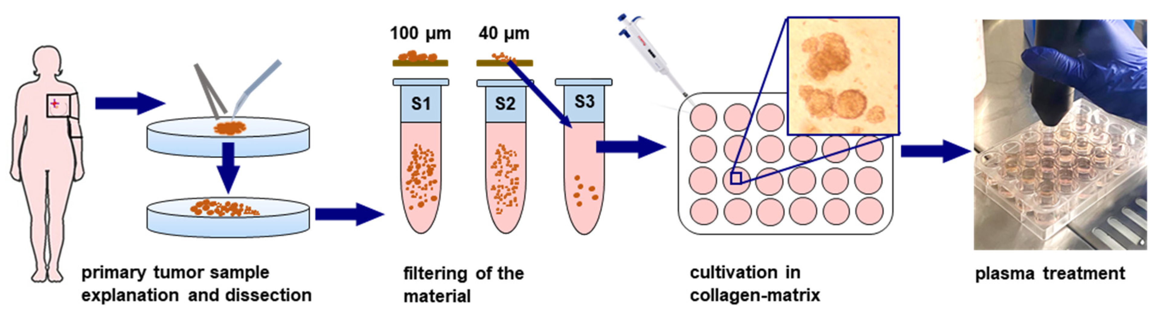
2.1. Breast Cancer Patient Tumor Material and Preparation

The local ethics committee approved the study (approval number IR.SBMU.RETECh.REC.1398.072). Twenty breast cancer patients at the Motamed Cancer Institute agreed to participate in the study with informed consent and according to the guidelines of the Declaration of Helsinki. The studied patients had no treatment of any kind (e.g., chemotherapy, hormone therapy, or radiotherapy) before surgery. The breast cancers were of stage I-II, and since the tumors have not been exposed to any form of therapeutic intervention, the cancer tissue was unlikely to have developed any resistance against standard therapies. All patients were female. The tumor samples were transferred to glass Petri dishes containing RPMI culture medium supplemented with 10% fetal bovine serum in a laminar flow hood under sterile conditions. Then, the tumor sample was cut into tiny pieces by a scalpel. During this process, it was ensured that the culture medium on the tissue was sufficient for proper nutrition. After the tissue was cut into proper sizes, 500 μL of type IV collagenases was poured onto the samples, followed by incubation at 37 °C for up to 30 min until the tissue lost its rigidity. The samples were filtered using a 100 µm cell strainer, and the eluent was filtered using a 40 µm cell strainer. The eluent containing single breast cancer cells and debris was discarded while the material on the filter, being 40–100 µm in size, was transferred to microtubes and washed. Subsequently, the tissue was mixed with type I collagen at a concentration of 2.8% and placed in an incubator for 30 min to form a gel. Fully supplemented cell culture medium was then added, and cultured tissues were placed in the incubator for 24 h and prior to plasma exposure.

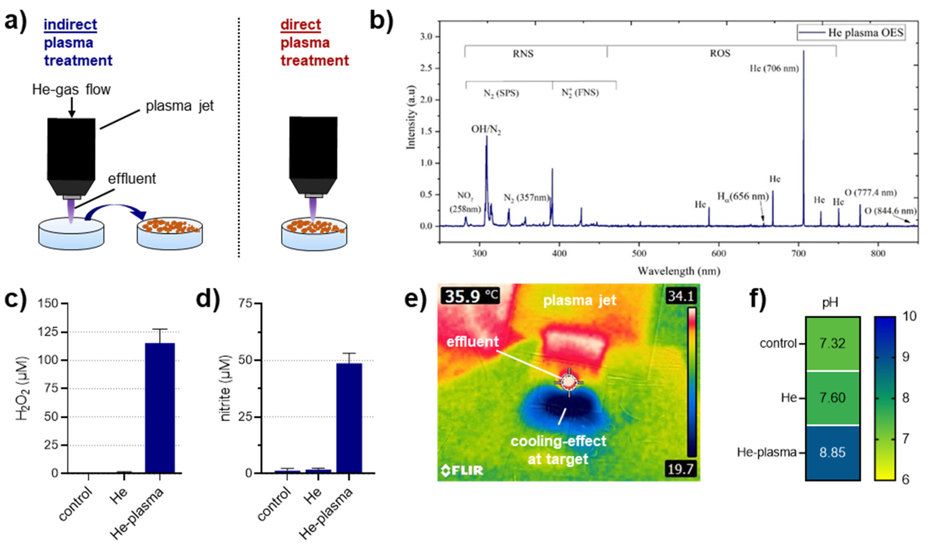
2.2. Plasma Source, Characterization, and Treatment

A sinusoidal power supply was used to generate the cold plasma. Helium with five standard liters per minute was used as feed gas supplemented with ten standard cubic centimeters per minute of molecular oxygen as carrier gases. The plasma jet was operated at 8 kV peak-to-peak voltage and 6 kHz frequency, resulting in a total dissipated power of 1 W. The distance between the plasma jet and the treated target was fixed to 10 mm. Optical emission spectroscopy (AvaSpec-ULS3648-USB2 with diffraction grating of 300 lines per mm, ten μm gap, and a wavelength range of 200 to 1100 nm) was employed to analyze the plasma gas. In plasma-treated liquids, hydrogen peroxide (H2O2) and nitrite (NO2) were measured using spectrophotometry as described before [38]. The pH of plasma-treated cell culture media was measured using a pH meter. The temperature of plasma-treated cell culture media was measured using a thermal infrared camera (FLIR E4 camera 80 by 60 pixels). For the treatment of collagen tissues, either direct or indirect plasma exposure was performed. In the direct treatment, the tumors embedded in collagen were directly exposed to physical plasma for 4 min while, in the indirect treatment, 1 mL of fully supplemented culture medium was exposed to plasma for 4 min and then transferred to collagen-tumor tissues. The control group remained untreated without any intervention and therapy. The plasma treatment was repeated 24 h later, and the collagen tissues were incubated for at least another 24 h after that. The plasma treatment time was identified to be suitable based on results obtained in pilot experiments.

2.3. Live Dead Assay

Twenty-four hours after the second plasma treatment, the culture medium was removed, and acridine orange/propidium iodide (AO/PI) staining solution was added and incubated for 20 min at 37 °C with the samples. AO enters all cells and stains their DNA, with a fluorescence emission at 520 nm. PI only enters dead cells and stains their DNA, with a fluorescence emission at 600 nm. Fluorescence quantification was conducted using MATLAB.

2.4. Immunofluorescence

Twenty-four hours after the second plasma treatment, the tissues were fixed with 4% paraformaldehyde and permeabilized with 0.1% Triton X-100 in PBS. Then, the samples were incubated with 3% BSA for 30 min to block nonspecific binding. Caspase-3 and E-cadherin antibodies were added to the samples and incubated at 4 °C overnight. The next day, the primary antibodies were washed away, and the samples were incubated with FITC-conjugated secondary antibodies for 2 h at room temperature in the dark. Finally, DAPI was added to counterstain the nuclei of the cells, and the images were recorded using fluorescence microscopy. The results were quantified using MATLAB.

2.5. Multiplex Supernatant Analysis

Supernatants from untreated and directly plasma-treated samples were collected and stored at −20 °C until analysis. Multiplex chemokine and cytokine analysis were performed as previously described [39]. Briefly, the supernatants were incubated with capture beads (LEGENDplex; BioLegend, Amsterdam, The Netherlands) according to the manufacturer’s instructions and measured using flow cytometry (CytoFLEX S; Beckman-Coulter, Krefeld, Germany). For quantification, data analysis LEGENDplex software (BioLegend, Amsterdam, The Netherlands) was used. For each analyte, separate standard curves were calculated using fifth-degree polynomials, paying attention to each analyte’s specific detection limits.

2.6. Statistical Analysis
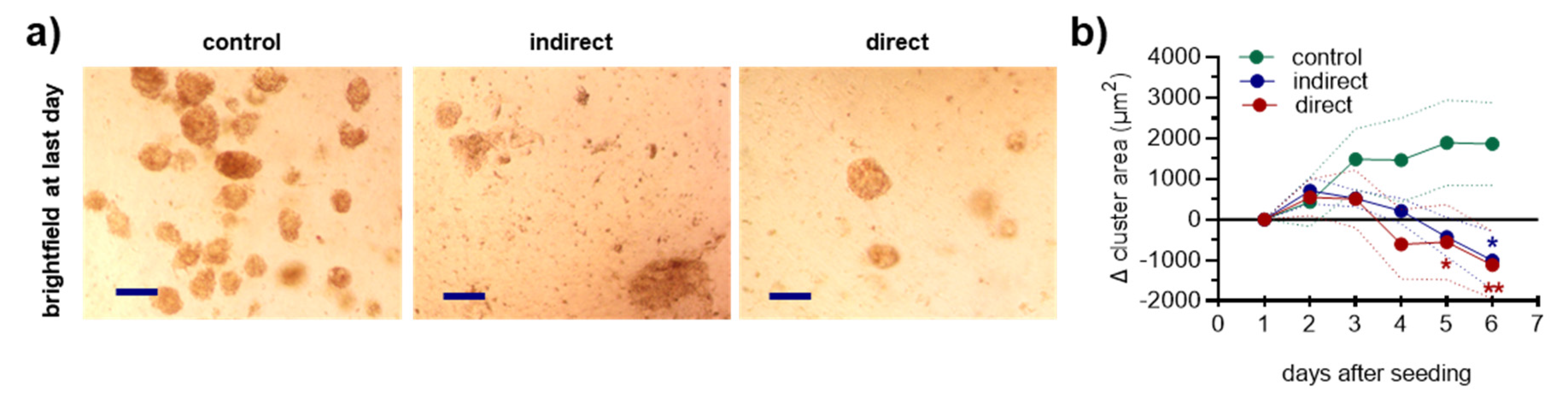
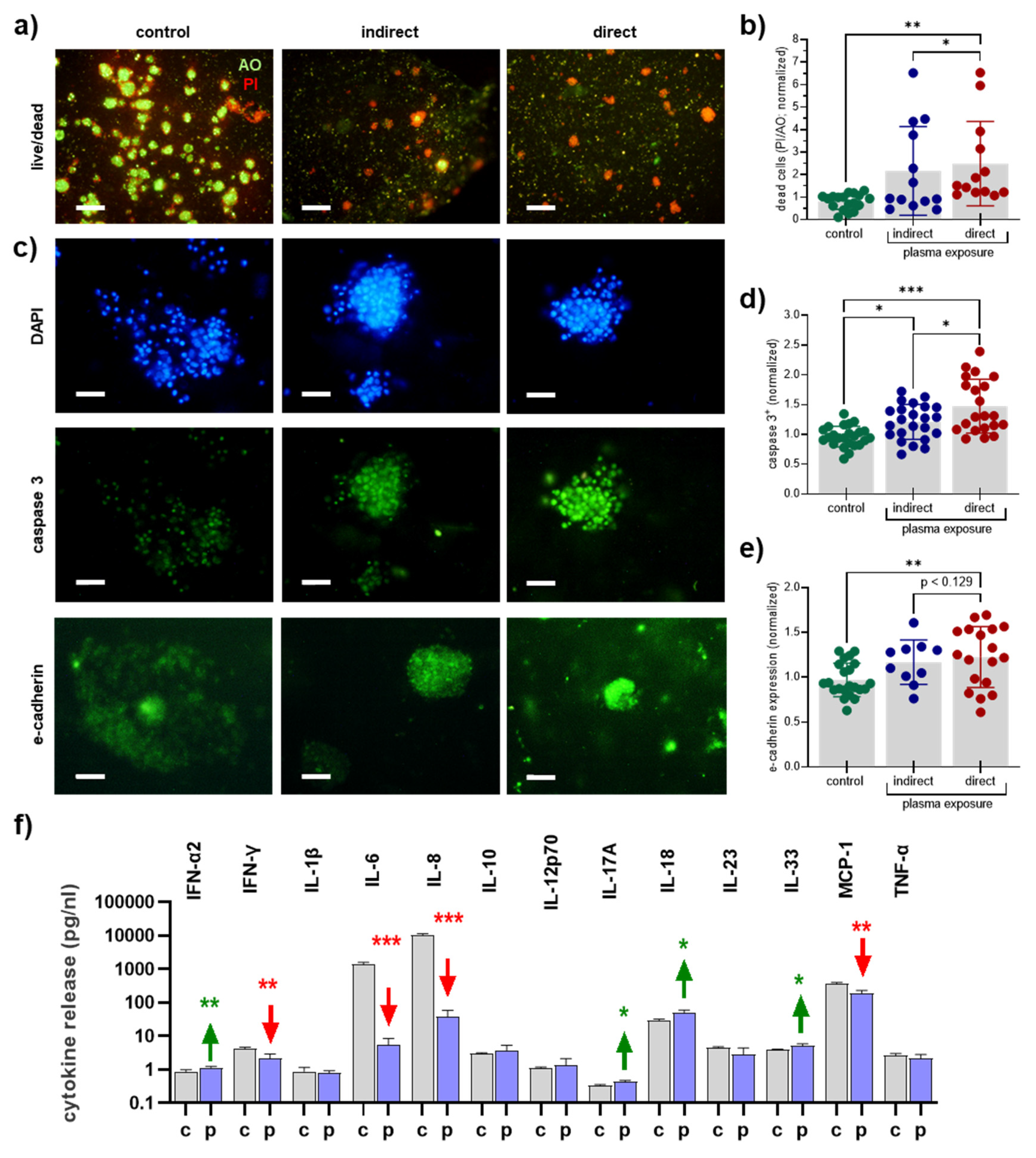
Graphing and statistical analysis (t-test, one-way ANOVA, or two-way ANOVA) were conducted using prism 9.1 (GraphPad Software, San Diego, CA, USA).

3. Results

In this study, direct and indirect plasma treatment was conducted (Figure 1a). The helium plasma jet emitted several reactive species, including ozone, atomic oxygen, hydroxyl radicals, and species of the second positive system of nitrogen (Figure 1b). In liquids, these species deteriorate to more stable oxidants such as hydrogen peroxide (H2O2; Figure 1c) and nitrite (NO2; Figure 1d) found in plasma-treated liquid but not helium gas-treated or untreated liquids. The plasma jet was engineered to be operated at about body temperature, leading to a surface heating of about 37 °C (Figure 1e). Exposure of fully supplemented cell culture medium led to a degassing effect of carbon dioxide and subsequent increase of the pH (Figure 1f), which quickly normalized after incubation in the humidified and CO2-containing incubator.
Human patient-derived breast cancer tissues were received after informed consent and processed in the laboratory (Figure 2). After dissection of the material, it was filtered to remove material larger than 100 µm followed by filtering through a 40 µm filter. The eluent of the later step including single cells and debris was discarded, and tumor microcolonies between 40–100 µm in size were mixed with collagen and added to multiwell dishes. The collagen-tumor matrix was plasma-treated directly or indirectly (Figure 3a), followed by allowing the colonies to grow through the course of incubation over several days. During this time, the colonies extended their volume due to the proliferation of individual cancer cell clones within the colonies. Non-malignant cells such as fibroblasts and immune cells of the TME do not proliferate because contact inhibition provided by the matrix inhibits their growth. Colony quantification revealed a significant decline of tumor growth in the collagen matrix for both direct and indirect plasma treatment (Figure 3b).
Exposure of the microtissue to helium gas alone did not give overall different results from untreated tissue in pilot experiments and hence was not included in the study. At 24 h after plasma treatment, the collagen-tumor-matrices were stained with the live/dead fluorescence dyes AO/PI (Figure 4a). Quantitative image analysis demonstrated a significantly increased presence of dead cells for both direct and indirect plasma treatment regimens (Figure 4b), showing significantly enhanced effects of the former over the latter. Next, the microtissues were fixed, and ultrathin sections were conducted. Staining was performed against active caspase or E-cadherin together with DAPI as nuclear counterstain (Figure 4c). The direct and indirect plasma treatment significantly increased the number of caspase 3-positive cells indicative of apoptosis (Figure 4d). Again, the direct treatment showed a more substantial effect compared to the indirect treatment. For the cell surface molecule E-cadherin, which is increasingly expressed in low-metastatic breast cancer tissues [40], a significantly increased presence was found for direct and indirect plasma treatment. These data suggested the indirect and especially the direct plasma treatment procedure to reduce tumor colony formation, increase apoptosis and terminal cell death, and elevated the expression of a marker associated with a more epithelial phenotype. Chemokine and cytokine release is another critical effector in the tumor microenvironment (TME). To better understand the effects of direct plasma treatment on the inflammatory profile of our collagen breast cancer microtissues, supernatants were collected 24 h after plasma treatment, and 13 different cytokines and chemokines were quantitatively assessed in these samples. Significantly increased levels were found for interferon (IFN)-α2, interleukin (IL)-17A, IL18, and IL33. Despite the significant increases, the absolute changes were of these targets were modest, however. For IFN-γ, IL-6, IL-6, and MCP-1, significantly decreased levels were determined, which was especially pronounced for IL-6 and IL-8. These data suggested that direct plasma treatment reduced cell viability and the expression of several cytokines and chemokines known to modulate the inflammatory profile of the TME critically.

4. Discussion

Gas plasma treatment is a promising and novel avenue to target cancer cells, including breast tumors. Albeit an array of in vitro and in vivo studies, data on primary tumor material from patients is scarce, hampering the translational value of this treatment and technology in oncology. To this end, we here investigated 20 clinical breast cancer samples using matrix-microtissues along with the functional consequences of gas plasma exposure in this tumor model of primary patient material.
We found an increase of apoptotic cells in our collagen-microtissue-matrices after plasma treatment accompanied by an increase of the adhesion marker E-cadherin associated with a less migratory activity of tumor cells [41]. Apoptosis and cell migration are two known effects of cold plasma in cancer therapy accompanied by consequences such as growth retardation and metastasis [42]. Tumor invasion and metastasis is the leading cause of death in cancer patients, including breast cancer [43]. The metastasis process is controlled and regulated by microenvironmental and systemic processes, including intercellular adhesion molecules. Weakening cell–cell and cell–matrix connections are essential for metastasis. Numerous families of adhesion molecules, including cadherins, integrins, adhesion molecules of the immunoglobulin family, selectins, and CD44 are identified [44]. The latter has been recently suggested to be affected by plasma treatment as well [45]. According to this hypothesis, where the lack or decreased expression of these molecules increases the rate of metastasis, these molecules can be used as markers of metastasis. E-cadherin is a transmembrane glycoprotein mediating calcium-dependent intracellular adhesion and is involved in epithelial cell–cell adhesion [46]. We had previously investigated metastasis on various pancreatic cancer cell lines by examining E-cadherin, EpCam, integrin α-2, integrin α-4, and ZEB-1, and concluded that the plasma treatment reduced metastasis in cancer cells [47], which is consistent with the results of cadherin expression in this study.
Initiation of apoptosis is a frequently described process in plasma-treated cancer cells. In vitro, it was found for several cancer types, such as colon [48,49,50], pancreas [51,52,53], head and neck [54,55,56], leukemia [57,58,59], glioblastoma [60,61,62], and melanoma [63,64,65]. For stage IV malignant melanoma patient-derived samples, we also have previously found augmented apoptosis within tissues after gas plasma exposure with the kINPen jet [66]. It is understood that this is a consequence of supraphysiological amounts of ROS/RNS delivered by the plasma system [67]. Nevertheless, there is a sensitivity gradient, as we have previously reported across 35 different tumor cell lines that have individual capacities to cope with the ROS/RNS [68]. The tumor-toxic activity of some of the long-lived species is retained in cell culture medium and other types of liquids, which can be subsequently used for cancer therapy [69]. Our present findings in primary human breast cancer tissue underline this notion, albeit the plasma-treated medium—called “indirect” in this work—was slightly less active than the direct gas plasma exposure. With plasma-treated cell culture medium, it should also be kept in mind that its utilization in preclinical cancer research is of limited translational value, as these solutions have not been accredited for use in humans as certified medical products [70].
In this study, 3D tissue culture technology has been used inside collagen to monitor plasma function on the tumoral tissue growing outside the body. Gel-like substances such as agarose have been used in other studies before the monitor the plasma-mediated ROS/RNS diffusion through different matrices [71,72,73]. Collagen has the added value that it is part of the in vivo TME of the tumors, making our microtissue model more realistic. A critical part of the TME is the release of various chemokines and cytokines, often rendering the micromilieu more tumorigenic. For instance, IL-6 has been reported to participate in a positive feedback loop in the breast cancer TME to fuel tumor growth by promoting immunosuppression [74]. IL-6 has also been found to increase the expression of the hyaluronic acid receptor CD44 that promotes breast cancer stem cell metastasis [75]. Along similar lines, the chemokine IL-8 (CXCL8) is linked with enhanced breast cancer stem cell activity and chemoresistance [76] and promotes the growth of patient-derived breast cancer growth in ex vivo models [77]. In our study, gas plasma exposure reduced levels of both IL-6 and IL-8 by a factor of 100, exemplifying the potency of this technology. Interestingly, we have recently identified a role of the CXCL8/HO-1 axis as critical mediators of plasma-induced cell death in leukemia cells [78]. Moreover, the chemokine MCP1 (CCL2) that was decreased with plasma treatment by a factor of two has been implemented in shaping a pro-tumorigenic TME as well [79]. More significantly changed soluble mediators were identified for the plasma treatment condition, but these were less pronounced in terms of absolute changes.
Nevertheless, gas plasma exposure was promising in elevating tumor cell death and E-cadherin expression and modulating inflammatory mediators in a favorable manner. Plasma technology is a surface treatment technology, and patients suffering from ulcerating breast cancers that are often infected and different to manage might benefit from repeated plasma exposure. The group of Philipp Wong has recently been able to demonstrate additive toxicity of plasma treatment with radiotherapy [16], which we could confirm for melanoma cells [80]. Electrochemotherapy (ECT) is another reported therapy for ulcerating breast cancer [81], and first reports have reported combined toxicity with plasma and ECT in vitro [82,83]. There is ample evidence that plasma treatment also combines with chemotherapy to promote cytotoxicity [84,85,86,87,88,89]. These considerations are both promising and guiding in advancing the therapeutic concept of adjuvant gas plasma therapy in breast cancer management.

5. Conclusions

Gas plasma exposure augmented cell death in three-dimensional microtissue-matrices of 20 patient-derived breast cancer tissues. These findings provide a translational value that complements several earlier and promising in vitro and in vivo studies on the tumor-toxic effects found in plasma-treated breast cancer cells. Despite the promising nature of the results, large-scale studies are needed to confirm the findings. Subsequent studies should use patient-derived cancer tissue models and clinically useful combination regimens to promote gas plasma-based cancer therapies in the future.

Author Contributions

Conceptualization, F.S., H.Z. and S.B.; methodology, Z.A., F.S., E.F., F.A. and S.B.; software, F.S., H.M., E.F. and S.B.; validation, Z.A. and F.S.; formal analysis, F.S.; investigation, Z.A. and F.S.; resources, F.S., B.S., H.Z. and S.B.; data curation, F.S.; writing—original draft preparation, F.S. and S.B.; writing—review and editing, S.B.; visualization, F.S., E.F. and S.B.; supervision, H.Z. and S.B.; project administration, H.Z. and S.B.; funding acquisition, H.Z. and S.B. All authors have read and agreed to the published version of the manuscript.

Funding

This study was supported by the German Federal Ministry of Education and Research (BMBF), grant numbers 03Z22DN11 and 03Z22Di1.

Institutional Review Board Statement

The study was conducted according to the guidelines of the Declaration of Helsinki, and approved by the Institutional Ethics Committee of Motamed Cancer Research Institute (protocol code IR.SBMU.RETECh.REC.1398.072 in April 2020).

Informed Consent Statement

Informed consent was obtained from all subjects involved in the study.

Data Availability Statement

Data are available from the corresponding author upon reasonable request.

Acknowledgments

Technical support by Felix Niessner is gratefully acknowledged.

Conflicts of Interest

The authors declare no conflict of interest.

References

  1. Curado, M.P. Breast cancer in the world: Incidence and mortality. Salud Publica Mex. 2011, 53, 372–384. [Google Scholar]
  2. Shulman, L.N.; Willett, W.; Sievers, A.; Knaul, F.M. Breast cancer in developing countries: Opportunities for improved survival. J. Oncol. 2010, 2010, 595167. [Google Scholar] [CrossRef]
  3. Ferlay, J.; Soerjomataram, I.; Dikshit, R.; Eser, S.; Mathers, C.; Rebelo, M.; Parkin, D.M.; Forman, D.; Bray, F. Cancer incidence and mortality worldwide: Sources, methods and major patterns in globocan 2012. Int. J. Cancer 2015, 136, E359–E386. [Google Scholar] [CrossRef]
  4. Cardoso, F.; Harbeck, N.; Barrios, C.; Bergh, J.; Cortés, J.; El Saghir, N.; Francis, P.; Hudis, C.; Ohno, S.; Partridge, A. Research needs in breast cancer. Ann. Oncol. 2017, 28, 208–217. [Google Scholar] [CrossRef]
  5. Penney, R.B.; Roy, D. Thioredoxin-mediated redox regulation of resistance to endocrine therapy in breast cancer. Biochim. Biophys. Acta 2013, 1836, 60–79. [Google Scholar] [CrossRef] [PubMed]
  6. Semenza, G.L. Hypoxia-inducible factors: Coupling glucose metabolism and redox regulation with induction of the breast cancer stem cell phenotype. EMBO J. 2017, 36, 252–259. [Google Scholar] [CrossRef]
  7. Blazquez-Castro, A. Direct (1)o2 optical excitation: A tool for redox biology. Redox Biol. 2017, 13, 39–59. [Google Scholar] [CrossRef]
  8. Glass, S.B.; Gonzalez-Fajardo, L.; Beringhs, A.O.; Lu, X. Redox potential and ros-mediated nanomedicines for improving cancer therapy. Antioxid. Redox Signal. 2019, 30, 747–761. [Google Scholar] [CrossRef]
  9. Liu, T.I.; Lu, T.Y.; Yang, Y.C.; Chang, S.H.; Chen, H.H.; Lu, I.L.; Sabu, A.; Chiu, H.C. New combination treatment from ros-induced sensitized radiotherapy with nanophototherapeutics to fully eradicate orthotopic breast cancer and inhibit metastasis. Biomaterials 2020, 257, 120229. [Google Scholar] [CrossRef]
  10. Privat-Maldonado, A.; Schmidt, A.; Lin, A.; Weltmann, K.D.; Wende, K.; Bogaerts, A.; Bekeschus, S. Ros from physical plasmas: Redox chemistry for biomedical therapy. Oxid. Med. Cell. Longev. 2019, 2019, 9062098. [Google Scholar] [CrossRef] [PubMed] [Green Version]
  11. Laroussi, M.; Lu, X.; Keidar, M. Perspective: The physics, diagnostics, and applications of atmospheric pressure low temperature plasma sources used in plasma medicine. J. Appl. Phys. 2017, 122, 020901. [Google Scholar] [CrossRef]
  12. Adil, B.H.; Al-Shammari, A.M.; Murbat, H.H. Breast cancer treatment using cold atmospheric plasma generated by the fe-dbd scheme. Clin. Plasma Med. 2020, 19–20. [Google Scholar] [CrossRef]
  13. Boisvert, J.-S.; Lafontaine, J.; Glory, A.; Coulombe, S.; Wong, P. Comparison of three radio-frequency discharge modes on the treatment of breast cancer cells in vitro. IEEE Trans. Radiat. Plasma Med. Sci. 2020, 644–654. [Google Scholar] [CrossRef]
  14. Jalili, A.; Irani, S.; Mirfakhraie, R. Combination of cold atmospheric plasma and iron nanoparticles in breast cancer: Gene expression and apoptosis study. Onco Targets Ther. 2016, 9, 5911–5917. [Google Scholar] [CrossRef]
  15. Jezeh, M.A.; Tayebi, T.; Khani, M.R.; Niknejad, H.; Shokri, B. Direct cold atmospheric plasma and plasma-activated medium effects on breast and cervix cancer cells. Plasma Process. Polym. 2020. [Google Scholar] [CrossRef]
  16. Lafontaine, J.; Boisvert, J.S.; Glory, A.; Coulombe, S.; Wong, P. Synergy between non-thermal plasma with radiation therapy and olaparib in a panel of breast cancer cell lines. Cancers 2020, 12, 348. [Google Scholar] [CrossRef] [Green Version]
  17. Lee, S.; Lee, H.; Jeong, D.; Ham, J.; Park, S.; Choi, E.H.; Kim, S.J. Cold atmospheric plasma restores tamoxifen sensitivity in resistant mcf-7 breast cancer cell. Free Radic. Biol. Med. 2017, 110, 280–290. [Google Scholar] [CrossRef] [PubMed]
  18. Liu, Y.; Tan, S.; Zhang, H.; Kong, X.; Ding, L.; Shen, J.; Lan, Y.; Cheng, C.; Zhu, T.; Xia, W. Selective effects of non-thermal atmospheric plasma on triple-negative breast normal and carcinoma cells through different cell signaling pathways. Sci. Rep. 2017, 7, 7980. [Google Scholar] [CrossRef] [PubMed]
  19. Park, S.; Kim, H.; Ji, H.W.; Kim, H.W.; Yun, S.H.; Choi, E.H.; Kim, S.J. Cold atmospheric plasma restores paclitaxel sensitivity to paclitaxel-resistant breast cancer cells by reversing expression of resistance-related genes. Cancers 2019, 11, 2011. [Google Scholar] [CrossRef] [Green Version]
  20. Wang, M.; Holmes, B.; Cheng, X.; Zhu, W.; Keidar, M.; Zhang, L.G. Cold atmospheric plasma for selectively ablating metastatic breast cancer cells. PLoS ONE 2013, 8, e73741. [Google Scholar] [CrossRef]
  21. Zhu, W.; Lee, S.J.; Castro, N.J.; Yan, D.; Keidar, M.; Zhang, L.G. Synergistic effect of cold atmospheric plasma and drug loaded core-shell nanoparticles on inhibiting breast cancer cell growth. Sci. Rep. 2016, 6, 21974. [Google Scholar] [CrossRef] [PubMed] [Green Version]
  22. Chauvin, J.; Gibot, L.; Griseti, E.; Golzio, M.; Rols, M.P.; Merbahi, N.; Vicendo, P. Elucidation of in vitro cellular steps induced by antitumor treatment with plasma-activated medium. Sci. Rep. 2019, 9, 4866. [Google Scholar] [CrossRef] [PubMed]
  23. Freund, E.; Liedtke, K.R.; van der Linde, J.; Metelmann, H.R.; Heidecke, C.D.; Partecke, L.I.; Bekeschus, S. Physical plasma-treated saline promotes an immunogenic phenotype in ct26 colon cancer cells in vitro and in vivo. Sci. Rep. 2019, 9, 634. [Google Scholar] [CrossRef]
  24. Miebach, L.; Freund, E.; Horn, S.; Niessner, F.; Sagwal, S.K.; von Woedtke, T.; Emmert, S.; Weltmann, K.D.; Clemen, R.; Schmidt, A.; et al. Tumor cytotoxicity and immunogenicity of a novel v-jet neon plasma source compared to the kinpen. Sci. Rep. 2021, 11, 136. [Google Scholar] [CrossRef] [PubMed]
  25. Plewa, J.M.; Yousfi, M.; Frongia, C.; Eichwald, O.; Ducommun, B.; Merbahi, N.; Lobjois, V. Low-temperature plasma-induced antiproliferative effects on multi-cellular tumor spheroids. New J. Phys. 2014, 16, 043027. [Google Scholar] [CrossRef]
  26. Hasse, S.; Meder, T.; Freund, E.; von Woedtke, T.; Bekeschus, S. Plasma treatment limits human melanoma spheroid growth and metastasis independent of the ambient gas composition. Cancers 2020, 12, 2570. [Google Scholar] [CrossRef]
  27. Freund, E.; Spadola, C.; Schmidt, A.; Privat-Maldonado, A.; Bogaerts, A.; von Woedtke, T.; Weltmann, K.D.; Heidecke, C.D.; Partecke, L.I.; Kading, A.; et al. Risk evaluation of emt and inflammation in metastatic pancreatic cancer cells following plasma treatment. Front. Phys. 2020, 8. [Google Scholar] [CrossRef]
  28. Mahdikia, H.; Saadati, F.; Freund, E.; Gaipl, U.S.; Majidzadeh, A.K.; Shokri, B.; Bekeschus, S. Gas plasma irradiation of breast cancers promotes immunogenicity, tumor reduction, and an abscopal effect in vivo. Oncoimmunology 2021, 10, 1859731. [Google Scholar] [CrossRef]
  29. Mizuno, K.; Shirakawa, Y.; Sakamoto, T.; Ishizaki, H.; Nishijima, Y.; Ono, R. Plasma-induced suppression of recurrent and reinoculated melanoma tumors in mice. IEEE TRPMS 2018, 2, 353–359. [Google Scholar] [CrossRef]
  30. Bekeschus, S.; Clemen, R.; Niessner, F.; Sagwal, S.K.; Freund, E.; Schmidt, A. Medical gas plasma jet technology targets murine melanoma in an immunogenic fashion. Adv. Sci. 2020, 7, 1903438. [Google Scholar] [CrossRef] [Green Version]
  31. Antoni, D.; Burckel, H.; Josset, E.; Noel, G. Three-dimensional cell culture: A breakthrough in vivo. Int. J. Mol. Sci. 2015, 16, 5517–5527. [Google Scholar] [CrossRef]
  32. Kapałczyńska, M.; Kolenda, T.; Przybyła, W.; Zajączkowska, M.; Teresiak, A.; Filas, V.; Ibbs, M.; Bliźniak, R.; Łuczewski, Ł.; Lamperska, K. 2d and 3d cell cultures–a comparison of different types of cancer cell cultures. Arch. Med. Sci. AMS 2018, 14, 910. [Google Scholar] [CrossRef]
  33. Edmondson, R.; Broglie, J.J.; Adcock, A.F.; Yang, L. Three-dimensional cell culture systems and their applications in drug discovery and cell-based biosensors. Assay Drug Dev. Technol. 2014, 12, 207–218. [Google Scholar] [CrossRef] [PubMed] [Green Version]
  34. Koo, H.; Yamada, K.M. Dynamic cell–matrix interactions modulate microbial biofilm and tissue 3d microenvironments. Curr. Opin. Cell Biol. 2016, 42, 102–112. [Google Scholar] [CrossRef] [Green Version]
  35. Cavo, M.; Fato, M.; Peñuela, L.; Beltrame, F.; Raiteri, R.; Scaglione, S. Microenvironment complexity and matrix stiffness regulate breast cancer cell activity in a 3d in vitro model. Sci. Rep. 2016, 6, 1–13. [Google Scholar] [CrossRef] [Green Version]
  36. Nicolas, J.; Magli, S.; Rabbachin, L.; Sampaolesi, S.; Nicotra, F.; Russo, L. 3d extracellular matrix mimics: Fundamental concepts and role of materials chemistry to influence stem cell fate. Biomacromolecules 2020, 21, 1968–1994. [Google Scholar] [CrossRef]
  37. Aref, A.R.; Campisi, M.; Ivanova, E.; Portell, A.; Larios, D.; Piel, B.P.; Mathur, N.; Zhou, C.; Coakley, R.V.; Bartels, A. 3d microfluidic ex vivo culture of organotypic tumor spheroids to model immune checkpoint blockade. Lab Chip 2018, 18, 3129–3143. [Google Scholar] [CrossRef] [Green Version]
  38. Girard, P.M.; Arbabian, A.; Fleury, M.; Bauville, G.; Puech, V.; Dutreix, M.; Sousa, J.S. Synergistic effect of h2o2 and no2 in cell death induced by cold atmospheric he plasma. Sci. Rep. 2016, 6, 29098. [Google Scholar] [CrossRef] [Green Version]
  39. Freund, E.; Miebach, L.; Clemen, R.; Schmidt, S.; Heidecke, A.; von Woedtke, T.; Weltmann, K.; Kersting, S.; Bekeschus, S. Large volume spark discharge and plasma jet-technology for generating plasma-oxidized saline targeting colon cancer in vitro and in vivo. J. Phys. D Appl. Phys. 2021, 129. [Google Scholar] [CrossRef]
  40. Aigner, K.; Dampier, B.; Descovich, L.; Mikula, M.; Sultan, A.; Schreiber, M.; Mikulits, W.; Brabletz, T.; Strand, D.; Obrist, P.; et al. The transcription factor zeb1 (deltaef1) promotes tumour cell dedifferentiation by repressing master regulators of epithelial polarity. Oncogene 2007, 26, 6979–6988. [Google Scholar] [CrossRef] [PubMed] [Green Version]
  41. Kowalski, P.J.; Rubin, M.A.; Kleer, C.G. E-cadherin expression in primary carcinomas of the breast and its distant metastases. Breast Cancer Res. 2003, 5, R217–R222. [Google Scholar] [CrossRef] [Green Version]
  42. Keidar, M.; Shashurin, A.; Volotskova, O.; Stepp, M.A.; Srinivasan, P.; Sandler, A.; Trink, B. Cold atmospheric plasma in cancer therapy. Phys. Plasmas 2013, 20, 057101. [Google Scholar] [CrossRef]
  43. Riggio, A.I.; Varley, K.E.; Welm, A.L. The lingering mysteries of metastatic recurrence in breast cancer. Br. J. Cancer 2021, 124, 13–26. [Google Scholar] [CrossRef]
  44. Edelman, G.M. Cell adhesion molecules. Science 1983, 219, 450–457. [Google Scholar] [CrossRef]
  45. Yusupov, M.; Privat-Maldonado, A.; Cordeiro, R.M.; Verswyvel, H.; Shaw, P.; Razzokov, J.; Smits, E.; Bogaerts, A. Oxidative damage to hyaluronan–cd44 interactions as an underlying mechanism of action of oxidative stress-inducing cancer therapy. Redox Biol. 2021, 43, 101968. [Google Scholar] [CrossRef] [PubMed]
  46. Canel, M.; Serrels, A.; Frame, M.C.; Brunton, V.G. E-cadherin–integrin crosstalk in cancer invasion and metastasis. J. Cell Sci. 2013, 126, 393–401. [Google Scholar] [CrossRef] [Green Version]
  47. Bekeschus, S.; Freund, E.; Spadola, C.; Privat-Maldonado, A.; Hackbarth, C.; Bogaerts, A.; Schmidt, A.; Wende, K.; Weltmann, K.D.; von Woedtke, T.; et al. Risk assessment of kinpen plasma treatment of four human pancreatic cancer cell lines with respect to metastasis. Cancers 2019, 11, 1237. [Google Scholar] [CrossRef] [Green Version]
  48. Choi, J.S.; Kim, J.; Hong, Y.J.; Bae, W.Y.; Choi, E.H.; Jeong, J.W.; Park, H.K. Evaluation of non-thermal plasma-induced anticancer effects on human colon cancer cells. Biomed. Opt. Express 2017, 8, 2649–2659. [Google Scholar] [CrossRef] [Green Version]
  49. Han, D.; Cho, J.H.; Lee, R.H.; Bang, W.; Park, K.; Kim, M.S.; Shim, J.H.; Chae, J.I.; Moon, S.Y. Antitumorigenic effect of atmospheric-pressure dielectric barrier discharge on human colorectal cancer cells via regulation of sp1 transcription factor. Sci. Rep. 2017, 7, 43081. [Google Scholar] [CrossRef] [PubMed] [Green Version]
  50. Ruwan Kumara, M.H.; Piao, M.J.; Kang, K.A.; Ryu, Y.S.; Park, J.E.; Shilnikova, K.; Jo, J.O.; Mok, Y.S.; Shin, J.H.; Park, Y.; et al. Non-thermal gas plasma-induced endoplasmic reticulum stress mediates apoptosis in human colon cancer cells. Oncol. Rep. 2016, 36, 2268–2274. [Google Scholar] [CrossRef] [Green Version]
  51. Hattori, N.; Yamada, S.; Torii, K.; Takeda, S.; Nakamura, K.; Tanaka, H.; Kajiyama, H.; Kanda, M.; Fujii, T.; Nakayama, G.; et al. Effectiveness of plasma treatment on pancreatic cancer cells. Int. J. Oncol. 2015, 47, 1655–1662. [Google Scholar] [CrossRef] [Green Version]
  52. Sato, Y.; Yamada, S.; Takeda, S.; Hattori, N.; Nakamura, K.; Tanaka, H.; Mizuno, M.; Hori, M.; Kodera, Y. Effect of plasma-activated lactated ringer’s solution on pancreatic cancer cells in vitro and in vivo. Ann. Surg. Oncol. 2018, 25, 299–307. [Google Scholar] [CrossRef] [PubMed]
  53. Zhen, X.; Sun, H.N.; Liu, R.; Choi, H.S.; Lee, D.S. Non-thermal plasma-activated medium induces apoptosis of aspc1 cells through the ros-dependent autophagy pathway. In Vivo 2020, 34, 143–153. [Google Scholar] [CrossRef] [PubMed]
  54. Welz, C.; Emmert, S.; Canis, M.; Becker, S.; Baumeister, P.; Shimizu, T.; Morfill, G.E.; Harreus, U.; Zimmermann, J.L. Cold atmospheric plasma: A promising complementary therapy for squamous head and neck cancer. PLoS ONE 2015, 10, e0141827. [Google Scholar] [CrossRef] [Green Version]
  55. Chang, J.W.; Kang, S.U.; Shin, Y.S.; Kim, K.I.; Seo, S.J.; Yang, S.S.; Lee, J.S.; Moon, E.; Baek, S.J.; Lee, K.; et al. Non-thermal atmospheric pressure plasma induces apoptosis in oral cavity squamous cell carcinoma: Involvement of DNA-damage-triggering sub-g(1) arrest via the atm/p53 pathway. Arch. Biochem. Biophys. 2014, 545, 133–140. [Google Scholar] [CrossRef]
  56. Clemen, R.; Heirman, P.; Lin, A.; Bogaerts, A.; Bekeschus, S. Physical plasma-treated skin cancer cells amplify tumor cytotoxicity of human natural killer (nk) cells. Cancers 2020, 12, 3575. [Google Scholar] [CrossRef]
  57. Barekzi, N.; Laroussi, M. Effects of low temperature plasmas on cancer cells. Plasma Process. Polym. 2013, 10, 1039–1050. [Google Scholar] [CrossRef]
  58. Moniruzzaman, R.; Rehman, M.U.; Zhao, Q.L.; Jawaid, P.; Mitsuhashi, Y.; Imaue, S.; Fujiwara, K.; Ogawa, R.; Tomihara, K.; Saitoh, J.I.; et al. Roles of intracellular and extracellular ros formation in apoptosis induced by cold atmospheric helium plasma and x-irradiation in the presence of sulfasalazine. Free Radic. Biol. Med. 2018, 129, 537–547. [Google Scholar] [CrossRef]
  59. Schmidt, A.; Rodder, K.; Hasse, S.; Masur, K.; Toups, L.; Lillig, C.H.; von Woedtke, T.; Wende, K.; Bekeschus, S. Redox-regulation of activator protein 1 family members in blood cancer cell lines exposed to cold physical plasma-treated medium. Plasma Process. Polym. 2016, 13, 1179–1188. [Google Scholar] [CrossRef]
  60. Akter, M.; Jangra, A.; Choi, S.A.; Choi, E.H.; Han, I. Non-thermal atmospheric pressure bio-compatible plasma stimulates apoptosis via p38/mapk mechanism in u87 malignant glioblastoma. Cancers 2020, 12, 245. [Google Scholar] [CrossRef] [PubMed] [Green Version]
  61. Cheng, X.Q.; Sherman, J.; Murphy, W.; Ratovitski, E.; Canady, J.; Keidar, M. The effect of tuning cold plasma composition on glioblastoma cell viability. PLoS ONE 2014, 9, e98652. [Google Scholar] [CrossRef] [PubMed] [Green Version]
  62. Vermeylen, S.; De Waele, J.; Vanuytsel, S.; De Backer, J.; Van der Paal, J.; Ramakers, M.; Leyssens, K.; Marcq, E.; Van Audenaerde, J.; Smits, E.L.J.; et al. Cold atmospheric plasma treatment of melanoma and glioblastoma cancer cells. Plasma Process. Polym. 2016, 13, 1195–1205. [Google Scholar] [CrossRef]
  63. Adhikari, M.; Kaushik, N.; Ghimire, B.; Adhikari, B.; Baboota, S.; Al-Khedhairy, A.A.; Wahab, R.; Lee, S.J.; Kaushik, N.K.; Choi, E.H. Cold atmospheric plasma and silymarin nanoemulsion synergistically inhibits human melanoma tumorigenesis via targeting hgf/c-met downstream pathway. Cell Commun. Signal. 2019, 17, 52. [Google Scholar] [CrossRef] [Green Version]
  64. Ishaq, M.; Bazaka, K.; Ostrikov, K. Pro-apoptotic noxa is implicated in atmospheric-pressure plasma-induced melanoma cell death. J. Phys. D Appl. Phys. 2015, 48, 464002. [Google Scholar] [CrossRef]
  65. Zucker, S.N.; Zirnheld, J.; Bagati, A.; DiSanto, T.M.; Des Soye, B.; Wawrzyniak, J.A.; Etemadi, K.; Nikiforov, M.; Berezney, R. Preferential induction of apoptotic cell death in melanoma cells as compared with normal keratinocytes using a non-thermal plasma torch. Cancer Biol. Ther. 2012, 13, 1299–1306. [Google Scholar] [CrossRef]
  66. Bekeschus, S.; Moritz, J.; Helfrich, I.; Boeckmann, L.; Weltmann, K.D.; Emmert, S.; Metelmann, H.R.; Stoffels, I.; von Woedtke, T. Ex vivo exposure of human melanoma tissue to cold physical plasma elicits apoptosis and modulates inflammation. Appl. Sci. 2020, 10, 1971. [Google Scholar] [CrossRef] [Green Version]
  67. Graves, D.B. Mechanisms of plasma medicine: Coupling plasma physics, biochemistry, and biology. IEEE Trans. Radiat. Plasma Med. Sci. 2017, 1, 281–292. [Google Scholar] [CrossRef]
  68. Bekeschus, S.; Liebelt, G.; Menz, J.; Berner, J.; Sagwal, S.K.; Wende, K.; Weltmann, K.D.; Boeckmann, L.; von Woedtke, T.; Metelmann, H.R.; et al. Tumor cell metabolism correlates with resistance to gas plasma treatment: The evaluation of three dogmas. Free Radic. Biol. Med. 2021. [Google Scholar] [CrossRef] [PubMed]
  69. Tanaka, H.; Bekeschus, S.; Yan, D.; Hori, M.; Keidar, M.; Laroussi, M. Plasma-treated solutions (pts) in cancer therapy. Cancers 2021, 13, 1737. [Google Scholar] [CrossRef]
  70. Freund, E.; Bekeschus, S. Gas plasma-oxidized liquids for cancer treatment: Pre-clinical relevance, immuno-oncology, and clinical obstacles. IEEE Trans. Radiat. Plasma Med. Sci. 2020, 1. [Google Scholar] [CrossRef]
  71. Busco, G.; Omran, A.V.; Ridou, L.; Pouvesle, J.-M.; Robert, E.; Grillon, C. Cold atmospheric plasma-induced acidification of tissue surface: Visualization and quantification using agarose gel models. J. Phys. D Appl. Phys. 2019, 52, 24LT01. [Google Scholar] [CrossRef]
  72. Kawasaki, T.; Sato, A.; Kusumegi, S.; Kudo, A.; Sakanoshita, T.; Tsurumaru, T.; Uchida, G.; Koga, K.; Shiratani, M. Two-dimensional concentration distribution of reactive oxygen species transported through a tissue phantom by atmospheric-pressure plasma-jet irradiation. Appl. Phys. Express 2016, 9, 076202. [Google Scholar] [CrossRef]
  73. Oh, J.-S.; Szili, E.J.; Ito, S.; Hong, S.-H.; Gaur, N.; Furuta, H.; Short, R.D.; Hatta, A. Slow molecular transport of plasma-generated reactive oxygen and nitrogen species and o2 through agarose as a surrogate for tissue. Plasma Med. 2015, 5, 125–143. [Google Scholar] [CrossRef]
  74. Hu, G.; Cheng, P.; Pan, J.; Wang, S.; Ding, Q.; Jiang, Z.; Cheng, L.; Shao, X.; Huang, L.; Huang, J. An il6-adenosine positive feedback loop between cd73(+) gammadeltatregs and cafs promotes tumor progression in human breast cancer. Cancer Immunol. Res. 2020, 8, 1273–1286. [Google Scholar] [CrossRef]
  75. Al-Hajj, M.; Wicha, M.S.; Benito-Hernandez, A.; Morrison, S.J.; Clarke, M.F. Prospective identification of tumorigenic breast cancer cells. Proc. Natl. Acad. Sci. USA 2003, 100, 3983–3988. [Google Scholar] [CrossRef] [Green Version]
  76. Dominguez, C.; McCampbell, K.K.; David, J.M.; Palena, C. Neutralization of il-8 decreases tumor pmn-mdscs and reduces mesenchymalization of claudin-low triple-negative breast cancer. JCI Insight 2017, 2, e94296. [Google Scholar] [CrossRef]
  77. Singh, J.K.; Farnie, G.; Bundred, N.J.; Simoes, B.M.; Shergill, A.; Landberg, G.; Howell, S.J.; Clarke, R.B. Targeting cxcr1/2 significantly reduces breast cancer stem cell activity and increases the efficacy of inhibiting her2 via her2-dependent and -independent mechanisms. Clin. Cancer Res. 2013, 19, 643–656. [Google Scholar] [CrossRef] [Green Version]
  78. Bekeschus, S.; Clemen, R.; Haralambiev, L.; Niessner, F.; Grabarczyk, P.; Weltmann, K.-D.; Menz, J.; Stope, M.; von Woedtke, T.; Gandhirajan, R.; et al. The plasma-induced leukemia cell death is dictated by the ros chemistry and the ho-1/cxcl8 axis. IEEE Trans. Radiat. Plasma Med. Sci. 2021, 5, 398–411. [Google Scholar] [CrossRef]
  79. Tsuyada, A.; Chow, A.; Wu, J.; Somlo, G.; Chu, P.; Loera, S.; Luu, T.; Li, A.X.; Wu, X.; Ye, W.; et al. Ccl2 mediates cross-talk between cancer cells and stromal fibroblasts that regulates breast cancer stem cells. Cancer Res. 2012, 72, 2768–2779. [Google Scholar] [CrossRef] [Green Version]
  80. Pasqual-Melo, G.; Sagwal, S.K.; Freund, E.; Gandhirajan, R.K.; Frey, B.; von Woedtke, T.; Gaipl, U.; Bekeschus, S. Combination of gas plasma and radiotherapy has immunostimulatory potential and additive toxicity in murine melanoma cells in vitro. Int. J. Mol. Sci. 2020, 21, 1379. [Google Scholar] [CrossRef] [Green Version]
  81. Cabula, C.; Campana, L.G.; Grilz, G.; Galuppo, S.; Bussone, R.; De Meo, L.; Bonadies, A.; Curatolo, P.; De Laurentiis, M.; Renne, M.; et al. Electrochemotherapy in the treatment of cutaneous metastases from breast cancer: A multicenter cohort analysis. Ann. Surg. Oncol. 2015, 22 (Suppl. 3), S442–S450. [Google Scholar] [CrossRef] [PubMed]
  82. Chung, T.H.; Stancampiano, A.; Sklias, K.; Gazeli, K.; Andre, F.M.; Dozias, S.; Douat, C.; Pouvesle, J.M.; Santos Sousa, J.; Robert, E.; et al. Cell electropermeabilisation enhancement by non-thermal-plasma-treated pbs. Cancers 2020, 12, 219. [Google Scholar] [CrossRef] [Green Version]
  83. Wolff, C.M.; Kolb, J.F.; Weltmann, K.D.; von Woedtke, T.; Bekeschus, S. Combination treatment with cold physical plasma and pulsed electric fields augments ros production and cytotoxicity in lymphoma. Cancers 2020, 12, 845. [Google Scholar] [CrossRef] [PubMed] [Green Version]
  84. Alimohammadi, M.; Golpur, M.; Sohbatzadeh, F.; Hadavi, S.; Bekeschus, S.; Niaki, H.A.; Valadan, R.; Rafiei, A. Cold atmospheric plasma is a potent tool to improve chemotherapy in melanoma in vitro and in vivo. Biomolecules 2020, 10, 1011. [Google Scholar] [CrossRef] [PubMed]
  85. Brulle, L.; Vandamme, M.; Ries, D.; Martel, E.; Robert, E.; Lerondel, S.; Trichet, V.; Richard, S.; Pouvesle, J.M.; Le Pape, A. Effects of a non thermal plasma treatment alone or in combination with gemcitabine in a mia paca2-luc orthotopic pancreatic carcinoma model. PLoS ONE 2012, 7, e52653. [Google Scholar] [CrossRef] [PubMed]
  86. Liedtke, K.R.; Freund, E.; Hermes, M.; Oswald, S.; Heidecke, C.D.; Partecke, L.I.; Bekeschus, S. Gas plasma-conditioned ringer’s lactate enhances the cytotoxic activity of cisplatin and gemcitabine in pancreatic cancer in vitro and in ovo. Cancers 2020, 12, 123. [Google Scholar] [CrossRef] [Green Version]
  87. Conway, G.E.; Casey, A.; Milosavljevic, V.; Liu, Y.; Howe, O.; Cullen, P.J.; Curtin, J.F. Non-thermal atmospheric plasma induces ros-independent cell death in u373mg glioma cells and augments the cytotoxicity of temozolomide. Br. J. Cancer 2016, 114, 435–443. [Google Scholar] [CrossRef] [PubMed]
  88. Sagwal, S.K.; Pasqual-Melo, G.; Bodnar, Y.; Gandhirajan, R.K.; Bekeschus, S. Combination of chemotherapy and physical plasma elicits melanoma cell death via upregulation of slc22a16. Cell Death Dis. 2018, 9, 1179. [Google Scholar] [CrossRef] [PubMed]
  89. Daeschlein, G.; Hillmann, A.; Gumbel, D.; Sicher, C.; von Podewils, S.; Stope, M.B.; Junger, M. Enhanced anticancer efficacy by drug chemotherapy and cold atmospheric plasma against melanoma and glioblastoma cell lines in vitro. IEEE Trans. Radiat. Plasma Med. Sci. 2018, 2, 153–159. [Google Scholar] [CrossRef]
Figure 1. Characteristics of the gas plasma jet used in this study. (a) scheme of the indirect vs. the direct treatment regimens; (b) optical emission spectroscopy of the plasma jet; (c) hydrogen peroxide (H2O2) production of the gas plasma jet; (d) nitrite production of the gas plasma jet; (e) thermal camera image of the gas plasma jet; (f) pH of cell culture medium exposed to the gas plasma jet. Data are representative or mean and standard deviation (c,d) of three experiments.
Applsci 11 04527 g001
Figure 2. Scheme of sample preparation and treatment. Human patient-derived breast cancer tissue was surgically removed and dissected ex vivo. Subsequently, the material was filtered several times to achieve microtissues with a size < 100 µm and >40 µm, prior to mixing with collagen. The patient-derived cancer tissue-collagen-matrix was cultured in microtiter plates and exposed to the gas plasma jet in a direct or indirect fashion.
Applsci 11 04527 g002
Figure 3. Gas plasma-treated human breast cancer tissue. (a) representative images of human patient-derived breast cancer tissue colonies in collagen 120 h after seeding and being either untreated or exposed to direct or indirect plasma treatment; (b) quantification of colonies with plasma treatment at day 2 and show mean and standard error of 7–10 patients, statistical analysis was performed using two-way ANOVA with (*) being p < 0.05 and (**) being p < 0.01. Scale bar is 250 µm.
Applsci 11 04527 g003
Figure 4. Microscopy and supernatant analysis. (a) representative fluorescence microscopy images of human patient-derived breast cancer tissue 24h after the second plasma treatment followed by staining with acridine orange (AO) and propidium iodide (PI); (b) quantification of dead cells; (c) representative fluorescence microscopy images of thin-sectioned patient-derived breast cancer tissues and staining with DAPI and anti-activated caspase 3 or anti E-cadherin antibodies; (d,e) quantification of caspase 3 positive cells (d) and E-cadherin expression (e) in untreated and direct or indirect plasma-treated human patient-derived breast cancer tissues; data (b,d,e) are mean and standard deviation of at least ten patients per group; (f) multiplex chemokine/cytokine analysis of supernatants from untreated and direct plasma-treated samples showing absolute levels in pg/mL of 13 targets. Statistical analysis was performed using one-way ANOVA (b,d,e) or t-tests (f), all with (*) being p < 0.05, (**) being p <0.01, and (***) being p < 0.001. Scale bars are 200 µm (AO/PI and E-cadherin) and 100 µm (DAPI and caspase 3). c = control, p = plasma.
Applsci 11 04527 g004
Publisher’s Note: MDPI stays neutral with regard to jurisdictional claims in published maps and institutional affiliations.

