
Cerium-Doped Self-Assembling Nanoparticles as a Novel Anti-Oxidant Delivery System Preserving Mitochondrial Function in Cortical Neurons Exposed to Ischemia-like Conditions
 , , and
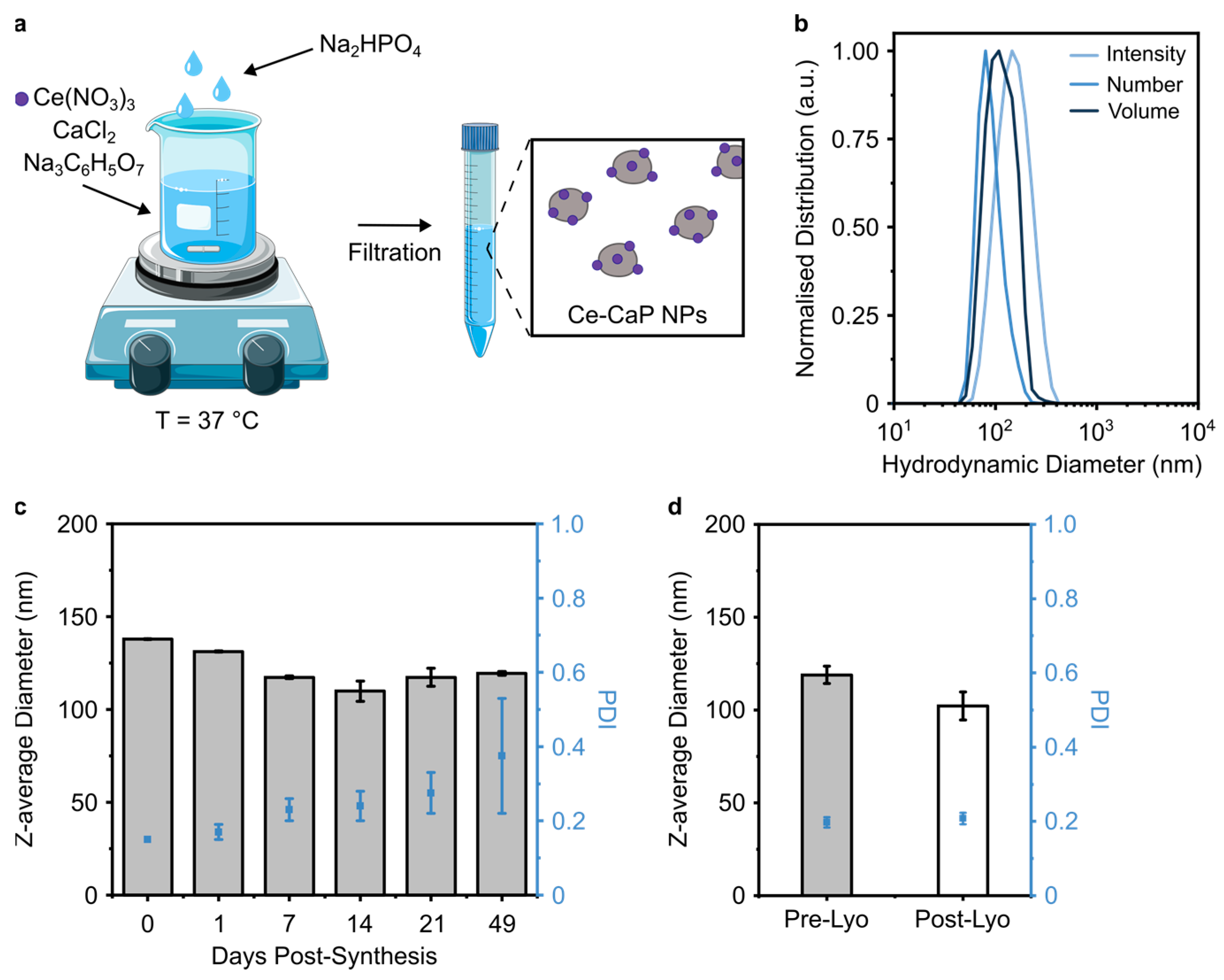
Abstract
:1. Introduction
2. Materials and Methods
2.1. Materials
2.2. Methods
2.2.1. Ce-CaP Nanoparticle Synthesis
2.2.2. Liposome Formulation
2.2.3. Ce-SANP Nanoparticle Formulation
2.2.4. Nanoparticle Physico-Chemical Characterization
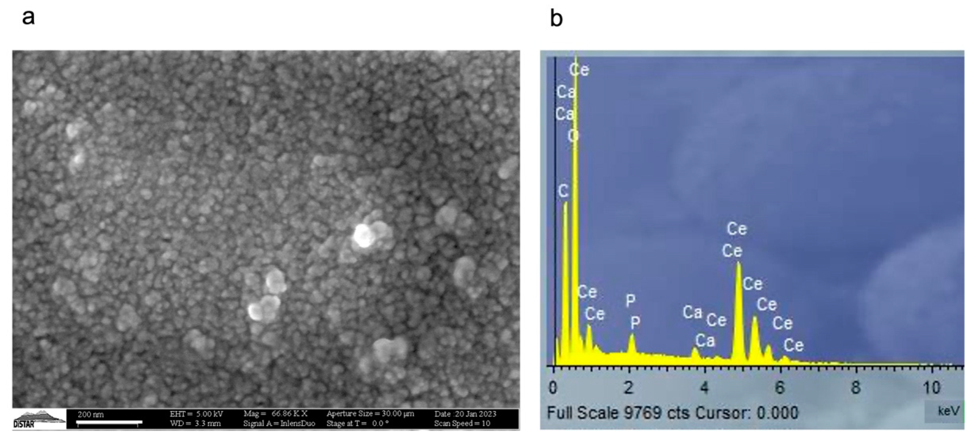
2.2.5. Scanning Electron Microscopy and Energy Dispersive X-ray Spectroscopy (SEM-EDX)
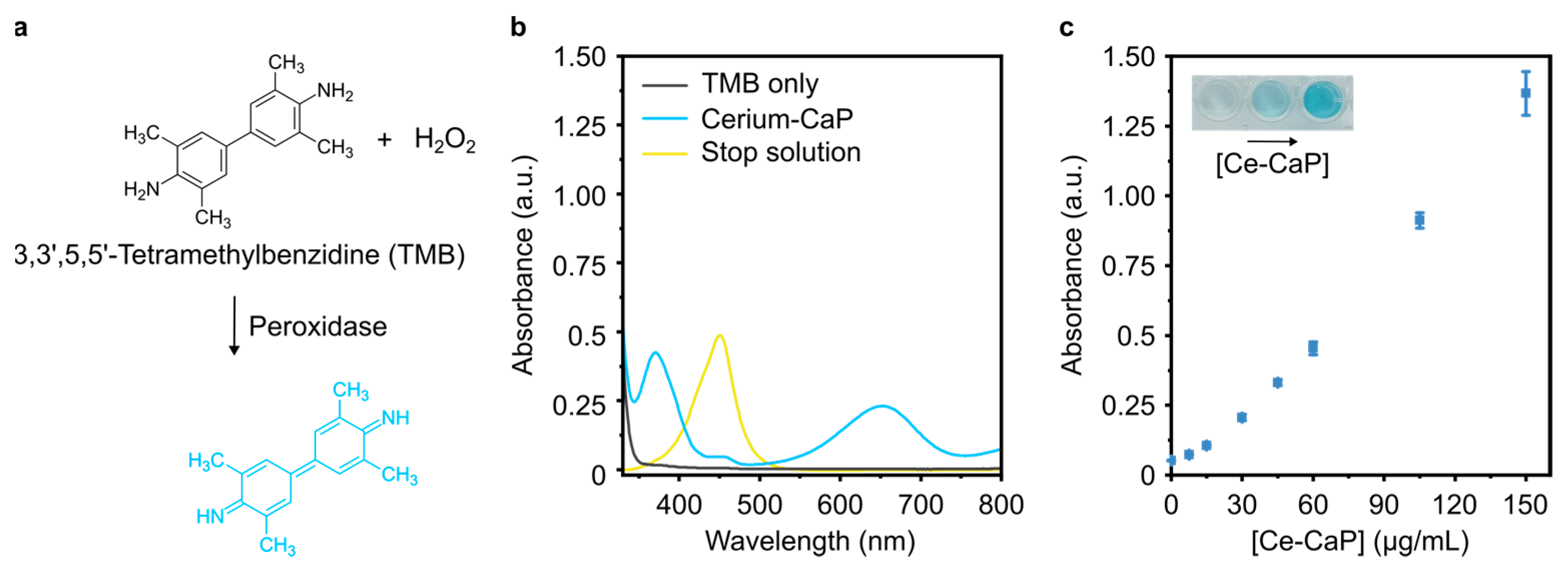
2.2.6. Peroxidase Assay
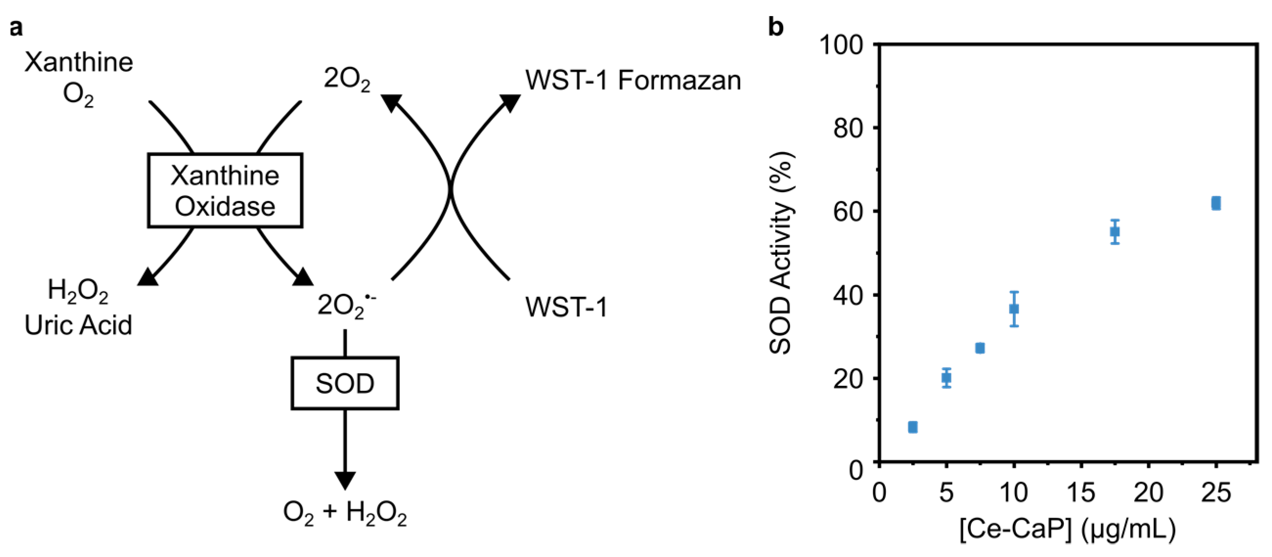
2.2.7. Superoxide Dismutase (SOD) Assay
2.2.8. Biological Characterization of Ce-SANP on Mitochondrial Function
2.2.9. Cell Culture
2.2.10. Chemical Hypoxia and Reoxygenation
2.2.11. Oxygen and Glucose Deprivation Followed by Reoxygenation (OGD/Rx)
2.2.12. Analysis of Mitochondrial activity
2.2.13. Measurement of ROS on Single-Cell
2.2.14. Quantification of ATP Level
2.2.15. [Ca2+]i Measurement
2.2.16. Statistical Analysis
3. Results and Discussion
3.1. Synthesis of Cerium-CaP Nanoparticles (Ce-CaP NPs)
3.2. SEM-EDS Analysis of Ce-CaP Nanoparticles
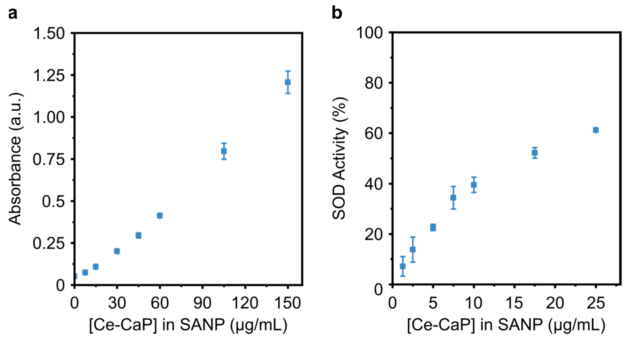
3.3. Characterization of Ce-CaP Nanoparticles: Enzyme-Like Activity
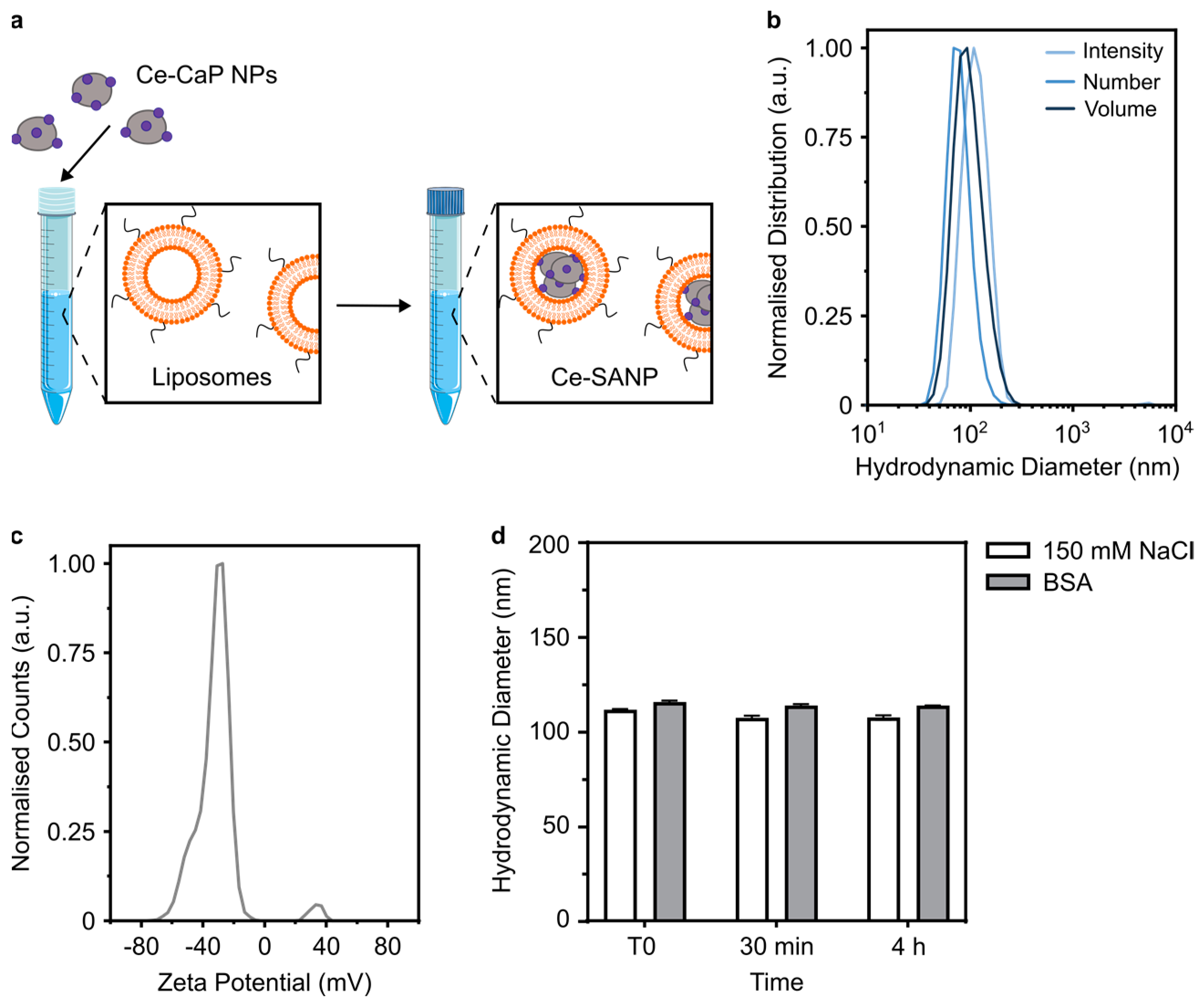
3.4. Formulation of Ce-SANP
3.5. Enzyme-Like Activity of Ce-SANP
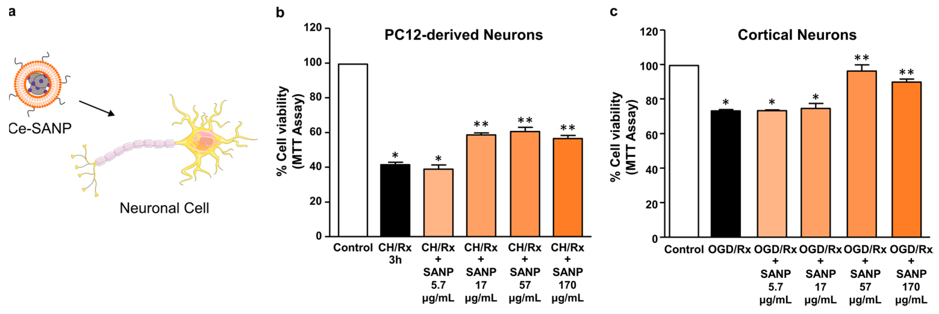
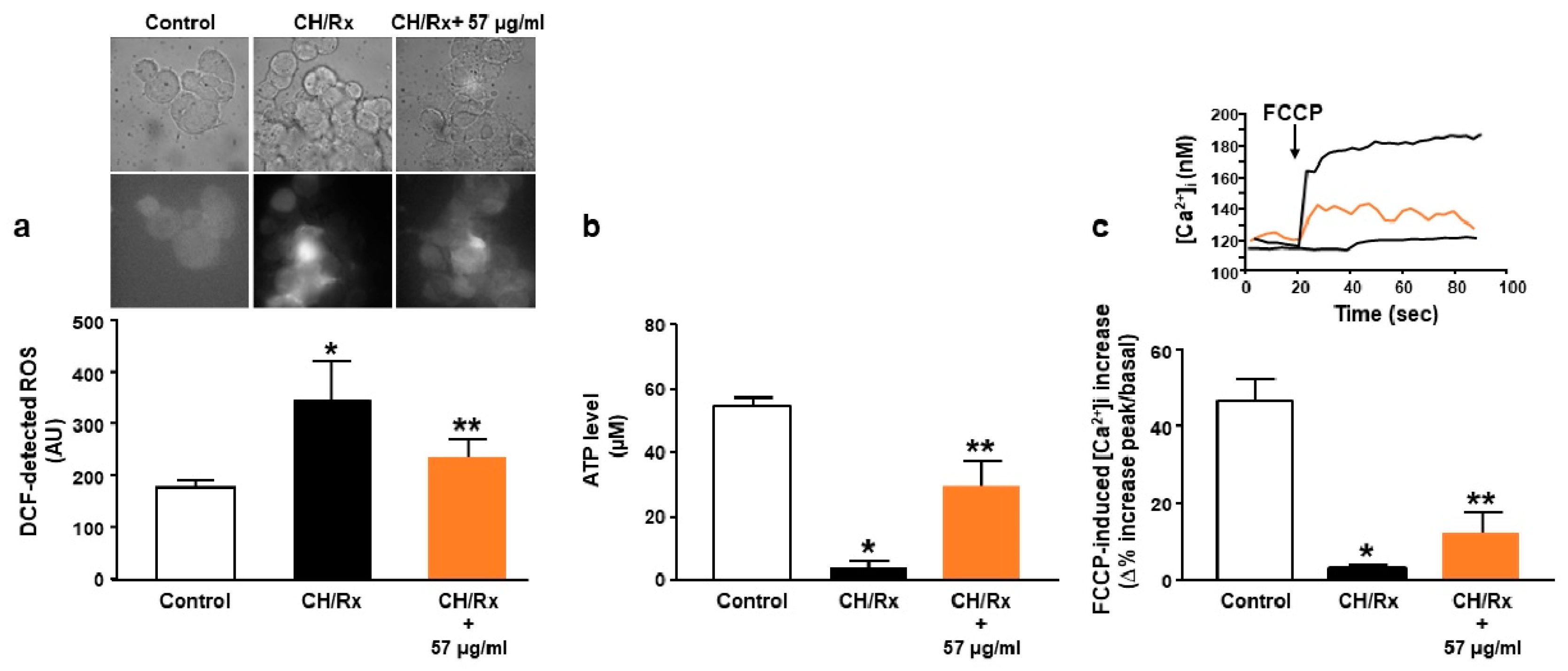
3.6. Effect of Ce-SANP on Mitochondrial Activity and Function in Primary Cortical Neurons Exposed to OGD Followed by Reoxygenation and Differentiated PC12 Cells Exposed to Chemical Hypoxia
4. Conclusions
Author Contributions
Funding
Institutional Review Board Statement
Informed Consent Statement
Data Availability Statement
Acknowledgments
Conflicts of Interest
References
- Gan, L.; Cookson, M.R.; Petrucelli, L.; la Spada, A.R. Converging Pathways in Neurodegeneration, from Genetics to Mechanisms. Nat. Neurosci. 2018, 21, 1300–1309. [Google Scholar] [CrossRef] [PubMed]
- Powers, W.J. Acute Ischemic Stroke. N. Engl. J. Med. 2020, 383, 252–260. [Google Scholar] [CrossRef] [PubMed]
- Hou, Y.; Dan, X.; Babbar, M.; Wei, Y.; Hasselbalch, S.G.; Croteau, D.L.; Bohr, V.A. Ageing as a Risk Factor for Neurodegenerative Disease. Nat. Rev. Neurol. 2019, 15, 565–581. [Google Scholar] [CrossRef]
- Forman, M.S.; Trojanowski, J.Q.; Lee, V.M.-Y. Neurodegenerative Diseases: A Decade of Discoveries Paves the Way for Therapeutic Breakthroughs. Nat. Med. 2004, 10, 1055–1063. [Google Scholar] [CrossRef]
- Andersen, J.K. Oxidative Stress in Neurodegeneration: Cause or Consequence? Nat. Med. 2004, 10, S18–S25. [Google Scholar] [CrossRef]
- Martinelli, C.; Pucci, C.; Battaglini, M.; Marino, A.; Ciofani, G. Antioxidants and Nanotechnology: Promises and Limits of Potentially Disruptive Approaches in the Treatment of Central Nervous System Diseases. Adv. Health Mater. 2020, 9, 1901589. [Google Scholar] [CrossRef]
- Wojsiat, J.; Zoltowska, K.M.; Laskowska-Kaszub, K.; Wojda, U. Oxidant/Antioxidant Imbalance in Alzheimer’s Disease: Therapeutic and Diagnostic Prospects. Oxid. Med. Cell. Longev. 2018, 2018, 6435861. [Google Scholar] [CrossRef]
- Yang, B.; Chen, Y.; Shi, J. Nanocatalytic Medicine. Adv. Mater. 2019, 31, 1901778. [Google Scholar] [CrossRef]
- Naz, S.; Beach, J.; Heckert, B.; Tummala, T.; Pashchenko, O.; Banerjee, T.; Santra, S. Cerium Oxide Nanoparticles: A ‘Radical’ Approach to Neurodegenerative Disease Treatment. Nanomedicine 2017, 12, 545–553. [Google Scholar] [CrossRef]
- Xiao, G.; Li, H.; Zhao, Y.; Wei, H.; Li, J.; Su, H. Nanoceria-Based Artificial Nanozymes: Review of Materials and Applications. ACS Appl. Nano. Mater. 2022, 5, 14147–14170. [Google Scholar] [CrossRef]
- Wang, W.; Jiang, X.; Chen, K. CePO4:Tb,Gd Hollow Nanospheres as Peroxidase Mimic and Magnetic–Fluorescent Imaging Agent. Chem. Commun. 2012, 48, 6839. [Google Scholar] [CrossRef] [PubMed]
- Vinothkumar, G.; Lalitha, A.I.; Suresh Babu, K. Cerium Phosphate–Cerium Oxide Heterogeneous Composite Nanozymes with Enhanced Peroxidase-Like Biomimetic Activity for Glucose and Hydrogen Peroxide Sensing. Inorg. Chem. 2019, 58, 349–358. [Google Scholar] [CrossRef] [PubMed]
- Shcherbakov, A.B.; Zholobak, N.M.; Baranchikov, A.E.; Ryabova, A.V.; Ivanov, V.K. Cerium Fluoride Nanoparticles Protect Cells against Oxidative Stress. Mater. Sci. Eng. C 2015, 50, 151–159. [Google Scholar] [CrossRef] [PubMed]
- Choi, S.W.; Kim, J. Recent Progress in Autocatalytic Ceria Nanoparticles-Based Translational Research on Brain Diseases. ACS Appl. Nano. Mater. 2020, 3, 1043–1062. [Google Scholar] [CrossRef]
- Sun, J.; Roy, S. Gene-Based Therapies for Neurodegenerative Diseases. Nat. Neurosci. 2021, 24, 297–311. [Google Scholar] [CrossRef] [PubMed]
- Martier, R.; Konstantinova, P. Gene Therapy for Neurodegenerative Diseases: Slowing Down the Ticking Clock. Front. Neurosci. 2020, 14, 580179. [Google Scholar] [CrossRef]
- Leavitt, B.R.; Tabrizi, S.J. Antisense Oligonucleotides for Neurodegeneration. Science 2020, 367, 1428–1429. [Google Scholar] [CrossRef]
- Roberts, T.C.; Langer, R.; Wood, M.J.A. Advances in Oligonucleotide Drug Delivery. Nat. Rev. Drug Discov. 2020, 19, 673–694. [Google Scholar] [CrossRef]
- Porru, M.; Zappavigna, S.; Salzano, G.; Luce, A.; Stoppacciaro, A.; Balestrieri, M.L.; Artuso, S.; Lusa, S.; de Rosa, G.; Leonetti, C.; et al. Medical Treatment of Orthotopic Glioblastoma with Transferrin-Conjugated Nanoparticles Encapsulating Zoledronic Acid. Oncotarget 2014, 5, 10446. [Google Scholar] [CrossRef]
- Salzano, G.; Zappavigna, S.; Luce, A.; D’Onofrio, N.; Balestrieri, M.L.; Grimaldi, A.; Lusa, S.; Ingrosso, D.; Artuso, S.; Porru, M.; et al. Transferrin-Targeted Nanoparticles Containing Zoledronic Acid as a Potential Tool to Inhibit Glioblastoma Growth. J. Biomed Nanotechnol. 2016, 12, 811–830. [Google Scholar] [CrossRef] [Green Version]
- Campani, V.; Zappavigna, S.; Scotti, L.; Abate, M.; Porru, M.; Leonetti, C.; Caraglia, M.; de Rosa, G. Hybrid Lipid Self-Assembling Nanoparticles for Brain Delivery of MicroRNA. Int. J. Pharm. 2020, 588, 119693. [Google Scholar] [CrossRef] [PubMed]
- Rispoli, C.; De Bonis, A.; Guarino, V.; Graziano, S.F.; Di Benedetto, C.; Esposito, R.; Morra, V.; Cappelletti, P. The ancient pozzolanic mortars of the Thermal complex of Baia (Campi Flegrei, Italy). J. Cult. Herit. 2019, 40, 143–154. [Google Scholar] [CrossRef]
- Secondo, A.; Petrozziello, T.; Tedeschi, V.; Boscia, F.; Vinciguerra, A.; Ciccone, R.; Pannaccione, A.; Molinaro, P.; Pignataro, G.; Annunziato, L. ORAI1/STIM1 Interaction Intervenes in Stroke and in Neuroprotection Induced by Ischemic Preconditioning Through Store-Operated Calcium Entry. Stroke 2019, 50, 1240–1249. [Google Scholar] [CrossRef] [PubMed]
- Pannaccione, A.; Secondo, A.; Scorziello, A.; Calì, G.; Taglialatela, M.; Annunziato, L. Nuclear Factor-ΚB Activation by Reactive Oxygen Species Mediates Voltage-Gated K+ Current Enhancement by Neurotoxic β-Amyloid Peptides in Nerve Growth Factor-Differentiated PC-12 Cells and Hippocampal Neurones. J. Neurochem. 2005, 94, 572–586. [Google Scholar] [CrossRef] [PubMed]
- Secondo, A.; Staiano, R.I.; Scorziello, A.; Sirabella, R.; Boscia, F.; Adornetto, A.; Valsecchi, V.; Molinaro, P.; Canzoniero, L.M.T.; di Renzo, G.; et al. BHK Cells Transfected with NCX3 Are More Resistant to Hypoxia Followed by Reoxygenation than Those Transfected with NCX1 and NCX2: Possible Relationship with Mitochondrial Membrane Potential. Cell Calcium. 2007, 42, 521–535. [Google Scholar] [CrossRef]
- Petrozziello, T.; Secondo, A.; Tedeschi, V.; Esposito, A.; Sisalli, M.; Scorziello, A.; di Renzo, G.; Annunziato, L. ApoSOD1 Lacking Dismutase Activity Neuroprotects Motor Neurons Exposed to Beta-Methylamino-L-Alanine through the Ca2+/Akt/ERK1/2 Prosurvival Pathway. Cell Death Differ. 2017, 24, 511–522. [Google Scholar] [CrossRef] [PubMed]
- Cocco, S.; Secondo, A.; del Viscovo, A.; Procaccini, C.; Formisano, L.; Franco, C.; Esposito, A.; Scorziello, A.; Matarese, G.; di Renzo, G.; et al. Polychlorinated Biphenyls Induce Mitochondrial Dysfunction in SH-SY5Y Neuroblastoma Cells. PLoS ONE 2015, 10, e0129481. [Google Scholar] [CrossRef]
- Grynkiewicz, G.; Poenie, M.; Tsien, R.Y. A New Generation of Ca2+ Indicators with Greatly Improved Fluorescence Properties. J. Biol. Chem. 1985, 260, 3440–3450. [Google Scholar] [CrossRef]
- Urbanczyk, J.; Chernysh, O.; Condrescu, M.; Reeves, J.P. Sodium-Calcium Exchange Does Not Require Allosteric Calcium Activation at High Cytosolic Sodium Concentrations. J. Physiol. 2006, 575, 693–705. [Google Scholar] [CrossRef]
- Cai, A.-Y.; Zhu, Y.-J.; Qi, C. Biodegradable Inorganic Nanostructured Biomaterials for Drug Delivery. Adv. Mater. Interfaces 2020, 7, 2000819. [Google Scholar] [CrossRef]
- di Mauro, V.; Iafisco, M.; Salvarani, N.; Vacchiano, M.; Carullo, P.; Ramírez-Rodríguez, G.B.; Patrício, T.; Tampieri, A.; Miragoli, M.; Catalucci, D. Bioinspired Negatively Charged Calcium Phosphate Nanocarriers for Cardiac Delivery of MicroRNAs. Nanomedicine 2016, 11, 891–906. [Google Scholar] [CrossRef] [PubMed]
- Heckman, K.L.; DeCoteau, W.; Estevez, A.; Reed, K.J.; Costanzo, W.; Sanford, D.; Leiter, J.C.; Clauss, J.; Knapp, K.; Gomez, C.; et al. Custom Cerium Oxide Nanoparticles Protect against a Free Radical Mediated Autoimmune Degenerative Disease in the Brain. ACS Nano 2013, 7, 10582–10596. [Google Scholar] [CrossRef]
- Kignelman, G.; Eyley, S.; Zhou, C.; Tunca, B.; Gonon, M.; Lahem, D.; Seo, J.W.; Thielemans, W. Colloidal Stability and Aggregation Mechanism in Aqueous Suspensions of TiO 2 Nanoparticles Prepared by Sol–Gel Synthesis. Langmuir 2021, 37, 14846–14855. [Google Scholar] [CrossRef] [PubMed]
- Yang, Y.; Mao, Z.; Huang, W.; Liu, L.; Li, J.; Li, J.; Wu, Q. Redox Enzyme-Mimicking Activities of CeO2 Nanostructures: Intrinsic Influence of Exposed Facets. Sci. Rep. 2016, 6, 35344. [Google Scholar] [CrossRef] [PubMed]
- Alizadeh, N.; Salimi, A.; Sham, T.-K.; Bazylewski, P.; Fanchini, G. Intrinsic Enzyme-like Activities of Cerium Oxide Nanocomposite and Its Application for Extracellular H 2 O 2 Detection Using an Electrochemical Microfluidic Device. ACS Omega 2020, 5, 11883–11894. [Google Scholar] [CrossRef] [PubMed]
- Baldim, V.; Bedioui, F.; Mignet, N.; Margaill, I.; Berret, J.-F. The Enzyme-like Catalytic Activity of Cerium Oxide Nanoparticles and Its Dependency on Ce 3+ Surface Area Concentration. Nanoscale 2018, 10, 6971–6980. [Google Scholar] [CrossRef]
- Baldim, V.; Yadav, N.; Bia, N.; Graillot, A.; Loubat, C.; Singh, S.; Karakoti, A.S.; Berret, J.-F. Polymer-Coated Cerium Oxide Nanoparticles as Oxidoreductase-like Catalysts. ACS Appl. Mater. Interfaces 2020, 12, 42056–42066. [Google Scholar] [CrossRef] [PubMed]
- Goujon, G.; Baldim, V.; Roques, C.; Bia, N.; Seguin, J.; Palmier, B.; Graillot, A.; Loubat, C.; Mignet, N.; Margaill, I.; et al. Antioxidant Activity and Toxicity Study of Cerium Oxide Nanoparticles Stabilized with Innovative Functional Copolymers. Adv. Health Mater. 2021, 10, 2100059. [Google Scholar] [CrossRef]
- Berret, J.-F.; Graillot, A. Versatile Coating Platform for Metal Oxide Nanoparticles: Applications to Materials and Biological Science. Langmuir 2022, 38, 5323–5338. [Google Scholar] [CrossRef]
- Asati, A.; Santra, S.; Kaittanis, C.; Perez, J.M. Surface-Charge-Dependent Cell Localization and Cytotoxicity of Cerium Oxide Nanoparticles. ACS Nano 2010, 4, 5321–5331. [Google Scholar] [CrossRef] [Green Version]
- Asati, A.; Santra, S.; Kaittanis, C.; Nath, S.; Perez, J.M. Oxidase-Like Activity of Polymer-Coated Cerium Oxide Nanoparticles. Angew. Chem. Int. Ed. 2009, 48, 2308–2312. [Google Scholar] [CrossRef] [PubMed]
- Lord, M.S.; Tsoi, B.; Gunawan, C.; Teoh, W.Y.; Amal, R.; Whitelock, J.M. Anti-Angiogenic Activity of Heparin Functionalised Cerium Oxide Nanoparticles. Biomaterials 2013, 34, 8808–8818. [Google Scholar] [CrossRef] [PubMed]
- Pramanik, N.; De, T.; Sharma, P.; Alakesh, A.; Jagirdar, S.K.; Rangarajan, A.; Jhunjhunwala, S. Surface-Coated Cerium Nanoparticles to Improve Chemotherapeutic Delivery to Tumor Cells. ACS Omega 2022, 7, 31651–31657. [Google Scholar] [CrossRef]
- Ristori, S.; Grillo, I.; Lusa, S.; Thamm, J.; Valentino, G.; Campani, V.; Caraglia, M.; Steiniger, F.; Luciani, P.; de Rosa, G. Structural Characterization of Self-Assembling Hybrid Nanoparticles for Bisphosphonate Delivery in Tumors. Mol. Pharm. 2018, 15, 1258–1265. [Google Scholar] [CrossRef]
- Nele, V.; Holme, M.N.; Kauscher, U.; Thomas, M.R.; Doutch, J.J.; Stevens, M.M. Effect of Formulation Method, Lipid Composition, and PEGylation on Vesicle Lamellarity: A Small-Angle Neutron Scattering Study. Langmuir 2019, 35, 6064–6074. [Google Scholar] [CrossRef]
- Battaglini, M.; Tapeinos, C.; Cavaliere, I.; Marino, A.; Ancona, A.; Garino, N.; Cauda, V.; Palazon, F.; Debellis, D.; Ciofani, G. Design, Fabrication, and In Vitro Evaluation of Nanoceria-Loaded Nanostructured Lipid Carriers for the Treatment of Neurological Diseases. ACS Biomater. Sci. Eng. 2019, 5, 670–682. [Google Scholar] [CrossRef]
- Cha, B.G.; Jeong, H.-G.; Kang, D.-W.; Nam, M.-J.; Kim, C.K.; Kim, D.Y.; Choi, I.-Y.; Ki, S.K.; Kim, S.I.; Han, J.h.; et al. Customized Lipid-Coated Magnetic Mesoporous Silica Nanoparticle Doped with Ceria Nanoparticles for Theragnosis of Intracerebral Hemorrhage. Nano Res. 2018, 11, 3582–3592. [Google Scholar] [CrossRef]
- Turovsky, E.A.; Varlamova, E.G. Mechanism of Ca2+-Dependent Pro-Apoptotic Action of Selenium Nanoparticles, Mediated by Activation of Cx43 Hemichannels. Biology 2021, 10, 743. [Google Scholar] [CrossRef]
- Kim, S.H.; Ryan, T.A. Synaptic Vesicle Recycling at CNS Synapses without AP-2. J. Neurosci. 2009, 29, 3865–3874. [Google Scholar] [CrossRef]


Disclaimer/Publisher’s Note: The statements, opinions and data contained in all publications are solely those of the individual author(s) and contributor(s) and not of MDPI and/or the editor(s). MDPI and/or the editor(s) disclaim responsibility for any injury to people or property resulting from any ideas, methods, instructions or products referred to in the content. |
© 2023 by the authors. Licensee MDPI, Basel, Switzerland. This article is an open access article distributed under the terms and conditions of the Creative Commons Attribution (CC BY) license (https://creativecommons.org/licenses/by/4.0/).
Share and Cite
Nele, V.; Tedeschi, V.; Campani, V.; Ciancio, R.; Angelillo, A.; Graziano, S.F.; De Rosa, G.; Secondo, A. Cerium-Doped Self-Assembling Nanoparticles as a Novel Anti-Oxidant Delivery System Preserving Mitochondrial Function in Cortical Neurons Exposed to Ischemia-like Conditions. Antioxidants 2023, 12, 358. https://doi.org/10.3390/antiox12020358
Nele V, Tedeschi V, Campani V, Ciancio R, Angelillo A, Graziano SF, De Rosa G, Secondo A. Cerium-Doped Self-Assembling Nanoparticles as a Novel Anti-Oxidant Delivery System Preserving Mitochondrial Function in Cortical Neurons Exposed to Ischemia-like Conditions. Antioxidants. 2023; 12(2):358. https://doi.org/10.3390/antiox12020358
Chicago/Turabian StyleNele, Valeria, Valentina Tedeschi, Virginia Campani, Raffaella Ciancio, Alessia Angelillo, Sossio Fabio Graziano, Giuseppe De Rosa, and Agnese Secondo. 2023. "Cerium-Doped Self-Assembling Nanoparticles as a Novel Anti-Oxidant Delivery System Preserving Mitochondrial Function in Cortical Neurons Exposed to Ischemia-like Conditions" Antioxidants 12, no. 2: 358. https://doi.org/10.3390/antiox12020358