Article

Characterization of Beta-Lactam Resistome of Escherichia coli Causing Nosocomial Infections

by
Rosalino Vázquez-López
1,
Tanya Hernández-Martínez
2,
Selene Ivonne Larios-Fernández
2,
Celia Piña-Leyva
2,
Manuel Lara-Lozano
2,
Tayde Guerrero-González
2,
Javier Martínez-Bautista
3,
Eduardo Gómez-Conde
4 and
Juan Antonio González-Barrios
2,*
1
Departamento de Microbiología, Centro de Investigación en Ciencias de la Salud (CICSA), Facultad de Ciencias de la Salud Universidad Anáhuac México Norte, Huixquilucan 52786, Mexico
2
Laboratorio de Medicina Genómica, Hospital Regional “Primero de Octubre”, ISSSTE, Av. Instituto Politécnico Nacional 1669, Lindavista, Gustavo A. Madero, Ciudad de México 07300, Mexico
3
Laboratorio de Microbiología, Hospital Regional “Primero de Octubre”, ISSSTE, Av. Instituto Politécnico Nacional 1669, Lindavista, Gustavo A. Madero, Ciudad de México 07300, Mexico
4
Departamento de Inmunobiología, Facultad de Medicina, Benemérita Universidad Autónoma de Puebla (BUAP), Puebla 72420, Mexico
*
Author to whom correspondence should be addressed.
Antibiotics 2023, 12(9), 1355; https://doi.org/10.3390/antibiotics12091355
Submission received: 14 July 2023 / Revised: 3 August 2023 / Accepted: 5 August 2023 / Published: 23 August 2023

Abstract

:
Nosocomial infections caused by Escherichia coli pose significant therapeutic challenges due to the high expression of genes encoding antimicrobial drug resistance. In this study, we investigated the conformation of the beta-lactam resistome responsible for the specific pattern of resistance against beta-lactam antibiotics. A total of 218 Escherichia coli strains were isolated from in-hospital patients diagnosed with nosocomial infections, obtained from various sources such as urine (n = 49, 22.48%), vaginal discharge (n = 46, 21.10%), catheter tips (n = 14, 6.42%), blood (n = 13, 5.96%), feces (n = 12, 5.50%), sputum (n = 11, 5.05%), biopsies (n = 8, 3.67%), cerebrospinal fluid (n = 2, 0.92%) and other unspecified discharges (n = 63, 28.90%). To characterize the beta-lactam resistome, all strains were subjected to antibiotic dilution tests and grown in beta-lactam antibiotics supplemented with Luria culture medium. Subsequently, multiplex PCR and next-generation sequencing were conducted. The results show a multi-drug-resistance phenotype, particularly against beta-lactam drugs. The primary determinant of this resistance was the expression of the blaTEM gene family, with 209 positive strains (95.87%) expressing it as a single gene (n = 47, 21.6%) or in combination with other genes. Common combinations included blaTEM + blaCTX (n = 42, 19.3%), blaTEM + blaCTX + blaSHV (n = 13, 6%) and blaTEM + blaCTX + blaBIL (n = 12, 5.5%), among others. The beta-lactam resistome of nosocomial Escherichia coli strains isolated from inpatients at the “October first” Regional Hospital of ISSSTE was predominantly composed of members of the blaTEM gene family, expressed in various configurations along with different members of other beta-lactamase gene families.

1. Introduction

In the past two decades, nosocomial infections have become a significant public health problem worldwide [1], representing a healthcare failure that leads to adverse effects during in-hospital stays and an increased risk of mortality among patients [2]. These infections are associated with various medical procedures such as invasive interventions, extensive surgeries and the use of indwelling medical devices, giving rise to central-line-associated bloodstream infections (CLABSIs), catheter-associated urinary-tract infections (CAUTIs), surgical-site infections (SSIs) and ventilator-associated pneumonia (VAP) [3].
Factors such as prolonged hospital stays also contribute to the acquisition of bloodstream infections not associated with central catheter use, gastrointestinal infections (GIs), urinary-tract infections (UTIs) not associated with catheter use and non-ventilator-associated hospital-acquired pneumonia (NV-HAP). Various body sites are affected by healthcare-associated infections (HAIs), including the ear, eye, nose, throat, lower respiratory tract, skin, soft tissue, cardiovascular system, bone, joints, central nervous system and reproductive tract. HAIs have diverse causes, including bacterial, viral or fungal agents. Among the main bacteria responsible for these infections are Gram-positive microorganisms like coagulase-negative Staphylococci, Staphylococcus aureus, Streptococcus species, Enterococcus faecalis, Enterococcus faecium and Clostridium difficile [4], as well as Gram-negative microorganisms such as some members of the Enterobacteriaceae family (e.g., Escherichia coli, Klebsiella pneumoniae, Klebsiella oxytoca, Proteus mirabilis), Enterobacter species (Pseudomonas aeruginosa, Acinetobacter baumanii and Burkholderia cepacian). These pathogens are associated with high morbidity and mortality rates in hospitalized patients due to their inherent multi-drug-resistant properties [5].
The extensive use of antibiotics in hospitals exerts significant selection pressure on nosocomial bacterial populations, especially on bacteria like Escherichia coli, Acinetobacter baumanii, Pseudomonas aeruginosa, Enterococcus faecium, Proteus mirabilis and Stenotrophomonas maltophilia, leading to the emergence of resistant, multi-resistant and pan-resistant nosocomial infections [6]. Escherichia coli, a pathogenic microorganism, can be found in various environmental sources and the intestines of humans and animals. While most strains are harmless, certain pathogenic strains have been associated with gastrointestinal diseases and extraintestinal tissue infections. In hospitals, Escherichia coli has been identified as the causative agent of various healthcare-associated infections like BSI, CLABSI, UTI, CAUTI, SSI, VAP, NV-HAP and GI [7].
Due to excessive and sometimes indiscriminate antibiotic use, Escherichia coli strains in hospitals have become highly multi-drug-resistant (MDR). This resistance is particularly common in beta-lactam antibiotics, severely limiting effective treatment options [8]. The resistance to beta-lactam antibiotics is encoded by different classes of beta-lactamases, including blaCTX, blaVIM, blaNMC, blaOXA, blaIMP, blaLAP, blaROB, blaDHA, blaVEB, blaCMY, blaLAP, blaSHV, blaTEM, blaSME, blaSFC, blaKPC, blaAmpC, blaGES, blaSME, blaPER and blaNDM [9,10,11,12,13,14,15,16,17,18], which confer a wide spectrum of resistance to beta-lactam antibiotics [19].
In this study, we focus on characterizing the beta-lactam resistome expressed by Escherichia coli-causing nosocomial infections in the “October first” Regional Hospital of ISSSTE in Mexico.

2. Materials and Methods

2.1. Primer Design

The bioinformatic analysis and primer design process were previously described and reported by our research group [20]. In summary, we retrieved beta-lactamase integron and DNA sequences from the GenBank of NCBI. All available sequences of beta-lactamases were gathered for the purpose of PCR primer design. The DNA sequences were aligned using ClustalW v.2 software [21]. Subsequently, we used FigTree V1.4.0 software [22] to generate phylogenetic trees based on the alignments. From the alignment results, conserved sequences were identified and used as a basis for designing a set of specific and degenerate primers. The PerPrimer v1.1.21 Software [23] was utilized, employing stringent criteria, including primer length (18–25 bp), a melting temperature (Tm) of 60–62 C, a GC content of 40–60%, with a Δ T of 1 C in the annealing temperature and an amplicon size ranging from 83 to 230 bp [20] (Table 1).

2.2. Biological Samples

A total of five-hundred-and-eighty-five clinical samples were collected from various secretions, including urethral, vaginal, nasal, diabetic foot ulcers, bronchial, surgical and pharyngeal wound secretions. Additionally, body fluids such as blood, dialysis fluid, cerebrospinal fluid, peritoneal, pleural and synovial fluid, urine, feces, sputum and semen, as well as catheter tips from patients diagnosed with nosocomial infections, were also included in the study. Among these samples, 218 Escherichia coli strains showing multi-drug resistance were cultured and selected for further analysis. The biochemical identification of these strains was performed by the Microbiology section of the Central Clinical Laboratory at the “October first” Regional Hospital (ISSSTE).

2.3. Escherichia coli Isolation Culture

This study was conducted at the “October first” Regional Hospital (ISSSTE) in Mexico City. The collected samples were cultured on various agar media, including Blood Agar (COS), MacConkey Agar, Chocolate Mueller Hilton Agar (PVX), Chromogenic Candida Agar, Sabouraud Agar, Triptone Soya Broth, Chromogenic Agar for Staphylococcus aureus identification in human specimens (SAID), Salted Mannitol Agar, Chromogenic Agar for the detection of Salmonella, SS Agar for Shigella and Salmonella and CromlDTM CPS Agar.
The cultured plates were incubated at 35 C for 18 to 24 h. All bacterial growth on the MacConkey agar plates was subjected to biochemical identification tests. However, only the cultures that were identified as Escherichia coli were further selected for antimicrobial susceptibility testing and genotyping.

2.4. Biochemical Identification

The biochemical identification of Escherichia coli strains was carried out using the VITEK 2XL system, an automated method provided by bioMérieux Inc. (Durham, NC, USA) Specifically, the VITEK 2XL GN 21341 cards were used, containing 21 biochemical tests. These tests included β -galactosidase (OPNG), arginine dehydrolase (ADH), lysine decarboxylase (LDC), ornithine decarboxylase (ODC), citrate utilization (CIT), hydrogen sulfide (H2S) production, urease (URE) activity, tryptophan deaminase (TDA) production, indole production (IND), Voges–Proskauer acetoin production (VP), gelatinase (GEL) activity and fermentation/oxidation tests for various sugars such as glucose (GLU), mannitol, inositol, sorbitol, ramnose, sucrose, melibiose, amygdalin (vitamin B17), arabinose and cytochrome oxidase (OX) activity.
To confirm the Escherichia coli identification, the strains must test positive for β -galactosidase (OPNG), lysine decarboxylase (LDL), ornithine decarboxylase (ODC), indole production (IND), glucose fermentation (GLU), mannitol (MAN), sorbitol (SOR), ramnose (RHA), sucrose (SAC), melibiose (MEL) and arabinose (ARA) tests among the set of biochemical tests conducted using the VITEK 2XL GN 21341 cards.

2.5. Antimicrobial Susceptibility Testing

Antibiotic susceptibility testing was performed using the VITEK 2XL automated system with ASTGN70 cards (REF 413 and 401) specifically designed for Gram-negative bacteria by BioMérieux Inc.(Durham, NC, USA) The following antibiotics were tested with varying concentrations (in μ g/mL): amikacin (8, 16, 64 μ g/mL), ampicillin (4, 8, 32 μ g/mL), ampicillin/sulbactam (4/2, 16/8, 32/16 μ g/mL), aztreonam (2, 8, 32 μ g/mL), cefazolin (4, 16, 64 μ g/mL), cefepime (2, 8, 16, 32 μ g/mL), cefotetan (2, 8, 32 μ g/mL), ceftazidime (1, 2, 8, 32 μ g/mL), ceftriaxone (1, 2, 8, 32 μ g/mL), cefuroxime (2, 8, 32 μ g/mL), ciprofloxacin (0.5, 2, 4 μ g/mL), gentamicin (4, 16, 32 μ g/mL), imipenem (2, 4, 16 μ g/mL), meropenem (0.5, 4, 16 μ g/mL), piperacillin/tazobactam (4/4, 16/4, 128/4 μ g/mL), ticarcillin/clavulanic acid (8/2, 32/2, 64/2 μ g/mL), tobramycin (8, 16, 64 μ g/mL), trimethoprim/sulfamethoxazole (0.5/9.5, 2/38, 16/304 μ g/mL), nitrofurantoin (16, 32, 64 μ g/mL) and gatifloxacin (1, 4, 8 μ g/mL).
The resistance or susceptibility profile of each strain was determined by measuring the minimum inhibitory concentration (MIC) of the antibiotics, which was assessed by measuring the optical density of bacterial cultures after incubation.

2.6. Beta-Lactam Selection

For genomic and plasmid DNA sequencing, it was necessary to obtain generic cultures of isolated pure Escherichia coli colonies. The growth of each colony was achieved by placing it in 10 mL of 25% lysogeny broth (LB) medium, which was then sterilized at 121 C for 15 min. Subsequently, each Escherichia coli strain was grown in a set of beta-lactam drugs, including ampicillin (10 μ g), ampicillin/sulbactam (10/10 μ g), carbenicillin (100 μ g), cefaclor (30 μ g), cefazolin (30 μ g), cefepime (30 μ g), cefoperazone (75 μ g), cefotetan (30 μ g), mezlocillin (75 μ g) and piperacillin/tazobactam (100/10 μ g). All of these beta-lactam drugs were obtained from Becton Dickinson (Franklin Lakes, NJ, USA). Each medium was incubated at 37 C for 24 h under aerobic conditions. The inoculum was adjusted using a Mac Farland 0.5 reading, which corresponds to an expected colony-forming units per milliliter (CFU/mL) of approximately 1.5 × 10⁸. The cultures were then placed in a Shaker Series Innova at 37 C and 240 rpm for 20 h.

2.7. DNA Extraction

2.7.1. Crude Extract

Total DNA was extracted using a modified version of the heat-shock method described by Maugeri et al. One milliliter of Luria medium containing a well-identified, multi-resistant Escherichia coli strain grown for 20 h at 35 C was used. The selected medium was lysed by heating it in a Thermoblock for 1 min at 100 C, followed by immediate placement in an ice bath for 5 min, inducing thermal shock and facilitating the release of genetic material. Two milliliters of the cell lysates containing DNA were purified using chelex (glass beads). The purified DNA served as a template for both real-time PCR assays and NGS (next-generation sequencing). The extracted DNA was stored at −80 C for further analysis and experimentation.

2.7.2. Plasmid DNA Extraction

Plasmid DNA extraction was conducted using a previously reported method [20]. The PureLink HiPure Plasmid DNA Purification Kit (Invitrogen, Carlsbad, CA, USA) was employed, following the manufacturer’s instructions. The extracted plasmid DNA solution was quantified using a Qubit Flex Fluorometer (Thermo Fisher Scientific, Wilmington, DE, USA) based on fluorometry. To verify the integrity of the plasmid DNA, electrophoresis was performed in 2% agarose gel. Subsequently, the samples were stored at −80 C until further use.

2.8. Real-Time PCR

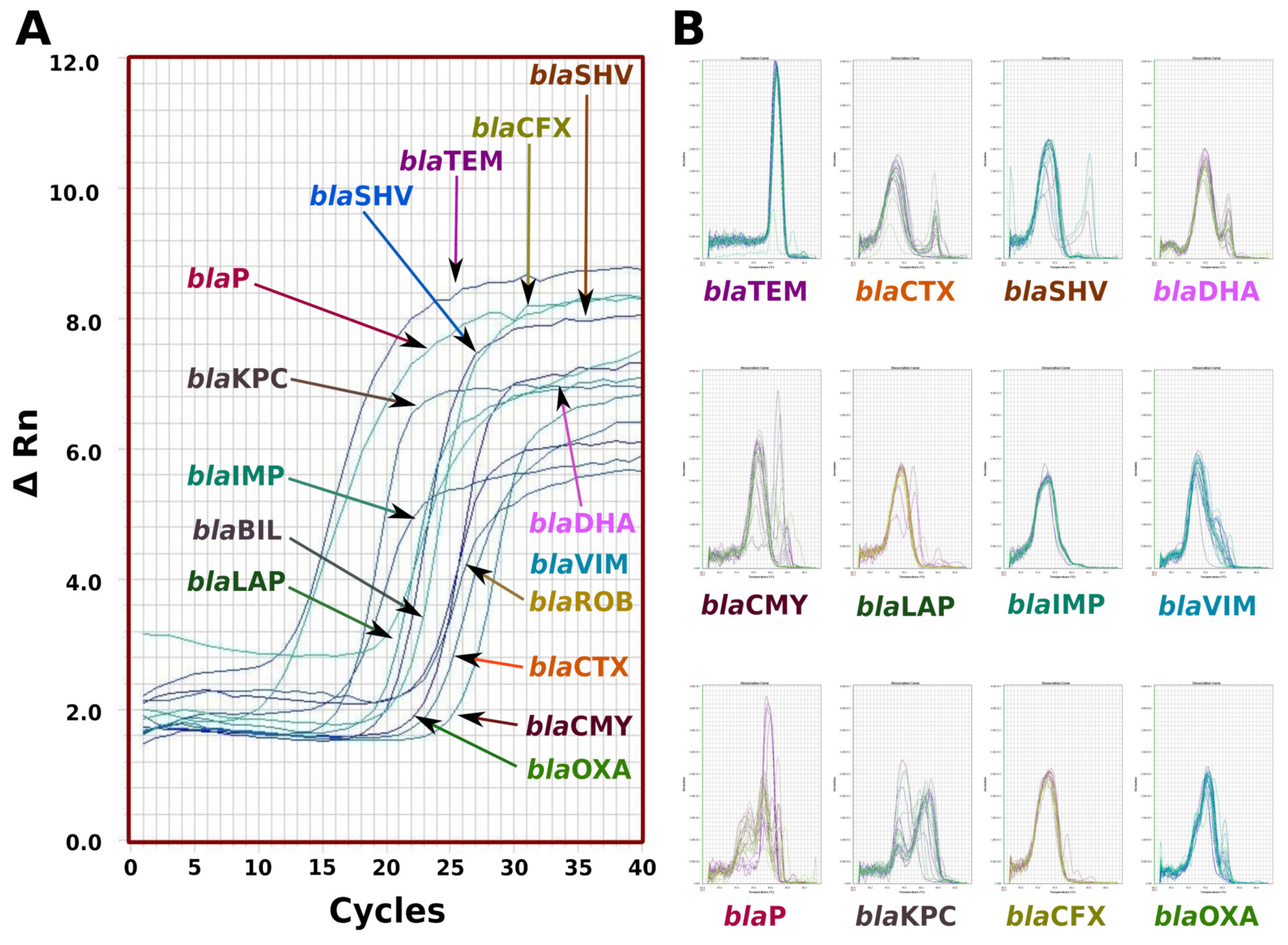
The beta-lactamases SYBR Green-based qPCR assays were performed on the Real-Time HT FAST 7900 system (Applied Biosystems, Waltham, MA, USA). Each reaction was conducted in a total volume of 10 μ L, comprising 5 μ L of 21.5 × SYBR Green PCR Master Mix (Applied Biosystems, Waltham, MA, USA), 10 mM of specific or degenerate primers and 3 μ L of the DNA template. The cycling conditions for both assays consisted of an initial denaturation step at 95 C for 5 min, followed by 45 cycles of denaturation at 95 C for 15 s, annealing at 50 C for 40 s and extension at 60 C for 40 s. To confirm the specificity of the qPCR products, the amplicons were analyzed using melting curves. Additionally, the products were visualized on a 2% agarose gel pre-stained with ethidium bromide and the resulting image was digitized using a GelLogistic 3000 photodocumenter.

2.9. Whole DNA Sequencing

For DNA sequencing, a previously reported method by the research group [20] was followed. Both genomic and plasmid DNA were mixed in a 1:1 proportion and used to prepare indexed libraries using the Illumina Nextera XT DNA Sample Preparation Kit (FC-131-1096) designed for small genomes. The libraries were then sequenced on the MiSeq platform (Illumina; San Diego, CA, USA). Adapters and barcodes were trimmed using the default settings in the Illumina experiment manager, resulting in 300 bp paired-end reads. The quality of the unprocessed reads was assessed using FastQC High-Throughput Sequence QC Report v:0.11.5 from Babraham Bioinformatics, Babraham Institute (Cambridge, UK) [24]. A minimum quality score (Q score) of more than 30 was achieved for at least 85% of all reads. For read mapping, the BWA-MEM aligner version 0.7.7-r441 from the Wellcome Trust Sanger Institute (Hinxton, UK) was used. Genome assembly was performed using the SPAdes Genome Assembler software version 3.11 from CAB, St. Petersburg State University (St. Petersburg, Russia). The reported ASM74325v1 genome of Escherichia coli strain 25922 ATCC with a size of 5.2 Mbp was used as the assembly reference [25]. The DNA metagenomic analysis for the taxonomic classification of the Escherichia coli bacteria was conducted using the Kraken taxonomic sequence classification system, version 0.10.5-beta, from CCB, Johns Hopkins University (Baltimore, MD, USA) [26]. For identifying beta-lactamase genes, a comparative analysis was performed using the Basic Local Alignment Search Tool (BLAST) from NCBI-NIH (Bethesda, MD, USA) [27].

2.10. Study Outcomes

The main objective of this study was to characterize the beta-lactam resistome expressed by Escherichia coli strains causing nosocomial infections. The focus was on understanding the patterns of antibiotic resistance induced by the expression of beta-lactamases, which often complicate the treatment of nosocomial infections. By examining the resistome and identifying the specific beta-lactamase genes responsible for the multi-drug resistance, this study aimed to provide valuable insights for the better management and treatment of nosocomial infections caused by these bacteria.

3. Results

3.1. Sampling Sources

We analyzed 585 clinical samples from various anatomopathological sources of in-hospital patients with nosocomial infections at the “October first” Regional Hospital in Mexico City. Of these, we isolated 218 multi-resistant Escherichia coli strains, with 63 (28.90%) being unspecified discharge, 49 (22.48%) from urine cultures, 46 (21.10%) from vaginal discharge, 14 (6.42%) from catheter tips, 13 (5.96%) from blood cultures, 12 (5.50%) from feces cultures, 11 (5.05%) from sputum, 8 (3.67%) from biopsies and 2 (0.92%) from cerebrospinal fluid.
The microbiology laboratory reported that all 218 strains were multi-drug-resistant, showing 100% resistance to ampicillin, ampicillin/sulbactam, cefazolin, ceftriaxone and cefepime. Additionally, 8.26% (n = 18) were resistant to ertapenem, 28.34% (n = 13) to meropenem, 58.26% (n = 217) to gentamicin, 50.00% (n = 109) to tobramycin, 91.28% (n = 199) to ciprofloxacin and 16.51% (n = 36) to nitrofurantoin. However, no resistance was found for amikasin and tigecycline (Table 2).

3.2. Multiple-Antibiotic-Resistance Index (MAR Index)

We calculated the multiple-antibiotic-resistance index (MAR index) for 218 Escherichia coli strains and found that 216 strains had an MAR index greater than 0.25. Among these, 167 strains had an MAR index ranging from 0.5 to 0.75. We did not find any strains with an MAR index of 0.05 to 0.2 or 0.95 to 1.0. The MAR index for all nosocomial Escherichia coli isolates was determined to be 0.57 ± 0.21 (Table 3).

3.3. Beta-Lactam Selection

We tested the Escherichia coli subculture in liquid beta-lactam selection medium and found that all 218 strains showed growth in medium supplemented with ampicillin, cefazolin and cefazolin. Furthermore, 87.16% (n = 190) of strains showed growth in amoxicillin/clavulanic acid, 85.32% (n = 190) in cefixime, 72.48% (n = 158) in cefuroxime, 71.10% (n = 155) in cephalothin, 67.43% (n = 158) in cefaclor, 64.68% (n = 141) in piperacillin, 63.30% (n = 138) in cefazolin and 52.29% (n = 114) in doxycycline. The lower resistance frequencies were 17.89% (n = 39) to piperacillin, 15.60% (n = 39) to ceftizoxime, 13.76% (n = 30) to meropenem and 11.01% (n = 24) to imipenem (Table 2). These results yielded a specific beta-lactam MAR index of 0.82 ± 0.09 (Table 4).

3.4. Sequencing Escherichia coli Identification and Genome Size

The automatic identification of 218 different strains of Escherichia coli was confirmed by performing whole-DNA sequencing using NGS (Table 4). The results showed 100% concordance with the automated bacterial identification. The genome sequence analysis revealed a wide diversity in the genome sizes of the Escherichia coli strains, with a standard genome size of 5,052,750 ± 218,517 bp. The minimum genome size was 4,566,280 bp and the maximum was 6,070,710 bp. The mode and median of genome size were 4,876,890 bp and 5,048,520 bp, respectively (Figure 1).

3.5. qPCR for Beta-Lactamases

The qPCR enabled the identification of beta-lactamase families, including blaTEM, blaCTX, blaSHV, blaBIL, blaDHA, blaIMP, blaLAP, blaP, blaVIM, blaKPC, blaCMY, blaOXA and blaROB (Figure 2).

3.6. Beta-Lactamases Gene Family Identification

The qPCR results revealed that all 218 Escherichia coli strains carried at least one beta-lactamase gene. We identified the transcribed gene families and the phenotype responsible for beta-lactam drug resistance. Specifically, 209 strains (95.87%) were positive for blaaTEM, 155 (71.10%) for blaCTX, 74 (33.94%) for blaSHV, 65 (29.82%) for blaBIL, 42 (19.27%) for blaDHA, 41 (18.81%) for blaCMY, 24 (11.01%) for blaIMP, 15 (6.88%) for blaLAP, 10 (14.59%) for blaP, 9 (4.13%) for blaVIM, 4 (1.82%) for blaCTX and 2 (0.92%) for blaCTX (Table 5).

3.7. Beta-Lactamase Phenotypes

Of the 218 cultures that tested positive for beta-lactam gene families, 84% of the strains carried the most common beta-lactam resistomes (Table 6). Specifically, 21.56% (n = 47) of strains carried only the blaTEM phenotype, 23.39% (n = 51) carried two different gene families, including the blaTEM + blaCTX, blaSHV + blaDHA, blaTEM + blaCMY, blaTEM + blaLAP, blaCTX + blaBIL and blaCTX + blaVIM phenotypes. In total, 29.36% (n = 64) carried three members of beta-lactamase gene families, including the blaTEM + blaSHV + blaCTX, blaTEM + blaCTX + blaBIL, blaaTEM + blaCTX + blaDHA, blaTEM + blaCTX + blaCMY, blaSHV + blaCTX + blaBIL, blaTEM + blaSHV + blaBIL, blaTEM + blaCTX + blaVIM, blaTEM + blaCTX + blaP, blaTEM + blaSHV + blaDHA, blaTEM + blaIMP + blaKPC and blaCTX + blaSHV + blaBIL phenotypes. In total, 4.13% (n = 9) carried four members of beta-lactamase gene families, including the blaTEM + blaCTX + blaSHV + blaBIL, blaTEM + blaCMY + blaVIM + blaSHV, blaTEM + blaCTX +blaDHA + blaBIL, blaTEM + blaCTX + blaSHV + blaIMP, blaSHV + blaCTX + blaCMY + blaDHA and blaTEM + blaCTX + blaBIL + blaP phenotypes. In total, 21.56% (n = 47) of all Escherichia coli strains carried from five to nine different gene families, which formed uncommon phenotypes (Table 7).

3.8. Beta-Lactamase Genotypes

Whole-genome sequencing using NGS was carried out to determine the specific genotypes for the beta-lactamase genes. For the blaTEM genotypes (n = 209), we found that they were composed of one of the following genes: blaTEM-1 (n = 116, 55.5%), blaTEM-2 (n = 21, 10.05%), blaTEM-10 (n = 9, 4.31%), blaTEM-12 (n = 29, 13.88%), blaTEM-24 (n = 15, 7.18%) and blaTEM-52. For the blaCTX genotypes (n = 155), we found that they were composed of one of the following genes: blaCTX-M-1 (n = 28, 18.06%), blaCTX-M-15 (n = 106, 68.39%), blaCTX-M-27 (n = 7, 4.52%) and blaCTX-M100 (n = 14, 9.03%). For the blaDHA genotypes (n = 42), we found that they were composed of one of the following genes: blaDHA-1 (n = 34, 80.95%), blaDHA-2 (n = 6, 14.29%) and blaDHA-7 (n = 2, 4.76%). For the blaLAP genotype (n = 15), we found that it was composed of one of the following genes: blaLAP-1 (n = 11, 73.33%) and blaLAP-2 (n = 4, 26.67%). For the blaSHV genotype (n = 74), we found that it was composed of one of the following genes: blaSHV-1 (n = 7, 9.46%), blaSHV-11 (n = 2, 2.70%), blaSHV-12 (n = 58, 78.38%), blaSHV-100 (n = 4, 5.41%) and blaSHV-121 (n = 3, 4.05%). For the blaCMY genotype (n = 41), we found that it was composed of one of the following genes: blaCMY-2 (n = 37, 90.24%), blaCMY-9 (n = 1, 2.44%), blaCMY-12 (n = 1, 2.44%) and blaCMY-38 (n = 2, 4.88%). For the blaIMP genotypes (n = 24), we found that they were composed of one of the following genes: blaIMP-1 (n = 2, 8.33%), blaIMP-2 (n = 1, 4.17%), blaIMP-4 (n = 1, 4.17%), blaIMP-6 (n = 1, 4.17%), blaIMP-11 (n = 2, 8.33%) and blaIMP-14 (n = 17, 70.83%). For the blaKPC genotypes (n = 2), we found that they were composed of blaKPC-2 (n = 1, 50%) and blaKPC3 (n = 1, 50%). For the blaBIL (n = 65), blaP (n = 10), blaVIM (n = 9) and blaCTX (n = 4) genotypes, we found only one member of each of these families, specifically the blaBIL-1, blaP, blaVIM-1, blaKPC-3 and blaCTX-M-15 genes (Table 6).

4. Discussion

Escherichia coli is a Gram-negative bacteria that has coexisted with humans since ancient times. It is one of the best-adapted microorganisms to the human environment and has become a member of the intestinal microbiota. It has a worldwide distribution and is known for its high genetic plasticity, which enables it to easily adapt to different environments. In our research, we found that 22.48% of all multi-resistant Escherichia coli were cultured from urine, 21.10% from vaginal discharge, 6.42% from catheter tips, 5.96% from blood cultures, 5.50% from feces cultures, 5.05% from sputum, 3.67% from biopsies and 0.92% from cerebrospinal fluid. Additionally, 28.90% were isolated from diverse discharge. These findings are consistent with previous reports where Escherichia coli was isolated from patients with nosocomial infections, urine infections, gut infections, neonatal meningitis, pneumonia, cholecystitis, peritonitis, osteomyelitis, infectious arthritis and even otitis. Moreover, Escherichia coli is the most frequent cause of bacteremia [28,29,30,31,32,33,34,35,36,37].

4.1. Escherichia coli Nosocomial Characterization

Escherichia coli has been identified as a member of the nosocomial bacteriome in hospitals worldwide [38]. In our study, we determined the nosocomial origin of Escherichia coli by quantifying the multiple-antibiotic-resistance index (MAR index) using dilution antibiogram data (Table 3). Our analysis revealed a wide range of antibiotic resistance in the Escherichia coli strains obtained from in-hospital patients, with 99.08% having an MAR index greater than 0.25 and a mean of 0.58 ± 0.21 for the antibiotics conforming to the general dilution antibiogram (Table 2). For the beta-lactam selection antibiogram, the MAR index was 0.82 ± 0.09 and for both sets of antibiotics, the MAR index was larger than 4, indicating that all Escherichia coli strains were isolated from an environment of high antibiotic use, specifically nosocomial sources.
Our findings are consistent with other reports where Escherichia coli isolated from tertiary hospitals in South-West Nigeria had an MAR index higher than 0.2 [39]. Similar results were found in Escherichia coli-causing urine infections in patients from a referral hospital in Eastern Nepal [40]. The high MAR index is a major health problem caused by uncontrolled antibiotic use worldwide, especially in the nosocomial environment. It has been identified as a risk factor for neonatal mortality and prolonged hospital stays [41]. Similar results have been published in adult in-hospital patients infected with Gram-negative bacilli that have a high MAR index, with these patients showing a significant increase in mortality, morbidity, length of hospitalization and healthcare costs [42].

4.2. Escherichia coli Characterization and Genome Size

The metagenomic DNA analysis of sequencing data confirmed that the Escherichia coli strains obtained from the microbiology section of the clinic laboratory of the “October first” Regional Hospital had a 100% identity (Table 5). All strains showed at least 60% of all sequencing reads that identified the species, with a mean of 85% of sequencing reads. These findings are consistent with previous reports by our research group [20].
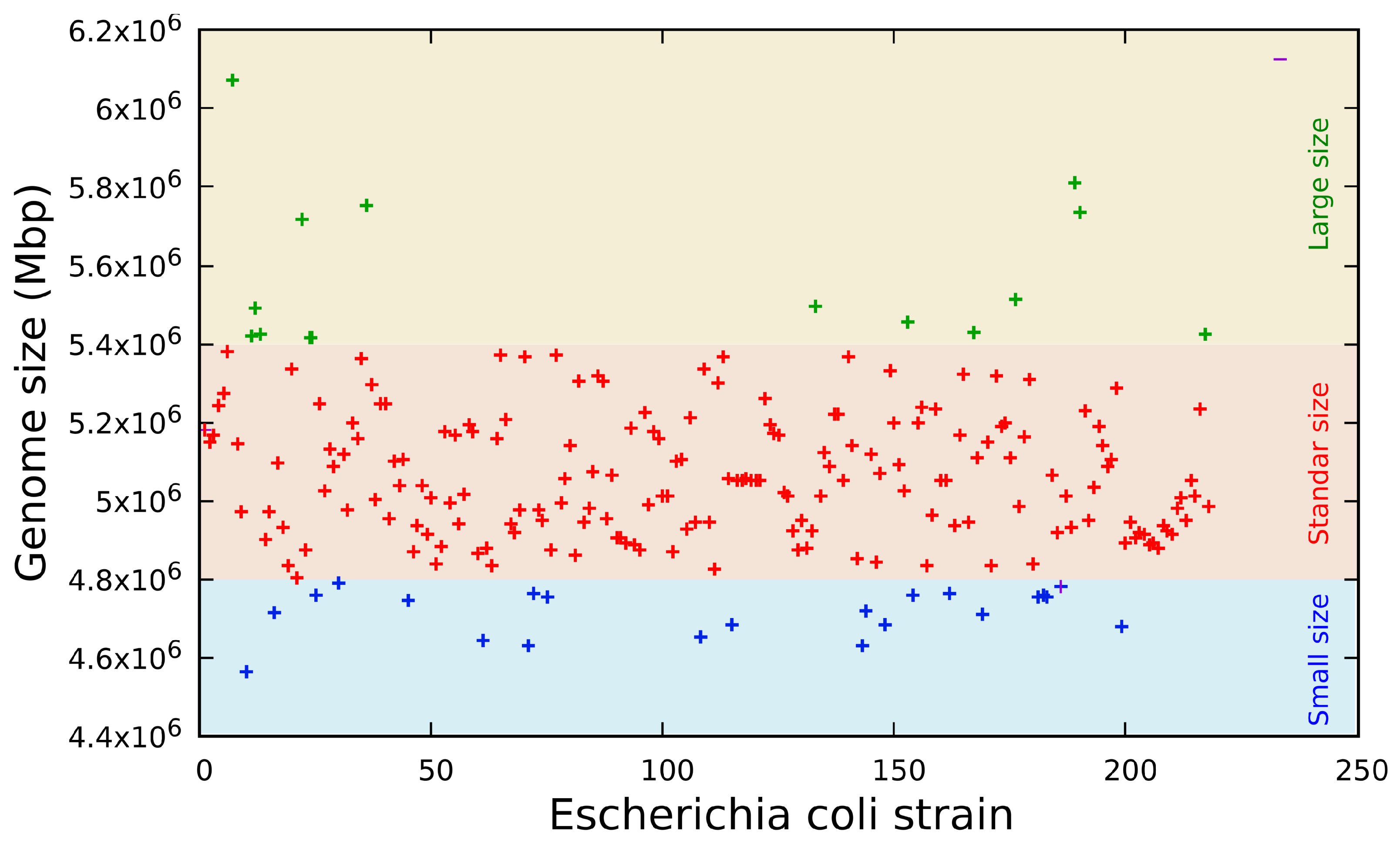
The genome sequence revealed three distinct groups (Figure 1) classified by size in base pairs: a short genome size composed of sequences from 4.56 to 4.83 Mbp (n = 10, 4.59%), a standard genome size composed of sequences from 4.84 to 5.27 Mbp (n = 176, 80.73%) and a long genome size composed of sequences from 5.28 to 6.07 Mbp (n = 32, 14.68%). These genome sizes are consistent with previous data published on the 61 genomes of Escherichia coli [43] and the data bank published by the SRA of NCBI [44].

4.3. Beta-Lactam Gene Families

The isolated Escherichia coli strains were cultured and selected in the presence of beta-lactam drugs in liquid culture media (Table 4). All strains were expected to be carriers of at least one member of the different beta-lactamase gene families. Real-time PCR was used to identify the gene families transcribed and the phenotype responsible for beta-lactam resistance. The results showed that 95.87% of the isolated strains were positive for blaTEM, 71.10% were positive for blaCTX, 33.94% were positive for blaSHV, 29.82% were positive for blaBIL, 19.27% were positive for blaDHA, 18.81% were positive for blaCMY, 11.01% were positive for blaIMP, 6.88% were positive for blaLAP, 14.59% were positive for blaP, 4.13% were positive for blaVIM, 1.82% were positive for blaCTX and 0.92% were positive for both blaKPC and blaCTX (Table 8). No Escherichia coli strains positive for members of the blaOXA and blaROB gene families were found in all obtained strains from patients with nosocomial infections. In this study, the blaTEM gene family was detected in 95.87% of the studied Escherichia coli bacteria from patients diagnosed with nosocomial infection in the“October first” Regional Hospital. This result is consistent with previous reports that found the blaTEM gene family in 74.5% of all multi-drug-resistant ESBL-producing Escherichia coli isolates from hospitals in Malaysia [45]. The seven most frequent gene families that code for beta-lactamases expressed by Escherichia coli that cause nosocomial infections are blaTEM, blaCTX, blaSHV, blaBIL, blaDHA, blaCMY and blaIMP, representing 93.85% of the beta-lactamase problem. However, the blaTEM, blaCTX and blaSHV families together represent the greatest problem with regard to resistance to beta-lactam drugs, as they are expressed in 67.38% of all multi-resistant Escherichia coli strains that cause nosocomial infections in the in-patients of the “October first” Regional Hospital of the ISSSTE. These results are consistent with previous works that found that Escherichia coli isolated from in-hospital patients expressed different members of the blaCTX, blaSHV and blaTEM gene families [46,47,48,49,50,51]. On the other hand, the gene families blaBIL, blaDHA, blaCMY, blaIMP, blaLAP, blaP, blaVIM, blaCTX and blaKPC represent 32.62% of the beta-lactam antibiotic resistance problem. These results are consistent with previous reports where the expression of the following gene families in Escherichia coli isolated from in-hospital patients was found: blaBIL [52], blaDHA [53], blaCMY [54], blaIMP, blaLAP, which has been associated with quinolone resistance gene expression [55] and blaP [56].

4.4. Beta-Lactamase Genome Family Resistome

Out of the 218 Escherichia coli strains resistant to beta-lactam drugs, isolated from 351 in-hospital patients with nosocomial infections, a total of 52 different combinations of gene families coding for beta-lactamases were identified as contributing to the beta-lactam resistome. Interestingly, 13 of the most frequently occurring combinations accounted for a substantial 72.9% of all beta-lactam resistomes (Table 6). On the other hand, the remaining 40 less common combinations were responsible just for 27.1% of all multi-resistance to beta-lactam drugs (Table 7). The common combinations consisted of one to six distinct gene families that encode beta-lactamases (Table 6), whereas the less common combinations were composed of two to nine different gene families coding for beta-lactamases (Table 7).
These observations lead us to postulate that the rare combinations represent natural trials in which beta-lactam resistomes are formed by integrating a large number of beta-lactamase gene families. These combinations are likely to be subjected to selection pressure and might potentially evolve into highly efficient resistomes in the future. However, currently, the most successful resistomes are the common ones composed of six or fewer gene families, as they are prevalent in the majority of nosocomial infections caused by multi-resistant Escherichia coli strains.

4.5. Genomic Beta-Lactamase Intrafamilial Variability

4.5.1. blaTEM Gene Family

The sequencing data revealed that 95.87% of all resistant Escherichia coli strains carried a member of the blaTEM gene family, with 21.56% of them exhibiting a unique beta-lactam resistance mechanism. In contrast, 74.31% of the strains (Table 6) showed multiple beta-lactam resistance mechanisms. These findings align with previous reports where the blaTEM gene family (n = 61 strains, 100%) was identified as the primary beta-lactam drug resistance mechanism in Escherichia coli isolated from the river Yamuna [57]. Characterization of the blaTEM members identified six different alleles of this gene family, including blaTEM-1 expressed in 55.5% of all isolated Escherichia coli strains, followed by blaTEM-12 in 13.88%, blaTEM-2 in 10.05%, blaTEM-52 in 9.09%, blaTEM-24 in 7.18% and blaTEM-10 in 4.31% (Table 6). These findings are consistent with another study where 24% (n = 48) of strains obtained from five hospitals in Tehran were found to carry the blaTEM-1 gene [58]. The blaTEM-1 enzyme is particularly noteworthy as it hydrolyzes ampicillin at a higher rate compared to carbenicillin, oxacillin and cephalothin but does not hydrolyze extended-spectrum cephalosporins. Furthermore, blaTEM-1 is considered an ancestral gene for blaTEM-10 and blaTEM-12, which are among the most commonly encountered blaTEM alleles in the United States of America [59].
Additionally, the blaTEM-2 allele exhibits similar hydrolytic activity to blaTEM-1 but possesses greater transcriptional activity [60]. Another less common beta-lactamase identified in this study includes blaTEM-24 and blaTEM-52, both of which have been described in bacteria-causing nosocomial infections in a Tunisian hospital [61]. These findings are consistent with similar results published by a Korean group from Clinical Isolates of Escherichia coli expressing the blaTEM-1, blaTEM-19, blaTEM-20 and blaTEM-52 genes [62], conferring resistance characteristics to penicillins, aztreonam and cephalosporins. The expression of all these genes has been previously cloned, sequenced and reported in Escherichia coli strains [63].

4.5.2. blaCTX Gene Family

The second most frequent gene family identified in our study was blaCTX (54.72%), which was expressed in six isoforms, distributed across three clusters. Within the blaCTX-M-1 cluster, we found two members: blaCTX-M-1, present in 18.06% and blaCTX-M-15, found in 68.39% of the samples. In the blaCTX-9 cluster, only theblaCTX-M-27 was detected in 4.52% of the strains. The blaCTX-M-25 cluster had the blaCTX-M-100 isoform as its sole member, accounting for 9.03% of the isolates. Notably, the blaCTX-M-15 isoform emerged as the most dominant among the clusters. These findings are consistent with results obtained from clinically significant Escherichia coli isolates in Kuwait Hospitals, where the blaCTX-M-15 isoform was present in 84.1% of all isolates (Table 6) [64]. Additionally, another study conducted in the UK using Escherichia coli-producing CTX-M extended-spectrum beta-lactamases identified the blaCTX-M-15 isoform in 95.9% of all studied strains [65]. In contrast, our study found the blaCTX-M-1 isoform in 18.06% of the samples. Interestingly, previous works published by three university hospitals in Tehran reported that 61.8% of 144 CTX-resistant Escherichia coli isolates carried the blaCTX-M-1 gene [66], conferring resistance characteristics to penicillin, aztreonam and cephalosporin. The expression of all these genes has been previously cloned, sequenced and reported in Escherichia coli strains [67].

4.5.3. blaSHV Gene Family

The third most frequently expressed beta-lactamase by Escherichia coli strains causing nosocomial infections was blaSHV, accounting for 33.94% of all analyzed multi-resistant strains. Within this gene family, five isoforms were identified, with blaSHV-12 being the most dominant (78.38%), followed by blaSHV-1 (9.46%), blaSHV-100 (5.41%), blaSHV-121 (4.05%) and blaSHV-11 (2.70%) (Table 6). Although all isoforms were expressed at low frequencies, blaSHV-12 stood out as the most prevalent member, isolated from diverse sources in a recent study conducted in Catalonia, where it was found at a frequency of 23% [68]. These isoforms confer characteristic resistance to penicillin, aztreonam and cephalosporin. The expression of all these gene isoforms has been previously cloned, sequenced and reported in Escherichia coli strains [69].

4.5.4. blaIMP Gene Family

The blaIMP gene family was detected in 11.01% of the strains and it comprises six different members, namely blaIMP-1 (8.33%; n = 2), blaIMP-2 (4.17%; n = 1), blaIMP-4 (4.17%; n = 1), blaIMP-6 (4.17%, n = 1), blaIMP-11 (8.33%; n = 2) and the dominant gene blaIMP-14 (8.33%; n = 17). These genes confer characteristic resistance to imipenem and carbapenem [55]. The expression of all these genes has been previously cloned and sequenced from Escherichia coli strains [70].

4.5.5. blaCTX-M, blaVIM and blaP Gene Family

All of these beta-lactamase gene families were expressed in only one isoform each. Specifically, the blaCTX-M-15 isoform was found in 1.82% (n = 4) of the strains, blaVIM-1 in 4.13% (n = 9) and blaP in 6.88% (n = 15) of the 218 Escherichia coli strains studied, isolated from patients attending the “October first” Regional Hospital in Mexico City. These specific isoforms of blaCTX-M-15, blaVIM-1 and blaP beta-lactamases were detected in the given samples, which might explain the observed differences from their more commonly reported counterparts worldwide, like blaCTX-M-1 and blaCTX-M-15, which are frequently identified extended-spectrum beta-lactamases (ESBLs) globally. The prevalence of specific beta-lactamase isoforms could be influenced by the sample type and origin.
It is worth noting that both blaCTX-M-1, blaVIM-1 and blaP beta-lactamases have been previously cloned and sequenced in Escherichia coli strains [71,72,73].

4.5.6. blaLAP and blaKPC Gene Families

The blaLAP gene was expressed in 6.88% of the isolated Escherichia coli strains, with only two detected isoforms: blaLAP-1 was the dominant gene expressed, accounting for 73.33% of all positive blaLAP strains, followed by the blaLAP-2 gene isoform, representing 26.67%. This gene, in combination with other beta-lactamases, contributes to the multi-drug resistance characteristics [74].
Similarly, the blaKPC gene family was expressed in two isoforms, namely blaKPC-2 and blaKPC-3, both of which have been previously reported as members of the Escherichia coli genotype [75,76]. These isoforms provide carbapenem resistance characteristics [77].

4.5.7. blaBIL, blaDHA and blaCMY Gene Families

The blaBIL gene family was the fourth most frequent, but only the blaBIL-1 isoform was identified. Previous reports have confirmed the expression of blaBIL-1 in Escherichia coli, conferring resistance characteristics to penicillin, carbenicillin, cefaclor and cephalosporin [52].
Additionally, the blaDHA (19.27%) and blaCMY (18.81%) gene families were expressed at similar frequencies. Within the blaDHA gene family, three isoforms were identified: blaDHA-1 (80.95%), blaDHA-2 (14.29%) and blaDHA-7 (4.76%). The blaCMY gene family was expressed in four isoforms: blaCMY-2 (90.24%), blaCMY-38 (4.88%), blaCMY-9 and blaCMY-12 (both expressed in 2.44%). The most dominant isoforms carried by Escherichia coli-causing nosocomial infections were blaDHA-1 and blaCMY-2. These genes were previously described as co-expressed with the blaCTX-M-15 isoform in a multi-resistant Escherichia coli strain causing the death of a puppy in Italy, indicating ESBL characteristics [78].
All of the blaBIL, blaDHA and blaCMY genes found in this study have been previously cloned and sequenced in Escherichia coli [52,79,80].

4.6. Beta-Lactam Resistome

The beta-lactam antibiotics, particularly penicillin, ampicillin and third-generation cephalosporin, play a crucial role in the treatment of nosocomial infections caused by Escherichia coli [81]. However, their extensive use in hospitals exerts significant selection pressure on the entire bacteriome, leading to the emergence of new beta-lactamases. This has become the most significant mechanism of resistance to beta-lactam antibiotics [82]. These resistance mechanisms are encoded in the bacterial chromosome, plasmids or transposons, facilitating the rapid spread of beta-lactamase genes among bacterial populations. As a result, the development of novel strategies to combat antibiotic resistance becomes essential to tackle the growing challenges posed by nosocomial infections.
In this study, our focus was on the identification of 14 of the most clinically important beta-lactamase families. These included the blaBIL, blaCFX, blaCMY, blaCTX, blaDHA, blaIMP, blaKPC, blaLAP, blaOXA, blaP, blaROB, blaSHV, blaTEM and blaVIM gene families, collectively covering 99% of the beta-lactam resistance reported in Escherichia coli strains causing various intestinal, extraintestinal and systemic pathologies [83,84]. By focusing on these specific gene families, we aimed to gain valuable insights into the prevalence and diversity of beta-lactam-resistance mechanisms in clinically relevant Escherichia coli strains, which is crucial for understanding and combating antibiotic resistance in medical settings.
Among the Escherichia coli strains analyzed, we observed that they express from one to nine members of beta-lactamase gene families. It is worth noting that the accumulation of four or more members of the beta-lactamase gene families is a rare occurrence, as they are often unique strains. Nonetheless, these strains remain of utmost importance as they represent potential sources for the expansion of pan-resistant Escherichia coli strains with resistance to all beta-lactam drugs. The presence and diversity of these beta-lactamase genes emphasize the urgent need for effective measures to control and combat antibiotic resistance in nosocomial infections
In our study, the dominant beta-lactamase genotypes in the analyzed Escherichia coli strains were formed by blaTEM (21.6%) and its combinations with blaCTX (19.3%), blaCTX and blaSHV (6%) and blaCTX and blaBIL (5.5%). Together, these four genotypes constituted 52.4% of all identified genotypes. Based on these findings, the analyzed Escherichia coli strains are classified as extended-spectrum beta-lactamase-producing (ESBL) strains. The remaining combinations of beta-lactamase gene families represented from 0.5% to 4.1% each, but they still represent pan-resistant Escherichia coli strains against beta-lactam drugs. Our method of evaluation using real-time PCR and corroborated by next-generation sequencing provides a comprehensive panorama of the beta-lactam drug resistome in the analyzed Escherichia coli strains.
These results are in line with previous reports indicating that Escherichia coli is the bacterial species that most frequently produces ESBLs [85]. Similarly, in Klebsiella pneumoniae, it has been reported that multiple beta-lactamase gene carriers express from two to four different members of the beta-lactamase gene families [86]. However, it is essential to note that our method provides a broader and more generalized perspective of the beta-lactam drug resistome in the studied Escherichia coli strains, allowing for a comprehensive understanding of the resistance mechanisms.
The molecular methods used for the identification and characterization of the Escherichia coli beta-lactam resistome are of critical importance in drug therapy. In nosocomial infections caused by ESBL-producing Escherichia coli, the available drug therapy options are significantly reduced due to the pan-resistant phenotype. This can lead to an increase in mortality rates, prolonged hospitalization time and higher healthcare costs [87,88]. Patients who have acquired nosocomial infections caused by ESBL-producing Escherichia coli have been reported to face challenges in treatment [89]. For instance, the identification of Escherichia coli strains producing ESBLs of the CTX-M type has been linked to treatment failure in urinary-tract infections (UTIs) when treated with commonly used antibiotics such as penicillin, ampicillin and first- to fourth-generation cephalosporins. Even next-generation antibiotics like carbapenem and imipenem may prove ineffective against these strains, as some of them have also developed resistance to beta-lactamase inhibitors [90,91].
Our study’s results are consistent with those published in a multicenter study conducted in eight hospitals in Mexico, where the dominant isolated microorganisms were ESBL-producing Escherichia coli strains (83.9%) and many carried genes encoding beta-lactamases of blaSHV and/or blaCTX-M types [92]. These findings emphasize the urgent need for robust molecular methods to identify and characterize beta-lactam resistance in Escherichia coli, as they play a crucial role in guiding appropriate and effective drug therapy decisions for nosocomial infections.

5. Conclusions

The multi-resistance phenotype for beta-lactam drugs observed in Escherichia coli-causing nosocomial infections for in-hospital patients at different services within the “October first” Regional Hospital is primarily determined by genes belonging to six beta-lactamase gene families: blaTEM, blaCTX, blaSHV, blaBIL, blaDHA and blaCMY. These gene families, when combined, account for the most frequent phenotypes of the beta-lactam resistome, irrespective of the sample type and tissue source. Among these, the genotypes comprising the genes blaTEM1, blaCTX-M-15, blaSHV-12, blaBIL-1, blaDHA-1 and blaCMY-2 are found to be the most prevalent in the third-level hospital located in Mexico City. These findings provide valuable insights into the molecular basis of beta-lactam resistance in nosocomial Escherichia coli infections, which is essential for optimizing treatment strategies and controlling the spread of antibiotic-resistant strains in the hospital setting.

Author Contributions

Conceptualization, J.A.G.-B.; methodology, R.V.-L., T.H.-M., S.I.L.-F., J.M.-B. and J.A.G.-B.; software, M.L.-L.; validation, R.V.-L., C.P.-L. and J.A.G.-B.; formal analysis, M.L.-L., T.G.-G. and J.A.G.-B.; investigation, J.A.G.-B.; resources, J.A.G.-B.; data curation, M.L.-L., E.G.-C. and J.A.G.-B.; writing—original draft preparation, T.H.-M.; writing—review and editing, J.A.G.-B.; project administration, J.A.G.-B.; funding acquisition, J.A.G.-B. All authors have read and agreed to the published version of the manuscript.

Funding

This work was supported by the National Council for Science and Technology (CONACYT) Mexico through the Sectoral Fund for Research in Health and Social Security (FOSISS), grant MOD.ORD./2012/SSA/IMSS/ISSSTE-CONACYT/000000000181384, and it was also supported by Centro de Investigación en Ciencias de la Salud (CICSA) of Facultad de Ciencias de la Salud Universidad Anáhuac México Norte Project Number 202324.

Institutional Review Board Statement

The protocol was approved by the Institutional Review Board of the “October 1st” Regional Hospital, ISSSTE, approval code E015-591.

Informed Consent Statement

Not applicable.

Data Availability Statement

The data presented in this study are available from the corresponding author.

Acknowledgments

The authors thankfully acknowledge Luz María Goti Rodríguez (Principal of Hospital Regional “Primero de Octubre”) and Tomás Barrientos (Faculty of Health Sciences University Anahuac) for providing the necessary laboratory facilities for the study.

Conflicts of Interest

The authors declare no conflict of interest.

Sample Availability

Samples of the compounds are available from the authors.

Abbreviations

The following abbreviations are used in this manuscript:
ADHarginine dehydrolase
AMampicillin
ANamikasin
ARAarabidosa
ATMaztreonam
blabeta-lactamase gene encoding
blaAmpCbeta-lactamase gene encoding chromosomal cephalosporinase
blaBILbeta-lactamase identification in Bilal patient
blaCMYbeta-lactamase gene encoding cephamycinase
blaCTXbeta-lactamase gene encoding cefotaximase, Munich
blaDHAbeta-lactamase gene discovered at Dhahran, Saudi Arabia
blaGESbeta-lactamase gene encoding Guiana—extended spectrum
blaIMPbeta-lactamase gene encoding active imipenem
blaKPCbeta-lactamase gene encoding Klebsiella pneumoniae carbapenemase
blaLAPbeta-lactamase gene encoding
blaNMCbeta-lactamase gene encoding not metalloenzyme carbapenemase
blaOXAbeta-lactamase gene encoding oxacilinases
blaROBbeta-lactamase gene named after patient
blaSFCbeta-lactamase gene identified in Serratia fonticola resistant to carbapenem
blaSHVbeta-lactamase gene encoding sulfhydryl reagent variable
blaSMEbeta-lactamase gene encoding Serratia marcescens enzyme
blaTEMbeta-lactamase gene encoding identified in Temoneira patient
blaVEBgene encoding Vietnam extended-spectrum beta-lactamase
blaVIMbeta-lactamase gene encoding Verona imipenemase
CANcefotetan
CAUTIcatheter-associated urinary-tract infections
CIPciprofloxacin
CITcitrate
CLABSIcentral line-associated bloodstream infections
CMEceftazidime
CROceftriaxone
CXIcefuroxime
CZcefazolin
FEPcefepime
FTnitrofurantoin
GANgatifloxacin
GELgelatinase
GIgastrointestinal infections
GLUfermentation/oxidation of glucose
GMgentamicin
H2Shydrogen sulfide
IEMimipenem
INDindole production
INDindole
LDClysine decarboxylase
LDLlysine decarboxylase
MANmannitose
MAR indexmultiple-antibiotic-resistance index
MDRmulti-drug-resistant bacteria
MELmelobilosa
MEMmeropenem
NV-HAPnon-ventilator-associated hospital-acquired pneumonia
ODCornithine decarboxylase
OPNGbeta-galactosides
OXcytochrome oxidase
PCRpolymerase chain reaction
PTZpiperacillin/tazobactam
Rresistant
RHArhamnose
Ssensitive
SACsucrose
SAMampicillin/sulbactam
SORsorbitol
SSIsurgical-site infections
STXtrimethoprim/sulfamethoxasol
TCAticarcillin/clavulanic acid
TDAtryptophan deaminase
TMtobramycin
UREurease
UTIurinary-tract infections
VAPventilator-associated pneumonia
VPVoges–Proskauer

References

  1. Khan, H.A.; Baig, F.K.; Mehboob, R. Nosocomial infections: Epidemiology, prevention, control and surveillance. Asian Pac. J. Trop. Biomed. 2017, 7, 478–482. [Google Scholar] [CrossRef]
  2. Bueno-Cavanillas, A.; Delgado-Rodríguez, M.; López-Luque, A.; Schaffino-Cano, S.; Gálvez-Vargas, R. Influence of nosocomial infection on mortality rate in an intensive care unit. Crit. Care Med. 1994, 22, 55–60. [Google Scholar] [CrossRef] [PubMed]
  3. Schabrun, S.; Chipchase, L. Healthcare equipment as a source of nosocomial infection: A systematic review. J. Hosp. Infect. 2006, 63, 239–245. [Google Scholar] [CrossRef] [PubMed]
  4. Magill, S.S.; O’Leary, E.; Janelle, S.J.; Thompson, D.L.; Dumyati, G.; Nadle, J.; Wilson, L.E.; Kainer, M.A.; Lynfield, R.; Greissman, S.; et al. Emerging Infections Program Hospital Prevalence Survey Team. Changes in prevalence of health care-associated infections in US hospitals. N. Engl. J. Med. 2018, 379, 1732–1744. [Google Scholar] [CrossRef] [PubMed]
  5. Magill, S.S.; Edwards, J.R.; Fridkin, S.K. Survey of health care-associated infections. N. Engl. J. Med. 2014, 370, 2542–2543. [Google Scholar] [CrossRef]
  6. Wen, X.M.; Ren, N.; Wu, A.H.; Xu, X.H. Distribution of pathogens causing nosocomial infection monitored by national nosocomial infection surveillance system and changing trend. Chin. J. Nosocomiol. 2011, 2, 350–355. [Google Scholar]
  7. Sikora, A.; Zahra, F. Nosocomial Infections. StatPearls Internet. 2021. Available online: https://www.ncbi.nlm.nih.gov/books/NBK559312/ (accessed on 23 September 2021).
  8. Ludden, C.; Coll, F.; Gouliouris, T.; Restif, O.; Blane, B.; Blackwell, G.A.; Kumar, N.; Naydenova, P.; Crawley, C.; Brown, N.M.; et al. Defining nosocomial transmission of Escherichia coli and antimicrobial resistance genes: A genomic surveillance study. Lancet Microbe 2021, 2, e472–e480. [Google Scholar] [CrossRef]
  9. Rocha, F.R.; Pinto, V.P.; Barbosa, F.C. The Spread of CTX-M-Type Extended-Spectrum β-Lactamases in Brazil: A Systematic Review. Microb. Drug Resist. 2016, 22, 301–311. [Google Scholar] [CrossRef]
  10. Nasser, M.; Palwe, S.; Bhargava, R.N.; Feuilloley, M.G.J.; Kharat, A.S. Retrospective Analysis on Antimicrobial Resistance Trends and Prevalence of beta-lactamases in Escherichia coli and ESKAPE Pathogens Isolated from Arabian Patients during 2000–2020. Microorganisms 2020, 8, 1626. [Google Scholar] [CrossRef]
  11. Zhu, M.; Yang, G.; Li, A.; Zong, L.; Dong, Z.; Lu, J.; Zhang, K.; Cheng, C.; Chang, Q.; Wu, X.; et al. Identification and molecular characterization of Escherichia coli blaSHV genes in a Chinese teaching hospital. Gene 2017, 600, 29–35. [Google Scholar] [CrossRef]
  12. Dizaji, A.S.; Fathi, R.; Sales, A.J. Molecular study of extended-spectrum beta-lactamase (TEM-1) gene in Escherichia coli isolates collected from Ostad Alinasab Hospital in Tabriz Iran. MMJ 2016, 29, 35–40. [Google Scholar] [CrossRef]
  13. Fonseca, F.; Sarmento, A.C.; Henriques, I.; Samyn, B.; Van Beeumen, J.; Domingues, P.; Domingues, M.R.; Saavedra, M.J.; Correia, A. Biochemical characterization of SFC-1, a class A carbapenem-hydrolyzing β-lactamase. Antimicrob. Agents Chemother. 2007, 51, 4512–4514. [Google Scholar] [CrossRef]
  14. Kocer, K.; Klein, S.; Hildebrand, D.; Krall, J.; Heeg, K.; Boutin, S.; Nurjadi, D. Pitfalls in genotypic antimicrobial susceptibility testing caused by low expression of blaKPC in Escherichia coli. J. Antimicrob. Chemother. 2021, 76, 2795–2801. [Google Scholar] [CrossRef] [PubMed]
  15. Alonso, N.; Miró, E.; Pascual, V.; Rivera, A.; Simó, M.; Garcia, M.C.; Xercavins, M.; Morera, M.A.; Espejo, E.; Gurguí, M.; et al. Molecular characterisation of acquired and overproduced chromosomal blaAmpC in Escherichia coli clinical isolates. Int. J. Antimicrob. Agents 2016, 47, 62–68. [Google Scholar] [CrossRef] [PubMed]
  16. Vourli, S.; Giakkoupi, P.; Miriagou, V.; Tzelepi, E.; Vatopoulos, A.C.; Tzouvelekis, L.S. Novel GES/IBC extended-spectrum β-lactamase variants with carbapenemase activity in clinical enterobacteria. FEMS Microbiol. Lett. 2006, 234, 209–231. [Google Scholar] [CrossRef]
  17. Zhang, Y.; Kashikar, A.; Brown, C.A.; Denys, G.; Bush, K. Unusual Escherichia coli PBP 3 insertion sequence identified from a collection of carbapenem-resistant Enterobacteriaceae tested in vitro with a combination of ceftazidime-, ceftaroline- or aztreonam-avibactam. Antimicrob. Agents Chemother. 2017, 61, e00389-17. [Google Scholar] [CrossRef]
  18. Bauernfeind, A.; Stemplinger, I.; Jungwirth, R.; Mangold, P.; Amann, S.; Akalin, E.; Ang, O.; Bal, C.; Casellas, J.M. Characterization of beta-lactamase gene blaPER-2, which encodes an extended-spectrum class A beta-lactamase. Antimicrob. Agents Chemother. 1996, 40, 616–620. [Google Scholar] [CrossRef]
  19. Bush, K.; Jacoby, G.A.; Medeiros, A.A. A functional classification scheme for beta-lactamases and its correlation with molecular structure. Antimicrob. Agents Chemother. 1995, 39, 1211–1213. [Google Scholar] [CrossRef]
  20. Vázquez-López, R.; Solano-Gálvez, S.; León-Chávez, B.A.; Thompson-Bonilla, M.R.; Guerrero-González, T.; Gómez-Conde, E.; Martínez-Fong, D.; González-Barrios, J.A. Characterization of gene families encoding beta-lactamases of gram-negative rods isolated from ready-to-eat vegetables in Mexico City. High-Throughput 2018, 7, 36. [Google Scholar] [CrossRef]
  21. ClustalW v.2 Software. Available online: http://www.clustal.org/clustal2/ (accessed on 23 September 2021).
  22. FigTree V1.4.0 Software. Available online: http://tree.bio.ed.ac.uk/software/figtree/ (accessed on 23 September 2021).
  23. PerPrimer v1.1.21 Software. Available online: http://perlprimer.sourceforge.net (accessed on 23 September 2021).
  24. Babraham Bioinformatics. Available online: https://www.bioinformatics.babraham.ac.uk/projects/fastqc/ (accessed on 23 September 2021).
  25. CodonCode Aligner. Available online: https://www.codoncode.com/aligner/ (accessed on 23 September 2021).
  26. Kraken Taxonomic Sequence Classification System. Available online: https://ccb.jhu.edu/software/kraken/ (accessed on 23 September 2021).
  27. Basic Local Alignment Search Tool. Available online: https://blast.ncbi.nlm.nih.gov/Blast.cgi (accessed on 23 September 2021).
  28. Bonten, M.; Johnson, J.R.; van den Biggelaar, A.H.; Georgalis, L.; Geurtsen, J.; de Palacios, P.I.; Gravenstein, S.; Verstraeten, T.; Hermans, P.; Poolman, J.T. Epidemiology of Escherichia coli bacteremia: A systematic literature review. Clin. Infect. Dis. 2021, 72, 1211–1219. [Google Scholar] [CrossRef]
  29. Garc’ıa-Morúa, A.; Hernández-Torres, A.; Salazar-de-Hoyos, J.L.; Jaime-Dávila, R.; Gómez-Guerra, L.S. Community-acquired urinary-tract infection etiology and antibiotic resistance in a Mexican population group. Rev. Mex. Urol. 2009, 69, 45–48. [Google Scholar]
  30. Lubbert, C. Antimicrobial therapy of acute diarrhoea: A clinical review. Expert Rev. Anti-Infect. Ther. 2016, 14, 193–206. [Google Scholar] [CrossRef] [PubMed]
  31. Zhu, M.L.; Mai, J.Y.; Zhu, J.H.; Lin, Z.L. Clinical analysis of 31 cases of neonatal purulent meningitis caused by Escherichia coli. Zhongguo dang dai er ke za zhi = Chin. J. Contemp. Pediatr. 2012, 14, 910–912. [Google Scholar]
  32. Liu, J.; Yan, Q.; Luo, F.; Shang, D.; Wu, D.; Zhang, H.; Shang, X.; Kang, X.; Abdo, M.; Liu, B.; et al. Acute cholecystitis associated with infection of Enterobacteriaceae from gut microbiota. Clin. Microbiol. Infect. 2015, 21, 851-e1. [Google Scholar] [CrossRef] [PubMed]
  33. Pérez-Fontán, M.; Lueiro, F. Escherichia coli peritonitis in patients undergoing peritoneal dialysis: A serious problem that may get worse. Perit. Dial. Int. 2006, 26, 174–177. [Google Scholar] [CrossRef]
  34. Shibata, S.; Tanizaki, R.; Watanabe, K.; Makabe, K.; Shoda, N.; Kutsuna, S.; Nagamatsu, M.; Oka, S.; Ohmagari, N. Escherichia coli vertebral osteomyelitis diagnosed according to broad-range 16S rRNA gene polymerase chain reaction (PCR). Intern. Med. 2015, 54, 3237–3240. [Google Scholar] [CrossRef]
  35. Keithlin, J.; Sargeant, J.; Thomas, M.K.; Fazil, A. Chronic sequelae of E. coli O157: Systematic review and meta-analysis of the proportion of E. coli O157 cases that develop chronic sequelae. Foodborne Pathog. Dis. 2014, 11, 79–95. [Google Scholar] [CrossRef]
  36. Kabir, M.S.; Joarder, A.H.; Ekramuddaula, F.M.; Uddin, M.M.; Islam, M.R.; Habib, M.A. Pattern of chronic suppurative otitis media. Mymensingh Med. J. MMJ 2012, 21, 270–275. [Google Scholar]
  37. Dale, A.P.; Woodford, N. Extra-intestinal pathogenic Escherichia coli (ExPEC): Disease, carriage and clones. J. Infect. 2015, 71, 615–626. [Google Scholar] [CrossRef]
  38. Sereia, A.F.; Christoff, A.P.; Cruz, G.N.; da Cunha, P.A.; da Cruz, G.C.; Tartari, D.C.; Zamparette, C.P.; Klein, T.C.; Masukawa, I.I.; Silva, C.I.; et al. Healthcare-Associated Infections-Related Bacteriome and Antimicrobial Resistance Profiling: Assessing Contamination Hotspots in a Developing Country Public Hospital. Front. Microbiol. 2021, 12, 711471. [Google Scholar] [CrossRef]
  39. Joseph, A.A.; Odimayo, M.S.; Olokoba, L.B.; Olokoba, A.B.; Popoola, G.O. Multiple antibiotic resistance iIndex of Escherichia coli isolates in a tertiary hospital in south-west Nigeria. Med. J. Zamb. 2017, 44, 225–232. [Google Scholar]
  40. Chakrawarti, A.; Dongol, P.; Khanal, H.; Subba, P.; Benerjee, J.J. Extended spectrum beta lactamases detection and multiple-antibiotic-resistance indexing of Escherichia coli from urine samples of patients from a referral hospital of eastern Nepal. Int. J. Appl. Sci. Biotechnol. 2015, 3, 423–426. [Google Scholar] [CrossRef]
  41. Peters, L.; Olson, L.; Khu, D.T.; Linnros, S.; Le, N.K.; Hanberger, H.; Hoang, N.T.; Tran, D.M.; Larsson, M. Multiple antibiotic resistance as a risk factor for mortality and prolonged hospital stay: A cohort study among neonatal intensive care patients with hospital-acquired infections caused by gram-negative bacteria in Vietnam. PLoS ONE 2019, 14, e0215666. [Google Scholar] [CrossRef] [PubMed]
  42. Cosgrove, S.E. The relationship between antimicrobial resistance and patient outcomes: Mortality, length of hospital stay and health care costs. Clin. Infect. Dis. 2006, 42 (Suppl. S2), S82–S89. [Google Scholar] [CrossRef]
  43. Lukjancenko, O.; Wassenaar, T.M.; Ussery, D.W. Comparison of 61 sequenced Escherichia coli genomes. Microb. Ecol. 2010, 60, 708–720. [Google Scholar] [CrossRef]
  44. SRA of NCBI. Available online: ttps://www.ncbi.nlm.nih.gov/sra (accessed on 23 September 2021).
  45. Lim, K.T.; Yasin, R.; Yeo, C.C.; Puthucheary, S.; Thong, K.L. Characterization of multidrug resistant ESBL-producing Escherichia coli isolates from hospitals in Malaysia. J. Biomed. Biotechnol. 2009, 2009, 165637. [Google Scholar] [CrossRef]
  46. Shahid, M.; Singh, A.; Sobia, F.; Rashid, M.; Malik, A.; Shukla, I.; Khan, H.M. blaCTX-M, blaTEM and blaSHV in Enterobacteriaceae from North-Indian tertiary hospital: High occurrence of combination genes. Asian Pac. J. Trop. Med. 2011, 4, 101–105. [Google Scholar] [CrossRef]
  47. Abdi, S.; Ranjbar, R.; Vala, M.H.; Jonaidi, N.; Bejestany, O.B.; Bejestany, F.B. Frequency of bla TEM, bla SHV, bla CTX-M and qnrA among Escherichia coli isolated from urinary-tract infection. Arch. Clin. Infect. Dis. 2014, 9, 1–5. [Google Scholar] [CrossRef]
  48. Bali, E.B.; Accedil, L.; Sultan, N. Phenotypic and molecular characterization of SHV, TEM, CTX-M and extended-spectrum-lactamase produced by Escherichia coli, Acinobacter baumannii and Klebsiella isolates in a Turkish hospital. Afr. J. Microbiol. Res. 2010, 4, 650–654. [Google Scholar]
  49. Nojoomi, F.; Ghasemian, A. Prevalence of ESBL phenotype, blaCTX-M-1, blaSHV and blaTEM genes among uropathogenic Escherichia coli isolates from 3 military hospitals of Tehran, Iran. J. Coast. Life Med. 2016, 4, 616–618. [Google Scholar] [CrossRef]
  50. Pishtiwan, A.H.; Khadija, K.M. Prevalence of blaTEM, blaSHV and blaCTX-M genes among ESBL-producing Klebsiella pneumoniae and Escherichia coli isolated from thalassemia patients in Erbil, Iraq. Mediterr. J. Hematol. Infect. Dis. 2019, 11, e2019041. [Google Scholar] [CrossRef]
  51. Ugbo, E.N.; Anyamene, C.O.; Moses, I.B.; Iroha, I.R.; Babalola, O.O.; Ukpai, E.G.; Chukwunwejim, C.R.; Egbule, C.U.; Emioye, A.A.; Okata-Nwali, O.D.; et al. Prevalence of blaTEM, blaSHV and blaCTX-M genes among extended spectrum beta-lactamase-producing Escherichia coli and Klebsiella pneumoniae of clinical origin. Gene Rep. 2020, 21, 100909. [Google Scholar] [CrossRef]
  52. Fosberry, A.P.; Payne, D.J.; Lawlor, E.J.; Hodgson, J.E. Cloning and sequence analysis of blaBIL-1, a plasmid-mediated class C beta-lactamase gene in Escherichia coli BS. Antimicrob. Agents Chemother. 1994, 38, 1182–1185. [Google Scholar] [CrossRef]
  53. Ding, H.; Yang, Y.; Lu, Q.; Wang, Y.; Chen, Y.; Deng, L.; Wang, A.; Deng, Q.; Zhang, H.; Wang, C.; et al. The prevalence of plasmid-mediated AmpC β-lactamases among clinical isolates of Escherichia coli and Klebsiella pneumoniae from five children’s hospitals in China. Eur. J. Clin. Microbiol. Infect. Dis. 2008, 27, 915–921. [Google Scholar] [CrossRef]
  54. Piazza, A.; Comandatore, F.; Romeri, F.; Pagani, C.; Mattioni Marchetti, V.; Brilli, M.; Panelli, S.; Migliavacca, R.; Ridolfo, A.; Olivieri, P.; et al. Detection of ST1702 Escherichia coli blaNDM-5 and blaCMY-42 genes positive isolates from a Northern Italian hospital. New Microbiol. 2018, 41, 230–231. [Google Scholar]
  55. Kizny Gordon, A.; Phan, H.T.; Lipworth, S.I.; Cheong, E.; Gottlieb, T.; George, S.; Peto, T.E.; Mathers, A.J.; Walker, A.S.; Crook, D.W.; et al. Genomic dynamics of species and mobile genetic elements in a prolonged bla IMP-4-associated carbapenemase outbreak in an Australian hospital. J. Antimicrob. Chemother. 2020, 75, 873–882. [Google Scholar] [CrossRef]
  56. Grossman, M.J.; Lampen, J.O. Purification and DNA binding properties of the blal gene product, repressor for the β-lactamase gene, blaP, of Bacillus licheniformis. Nucleic Acids Res. 1987, 15, 6049–6062. [Google Scholar] [CrossRef]
  57. Singh, N.S.; Singhal, N.; Virdi, J.S. Genetic environment of blaTEM-1, blaCTX-M-15, blaCMY-42 and characterization of integrons of Escherichia coli isolated from an Indian urban aquatic environment. Front. Microbiol. 2018, 9, 382. [Google Scholar] [CrossRef]
  58. Shah, C.f.; Nasiri, S.; Noveyri, H. Detection of blaTEM & blaSHV genes among clinical isolates of E. coli from Tehran hospitals. Iran. J. Med. Microbiol. 2007, 1, 1–8. [Google Scholar]
  59. Paterson, D.L.; Bonomo, R.A. Extended-spectrum β-lactamases: A clinical update. Clin. Microbiol. Rev. 2005, 18, 657–686. [Google Scholar] [CrossRef]
  60. Ali, T.; Ali, I.; Khan, N.A.; Han, B.; Gao, J. The growing genetic and functional diversity of extended spectrum beta-lactamases. BioMed Res. Int. 2018, 2018, 9519718. [Google Scholar] [CrossRef]
  61. Mahrouki, S.; Chihi, H.; Bourouis, A.; Ayari, K.; Ferjani, M.; Moussa, M.B.; Belhadj, O. Nosocomial dissemination of plasmids carrying blaTEM-24, blaDHA-1, aac (6’)-Ib-cr and qnrA6 in Providencia spp. strains isolated from a Tunisian hospital. Diagn. Microbiol. Infect. Dis. 2015, 81, 50–52. [Google Scholar] [CrossRef] [PubMed]
  62. Jeong, S.H.; Bae, I.K.; Lee, J.H.; Sohn, S.G.; Kang, G.H.; Jeon, G.J.; Kim, Y.H.; Jeong, B.C.; Lee, S.H. Molecular characterization of extended-spectrum beta-lactamases produced by clinical isolates of Klebsiella pneumoniae and Escherichia coli from a Korean nationwide survey. J. Clin. Microbiol. 2004, 42, 2902–2906. [Google Scholar] [CrossRef] [PubMed]
  63. NCBI-Gene blaTEM. Available online: https://www.ncbi.nlm.nih.gov/pathogens/refgene/#gene_family:(blaTEM) (accessed on 7 October 2021).
  64. Al-Hashem, G.; Al Sweih, N.; Jamal, W.; Rotimi, V.O. Sequence analysis of blaCTX-M genes carried by clinically significant Escherichia coli isolates in Kuwait hospitals. Med. Princ. Pract. 2011, 20, 213–219. [Google Scholar] [CrossRef]
  65. Woodford, N.; Ward, M.E.; Kaufmann, M.E.; Turton, J.; Fagan, E.J.; James, D.; Johnson, A.P.; Pike, R.; Warner, M.; Cheasty, T.; et al. Community and hospital spread of Escherichia coli producing CTX-M extended-spectrum β-lactamases in the UK. J. Antimicrob. Chemother. 2004, 54, 735–743. [Google Scholar] [CrossRef]
  66. Najar Peerayeh, S.; Eslami, M.; Memariani, M.; Siadat, S.D. High prevalence of blaCTX-M-1 group extended-spectrum β-lactamase genes in Escherichia coli isolates from Tehran. Jundishapur J. Microbiol. 2013, 6, e6863. [Google Scholar] [CrossRef]
  67. NCBI-Gene blaCTX. Available online: https://www.ncbi.nlm.nih.gov/pathogens/refgene/#gene_family:(blaCTX-M) (accessed on 7 October 2021).
  68. Darwich, L.; Vidal, A.; Seminati, C.; Albamonte, A.; Casado, A.; López, F.; Molina-López, R.A.; Migura-Garcia, L. High prevalence and diversity of extended-spectrum β-lactamase and emergence of OXA-48 producing Enterobacterales in wildlife in Catalonia. PLoS ONE 2019, 14, e0210686. [Google Scholar] [CrossRef]
  69. NCBI-Gene blaSHV. Available online: https://www.ncbi.nlm.nih.gov/pathogens/refgene/#gene_family:(blaSHV) (accessed on 7 October 2021).
  70. NCBI-Gene blaIMP. Available online: https://www.ncbi.nlm.nih.gov/pathogens/refgene/#gene_family:(blaIMP) (accessed on 7 October 2021).
  71. NCBI-Gene blaCTX-M. Available online: https://www.ncbi.nlm.nih.gov/pathogens/refgene/#gene_family:(blaCTX-M%20blaKLUC) (accessed on 11 October 2021).
  72. NCBI-Gene blaVIM. Available online: https://www.ncbi.nlm.nih.gov/pathogens/refgene/#gene_family:(blaVIM) (accessed on 11 October 2021).
  73. NCBI-Gene blaP. Available online: https://www.ncbi.nlm.nih.gov/pathogens/refgene/#gene_family:(blaP) (accessed on 11 October 2021).
  74. Zhou, D.; Sun, Z.; Lu, J.; Liu, H.; Lu, W.; Lin, H.; Zhang, X.; Li, Q.; Zhou, W.; Zhu, X.; et al. Characterization of a novel chromosomal class c-lactamase, yoc-1 and comparative genomics analysis of a multidrug resistance plasmid in Yokenella regensburgei W13. Front. Microbiol. 2020, 11, 2021. [Google Scholar] [CrossRef]
  75. NCBI-Gene blaLAP. Available online: https://www.ncbi.nlm.nih.gov/pathogens/refgene/#gene_family:(blaLAP) (accessed on 12 October 2021).
  76. NCBI-Gene blaKPC. Available online: https://www.ncbi.nlm.nih.gov/pathogens/refgene/#gene_family:(blaKPC) (accessed on 12 October 2021).
  77. Xia, L.N.; Tao, X.Q.; Shen, J.Z.; Dai, L.; Wang, Y.; Chen, X.; Wu, C.M. A survey of β-lactamase and 16S rRNA methylase genes among fluoroquinolone-resistant Escherichia coli isolates and their horizontal transmission in Shandong, China. Foodborne Pathog. Dis. 2011, 8, 1241–1248. [Google Scholar] [CrossRef]
  78. Mattioni Marchetti, V.; Bitar, I.; Mercato, A.; Nucleo, E.; Marchesini, F.; Mancinelli, M.; Prati, P.; Scarsi, G.S.; Hrabak, J.; Pagani, L.; et al. Deadly puppy infection caused by an MDR Escherichia coli O39 blaCTX–M–15, blaCMY–2, blaDHA–1 and aac (6)-Ib-cr–positive in a breeding kennel in Central Italy. Front. Microbiol. 2020, 11, 584. [Google Scholar] [CrossRef]
  79. NCBI-Gene blaDHA. Available online: https://www.ncbi.nlm.nih.gov/pathogens/refgene/#gene_family:(blaDHA) (accessed on 12 October 2021).
  80. NCBI-Gene blaCMY. Available online: https://www.ncbi.nlm.nih.gov/pathogens/refgene/#gene_family:(blaCFE%20blaCMY%20blaLAT) (accessed on 12 October 2021).
  81. De Angelis, G.; Del Giacomo, P.; Posteraro, B.; Sanguinetti, M.; Tumbarello, M. Molecular mechanisms, epidemiology and clinical importance of β-lactam resistance in enterobacteriaceae. Int. J. Mol. Sci. 2020, 21, 5090. [Google Scholar] [CrossRef]
  82. Livermore, D.M.; Woodford, N. The β-lactamase threat in Enterobacteriaceae, Pseudomonas and Acinetobacter. Trends Microbiol. 2006, 14, 413–420. [Google Scholar] [CrossRef]
  83. Gniadkowski, M. Evolution of extended spectrum β-lactamases by mutation. Clin. Microbiol. Infect. 2008, 14, 11–32. [Google Scholar] [CrossRef]
  84. Ghafourian, S.; Sadeghifard, N.; Soheili, S.; Sekawi, Z. Extended spectrum beta-lactamases: Definition, classification and epidemiology. Curr. Issues Mol. Biol. 2015, 17, 11–22. [Google Scholar] [CrossRef]
  85. Olsen, R.H.; Bisgaard, M.; Löhren, U.; Robineau, B.; Christensen, H. Extended-spectrum β-lactamase-producing Escherichia coli isolated from poultry: A review of current problems, illustrated with some laboratory findings. Avian Pathol. 2014, 43, 199–208. [Google Scholar] [CrossRef]
  86. Carvalho, I.; Chenouf, N.S.; Carvalho, J.A.; Castro, A.P.; Silva, V.; Capita, R.; Alonso-Calleja, C.; Enes Dapkevicius, M.D.; Igrejas, G.; Torres, C.; et al. Multidrug-resistant Klebsiella pneumoniae harboring extended spectrum β-lactamase encoding genes isolated from human septicemias. PLoS ONE 2021, 16, e0250525. [Google Scholar] [CrossRef]
  87. Cisneros, J.M.; Cordero, E. Relevancia de las BLEE en el pronóstico y coste de las infecciones. Enfermedades Infecc. Microbiol. Clin. 2007, 25, 48–53. [Google Scholar]
  88. Lautenbach, E.; Patel, J.B.; Bilker, W.B.; Edelstein, P.H.; Fishman, N.O. Extended-spectrum beta-lactamase-producing Escherichia coli and Klebsiella pneumoniae: Risk factors for infection and impact of resistance on outcomes. Clin. Infect. Dis. 2001, 32, 1162–1171. [Google Scholar] [CrossRef]
  89. Martínez, J.L.; Baquero, F. Interactions among strategies associated with bacterial infection: Pathogenicity, epidemicity and antibiotic resistance. Clin. Microbiol. Rev. 2002, 15, 647–679. [Google Scholar] [CrossRef]
  90. Ben-Ami, R.; Schwaber, M.J.; Navon-Venezia, S.; Schwartz, D.; Giladi, M.; Chmelnitsky, I.; Leavitt, A.; Carmeli, Y. Influx of extended-spectrum β-lactamase-producing Enterobacteriaceae into the hospital. Clin. Infect. Dis. 2006, 42, 925–934. [Google Scholar] [CrossRef]
  91. Coque, T.M.; Baquero, F.; Canton, R. Increasing prevalence of ESBL-producing Enterobacteriaceae in Europe. Eurosurveillance 2008, 13, 19044. [Google Scholar] [CrossRef] [PubMed]
  92. Silva, J.; Aguilar, C.; Becerra, Z.; Lopez-Antunano, F.; Garcia, R. Extended-spectrum β-lactamases in clinical isolates of enterobacteria in Mexico. Microb. Drug Resist. 1999, 5, 189–193. [Google Scholar] [CrossRef] [PubMed]
Figure 1. Genome size distribution of 218 Escherichia coli strains found to cause nosocomial infection, green points = large genomes, red points = standard genomes, blue points = small genomes.
Antibiotics 12 01355 g001
Figure 2. Characterization of the real-time PCR of the different families of genes encoding for beta-lactamases: (A) amplification curves, (B) dissociation curves. In both curves SYBR-GREEN was used as a fluorescent dye to show the qPCR amplification and dissociation.
Antibiotics 12 01355 g002
Table 1. Primer sequences and amplicon size for beta-lactamase gene families [20].
Gene FamilyPrimer NamePrimer Sequence (5 to 3 ) *Tm ( C)PositionAmplicon (bp)
blaOXAblaOXA-FWGGTTTCGGTAATGCTGAAATTGG61.18214–236114
blaOXA-RWGCTGTGTATGTGCTAATTGGGA61.19327–306
blaVIMblaVIM-FWCGACAGTCARCGAAATTCC61.39105–123133
blaVIM-RWCAATGGTCTSATTGTCCGTG61.34238–219
blaSHVblaSHV-FW1CGTAGGCATGATAGAAATGGATC61.04133–155106
blaSHV-RW1CGCAGAGCACTACTTTAAAGG61.33239–218
blaSHV-FW2GCCTCATTCAGTTCCGTTTC61.62399–418141
blaSHV-RW2CCATTACCATGAGCGATAACAG61.22540–518
blaTEMblaTEM-FWGCCAACTTACTTCTGACAACG61.81699–1719213
blaTEM-RWCGTTTGGAATGGCTTCATTC60.131912–1892
blaIMPblaIMP-FW1GGAATAGARTGGCTTAAYTCTCG60.92319–332183
blaIMP-RW1CYASTASGTTATCTKGAGTGTG62.45502–480
blaIMP-FW2GGTGGAATAGARTGGCTTAAYTC61.11316–339192
blaIMP-RW2CCAAACCACTACGTTATCTKGAG61.29508–485
blaROBblaROB-FWCCAACATCGTGGAAAGTGTAG61.27718–739126
blaROB-RWGTAAATTGCGTACTCATGATTGC60.9844–821
blaKPCblaKPC-FWGCTAAACTCGAACAGGACTTTG61.79100–121117
blaKPC-RWCTTGAATGAGCTGCACAGTG61.9216–197
blaCTXblaCTX-FW1GATACCGCAGATAATACGCAG60.79161–181116
blaCTX-RW1CGTTTTGCGTTTCACTCTG60.28276–258
blaCTX-FW2GCTGATTCTGGTCACTTACTTC61.02789–81083
blaCTX-RW2CGCCGACGCTAATACATC60.69855–872
blaCTX-FW3CTGCTTAACTACAATCCSATTGC62.17314–336226
blaCTX-RW3GGAATGGCGGTATTKAGC60.86539–522
blaCMYblaCMY-FW1GTTTGAGCTAGGATCGGTTAG60.25337–357123
blaCMY-RW1CTGTTTGCCTGTCAGTTCTG61.48460–441
blaCMY-FW2GAACGAAGGCTACGTAGCT61.71213–231160
blaCMY-RW2CTGAAACGTGATTCGATCATCA61.08372–351
blaDHAblaDHA-FW1GCATATTGATCTGCATATCTCCAC61.6399–422200
blaDHA-RW1GCTGCTGTAACTGTTCTGC61.62598–580
blaDHA-FW2GCGGATCTGCTGAATTTCTATC61.54464–485147
blaDHA-RW2GCAGTCAGCAACTGCTCATAC61.05610–591
blaDHA-FW3GTAAGATTCCGCATCAAGCTG61.74430–450117
blaDHA-RW3GGGTTATCTCACACCTTTATTACTG61.08546–522
blaPblaP-FWGGAGAATATTGGGATTACAATGGC61.74271–294204
blaP-RWCGCATCATCGAGTGTGATTG61.8474–455
blaCFXblaCFX-FWCCAGTCATATCATTGACAGTGAG60.86437–459177
blaCFX-RWGACATTTCCTCTTCCGTATAAGC61.16613–591
blaLAPblaLAP-FWAGGGCTTGAACAACTTGAAC61.07249–268126
blaLAP-RWGTAATGGCAGCATTGCATAAC60.59374–354
blaBILblaBIL-FWGCCGATATCGTTAATCGCAC61.65100–119128
blaBIL-RWGTTATTGGCGATATCGGCTTTA60.98227–206
* All primers were designed to run at the same temperature.
Table 2. Representative antibiotic-resistance pattern of Escherichia coli causal of nosocomial infection in in-hospital patients in the “October first” Regional Hospital (ISSSTE) in Mexico city.
Sample IDAntibiotic Drugs
AMANSAMATMCZFEPCANCMECROCXICIPGMIEMMEMPTZTCATMSTXFTGAN
21.242.01RSRRRRRRRSRRSSRSSRSS
29.269.39RSRRRRRRRRRSSSRSSSSR
16.231.77RSRRRRSRRSRRSSSRRSSR
14.074.98RSRRRRRRRRRSSSRSSRSS
16.242.17RSRRRRSRRRRRSSSSRRSR
9.312.99RSRRRRRRRSRRRSRSRRSS
25.012.34RSRRRRSRRRRSSSSSRRSR
25.275.37RSRRRRRRRSRRSSRSSRSR
30.318.09RSRRRRRRRRSSSSRRSRSS
23.089.11RSRRRRSRRRRRSSSSRRRR
30.343.91RSRRRRRRRSRSSRRSSRSR
13.223.15RSRRRRSRRRRRSSSSRRRS
13.330.01RSRRRRRRRRRSSSRSSRSS
15.286.98RRRRRRRRRRRSSSSSRRSR
14.273.94RSRRRRRRRSRSRRRSSRSR
8.204.99RRRRRRSRRRRSSSSSRRSS
14.001.99RRRRRRSRRSRSSRRSRRRR
3.296.34RSRRRRRRRRRRSSRSSRSS
21.045.37RRRRRRRRRSRSSRSRRRSR
20.315.10RSRRRRRRRRRSSRRSSRSR
3.286.91RSRRRRRRRRRSRRSSRRSR
R = resistant, S = sensitive, AM = ampicillin, AN = amikasin, SAM = ampicillin/sulbactam, ATM = aztreonam, CZ = cefazolin, FEP = cefepime, CAN = cefotetan, CME = ceftazidime, CRO = ceftriaxone, CXI = cefuroxime, CIP = ciprofloxacin, GM = gentamicin, IEM = imipenem, MEM = meropenem, PTZ = piperacillin/tazobactam, TCA = ticarcillin/clavulanic acid, TM = tobramycin, STX = trimethoprim/sulfamethoxasol, FT = nitrofurantoin and GAN = gatifloxacin.
Table 3. Multiple-antibiotic-resistance indices (MAR indices) of Escherichia coli strains causing nosocomial infection in the “October first” Regional Hospital (ISSSTE) in Mexico city.
Number of AntibioticsMAR Index *FrequencyNumber of AntibioticsMAR IndexFrequency
10.050 (0.00%)110.5510 (4.59%)
20.100 (0.00%)120.6032 (14.68%)
30.150 (0.00%)130.6534 (15.60%)
40.200 (0.00%)140.7026 (11.93%)
50.252 (0.92%)150.7537 (16.97%)
60.305 (2.29%)160.8013 (5.96%)
70.3510 (4.59%)170.855 (2.29%)
80.409 (4.13%)180.903 (1.38%)
90.4513 (5.96%)190.950 (0.00%)
100.5019 (8.72%)201.000 (0.00%)
* The MAR index was calculated as a function of all beta-lactam antibiotics used in the antibiogram.
Table 4. Frequency of Escherichia coli * coli multi-resistant strains to beta-lactam antibiotics in the “October first” Regional Hospital (ISSSTE) in Mexico city.
Beta-Lactam AntibioticsEscherichia coli
Frequency%
Amoxicillin/clavulanic acid (AmC-30)19087.16
Piperacillin (PIP-100)14164.68
Piperacillin/tazobactam (TZP-110)3917.89
Doxycycline (D-30)11452.29
Ampicillin 10(AM-10)218100
Cephalothin 1 (CF-30)218100
Cefazolin 1 (CZ-30)218100
Cefaclor 2 (CEC-30)14767.43
Cefuroxime 2 (CXM-30)15872.48
Cefixime 3 (CFM-5)18685.32
Ceftizoxime 3 (ZOX-30)3415.6
Meropenem (MEM-10)1328.34
Imipenem (IPM-10)2411.01
* All strands were cultured at the Microbiology Laboratory of the “October first” Regional Hospital.
Table 5. Frequency of beta-lactamase gene families identified by real-time PCR in multi-resistant Escherichia coli strains causing nosocomial infection in the “October first” Regional Hospital.
Gene FamilyPositive Strains
(n)
%Gene FamilyPositive Strains
(n)
%
blaTEM20995.87blaIMP2411.01
blaCTX15571.1blaLAP156.88
blaSHV7433.94blaP104.59
blaBIL6529.82blaVIM94.13
blaDHA4219.27blaCTX41.82
blaCMY4118.81blaKPC20.92
Table 6. Uncommon beta-lactam resistomes carried by multi-resistant Escherichia coli strains causing nosocomial infection in the “October first” Regional Hospital (ISSSTE) in Mexico city.
Beta-Lactam Gene Family Phenotypen%
blaTEM + blaSHV + blaCTX20.9
blaTEM + blaCTX + blaSHV + blaDHA + blaCMY + blaLAP20.9
blaSHV + blaDHA20.9
blaSHV + blaCTX + blaBIL20.9
blaTEM + blaCMY10.5
blaTEM + blaLAP10.5
blaCTX + blaBIL10.5
blaTEM + blaSHV + blaBIL10.5
blaTEM + blaCTX + blaP10.5
blaTEM + blaSHV + blaBIL10.5
blaTEM + blaCTX + blaDHA10.5
blaTEM + blaCTX + blaIMP10.5
blaTEM + blaCFX + blaSHV10.5
blaTEM + blaCMY + blaBIL10.5
blaTEM + blaCMY + blaP10.5
blaTEM + blaSHV + blaDHA10.5
blaTEM + blaIMP + blaKPC10.5
blaCTX + blaSHV + blaBIL10.5
blaTEM + blaCMY + blaVIM + blaSHV10.5
blaTEM + blaCTX + blaDHA + blaBIL10.5
blaTEM + blaCTX + blaSHV + blaIMP10.5
blaSHV + blaCTX + blaCMY + blaDHA10.5
blaTEM + blaCTX + blaBIL + blaP10.5
blaTEM + blaCTX + blaSHV + blaCMY + blaVIM10.5
blaTEM + blaCTX + blaSHV + blaCMY + blaDHA10.5
blaTEM + blaCTX + blaSHV + blaBIL + blaP10.5
blaTEM + blaCTX + blaCMY + blaIMP + blaDHA10.5
blaTEM + blaCTX + blaDHA + blaIMP + blaBIL + blaKPC10.5
blaTEM + blaCTX + blaCFX + blaDHA + blaBIL + blaP10.5
blaTEM + blaCTX + blaSHV + blaIMP + blaDHA + blaBIL10.5
blaTEM + blaCTX + blaSHV + blaCMY + blaDHA + blaBIL10.5
blaTEM + blaCTX + blaSHV + blaIMP + blaDHA + blaBIL10.5
blaTEM + blaCTX + blaCFX + blaSHV + blaIMP + blaBIL10.5
blaTEM + blaCTX + blaSHV + blaCMY + blaDHA + blaP10.5
blaTEM + blaCTX + blaSHV + blaIMP + blaDHA + blaBIL + blaP10.5
blaTEM + blaCTX + blaSHV + blaCFX + blaIMP + blaBIL + blaP10.5
blaTEM + blaCTX + blaCMY + blaSHV + blaIMP + blaDHA + blaBIL + blaP10.5
blaTEM + blaCTX + blaSHV + blaCFX + blaIMP + blaDHA + blaBIL + blaLAP10.5
blaTEM + blaCTX + blaCMY + blaSHV + blaIMP + blaBIL + blaDHA + blaP + blaBIL10.5
Table 7. Beta-lactamase-specific genes expressed by multi-resistant Escherichia coli strains causing nosocomial infection in the “October first” Regional Hospital (ISSSTE) in Mexico city.
Gene FamilyGenotypeFrequency%Gene FamilyGenotypeFrequency%
blaTEM (n = 209)blaTEM-111655.5blaSHV (n = 74)blaSHV-179.46
blaTEM-22110.05 blaSHV-1122.7
blaTEM-1094.31 blaSHV-125878.38
blaTEM-122913.88 blaSHV-10045.41
blaTEM-24157.18 blaSHV-12134.05
blaTEM-52199.09blaBIL (n = 65)blaBIL-165100
blaCTX (n = 155)blaCTX-M-12818.06blaCMY (n = 41)blaCMY-23790.24
blaCTX-M-1510668.39 blaCMY-912.44
blaCTX-M-2774.52 blaCMY-1212.44
blaCTX-M100149.03 blaCMY-3824.88
blaDHA (n = 42)blaDHA-13480.95blaIMP (n = 24)blaIMP-128.33
blaDHA-2614.29 blaIMP-214.17
blaDHA-724.76 blaIMP-414.17
blaLAP (n = 15)blaLAP-11173.33 blaIMP-614.17
blaLAP-2426.67 blaIMP-1128.33
blaP (n = 10)blaP10100 blaIMP-141770.83
blaVIM (n = 9)blaVIM-19100blaKPC (n = 2)blaKPC-2150
blaCTX (n = 4)blaCTX-M-154100 blaKPC-3150
All beta-lactamase genes were confirmed using NGS data.
Table 8. Frequent beta-lactam resistomes carried by multi-resistant Escherichia coli strains causing nosocomial infection in the “October first” Regional Hospital (ISSSTE) in Mexico city.
Beta-Lactam Gene Family Phenotypen%
blaTEM4721.6
blaTEM + blaCTX4219.3
blaTEM + blaSHV + blaCTX136
blaTEM + blaCTX + blaBIL125.5
blaTEM + blaCTX + blaCMY83.7
blaTEM + blaCTX + blaSHV + blaCMY + blaBIL83.7
blaTEM + blaCTX + blaSHV + blaDHA + blaBIL73.2
blaTEM + blaIMP + blaSHV + blaDHA + blaBIL62.8
blaCTX + blaVIM41.8
blaTEM + blaCTX + blaSHV + blaBIL41.8
blaTEM + blaSHV + blaCTX + blaCMY + blaBIL41.8
blaTEM + blaCTX + blaCMY + blaSHV + blaIMP + blaBIL41.8
blaTEM + blaCTX + blaVIM31.4
Disclaimer/Publisher’s Note: The statements, opinions and data contained in all publications are solely those of the individual author(s) and contributor(s) and not of MDPI and/or the editor(s). MDPI and/or the editor(s) disclaim responsibility for any injury to people or property resulting from any ideas, methods, instructions or products referred to in the content.

