Article

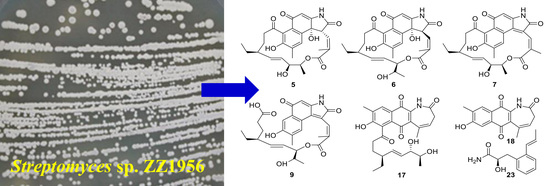
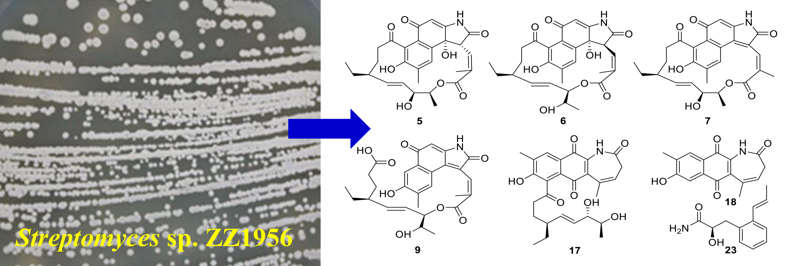
New Hygrocins K–U and Streptophenylpropanamide A and Bioactive Compounds from the Marine-Associated Streptomyces sp. ZZ1956

1
Ocean College, Zhoushan Campus, Zhejiang University, Zhoushan 316021, China
2
Zhejiang Marine Development Research Institute, Zhoushan 316000, China
3
College of Pharmaceutical Sciences, Zhejiang University, Hangzhou 310058, China
*
Authors to whom correspondence should be addressed.
Antibiotics 2022, 11(11), 1455; https://doi.org/10.3390/antibiotics11111455
Submission received: 1 October 2022 / Revised: 19 October 2022 / Accepted: 21 October 2022 / Published: 22 October 2022
(This article belongs to the Special Issue Discovery and Development of the Novel Antimicrobial Agent)

Abstract

:
Marine-derived Streptomyces actinomycetes are one of the most important sources for the discovery of novel bioactive natural products. This study characterized the isolation, structural elucidation and biological activity evaluation of thirty compounds, including twelve previously undescribed compounds, namely hygrocins K–U (513, 17 and 18) and streptophenylpropanamide A (23), from the marine-associated actinomycete Streptomyces sp. ZZ1956. Structures of the isolated compounds were determined by a combination of extensive NMR spectroscopic analyses, HRESIMS data, the Mosher’s method, ECD calculations, single crystal X-ray diffraction and comparison with reported data. Hygrocins C (1), D (2), F (4), N (8), Q (11) and R (12), 2-acetamide-6-hydroxy-7-methyl-1,4-naphthoquinone (22), echoside C (27), echoside A (28) and 11,11′-O-dimethylelaiophylin (30) had antiproliferative activity (IC50: 0.16–19.39 μM) against both human glioma U87MG and U251 cells with hygrocin C as the strongest active compound (IC50: 0.16 and 0.35 μM, respectively). The analysis of the structure–activity relationship indicated that a small change in the structures of the naphthalenic ansamycins had significant influence on their antiglioma activities. Hygrocins N (8), O (9), R (12), T (17) and U (18), 2-amino-6-hydroxy-7-methyl-1,4-naphthoquinone (21), 2-acetamide-6-hydroxy-7-methyl-1,4-naphthoquinone (22), 3′-methoxy(1,1′,4′,1″-terphenyl)-2′,6′-diol (26), echoside C (27) and echoside A (28) showed antibacterial activity against methicillin-resistant Staphylococcus aureus and Escherichia coli with MIC values of 3–48 μg/mL.

Graphical Abstract

1. Introduction

Compound 3-amino-5-hydroxy benzoic acid (3,5-AHBA) is the precursor of a big group of natural products, including the ansamycins, the mitomycins and the unique saliniketals (degraded ansamycins) [1]. The ansamycins have two characteristic structural features: an aromatic core and a so-called ansa bridge containing a lactam moiety, whose two ends link to two nonadjacent positions of the aromatic core [2]. The ansamycins can be divided into naphthalenic or benzenic depending on the nature of their aromatic ring. The naphthalenic ansamycins include rifamycins, ansalactams, chaxamycins, divergolides, hygrocins, naphthomycins, rubradirins and streptovaricins. Their structural characteristic is a 1,4-naphtoquinone or a 1,4-hydroxynaphtalene chromophore. Benzenic ansamycins have a 1,4-benzoquinone or a 1,4-hydrobenzoquinone chromophore and include ansatrienins, cebulactams, cytotrienins, geldanamycins, herbimycins, macbecins, maytansines (ansamitocins) and tetrapetalones. Precursor feeding experiments and genetic and biochemical methods have been applied to investigate the biosynthesis of the ansamycins, demonstrating that AHBA is the source of the chromophore and the aliphatic ansa chains are derived from acetate, propionate, isobutyrate or glycolate units [1,2].
While most of the ansamycins were isolated from actinomycetes, the class of maytansines was also found in higher plants and mosses [3,4]. However, the first maytansines found in plants [4] are now known to be produced by the interplay amongst bacteria in the root system [5]. It was reported that nearly 300 ansamycins have been identified from natural sources [6] and more and more ansamycins continue to be reported, such as the recently described olimycins from the ovmO-inactivated mutant strain Streptomyces olivaceus SCSIO T05 [7] and ansaseomycins from a heterologous mutant strain of Streptomyces seoulensis [8]. The ansamycins exhibit diverse biological activities, such as antibacterial (naphthomycin A, rifamycin, rubradirin and streptovaricin A), anticancer (ansamitocins P-3, ansatrienins A, geldanamycin, mitomycin C, saliniketals A and B), lipoxygenase inhibitory (tetrapetalones A and B) and antiviral (divergolide O) activities [1,9]. The well-known drugs of the ansamycin family were the first-line anti-tuberculous drug rifamycin and the antibody–drug conjugate Kadcyla (emtansine).
As part of an ongoing project to discover novel antiglioma natural products from marine microorganisms [10,11,12,13,14,15,16,17,18,19,20], we isolated an actinomycete from a sediment sample collected from an intertidal mangrove area at the Pacific Ocean close to South Sulawesi, Indonesia. This actinomycete was assigned as Streptomyces sp. ZZ1956 based on its 16S rDNA sequence analysis (Figure S1 and Table S1). An extract prepared from the culture of the strain ZZ1956 in GYM liquid medium exhibited inhibitory activity against the proliferation of glioma U251 and U87MG cells with inhibition rates of over 90%. Chemical investigation of this active extract resulted in the isolation and identification of thirty compounds 130, including eleven new naphthalenic ansamycin analogues hygrocins K–U (513, 17, 18) and one new phenylpropanamide derivative streptophenylpropanamide A (23) (Figure 1). Herein, we described the isolation and culture of the strain ZZ1956 as well as the isolation, structure elucidation and bioactive evaluation of these isolated compounds.

2. Results and Discussion

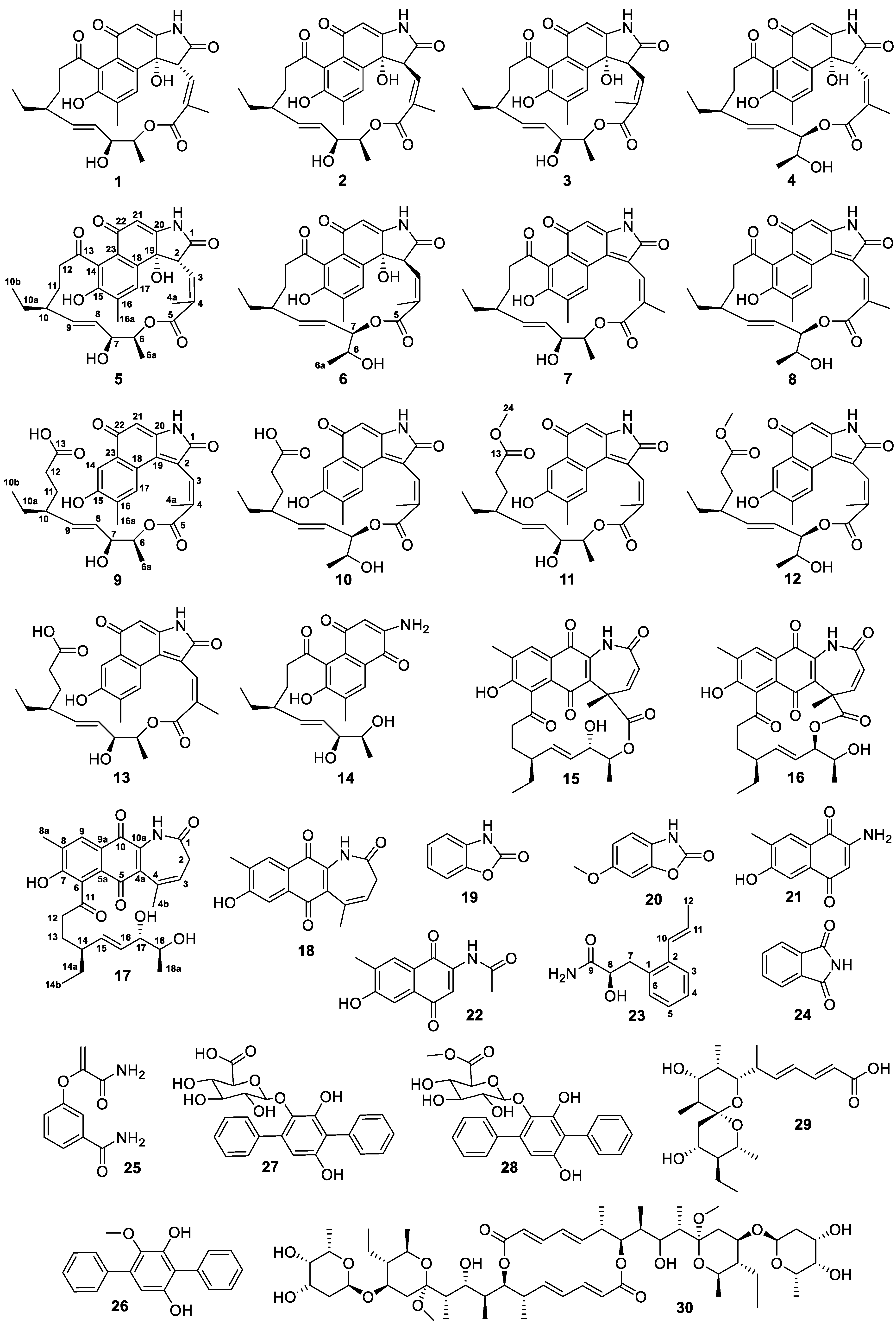
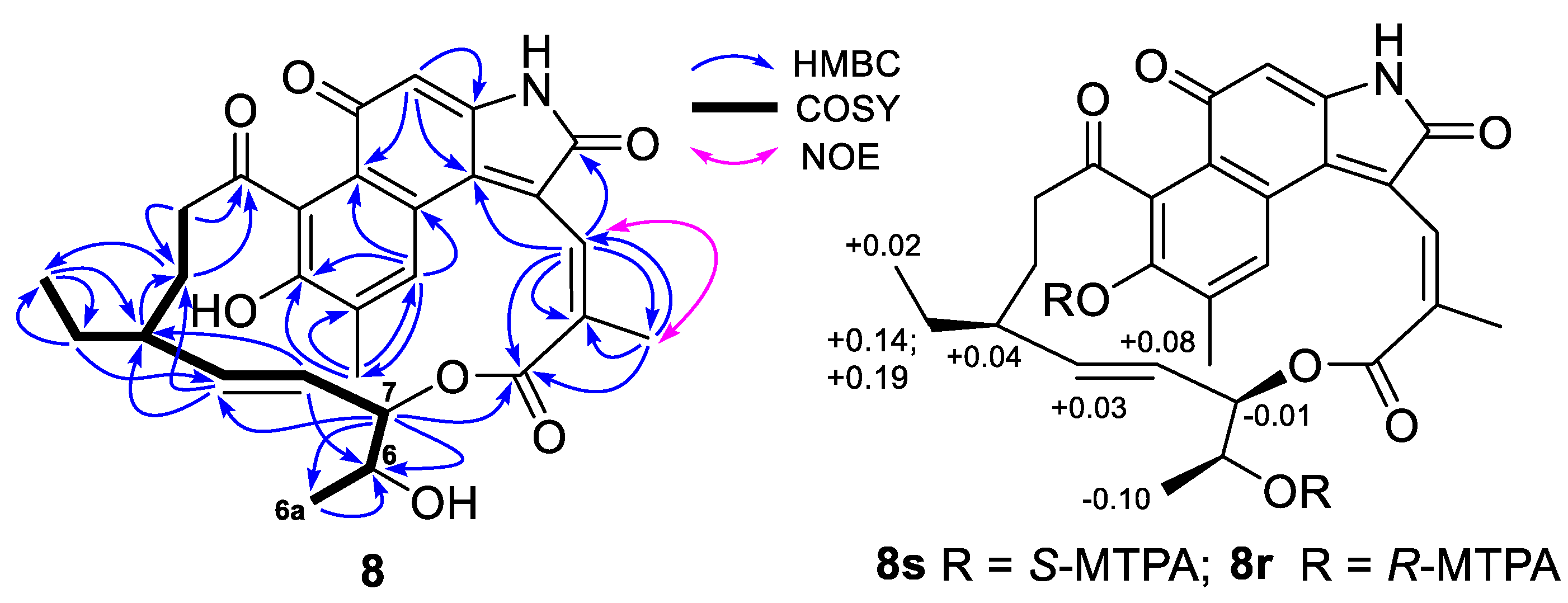
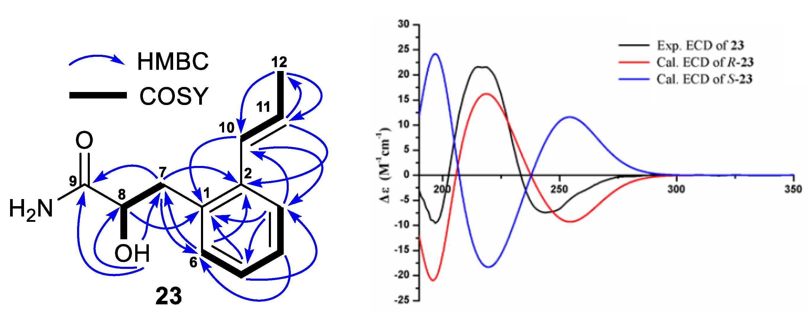
2.1. Structure Elucidation of the Isolated Compounds

After analyses of the NMR spectroscopic data and comparison with related data of references, eighteen known compounds were elucidated to be hygrocins C–F (14) [21], degrahygrocin A (14) [22], hygrocin B (15) [22], hygrocin G (16) [21], benzoxazolone (19) [23], coixol (20) [24], 2-amino-6-hydroxy-7-methyl-1,4-naphthoquinone (21) [25], 2-acetamide-6-hydroxy-7-methyl-1,4-naphthoquinone (22) [26], 1H-isoindole-1,3(2H)-dione (24) [27], 3-(3′-amino-3′-oxoprop-1′-en-2′-yl)oxy benzamide (25) [28], 3′-methoxy(1,1′,4′,1″-terphenyl)-2′,6′-diol (26) [29], echoside C (27) [30], echoside A (28) [30], pteridic acid hydrate (29) [31] and 11,11′-O-dimethylelaiophylin (30) [32]. The structure of hygrocin C (1) was confirmed by single crystal X-ray diffraction (Table S2). Degrahygrocin A (14) was previously reported as an alkaline hydrolytic product of hygrocin A [22]; while 2-acetamide-6-hydroxy-7-methyl-1,4-naphthoquinone (22) was an intermediate compound of chemical synthesis of ansalactam A [26]. Therefore, both compounds 14 and 22 are reported as natural products for the first time. The NMR data of these known compounds are presented in Tables S3–S14.
Compound 5 had the same molecular formula C28H31NO8 and very similar UV characteristic absorptions as hygrocins C–F (14), indicating that they are isomers. Careful analyses of the 1H, 13C, HMQC, COSY, HMBC and NOESY NMR spectra of 5 demonstrated that its structure was different from those of 14 in the configurations at C-2 and C3-C4 double bond as well as the position of the lactone ring formation. The configuration at C-2 was established as R based on a strong NOE correlation observed between H-2 and H3-6a (Figure 2). A strong NOE correlation of H-2 with H3-6a was an indication of 2R-configuration in 1 and 4, compared to the 2S-configuration in 2 and 3 without the NOE correlation of H-2 and H3-6a [21]. The chemical shift value (δC 13.7) of C-4a in 5 indicated a 3E-configuration, compared to the downfield chemical shift values (δC 21.1–22.2) (Tables S3 and S4) of C-4a for a 3Z-configuration in 1, 2 and 4 [21]. Observed strong NOE (for 7, 8, 13), no NOE (for 1012) or weak NOE (5, 6, 9) correlation between H-3 and H3-4a also supported the assignment of the 3E- or 3Z-configuration. In addition, the trans-coupling constant value of 16.0 Hz (3JH8/H9) indicated an 8E-configuration and the small vicinal coupling constant value of 2.8 Hz (3JH6/H7) suggested a syn orientation between H-6 and H-7 [21]. HMBC correlations (Figure 2) of H-6 (δH 4.65) with C-5 (δC 167.7) established the linkage of C5 and C6 through an oxygen to form the lactone ring. It is known that hygrocins C–E (13) have the same 6S, 7S, 10S, 19R-configuration. Therefore, we proposed compound 5 to have the same 6S, 7S, 10S, 19R-configuration as 13 based on a common biosynthetic origin. Based on the above analyses, it can be concluded that the structure of 5 is similar to that of 1 with the only difference being the configuration of the C3–C4 double bond. Therefore, the structure of 5 was elucidated as a previously undescribed member of the naphthoquinone ansamycins, named hygrocin K. Its 13C and 1H NMR data are reported in Table 1 and Table 2.
Compound 6 had the same molecular formula C28H31NO8 as 15 deduced from its HRESIMS ion peak at m/z 508.1979 [M–H] and 13C NMR data. Careful analyses of its 1D- and 2D-NMR spectra determined that 6 and hygrocins F (4) had the same planar structure. As described above for 5, no NOE correlation between H-2 with H3-6a and weak NOE correlation between H-3 with H3-4a as well as the relative upfield shift value at δC 13.6 for C-4a indicated that 6 had 2S- and 3E-configurations [21]. The structure of 6 was thus identified as a previously undescribed naphthoquinone ansamycin, named hygrocin L. Its 13C and 1H NMR data (Table 1 and Table 2) were assigned based on the HMQC, COSY and HMBC correlations (Figure 2).
Compounds 7 and 8 had very similar UV absorptions and the same molecular formula C28H29NO7 deduced from their HRESIMS ion peaks at m/z 490.1875 and 490.1871 [M–H], respectively, 18 mass units lower than that of 16, corresponding to the loss of a H2O molecule. Detailed analyses of the 1D- and 2D-NMR spectra of 7 and 8 as well as comparison of their NMR data with those of 16 demonstrated that the methine at C-2 and the non-protonated oxygenated carbon at C-19 in 16 were replaced by two non-protonated olefinic carbons at C-2 (δC 121.9 in 7 or δC 122.6 in 8) and C-19 (δC 134.8 in 7 or δC 136.4 in 8). Therefore, both 7 and 8 had a C2-C19 double bond. The downfield shift values of C-4a at δC 21.1 in 7 or δC 22.1 in 8 and the strong NOE correlation between H-3 and H3-4a (Figure 2 and Figure 3) indicated that they had a 3Z-configuration; while the trans-coupling constant values of 15.1 Hz in 7 and 15.6 Hz in 8 between H-8 and H-9 suggested that both 7 and 8 had an 8E-configuration. HMBC correlations of H-6 (δH 4.87) with C-5 (δC 167.6) in 7 and H-7 (δH 4.97) with C-5 (δC 168.3) in 8 established the position of the lactone ring formation. The Mosher’s method was used to determine the configuration at C-6 in 8. The results (Figure 3 and Table S15) indicated a 6S-configuration for 8. Therefore, compound 7 should have the same 6S, 7S-configuration as compounds 13 and 5 and compound 8 should have the same 6S, 7R-configuration as compounds 4, 6 and 16 [21] based on their shared biogenesis, the structures of the reported compounds and the Mosher’s method results of 8. The structures of 7 and 8 were thus elucidated as two previously unreported naphthoquinone ansamycins, named hygrocin M (7) and hygrocin N (8). The 13C and 1H NMR data (Table 1 and Table 2) of 7 and 8 were assigned based on the HMQC, COSY and HMBC correlations (Figure 2 and Figure 3).
Compounds 9 and 10 were obtained as a red amorphous powder and had very similar UV characteristic absorptions (around 201 and 335 nm) to those of 7 and 8, suggesting that they were analogues. Both 9 and 10 had the same molecular formula C28H31NO8 deduced from their 13C NMR data and HRESIMS ion peaks at m/z 508.1975 [M–H] in 9 and 508.1974 [M–H] in 10, 18 mass units higher than those of 7 and 8. Compared to 7 and 8, one additional aromatic hydrogen signal at δH 7.43 (s) in 9 or δH 7.42 (s) in 10 was observed in their 1H NMR spectra. However, the 13C NMR signal at δC 212.9 in 7 or δH 212.0 in 8 for the ketone group at C-13 was replaced in both 9 and 10 by upfield shifted signals at δC 177.7 in 9 or δC 177.8 in 10. Further analyses of their HMQC, COSY and HMBC correlations (Figure 4) as well as consideration of their molecular formula and 14 degrees of unsaturation required by the molecular formula demonstrated that 9 and 10 were derivatives of 7 and 8, respectively, with ring opening between C-13 and C-14. The chemical shift at δC 16.4 for C-4a and no NOE or weak NOE correlation between H-3 and H3-4a in 9 or 10 indicated a 3E-configuration, compared to the downfield shift values at δC 21.1 and 22.1 (Table 1) for the C-4a and the strong NOE correlation between H-3 and H3-4a in 7 and 8 with a 3Z-configuration. Therefore, the structures of 9 and 10 were elucidated as two previously reported naphthoquinone ansamycins, named hygrocin O (9) and hygrocin P (10). Their 13C and 1H NMR data are reported in Table 1 and Table 3.
Compounds 11 and 12 were also obtained as a red amorphous powder and had same molecular formula C29H33NO8 deduced from their 13C NMR data and HRESIMS ion peaks at m/z 522.2125 [M–H] in 11 and 522.2132 [M–H] in 12, 14 mass units higher than those of 9 and 10. Compared to the 13C and 1H NMR data of 9 and 10, both 11 and 12 had additional NMR signals for a methoxy group at δC 52.1 and δH 3.57 (3H, s) in 11 and δC 52.2 and δH 3.65 (3H, s) in 12. A HMBC correlation of H3-24 (δH 3.57) with C-13 (δC 176.0) in 11 and H3-24 (δH 3.65) with C-13 (δC 176.0) in 12 established the position of the methoxy group. Further analyses of their HMQC, COSY, HMBC and NOE correlations (Figure 4) demonstrated that 11 and 12 were the methyl esters of 9 and 10, respectively. The structures of 11 and 12 were thus identified as two previously undescribed naphthoquinone ansamycins, named hygrocin Q (11) and R (12). The 13C and 1H NMR data of 11 and 12 are reported in Table 1 and Table 3. It should be noted that 11 and 12 may be the artificial products of methyl esterification of 9 and 10, respectively, originated in the extraction and separation process.
The HRESIMS spectrum of compound 13 gave an ion peak at m/z 508.1975 [M–H], corresponding to a molecular formula C28H31NO8, which was the same as those of 9 and 10. Detailed analyses of the 1D- and 2D-NMR spectra of 13 determined that 9 and 13 had the same planar structure and their structural difference was only the different configuration of the C3-C4 double bond. The downfield shift value at δC 21.1 for C-4a and a strong NOE correlation between H-3 and H3-4a (Figure 5) suggested a 3Z-configuration in 13. The structure of 13 was thus assigned as a previously undescribed naphthoquinone ansamycin, named hygrocin S (13). Its 13C and 1H NMR data (Table 1 and Table 3) were assigned based on the HMQC, COSY and HMBC correlations (Figure 5).
Compound 17 was obtained as a yellow amorphous powder and its molecular formula C27H31NO7 was determined based on the HRESIMS ion peak at m/z 480.2024 [M–H] and 13C NMR data. Interpretation of the 1H, 13C and HMQC NMR spectra of 17 indicated that its twenty-seven carbons (Table 4) were assigned to four carbonyls, six pairs of double bonds, two oxymethines, one methine, four methylenes and four methyls. These carbon types of 17 were very similar to those of hygrocin B (15). Compared to 15, the NMR spectra of 17 showed additional signals for one non-protonated olefinic carbon and one methylene group at δC 37.7 and δH 2.90 (2H, d, J = 7.2 Hz) (Table 4) and lacked the signals for one carbonyl carbon, one protonated olefinic carbon and the non-protonated carbon at δC 52.6 (C-4) (Table S7), which were observed in the NMR spectra of 15. Further analyses of the HMQC, COSY and HMBC correlations (Figure 5) of 17 indicated that 17 had a C3-C4 double bond, but did not have the lactone structure existed in 15. Therefore, compound 17 was a seco-derivative of 15. A shared biogenesis suggested that 17 and 15 should have the same 14S, 17S, 18S-configuration. Analyses described above resulted in the identification of 17 as a previously undescribed naphthoquinone ansamycin, named hygrocin T. Its 13C and 1H NMR data (Table 4) were assigned based on the HMQC, COSY and HMBC correlations (Figure 5).
Compound 18 was obtained as a yellow amorphous powder and its HRESIMS gave an ion peak at m/z [M–H] 282.0770, corresponding to a molecular formula C16H13NO4 with eleven degrees of unsaturation. Based on the analyses of its 1H, 13C, DEPT and HMQC NMR spectra, the sixteen carbons were assigned to three carbonyls (δC 184.2, 177.4, 169.3), five pairs of double bonds, one methylene (δC 36.4) and two methyl groups (δC 20.8, 16.3) (Table 5). The three carbonyl and five pairs of double bonds accounted for eight out of the eleven degrees of unsaturation required by the molecular formula, suggesting that 18 had a structure with three rings. Above evidence, together with further analyses of its COSY and HMBC correlations (Figure 5), demonstrated that the core structure of 18 was similar to that of 17 with the only difference being the absence in 18 of the side chain attached. Therefore, the structure of 18 was elucidated as a previously unreported naphthoquinone ansamycin, named hygrocin U. The 13C and 1H NMR data (Table 5) assignment of 18 was made based on the HMQC, COSY and HMBC correlations (Figure 5).
The molecular formula C12H15NO2 of 23 was determined by its HRESIMS ion peaks at m/z 206.1175 [M+H]+ and 228.0995 [M+Na]+ as well as 13C NMR data. The twelve carbons in 23 were assigned to one carbonyl (δC 176.0), eight olefinic carbons, one oxymethine (δC 71.9), one methylene (δC 37.5) and one methyl group (δC 18.7). COSY correlations (Figure 6) of H-11 (δH 6.09, 1H, m) with H-10 (δH 6.74, 1H, dd, 15.8, 1.8 Hz) and H3-12 (δH 1.86, 3H, dd, 6.5, 1.8 Hz) as well as HMBC correlations (Figure 6) of H-12 with C-10 (δC 128.6) and C-11 (δC 126.7) indicated the existence of a 1-propen-1-yl group. Similarly, a “-CH2-CH(OH)-CO-” structural fragment was established based on the COSY correlations of H-8 (δH 3.92, 1H, ddd, 9.3, 6.2, 3.6 Hz) with H-7 (δH 3.08, 1H, dd, 14.1, 3.6 Hz; 2.64, 1H, dd, 14.1, 9.3 Hz) and OH-8 (δH 5.42, 1H, d, 6.2 Hz) as well as HMBC correlations of H-7 with C-9 (δC 176.0) and OH-8 with C-7 (δC 37.5), C-8 (δC 71.9) and C-9. In the downfield area (δH 6.09–7.60) of the 1H NMR spectrum of 23, there were signals for six olefinic protons. The 1-propen-1-yl group accounted for two olefinic protons and two olefinic carbons and the remaining four olefinic protons and six olefinic carbons were assigned to an aromatic ring. HMBC correlations of H-10 with C-1 (δC 135.6), C-2 (δC 136.6) and C-3 (δC 125.3) and H-3 (δH 7.41, 1H, d, 7.5 Hz) with C-10 established the linkage of the 1-propen-1-yl at C-2. In the same way, the positioning of the “-CH2-CH(OH)-CO-” group at C-1 was indicated by HMBC correlations of H-6 (δH 7.15, 1H, m) with C-7, H-7 with C-2 and C-6 (δC 130.7) and H-8 with C-1. In addition, the 1H NMR spectrum of 23 showed two noncarbonated proton signals at δH 7.25 (1H, s) and 7.16 (1H, s), which were assigned to NH2-9. The HRESIMS data also supported a -NH2 group at C-9, rather than a -OH group. A trans-coupling constant value of 15.8 Hz (3JH10/H11) indicated a 10E-configuration, while the absolute configuration at C-8 was determined based on the results (Figure 6 and Tables S16–S19) from ECD calculations. The ECD spectrum of 23 displayed positive and negative Cotton effects at 215 and 244 nm, respectively, which closely matched those of the ECD curve calculated for 8R-23. Based on the foregoing evidence, the structure of 23 was identified as a previously undescribed phenylpropanamide analogue, named streptophenylpropanamide A. Its 13C and 1H NMR data are reported in Table 5.

2.2. Biological Activity Evaluation

Sulforhodamine B (SRB) assay was applied to determine the activity of all thirty isolated compounds (130) against the proliferation of glioma cells. Doxorubicin was used as a positive control. The results (Table 6) indicated that compounds 1, 2, 4, 8, 11, 12, 22 and 30 showed potent antiproliferative activity against both glioma U87MG and U251 cells with IC50 values ranging from 0.16 to 10.46 μM. Compounds 27 and 28 also had activity in inhibiting the proliferation of glioma U87MG and U251 cells with IC50 values of 11.18 and 19.39 μM, respectively. Among all the active compounds, hygrocin C (1) showed the strongest activity (IC50: 0.16 and 0.35 μM), followed by hygrocin D (2) (IC50: 0.39 and 2.63 μM). Compounds 118 were eighteen naphthoquinone ansamycins. It was noted that the active ring closed compounds hygrocins C (1), D (2) and F (4) had a 3Z-configuration, compared to the inactive ring closed compounds hygrocins E (3), K (5) and L (6) with a 3E-configuration. However, although both hygrocins M (7) and N (8) had the 3Z-configuration, they exhibited significantly different activities due to the different positioning of the ring closure at the C-6 or C-7 position. In addition, most of the ring open compounds hygrocins O (9), P (10), S (13) and T (17) and degrahygrocin A (14) were inactive. However, the ring open compounds hygrocins Q (11) and R (12), the methyl esters of hygrocins O (9) and P (10), respectively, were active. These analyses of the structure–activity relationship indicated that a small change in the structure of this class of compounds had significant influence on their antiglioma activities.
The activity of compounds 130 in inhibiting the growth of methicillin-resistant Staphylococcus aureus (MRSA) and Escherichia coli was also evaluated. The results (Table 6) showed that compounds 8, 9, 12, 17, 18, 21, 22 and 2628 exhibited antibacterial activity against both MRSA and E. coli with MIC values of 3–48 μg/mL.

3. Experimental Section

3.1. General Procedures

Optical rotation (OR), ultraviolet–visible (UV), electronic circular dichroism (ECD) and infrared (IR) spectra were recorded on an Autopol I polarimeter (Rudolph Research Analytical, Hackettstown, NJ, USA), a METASH UV-8000 spectrometer (Shanghai METASH Instruments Co. Ltd., Shanghai, China), a JASCO J-815 spectropolarimeter (JASCO Co., Tokyo, Japan) and a NicoletTM ISTM 10 FT-IR spectrometer (Thermo Fisher Scientific, Waltham, MA, USA), respectively. HRESIMS data were acquired on an Agilent 6230 TOF LC/MS spectrometer (Agilent Technologies Co. Ltd., Santa Clara, CA, USA). NMR spectra were obtained on a JEOL 600 spectrometer (JEOL Co. Ltd., Tokyo, Japan) using standard programs and acquisition parameters and chemical shift values were expressed in δ (ppm) relative to DMSO-d6 (δC 39.5, δH 2.50), MeOH-d4 (δC 49.15, δH 3.31) or acetone-d6 (δC 29.8, δH 2.05). Diaion HP-20 (Mitsubishi Chemical, Tokyo, Japan), silica gel (100–200 mesh, Qingdao Marine Chemical Co., Ltd., Qingdao, China), octadecyl-functionalized silica gel (ODS, Cosmosil 75C18-Prep, Nacalai Tesque Inc., Kyoto, Japan) and sephadex LH-20 (GE Healthcare, Waukesha, WI, USA) were used for column chromatography. HPLC separation was performed on a CXTH LC-3000 preparative HPLC system (Beijing Chuangxin Tongheng Science & Technology Co. Ltd., Beijing, China) with column A (CT-30, 280 × 30 mm, 10 µm, Fuji-C18) and an Agilent 1260 infinity HPLC system (Agilent Technologies Co. Ltd., Santa Clara, CA, USA) using Zorbax SB-C18 columns (column B: 250 × 9.4 mm, 5 µm or column C: 250 × 4.6 mm, 5 µm) with a DAD detector. All solvents used for this study were purchased from the Sinopharm Chemical Reagent Co. Ltd. (Shanghai, China). Glioma U87MG (JDS-2568) and U251 (XB-0439) cells used in the experiment were purchased from the Cell Bank of the Chinese Academy of Sciences (Shanghai, China). Methicillin-resistant Staphylococcus aureus (MRSA) ATCC 43300 and Escherichia coli ATCC 25922 were gifts from Dr. Zhongjun Ma and Dr. Pinmei Wang, respectively. Doxorubicin (DOX) was purchased from Solarbio Science & Technology Co. Ltd. (Beijing, China). Vancomycin and gentamicin were ordered from Meilune Biotechnology Co., Ltd. (Dalian, China). Gauze’s agar medium was ordered from the Guangdong Huankai Microbial Science and Technology Co., Ltd. (Guangzhou, China). GYM liquid medium (glucose 4 g, yeast extract 10 g, malt extract 10 g, tap water 1.0 L) was made in the authors’ laboratory.

3.2. Isolation and Taxonomic Identity of Streptomyces sp. ZZ1956

The Streptomyces sp. ZZ1956 strain was isolated from a marine mud sample collected from the mangrove area (4.15° S, 119.61° E) of Pangkep District South Sulawesi Province, Indonesia in September 2018. Briefly, the sample (1.0 g) was suspended in sterile water to make dilutions of 10−2, 10−3 and 10−4 g/mL. Each dilution of 200 µL was spread over the surface of solid Gauze’s medium in a Petri dish and then incubated for 10 days at 28 °C. The single ZZ1956 colony from a Petri dish with the 10−2 g/mL dilution was transferred to a Gauze’s agar plate. After growth for another 7 days at 28 °C, the pure strain ZZ1956 colony (Figure S2) was transferred onto Gauze′s agar slants and stored at 4 °C for later use. The 16S rDNA sequence analysis of the strain ZZ1956 was conducted by Legenomics (Hangzhou, China). The 16S rDNA sequence of the strain ZZ1956 was deposited in GenBank with an accession number of MT672495. The voucher strain of Streptomyces sp. ZZ1956 was preserved at the Laboratory of the Institute of Marine Biology and Pharmacology, Ocean College, Zhoushan campus, Zhejiang University, Zhoushan, China.

3.3. Mass Culture of the Strain ZZ1956

Colonies of the strain ZZ1956 from the Gauze’s agar plate were inoculated into 500 mL Erlenmeyer flasks, each containing 250 mL of sterile GYM liquid medium and then incubated at 28 °C for 3 days on a rotary shaker (180 rpm) to prepare the seed broth. The seed broth (10 mL) was then transferred into a 500 mL Erlenmeyer flask containing 250 mL sterilized GYM liquid medium. A total of 60 L (240 bottles) of culture was prepared for this study and incubated at 28 °C for 15 days under shaking (180 rpm) condition.

3.4. Extraction and Isolation of Compounds 130

The 60-L culture of strain ZZ1956 was centrifuged to yield supernatant and mycelia. The mycelia were extracted with MeOH three times (3 L, each time) to give a MeOH extract solution. The supernatant was applied to a Dianion HP-20 column eluted with water and then MeOH to obtain a MeOH elution. The MeOH extract solution and MeOH elution were combined and dried in vacuo to give a crude extract, which was further partitioned with EtOAc three times to give an EtOAc extract (24 g). The EtOAc extract was subjected to a column of silica gel eluted with mixtures of cyclohexane/EtOAc (10/1, 8/1, 5/1, 2/1, 1/1, v/v), EtOAc, and MeOH to give ten fractions (Frs. A–J) based on the results of TLC and HPLC analyses.
Fr. A was purified by HPLC using column C (mobile phase: MeCN/H2O, 65/35; flow rate: 0.8 mL/min; UV detection: 210 nm) to give 26 (4.8 mg, tR 15.4 min). Fractions B and D were separated on HPLC column B (flow rate: 1 mL/min; UV detection: 210 nm) to give 24 (3.0 mg, tR 25.0 min, MeOH/H2O, 40/60) and 22 (4.4 mg, tR 24.4 min, MeOH/H2O, 67/37), respectively.
Fr. C was subjected to a sephadex LH-20 column eluted with 70% MeOH to yield three subfractions (Frs. C1–C3). Fr. C1 was further separated on column C (mobile phase: MeOH/H2O, 34/66; flow rate: 0.8 mL/min; UV detection: 210 nm) to give 19 (1.2 mg, tR 15.7 min) and 20 (3.8 mg, tR 18.6 min). Compound 18 (1.7 mg, tR 25.5 min) was obtained from Fr. C2 through HPLC purification using column B (mobile phase: MeOH/H2O, 67/33; flow rate: 1 mL/min; UV detection: 280 nm).
Each of Fr. E, Fr. F and Fr. H was separated by preparative HPLC using column A (flow rate: 10 mL/min; UV detection: 210 nm). Compounds 21 (4.9 mg, tR 19.5 min), 15 (21.0 mg, tR 24.3 min) and 16 (7.4 mg, tR 44.1 min) were obtained from Fr. E using a mobile phase (MeOH/0.1% HOAc in H2O, 70/30), 14 (8.0 mg, tR 26.2 min, MeOH/0.1% HOAc in H2O, 65/35) was purified from Fr. H and five subfractions (Frs. F1–F5) were obtained from Fr. F using a gradient mobile phase (MeOH/0.1% HOAc in H2O, 30/70–100/0) in 40 min. Each of Frs. F1–F5 was purified by HPLC column B (flow rate: 1 mL/min; UV detection: 210 nm) to give 1 (28.0 mg, tR 23.1 min, MeCN/H2O, 33/67), 7 (3.3 mg, tR 23.7 min, MeOH/H2O, 55/45), 3 (6.0 mg, tR 57.5 min, MeCN/H2O, 30/70), 2 (19.0 mg, tR 26.3 min, MeCN/H2O, 45/55) and 8 (3.3 mg, tR 33.3 min, MeOH/H2O, 66/34).
Fr. G was fractionated by an ODS column eluted with 65%, 75% and 100% MeOH to give three subfractions (Frs. G1–G3) based on the results of TLC and HPLC analyses. Compound 29 (4.4 mg, tR 28.2 min) was obtained from Fr. G2 through HPLC purification using column B (mobile phase: MeCN/0.1% HOAc in H2O, 30/70; flow rate: 1 mL/min; UV detection: 256 nm). Fr. G3 was further separated by HPLC column A (mobile phase: MeOH/0.1% HOAc in H2O, 70/30; flow rate: 10 mL/min; UV detection: 210 nm) to give six subfractions (Frs. G3a–G3f). Fr. G3a continued to be separated on the same column A with the same flow rate and UV detection to give Fr. G3aa and Fr. G3ab (MeOH/0.1% HOAc in H2O, 55/45). Further purification by HPLC column B (flow rate: 1 mL/min; UV detection: 256 nm) yielded compounds 5 (5.9 mg, tR 48.8 min, MeCN/H2O, 27/73) from Fr. G3aa, 6 (2.3 mg, tR 43.7 min, MeCN/H2O, 33/67) from Fr. G3ab, 23 (2.0 mg, tR 21.9 min, MeOH/0.1% HOAc in H2O, 65/35) from Fr. G3b, 4 (16.0 mg, tR 24.1 min, MeCN/H2O, 45/55) from Fr. G3c, 13 (3.0 mg, tR 32.3 min) and 17 (8.0 mg, tR 37.1 min, MeCN/0.1% HOAc in H2O, 39/61) from Fr. G3d, 9 (2.8 mg, tR 30.0 min), 10 (2.8 mg, tR 38.5 min), 12 (2.5 mg, tR 64.2 min) and 11 (2.2 mg, tR 77.1 min, MeOH/0.1% HOAc in H2O, 70/30) from Fr. G3f.
Similarly, Fr. I was also applied to an ODS column eluted with 50%, 70% and 100% MeOH to yield three subfractions (Frs. I1–I3). Fr. I1 was further separated on HPLC column A (flow rate: 10 mL/min; UV detection: 210 nm) with a gradient mobile phase from 40% to 100% MeOH in 40 min to give 25 (2.0 mg, tR 15.8 min); while compound 30 (20.0 mg, tR 32.4 min) was obtained from Fr. I3 by separating on column B (mobile phase: MeOH/H2O, 95/5; flow rate: 1 mL/min; UV detection: 256 nm).
Finally, Fr. J was fractionated on an ODS column eluted with 30%, 40%, 60%, 70% and 100% MeOH to give Fr. J1 and Fr. J2 based on the results of HPLC analyses. Fr. J1 was further separated on column A (flow rate: 10 mL/min; UV detection: 210 nm) with a gradient mobile phase from 40% to 100% MeOH in 40 min to give Fr. J1a and Fr. J1b. Compounds 27 (9.0 mg, tR 24.3 min) and 28 (2.0 mg, tR 42.3 min) were obtained from Fr. J1a through HPLC purification using column B (mobile phase: ACN/H2O, 33/67; flow rate: 1 mL/min; UV detection: 210 nm).

3.5. Compound Characterization Data

Hygrocin K (5): Light yellow oil; molecular formula C28H31NO8; [α]D20 –65.5 (c 0.10, MeOH); UV (MeOH) λmax (log ε) 203 (4.39), 271 (4.04), 304 (4.00) nm; IR (ATR) νmax 3320, 2962, 2929, 2870, 1662, 1627, 1569, 1322, 1237, 1190, 1131, 1051, 976, 857, 733 cm−1; 13C NMR data (150 MHz), Table 1, 1H NMR data (600 MHz), Table 2; HRESIMS m/z 508.1969 [M–H] (calcd for C28H30NO8, 508.1971).
Hygrocin L (6): Light yellow oil; molecular formula C28H31NO8; [α]D20 +166.6 (c 0.10, MeOH); UV (MeOH) λmax (log ε) 203 (4.21), 270 (4.04), 304 (4.03) nm; IR (ATR) νmax 3314, 2963, 2928, 2874, 1698, 1657, 1627, 1567, 1239, 1188, 1108, 1029, 976, 857, 810, 735 cm−1; 13C NMR data (150 MHz), Table 1, 1H NMR data (600 MHz), Table 2; HRESIMS m/z 508.1979 [M–H] (calcd for C28H30NO8, 508.1971).
Hygrocin M (7): Light yellow amorphous powder; molecular formula C28H29NO7; [α]D20 +166.7 (c 0.10, MeOH); UV (MeOH) λmax (log ε) 201 (4.52), 333 (4.11) nm; IR (ATR) νmax 3362, 2960, 2930, 2868,1714, 1655, 1626, 1559, 1446, 1346, 1285, 1134, 1044, 860 cm−1; 13C NMR data (150 MHz), Table 1, 1H NMR data (600 MHz), Table 2; HRESIMS m/z 490.1875 [M–H] (calcd for C28H28NO7, 490.1866).
Hygrocin N (8): Light yellow amorphous powder; molecular formula C28H29NO7; [α]D20 +270.0 (c 0.10, MeOH); UV (MeOH) λmax (log ε) 201 (4.55), 336 (4.26) nm; IR (ATR) νmax 3275, 2963, 2927, 2877, 1709, 1650, 1617, 1567, 1466, 1338, 1282, 1249, 1132, 1079, 859, 736 cm−1; 13C NMR data (150 MHz), Table 1, 1H NMR data (600 MHz), Table 2; HRESIMS m/z 492.2010 [M+H]+ (calcd for C28H30NO7, 492.2022), 514.1832 [M+Na]+ (calcd for C28H29NNaO7, 514.1842) and 490.1871 [M–H] (calcd for C28H28NO7, 490.1866).
Hygrocin O (9): Red amorphous powder; molecular formula C28H31NO8; [α]D20 –21.5 (c 0.10, MeOH); UV (MeOH) λmax (log ε) 201 (4.65), 334 (4.33) nm; IR (ATR) νmax 3257, 2959, 2926, 2872, 1697, 1652, 1600, 1580, 1343, 1257, 1204, 1135, 1022, 978 cm−1; 13C NMR data (150 MHz), Table 1, 1H NMR data (600 MHz), Table 3; HRESIMS m/z 508.1975 [M–H] (calcd for C28H30NO8, 508.1971).
Hygrocin P (10): Red amorphous powder; molecular formula C28H31NO8; [α]D20 –55.5 (c 0.10, MeOH); UV (MeOH) λmax (log ε) 201 (4.29), 334 (4.07) nm; IR (ATR) νmax 3229, 2966, 2926, 2875, 1705, 1651, 1622, 1576, 1467, 1339, 1263, 1245, 1120, 1063, 1018, 976, 855, 737 cm−1; 13C NMR data (150 MHz), Table 1, 1H NMR data (600 MHz), Table 3; HRESIMS m/z 508.1974 [M–H] (calcd for C28H30NO8, 508.1971).
Hygrocin Q (11): Red amorphous powder; molecular formula C29H33NO8; [α]D20 –16.0 (c 0.10, MeOH); UV (MeOH) λmax (log ε) 201 (4.22), 335 (4.10) nm; IR (ATR) νmax 3345, 2960, 2925, 2875, 1714, 1655, 1629, 1598, 1573, 1435, 1349, 1263, 1188, 1149, 1020, 978, 851, 755 cm−1; 13C NMR data (150 MHz), Table 1, 1H NMR data (600 MHz), Table 3; HRESIMS m/z 522.2125 [M–H] (calcd for C29H32NO8, 522.2128).
Hygrocin R (12): Red amorphous powder; molecular formula C29H33NO8; [α]D20 –40.0 (c 0.10, MeOH); UV (MeOH) λmax (log ε) 201 (4.14), 335 (4.09) nm; IR (ATR) νmax 3376, 2959, 2927, 2872, 1696, 1653, 1596, 1578, 1441, 1339, 1261, 1202, 1136, 1064, 976, 851, 802, 728 cm−1; 13C NMR data (150 MHz), Table 1, 1H NMR data (600 MHz), Table 3; HRESIMS m/z 522.2132 [M–H] (calcd for C29H32NO8, 522.2128).
Hygrocin S (13): Red amorphous powder; molecular formula C28H31NO8; [α]D20 –21.6 (c 0.10, MeOH); UV (MeOH) λmax (log ε) 201 (4.13), 333 (4.06) nm; IR (ATR) νmax 3240, 2962, 2870, 1709, 1655, 1574, 1350, 1222, 1130, 1068, 976, 855 cm−1; 13C NMR data (150 MHz), Table 1, 1H NMR data (600 MHz), Table 3; HRESIMS m/z 508.1975 [M–H] (calcd for C28H30NO8, 508.1971).
Hygrocin T (17): Yellow amorphous powder; molecular formula C27H31NO7; [α]D20 –34.0 (c 0.10, MeOH); UV (MeOH) λmax (log ε) 204 (4.35), 211 (4.35), 279 (4.36), 308 (4.10) nm; IR (ATR) νmax 3324, 2961, 2925, 2874, 1694, 1655, 1567, 1457, 1339, 1261, 1188, 1151, 1012, 973, 806, 735 cm−1; 13C NMR (150 MHz) and 1H NMR (600 MHz) data, Table 4; HRESIMS m/z 480.2024 [M–H] (calcd for C27H30NO7, 480.2022).
Hygrocin U (18): Light yellow amorphous powder; molecular formula C16H13NO4; UV (MeOH) λmax (log ε) 201 (4.21), 211 (4.12), 280 (4.17), 307 (3.81) nm; IR (ATR) νmax 3319, 2956, 2920, 2849, 1674, 1651, 1567, 1466, 1327, 1265, 1206, 1146 cm−1; 13C NMR (150 MHz) and 1H NMR (600 MHz) data, Table 5; HRESIMS m/z 282.0770 [M–H] (calcd for C16H12NO4, 282.0766).
Streptobenzenepropanamide A (23): White amorphous powder; molecular formula C12H15NO2; [α]D20 +12.4 (c 0.10, MeOH); ECD (15 mg/L, MeOH) λmax (Δε) 215 (+20.14), 244 (–6.93) nm; UV (MeOH) λmax (log ε) 202 (4.16), 248 (3.91) nm; IR (ATR) νmax 3333, 2963, 2925, 2852, 1665, 1578, 1447, 1406, 1380, 1261, 1094, 1074, 965, 802, 751 cm−1; 13C NMR (150 MHz) and 1H NMR (600 MHz) data, Table 5; HRESIMS m/z 206.1175 [M+H]+ (calcd for C12H16NO2+, 206.1181) and 228.0995 [M+Na]+ (calcd for C12H15NNaO2+, 228.1000).

3.6. MTPA Esterification Hygrocin N (8)

Hygrocin N (8, 3 mg) was dissolved in 2 mL anhydrous pyridine. Half of the sample solution was added either (R)- or (S)-α-methoxy-α-(trifluoromethyl)-phenylacetyl chloride (MTPA-Cl, 45 μL). The mixture was stirred at room temperature for 24 h and then added MeOH (0.5 mL) to stop the reaction. The reaction products were separated by HPLC using column B with a flow rate of 1 mL/min and UV detection of 210 nm to furnish (S)-MTPA ester 8s (1.0 mg, tR 22.5 min, MeOH/H2O, 92/8) or (R)-MTPA ester 8r (1.2 mg, tR 22.5 min, MeOH/H2O, 92/8).
(S)-MTPA ester 8s: 1H NMR data (600 MHz, in MeOH-d4), Table S15; HRESIMS m/z 924.2815 [M+H]+ (calcd for C48H44F6NO11+, 924.2819) and 946.2640 [M+Na]+ (calcd for C48H43F6NNaO11+, 946.2638).
(R)-MTPA ester 8r: 1H NMR data (600 MHz, in MeOH-d4), Table S15; HRESIMS m/z 924.2824 [M+H]+ (calcd for C48H44F6NO11+, 924.2819) and 946.2628 [M+Na]+ (calcd for C48H43F6NNaO11+, 946.2638).

3.7. ECD Calculations

The details of ECD calculations for compound 23 were described as our previous publications [19,20].

3.8. Sulforhodamine B (SRB) Assay

The culture of human glioma cells and the SRB assay were according to our previous reports [19,33].

3.9. Antibacterail Activity Determination

The antibacterial activity of the tested compounds against MRSA and E. coli was evaluated by the micro broth dilution method [34] using vancomycin and gentamicin as positive controls and DMSO as a negative control.

4. Conclusions

Marine-derived actinomycetes from the genus Streptomyces continue to be one of the main resources for the discovery of novel bioactive natural products. A chemical investigation of the extract prepared from a scaled-up culture of the marine-derived actinomycete Streptomyces sp. ZZ1956 in GYM liquid medium resulted in the isolation and identification of thirty compounds (130), including twelve previously undescribed compounds, namely, hygrocins K–U (513, 17, 18) and streptophenylpropanamide A (23). Compounds 118 were naphthalenic ansamycin derivatives and a small change in their structures significantly influenced their antiglioma activity. Hygrocins C (1), D (2) and F (4) structurally characterized with ring closing and 3Z-configuration exhibited potent antiproliferative activity against both human glioma U87MG and U251 cells. Hygrocins N (8), O (9), R (12), T (17) and U (18), 2-amino-6-hydroxy-7-methyl-1,4-naphthoquinone (21), 2-acetamide-6-hydroxy-7-methyl-1,4-naphthoquinone (22), 3′-methoxy(1,1′,4′,1″-terphenyl)-2′,6′-diol (26), echoside C (27) and echoside A (28) exhibited antibacterial activity against MRSA and E. coli. The data from this study greatly enrich the chemical and bioactive diversities of the ansamycin antibiotics.

Supplementary Materials

The following supporting information can be downloaded at: https://www.mdpi.com/article/10.3390/antibiotics11111455/s1, Table S1: Sequences producing significant alignments; Table S2: Crystallographic data and structure refinement parameters of hygrocin C (1); Tables S3–S14: 13C and 1H NMR data of the known compounds; Table S15: 1H NMR data of compound 8s and 8r; Tables S16–S19: Data of the ECD calculations of streptobenzenepropanamide A (23); Figure S1: 16S rDNA sequence of Streptomyces sp. ZZ1956; Figure S2: Colony of strain ZZ1956 cultured in GYM medium; Figures S3–S19: NMR, HRESIMS, UV and IR spectra of hygrocin K (5); Figures S20–S36: NMR, HRESIMS, UV and IR spectra of hygrocin L (6); Figures S37–S52: NMR, HRESIMS, UV and IR spectra of hygrocin M (7); Figures S53–S68: NMR, HRESIMS, UV and IR spectra of hygrocin N (8); Figures S69–S72: 1H NMR and HRESIMS spectra of 8s and 8r; Figures S73–S88: NMR, HRESIMS, UV and IR spectra of hygrocin O (9); Figures S89–S104: NMR, HRESIMS, UV and IR spectra of hygrocin P (10); Figures S105–S118: NMR, HRESIMS, UV and IR spectra of hygrocin Q (11); Figures S119–S132: NMR, HRESIMS, UV and IR spectra of hygrocin R (12); Figures S133–S146: NMR, HRESIMS, UV and IR spectra of hygrocin S (13); Figures S147–S160: NMR, HRESIMS, UV and IR spectra of hygrocin T (17); Figures S161–S169: NMR, HRESIMS, UV and IR spectra of hygrocin U (18); Figures S170–S182: NMR, HRESIMS, UV and IR spectra of streptobenzenepropanamide A (23).

Author Contributions

X.-Y.L. and Z.Z. conceived and designed the experiments; W.Y., A.W.N., K.Y. and M.M. performed the experiments; X.-Y.L. and Z.Z. analyzed the data and wrote the paper. All authors have read and agreed to the published version of the manuscript.

Funding

This study was financially supported by the National Natural Science Foundation of China (No. 81773587) and the HPC Center of Zhejiang University (Zhoushan Campus).

Institutional Review Board Statement

Not applicable.

Informed Consent Statement

Not applicable.

Data Availability Statement

All the data in this research are presented in manuscript and supplementary material.

Acknowledgments

We thank Chelsea Zhang in New York (NY, USA) for English editing.

Conflicts of Interest

The authors declare no conflict of interest.

References

  1. Kang, Q.J.; Shen, Y.M.; Bai, L.Q. Biosynthesis of 3,5-AHBA-derived natural products. Nat. Prod. Rep. 2012, 29, 243–263. [Google Scholar] [CrossRef]
  2. Skrzypczak, N.; Przybylski, P. Structural diversity and biological relevance of benzenoid and atypical ansamycins and their congeners. Nat. Prod. Rep. 2022, 39, 1678–1704. [Google Scholar] [CrossRef]
  3. Funayama, S.; Cordell, G.A. Ansamycin antibiotics, discovery, classification, biosynthesis and biological activities. Stud. Nat. Prod. Chem. 2000, 23, 51–106. [Google Scholar]
  4. Eckelmann, D.; Kusari, S.; Spiteller, M. Occurrence and spatial distribution of maytansinoids in Putterlickia pyracantha, an unexplored resource of anticancer compounds. Fitoterapia 2016, 113, 175–181. [Google Scholar] [CrossRef] [PubMed]
  5. Kusari, S.; Lamshöft, M.; Kusari, P.; Gottfried, S.; Zühlke, S.; Louven, K.; Hentschel, U.; Kayser, O.; Spiteller, M. Endophytes are hidden producers of maytansine in Putterlickia roots. J. Nat. Prod. 2014, 77, 2577–2584. [Google Scholar] [CrossRef] [PubMed] [Green Version]
  6. Wei, F.F.; Wang, Z.S.; Lu, C.H.; Li, Y.Y.; Zhu, J.; Wang, H.X.; Shen, Y.M. Targeted discovery of pentaketide ansamycin aminoansamycins A–G. Org. Lett. 2019, 21, 7818–7822. [Google Scholar] [CrossRef] [PubMed]
  7. Zhang, C.Y.; Zhang, H.R.; Ju, J.H. On-PKS Baeyer-Villiger-type O-atom insertion catalyzed by luciferase-like monooxygenase ovmO during olimycin biosynthesis. Org. Lett. 2020, 22, 1780–1784. [Google Scholar] [CrossRef] [PubMed]
  8. Liu, S.H.; Wei, Y.Y.; Xing, Y.N.; Chen, Y.; Wang, W.; Wang, K.B.; Liang, Y.; Jiao, R.H.; Zhang, B.; Ge, H.M. A BBE-like oxidase, asmF, dictates the formation of naphthalenic hydroxyl groups in ansaseomycin biosynthesis. Org. Lett. 2021, 23, 3724–3728. [Google Scholar] [CrossRef] [PubMed]
  9. Nong, X.H.; Tu, Z.C.; Qi, S.H. Ansamycin derivatives from the marine-derived Streptomyces sp. SCSGAA 0027 and their cytotoxic and antiviral activities. Bioorganic Med. Chem. Lett. 2020, 30, 7168. [Google Scholar] [CrossRef] [PubMed]
  10. Ye, X.W.; Anjum, K.; Song, T.F.; Wang, W.L.; Liang, Y.; Chen, M.X.; Huang, H.C.; Lian, X.Y.; Zhang, Z.Z. Antiproliferative cyclodepsipeptides from the marine actinomycete Streptomyces sp. P11-23B downregulating the tumor metabolic enzymes of glycolysis, glutaminolysis, and lipogenesis. Phytochemistry 2017, 135, 151–159. [Google Scholar] [CrossRef] [PubMed]
  11. Chen, L.; Chai, W.Y.; Wang, W.L.; Song, T.F.; Lian, X.Y.; Zhang, Z.Z. Cytotoxic bagremycins from mangrove-derived Streptomyces sp. Q22. J. Nat. Prod. 2017, 80, 1450–1456. [Google Scholar] [CrossRef] [PubMed]
  12. Chen, M.X.; Chai, W.Y.; Song, T.F.; Ma, M.Z.; Lian, X.Y.; Zhang, Z.Z. Anti-glioma natural products downregulating tumor glycolytic enzymes from marine actinomycetes Streptomyces sp. ZZ406. Sci. Rep. 2018, 8, 72. [Google Scholar] [CrossRef] [PubMed] [Green Version]
  13. Song, T.F.; Chen, M.X.; Chai, W.Y.; Zhang, Z.Z.; Lian, X.Y. New bioactive pyrrospirones C-I from a marine-derived fungus Penicillium sp. ZZ380. Tetrahedron 2018, 74, 884–891. [Google Scholar] [CrossRef]
  14. Song, T.F.; Chen, M.X.; Ge, Z.W.; Chai, W.Y.; Li, X.C.; Zhang, Z.Z.; Lian, X.Y. Bioactive penicipyrrodiether A, an adduct of GKK1032 analogue and phenol A derivative, from a marine-sourced fungus Penicillium sp. ZZ380. J. Org. Chem. 2018, 83, 13395–13401. [Google Scholar] [CrossRef] [PubMed]
  15. Song, T.F.; Tang, M.M.; Ge, H.J.; Chen, M.X.; Lian, X.Y.; Zhang, Z.Z. Novel bioactive penicipyrroether A and pyrrospirone J from the marine-derived Penicillium sp. ZZ380. Mar. Drugs 2019, 17, 292. [Google Scholar] [CrossRef] [PubMed] [Green Version]
  16. Zhang, D.; Yi, W.W.; Ge, H.J.; Zhang, Z.Z.; Wu, B. Bioactive streptoglutarimides A–J from the marine-derived Streptomyces sp. ZZ741. J. Nat. Prod. 2019, 82, 2800–2808. [Google Scholar] [CrossRef] [PubMed]
  17. Qin, L.; Yi, W.W.; Lian, X.Y.; Zhang, Z.Z. Bioactive alkaloids from the actinomycete Actinoalloteichus sp. ZZ1866. J. Nat. Prod. 2020, 83, 2686–2695. [Google Scholar] [CrossRef] [PubMed]
  18. Ge, H.J.; Zhang, D.; Shi, M.R.; Lian, X.Y.; Zhang, Z.Z. Antiproliferative activity and potential mechanism of marine-sourced streptoglutarimide H against lung cancer cells. Mar. Drugs 2021, 19, 79. [Google Scholar] [CrossRef] [PubMed]
  19. Yong, K.; Kaleem, S.; Wu, B.; Zhang, Z.Z. New antiproliferative compounds against glioma cells from the marine-sourced fungus Penicillium sp. ZZ1750. Mar. Drugs 2021, 19, 483. [Google Scholar] [CrossRef]
  20. Yi, W.W.; Lian, X.Y.; Zhang, Z.Z. Cytotoxic metabolites from the marine-associated Streptomyces sp. ZZ1944. Phytochemistry 2022, 201, 113292. [Google Scholar] [CrossRef] [PubMed]
  21. Lu, C.H.; Li, Y.Y.; Deng, J.J.; Li, S.R.; Shen, Y.; Wang, H.X.; Shen, Y.M. Hygrocins C–G, cytotoxic naphthoquinone ansamycins from gdmAI-disrupted Streptomyces sp. LZ35. J. Nat. Prod. 2013, 76, 2175–2179. [Google Scholar] [CrossRef] [PubMed]
  22. Cai, P.; Kong, F.M.; Ruppen, M.E.; Glasier, G.; Carter, G.T. Hygrocins A and B, naphthoquinone macrolides from Streptomyces hygroscopicus. J. Nat. Prod. 2005, 68, 1736–1742. [Google Scholar] [CrossRef] [PubMed]
  23. Sánchez-Ferrando, F. Assignment of quaternary carbons via two-bond heteronuclear selective NOE difference, a useful structure elucidation aid. Magn. Reson. Chem. 1985, 23, 185–191. [Google Scholar] [CrossRef]
  24. Nagao, T.; Otsuka, H.; Kohda, H.; Sato, T.; Yamasaki, K. Benzoxazinones from Coix lachryma-jobi var. ma-yuen. Phytochemistry 1985, 24, 2959–2962. [Google Scholar] [CrossRef]
  25. Supong, K.; Sripreechasak, P.; Tanasupawat, S.; Danwisetkanjana, K.; Rachtawee, P.; Pittayakhajonwut, P. Investigation on antimicrobial agents of the terrestrial Streptomyces sp. BCC71188. Appl. Microbiol. Biotechnol. 2017, 101, 533–543. [Google Scholar] [CrossRef]
  26. Hager, A.; Kuttruff, C.A.; Herrero-Gómez, E.; Trauner, D. Toward the total synthesis of ansalactam A. Tetrahedron Lett. 2014, 55, 59–62. [Google Scholar] [CrossRef]
  27. Spiessens, L.I.; Anteunis, M.J.O. NMR studies on imidines. III. 1H and 13C nuclear magnetic resonance study on the tautomerism and geometrical isomerism of 3-iminoisoindolinone and some alkylated derivatives. Observation of E and Z isomers about a free imino grouping. Bull. Des Sociétés Chim. Belg. 1983, 92, 965–993. [Google Scholar] [CrossRef]
  28. Chang, S.-S.; Chen, M.-H.; Wang, R.-Z.; Wu, Y.-X.; Yang, G.-H.; Dong, B.; Yu, L.-Y.; Gao, Z.-P.; Si, S.-Y. A new benzamide derivative from rice fermentation of Streptomyces sp. CPCC 202950. China J. Chin. Materia Med. 2017, 42, 2097–2101. [Google Scholar]
  29. Biggins, J.B.; Liu, X.F.; Feng, Z.Y.; Brady, S.F. Metabolites from the induced expression of cryptic single operons found in the genome of Burkholderia pseudomallei. J. Am. Chem. Soc. 2011, 133, 1638–1641. [Google Scholar] [CrossRef] [Green Version]
  30. Deng, J.J.; Lu, C.H.; Li, S.R.; Hao, H.L.; Li, Z.U.; Zhu, J.; Li, Y.Y.; Shen, Y.M. p-Terphenyl O-β-glucuronides, DNA topoisomerase inhibitors from Streptomyces sp. LZ35ΔgdmAI. Bioorganic Med. Chem. Lett. 2014, 24, 1362–1365. [Google Scholar] [CrossRef]
  31. Han, B.; Li, W.X.; Cui, C.B. Pteridic acid hydrate and pteridic acid C produced by Streptomyces pseudoverticillus YN17707 induce cell cycle arrest. Chin. J. Nat. Med. 2015, 13, 467–470. [Google Scholar] [PubMed]
  32. Ritzau, M.; Heinze, S.; Fleck, W.F.; Dahse, H.M.; Graefe, U. New macrodiolide antibiotics, 11-O-monomethyl- and 11,11’-O-dimethylelaiophylins, from Streptomyces sp. HKI-0113 and HKI-0114. J. Nat. Prod. 1998, 61, 1337–1339. [Google Scholar] [CrossRef] [PubMed]
  33. Xin, W.X.; Ye, X.W.; Yu, S.R.; Lian, X.Y.; Zhang, Z.Z. New capoamycin-type antibiotics and polyene acids from marine Streptomyces fradiae PTZ0025. Mar. Drugs 2012, 10, 2388–2402. [Google Scholar] [CrossRef] [PubMed] [Green Version]
  34. Ye, X.W.; Anjum, K.; Song, T.F.; Wang, W.L.; Yu, S.R.; Huang, H.C.; Lian, X.Y.; Zhang, Z.Z. A new curvularin glycoside and its cytotoxic and antibacterial analogues from marine actinomycete Pseudonocardia sp. HS7. Nat. Prod. Res. 2016, 30, 1156–1161. [Google Scholar] [CrossRef] [PubMed]
Figure 1. Structure of isolated compounds 130.
Antibiotics 11 01455 g001
Figure 2. COSY, key HMBC and NOE correlations of hygrocins K–M (57).
Antibiotics 11 01455 g002
Figure 3. COSY and key HMBC correlations of hygrocin N (8) and the ΔδS-R values for the MTPA esters (8s and 8r) of hygrocin N (8).
Antibiotics 11 01455 g003
Figure 4. COSY and key HMBC correlations of hygrocins O–R (912).
Antibiotics 11 01455 g004
Figure 5. COSY and key HMBC correlations of hygrocins S–U (13, 17, 18).
Antibiotics 11 01455 g005
Figure 6. COSY and key HMBC correlations of streptobenzenepropanamide A (23) and the experimental ECD spectrum of streptobenzenepropanamide A (23) and the calculated ECD curves of the model molecules of R-23 and S-23 at the b3lyp/6-311+g (d, p) level.
Antibiotics 11 01455 g006
Table 1. 13C NMR data of compounds 513 (150 MHz, in MeOH-d4, δC).
No.5678910111213
1177.3, C177.1, C172.5, C173.1, C171.6, C171.6, C171.5, C171.7, C171.7, C
256.9, CH56.9, CH121.9, C122.6, C126.8, C126.8, C126.8, C126.8, C126.6, C
3133.1, CH133.6, CH128.9, CH127.5, CH129.7, CH129.7, CH129.7, CH129.7, CH129.7, C
4133.9, C133.5, C137.5, C137.8, C137.6 a, C137.7 a, C137.6 a, C137.7 a, C138.3, C
4a13.7, CH313.6, CH321.1, CH322.1, CH316.4, CH316.4, CH316.4, CH316.4, CH321.1, CH3
5167.7, C167.3, C167.6, C168.3, C168.1, C167.9, C168.1, C167.9, C168.5, C
675.3, CH69.5, CH74.7, CH68.5, CH75.6, CH69.9, CH75.5, CH69.9, CH75.2, CH
6a15.2, CH319.1, CH313.5, CH319.1, CH316.4, CH319.6, CH316.2, CH319.6, CH315.0, CH3
774.2, CH77.9, CH71.3, CH80.6, CH75.4, CH81.4, CH75.3, CH81.2, CH74.4, CH
8128.2, CH125.3, CH128.7, CH124.4, CH131.5, CH 127.9, CH 131.5, CH127.9, CH 130.7, CH
9138.3, CH136.5, CH137.3, CH139.9, CH138.3, CH141.3, CH138.1, CH140.9, CH137.9, CH
1041.9, CH41.9, CH43.8, CH43.4, CH45.6, CH45.8, CH45.6, CH45.8, CH45.5, CH
10a26.3, CH226.6, CH226.6, CH229.2, CH229.2, CH229.1, CH229.2, CH229.1, CH229.1, CH2
10b10.6, CH312.4, CH312.7, CH311.5, CH312.3, CH312.3, CH312.3, CH312.3, CH312.2, CH3
1129.2, CH233.0, CH232.1, CH228.9, CH231.1, CH231.2, CH231.0, CH231.0, CH230.9, CH2
1241.8, CH242.2, CH239.4, CH242.5, CH233.0, CH233.2, CH233.0, CH233.0, CH233.0, CH2
13212.0, C211.2, C212.9, C212.0, C177.7, C177.8, C176.0, C176.0, C177.7, C
14130.1, C127.2, C129.9, C129.2, C113.9, CH113.9, CH113.9, CH113.9, CH113.7, CH
15153.0, C153.7, C158.7, C158.0, C159.8, C159.8, C159.9, C159.9, C159.5, C
16133.1 a, C133.4, C133.3, C133.7, C131.9 b, C131.9, C131.9, C131.9, C132.2, C
16a17.0, CH316.9, CH317.3, CH317.2, CH316.8, CH316.8, CH316.8, CH316.8, CH316.7, CH3
17131.5, CH132.1, CH131.3, CH130.7, CH131.7 b, CH131.6, CH131.6, CH131.6, CH131.7, CH
18133.0 a, C134.0, C131.2, C130.1, C122.8, C122.8, C122.7, C122.8, C123.2, C
1975.4, C75.8, C134.8, C136.4, C137.4 a, C137.4 a, C137.4 a, C137.4 a, C139.6, C
20164.6, C164.5, C154.8, C155.1, C154.0, C154.0, C154.0, C154.0, C154.5, C
21105.1, CH105.3 CH106.0, CH105.8, CH106.3, CH106.4, CH 106.3, CH106.4, CH106.0, CH
22185.6, C185.9, C187.4, C187.0, C186.5, C186.4, C186.4, C186.4, C186.7, C
23129.7, C131.1, C129.3, C128.4, C131.9 b, C131.9, C131.9, C131.9, C131.7, C
24------52.1, CH352.2, CH3-
a,b The data with the same labels in each column may be interchanged.
Table 2. 1H NMR data of compounds 58 (600 MHz, in MeOH-d4, δH, multi., J in Hz).
No.5678
24.06, d (11.1)4.04, d (11.1)--
36.09, dd (11.1, 1.8)6.10, dd (11.1, 1.4)6.90, s6,81, s
4a2.06, d (1.8)2.03, s2.21, s2.19, s
64.65, m3.67, m4.87 a, m3.69, m
6a1.15, d (6.4)1.02, d (6.7)0.91, d (6.0)0.95, d (6.4)
73.90, dd (6.8, 2.8)5.11, t (5.0)3.86, m4.97, t (5.8)
84.92, dd (16.0, 6.8)5.26, dd (16.1, 5.0)4.59, d (15.1)4.41, dd (15.6, 6.6)
95.50, dd (16.0, 5.5) 5.06, dd (16.1, 6.4)5.30, dd (15.1, 9.2)5.36, dd (15.6, 5.8)
101.84, m1.69, m1.55, m1.56, m
10a1.49, m; 1.23, m1.32, m1.48, m; 0.92, m1.30, m; 1.14, m
10b0.71, t (7.3)0.82, t (7.4)0.69, t (7.2)0.76, t (7.2)
111.45, m; 1.19, m1.55, m; 1.38, m1.28, m; 1.18, m1.53, m; 1.35, m
122.71, m; 2.59, m2.67, m; 2.49, m2.75, d (11.6)2.81, dd (16.8, 8.5);
2.43, dd (16.8, 10.0)
16a2.29, s2.27, s2.21, s2.15, s
177.22, s7.28, s7.57, s7.65, s
215.87, s5.93, s5.85, s5.81, s
a The data was overlapped with that of H2O.
Table 3. 1H NMR data of compounds 913 (600 MHz, in MeOH-d4, δH, multi., J in Hz).
No.910111213
37.53, s7.54, s7.51, s7.54, d (1.4)7.54, s
4a1.92, s1.92, s1.92, s1.93, d (1.4)2.24, s
65.05, m3.93, m5.06, m3.92, m4.84, m
6a1.34, d (6.4)1.24, d (6.6)1.33, d (6.4)1.24, d (6.4)1.14, d (6.4)
74.19, t (6.3)5.23, t (6.6)4.19, t (6.1)5.24, t (6.3)4.03, t (6.0)
85.54, dd (15.5, 6.3)5.57, m5.52, dd (15.5, 6.1)5.56, m5.30, dd (15.5, 6.0)
95.48, dd (15.5, 8.6)5.57, m5.45, dd (15.5, 8.6)5.57, m5.24, dd (15.5, 8.8)
101.95, m1.95, m1.90, m1.96, m1.71, m
10a1.43, m; 1.28, m1.49, m; 1.31, m1.45, m; 1.26, m1.48, m; 1.29, m1.26, m; 1.18, m
10b0.87, t (7.1)0.86, t (7.3)0.86, t (7.2)0.86, t (7.4)0.76, t (7.4)
111.74, m; 1.46, m1.76, m; 1.47, m1.72, m; 1.41, m1.77, m; 1.52, m1.57, m; 1.09, m
122.30, m; 2.21, m2.30, m; 2.23, m2.28, m; 2.24, m2.30, m; 2.26, m2.10, m; 2.02, m
147.43, s7.42, s7.43, s7.43, s7.46, s
16a2.24, s2.21, s2.23, s2.21, s2.28, s
177.42, s7.39, s7.40, s7.40, s6.83, s
215.88, s5.88, s5.87, s5.88, s5.88, s
24--3.57, s3.65, s-
Table 4. 13C NMR (150 MHz) and 1H NMR (600 MHz) data of compound 17 (in DMSO-d6).
No.δC, TypeδH, Multi. (J in Hz)No.δC, TypeδH, Multi. (J in Hz)
1171.8, C-10180.1, C-
237.7, CH22.90, d (7.2)10a138.6, C-
3125.5, CH5.83, td (7.2, 1.2) 11208.5, C-
4136.4, C-1242.5, CH22.76, m; 2.67, m
4a130.1, C-1329.7, CH21.95, m; 1.67, m
4b21.4, CH32.13, s1445.2, CH2.00, m
5185.4, C-14a29.2, CH21.49, m; 1.28, m
5a131.8, C-14b12.3, CH30.90, t (7.5)
6124.3, C-15138.6, CH5.47, dd (15.8, 6.8)
7158.8, C-16131.3 a, CH5.43, dd (15.8, 6.3)
8133.0, C-1778.6, CH3.76, t (6.3)
8a17.0, CH32.35, s1872.0, CH3.53, m
9131.3 a, CH7.92, s18a19.4, CH31.05, d (6.3)
9a131.2 a, C-
a Interchangeable chemical shifts.
Table 5. 13C NMR (150 MHz) and 1H NMR (600 MHz) data of compounds 18 and 23 (in DMSO-d6).
No.18No.23
δC, TypeδH, Multi. (J in Hz)δC, TypeδH, Multi. (J in Hz)
1169.3, C-1135.6, C-
236.4, CH22.83, d (7.1)2136.6, C-
3123.7, CH5.79, t (7.1)3125.3, CH7.41, d (7.5)
4134.2, C-4126.4 b, CH7.13, m
4a127.8, C-5126.3 b, CH7.12, m
4b20.8, CH32.09, s6130.7, CH7.15, m
5184.2, C-737.5, CH23.08, dd (14.1, 3.6);
2.64, dd (14.1, 9.3)
5a132.9, C-871.9, CH3.92, ddd (9.3, 6.2, 3.6)
6112.1, CH7.19, s9176.0, C-
7165.6 a, C-10128.6, CH6.74, dd (15.8, 1.8)
8131.1, C-11126.7, CH6.09, dq (15.8, 6.5)
8a16.3, CH32.19, s1218.7, CH31.86, dd (6.5, 1.8)
9129.1, CH7.75, sOH-8-5.42, d (6.2)
9a119.6 a, C-NH2-9-7.25, s; 7.16, s
10177.4, C-
10a137.6, C-
OH-7-9.34, 1H, s
a The data were observed from HMBC correlations; b The data may be interchanged.
Table 6. Antiglioma and antibacterial activities of compounds.
CompoundsGlioma Cells (IC50: μM)Microorganisms (MIC: μg/mL)
U87MGU251MRSAEscherichia coli
10.16 ± 0.010.35 ± 0.01NANA
20.39 ± 0.042.63 ± 0.47NANA
40.57 ± 0.097.33 ± 0.20NANA
88.17 ± 0.177.04 ± 0.28158
9NANA2420
118.81 ± 0.8010.46 ± 0.27NANA
128.32 ± 0.387.86 ± 0.26916
17NANA4425
1834.68 ± 0.58>5036
21NANA1016
226.18 ± 0.188.13 ± 0.5638
26NANA512
2711.18 ± 0.9214.64 ± 1.73628
2819.39 ± 0.6713.42 ± 1.71848
301.64 ± 0.061.35 ± 0.05NANA
Doxorubicin0.43 ± 0.014.18 ± 0.39NTNT
VancomycinNTNT0.25NT
GentamicinNTNT0.500.25
NA: No activity at a concentration of 50 μM or 50 μg/mL; NT: No testing.
Publisher’s Note: MDPI stays neutral with regard to jurisdictional claims in published maps and institutional affiliations.

