A Monosodium Iodoacetate Osteoarthritis Lameness Model in Growing Pigs


1
Department of Clinical Sciences of Companion Animals, Faculty of Veterinary Medicine, Utrecht University, Yalelaan 108, NL-3584 CM Utrecht, The Netherlands
2
UMC Utrecht Brain Center, University Medical Center Utrecht, Utrecht University, Universiteitsweg 100, NL-3584 CG Utrecht, The Netherlands
3
Behavior and Welfare Group, Department of Farm Animal Health, Faculty of Veterinary Medicine, Utrecht University, Yalelaan 107, NL-3584 CL Utrecht, The Netherlands
*
Author to whom correspondence should be addressed.
†
Current affiliation: Animal Research Institute AMC, Amsterdam UMC, Meibergdreef 67, NL-1105 BK Amsterdam, The Netherlands.
Animals 2019, 9(7), 405; https://doi.org/10.3390/ani9070405
Submission received: 31 May 2019
/
Revised: 28 June 2019
/
Accepted: 28 June 2019
/
Published: 1 July 2019
(This article belongs to the Special Issue Lameness in Livestock)
Abstract
:Simple Summary
Osteoarthritis is a cause of lameness in pigs. It causes pain for affected pigs and reduces profit for the farmer. From an acute problem, it progresses into a chronic condition. To study treatments for osteoarthritis, a model that mimics the functional and structural aspects of osteoarthritis is needed. To induce osteoarthritis, we injected a chemical compound (Monosodium Iodoacetate) into the carpal joint of 10 pigs (treatment group). Ten other pigs were injected with an innocuous substance (control group). We assessed their gait by visual inspection and with a device that measures weight-bearing on each limb. After 68 days, we euthanized the pigs and examined the tissues of injected joints microscopically. We could confirm structural joint changes resembling osteoarthritis in 8 of 10 pigs in the treatment group These pigs also placed less weight on their affected limb compared to the control group on day 3, 14, 28, and 56. Visually, these pigs were only more lame on day 1. Treatment with Monosodium Iodoacetate caused joint changes and lameness resembling those of naturally-occurring osteoarthritis. Although the model needs improvement for use in visual lameness, it enables the study of (drug) intervention on objective movement–pain behavior and structural joint changes.
Abstract
Lameness is a common problem in pigs, causing welfare issues in affected pigs and economic losses for farmers. It is often caused by osteoarthrosis (OA) in its acute or chronic form. We assessed face and construct validity of a potential model for naturally-occurring OA and its progression to chronic OA. Such a model would allow the assessment of possible interventions. Monosodium-iodoacetate (MIA) or isotonic saline was deposited in the intercarpal joint of 20 growing pigs. Functional effects were assessed using subjective (visual lameness scoring) and objective (kinetic gait analysis) techniques at several timepoints. Structural effects were assessed by histopathology at 68 days. Eight out of 10 MIA treated animals had histopathological OA lesions confirmed in the target joint, while for all saline treated animals the target joint was judged to be normal. Pressure mat analysis revealed increased asymmetric weight bearing in these animals compared to the control group on day 3, 14, 28 and 56. Visual scoring only showed a difference between groups on day 1. MIA did not cause prolonged visible lameness, thus face validity for OA under field conditions was not entirely met. Since objective gait parameters showed decreased weightbearing as a behavioral expression of pain, it may be used as a general model for movement-induced pain in pigs.
1. Introduction
Lameness is a common problem in swine husbandry [1]. It causes welfare issues in affected pigs, such as pain and diminished competitive capabilities. Financial damages caused by lameness in livestock may also be substantial [2,3,4,5].
Osteoarthritis (OA) is a frequent cause of pain-induced lameness in pigs. The lameness is mostly due to pain, although in advanced stages extensive bony changes may also cause mechanical impairment. Like the changing views on the pathogenesis of OA in humans, OA in pigs should not be considered a single disorder but as a group of different or partially overlapping disease phenotypes that share certain clinical and pathological features [6]. OA in pigs is most often caused by infectious agents, physical injury, or disrupted skeletal development in the form of osteochondrosis [7]. Regardless of the starting point of synovial tissue insult, the reaction patterns of these synovial tissues are limited. If the initial insult or ensuing synovial tissue damage is not resolved, progression of the condition will follow along convergent lines toward end stage OA, albeit with variable timelines. As the interest in pig OA is just starting to increase there is a paucity of studies published, and the age-related OA phenotype has only recently been described [8].
OA presents itself as articular cartilage degeneration or damage, with a mild or excessive synovial inflammatory response, which may be accompanied with marked joint distention. Articular cartilage will start to show fissures and ulceration and eventually subchondral bone becomes exposed. The subcortical plate thickens, and osteophytes may form [8].
To minimize the consequences of OA in affected animals, the condition must be detected as early as possible [9]. This maximizes the window to slow down or, provided effective disease modifying interventions become available, halt progression towards clinical end-stage OA.
To study the effects of interventions, either animals with naturally-occurring disease or animals in which the disease is modelled experimentally can be used. Naturally-occurring disease has the advantage that all aspects of the disease are present, including its underlying pathophysiological processes. There are however also disadvantages. The occurrence of the disease may be unpredictable, as is the location of the lesions. It may also be difficult to diagnose early phases of the condition in vivo, because of a lack of validated sensitive diagnostic methods to identify appropriate animals before overt behavioral changes are apparent. The systematic study of the effects of interventions on the progression of OA therefore warrant the use of an experimental model mimicking both the acute and chronic phase of OA.
A single animal model is unlikely to be appropriate to study all aspects of a condition. The validity for studying certain aspects of a condition in the model being considered therefore needs to be established before it can be used. In biomedical research, the validity of a model is commonly evaluated using three distinctive validation criteria that address face, predictive, and construct validity [10].
Face validity refers to what the model should resemble on general impression or easily discernible presentation, i.e., it should be phenotypically similar to the condition it is supposed to model. However, this does not automatically imply that the underlying pathobiology is indeed the same or even comparable to that of the modelled disease state. Predictive validity of an animal model is inferred from the ability of the model to predict a specified outcome, such as behavior or histopathology, or the efficacy of a therapeutic intervention in the specific situation it is supposed to mimic; the latter being predictive validity in the narrow sense such as used in pharmacological research. These predictions would then allow extrapolation of an observed outcome to other situations.
Construct validity refers to the legitimacy of either the experimental manipulations used or the measurements that are performed in a model to assess a certain aspect of the condition. A model with strong construct validity therefore shows a strong relationship between the best theory available regarding the etiology or pathogenesis of the target disorder [11].
The validation of a putative animal model is an iterative process in which several cycles of building, assessing and evaluating the model may be needed until the validity of a model is established [11]. As can be inferred from the previous descriptions of different aspects of validity, it is very important to decide and report on the intended use of the model and the specific aspects of the condition it is supposed to model. In this study, the goal was to develop an inducible model that in standardized laboratory settings leads to predictable pathological changes in the joint with the associated behavioral expression lameness. This would enable the assessment of interventions that minimize the welfare issues associated with OA in pigs, which are mostly due to pain and the resulting lameness. A model would therefore not only have to induce histopathological changes that resemble OA, but also produce pain-induced lameness.
Osteoarthritis is depending on the stage characterized by at least two distinct types of measurable pain. That these two types of pain are distinct types is demonstrated by the differential sensitivity to (analgesic) drug treatment [12,13]. One is non-ambulatory pain, which occurs spontaneously or due to nociceptive sensitization processes both peripherally and more centrally. With the latter, a non-noxious movement unrelated stimulus like heat or gentle pressure (termed quantitative sensory testing) may provoke the display of nocifensive reflexes or outwardly expressed conscious pain behavior. However, nocifensive reflexes may not represent pain per sé and detecting conscious pain behavior requires knowledge of species-specific pain behavior, which for pigs was recently reviewed [14].
The second distinct measurable pain type is movement induced pain, or in non-verbal subjects movement induced nociception, when outward behavioral signs of conscious pain are not readily detected. With this pain type, sensory input provoked by movement and/or dynamic weight bearing results in detectable changes in locomotor patterns [15,16] sometimes accompanied with an outwardly display of conscious pain behavior.
To quantify lameness in pigs, several methods are available, of which visual scoring is the simplest, fastest, and least expensive. This is also the method that is used in the field by farmers and veterinarians for assessing locomotion in pigs [17]. However, it is subjective [18], suffers from only moderate inter-observer agreement [19,20] and does not always correlate well with either objective kinetic and kinematic measurements or joint pathology [21,22]. Therefore, we also assessed limb loading using kinetic pressure mat analysis [23,24,25]. To induce OA, we required a model in pigs that would result in (1) an adequate timespan of pain-induced lameness, (2) discernable reproducible histopathology, and (3) in which the impact of an early intervention on the progression of the condition could be assessed. For the pig, only a mild, reversible inducible synovitis model has been described [23]. In rats, intra-articular injection with monosodium-iodoacetate (MIA) has been established as a robust model for osteoarthritic joint change and pain-associated lameness, with the desired reproducible progression and time-span [25,26,27].
Intra-articular deposited MIA interferes with cartilage metabolism by inhibition of chondrocyte glycolysis [28,29]. Over time, it induces dose-dependent changes in chondral and subchondral tissue, and with higher doses results in clinical manifestations of osteoarthritis, including decreased weight bearing [26], decreased spontaneous locomotor activity and secondary progressive long-term loss of spontaneous mobility [25]. Consequently, we expect that MIA is a potentially useful method to induce OA in a porcine model.
In this study, we therefore assessed face and construct validity of MIA-induced lameness as a model for naturally occurring lameness due to OA in pigs, using gait scores, pressure mat analysis, and histopathology. We expected that the pigs showed phenotypically a symptomatology that is comparable to that of naturally occurring OA (face validity). For the model to have construct validity, the (histopathological) changes in the animal that cause the altered phenotype (in this case, lameness) should be comparable to that in naturally-occurring OA. Therefore, we expect to see cartilage damage, inflammation of the synovial membrane and damage to subchondral bone. Also, we expected that these changes induced pain in the affected joint which in turn caused lameness. We did not intend to mimic the naturally occurring processes that induce OA.
2. Materials and Methods
2.1. Ethical Statement
The study design, including a standard rescue analgesia protocol for severely lame animals as well as an exploratory dose-response study in 8 piglets (please, see Supplementary Material B, including Tables S1–S2, Figure S4, Equation (S1)), was reviewed and approved by the local ethical committee of Utrecht University (no. 2014.I.11.085, date of approval 17 December 2014), The Netherlands, and was conducted in accordance with the recommendations of EU directive 86/609/EEC. All effort was taken to minimize the number of animals used and their suffering. The analgesic rescue protocol for severely lame animals consisted of 0.01 mg/kg buprenorphine IM (0.3 mg/mL, Temgesic, Reckitt Bensicker BV, Hoofddorp, The Netherlands) every 12 hours as required.
2.2. Animals
At weaning at the age of 28 days, 20 piglets (Topigs 20 × Pietrain cross), born at the teaching farm of Utrecht University, were selected from a total of 5 litters (Table 1). All piglets underwent a general clinical examination and visual assessment of locomotion by a veterinarian at the time of recruitment. The assessment was based on the behavior, posture, gait, nutritional status, upkeep of coat, demeanor, and absence of prominent clinical abnormalities, as described in detail in a standard textbook [17]. Only healthy, sound piglets were selected. The selected piglets (4 per litter) were transported to the research stable of the Behavior and Welfare group.
2.3. Housing
Piglets were group-housed with littermates in pens with solid concrete floors and deep straw bedding. Each pen contained an adjustable covered nest area, fitted with rubber flaps to provide extra shelter during the first weeks. Each nest area had one heat lamp per 2 m2 and the floor was covered with a rubber mat with abundant straw bedding on top. Enrichment, consisting of metal chains, balls, and rubber chewing sticks, was provided for the entire duration of the study. Water was available ad libitum through a drinking nipple. The pigs were fed commercial standard food for growing pigs (Stimulans, De Heus, The Netherlands) ad libitum in a large food trough. A radio was playing softly in the stable day and night, including during testing.
2.4. Handling and Habituation
All husbandry related activities and general welfare checks were performed daily by qualified personnel that the pigs were familiar with. Additionally, personnel spent at least one hour sitting quietly in the pen, touching and petting pigs that approached voluntarily, until all pigs could be touched without evoking avoidance reactions by the pigs. Piglets were habituated to the experimental set-up with their entire group first. When they did not show any outward signs of distress such as vocalizations or escape attempts the groups were made progressively smaller until animals were able be alone in the experimental set-up 3 weeks later. To minimize the time needed to collect successful trots for the videotaped open space locomotion as well as the for the pressure mat runway, the pigs were trained before the start of the experiment using positive reinforcement with clickers and sugar-coated milk chocolate treats (M&M’s®, Mars Chocolate, Veghel, Netherlands) as rewards. Normal exploratory behavior across the runway was shaped until each pig was able to trot down the runway in a straight line at constant speed without stopping. Training sessions for piglets isolated from their pen mates lasted for a maximum of 10 minutes per pig per session.
2.5. Experimental Design
Piglets from each litter were randomly assigned at day 0 (envelope method) to either the MIA treatment (MIA-group; n = 2 per litter, total over 5 litters: n = 10) or the isotonic saline control group (SC-group; n = 2 per litter, total over 5 litters: n = 10). All personnel, with the exception the researcher performing the intra-articular injections (JU) remained blinded for treatment.
Pressure mat measurements as well as videos of the locomotion of the pigs were collected on several timepoints (Table 2). Visual scoring was performed off-line from the videos to keep the observers blinded with respect to the timepoint and group.
2.5.1. Intra-Articular Injection
Intra-articular injection technique was practiced with methylene blue on post-mortem material of pigs of the same size as the experimental animals (Supplementary Material A, Figures S1–S3). The dose of MIA that was injected into the intercarpal joint (20 mg as an 80 mg/mL solution) was based on an in vivo pilot study using three doses: 10 mg, 20 mg and 40 mg (Supplementary Material B, Tables S1–S2, Figure S4, Equation (S1)).
Anaesthesia, aseptic intra-articular injections and recovery were all carried out in dedicated rooms separated from the housing pens. At day 0, animals were guided from their housing pen into a dedicated pen for induction of anaesthesia. Animals were sedated 2 at a time with dexmedetomidine IM (15 µg/kg, Dexdomitor 0.5 mg/mL, Orion Pharma, Espoo, Finland). Following clinical sedation, general anaesthesia was induced using ketamine (10 mg/kg, Narketan 10, Vétoquinol S.A., Magny Vernois, France) and midazolam (IM; 0.5 mg/kg, Midazolam Actavis, 5 mg/mL, Actavis Group PTC, Hafnarfjördur, Iceland) in the same syringe. Supplemental flow by oxygen (8 L O2/min) was provided during the procedure and in recovery. Under general anaesthesia, the animal was transported to the procedure room. The left carpal joint was aseptically prepared by clipping, followed by a 2-step removal of hair and debris using an iodine-based scrub solution, and disinfection with 70% alcohol and chlorhexidine (Hibisol, Regent Medical Ltd., Nottingham, UK). Pigs received a slow intra-articular injection with either 0.25 mL MIA (80 mg/mL solution, sodium iodoacetate BioUltra 98%, Sigma Aldrich, St. Louis, MO, USA) or 0.25 mL of sterile NaCl 0.9% (B. Braun Melsungen AG, Melsungen, Germany). The left carpus was slightly bended in a 100° to 110° angle, under gentle distention, and the needle was inserted dorsally, just medial from the central axis, 2 to 3 mm deep, into the proximal intercarpal joint. After deposition of the injectate, the carpus was slowly flexed and distended, before animals received atipamezole (IM; 0.3 mg/kg Atipam 5 mg/mL, Eurovet Animal Health, Bladel, Netherlands) and were transported to one of 2 adjacent pens dedicated for recovery. Soft flooring and external warmth were provided until animals were able to walk back to their housing pen.
2.5.2. Locomotion and Gait Measurements
Visual Scoring
Two experienced observers, one board-certified porcine health specialist/ECPHM (European College of Porcine Health Management) diplomate and one ECPHM resident, visually scored all pigs for lameness. They were blinded for both treatment and experimental day by providing them with off-line 2–3 min movie clips of each pig at baseline, day 1, 3 and 28, striped of ID and timepoint. Animals were encouraged to stand, walk and trot over a 10 to 15 m distance and were filmed from all sides. Observers could watch the movies as often as they needed to reach a decision on the score, but they could not use slow motion or freeze the movie at any time. Visual scoring was performed according to a protocol modified from Main et al. [30] (see Table 3). To maintain blinding for pig ID however, the protocol was adapted in which all descriptors that incorporated behavior (requiring pig ID within the home pen) were removed.
Pressure Mat Analysis
A Footscan® 3D Gait Scientific 2 m pressure mat system (RSscan International, Olen, Belgium) with an active sensor surface of 1.95 × 0.32 m containing 16,384 sensors (2.6 sensors per cm2), with a sensitivity of 0.27–127 N/cm2 and a measuring frequency of 126 Hz was connected to a laptop with dedicated software (Footscan Scientific Gait 7 gait 2nd generation, RSscan International, Olen, Belgium). Calibration according to the manufacturer’s specifications using a person weighing 62 kg preceded each measuring session. The mat was mounted flush with a 40 × 1000 cm runway [31]. A holding pen with a guillotine door was located at both ends of the runway (Figure 1). The entire runway was covered with a rubber mat (5 mm thick, shore value 65° ± 5). All runs were videotaped using 2 action cams (JVC-GC-XA1, JVC, Kawagana-ku, Yokohama, Japan) on both ends of the runway. The cameras were mounted 2 meters above the runway and each faced down to the middle of the runway, while capturing the entire runway ‘in frame’. A third camera (Panasonic HDC-SD9, Matsushita Electric Industrial Co. Ltd, Kadoma, Japan) was used to videotape the lateral view of the trot for visual scoring.
On measuring days, pigs from one pen were placed in a holding area adjacent to the runway. The pig in the runway was able to hear and smell its penmates but could not see them. Pigs were let into the runway one by one and tested in the order they presented themselves in. Two people at either side of the runway operated the guillotine doors and used clickers and chocolate treats to reward the pigs. A third investigator, blinded to treatment, operated the software and judged each run on the following criteria: Pigs had to trot the entire length of the runway at a visually steady pace in either direction in a straight line and looking straight ahead. Runs that fulfilled these criteria were saved and the process was repeated until 4 runs meeting these criteria were collected. All pigs performed the required 4 valid runs within 5 min.
2.5.3. Euthanasia
General anaesthesia of the animal was induced under identical conditions as described preceding the intra-articular injections. When clinical depth of anaesthesia was judged as adequate, IV access (ear vein, jugular or superficial abdominal vein) was secured using an IV cannula and the pigs were euthanized by IV injection of 50 mL of Pentobarbitone (400 mg/mL, Euthanimal, Alfasan, Woerden, The Netherlands). After euthanasia, the pigs were transported to the Department of Pathobiology of the Faculty of Veterinary Medicine of Utrecht University.
2.5.4. Necropsy and Histopathology
Within hours of euthanasia, a full necropsy including opening of left and right carpal joints, shoulder joint, elbow joints, knees, and tarsal joints was performed. Samples of ca. 4 mm thick were taken of both radiocarpal joints using a K430 band saw (Kolbe GmbH FOODTEC, Elchingen, Germany) equipped with 16 × 0.4-4TPI blades (AB Munkforssåger, Munkfors, Sweden). These samples were placed in 10% neutral buffered formalin and stored at room temperature until sufficiently fixated. After fixation the samples of the joints were decalcified in 10% ethylene-diamine-tetra-acetic acid (EDTA, Sigma Aldrich, St. Louis, MO, USA). Decalcification time varied between 2 and 15 weeks. Hereafter the samples were embedded in paraffin, cut into sections of 3 micrometer, stained with haematoxylin and eosin (HE) and evaluated by light microscopy (Olympus BX-45, Zoeterwoude, The Netherlands) by a board-certified veterinary pathologist.
2.6. Data Preparation
Claw strikes were manually assigned to left front (LF), right front (RF), left hind (LH), and right hind (RH) limbs using the Footscan software. Peak vertical force (PVF (N)) and Vertical Impulse (VI (Ns)) were calculated from the raw data by the program. A left-right asymmetry index (ASI) comparing the two front limbs was calculated using the following equations:
An ASI of 0 indicated perfect symmetry and the extreme values of −200 or +200 indicated non-weight-bearing lameness on the left and right side, respectively.
2.7. Statistical Analysis
A linear mixed effects model was used to evaluate the effect of day, treatment, litter, and gender and their interactions as fixed factors on ASI of PVF and VI. Pig was treated as random effect. Model selection was based on Akaike’s information Criterion (AIC), and the appropriateness of the models was evaluated by a visual inspection of graphs and residues for normality and homoscedasticity.
Agreement between two observers assigning visual scores was assessed using Cohen’s weighted ƙ with squared weights of differences.
The results of visual scoring were compared between groups for each timepoint using Fishers’ exact test.
All analyses were performed using R Statistical software version 3.1.2 (R. Foundation of statistical Computing, Vienna, Austria) [32] with package nlme [33].
Statistical significance was set at p < 0.05. Unless indicated otherwise, results are presented as mean +/− SD.
3. Results
All animals were judged to be clinically healthy and of sound locomotion at the time of recruitment up to the point of the baseline Footscan measurement.
Marked clinical lameness (lameness score 4 or higher) was observed 4–5 h after intra-articular injection in 8 animals, upon which they received as per protocol rescue analgesia consisting of 0.01 mg/kg of IM buprenorphine (0.3 mg/mL, Temgesic, Reckitt Bensicker BV, Hoofddorp, Netherlands) None of the animals met the criteria for subsequent doses of rescue analgesia during the study. After unblinding, these 8 animals were found to be in the MIA-group.
3.1. Pathology
3.1.1. Gross Pathology
All 20 animals were found to be in excellent conditions and no visual signs of internal organ changes were observed. In the control group, in 4 animals a singular slight depression of articular cartilage was observed macroscopically in the opened left stifle (n = 3) and right shoulder (n = 1).
None of the 20 target joints showed an increase in synovial fluid. In the MIA treated group, 1 animal did not show osteoarthritic changes in the target joint. In the remaining 9 animals, macroscopically the left radiocarpal joint surface was severely irregular and dull (cartilage necrosis), with multifocal red discolorations (Supplementary Material C; Figure S4, panel b). In the 9 damaged target joints, synovial membrane thickening (n = 3), thickened joint capsule (n = 1) and subchondral bone necrosis (n = 4) were observed macroscopically, but all in different affected target joints. Macroscopically, none of the animals in the MIA groups showed changes in other opened joints.
3.1.2. Histopathology of Target Joints
In 5 control animals, either in the left (n = 2) or both (n = 3) carpi focal changes were detected within the articular cartilage, characterized by solitary necrotic chondrocytes, shrunken and hypereosinophilic chondrocytes and loss of chondrocytes, compatible with spontaneous osteochondrosis latens. In the MIA treated group, 1 animal did not show histopathological lesions. This was the same animal that also did not show macroscopic lesions. From another animal, showing macroscopic lesions, the sample for histopathological evaluation could not be recovered, due to technical difficulties during a much prolonged decalcification and preparation phase. In the remaining 8 macroscopically damaged carpi of which a sample for histopathology was available, the joint surfaces were moderately irregular. The articular cartilage lost its hyperchromatic basophilic staining, and multifocally chondrocytes were either shrunken and hypereosinophilic, or not present at all (necrosis). Occasionally there was formation of fissures and cystic spaces in the necrotic cartilage. These necrotic areas were multifocally surrounded by vital cartilage in which clusters of hyperplastic chondrocytes (chondrones) were visible. Multifocally the subchondral bone was replaced by hypereosinophilic amorphic material in which shrunken, hypereosinophilic, necrotic osteocytes were present. Within the bone marrow surrounding these areas mildly increased amounts of osteoclast and moderately increased amounts of osteoblast were seen. Multifocally both cartilage and subchondral bone were replaced by fibrovascular tissue.
In summary, changes in these joints were characterized by chondral erosions, chondrocyte necrosis, the formation of chondrones and exposure of subchondral bone.
3.2. Gait Analyses
3.2.1. Visual Scoring
Due to technical difficulties, one movie was not available for scoring lameness. This was a movie of a pig from the MIA group on day 3.
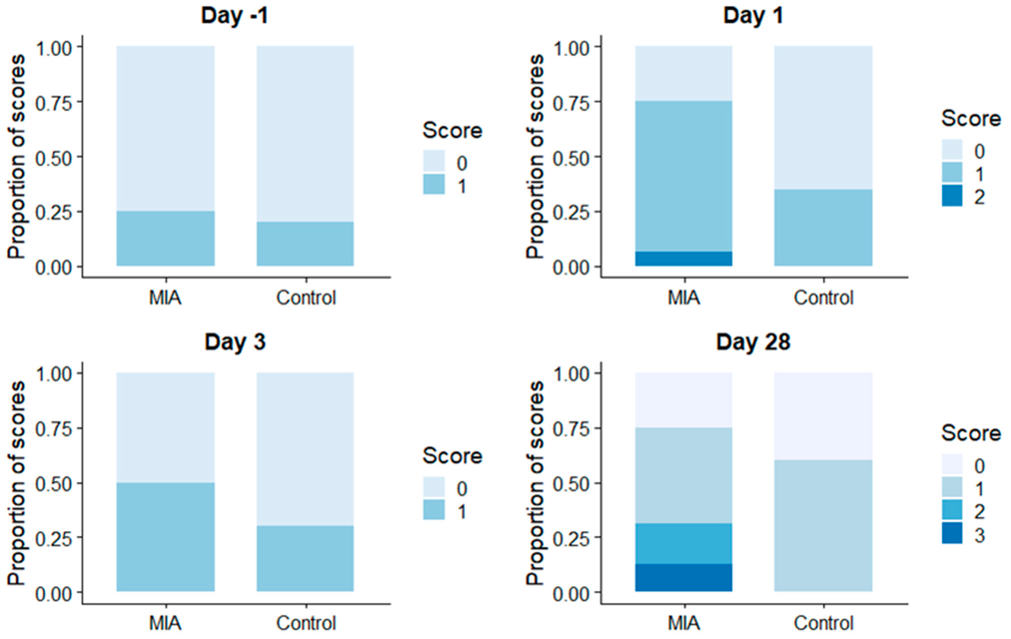
There was substantial agreement between the two observers, ƙ = 0.614, p < 0.005.
Lameness scores 1 and 2 were attributed to pigs in both the MIA and SC groups and on all days. Lameness score 3 and 4 were only given to pigs in the MIA group (Figure 2). Fishers exact test showed a difference in visual scores between groups only on day 1, with visual scores being higher in the MIA group (0.81+/−0.54) compared to the SC group (0.35+/−0.48). The pig from the MIA group that did not show any (histo-)pathological lesions had been assigned a gait score of 0 for all timepoints by both observers. The pig with macroscopic lesions but a histological sample being unavailable due to technical difficulties, had been assigned gait score 1 at day 3 and 28 by 1 observer, and score 0 for all other timepoints, including those assigned by the other observer.
3.2.2. Pressure Mat Analyses
Data from two animals from the MIA group were excluded from analysis, on account of no histopathological joint changes (n = 1) and no histopathological sample available (n = 1) to verify osteoarthritic changes observed on macroscopy. Pre-injection, ASI of VI and PVF in both groups were close to 0 (0 indicating perfect symmetry). Apart from ASI of PVF on day 7, when ASI scores were similar (and in the control group, close to 0), PVF and VI values were lower on all days post-injection (i.e., the left front limb was loaded less than the right front limb). In addition, the MIA group had lower ASI’s of PVF and VI than the SC group on day 3, 14, 28, and 56 (Table 4, Table 5 and Table 6).
4. Discussion
Choosing an appropriate in vivo animal model to study OA depends on which aspects of OA development are targeted and what the target species are [8]. We selected the pig as model species for studying pain related lameness under experimentally controlled conditions, with a clear veterinary focus. However, an OA model in the pig might also be useful to study the behavior of peri-articular structures in human OA, as proportions and intra-articular cartilage and associated ligaments are considered to have characteristics close to the analogous structures in humans [34]. Furthermore, pigs have a digestive tract more similar to humans than most other species used in in vivo animal OA research. Translational aspects from pig to human might therefor be more successful in the case of orally administered medication as interventional therapy [34].
In our study, we injected MIA to induce osteoarthritis in a single joint of pigs as a controlled experimental intervention. We tracked functional characteristics relative to baseline and a saline- control group using both visual scoring and limb loading kinetics up to day 56 after injection, with subsequent full necropsy and histopathology one week following completion of data collection. It has previously been shown in pigs that decreased weight-bearing is a useful measure in both experimentally-induced [23,24] and naturally-occurring [15,31] lameness. To our knowledge, the effects of MIA on weight-bearing in pigs have not been described before.
As between species translational aspects of OA are not the focus of the present study, the interested reader is referred elsewhere [35,36].
Next to face and construct validity of our model, the longitudinal within subject design of the present study also merits a limited discussion of predictive validity for naturally occurring lameness in pigs.
4.1. Face Validity
Face validity refers to how well the model resembles aspects of the disease condition phenotypically. We focused on histopathological joint alterations as the main histological endpoint, and altered weight bearing as the main clinical endpoint.
4.1.1. Histopathological Presentation of Joint Changes
Eight out of ten animals which were administered MIA showed histopathological changes in the injected joint, corroborating the MIA-induced synovial tissue changes described in other species [25,27,37]. MIA injection failed to induce histopathological changes consistent with OA in one animal and in another animal a sample for histopathological evaluation was lost due to technical difficulties. In the present study, histopathology was performed at a single timepoint after intra-articular injection, therefore dynamic changes in the injected joint caused by MIA could not be assessed.
When induction of MIA-associated histopathological OA joint changes is depending on a single MIA injection, the total dose of MIA administered [38] and intensity of exercise regime [39,40] have been shown to severely limit the extent or even completely reverse typical histopathological joint changes associated with MIA observed on necropsy. In our present study, the pigs received more exercise than an average pig on a commercial farm. Pressure mat measurements took on average 5–10 min per pig, although most of this time was spent consuming the rewards and waiting until the measuring equipment was ready for another run. Additionally, the pigs were housed under enriched conditions with straw bedding and more space than commercially housed pigs, which may have resulted in more activity and thus more exercise.
Although the effect of an exercise regime in our prospected pig model warrants further study, we argue that the most likely explanation for the absence of typical MIA -associated histopathology in two animals is failure to introduce or contain a sufficient dose of intra-articular MIA. This despite diligent practice of the intra-articular injection technique on same size piglet cadavers before applying this technique in the animals in our study.
4.1.2. Altered Weight Bearing (Lameness Assessment)
We assessed lameness by two methods: Subjective evaluation by veterinarians using a visual scoring system [30], and objective quantification of limb loading using a pressure mat [31]. Visual scoring is the only method routinely used in commercially housed pigs, and therefore relevant to daily practice. Until more sensitive diagnostic methods become widely available, pigs that are visually clearly lame will be identified by farmers and veterinarians and only this subpopulation with pronounced clinical lameness will be considered for treatment. Subclinical stages of OA are not detectable using this method. In order to also evaluate objective parameters for lameness in our model, pressure mat analysis was performed, a method that was expected to be more sensitive than the visual scoring method [41].
Visual Scoring
The visual scoring data only detected lameness between in the MIA-group compared to SC-group on day 1. The agreement between the two blinded observers using the adapted scoring-system was ƙ = 0.614. In the publication on the original scoring system, agreement varied between virtually no agreement (ƙ = 0.01) and high agreement (ƙ = 0.90), depending on familiarity with the scoring system and experience [30]. Although the two observers in our study were very experienced veterinarians, they had not used the scoring system before, which may explain the lower agreement compared to the trained observers in the original study. Also, the visual scoring system was modified, and several criteria present in the original scoring system by Main et al. [30] were removed to preserve true blinding of the video-assessors (Table 2). The items “initial response to human presence”, “pig’s response after opening gate” and “behaviour of individual within group” from the original protocol were not used, since this would require marking individuals to identify them within the group. Observers would then be able to recognize individuals on subsequent timepoints which may bias their scoring. The usability and sensitivity of visual scoring is enhanced by the addition of these behavioural criteria, although this was not confirmed in spontaneous OA in pigs [8]. Disuse of these criteria for scoring lameness may have resulted in the reduced performance of visual scoring. It is possible that particular aspects of lameness such as head bobbing are difficult to observe in pigs [30].
Another potential confounding factor, is the extensive training of the piglets to evenly trot for a click and food reward. Locomotion induced pain and a sweet treat reward at the destination, especially in a species as the pigs can pose two strongly opposing motivators to display outwardly visible pain behavioural. During the training, initially several animals were trained together, which may have induced fierce competition for the sweet treat reward, shaping getting to the sweet treat as a very strong motivator. Perhaps this point is best illustrated with a standard practice in neonatal care, where a sweet tasting liquid is administered orally, just prior to an invasive painful stimulus (like a heel prick) [42]. This on account of these (premature) neonates not showing, or showing to a lesser extent the characteristic pain face when being administered sucrose or a sweet tasting liquid. Similar to ambulation, a neonatal pain face originates from a highly shaped pattern of muscle contractions of which the coordination is mainly spinally mediated, with top down cerebral modulation [42]. This at least raises the possibility that outwardly displayed pain behaviour is susceptible to cerebral modulation induced by a “tug of war” between two strong yet opposing motivators.
Scoring from video recordings could also be more difficult than expected, however, movie clips have been used to score lameness in several other studies [43,44,45,46]. Observers commented on some of the recordings as “difficult to score”, which may mean that some of the errors are due to having to score from movies instead from live observations. On the other hand, in veterinary practice conditions are usually much worse, with low lighting intensities and crowded stables. The identification of lame animals by visual inspection is likely even more difficult under on-farm conditions than under the optimised experimental conditions of the present study. Consequently, we expect that under on-farm conditions, many (sub)clinically lame animals probably remain undetected.
MIA injection did not result in a lasting clinical lameness when assessing it visually. This means that if OA is induced using MIA, most animals will not be lame enough to be selected as candidates for treatment by the farmer or veterinarian.
When scoring lameness in pigs visually, the stoic nature of the species [8] likely contributed to the low predictability and correlation between the degree of joint damage induced and degree of lameness scored.
Eight animals from the MIA group needed rescue analgesia once due to clinical lameness scores of 4 or higher several hours after intra-articular injections were administered. It is possible that rescue analgesia masked more extreme differences on the first day after injection. The aim of this study, however, was to induce clinical lameness which would progress into a chronic condition. It is unlikely that a single clinical dose of buprenorphine would influence lameness scores for more than 12 h, let alone for 28 days afterwards. The pain behaviour suppressive effect of buprenorphine reported in experimental pain studies is dose dependent [47]. For the dose administered as rescue analgesia in this study the expected duration of effect is around 7 h. Although excessive dose escalation may inconsistently increase duration of effect considerably [48], in the clinical dose range the duration of effects does not exceed 12 h [49].
Another possible explanation for visual scoring not being able to detect severe lameness over a longer period is that the dose of MIA used in this study was not high enough to induce more severe lameness. Data from horses and dogs [50,51] indicated that MIA should not be dosed on body weight. Therefore, the MIA-dose we administered to our pigs (20 mg in an 80 mg/mL solution) was based on a pilot study using three doses: 10 mg, 20 mg or 40 mg (Supplementary Material B, Tables S1–S2, Figure S4, Equation (S1)). Both functional impairments and histological evidence of osteoarthritis development at day 11 were seen in the 20 and 40 mg dose, however, changes in the 40 mg dose were more severe and expected to progress to unacceptable levels of lameness and possibly ankyloses during the intended study period of 68 days post intra-articular injection. The 20 mg dose induced changes that were more comparable to previously reported changes in rats on day 10–14 [52] and was therefore selected for the study reported here.
Kinetic Data
Our criterion for face validity in a pig model of MIA induced OA concerning kinetic characteristics was that pigs should show decreased weight-bearing in both the acute and chronic phase in the leg with experimentally induced OA. The kinetic data in our study showed a biphasic decrease in weight-bearing in the MIA injected group. This is consistent with the well-characterized development of MIA-induced functional osteoarthritic changes in rats [53]. Immediately after intra-articular MIA-injection in the knee, rats showed a marked decrease in weight-bearing that was most pronounced at 4 days post-injection. By 7 days post-injection, weight-bearing had almost entirely normalized. At day 14 post-injection, however, a second less pronounced stage of decreased weight-bearing developed which did not resolve for the remainder of the study (28 days post-injection). This biphasic decrease in weight-bearing was also observed in rats by Fernihough et al. [54].
In conclusion, our criterion for face validity was partly fulfilled. Weight-bearing measured with an objective method decreased, however visual inspection did not identify a significant increase in lameness after day 1. This means that although intra-articular MIA injection can be used to induce lameness, this lameness is not consistently severe enough to model animals that are identified as candidates for treatment under on-farm conditions.
4.2. Predictive Validity
Longitudinal kinetic gait assessment data of our model was consistent with the rat MIA-model, which is favoured as a model to assess disease modifying (oral) drug (DMOD) therapies in OA [35]. Although predictive validity of our pig model was not part of the present study, we recently performed a study using this animal model [55]. We investigated, using the present pig model, whether a disease modifying oral drug (DMOD) therapy is a feasible option for intervention. Despite caution for non-steroid anti-inflammatory drug (NSAID) treatment in conditions requiring bone remodeling is sometimes advocated [56], no adverse side-effects of NSAID (Meloxicam) treatment were detected in our pig model. Daily treatment for 48 consecutive days with oral Meloxicam (Metacam 15 mg/mL oral suspension for pigs, Boehringer Ingelheim Vetmedica, Rohrdorf, Germany), a potential osteoarthritis DMOD, in healthy control animals (intra-articular saline injections) did not affect gait, and further did not reveal adverse effects in growing pigs. Weight gain, gait, trabecular bone parameters, growth plate morphology, gastrointestinal integrity, and kidney histology were al shown to be unaffected by long-term Meloxicam treatment. These findings are not yet supportive evidence for the feasibility of daily DMOD therapy with Meloxicam in porcine osteoarthritis. However, when Meloxicam as DMOD therapy following intra-articular MIA injections would be assessed in our model, confounding pain or gait abnormalities due to aberrant skeletal development would not be expected. As part of the iterative process of model evaluation, other putative therapeutics should be tested in this model to further establish its predictive validity.
4.3. Construct Validity
For the MIA model to have construct validity, the mechanisms underlying the main clinical symptom lameness should be comparable to the naturally-occurring disease.
We only performed morphological assessment of joint condition at necropsy on day 68 and therefore we do not have information regarding the development of structural joint changes prior to day 68. A non-invasive imaging technique such as MRI may have provided insight into the dynamics of morphological joint changes. This would have enabled comparison with the kinetic measurements, without losing animals for the later timepoints, as is the case with necropsy of some animals at an earlier timepoint. In a pilot study (Supplementary Material B, Tables S1–S2), a board-certified veterinary radiologist blinded for treatment assessed the carpal joint of pigs injected with MIA and saline solution 11 days previously with a 1.5 Tesla closed model MRI post-mortem just prior to histopathology. It proved difficult to relate the MRI images of the freshly obtained affected and control carpal joints with the histopathology performed the same day. Furthermore, anaesthesia required for imaging during a longitudinal in vivo experiment might have caused residual spinal anti-nociceptive effects [57,58]. This may interfere with subsequent clinical scoring of lameness, particularly in the earlier time-points, when interval times are short. We therefore decided not to include imaging techniques requiring anaesthesia in our final study design.
In the case of OA, lameness is initially due to (inflammatory) pain. In advanced cases, altered range of motion due to bony changes is an additional cause for impaired locomotion [8]. Although inflammation and joint damage are generally considered to contribute to joint pain, radiographic grading of the severity of osteoarthritis in humans often does not match the amount of pain reported [12,59].
Joint pain associated with OA in prone individuals is a progressive, fluctuating, mixed, and ultimately severely debilitating chronic pain complex [60,61,62,63]. Generally, initial acute inflammatory pain with straightforward relation between tissue damage and pain sensation becomes progressively unhinged due to functional and structural changes in the nociceptive pathways. Around the time that subchondral bone becomes exposed due to full thickness cartilage erosion, a neuropathic pain component [64] accompanies and interacts with the inflammatory and mechanical pain component [9]. A complex, mixed chronic progressive pain syndrome ensues. Supraspinal mechanisms meant to modulate ascending nociceptive input will ultimately fail and drive the affective-motivational and cognitive-evaluative components of the pain experience towards a debilitating condition often contributing to a general anhedonic and depressive state of mind [60]. As such a state is known to influence the perception of evoked (experimental) pain (such as is tested during human OA staging and OA pain studies) [59], it is hardly a surprise that poor correlation is consistently reported [59,65] between anatomical joint scores of affected joints and associated joint pain. Although decreased weightbearing assessed by kinetic analysis in the MIA group followed a bi-phasic pattern in agreement with the data in rat, at present we do not know if structural changes are the cause of this decrease.
Kinetic gait assessment revealed significant alteration in locomotion at timepoints representing the more acute as well as the chronic phase of MIA induced OA. However, with the visual scoring system used only the acute phase of pain-associated lameness was detected against the background of isotonic saline control treatment. Potentially, increasing the dose of intra-articular MIA could improve the performance of our model with regards to visual lameness scoring during the more chronic phase of MIA-induced OA.
5. Conclusions
Although we feel that naturally-occurring animal models are the best reflection of the actual disease in both humans and animals, they have clear limitations [10]. Especially in larger animals and with OA, disease development, and progression, may take months to years. It may affect multiple sites in the body and during development uncontrolled comorbidities may ensue, negatively impacting a controlled study of an isolated site of disease manifestation. And first and foremost, in many animals the disease will never develop at all [35].
Although the MIA model evaluated in this study may be useful for some applications, it also has limitations. The aim was to develop a model the acute and chronic phase of OA to ultimately assess the effects of interventions on lameness in commercially housed pigs. Although objective parameters for weight-bearing showed that lameness was present in MIA-treated animals in both the acute and chronic phase of induced OA, visually many animals were considered “sound” by two experienced observers. Therefore, it is unlikely that the detected level of lameness by kinetic analysis in our model would ever be identified on-farm as in need of intervention. The decreased weight-bearing however, can be interpreted as a behavioral indicator of pain [8] and therefore the MIA model as reported here may have its merit as a general model for pain in pigs.
Supplementary Materials
The following are available online at https://www.mdpi.com/2076-2615/9/7/405/s1, Material A: Optimization of intra-articular injection technique on cadaver material; Figure S1: Presentation of the dorsal aspect of the left proximal carpal joint, Figure S2: Injection site, angle and depth of needle advancement for intra-articular deposition of methylene blue into the intercarpal joint, Figure S3: The distribution of blue dye in the carpal joint sac. Material B: Pilot study: In Vivo intra-articular Monododium Iodoacetate dose finding study; Table S1: Litter of origen, gender, and treatment group of selected piglets, Table S2: Experimental procedures, Equation (S1): Asymmetry index of stance time; Material C: Representative macroscopic intercarpal joint morphology 68 days following intra-articular injection of isotonic saline or 20 mg Monosodium Iodoacetate: Figure S4: Macroscopic presentation of the proximal aspect (looking in a ventro-dorsal direction) of the intercarpal joint on necropsy.
Author Contributions
Conceptualization, J.U., E.M. and F.J.v.d.S.; Methodology, E.M., J.U. and F.J.v.d.S.; Formal analysis, E.M. and F.J.v.d.S.; Investigation, J.U. and E.M.; Resources F.J.v.d.S.; Writing—original draft preparation, J.U., E.M. and F.J.v.d.S.; writing—review and editing, E.M., J.U. and F.J.v.d.S.; visualization, E.M., F.J.v.d.S. and J.U.; Supervision, F.J.v.d.S.; Project administration, F.J.v.d.S.
Funding
This research received no external funding.
Acknowledgments
The authors wish to express their gratitude and acknowledge the substantial contributions towards animal care, training the animals and assistance in conducting the experimental measurements: A. van Dam, M.M.M. Wetzels and T. Verhoeve. For the pilot study we would further like to thank E.C.A. Waas and M. Kranenburg. The authors further would like to extend their gratitude toward L. Dieste Pérez and A. van Nes for their willingness to score the video recordings of locomotion and H. Vernooij for consultation with the statistical analysis.
Conflicts of Interest
The authors declare no conflict of interest.
References
- Kilbride, A.L.; Gillman, C.E.; Green, L.E. A cross-sectional study of the prevalence of lameness in finishing pigs, gilts and pregnant sows and associations with limb lesions and floor types on commercial farms in England. Anim. Welf. 2009, 18, 215–224. [Google Scholar]
- Anil, S.; Anil, L.; Deen, J. Effect of Lameness in Pigs in Terms of “Five Freedoms”. J. Appl. Anim. Welf. Sci. 2009, 12, 144–145. [Google Scholar] [CrossRef]
- Heinonen, M.; Peltoniemi, O.; Valros, A. Impact of lameness and claw lesions in sows on welfare, health and production. Livest. Sci. 2013, 156, 2–9. [Google Scholar] [CrossRef]
- Jensen, T.B.; Baadsgaard, N.P.; Houe, H.; Toft, N.; Østergaard, S. The effect of lameness treatments and treatments for other health disorders on the weight gain and feed conversion in boars at a Danish test station. Livest. Sci. 2007, 112, 34–42. [Google Scholar] [CrossRef]
- Jensen, T.B.; Kristensen, H.H.; Toft, N. Quantifying the impact of lameness on welfare and profitability of finisher pigs using expert opinions. Livest. Sci. 2012, 149, 209–214. [Google Scholar] [CrossRef]
- Lane, N.E.; Brandt, K.; Hawker, G.; Peeva, E.; Schreyer, E.; Tsuji, W.; Hochberg, M.C. OARSI-FDA initiative: defining the disease state of osteoarthritis. Osteoarthritis Cartilage 2011, 19, 478–482. [Google Scholar] [CrossRef] [PubMed] [Green Version]
- Jensen, T. Causes of and predisposing risk factors for leg disorders in growing-finishing pigs. CAB Rev. Perspect. Agric. Vet. Sci. Nutr. Nat. Resour. 2009, 4, 1–8. [Google Scholar] [CrossRef]
- Macfadyen, M.A.; Daniel, Z.; Kelly, S.; Parr, T.; Brameld, J.M.; Murton, A.J.; Jones, S.W. The commercial pig as a model of spontaneously-occurring osteoarthritis. BMC Musculoskelet. Disord. 2019, 20, 70. [Google Scholar] [CrossRef]
- Deveza, L.A.; Hunter, D.J. Editorial: Unraveling Osteoarthritis Pathogenesis: New Insights Into Preradiographic Disease and Patient Phenotypes. Arthritis Rheumatol. 2015, 67, 3097–3100. [Google Scholar] [CrossRef] [Green Version]
- Van der Staay, F.J. Animal models of behavioral dysfunctions: basic concepts and classifications, and an evaluation strategy. Brain Res. Rev. 2006, 52, 131–159. [Google Scholar] [CrossRef]
- Maximino, C.; van der Staay, F.J. Behavioral models in psychopathology: epistemic and semantic considerations. Behav. Brain Funct. BBF 2019, 15, 1. [Google Scholar] [CrossRef] [PubMed] [Green Version]
- Dieppe, P.A.; Lohmander, L.S. Pathogenesis and management of pain in osteoarthritis. Lancet 2005, 365, 965–973. [Google Scholar] [CrossRef]
- Ishikawa, G.; Nagakura, Y.; Takeshita, N.; Shimizu, Y. Efficacy of drugs with different mechanisms of action in relieving spontaneous pain at rest and during movement in a rat model of osteoarthritis. Eur. J. Pharmacol. 2014, 738, 111–117. [Google Scholar] [CrossRef] [PubMed]
- Ison, S.H.; Clutton, R.E.; Di Giminiani, P.; Rutherford, K.M.D. A Review of Pain Assessment in Pigs. Front. Vet. Sci. 2016, 3, 108. [Google Scholar] [CrossRef] [PubMed]
- Meijer, E.; van Nes, A.; Back, W.; van der Staay, F.J. Clinical effects of buprenorphine on open field behaviour and gait symmetry in healthy and lame weaned piglets. Vet. J. Lond. Engl. 1997 2015, 206, 298–303. [Google Scholar] [CrossRef] [PubMed]
- Ferreira-Gomes, J.; Adães, S.; Mendonça, M.; Castro-Lopes, J.M. Analgesic effects of lidocaine, morphine and diclofenac on movement-induced nociception, as assessed by the Knee-Bend and CatWalk tests in a rat model of osteoarthritis. Pharmacol. Biochem. Behav. 2012, 101, 617–624. [Google Scholar] [CrossRef] [PubMed]
- Clinical Examination of Farm Animals: Jackson/Clinical; Jackson, P.G.G.; Cockcroft, P.D. (Eds.) Blackwell Science Ltd.: Oxford, UK, 2002; ISBN 978-0-470-75242-5. [Google Scholar]
- Arkell, M.; Archer, R.M.; Guitian, F.J.; May, S.A. Evidence of bias affecting the interpretation of the results of local anaesthetic nerve blocks when assessing lameness in horses. Vet. Rec. 2006, 159, 346–349. [Google Scholar] [CrossRef] [PubMed]
- D’Eath, R.B. Repeated locomotion scoring of a sow herd to measure lameness: consistency over time, the effect of sow characteristics and inter-observer reliability. Anim. Welf. 2012, 21, 219–231. [Google Scholar] [CrossRef]
- Petersen, H.H.; Enøe, C.; Nielsen, E.O. Observer agreement on pen level prevalence of clinical signs in finishing pigs. Prev. Vet. Med. 2004, 64, 147–156. [Google Scholar] [CrossRef]
- Etterlin, P.E.; Morrison, D.A.; Österberg, J.; Ytrehus, B.; Heldmer, E.; Ekman, S. Osteochondrosis, but not lameness, is more frequent among free-range pigs than confined herd-mates. Acta Vet. Scand. 2015, 57, 63. [Google Scholar] [CrossRef] [Green Version]
- Quinn, M.M.; Keuler, N.S.; Lu, Y.; Faria, M.L.E.; Muir, P.; Markel, M.D. Evaluation of agreement between numerical rating scales, visual analogue scoring scales, and force plate gait analysis in dogs. Vet. Surg. VS 2007, 36, 360–367. [Google Scholar] [CrossRef] [PubMed]
- Karriker, L.A.; Abell, C.E.; Pairis-Garcia, M.D.; Holt, W.A.; Sun, G.; Coetzee, J.F.; Johnson, A.K.; Hoff, S.J.; Stalder, K.J. Validation of a lameness model in sows using physiological and mechanical measurements. J. Anim. Sci. 2013, 91, 130–136. [Google Scholar] [CrossRef] [PubMed]
- Pairis-Garcia, M.D.; Johnson, A.K.; Abell, C.A.; Coetzee, J.F.; Karriker, L.A.; Millman, S.T.; Stalder, K.J. Measuring the efficacy of flunixin meglumine and meloxicam for lame sows using a GAITFour pressure mat and an embedded microcomputer-based force plate system. J. Anim. Sci. 2015, 93, 2100–2110. [Google Scholar] [CrossRef] [PubMed]
- Guingamp, C.; Gegout-Pottie, P.; Philippe, L.; Terlain, B.; Netter, P.; Gillet, P. Mono-iodoacetate-induced experimental osteoarthritis: a dose-response study of loss of mobility, morphology, and biochemistry. Arthritis Rheum. 1997, 40, 1670–1679. [Google Scholar] [CrossRef] [PubMed]
- Bove, S.E.; Calcaterra, S.L.; Brooker, R.M.; Huber, C.M.; Guzman, R.E.; Juneau, P.L.; Schrier, D.J.; Kilgore, K.S. Weight bearing as a measure of disease progression and efficacy of anti-inflammatory compounds in a model of monosodium iodoacetate-induced osteoarthritis. Osteoarthritis Cartilage 2003, 11, 821–830. [Google Scholar] [CrossRef] [Green Version]
- Combe, R.; Bramwell, S.; Field, M.J. The monosodium iodoacetate model of osteoarthritis: A model of chronic nociceptive pain in rats? Neurosci. Lett. 2004, 370, 236–240. [Google Scholar] [CrossRef] [PubMed]
- Kalbhen, D.A. Chemical model of osteoarthritis—A pharmacological evaluation. J. Rheumatol. 1987, 14, 130–131. [Google Scholar] [PubMed]
- Van der Kraan, P.M.; Vitters, E.L.; van de Putte, L.B.; van den Berg, W.B. Development of osteoarthritic lesions in mice by “metabolic” and “mechanical” alterations in the knee joints. Am. J. Pathol. 1989, 135, 1001–1014. [Google Scholar]
- Main, D.C.J.; Clegg, J.; Spatz, A.; Green, L.E. Repeatability of a lameness scoring system for finishing pigs. Vet. Rec. 2000, 147, 574–576. [Google Scholar] [CrossRef]
- Meijer, E.; Oosterlinck, M.; van Nes, A.; Back, W.; van der Staay, F.J. Pressure mat analysis of naturally occurring lameness in young pigs after weaning. BMC Vet. Res. 2014, 10, 193. [Google Scholar]
- R Development Core Team. R: A Language and Environment for Statistical Computing; R. Foundation for statistical Computing: Vienna, Austria, 2008. [Google Scholar]
- Pinheiro, J.; Bates, D.; DebRoy, S.; Sarkar, D. nlme: Linear and Nonlinear Mixed Effects Models. Available online: https://cran.r-project.org/web/packages/nlme/index.html (accessed on 1 July 2019).
- Swindle, M.M.; Makin, A.; Herron, A.J.; Clubb, F.J.; Frazier, K.S. Swine as Models in Biomedical Research and Toxicology Testing. Vet. Pathol. Online 2012, 49, 344–356. [Google Scholar] [CrossRef] [PubMed]
- Teeple, E.; Jay, G.D.; Elsaid, K.A.; Fleming, B.C. Animal Models of Osteoarthritis: Challenges of Model Selection and Analysis. AAPS J. 2013, 15, 438–446. [Google Scholar] [CrossRef] [PubMed] [Green Version]
- Lampropoulou-Adamidou, K.; Lelovas, P.; Karadimas, E.V.; Liakou, C.; Triantafillopoulos, I.K.; Dontas, I.; Papaioannou, N.A. Useful animal models for the research of osteoarthritis. Eur. J. Orthop. Surg. Traumatol. 2014, 24, 263–271. [Google Scholar] [CrossRef] [PubMed]
- Guzman, R.E.; Evans, M.G.; Bove, S.; Morenko, B.; Kilgore, K. Mono-iodoacetate-induced histologic changes in subchondral bone and articular cartilage of rat femorotibial joints: An animal model of osteoarthritis. Toxicol. Pathol. 2003, 31, 619–624. [Google Scholar] [CrossRef] [PubMed]
- Stobie, D.; Lipowitz, A.J.; Greer, N.L. Failure of monoiodoacetate to induce lameness in dogs. Vet. Comp. Orthop. Traumatol. VCOT 1994, 7, 91–93. [Google Scholar] [CrossRef]
- Galois, L.; Etienne, S.; Grossin, L.; Watrin-Pinzano, A.; Cournil-Henrionnet, C.; Loeuille, D.; Netter, P.; Mainard, D.; Gillet, P. Dose-response relationship for exercise on severity of experimental osteoarthritis in rats: A pilot study. Osteoarthritis Cartilage 2004, 12, 779–786. [Google Scholar] [CrossRef] [PubMed]
- Fallah Mohammadi, M.; Hajizadeh Moghaddam, A.; Mirkarimpur, H. The Effects of a Moderate Exercise Program on Knee Osteoarthritis in Male Wistar Rats. Iran. J. Basic Med. Sci. 2013, 16, 683–688. [Google Scholar] [PubMed]
- Waxman, A.S.; Robinson, D.A.; Evans, R.B.; Hulse, D.A.; Innes, J.F.; Conzemius, M.G. Relationship between objective and subjective assessment of limb function in normal dogs with an experimentally induced lameness. Vet. Surg. VS 2008, 37, 241–246. [Google Scholar] [CrossRef] [PubMed]
- Fitzgerald, M. What do we really know about newborn infant pain? Exp. Physiol. 2015, 100, 1451–1457. [Google Scholar] [CrossRef] [PubMed]
- Fuller, C.J.; Bladon, B.M.; Driver, A.J.; Barr, A.R.S. The intra- and inter-assessor reliability of measurement of functional outcome by lameness scoring in horses. Vet. J. Lond. Engl. 1997 2006, 171, 281–286. [Google Scholar] [CrossRef]
- Hewetson, M.; Christley, R.M.; Hunt, I.D.; Voute, L.C. Investigations of the reliability of observational gait analysis for the assessment of lameness in horses. Vet. Rec. 2006, 158, 852–857. [Google Scholar] [CrossRef] [PubMed]
- Kaler, J.; Wassink, G.J.; Green, L.E. The inter- and intra-observer reliability of a locomotion scoring scale for sheep. Vet. J. Lond. Engl. 1997 2009, 180, 189–194. [Google Scholar] [CrossRef] [PubMed] [Green Version]
- Keegan, K.G.; Wilson, D.A.; Wilson, D.J.; Smith, B.; Gaughan, E.M.; Pleasant, R.S.; Lillich, J.D.; Kramer, J.; Howard, R.D.; Bacon-Miller, C.; et al. Evaluation of mild lameness in horses trotting on a treadmill by clinicians and interns or residents and correlation of their assessments with kinematic gait analysis. Am. J. Vet. Res. 1998, 59, 1370–1377. [Google Scholar] [PubMed]
- Rodriguez, N.A.; Cooper, D.M.; Risdahl, J.M. Antinociceptive activity of and clinical experience with buprenorphine in swine. Contemp. Top. Lab. Anim. Sci. Am. Assoc. Lab. Anim. Sci. 2001, 40, 17–20. [Google Scholar]
- Hermansen, K.; Pedersen, L.E.; Olesen, H.O. The analgesic effect of buprenorphine, etorphine and pethidine in the pig: A randomized double blind cross-over study. Acta Pharmacol. Toxicol. (Copenh.) 1986, 59, 27–35. [Google Scholar] [CrossRef] [PubMed]
- Tranquilli, W.J.; Thurmon, J.C.; Grimm, K.A. Chapter 29- Swine. In Veterinary Anesthesia and Analgesia; 4th ed.; Blackwell Puyblishing: Oxford, UK, 2007; pp. 747–763. ISBN 978-0-7817-5471-2. [Google Scholar]
- Dowling, B.; Dart, A.; Matthews, S. Chemical arthrodesis of the distal tarsal joints using sodium monoiodoacetate in 104 horses. Aust. Vet. J. 2004, 82, 38–42. [Google Scholar] [CrossRef] [PubMed]
- Goranov, N. Clinical changes in sodium monoiodoacetate induced stifle osteoarthritis model in dogs. Vet. World 2012, 5, 138–144. [Google Scholar] [CrossRef]
- Ivanavicius, S.P.; Ball, A.D.; Heapy, C.G.; Westwood, F.R.; Murray, F.; Read, S.J. Structural pathology in a rodent model of osteoarthritis is associated with neuropathic pain: Increased expression of ATF-3 and pharmacological characterisation. PAIN 2007, 128, 272–282. [Google Scholar] [CrossRef]
- Pomonis, J.D.; Boulet, J.M.; Gottshall, S.L.; Phillips, S.; Sellers, R.; Bunton, T.; Walker, K. Development and pharmacological characterization of a rat model of osteoarthritis pain. Pain 2005, 114, 339–346. [Google Scholar] [CrossRef]
- Fernihough, J.; Gentry, C.; Malcangio, M.; Fox, A.; Rediske, J.; Pellas, T.; Kidd, B.; Bevan, S.; Winter, J. Pain related behaviour in two models of osteoarthritis in the rat knee. Pain 2004, 112, 83–93. [Google Scholar] [CrossRef]
- Gorissen, B.M.C.; Uilenreef, J.J.; Bergmann, W.; Meijer, E.; van Rietbergen, B.; van der Staay, F.J.; van Weeren, P.R.; Wolschrijn, C.F. Effects of long-term use of the preferential COX-2 inhibitor meloxicam on growing pigs. Vet. Rec. 2017, 181, 564. [Google Scholar] [CrossRef] [PubMed]
- Pountos, I.; Georgouli, T.; Calori, G.M.; Giannoudis, P.V. Do nonsteroidal anti-inflammatory drugs affect bone healing? A critical analysis. Sci. World J. 2012, 2012, 606404. [Google Scholar] [CrossRef]
- Ishii, H.; Kohno, T.; Yamakura, T.; Ikoma, M.; Baba, H. Action of dexmedetomidine on the substantia gelatinosa neurons of the rat spinal cord. Eur. J. Neurosci. 2008, 27, 3182–3190. [Google Scholar] [CrossRef] [PubMed] [Green Version]
- Christoph, T.; Schiene, K.; Englberger, W.; Parsons, C.G.; Chizh, B.A. The antiallodynic effect of NMDA antagonists in neuropathic pain outlasts the duration of the in vivo NMDA antagonism. Neuropharmacology 2006, 51, 12–17. [Google Scholar] [CrossRef] [PubMed]
- Arendt-Nielsen, L.; Egsgaard, L.L.; Petersen, K.K.; Eskehave, T.N.; Graven- Nielsen, T.; Hoeck, H.C.; Simonsen, O. A mechanism-based pain sensitivity index to characterize knee osteoarthritis patients with different disease stages and pain levels. Eur. J. Pain 2015, 19, 1406–1417. [Google Scholar] [CrossRef] [PubMed]
- Neogi, T. The epidemiology and impact of pain in osteoarthritis. Osteoarthritis Cartilage 2013, 21, 1145–1153. [Google Scholar] [CrossRef] [PubMed] [Green Version]
- Thakur, M.; Dawes, J.M.; McMahon, S.B. Genomics of pain in osteoarthritis. Osteoarthritis Cartilage 2013, 21, 1374–1382. [Google Scholar] [CrossRef] [PubMed] [Green Version]
- King, C.D.; Sibille, K.T.; Goodin, B.R.; Cruz-Almeida, Y.; Glover, T.L.; Bartley, E.; Riley, J.L.; Herbert, M.S.; Sotolongo, A.; Schmidt, J.; et al. Experimental pain sensitivity differs as a function of clinical pain severity in symptomatic knee osteoarthritis. Osteoarthritis Cartilage 2013, 21, 1243–1252. [Google Scholar] [CrossRef] [PubMed] [Green Version]
- Creamer, P.; Lethbridge-Cejku, M.; Hochberg, M.C. Factors associated with functional impairment in symptomatic knee osteoarthritis. Rheumatology 2000, 39, 490–496. [Google Scholar] [CrossRef] [Green Version]
- Dimitroulas, T.; Duarte, R.V.; Behura, A.; Kitas, G.D.; Raphael, J.H. Neuropathic pain in osteoarthritis: A review of pathophysiological mechanisms and implications for treatment. Semin. Arthritis Rheum. 2014, 44, 145–154. [Google Scholar] [CrossRef]
- Shimura, Y.; Kurosawa, H.; Sugawara, Y.; Tsuchiya, M.; Sawa, M.; Kaneko, H.; Futami, I.; Liu, L.; Sadatsuki, R.; Hada, S.; et al. The factors associated with pain severity in patients with knee osteoarthritis vary according to the radiographic disease severity: A cross-sectional study. Osteoarthritis Cartilage 2013, 21, 1179–1184. [Google Scholar] [CrossRef] [PubMed]
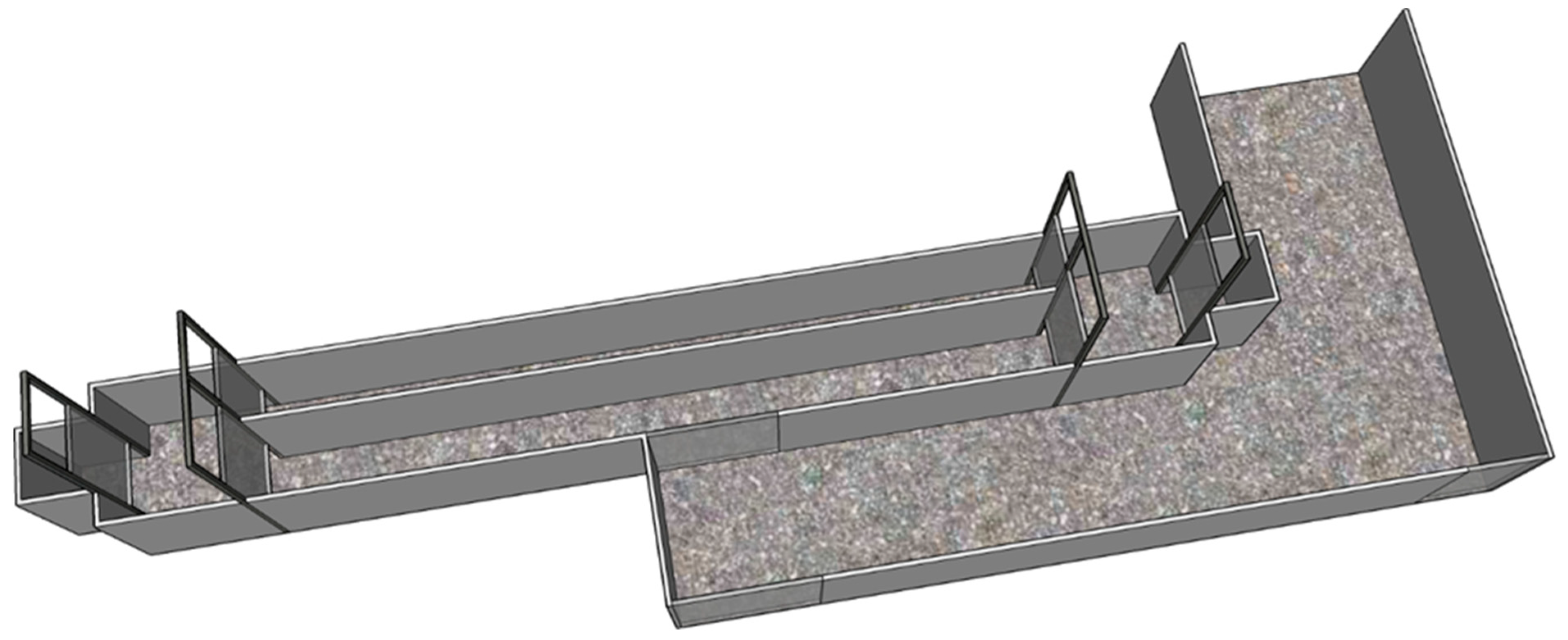
Figure 1.
Schematic illustration of the experimental setup of the runway containing a pressure mat. The pressure mat was positioned exactly half-way in the runway at the back (furthest away from the penmates in the holding area). The cameras were positioned on the top of the frames of the guillotine doors on either side of the runway.

Figure 2.
Proportion of different visual lameness scores modified from (see Table 2 for scored parameters) on specified timepoints during the experiment. The time-point designated panels represent blinded observer assigned offline lameness score distribution in the MIA treatment (MIA) and Saline Control treatment (Controls) groups. Day-1: on the day before intra-articular injection; Day 1, Day 3 and Day 28: one day, 3 days, and 28 days respectively after intra-articular injections.


Table 1.
Litter of origin, gender and treatment group of selected piglets.
| ID | Litter | Gender | Group |
|---|---|---|---|
| 1 | 1 | F 1 | MIA 3 |
| 2 | 1 | M 2 | MIA |
| 3 | 2 | F | MIA |
| 4 | 2 | F | MIA |
| 5 | 3 | M | MIA |
| 6 | 3 | F | MIA |
| 7 | 4 | F | MIA |
| 8 | 4 | F | MIA |
| 9 | 5 | F | MIA |
| 10 | 5 | F | MIA |
| 11 | 1 | M | SC 4 |
| 12 | 1 | F | SC |
| 13 | 2 | M | SC |
| 14 | 2 | M | SC |
| 15 | 3 | F | SC |
| 16 | 3 | M | SC |
| 17 | 4 | F | SC |
| 18 | 4 | M | SC |
| 19 | 5 | M | SC |
| 20 | 5 | F | SC |
1 F = female; 2 M = Male; 3 MIA = Monosodium Iodoacetate treatment; 4 SC = Saline Control treatment.
Table 2.
Experimental procedures.
| Day Relative to Joint Injection | Age of Animals (days) | Performed Procedures |
|---|---|---|
| –21 | 28 | Selection of litters and piglets; Weaning and transportation to experimenteal facility |
| –21 to –1 | 28–49 | Habitation and training (to personnel and being alone in the different test set-ups) |
| –1 | 49 | Baseline measurements:
|
| 0 | 50 | General anaesthesia for intra-articular injection:
|
| ||
| 1 | 51 | Day 1 measurements:
|
| 3 | 53 | Day 3 measurements:
|
| 7 | 57 | Day 7 measurements:
|
| 14 | 64 | Day 14 measurements:
|
| 21 | 71 | Day 21 measurements:
|
| 28 | 78 | Day 28 measurements:
|
| 56 | 106 | Day 56 measurements:
|
| 68 | 118 | General anaesthesia for euthanasia; necropsy |
1 ♂ being an intact male; 2 ♀ being an intact female.
Table 3.
Visual scoring protocol modified from Main et al. [30].
Table 3.
Visual scoring protocol modified from Main et al. [30].
| Lameness Score | Standing Posture | Gait |
|---|---|---|
| 0 | Pig stands squarely on 4 legs. | Even strides1. Caudal body sways slightly while walking. Pig is able to accelerate and change direction rapidly |
| 1 | As for score 0. | Abnormal stride length (not easily identified). Movements no longer fluent (pig appears stiff). Pig still able to accelerate and change direction. |
| 2 | Uneven posture. | Shortened stride. Lameness detected. Swagger of caudal body while walking. No hindrance in pig’s agility. |
| 3 | Uneven posture. Will not bear weight on affected limb (appears to be standing on toes). | Shortened stride. Minimum weight-bearing on affected limb. Swagger of caudal body while walking. Will still trot and gallop. |
| 4 | Affected limb elevated off floor. Pig appears visibly distressed. | Pig may not place affected limb on the floor while moving. |
| 5 | Will not stand unaided. | Does not move. |
1Bold type identifies the defining criteria that must be present to assign a score. Normal type shows supporting criteria that are useful for assigning a score.
Table 4.
Mean values ± SD on each day for asymmetry indices (ASI) of Peak Vertical Force (PVF) and Vertical Impulse (VI) in the MIA (n = 8) and SC (n = 10) groups.
Table 4.
Mean values ± SD on each day for asymmetry indices (ASI) of Peak Vertical Force (PVF) and Vertical Impulse (VI) in the MIA (n = 8) and SC (n = 10) groups.
| Day | Parameter | MIA | SC |
|---|---|---|---|
| 1 | PVF | 2.94 ± 20.59 | −0.27 ± 18.88 |
| VI | 7.36 ± 24.55 | 1.52 ± 20.30 | |
| 1 | PVF | −36.79 ± 22.94 | −28.55 ± 25.42 |
| VI | −43.47 ± 22.98 | −28.06 ± 27.59 | |
| 3 | PVF | −27.08 ± 25.41 1 | -0.38 ± 16.77 |
| VI | −33.16 ± 19.28 1 | −3.44 ± 19.10 | |
| 7 | PVF | −18.32 ± 30.12 | −3.64 ± 25.02 |
| VI | −19.90 ± 30.58 | −4.99 ± 22.05 | |
| 14 | PVF | 32.84 ± 23.58 1 | 2.31 ± 17.82 |
| VI | −33.46 ± 26.89 1 | 4.21 ± 18.63 | |
| 28 | PVF | −32.84 ± 32.67 1 | 14.69 ± 12.03 |
| VI | −32.58 ± 34.30 1 | 16.84 ± 8.63 | |
| 56 | PVF | −44.14 ± 50.09 1 | −3.10 ± 14.70 |
| VI | −40.03 ± 56.93 1 | −3.83 ± 17.06 |
1 Significant difference from SC group on the same day.
Table 5.
Estimates and standard errors for intercept and differences with reference category Peak Vertical Force (PVF) for each explanatory variable in the final model.
Table 5.
Estimates and standard errors for intercept and differences with reference category Peak Vertical Force (PVF) for each explanatory variable in the final model.
| Explanatory Variable | Estimate | Standard Error | p-Value |
|---|---|---|---|
| (Intercept) 1 | 2.94 | 8.71 | 0.737 |
| Day 1 | −39.73 | 11.58 | 0.001 |
| Day3 | −30.01 | 11.58 | 0.011 |
| Day7 | −21.26 | 11.58 | 0.070 |
| Day 14 | −35.77 | 11.58 | 0.003 |
| Day28 | −35.78 | 11.58 | 0.003 |
| Day56 | −47.08 | 11.58 | 0.000 |
| SC group | −3.21 | 11.69 | 0.787 |
| Day 1 × SC group | 11.45 | 15.54 | 0.463 |
| Day 3 × SC group | 29.90 | 15.54 | 0.057 |
| Day 7 × SC group | 17.89 | 15.54 | 0.253 |
| Day14 × SC group | 38.35 | 15.54 | 0.015 |
| Day 28 × SC group | 50.75 | 15.54 | 0.002 |
| Day56 × SC group | 44.25 | 15.54 | 0.005 |
1 Mean PVF for reference category (Day-1, MIA group); SC = saline control
Table 6.
Estimates and standard errors for intercept and differences with reference category Vertical Impulse (VI) for each explanatory variable in the final model.
Table 6.
Estimates and standard errors for intercept and differences with reference category Vertical Impulse (VI) for each explanatory variable in the final model.
| Explanatory Variable | Estimate | Standard Error | p-Value |
|---|---|---|---|
| (Intercept) 1 | 8.382.94 | 8.50 | 0.326 |
| Day 1 | −50.83 | 11.97 | 0.000 |
| Day3 | −40.52 | 11.98 | 0.001 |
| Day7 | −27.26 | 12.02 | 0.026 |
| Day 14 | −40.83 | 12.16 | 0.001 |
| Day28 | −39.94 | 12.66 | 0.002 |
| Day56 | −47.40 | 14.46 | 0.002 |
| SC group | −1.96 | 11.69 | 0.869 |
| Day 1 × SC group | 21.24 | 16.06 | 0.189 |
| Day 3 × SC group | 35.54 | 16.08 | 0.029 |
| Day 7 × SC group | 20.74 | 16.13 | 0.202 |
| Day14 × SC group | 43.51 | 16.31 | 0.009 |
| Day 28 × SC group | 55.25 | 16.99 | 0.002 |
| Day56 × SC group | 42.04 | 19.41 | 0.033 |
1 Mean VI for reference category (Day-1, MIA group); SC = saline control
© 2019 by the authors. Licensee MDPI, Basel, Switzerland. This article is an open access article distributed under the terms and conditions of the Creative Commons Attribution (CC BY) license (http://creativecommons.org/licenses/by/4.0/).
Share and Cite
MDPI and ACS Style
Uilenreef, J.; van der Staay, F.J.; Meijer, E. A Monosodium Iodoacetate Osteoarthritis Lameness Model in Growing Pigs. Animals 2019, 9, 405. https://doi.org/10.3390/ani9070405
AMA Style
Uilenreef J, van der Staay FJ, Meijer E. A Monosodium Iodoacetate Osteoarthritis Lameness Model in Growing Pigs. Animals. 2019; 9(7):405. https://doi.org/10.3390/ani9070405
Chicago/Turabian StyleUilenreef, Joost, Franz Josef van der Staay, and Ellen Meijer. 2019. "A Monosodium Iodoacetate Osteoarthritis Lameness Model in Growing Pigs" Animals 9, no. 7: 405. https://doi.org/10.3390/ani9070405
Note that from the first issue of 2016, this journal uses article numbers instead of page numbers. See further details here.