Article

Prevalence of Gastrointestinal Parasites in Zoo Animals and Phylogenetic Characterization of Toxascaris leonina (Linstow, 1902) and Baylisascaris transfuga (Rudolphi, 1819) in Jiangsu Province, Eastern China

1
College of Veterinary Medicine, Yangzhou University, Yangzhou 225009, China
2
Jiangsu Co-Innovation Center for Prevention and Control of Important Animal Infectious Diseases and Zoonoses, Yangzhou University, Yangzhou 225009, China
3
Jiangsu Key Laboratory of Zoonosis, Yangzhou University, Yangzhou 225009, China
4
Yangzhou Zhuyuwan Zoo, Yangzhou 225009, China
*
Author to whom correspondence should be addressed.
Animals 2024, 14(3), 375; https://doi.org/10.3390/ani14030375
Submission received: 27 November 2023 / Revised: 12 January 2024 / Accepted: 22 January 2024 / Published: 24 January 2024

Abstract

:

Simple Summary

A study in eastern China has found that over 40% of zoo animals are infected with gastrointestinal parasites, which pose a threat to their welfare and the health of visitors and veterinarians. More than 11 parasite species were identified in the study conducted at Zhuyuwan Zoo, including two species of Ascaris, and they detected Paramphistomum spp. eggs in the captive Père David’s deer and Fasciola spp. eggs in sika deer, which had not been previously reported in Chinese zoos. The study highlights the need for prevention and control measures to be implemented to tackle parasitic diseases in zoo animals.

Abstract

The burden of gastrointestinal parasites in zoo animals has serious implications for their welfare and the health of veterinarians and visitors. Zhuyuwan Zoo is located in the eastern suburb of Yangzhou city in eastern China, in which over 40 species of zoo animals are kept. In order to understand the infection status of GI parasites in Zhuyuwan Zoo, a total of 104 fresh fecal samples collected randomly from birds (n = 19), primates (n = 19), and non-primate mammals (n = 66) were analyzed using the saturated saline flotation technique and nylon sifter elutriation and sieving method for eggs/oocysts, respectively. Two Ascaris species were molecularly characterized. The results showed that the overall prevalence of parasitic infection was 42.3% (44/104). The parasitic infection rate in birds, primates, and non-primate mammals were 26.3% (5/19), 31.6% (6/19), and 50.0% (33/66), respectively. A total of 11 species of parasites were identified, namely, Trichostrongylidae, Capillaria sp., Trichuris spp., Strongyloides spp., Amidostomum sp., Toxascaris leonina, Baylisascaris transfuga, Parascaris equorum, Paramphistomum spp., Fasciola spp., and Eimeria spp. Paramphistomum spp. eggs were first detected from the captive Père David’s deer, and Fasciola spp. eggs were first reported from sika deer in zoo in China. A sequence analysis of ITS-2 and cox1 showed that the eggs isolated from the African lion (Panthera leo Linnaeus, 1758) were T. leonina, and the eggs from the brown bear (Ursus arctos Linnaeus, 1758) were B. transfuga. The public health threat posed by these potential zoonotic parasitic agents requires attention. These results lay a theoretical foundation for prevention and control of wild animal parasitic diseases at zoos in China.

1. Introduction

Wild animals are not only important assets to the natural world but are also considered companions to humans. Nonetheless, the rapid increase in the global human population over the past six decades has resulted in significant ecological changes and a loss of wildlife habitats [1]. Consequently, the survival of wild animals has been jeopardized, with certain species teetering on the brink of extinction [2,3]. Additionally, both captive and wild animals contribute to the spread of various diseases. Keeping wild animals in zoos can worsen the problem of parasitic infections, posing a significant threat to endangered species and sometimes causing unexpected declines in local populations [4].
The prevalence of GI parasites in zoo animals poses a significant health concern, with symptoms such as apathy, colic, diarrhea, malaise, and weight loss [5]. Despite the global prevalence of GI parasites in wildlife [6], there has been limited research conducted on their prevalence in zoo animals within eastern China. Prevention and control of parasitic diseases in wildlife is the responsibility of zoo veterinarians [7]. To effectively evaluate and regulate the impact of intestinal parasites on animal populations, including zoonotic pathogens, it is essential to assess their prevalence within wildlife populations. This becomes even more crucial during the COVID-19 pandemic, as concerns regarding emerging zoonoses have heightened [8]. Furthermore, such assessments are vital for ensuring the health and safety of zoo veterinarians and tourists [9].
Traditionally, the detection and identification of Ascarididae eggs have relied on microscopy, which is a method that requires expertise and knowledge but can sometimes lead to identification errors [10,11]. For example, there is misidentification between Toxocara canis (Werner, 1782) eggs and Toxocara cati (Schrank, 1788) eggs, and some pollens are morphologically similar to Ascarididae eggs. However, in recent years, molecular techniques have emerged as valuable tools for distinguishing and classifying Ascarididae species [12]. Particularly, several genetic markers such as internal transcribed spacer 2 (ITS-2) in the nuclear ribosomal DNA (rDNA) region and cytochrome c oxidase subunit 1 (cox1) in the mitochondrial DNA (mtDNA) region have proven to be useful in investigating genetic diversity within the family Ascarididae [13,14,15]. However, Blouin [16] suggests that cox1 may provide more reliable results for genetic comparison compared to ITS in closely related nematode species.
In the present study, we aimed to determine the occurrence and variety of intestinal parasites in fecal samples collected from wild animals residing in the Zhuyuwan Zoo, located in the Yangzhou region of eastern China, and we utilized molecular genetic techniques to identify two Ascarididae species at the ITS-2 locus and subsequently compared their genetic divergence within an evolutionary tree at the cox1 locus. These results lay a foundation for prevention and control of wild animal parasitic diseases at a zoo in Jiangsu Province, eastern China.

2. Materials and Methods
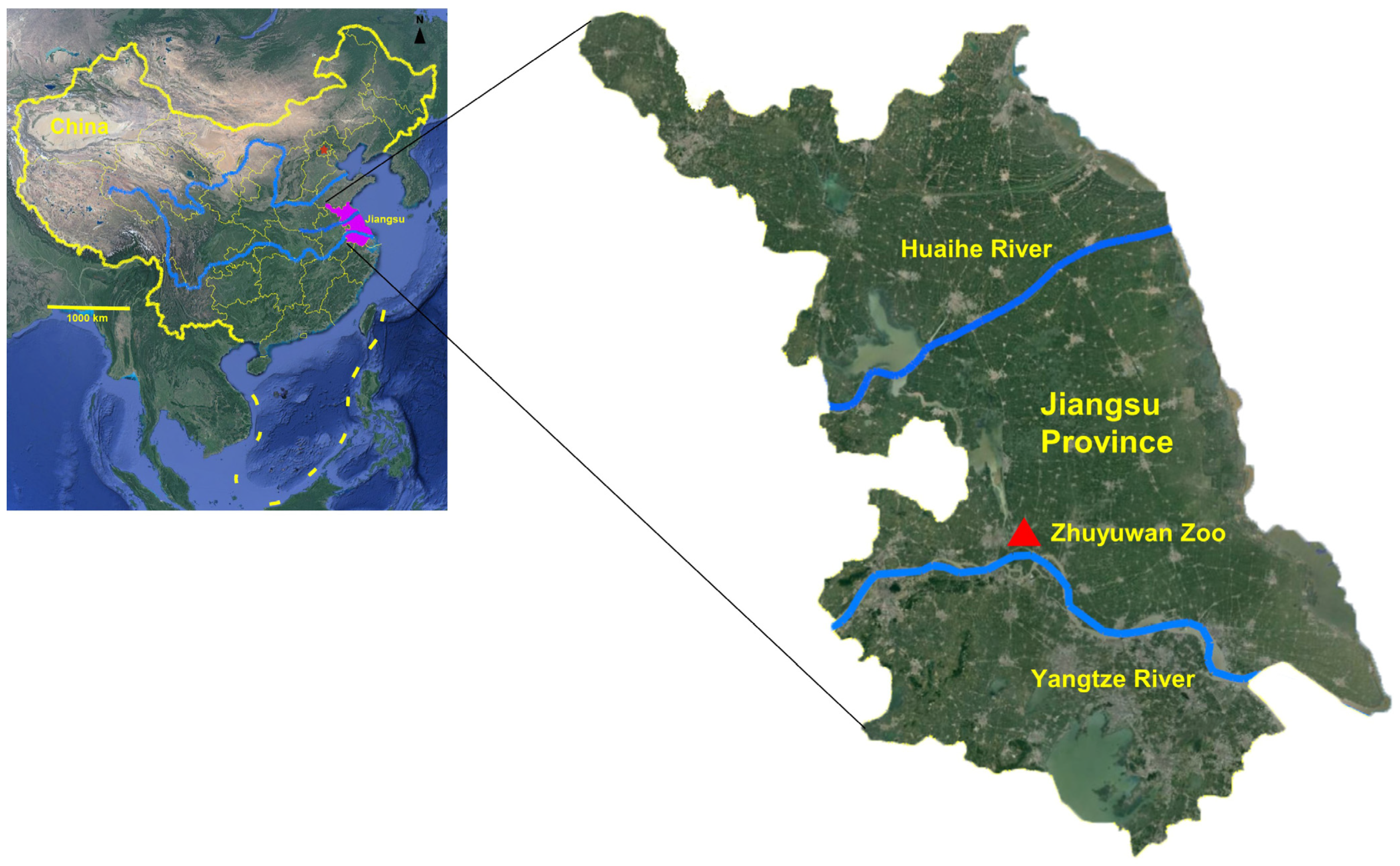
2.1. Study Site

The study was conducted at Zhuyuwan Zoo in Yangzhou city, Jiangsu Province, located in eastern China (Figure 1). Yangzhou city, situated on the lower reaches of the Yangtze River, has a temperate continental monsoon climate with four distinct seasons. The area has a mild climate with ample sunshine and rainfall, which creates favorable conditions for the occurrence and transmission of parasitic diseases. The zoo relocated from Slender West Lake to Zhuyuwan in 2004. Currently, the zoo accommodates more than 40 species of mammals, birds, and amphibians from Asia, Africa, and the Americas.

2.2. Sample Collection
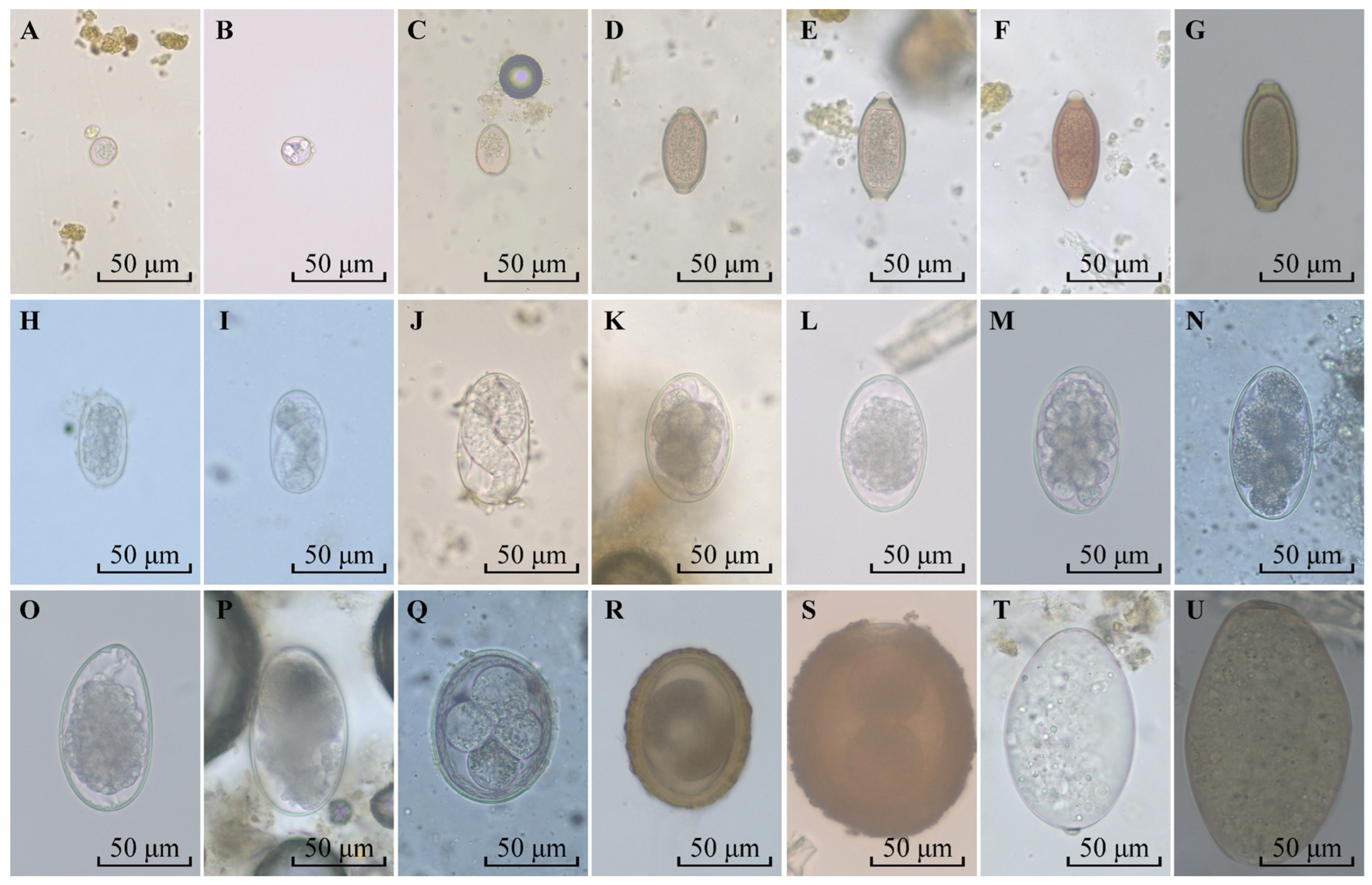
In 2021, a total of 104 fecal samples were randomly collected from different animals living in Zhuyuwan Zoo. Of them, 19 samples were collected from 15 species of birds, 19 from 8 species of primates, and 66 from 17 species of non-primate mammals. Trained animal handlers conducted the collection of fecal samples (~50 g) in the morning. The samples were stored in zipper bags and placed in a ~4 °C sample box for storage, and processed within 6 h of collection.

2.3. Microscopy

Nematode eggs and coccidian oocysts were detected using the saturated saline floatation method [17]. Briefly, 10 g of feces were diluted with 15 mL of saturated saline, filtered through a filter with a pore size of 250 μm, and the filtrate was centrifuged for 5 min at 800× g. A coverslip was placed over the surface of the supernatant, and it was viewed under a microscope after 3 min. The eggs of trematodes were examined using the nylon sieve washing method [17], i.e., 10 g feces diluted with water passed through 60 mesh (aperture = 250 μm) sieve and 260 mesh (aperture = 57 μm) sieve successively, and then the filter residue in the 260 mesh sieve was washed with water until the final filtrate was clear. Finally, the sediment in the sieve mesh was observed under a microscope. The eggs of nematode and trematode were observed and identified under a 40× objective lens. Unsporulated oocysts were incubated in 2.5% potassium dichromate (K2Cr2O7) for 5–7 days. After centrifugation at 800× g for 8 min, the supernatant was discarded, and the precipitate was resuspended in saturated saline and centrifuged at 700× g for 8 min. The sporulation oocysts from the supernatant were observed under the 40× objective. All identifications were performed as previously described [17,18,19,20]. Among nematode eggs, Trichuris and Strongyloides egg are easy to be identified according to their characteristic morphology and structure, in which Trichuris eggs are lemon shaped, yellow, or brown in color, with a thick smooth shell and a conspicuous polar plug at both ends, whereas Strongyloides egg are oval, thin-shelled and with larvae. In addition, the eggs of Haemonchus spp., Ostertagia spp., Trichostronglus spp. (belonging to Trichostrongylidae), and Oesophagostomum spp. (belonging to Cyathostomidae) possess similar sizes (73~95 μm × 34~50 μm), morphologies (ovoid in shape), and structures (containing numbers of embryo cells); it is difficult to distinguish between these species [17,18]. In this study, therefore, eggs with the above similar morphological structure were classified as Trichostrongylidae parasites.

2.4. DNA Isolation and PCR Amplification

About 100 eggs recovered from the African lions (Panthera leo) and the brown bears (Ursus arctos) were ground five times in liquid nitrogen, each time for 1 min. The subsequent steps for DNA extraction followed the guidelines outlined in the MiniBEST Universal Genomic DNA Extraction Kit Ver.5.0 (TaKaRa, Tokyo, Japan).
A standard PCR-based sequencing technique targeting ITS-2 locus (~300 bp, specific primers) was used to detect the Toxascaris leonina and Baylisascaris transfuga in the feces from an African lion and a brown bear, as previously reported [21,22]. Additionally, a standard PCR protocol was used to amplify the cox1 gene sequence, utilizing generic primers reported by Gasser et al. [19]. The expected size of the PCR product (cox1) was approximately 450 bp (Table 1). The PCR amplification process followed these cycling conditions: an initial denaturation step at 94 °C for 5 min, followed by 35 cycles of denaturation at 94 °C for 30 s, annealing at 60 °C (for ITS-2) or 50 °C (for cox1) for 30 s, extension at 72 °C for 60 s, and a final extension step at 72 °C for 5 min. Subsequently, we purified the PCR products using the MiniBEST DNA Fragment Purification Kit Ver.4.0 (TaKaRa, Tokyo, Japan) and subjected them to 1.0% agarose gel electrophoresis, followed by staining with ethidium bromide. The gel was then transilluminated and photographed using a gel imaging system (Bio-Rad, Hercules, CA, USA).

2.5. Sequence and Phylogenetic Analysis

All PCR products exhibited a single band. The purified PCR products were sent to Beijing Genomics Institute (BGI, Beijing, China) for Sanger sequencing. Subsequently, the sequencing results were analyzed using MegAlign with the clustal/W method and BLAST using highly similar sequences.
Phylogenetic analysis of T. leonina and B. transfuga was conducted based on the cox1 loci, and additional isolates from GenBank were included. The phylogenetic trees were constructed using the MEGA 5 software [23]. To determine the most suitable model, ModelTest in MEGA 5 was employed, and the Tamura-Nei [24,25] was utilized for Maximum Likelihood (ML), Neighbor-Joining (NJ), and Maximum Parsimony (MP) analyses. The reliability of the results was assessed through bootstrap analyses comprising 1000 replicates. Trichuris suis was selected as an outgroup.

3. Results

3.1. Occurrence of Intestinal Parasites

Based on egg morphology, a total of 11 species of parasites were identified, including Fasciola spp. in family Fasciolidae, Paramphistomum spp. in family Paramphistomatidae, Capillaria sp. in family Capillariidae, Trichuris spp. in family Trichuridae, Strongyloides spp. in family Strongyloididae, Amidostomum sp. in family Amidostomatidae, Trichostrongylidae, T. leonina, B. transfuga and Parascaris equorum (Goeze, 1782) in family Ascarididae, and Eimeria spp. in family Eimeriidae (Figure 2).
Of 11 species of parasites, Trichostrongylidae had the highest infection rate of 34.8% (23/104), followed by Trichuris spp. (5.8%, 6/104), Eimeria spp. (5.8%, 6/104), and Strongyloides spp. (3.8%, 4/104). The infection rate with other parasites was 0.9% (1/106). The positive rates for birds, primates, and non-primate mammals were 26.3% (5/19), 31.6% (6/19), and 50.0% (33/66), respectively (Table 2, Table 3 and Table 4). The occurrence of helminths and protozoans was 39.4% (41/104) and 5.8% (6/104), respectively (Table 5). Five fecal samples were mixed with two or more parasites with 5% (5/104) positivity rate.

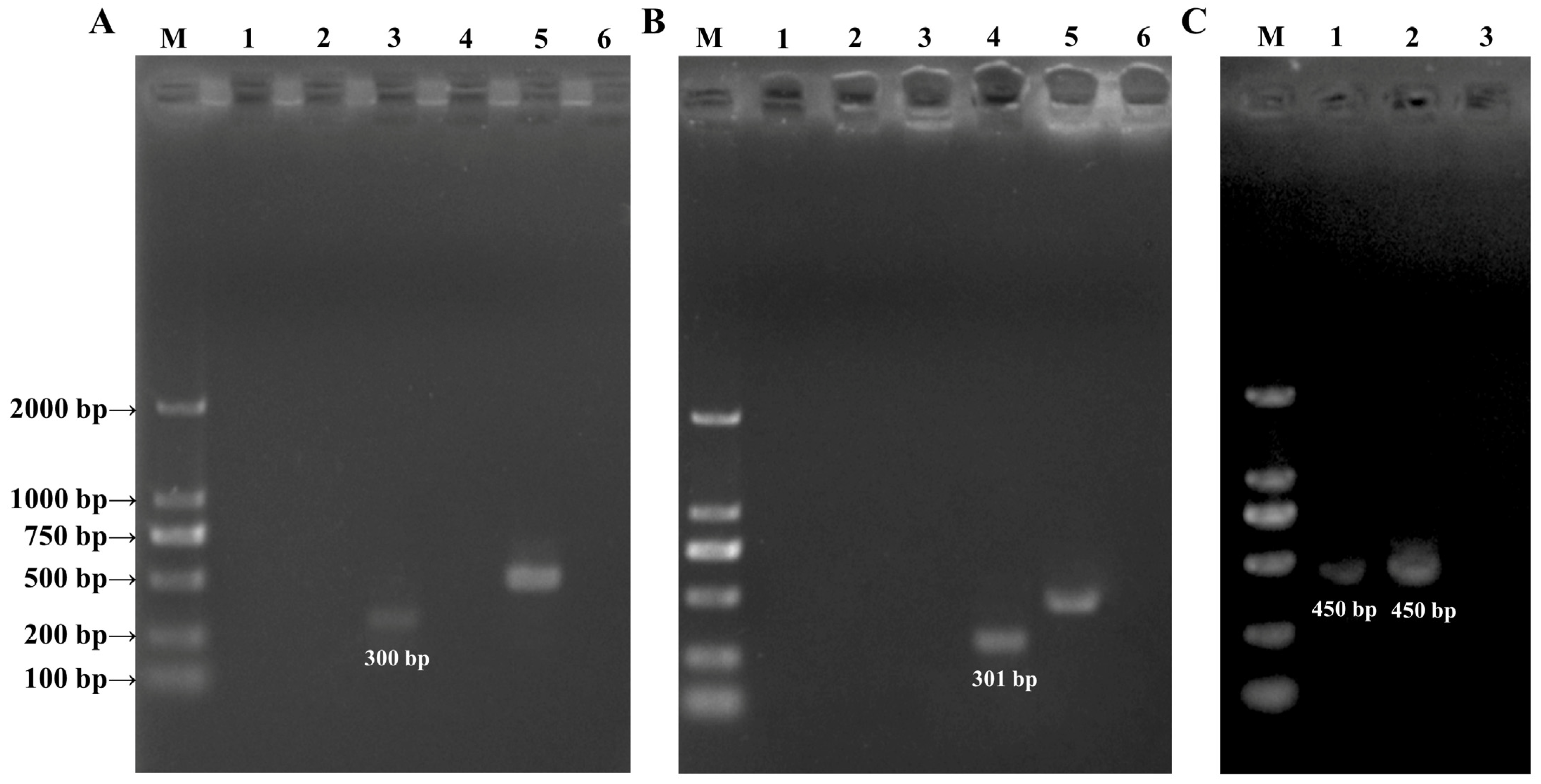
3.2. PCR Amplification Analysis

A total of 23.9 ng/μL and 7.8 ng/μL of DNA were extracted from about 100 eggs collected from an African lion and a brown bear, respectively. At the ITS-2 locus, a band of 300 bp in size was amplified only using TleoF-NC2R specific primers from the eggs from an African lion (Figure 3A). Similarly, a band of 301 bp was amplified only using NITSF-NITSR primers from the eggs isolated from a brown bear (Figure 3B). The NC13-NC2 specific primers were employed as a control to verify the presence of parasite DNA in each sample. At the cox1 locus, a band of about 450 bp in size were successfully amplified using JB3-JB4.5 generic primers from Toxascaris leonina (African lion) and Baylisascaris transfuga (brown bear) and sequenced, respectively (Figure 3C). Afr L refers to samples recovered from African lion feces and Bn B refers to samples recovered from brown bear feces.

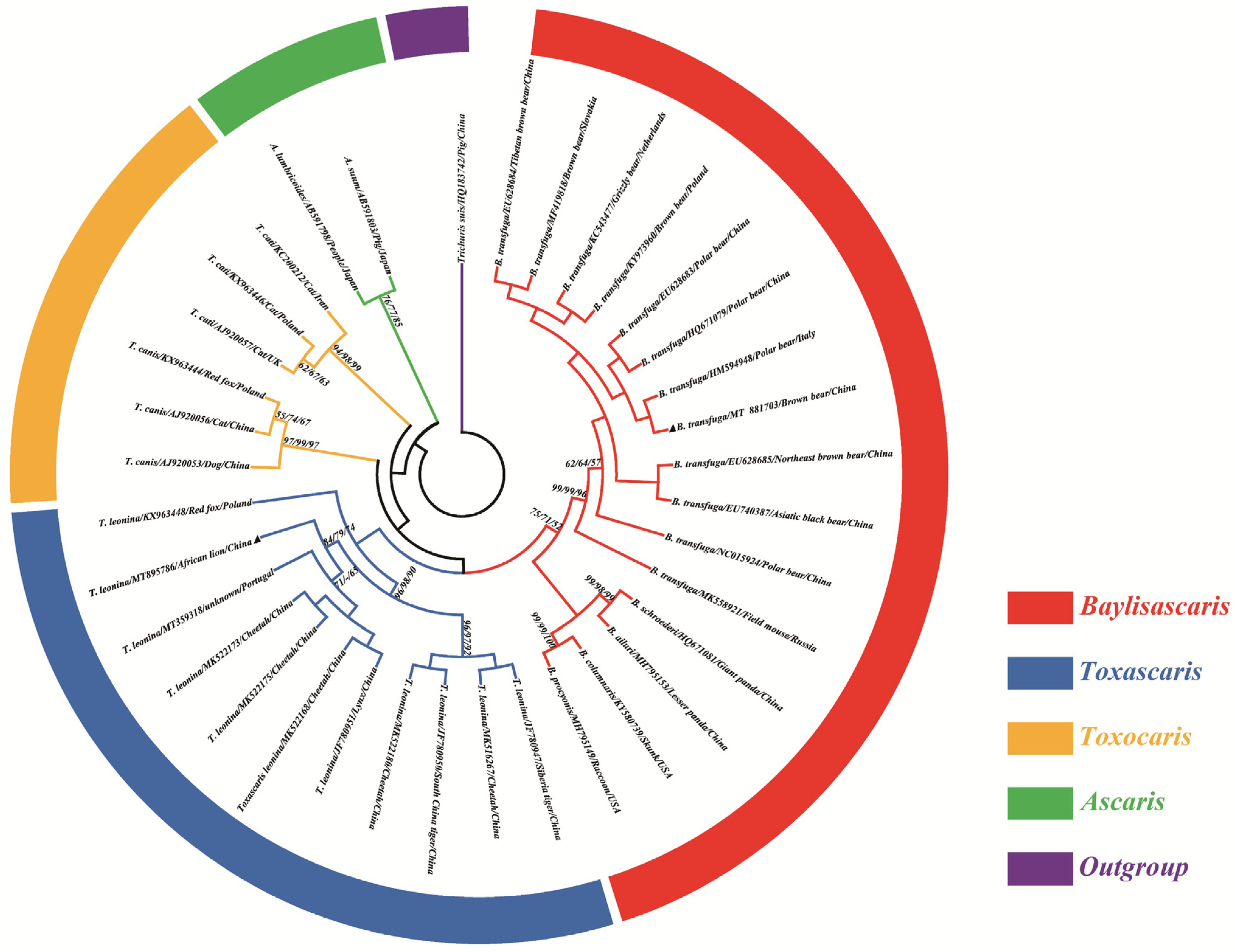
3.3. Phylogenetic Analysis

The homology analysis showed that the percentage of identity was 92.4% between T. leonina (MT895786) and B. transfuga (MT881703). Toxascaris leonina in this study (MT895786) shared 98.1% similarity with T. leonina from Changsha of China (MK522168, MK522175). The phylogenetic analysis revealed that T. leonina (MT895786) was grouped together with T. leonina (MT359318, MK522173, MK522175, MK522168 and JF780951) to form a sub-clade in the cox1 phylogenetic tree (Figure 4). Baylisascaris transfuga (MT881703) shared 100% similarity with B. transfuga (HM594948), and phylogenetic analysis revealed that B. transfuga (MT881703) was grouped together with B. transfuga (HM594948, HQ671079, EU628683, KY973960, KC543477, MF419818, EU628684) to form a sub-clade in the cox1 phylogenetic tree (Figure 4). Although the three algorithms (ML/NJ/MP) differed slightly in topology, all analyses yielded a consistent, robust phylogenetic resolution for T. leonina and B. transfuga and their congeneric species in the genera Toxascaris and Baylisascaris, repectively. The ML method was eventually adopted to construct this evolutionary tree. Trichuris suis (HQ183742) was used as an outgroup (Figure 4).

4. Discussion

The overall prevalence of GI parasites in animals living in Zhuyuwan Zoo was 42.3% (44/104), which is similar to the results observed in zoo animals in Xining of China [20], Slovenia (45%, 337/741) [21], and Poland (48%, 34/71) [22]. However, the infection rate of GI parasites in the present study was higher than that in twenty-four zoological gardens of China (26.51%, 317/1196) [23], Nepal (19.54%, 17/87) [24], and France (32.2%, 99/307) [25], and lower than that in Sichuan of China [26], Italy (80%, 24/30) [27], and Brazil (71.1%, 27/38) [28]. These differences may be related to the ecological environment and sampling season. The present study found a higher occurrence of helminths (39.4%, 41/104) compared to protozoans (5.8%, 6/104), similar to the observations in a zoological garden in Kenya, which revealed a higher occurrence of helminths (64.4%, 203/315) and a lower occurrence of protozoans (17.1%, 54/315) [29]. Interestingly, the prevalence of protozoa is lower than that of helminths in primates and non-primate mammals, whereas in birds the prevalence of protozoa infection is higher than that of helminths. This may be related to the environment in which these birds and primates and non-primate mammals live. These birds are wading birds whose feeding environment relies heavily on silt, which contains some fecal matter and some infectious Eimeria coccidia, thus making them susceptible to disease transmission [30]. Of course, it may also be due to birds being more susceptible to Eimeria, as Eimeria coccidia causes the most severe damage to poultry (belonging to birds). Additionally, our results revealed that the positive rates of GI parasites in birds, primates, and non-primate mammals were 26.3% (5/19), 31.6% (6/19), and 50.0% (33/66), respectively. Interestingly, the positive rate of mixed infection with two or more parasites was only 5% (5/104), which is consistent with previous reports (7%, 55/741) [21]. In addition, no tapeworms were detected in this study.
In this study, Trichuris spp. had a 5.8% (6/104) infection rate, second only to Trichostrongylidae (22.1%, 23/104). Strongyloides eggs were detected in fecal samples from the Ring-tailed lemurs (Lemur catta) and red pandas (Ailurus fulgens). Strongyloides parasites can infect their hosts through skin penetration or ingestion [31] and reproduce asexually in the host’s intestinal wall, contributing to their high infection rates [32]. Rondon, Ortiz, Leon, Galvis, Link and Gonzalez [33] reported that 21.6% (40/185) of fecal samples from neotropical primates were positive for Strongyloides spp. Studies on parasites infecting nonhuman primates are essential for better understanding the potential threat of zoonotic transmission to humans [34], especially given the ongoing processes of pervasive land use change and biodiversity loss [35] and the COVID-19 pandemic [36]. In addition, Trichuris spp., as a zoonotic parasite, may increase the risk of mixed infection with intestinal pathogens [37]. Therefore, Zhuyuwan Zoo needs to pay more attention to prevention and control of Trichuris spp. and Strongyloides spp.
The Père David’s deer (Elaphurus davidianus) and the sika deer (Cervus nippon) are National Protected Animals in China [38]. Here, Paramphistomum spp. eggs were first detected in the captive Père David’s deer in a zoo in China, and Fasciola spp. eggs were detected from the feces of sika deer. Paramphistomum spp. and Fasciola spp. have different degrees of pathogenicity to animals, and Fasciola parasites are an important zoonotic pathogen. So, Zhuyuwan Zoo needs to pay special attention to prevention and control of Paramphistomum spp. and Fasciola spp.
For decades, there has been considerable debate surrounding the systematics of members of the Ascaridomorpha (including Anisakidae, Ascarididae, Ascaridiidae and Toxocaridae) [39,40]. With the increasing utilization of molecular biology methods, there has been a growing ability to examine and identify ascarids [41]. This is particularly important because the eggs of many ascarid species share similar morphological characteristics, potentially leading to misidentification [42]. In the present study, two Ascaris species were identified using molecular biological methods in this study. It was found that T. leonina could only be amplified using TleoF-NC2R primers and B. transfuga could only be amplified using NITSF-NITSR primers, respectively. The results of a phylogenetic tree showed that T. leonina was closer to T. canis than to T. cati at cox1 locus, which was similar to the result reported by Xie et al. [15].
Toxascaris spp. and Baylisascais spp., belonging to the family Ascarididae, are common GI parasites parasitized in carnivores such as members of Canidae, Felidae, and Ursidae. T. leonina mainly resides in the gastrointestinal tract of dogs, cats, and lions [43,44], which carries the potential risk to infect humans due to their close relationship with zoonotic ascarids like Toxocara canis and Toxocara cati [45,46]. Baylisascaris transfuga has been documented in all extant members of the family Ursidae worldwide, except for the spectacled bear [47,48]. The possibility of B. transfuga causing illness in humans has been suggested, and it poses a threat to public health [42,49]. Previous research has shown that B. transfuga larvae can migrate within the tissues of various animals like chickens [50], rabbits [51], mice [52], and Mongolian jirds [53] from their original locations in the intestine. However, the occurrence of visceral larva migrans (VLM) has only been observed in mammals [49]. Therefore, there is a potential risk for humans who come into contact with infectious eggs of B. transfuga in the environment [48]. In addition, the other five GI parasites detected in this study also are potentially dangerous for humans. A previous study has shown that anthelmintic drugs could effectively control soil-transmitted helminth infections [54]. Therefore, we recommend that zoos appropriately use albendazole or praziquantel for helminths eradication to reduce the risk of infection to tourists and veterinarians.

5. Conclusions

The prevalence of GI parasites was 42.3% (44/104) in Zhuyuwan Zoo in China. Paramphistomum spp. eggs were first detected from the captive Père David’s deer and Fasciola spp. eggs were first reported from sika deer in zoo in China. Ascaridoid parasite eggs from the African lions were identified as T. leonina, and those from the brown bear were identified as B. transfuga. Of 11 GI parasites detected in this study, seven are potentially dangerous for humans. The public health threat posed by these potential zoonotic parasitic agents requires attention.

Author Contributions

Conceptualization, J.T.; methodology, W.C.; formal analysis and investigation, W.C., Y.Z. and F.W.; resources, X.X.; data curation, Q.F., Z.Z. and N.X.; writing—original draft preparation, W.C.; writing—review and editing, J.T.; visualization, W.C.; supervision, Z.H. and J.X.; project administration, J.T. and D.L.; funding acquisition, J.T. All authors have read and agreed to the published version of the manuscript.

Funding

This study was supported by the National Natural Science Foundation of China (31972698 to JT), the 111 Project D18007, and the Priority Academic Program Development of Jiangsu Higher Education Institutions (PAPD). The funders had no role in study design, data collection and analysis, the decision to publish, or preparation of the manuscript.

Institutional Review Board Statement

This study was approved by the Animal Ethics Committee of Yangzhou University (NSFC2020-SYXY-27).

Informed Consent Statement

Not applicable.

Data Availability Statement

Data are contained within the article.

Acknowledgments

The author would like to thank Xu Xun, a veterinary of Zhuyuwan Zoo, for his help in collecting fecal samples, as well as his professional treatment of wildlife.

Conflicts of Interest

The authors declare no conflicts of interest.

References

  1. Christe, P. Loss of biodiversity: A one-way ticket to new emerging diseases? Rev. Med. Suisse 2023, 19, 842–844. [Google Scholar] [PubMed]
  2. Edwards, K.L.; Edes, A.N.; Brown, J.L. Stress, well-being and reproductive success. Adv. Exp. Med. Biol. 2019, 1200, 91–162. [Google Scholar] [PubMed]
  3. Serota, M.W.; Barker, K.J.; Gigliotti, L.C.; Maher, S.M.L.; Shawler, A.L.; Zuckerman, G.R.; Xu, W.; Verta, G.; Templin, E.; Andreozzi, C.L.; et al. Incorporating human dimensions is associated with better wildlife translocation outcomes. Nat. Commun. 2023, 14, 2119. [Google Scholar] [CrossRef] [PubMed]
  4. Booth, M. Climate change and the neglected tropical diseases. Adv. Parasitol. 2018, 100, 39–126. [Google Scholar] [PubMed]
  5. Locklear, T.R.; Videla, R.; Breuer, R.M.; Mulon, P.Y.; Passmore, M.; Mochel, J.P.; Gerhold, R.; Schaefer, J.J.; Smith, J.S. Presentation, clinical pathology abnormalities, and identification of gastrointestinal parasites in camels (Camelus bactrianus and Camelus dromedarius) presenting to two north American veterinary teaching hospitals. A retrospective study: 1980–2020. Front. Vet. Sci. 2021, 8, 651672. [Google Scholar] [CrossRef] [PubMed]
  6. Trogu, T.; Formenti, N.; Marangi, M.; Viganò, R.; Bionda, R.; Giangaspero, A.; Lanfranchi, P.; Ferrari, N. Detection of zoonotic Cryptosporidium ubiquitum in alpine wild ruminants. Pathogens 2021, 10, 655. [Google Scholar] [CrossRef]
  7. Espinosa García-San Román, J.; Quesada-Canales, Ó.; Arbelo Hernández, M.; Déniz Suárez, S.; Castro-Alonso, A. Veterinary Education and Training on Non-Traditional Companion Animals, Exotic, Zoo, and Wild Animals: Concepts Review and Challenging Perspective on Zoological Medicine. Vet. Sci. 2023, 10, 357. [Google Scholar] [CrossRef]
  8. Bedoya-Pérez, M.A.; Ward, M.P.; Loomes, M.; McGregor, I.S.; Crowther, M.S. The effect of COVID-19 pandemic restrictions on an urban rodent population. Sci. Rep. 2021, 11, 12957. [Google Scholar] [CrossRef]
  9. Aguirre, A.A. Essential veterinary education in zoological and wildlife medicine: A global perspective. Rev. Sci. Tech. 2009, 28, 605–610. [Google Scholar] [CrossRef]
  10. Vlaminck, J.; Levecke, B.; Vercruysse, J.; Geldhof, P. Advances in the diagnosis of Ascaris suum infections in pigs and their possible applications in humans. Parasitology 2014, 141, 1904–1911. [Google Scholar] [CrossRef]
  11. Jourdan, P.M.; Lamberton, P.H.L.; Fenwick, A.; Addiss, D.G. Soil-transmitted helminth infections. Lancet 2018, 391, 252–265. [Google Scholar] [CrossRef] [PubMed]
  12. Lamberton, P.H.; Jourdan, P.M. Human Ascariasis: Diagnostics Update. Curr. Trop. Med. Rep. 2015, 2, 189–200. [Google Scholar] [CrossRef]
  13. Li, M.W.; Lin, R.Q.; Chen, H.H.; Sani, R.A.; Song, H.Q.; Zhu, X.Q. PCR tools for the verification of the specific identity of ascaridoid nematodes from dogs and cats. Mol. Cell Probes 2007, 21, 349–354. [Google Scholar] [CrossRef] [PubMed]
  14. Fogt-Wyrwas, R.; Mizgajska-Wiktor, H.; Pacoń, J.; Jarosz, W. Intraspecific variation between the ITS sequences of Toxocara canis, Toxocara cati and Toxascaris leonina from different host species in south-western Poland. J. Helminthol. 2013, 87, 432–442. [Google Scholar] [CrossRef] [PubMed]
  15. Xie, Y.; Li, Y.; Gu, X.; Liu, Y.; Zhou, X.; Wang, L.; He, R.; Peng, X.; Yang, G. Molecular characterization of ascaridoid parasites from captive wild carnivores in China using ribosomal and mitochondrial sequences. Parasit. Vectors 2020, 13, 382. [Google Scholar] [CrossRef] [PubMed]
  16. Blouin, M.S. Molecular prospecting for cryptic species of nematodes: Mitochondrial DNA versus internal transcribed spacer. Int. J. Parasitol. 2002, 32, 527–531. [Google Scholar] [CrossRef] [PubMed]
  17. Taylor, M.A.; Coop, R.L.; Wall, R.L. Veterinary Parasitology; Blackwell Publishing: Oxford, UK, 2007. [Google Scholar]
  18. Ministry of Agriculture, Fisheries and Food. Manual of Veterinary Parasitological Laboratory Techniques, 2nd ed.; Technical Bulletin No.18.; Her Majesty’s Stationary Office: London, YK, 1977. [Google Scholar]
  19. Gasser, R.B.; Zhu, X.; McManus, D.P. NADH dehydrogenase subunit 1 and cytochrome c oxidase subunit I sequences compared for members of the genus Taenia (Cestoda). Int. J. Parasitol. 1999, 29, 1965–1970. [Google Scholar] [CrossRef]
  20. Zhao, W.; Wang, X. Investigation of intestinal parasites in wild animals at Xining Wildlife Park. Heilongjiang Anim. Sci. Vet. Med. 2014, 2, 139–140. [Google Scholar]
  21. Kvapil, P.; Kastelic, M.; Dovc, A.; Bartova, E.; Cizek, P.; Lima, N.; Strus, S. An eight-year survey of the intestinal parasites of carnivores, hoofed mammals, primates, ratites and reptiles in the Ljubljana zoo in Slovenia. Folia Parasitol. 2017, 64, 13. [Google Scholar] [CrossRef]
  22. Maesano, G.; Capasso, M.; Ianniello, D.; Cringoli, G.; Rinaldi, L. Parasitic infections detected by FLOTAC in zoo mammals from Warsaw, Poland. Acta Parasitol. 2014, 59, 343–353. [Google Scholar] [CrossRef]
  23. Li, M.; Zhao, B.; Li, B.; Wang, Q.; Niu, L.; Deng, J.; Gu, X.; Peng, X.; Wang, T.; Yang, G. Prevalence of gastrointestinal parasites in captive non-human primates of twenty-four zoological gardens in China. J. Med. Primatol. 2015, 44, 168–173. [Google Scholar] [CrossRef]
  24. Dhakal, P.; Sharma, H.P.; Shah, R.; Thapa, P.J.; Pokheral, C.P. Copromicroscopic study of gastrointestinal parasites in captive mammals at Central Zoo, Lalitpur, Nepal. Vet. Med. Sci. 2023, 9, 457–464. [Google Scholar] [CrossRef] [PubMed]
  25. Cian, A.; El Safadi, D.; Osman, M.; Moriniere, R.; Gantois, N.; Benamrouz-Vanneste, S.; Delgado-Viscogliosi, P.; Guyot, K.; Li, L.L.; Monchy, S.; et al. Molecular Epidemiology of Blastocystis sp. in Various Animal Groups from Two French Zoos and Evaluation of Potential Zoonotic Risk. PLoS ONE 2017, 12, e0169659. [Google Scholar] [CrossRef] [PubMed]
  26. Qi, M.; Dong, H.; Hu, L.; Peng, G.; Chen, Z.; Zhou, Z.; Wan, J.; Jian, F.; Zhang, L. Investigation on the Prevalence of Intestinal Parasites in Wild Animal Zoo. China Anim. Husb. Vet. Med. 2011, 38, 188–190. [Google Scholar]
  27. Capasso, M.; Maurelli, M.P.; Ianniello, D.; Alves, L.C.; Amadesi, A.; Laricchiuta, P.; Silvestre, P.; Campolo, M.; Cringoli, G.; Rinaldi, L. Use of Mini-FLOTAC and Fill-FLOTAC for rapidly diagnosing parasitic infections in zoo mammals. Rev. Bras. Parasitol. Vet. 2019, 28, 168–171. [Google Scholar] [CrossRef]
  28. Holsback, L.; Cardoso, M.J.; Fagnani, R.; Patelli, T.H. Natural infection by endoparasites among free-living wild animals. Rev. Bras. Parasitol. Vet. 2013, 22, 302–306. [Google Scholar] [CrossRef] [PubMed]
  29. Munene, E.; Otsyula, M.; Mbaabu, D.A.; Mutahi, W.T.; Muriuki, S.M.; Muchemi, G.M. Helminth and protozoan gastrointestinal tract parasites in captive and wild-trapped African non-human primates. Vet. Parasitol. 1998, 78, 195–201. [Google Scholar] [CrossRef] [PubMed]
  30. Albuixech-Martí, S.; Lynch, S.A.; Culloty, S.C. Connectivity dynamics in Irish mudflats between microorganisms including Vibrio spp., common cockles Cerastoderma edule, and shorebirds. Sci. Rep. 2021, 11, 22159. [Google Scholar] [CrossRef]
  31. Parr, N.A.; Fedigan, L.M.; Kutz, S.J. A coprological survey of parasites in white-faced capuchins (Cebus capucinus) from Sector Santa Rosa, ACG, Costa Rica. Folia Primatol. 2013, 84, 102–114. [Google Scholar] [CrossRef]
  32. Barutzki, D.; Schaper, R. Results of parasitological examinations of faecal samples from cats and dogs in Germany between 2003 and 2010. Parasitol. Res. 2011, 109 (Suppl. S1), S45–S60. [Google Scholar] [CrossRef]
  33. Rondon, S.; Ortiz, M.; Leon, C.; Galvis, N.; Link, A.; Gonzalez, C. Seasonality, richness and prevalence of intestinal parasites of three neotropical primates (Alouatta seniculus, Ateles hybridus and Cebus versicolor) in a fragmented forest in Colombia. Int. J. Parasitol. Parasites Wildl. 2017, 6, 202–208. [Google Scholar] [CrossRef] [PubMed]
  34. García-Sánchez, A.M.; Miller, A.Z.; Caldeira, A.T.; Cutillas, C. Bacterial communities from Trichuris spp. A contribution to deciphering the role of parasitic nematodes as vector of pathogens. Acta Trop. 2022, 226, 106277. [Google Scholar] [CrossRef] [PubMed]
  35. Brodie, J.F.; Mohd-Azlan, J.; Chen, C.; Wearn, O.R.; Deith, M.C.M.; Ball, J.G.C.; Slade, E.M.; Burslem, D.; Teoh, S.W.; Williams, P.J.; et al. Landscape-scale benefits of protected areas for tropical biodiversity. Nature 2023, 620, 807–812. [Google Scholar] [CrossRef] [PubMed]
  36. Davis, H.E.; McCorkell, L.; Vogel, J.M.; Topol, E.J. Long COVID: Major findings, mechanisms and recommendations. Nat. Rev. Microbiol. 2023, 21, 133–146. [Google Scholar] [CrossRef] [PubMed]
  37. Polakovicova, N.; Adji, A.V.; Myhill, L.J.; Williams, A.R. Whipworm Infection in Mice Increases Coinfection of Enteric Pathogens but Promotes Clearance of Ascaris Larvae from the Lungs. J. Infect. Dis. 2023, 227, 1428–1432. [Google Scholar] [CrossRef] [PubMed]
  38. Elaphurus Davidianus. The IUCN Red List of Threatened Species. Available online: https://dx.doi.org/10.2305/IUCN.UK.2016-2.RLTS.T7121A22159785.en (accessed on 1 March 2021).
  39. Gu, X.H.; Guo, N.; Chen, H.X.; Sitko, J.; Li, L.W.; Guo, B.Q.; Li, L. Mitogenomic phylogenies suggest the resurrection of the subfamily Porrocaecinae and provide insights into the systematics of the superfamily Ascaridoidea (Nematoda: Ascaridomorpha), with the description of a new species of Porrocaecum. Parasit. Vectors 2023, 16, 275. [Google Scholar] [CrossRef] [PubMed]
  40. Xiong, L.; Chen, L.; Chen, Y.; Shen, N.; Hua, R.; Yang, G. Evaluation of the immunoprotective effects of eight recombinant proteins from Baylisascaris schroederi in mice model. Parasit. Vectors 2023, 16, 254. [Google Scholar] [CrossRef]
  41. Li, Y.; Niu, L.; Wang, Q.; Zhang, Z.; Chen, Z.; Gu, X.; Xie, Y.; Yan, N.; Wang, S.; Peng, X.; et al. Molecular characterization and phylogenetic analysis of ascarid nematodes from twenty-one species of captive wild mammals based on mitochondrial and nuclear sequences. Parasitology 2012, 139, 1329–1338. [Google Scholar] [CrossRef]
  42. Testini, G.; Papini, R.; Lia, R.P.; Parisi, A.; Dantas-Torres, F.; Traversa, D.; Otranto, D. New insights into the morphology, molecular characterization and identification of Baylisascaris transfuga (Ascaridida, Ascarididae). Vet. Parasitol. 2011, 175, 97–102. [Google Scholar] [CrossRef]
  43. Okulewicz, A.; Lonc, E.; Borgsteede, F.H. Ascarid nematodes in domestic and wild terrestrial mammals. Pol. J. Vet. Sci. 2002, 5, 277–281. [Google Scholar]
  44. Miller, A.D. Pathology of larvae and adults in dogs and cats. Adv. Parasitol. 2020, 109, 537–544. [Google Scholar]
  45. Macpherson, C.N. The epidemiology and public health importance of toxocariasis: A zoonosis of global importance. Int. J. Parasitol. 2013, 43, 999–1008. [Google Scholar] [CrossRef] [PubMed]
  46. Jenkins, E.J. Toxocara spp. in dogs and cats in Canada. Adv. Parasitol. 2020, 109, 641–653. [Google Scholar] [PubMed]
  47. Figueroa, J. New records of parasites in free-ranging Andean bears from Peru. Ursus 2015, 26, 21–27. [Google Scholar] [CrossRef]
  48. Sapp, S.G.H.; Gupta, P.; Martin, M.K.; Murray, M.H.; Niedringhaus, K.D.; Pfaff, M.A.; Yabsley, M.J. Beyond the raccoon roundworm: The natural history of non-raccoon Baylisascaris species in the New World. Int. J. Parasitol. Parasites Wildl. 2017, 6, 85–99. [Google Scholar] [CrossRef] [PubMed]
  49. Bauer, C. Baylisascariosis—Infections of animals and humans with ’unusual’ roundworms. Vet. Parasitol. 2013, 193, 404–412. [Google Scholar] [CrossRef] [PubMed]
  50. Papini, R.; Cavicchio, P.; Casarosa, L. Experimental infection in chickens with larvae of Baylisascaris transfuga (Nematoda: Ascaridoidea). Folia Parasitol. 1993, 40, 141–143. [Google Scholar]
  51. Papini, R.; Demi, S.; Croce, G. Observations on the migratory behaviour of Baylisascaris transfuga larvae in rabbits. Rev. Med. Vet. 1996, 147, 893–896. [Google Scholar]
  52. Juránková, J.; Hofmannová, L.; Frgelecová, L.; Daněk, O.; Modrý, D. Baylisascaris transfuga (Ascaridoidea, Nematoda) from European brown bear (Ursus arctos) causing larva migrans in laboratory mice with clinical manifestation. Parasitol. Res. 2022, 121, 645–651. [Google Scholar] [CrossRef]
  53. Cho, S.; Egami, M.; Ohnuki, H.; Saito, Y.; Chinone, S.; Shichinohe, K.; Suganuma, M.; Akao, N. Migration behaviour and pathogenesis of five ascarid nematode species in the Mongolian gerbil Meriones unguiculatus. J. Helminthol. 2007, 81, 43–47. [Google Scholar] [CrossRef]
  54. Moser, W.; Schindler, C.; Keiser, J. Efficacy of recommended drugs against soil transmitted helminths: Systematic review and network meta-analysis. BMJ 2017, 358, j4307. [Google Scholar] [CrossRef]
Figure 1. Geographical location of the sampling site, Zhuyuwan Zoo.
Animals 14 00375 g001
Figure 2. Parasites identified in stool samples from zoo animals. (AC): Eimeria spp.; (DF): Trichuris spp.; (G): Capillaria sp.; (H): Amidostomum sp.; (I,J): Strongyloides spp.; (KP): Trichostrongylidae; (Q): Toxascaris leonina; (R): Baylisascaris transfuga; (S): Parascaris equorum; (T): Paramphistomum spp.; (U): Fasciola spp.
Animals 14 00375 g002
Figure 3. Results of amplification using PCR. (Panel A): amplification of SZ (300 bp) using ITS-2 primers; Lane 1: TocanF-NC2R, Lane 2: TocatF-NC2R, Lane 3: TleoF-NC2R, Lane 4: NITSF-NITSR; Lane 5: NC13F-NC2R; Lane 6: negative control. (Panel B): amplification of ZX (301 bp) using ITS-2 primers; Lane 1: TocanF-NC2R, Lane 2: TocatF-NC2R, Lane 3: TleoF-NC2R, Lane 4: NITSF-NITSR; Lane 5: NC13F-NC2R; Lane 6: negative control. (Panel C): amplification of SZ (450 bp) and ZX (450 bp) using cox1 primers; Lane 1: JB3-JB4.5; Lane 2 JB3-JB4.5; Lane 3: negative control.
Animals 14 00375 g003
Figure 4. Evolutionary relationships of T. leonina and B. transfuga inferred using ML, NJ and MP analyses of cox1 sequences. Toxascaris leonina recovered from the African lion was genetically divided into the general Toxascaris and B. transfuga recovered from the brown bear was genetically divided into the general Baylisascaris which were indicated with blue and red rings, respectively. The general Toxocaris and Ascaris were indicated with yellow and green rings, and the Outgroup was purple, respectively. The numbers along branches indicate bootstrap values derived from different analyses in the order: ML/NJ/MP. Values lower than 50 are shown as “-”. Black triangles represent the sequencing results of this experiment.
Animals 14 00375 g004
Table 1. Sequences of primers.
Name of PrimerSequence (5′ to 3′)
For ITS-2
  TcanFAGTATGATGGGCGCCAAT
  NC2RTTAGTTTCTTTTCCTCCGCT
  TcatFGGAGAAGTAAGATCGTGGCACGCGT
  NC2RTTAGTTTCTTTTCCTCCGCT
  TaleoFCGAACGCTCATATAACGGCATACTC
  NC2RTTAGTTTCTTTTCCTCCGCT
  NITSFTTATGAATTTTCAACATGGC
  NITSRGTTAGATGCTTAAATTCAGC
For cox1
  JB3TTTTTTGGGCATCCTGAGGTTTAT
  JB4.5TAAAGAAAGAACATAATGAAAATG
Table 2. Prevalence of intestinal parasites in birds at Zhuyuwan Zoo.
SpeciesnNo. (%) of Positive Samples for Parasite Species
Capillaria sp.Amidostomum sp.Eimeria spp.
Birds191 (5.3)1 (5.3)3 (15.8)
 Family Dromaiidae
  Emu (Dromaius novaehollandiae)1
 Family Struthioidae
  Common ostrich (Struthio camelus)2
 Family Phoenicopteridae
  Greater flamingo (Phoenicopterus roseus)1
 Family Gruidae
  Manchurian crane (Grus japonensis)2
  Hooded crane (Grus monacha)11 (100)
  White-naped crane (Grus vipio)1
  Siberian crane (Grus leucogeranus)1
  Black crowned-crane (Balearica pavonina)21 (50)
  Black-necked crane (Grus nigricollis)1 1 (100)
  Demoiselle crane (Anthropoides virgo)1
  Common crane (Grus grus)1
 Family Ciconiidae
  Oriental white stork (Ciconia boyciana)11 (100)
 Family Phasianidae
  Green peafowl (Pavo muticus)11 (100)
 Family Anatidae
  Northern pintail (Anas acuta)2
 Family Accipitridae
  Black vulture (Aegypius monachus)1
n = number of samples collected and examined (the same below).
Table 3. Prevalence of intestinal parasites in primates at Zhuyuwan Zoo.
SpeciesnNo. (%) of Positive Samples for Parasite Species
Trichuris spp.Strongyloides spp.
Primates193 (15.8)3 (15.8)
 Family Cebidae
  Black-capped capuchins (Cebus apella)2
  Squirrel monkey (Saimiri sciureus)3
 Family Lemuridae
  Ring-tailed lemur (Lemur catta)43 (75)
 Family Cercopithecidae
  François’s leaf monkey (Trachypithecus francoisi)2
  Golden monkey (Rhinopithecus)22 (100)
  Patas monkey (Erythrocebus patas)11 (100)
 Family Hylobatidae
  Lar gibbon (Hylobates lar)3
 Family Hominidae
  Chimpanzee (Pan troglodytes)2
Table 4. Prevalence of intestinal parasites in non-primate mammals at Zhuyuwan Zoo.
SpeciesnNo. (%) of Positive Samples for Parasite Species
TrichostrongylidaeTrichuris spp.Strongyloides spp.Toxascaris leoninaBaylisascaris transfugaParascaris equorumParamphistomum spp.Fasiola spp.Eimeria spp.
Non-primate mammals6623 (34.8)3 (10.7)1 (3.6)1 (3.6)1 (3.6)1 (3.6)1 (1.5)1 (1.5)3 (4.5)
 Family Camelidae
  Llama (Lama glama)21 (50)
  Alpaca (Vicugna pacos)11 (100)1 (100)
 Family Bovidae
  Blue wildebeest (Connochaetes taurinus)114 (36.4)
  Gemsbok (Oryx gazella)1010 (100)2 (100)2 (100)
  Scimitar-horned oryx (Oryx dammah)44 (100)
  Family Equidae
  Pony (Equus spp., pony)2
  Common zebra (Equus quagga) 103 (30)1 (10)
  Family Cervidae
  Père David’s deer (Elaphurus davidianus)11 (100)
  Sika deer (Cervus nippon)101 (10)1 (10)
  Family Ursidae
  Giant panda (Ailuropoda melanoleuca)2
  Brown bear (Ursus arctos)21 (50)
 Family Ailuridae
  Red panda (Ailurus fulgens)21 (50)
 Family Felidae
  Siberian tiger (Panthera tigris ssp. altaica)2
  African lion (Panthera leo)21 (50)
  Leopard (Panthera pardus)1
 Family Canidae
  Common wolf (Canis lupus)2
 Family Macropodidae
  Gray kangaroo (Macropus giganteus)2
Table 5. The overall occurrence of intestinal parasitic infections among various animals at Zhuyuwan Zoo.
AnimalsSamplesHelminth Positive (%)Protozoan Positive (%)Total
Birds192 (10.5)3 (15.8)5 (26.3)
Primates196 (31.6)0 (0)6 (31.6)
Mammals6633 (50.0)3 (4.5)33 (50.0)
Total10441 (39.4)6 (5.8)44 (42.3)
Disclaimer/Publisher’s Note: The statements, opinions and data contained in all publications are solely those of the individual author(s) and contributor(s) and not of MDPI and/or the editor(s). MDPI and/or the editor(s) disclaim responsibility for any injury to people or property resulting from any ideas, methods, instructions or products referred to in the content.

