Article

Modelling the Unidentified Abortion Burden from Four Infectious Pathogenic Microorganisms (Leptospira interrogans, Brucella abortus, Brucella ovis, and Chlamydia abortus) in Ewes Based on Artificial Neural Networks Approach: The Epidemiological Basis for a Control Policy

by
Gabriel Arteaga-Troncoso
1,2,
Miguel Luna-Alvarez
3,
Laura Hernández-Andrade
4,
Juan Manuel Jiménez-Estrada
5,
Víctor Sánchez-Cordero
6,
Francisco Botello
6,
Roberto Montes de Oca-Jiménez
7,
Marcela López-Hurtado
8 and
Fernando M. Guerra-Infante
8,9,*
1
Department of Cellular Biology and Development, Instituto Nacional de Perinatología, Ciudad de Mexico 11000, Mexico
2
Military School of Health Officers, University of the Mexican Army and Air Force, SEDENA, Ciudad de Mexico 11650, Mexico
3
Laboratory of Leptospirosis, National Centre for Disciplinary Research in Animal Health, and Food Safety (CENID-SAI, INIFAP), Ciudad de Mexico 05110, Mexico
4
Laboratory of Bacteriology, National Centre for Disciplinary Research in Animal Health, and Food Safety (CENID-SAI, INIFAP), Ciudad de Mexico 05110, Mexico
5
Laboratory of Molecular Biology, Public Health Laboratory of State of Mexico, ISEM, Toluca 50180, Mexico
6
Department of Zoology and National Pavilion of Biodiversity, Institute of Biology, National Autonomous University of Mexico, Ciudad de Mexico 04510, Mexico
7
Faculty of Veterinary Medicine, Universidad Autónoma del Estado de Mexico, UAEM, Toluca 50295, Mexico
8
Department of Infectology and Immunology, Instituto Nacional de Perinatología, Ciudad de Mexico 11000, Mexico
9
Department of Veterinary Microbiology, Escuela Superior de Ciencias Biológicas, IPN, Ciudad de Mexico 11340, Mexico
*
Author to whom correspondence should be addressed.
Animals 2023, 13(18), 2955; https://doi.org/10.3390/ani13182955
Submission received: 25 July 2023 / Revised: 23 August 2023 / Accepted: 7 September 2023 / Published: 18 September 2023
(This article belongs to the Section Small Ruminants)

Abstract

:

Simple Summary

Since the beginning of the Cenozoic era, microorganisms have circulated worldwide, many of them cause significant morbidity and mortality in animals and humans. Ecological changes may favor transmission, and modifying of host–environment/pathogen interactions and leptospirosis is a good example of this, as it evolved from pathogens circulating in wildlife. Using data generated from an epidemiological survey and from the lab, the abortion burden of multiple microorganisms in sheep was predicted according to the artificial neural network approach and Generalized Linear Model (GLM) in a geographic area of the Mexican highlands. The results showed that the best GLM is integrated by the serological detection of Leptospira interrogans serovar Hardjo and Brucella ovis in animals on the slopes with elevation between 2600 and 2800 masl in the municipality of Xalatlaco. The sheep pen built with materials of metal grids and untreated wood, dirt and concrete floors, bed of straw, and the well water supply were also remained independently associated with infectious abortion. We suggest that sensitizing stakeholders on good agricultural practices could improve public health surveillance.

Abstract

Unidentified abortion, of which leptospirosis, brucellosis, and ovine enzootic abortion are important factors, is the main cause of disease spread between animals and humans in all agricultural systems in most developing countries. Although there are well-defined risk factors for these diseases, these characteristics do not represent the prevalence of the disease in different regions. This study predicts the unidentified abortion burden from multi-microorganisms in ewes based on an artificial neural networks approach and the GLM. Methods: A two-stage cluster survey design was conducted to estimate the seroprevalence of abortifacient microorganisms and to identify putative factors of infectious abortion. Results: The overall seroprevalence of Brucella was 70.7%, while Leptospira spp. was 55.2%, C. abortus was 21.9%, and B. ovis was 7.4%. Serological detection with four abortion-causing microorganisms was determined only in 0.87% of sheep sampled. The best GLM is integrated via serological detection of serovar Hardjo and Brucella ovis in animals of the slopes with elevation between 2600 and 2800 meters above sea level from the municipality of Xalatlaco. Other covariates included in the GLM, such as the sheep pen built with materials of metal grids and untreated wood, dirt and concrete floors, bed of straw, and the well water supply were also remained independently associated with infectious abortion. Approximately 80% of those respondents did not wear gloves or masks to prevent the transmission of the abortifacient zoonotic microorganisms. Conclusions: Sensitizing stakeholders on good agricultural practices could improve public health surveillance. Further studies on the effect of animal–human transmission in such a setting is worthwhile to further support the One Health initiative.

1. Introduction

Decisions to initiate a public health program in the community are largely based on accurate estimates of the burden of disease. Abortion in sheep, including early fetal loss and stillbirth of lambs, is a major cause of economic loss for farm workers and farming communities. Naturally, abortion can be a common source of infection in humans and therefore of public health importance when caused by zoonotic microorganisms [1]. While many infectious causes of abortions occur worldwide, etiologic data based on robust laboratory-confirmed diagnoses are subject to studies of farming systems in high- and middle-income countries with demographic characteristics not observed in low-income settings [2]. A comparison of etiologies between countries and geographic areas is also not feasible for use in public health policy since epidemiological studies vary in methodologies and in the prevalence of the specific pathogens that are investigated. Therefore, it is necessary to implement methods that balance the economic costs allocated to the programs by increasing the accuracy and feasibility (efficiency) of the sampling [3].
In Mexico, as in other developing countries, data on the prevalence of zoonotic infectious agents are generally obtained through cross-sectional studies. Due to the limited availability of sampling frames and the high costs of transport to the sampling site, it is impossible and impractical to select a simple random sample (SRS) of the animals in the population solution for most of these studies is to obtain a cluster sample by randomly selecting flocks from a checklist and then randomly reselecting a defined number of animals within each herd [4]. In addition, new epidemiological surveillance tools, such as artificial intelligence and satellite geoprocessing, will greatly complement public health systems [5,6].
The relationship between infectious agents and host to human transmission is influenced by many environmental factors. Leptospira, Brucella, and Chlamydia are some of those endemic infectious agents in many countries without the availability of epidemiological surveillance systems or adequate diagnostic laboratories [7,8]. Rural areas tend to be a higher risk compared to urban areas due to a larger number of animal reservoirs in agricultural and forestall areas, as well as a higher level of transmission between domestic and wild animals [9,10]. On the contrary, urban leptospirosis, for example, is relatively easier to control through the implementation of anti-epizootic measures such as controlling the reproduction of rats by avoiding their availability of food and shelter [11]. A dirty environment and rodents will always be associated with the transmission mechanisms of leptospirosis due to the possible presence of garbage and contaminated water and soil [12,13,14]. Many studies have reported that rural areas with limited access to clean drinking water and sanitation are more conducive to human infection [15]. Furthermore, leptospirosis has been identified as an occupational disease where humans acquire the infection mainly through exposure to livestock, agricultural, and military activities [16].
Artificial intelligence algorithms can be used for interdisciplinary applications, such as configuring robotic systems or validating biological systems that provide solutions to complex real-world problems in the current era of digital healthcare systems [17]. Predictive learning tries to build suitable prediction rules using the learning algorithms specific only by processing the data without any knowledge of it. Relevant information during problem solving is supposed to be contained in the available data, and it is the responsibility of the learning algorithm to automatically extract and organize this information to obtain a prediction rule.
In artificial neural network models, the artificial neuron simulates the essence of neural biological systems by imitating their behavior. Each neuron takes in input from other neurons and processes it using an activation function to pass on the output to the next neurons, and each information passes around through a connection that has a specific strength or weight equivalent to a biological neuron’s synaptic efficiency. Each artificial neuron also has a particular threshold value, and the difference between the weighted sum of the inputs and the threshold value comprising the neuron’s activation (post-synaptic potential). The neuronal activation signal passing through an activation or transfer function (non-linear algebra component) produces the neuron’s output. This non-linear activation function limits the range of values that the output variable of a neuron can take. Therefore, each neuron’s activity level is a function of inputs it receives and a result that is sent as a signal through its connections with other neurons [18].
Understanding the risk of infection in agropastoral settings where herds mix with each other and where different pathogens coexist with ease of transmission to humans living with sheep remains a major challenge. Field studies in this area have reported risk factors for within-flock transmission of Leptospira in the valley region, as well as detection of Chlamydia abortus via molecular testing in fetuses and abortive products in sheep [19,20]. The purpose of the study is to estimate the unidentified abortion burden from Leptospira serovars, smooth Brucella species (smooth Brucella spp.), Brucella ovis (B. ovis) and Chlamydia abortus (C. abortus), as well as the identification of putative factors of abortion in sheep. This is intended to determine risk areas to identify possible new outbreaks towards the development of a regional zoonotic disease surveillance program.

2. Materials and Methods

2.1. Ethical Considerations

Approval for conducting this study was obtained from the Institutional Committee for Research and Advanced Studies at the UAEM Animal Health Center, Toluca, Mexico, whose protocol number was 2230/2006U. We analyze data collected from a field survey as well as serological screening of leptospirosis, brucellosis, and ovine enzootic abortion of unvaccinated ewes, and flocks were the primary sampling unit. Sampling was performed from January to December 2018. Location of sites of grazing sheep under traditional silvopastoral system with communal use of land resource required global positioning system equipment and a digital camera to take pictures of the surrounding grazing areas. To reduce the risk of injury and death in sheep, as well as to guarantee the safety of keepers and researchers, the good practice guides, standards, and recommendations issued by Teagasc—Agriculture and Food Development Authority were followed [21].

2.2. Owners’ Participation

To facilitate unbiased estimates, a two-stage cluster survey design was conducted. In the first stage, all sheep owners belonging to the Regional Association of Sheep Breeders were invited to participate. All owners were registered with a consecutive registration number. Collaboration for this research was encouraged through participation in educational forums with a focus on health promotion and prevention of zoonotic diseases. The confidentiality of the results of the study and offer free serologic testing to producers that were selected were agreed. The owners of flocks were randomly selected using the lottery method. An invitation letter and a proposed sampling schedule were sent to each of the 35 selected owners. The farmers answered a set of questions regarding herd structure, land use, animal production, flock size, breeding, drinking water, health trait, animal production, performance of activities in lambing sheep, farm building and equipment, and farmer profile topics (see Table S4 for questionnaire). The questions were answered during a personal interview with the owner or service farm workers, always avoiding mentioning economic aspects. Samples for serological screening were collected, and the exact geographic distribution of the animal groups was demarcated. We obtained verbal consent from the owners/farmers due to the high level of trust.

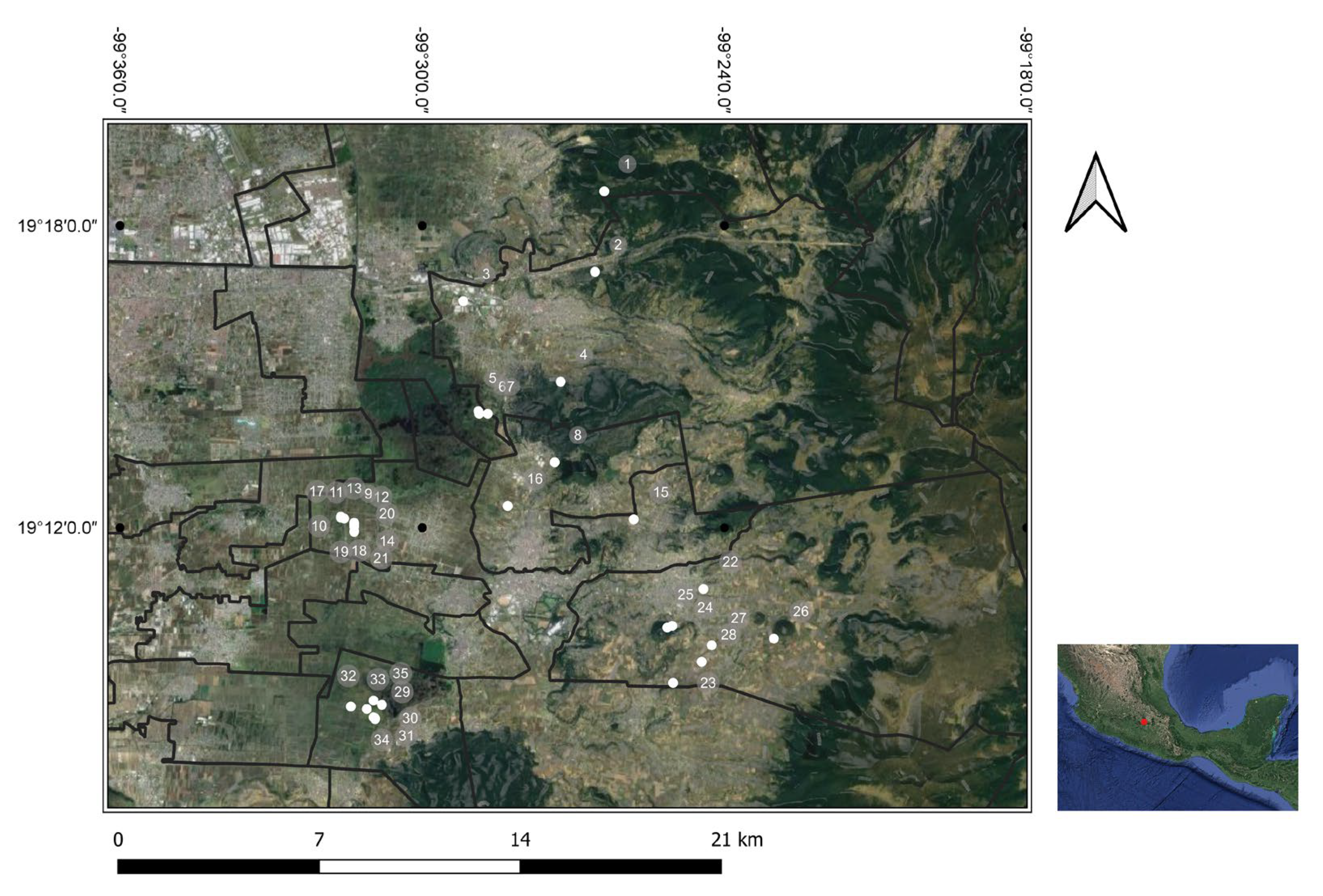
2.3. Geographic Distribution of Selected Groups

To identify the geographic location of the animal groups and the areas where the sheep graze, a satellite positioning system (Global Positioning System, GPS; Magellan Meridian, Thales Navigation, San Dimas, CA, USA) was used. The coordinates of latitude, longitude, and altitude of the position, time, and satellites of each of the animal groups selected herds were recorded in the field log. The signal received from three satellites was considered valid. A geographic information system (GIS) was developed to store and analyze of geographical and spatial data for the study area. Climatic and environmental data, including topography, hydrography, average annual precipitation, and maximum and minimum temperatures recorded, as well as the distribution of human communities, were obtained from INEGI-2017 [22], and they are summarized in Table S1. A map of study the site was build using QGIS, version 3.0 (QGIS.org; https://qgis.org/es/site/, accessed on 18 March 2023). The vectorial layer of the municipalities was based on INEGI-2005 [23], and Google Satellite and Terrain platforms, respectively. The elevational gradient (2572–2995 masl) was established along the southeastern slopes of the State of Mexico in central Mexico, where animal sampling was conducted at thirty-five study sites within elevational belts of approximately 200 m each.

2.4. Serological Screening

In the second stage of sampling, it was necessary to randomly select an initial animal and then another 9 more animals in each of the first-stage clusters. Blood samples from 345 ewes of reproductive age were collected. The blood samples were obtained through puncturing of the jugular vein. The serum samples were obtained by centrifuging the test tubes containing the blood samples at 1000× g. The samples were then kept frozen at −20 °C until use.
To detect the presence of abortion-causing microorganisms in ewes and obtain a preliminary picture of their epidemiology, a serological survey for antibodies against Leptospira spp., smooth Brucella spp., B. ovis, and C. abortus was carried out. Lytic/agglutinating activity of antibodies against Leptospira serovars was performed using a microscopic agglutination test (MAT) as described in the OIE Diagnostic Tests and Vaccines for Terrestrial Animals [24]. Antigens used in the MAT included ten serovars usually detected in this area. For the study panel, the H-89 (Hardjo genotype hardjoprajitno), Sinaloa ACR (Portland-vere), and Palo Alto (Icterohaemorrhagiae) strains isolated in Mexico and kindly classified for CA Bolin from Ames, IA, USA, were included. The cut-off titer (≥1:100) was considered positive. The end-point titer was the highest serum dilution showing agglutination of at least 50% of the leptospires—it was also included in MAT, positive reference controls for each strain.
The detection of anti-smooth Brucella spp. antibodies was analyzed with a rapid serum agglutination test (Rose Bengal Plate Test, RBPT) using B. abortus biovar 1 strain 1119-3 antigen with cellular concentration at 3% that covers the reactivity of B. melitensis and B. abortus. The RBPT result showed only two classifications: (a) positive reaction and (b) negative reaction depending on the presence or absence of agglutination. The sensitivity and specificity of RBT calculated for diagnosing Brucella in goats are 59 % and 98%, respectively [25].
The Oüchterlony double immunodiffusion (AGID) test was used to detect anti-bodies against B. ovis [26]. The appearance of precipitation lines of complete identity with the control serum lines was considered positive serum for antibodies against B. ovis detection. The absence of precipitation lines of complete identity was considered as a negative result. The AGID antigens are highly sensitive (70.1%) and specific (100%) for the serological diagnosis of B. ovis infection in rams [27].
Also, serum samples were analyzed for detection of antibodies against C. abortus using a recombinant commercial ELISA test (Chlamydia abortus serum verification, P00700/05-25/05/04. Institute Pouquier, Montpellier, France) used in accordance with the manufacturer’s instructions. The ELISA assay for diagnosing C. abortus infection in sheep has shown sensitivity and specificity of 93.5% and 98.5%, respectively [28].

2.5. Statistical Analysis

The overall seroprevalence of each one of the four abortion-causing microorganisms and the 95% confidence interval (CI) were calculated based on the following equation:
p ^ = N M n i = 1 n y i
where yi = ni (d) is the number of animals sampled and proportion of disease-positive animal proportion in the cluster, N is the number of clusters, nc is the number of cluster sampling, Mi is the number of animals in cluster, and ρh is the prevalence amongst flocks of the population.
The variance estimated for number of clusters is given by:
v ^ p ^ = N 2 M 2 1 n N s ^ b 2 n
s ^ b 2 = 1 N 1 i = 1 N y i y ¯ 2
All categorical demographics and characteristics from sheep groups were expressed as weighted percentages with 95% confidence intervals (CI). In all cases, the analysis (two-sided test) was considered statistically significant at p < 0.05. For continuous variables, a statistically significant difference was determined using Student’s t-test or the Mann–Whitney U test in the case of nonnormality. Categorical data were evaluated using Fisher’s exact test, and correlations between categorical and continuous variables were examined using Spearman’s rho. The Kolmogorov–Smirnov test was used to verify data normality.

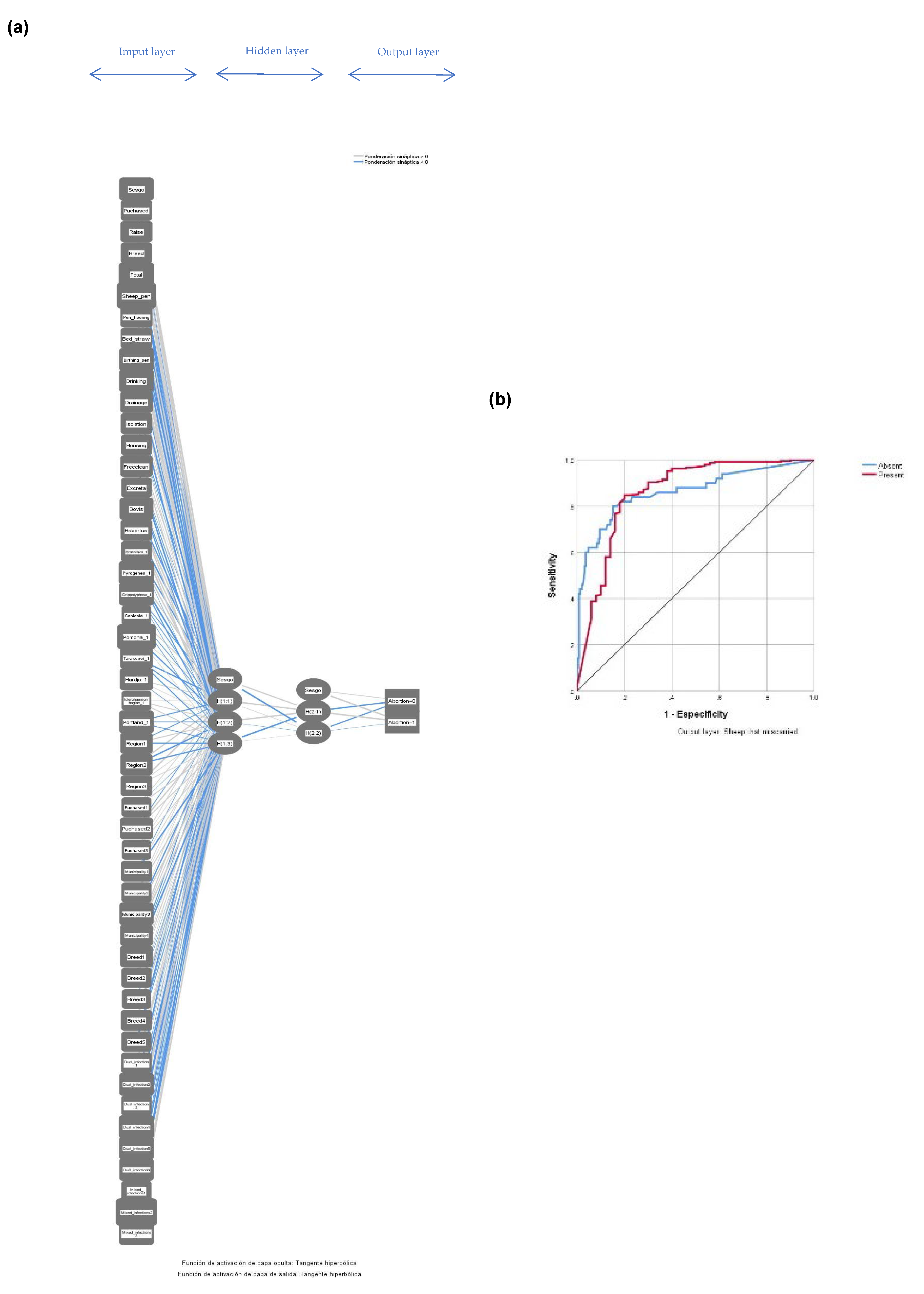
2.5.1. Artificial Neural Networks Model (ANNM)

The statistical patterns recognition from machine learning was performed with multilayer perceptron algorithm. The creation of topology and training of the network from various combinations of variables obtained from data of field survey and serological status of microorganisms considered the hidden layers, training cycles, and the parameters of the mathematical training function. The statistical properties of the training (70%), validation (20%), and test (10%) data of the ANNM allowed the prediction and classification of variables. Prior to exporting the data matrix to the ANNM for training, normalization of the data was performed to restrict the data range within a 0 to 1 scalar since the sigmoid activation function was assigned for each neuron in the middle layer. The weight of 0.5 corresponding to the activated value allows a better neural response. Also, categorical variables were recoded into “dummy” variables to improve response.
The performance criteria were decided based on the system-estimated values that occurred during training. The prediction accuracy was estimated from the area under the curve obtained by the receiver operating characteristic curve (ROC). A plot of power as a function of the type I error of the decision rule illustrated the performance of the binary model as its discrimination threshold is varied.

2.5.2. Generalized Linear Model (GLM)

The relationship between abortion and risk factors, protective or confounding, was determined by preprocessing variables obtained using the multilayer perceptron neural network model. We used a type of GLM and assumed that ewe abortion followed a binomial distribution. The use of logit link function facilitated the estimated contribution from covariates to be additive. A covariate selection algorithm was used to facilitate incorporation into the final GLM. To select the best model, we use a modified backward elimination procedure [29]. The model was built in three steps. (1) All covariates predicted by ANNM according to normalized significance were entered. (2) It started with a complex model verifying if the interaction terms were necessary with the elimination of the terms successively. The likelihood ratio statistic with p value < 0.3 suggested integration of the individual covariate. The next stage considered dropping a term from the main effects model. One by one of the covariates were eliminated from the initial model according to the highest p-value comparing the current model with the previous one. The Bayesian Information Criterion (BIC) and Akaike (AIC) index were used to choose between two or more alternative GLMs. (3) A receiver operating characteristic (ROC) curve was used as plot of sensitivity as a function of (1—specificity) for evaluating predictive power of all possible models. Statistical analyses were performed with Epi-Info v7.2.4.0 software (Centers for Disease Control and Prevention, CDC: Atlanta, GA, USA), and IBM SPSS v25 software (IBM Corp: Armonk, NY, USA).

3. Results

3.1. Places of Sampling

The study was carried out in a lacustrine zone of the trans-Mexican neovolcanic belt to the south-east of the State of Mexico, Mexico (19° N; 99° W). Based on the GPS readings recorded, the sampling sites were in three altitudinal zones of the mountainous region: zone 1 (elevations up to 2600 masl); zone 2 (2601 to 2800 masl); and zone 3 (>2800 masl) (Table S2; Figure S1). The map of the sampling sites and the geographical coordinates of study area is shown in Figure 1.

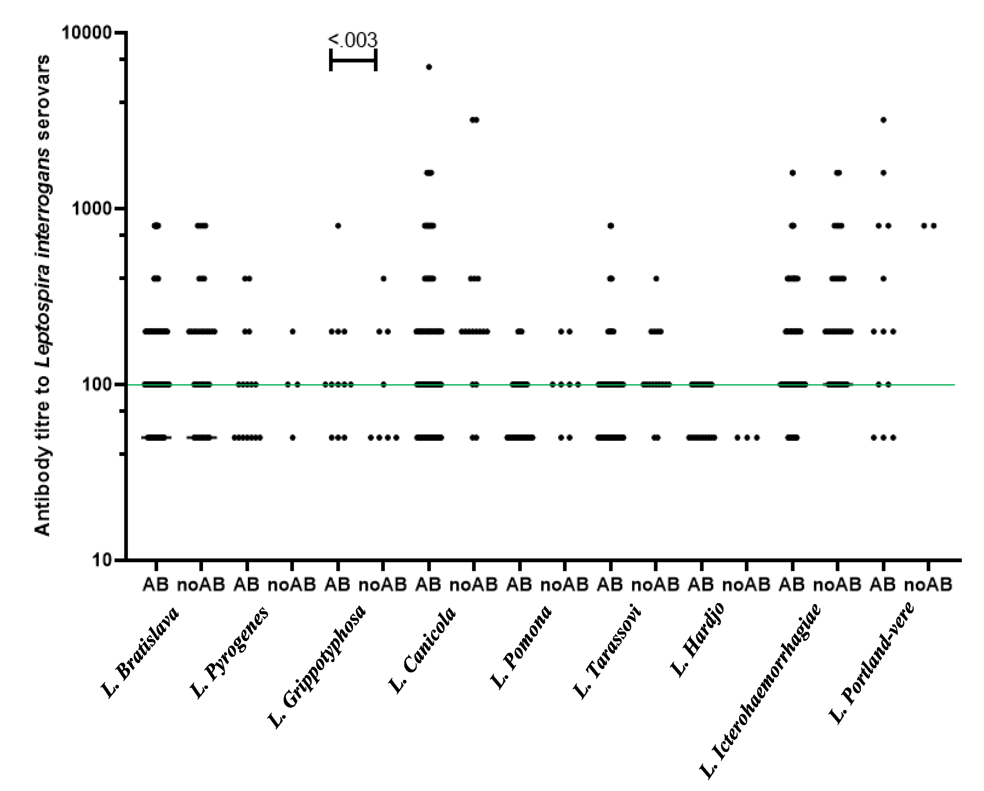
3.2. Antibody Prevalence of Multi-Pathogens and History of Abortion

The 345 sera available from sheep groups were screened for causing abortions multi-microorganisms. An infectious etiology was determined in 83.8% (289/345) of the ewes with a history of abortions in which the serological diagnoses were determined. Overall seroprevalence of smooth Brucella spp. was 70.7% (95% CI 65.7–75.8), while Leptospira spp. was 55.2% (95% CI 46.9–63.4), C. abortus was 21.9% (95% CI 5.3–38.5), and B. ovis was 7.4% (95% CI 0.0–28.1) according to the RBPT, MAT, ELISA, and AGID tests, respectively. The weighted seroprevalence of multiple organisms was only higher for smooth Brucella spp., but not for Leptospira spp., B. ovis, and C. abortus in aborted ewes. The weighted seroprevalence of smooth Brucella spp. was higher in aborted animals than in animals without a history of abortion (83.3% vs. 67.7%, respectively) (p < 0.007). Concerning seroprevalence by geographic regions, only seroprevalence of C. abortus for those aborted ewes from slopes with elevations up to 2600 masl was higher than for aborted ewes from slopes with elevations above 2800 masl (75.8% vs. 41.7%, respectively) (OR = 4.4; 95% CI 1.2–15.9; p < 0.02).
The probability of abortion due to Brucella infection with a positive-RBPT test was estimated to be around 98% (uncertainty coefficient 0.02), while C. abortus infection with a positive ELISA test was 82% (uncertainty coefficient 0.18). All sheep groups showed seropositivity to smooth Brucella spp., while 94.3% (33/35) of the investigated flocks showed antibodies against Leptospira spp., C. abortus, and B. ovis. At the individual level, the seroprevalence with two and three abortifacient microorganisms in the sampled sheep was higher from slopes with elevations up to 2600 masl than animals from slopes with elevations above 2600 masl. Serological detection with the four abortion-causing microorganisms was determined only in 0.87% (3/345) of sheep sampled. Table 1 shows the seropositivity of Leptospira spp., smooth Brucella spp., B. ovis, C. abortus antibodies in the ewe population sampled varied by altitudinal zonation. In contrast, Table 2 shows the weighted seropositive of Leptospira serovars in ewes sampled with history of abortion. Antibodies against Leptospira were detected in the sera of 188 ewes. The MAT results only showed questionable titers in 5.3% (166/3,105) of the total reactions (1:50 reactivity in sera that tested negative). The prevalence of agglutinins was detected in the sera of aborted ewes, mostly against serovar Icterohaemorrhagiae (37.2%, 95% CI 31.5–42.9), Bratislava (27%, 95% CI 21.8–32.2), and Canicola (12.3%, 95% CI 8.5–16.1). Nevertheless, serological profiles of the Leptospira serovars of the abortive and nonabortive animals, taken individually throughout the study, were only significantly different for serovar Grippotyphosa (p < 0.003). All sera that tested positive had antibody titers from 1:100 to 1:6400, with the strongest reactions to type strain Hond Utrecht IV and Portland-vere (Figure 2).

3.3. Validation of the Multilayer Perceptron Algorithm for Abortion in Ewes

The structure of infectious abortion prediction model based on the machine learning of multilayer perceptron algorithm is shown in Figure 3. The multilayer perceptron model allowed us to identify the patterns of predictive variables among the universe of non-linear variables related to abortion in sheep after training and the validation of the neural network. The neural network architecture showed a minimum number of hidden neurons to avoid the problem of overfitting, with which the perceptron algorithm obtains the best performance (Figure 3a), while the evaluation of the performance of the multilayer perceptron algorithm based on the area under the curve of the ROC curve showed that 80% of the ewes with a history of abortion were correctly classified (Figure 3b). The relative importance of input neurons for the prediction of abortion in sheep obtained from the multilayer perceptron model (Figure 4). The importance plot showed that the results are dominated by mixed infections with Leptospira spp.-C. abortus-smooth Brucella spp.-Brucella ovis (100%), followed by whether Leptospira serovar Canicola was identified exclusively (79.3%), with other infection predictors following far behind. The importance of mixed infections was clearly visible in the chart and much less visible for smooth Brucella spp. infection.

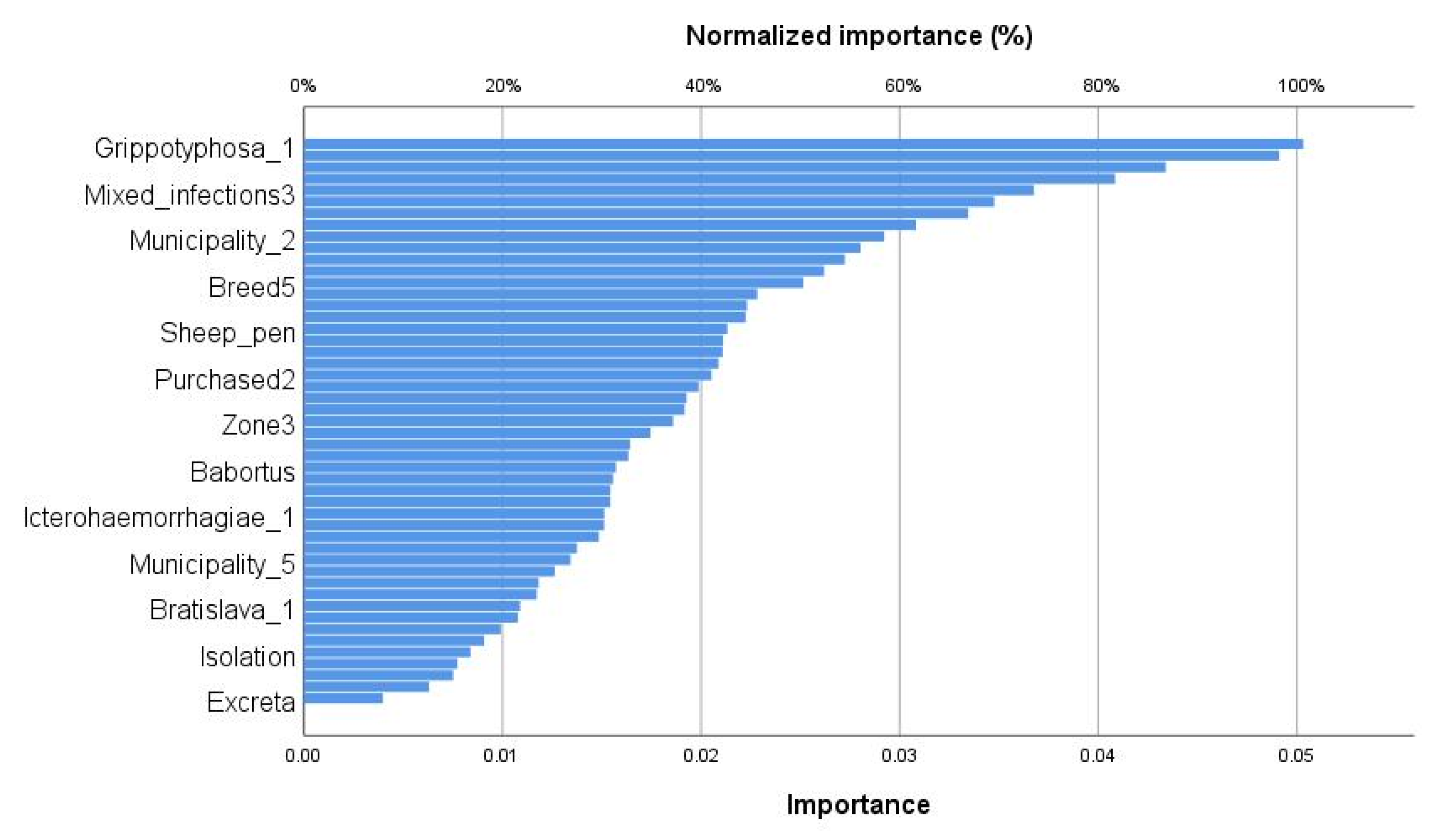
3.4. Determinants of Infectious Abortion in Ewes

The relationship between infectious abortion and preprocessing variables obtained by the multilayer perceptron neural network model showed 28 possible GLM models that can support prevention and control activities in different geographical areas (Table S2). The best model for infectious abortion, according to BIC and AIC, involving all four infectious pathogens showed several leptospiral serovars with a significant effect (Table 3). Serological detection of leptospirosis, which includes serovar Hardjo, as well as brucellosis caused by B. ovis, have a positive impact on sheep abortion (p < 0.001). On the other hand, the identification of serovar Grippotyphosa and Tarassovi had a significant negative effect on the outcome. The GLM additionally revealed that from the 30 analyzed exposure factors, 14 (46.7%) contribute significantly to ovine abortions. The determinants obtained from the generalized linear model were significantly different from zero, and the deviance residuals showed good model fit due to the ROC curve result (AUC: 0.89, 95% CI: 0.85–0.93; p < 2.94 × 10⁻²⁰). The AUC showed the probability that a randomly chosen aborted sheep would classify higher than a randomly chosen non-aborted sheep.

4. Discussion

Among the etiologies of abortion in sheep there are the presence of infectious agents, including many zoonotic microorganisms, and non-infectious causes such as nutritional, genetic, hormonal, toxic, and clinical (trauma, dystocia, prolapse) [30]. The results of our study demonstrate the infectious etiology in 82.6% (285/345) of the ewes with a history of abortions in which the serological diagnoses were determined.
It is widely accepted that seroprevalence of infectious agents causing abortion in small ruminants may vary according to geographic region, and zoonotic pathogens such as Leptospira spp., smooth Brucella spp., B. ovis, and C. abortus are among the most important microorganisms [31,32]. Although notification to the World Organization for Animal Health (OIE) is mandatory for many abortive pathogens of sheep due to restrictions on international trade, in Mexico, there is a lack of an effective epidemiological surveillance system that allows the development of strategies to prevent and control reproductive losses due to abortions and stillbirths of lambs, as well as to assess the prevalence of endemic diseases according to the diversity of ecosystems, with the subsequent reduction in risks to public health.

4.1. Towards the Strengthening of Regional Surveillance

The overall seroprevalence of microorganisms causing abortion in sheep appears to be very high, with 70.7% of animals testing positive for smooth Brucella spp. (B. melitensis, B. abortus, and B. suis) and 55.2% of animals positive for Leptospira spp., followed by the seroprevalence of C. abortus (21.9%) and B. ovis (7.4%). These findings show previous exposure is strikingly higher compared to previously reported seroprevalences of brucellosis in countries such as Iran (29.1%) and Egypt (16.3%), and less than 1% in the Arabian Gulf region, countries characterized by desert climates in summer and mild in winter [33,34,35]. Leptospira serovar-specific antibodies have been detected in 24.7% of ewes with a history of abortions in Brazil, 8.5% in Iran, and 4.5% in Italy [36,37,38].
Leptospirosis and brucellosis are the most widespread neglected diseases throughout the world, except Antarctica [39]. Climate changes, changes in ecological niches, and the appearance of new potential maintenance hosts could represent the most important factors involved in the epidemiology of abortifacient microorganisms. The environmental and geographical characteristics of Southern region of the State of Mexico can be considered as the optimal conditions for Leptospira spp. and Brucella spp. spreading among sheep and other animals, and eventually among humans.
The high seroprevalence of leptospirosis and brucellosis in ewes is not consistent with the small number of cases of human leptospirosis and brucellosis in the State of Mexico, Mexico. Mexican population data from 2012–2022 showed 17 human confirmed cases with Leptospira positive serological reaction and 655 confirmed cases with Brucella were recorded by the Mexican Ministry of Health [40]. Owing to the lack of diagnostic laboratories and a limited reporting system, leptospirosis and brucellosis are one of several neglected diseases in Mexico, and this may be one of the reasons why few cases were identified over this period, despite the high carriage of multi microorganisms in animals. The occurrence of human leptospirosis cases is more common in the tropics, especially in South America and Asia [41,42], and in regions where brucellosis is endemic, deleterious effects are seen in both humans and domestic animals in the developing nations of Africa, South/Southeast Asia, and Latin America [43]. The appearance of zoonotic disease in new localizations, as well as the sources of transmission between wild and domestic animals, is of great importance in terms of the epidemiological dimension. For many years, small ruminants had been considered as accidental hosts of leptospires, but several studies have shown that leptospiral infection in goats and sheep is common, and these species can also act as only maintenance hosts for serovars and carriers of leptospires eliminating the microorganisms on the environment for long time periods [44]. The maintenance hosts tend to be infected by serovars that colonize the kidneys and are shed in the urine. This hosts may act as chronic selective carriers of Leptospira serovars in a range of ecosystems and possibly transmit the pathogen to accidental hosts [45]. Detection of serovar Canicola 20.3% of animals sampled and Portland vere-type strain in 3.5% observed in our study suggests the presence of a selective host such as dogs that can cause infection in sheep and possibly cause accidental infections in humans. It should be noted that the clinical differences of the disease in dogs are based on the signs associated with non-icterogenic Canicola serovar like that observed in humans as "Stuttgart disease" [46,47,48]. Leptospirosis and brucellosis in sheep pose major threats to public health from direct contact with infected animals or their contaminated biological secretions (e.g., Amniotic fluid or vaginal discharge, and aborted fetuses or placentae), as well as the consumption of meat, unpasteurized milk, and dairy products produced with consequent economic loss from restrictions on contaminated dairy products [49].
Previous epidemiological investigations reported the circulation of Leptospira serovars in this mountainous region with an overall seroprevalence of 54.5% and detection the most likely infecting serovars as Icterohaemorrhagiae (54.5%), Bratislava (40%), Canicola (19%), and Tarassovi (15.8%) [19]. In this study, the overall seroprevalence of 55.2% is consistent with previously reported; but serological detection against the serovars as Pomona (5.2%), Grippotyphosa (3.8%), Pyrogenes (3.5%), and Portland vere-type strain (3.5%) suggest the possibility of investigating new serovars from wild reservoirs or sheep of other environmental settings. Wild rodents are the main reservoirs for pathogenic Leptospira species as serovar Grippotyphosa, which cause leptospirosis in sheep [50]. Transmission of Leptospira serovars requires a continuous enzootic circulation of the organism between animals, although with the possibility of introduction of new serovars from animal reservoirs, both wildlife and domestic animals [44]. According to Guedes et al. [51], the microscopic agglutination test is a good serological technique for the detection of antibodies against Leptospira serovars, but cross-reactions and paradoxical reactions are frequently observed with MAT. Serological paradoxical reactions and cross-reactions between serogroups were observed using MAT in our study, but the presence of high-titer Leptospira seropositivity (>1:200) in 36.6% of Bratislava seropositive sheep, 31.3% of Icterohaemorrhagiae, 14.5% of Canicola, 4.9% of Tarassovi, 2.0% of Grippotyphosa, 1.4% of Pomona, and 1.3% of Pyrogenes suggests the possibility of infection with these serovars. Antibody titers >1:100 detected in these animal sera probably resulted in an overestimation of overall seroprevalence of leptospirosis.

4.2. Epidemiological Control of Unidentified Abortion

Data obtained in our study have allowed the successful implementation of an ANNM to model mixed infection of four-abortive agents in sheep with a history of abortion as in other epidemiological studies [52,53]. Compared to stochastic models, the multilayer perceptron algorithm provided adequate prediction of abortion cases without the need for prior statistical correlations, or the assumptions required by common epidemiological models. A multilayer perceptron is a neural network that learns the relationship between linear and non-linear data and is considered an easy tool for the prediction of different diseases. The multilayer perceptron algorithm facilitated the identification of the statistical patterns among the infinite non-linear combinations related to abortion in sheep after training and validation. This is the first article in a series of possible ones that will use deep machine learning in the prognosis of diseases in animals. Deep learning has gained attention in recent decades for its innovative application in areas such as image classification using only pixels and labels as input layers, speech recognition, and automatic translation of text from one language to another without human involvement.
Methodological strategy based on machine learning algorithms allowed the identification of the preprocessed variables associated with abortion in sheep. The percentage of predictive values in the training and test performance in the aborted sheep classification from the multilayer perceptron algorithm was 89.4% and 88.2%, respectively. The adequate performance of the algorithm was obtained by the ROC curve that demonstrated an area under the curve to correctly predict abortion in sheep of 86.2%. Based on machine learning, the normalized importance values were obtained, which served to integrate the variables of GLM initial model. The final GLM appeared to fit the data well (overdispersion coefficient statistic = 0.83). If the value of the overdispersion coefficient is >1, this would show that meaning the variance is much larger than the mean, and so the GLM model is not appropriate. The area under the ROC curve (0.89) was significantly different from 0.5, since the p value was <0.001, indicating that the GLM classified the group of aborted ewes significantly better than chance. The final GLM showed a high predictive capacity (89%); in other words, 307 of the 345 sheep sampled were correctly classified. The result obtained via GLM allows us to know the exact extent of the abortion burden of zoonotic diseases in the region of the trans-Mexican neo-volcanic belt. The detection of serovar Hardjo and Brucella ovis in animals of the slopes with elevation between 2600 and 2800 meters above sea level from the municipality of Xalatlaco were independently and significantly related to an increased risk of abortion. The water well supply, sheep pen built with materials of metal grids and untreated wood, dirt and concrete floor, and bed of straw were also independently associated to a risk of abortion. The results of our study provide important data for use in regional public health policy. The strengthening of epidemiological surveillance and risk assessment in these remote rural areas will allow the optimal implementation of prevention measures in the first line of health services where people are cared for. Physicians and those with less experience in remote rural areas also need to recognize the diseases of animals that affect humans through regular and structured training and supervision on identifying variations that could indicate novel outbreaks of zoonotic diseases in the human population. There is a paradox that often less-experienced physicians, nurses, and midwives are deployed to rural settings, which are viewed as undesirable, and these new graduates are provided with little ongoing support or mentoring.

4.3. Strength and Limitations of Study

The strength of our study includes complete information on the management of the herd and the individual animal based on the factors or characteristics that have been related to abortion in sheep, as well as the laboratory results of the serological samples obtained. Epidemiological indicators of seroprevalence of microorganisms that cause abortion in sheep to achieve its goal of providing factual, objective, reliable, and comparable information with a high precision based on clustered sampling. Also, the main factors and the less important factors in the prediction of abortion in sheep are reported by artificial intelligence learning algorithms using the multilayer perceptron model. The limitations of our study were those related to the detection of other foodborne zoonoses such as Salmonella abortusovis, Campylobacter spp., Toxoplasma gondii, Listeria spp., Coxiella burnetii, and Yersinia pseudotuberculosis which can be disseminated among animals causing abortion, as well as contaminate vegetables and fruits for human consumption. Although studies have shown that T. gondii has been recognized as a major cause of infectious sheep abortion in New Zealand, Australia, the United Kingdom, Norway, and the United States, the presence of the disease is usually sporadic, and serological testing is not specific for the detection of viable Toxoplasma oocysts. Evidence for abortion has been based on the detection of the DNA of T. gondii from fetal tissues, but these findings do not show a relationship with maternal serological results [54]. Other studies show congenital transmission from the molecular detection of T. gondii in the brain from aborted fetuses with low maternal seroconversion; however, in fetal brain samples from negative lambs, high maternal serology is observed [55]. The usefulness of detecting antibodies for the diagnosis of T. gondii in sheep abortion is currently ambivalent and controversial because there may be cross-reactivity with oocytes from other parasites, so more studies are required in this regard. Unfortunately, the problem of diagnosis of C. burnetti was not addressed due to the limited commercially available serological tests for using in animals. A purposeful search for C. burnetii infection with appropriate serological testing is required to understand the course of the disease in small ruminants, its epidemiology, and the risk of transmission to humans. Since there are few reports in Mexico in both animal and human populations that demonstrate reliable evidence of the presence of C. burnetii, this may not reflect the current situation of emerging rickettsial diseases [56]. Q fever, so far, is a disease considered exotic in our country.
Other difficulties were related to the budget; however, the application of the cluster sampling design balanced the feasibility of the abortion research project with the precision of the epidemiological impact measures, complemented by the data analysis based on the machine learning algorithm.

5. Conclusions

Our neural network approach has been able to identify multiple microorganisms and putative predictors of infectious abortion in sheep. This report includes the measurable factors to be monitored in future epidemiological studies to improve public health surveillance. Since most abortion can be prevented by the sanitation measures identified here, it only remains to propose the use of existing or currently under development vaccines to reduce the risk of animal–human transmission of zoonotic infectious agents [57]. The results of the study are expected to help establish priorities and to tailor specific public health interventions, including vaccination, to the etiology of infectious abortion in sheep from the mountainous region of Mexico.

Supplementary Materials

The following supporting information can be downloaded at: https://www.mdpi.com/article/10.3390/ani13182955/s1, Table S1: Climatic and environmental data from study area; Table S2: The geographic distribution of clustered sampling and group size in sheep transhumance systems; Table S3: Schwarz’s and Akaike’s Bayesian Information Criteria for 28 generalized linear models of infectious abortion in ewes; Table S4: Structured questionnaire to collect information about the flocks, animals, and risks factors related to ewes abortion; Figure S1: The view of the flocks of sheep in their habitats where they have shelter, water, and a large grassy pasture. (a) zone 1 (elevations up to 2600 masl.); (b) zone 2 (2601 to 2800 masl.); and (c) zone 3 (>2800 masl.).

Author Contributions

Conceptualization, G.A.-T. and J.M.J.-E.; methodology, M.L.-A., L.H.-A. and M.L.-H.; formal analysis, G.A.-T., V.S.-C. and F.B.; data curation, G.A.-T., M.L.-A., L.H.-A. and M.L.-H.; writing—original draft preparation, G.A.-T.; writing—review and editing, G.A.-T., M.L.-A., L.H.-A., J.M.J.-E., V.S.-C., F.B., R.M.d.O.-J., M.L.-H. and F.M.G.-I.; methodology, M.L.-A., L.H.-A. and M.L.-H.; supervision, F.M.G.-I. and R.M.d.O.-J.; project administration, G.A.-T.; funding acquisition, F.M.G.-I. and R.M.d.O.-J. All authors have read and agreed to the published version of the manuscript.

Funding

This research received no external funding. This research received no specific grant from any funding agency, commercial or not-for-profit sectors.

Institutional Review Board Statement

The project was approved by the Institutional Committee for Research and Advanced Studies, Faculty of Veterinary Medicine, Universidad Autónoma del Estado de Mexico, UAEM, Toluca, Mexico (n°2230/2006U).

Informed Consent Statement

Not applicable.

Data Availability Statement

The datasets used and/or analyzed during the current study are available from the corresponding author upon reasonable request.

Acknowledgments

The authors thank MVZ. V. Banda, and Ing. A. Arteaga for technical advice, and CA. Bolin from Ames, IA, USA for classifying type strains. We are grateful to the owners of the affected properties for their cooperation and assistance with the investigation.

Conflicts of Interest

The authors declare that the research was conducted in the absence of any commercial or financial relationships that could be construed as a potential conflict of interest.

References

  1. Guernier, V.; Allan, K.J.; Goarant, C. Advances and challenges in barcoding pathogenic and environmental Leptospira. Parasitology 2018, 145, 595–607. [Google Scholar] [CrossRef] [PubMed]
  2. Costa, F.; Hagan, J.E.; Calcagno, J.; Kane, M.; Torgerson, P.; Martinez-Silveira, M.S.; Stein, C.; Abela-Ridder, B.; Ko, A.I. Global morbidity and mortality of leptospirosis: A systematic review. PLoS Negl. Trop. Dis. 2015, 9, e0003898. [Google Scholar] [CrossRef] [PubMed]
  3. World Health Organization WHO. Report of the Second Meeting of the Leptospirosis Burden Epidemiology Reference Group. 2011. Available online: http://apps.who.int/iris/handle/10665/44588 (accessed on 18 March 2023).
  4. Levett, P.N. Leptospirosis: A forgotten zoonosis? Clin. Appl. Immunol. Rev. 2004, 4, 435–448. [Google Scholar] [CrossRef]
  5. Comia, I.; Madureira, A.C.; Schooley, R.T.; Vieira, M.L.; Noormahomed, E.V. Molecular Detection of Leptospira spp. in Rodents Trapped in the Mozambique Island City, Nampula Province, Mozambique. EC Microbiol. 2018, 14, 813–821. [Google Scholar] [PubMed]
  6. Gamage, C.D.; Sato, Y.; Kimura, R.; Yamashiro, T.; Toma, C. Understanding leptospirosis eco-epidemiology by environmental DNA metabarcoding of irrigation water from two agro-ecological regions of Sri Lanka. PLoS Negl. Trop. Dis. 2020, 14, e0008437. [Google Scholar] [CrossRef] [PubMed]
  7. Torgerson, P.R.; Hagan, J.E.; Costa, F.; Calcagno, J.; Kane, M.; Martinez-Silveira, M.S.; Goris, M.G.A.; Stein, C.; Ko, A.I.; Abela-Ridder, B. Global Burden of Leptospirosis: Estimated in Terms of Disability Adjusted Life Years. PLoS Negl. Trop. Dis. 2015, 9, e0004122. [Google Scholar] [CrossRef] [PubMed]
  8. Dorsch, M.A.; Francia, M.E.; Tana, L.R.; González, F.C.; Cabrera, A.; Calleros, L.; Sanguinetti, M.; Barcellos, M.; Zarantonelli, L.; Ciuffo, C.; et al. Diagnostic Investigation of 100 Cases of Abortion in Sheep in Uruguay: 2015–2021. Front. Vet. Sci. 2022, 9, 904786. [Google Scholar] [CrossRef] [PubMed]
  9. Hassell, J.M.; Begon, M.; Ward, M.J.; Fèvre, E.M. Urbanization and Disease Emergence: Dynamics at the Wildlife-Livestock-Human Interface. Trends Ecol. Evol. 2017, 32, 55–67. [Google Scholar] [CrossRef]
  10. Abdul Mutalip, M.H.; Mahmud, M.A.F.; Lodz, N.A.; Yoep, N.; Muhammad, E.N.; Ahmad, A.; Hashim, M.H.; Muhamad, N.A. Environmental risk factors of leptospirosis in urban settings: A systematic review protocol. BMJ. Open 2019, 9, e023359. [Google Scholar] [CrossRef]
  11. Suwannarong, K.; Soonthornworasiri, N.; Maneekan, P.; Yimsamran, S.; Balthip, K.; Maneewatchararangsri, S.; Saisongkorh, W.; Saengkul, C.; Sangmukdanun, S.; Phunta, N.; et al. Rodent-Human Interface: Behavioral Risk Factors and Leptospirosis in a Province in the Central Region of Thailand. Vet. Sci. 2022, 9, 85. [Google Scholar] [CrossRef]
  12. Mendoza, M.V.; Rivera, W.L. Identification of Leptospira spp. from environmental sources in areas with high human leptospirosis incidence in the Philippines. Pathog. Glob. Health 2019, 113, 109–116. [Google Scholar] [CrossRef] [PubMed]
  13. Sun, J. Dirty environment for adult life: The bad, the good, the unknown. Genes Dis. 2016, 3, 167–168. [Google Scholar] [CrossRef] [PubMed]
  14. Li, Y.; Baldridge, M.T. Modelling human immune responses using microbial exposures in rodents. Nat. Microbiol. 2023, 8, 363–366. [Google Scholar] [CrossRef] [PubMed]
  15. Aikowe, J.O.; Mazancová, J. Barriers to Water Access in Rural Communities: Examining the Factors Influencing Water Source Choice. Water 2021, 13, 2755. [Google Scholar] [CrossRef]
  16. Haake, D.A.; Levett, P.N. Leptospirosis in humans. Curr. Trop. Microbiol. Immunol. 2015, 387, 65–97. [Google Scholar]
  17. Ghaffar Nia, N.; Kaplanoglu, E.; Nasab, A. Evaluation of artificial intelligence techniques in disease diagnosis and prediction. Discov. Artif. Intell. 2023, 3, 5. [Google Scholar] [CrossRef]
  18. Montesinos López, O.A.; Montesinos López, A.; Crossa, J. Fundamentals of artificial neural networks and deep learning. In Multivariate Statistical Machine Learning Methods for Genomic Prediction; Springer: Cham, Switzerland, 2022. [Google Scholar]
  19. Arteaga-Troncoso, G.; Jiménez-Estrada, J.; Montes de Oca, R.; López-Hurtado, M.; Luna-Alvarez, M.; Hernandez-Andrade, L.; Moreno-Alfaro, A.; Galan-Herrera, J.F.; Guerra-Infante, F.M. Seroprevalence and risk factors associated with within-flock transmission of Leptospira interrogans in transhumant farming systems in Mexico. Epidemiol. Infect. 2015, 143, 2894–2902. [Google Scholar] [CrossRef]
  20. Jiménez-Estrada, J.M.; Escobedo-Guerra, M.R.; Arteaga-Troncoso, G.; López-Hurtado, M.; De Haro-Cruz, M.D.J.; Jiménez, R.M.D.O.; Guerra-Infante, F.M. Detection of Chlamydophila abortus in sheep (Ovis aries) in Mexico. Am. J. Anim. Vet. Sci. 2008, 3, 91–95. [Google Scholar]
  21. Teagasc—Agriculture and Food Development Authority of Ireland. A Guide to Designing a Sheep Handling Unit. 2020. Available online: https://www.teagasc.ie/media/website/publications/2020/A-Guide-to-Designing-a-Sheep-Handling-Unit.pdf (accessed on 18 March 2023).
  22. INEGI. Carta de Uso del Suelo y Vegetación; Serie VI, escala 1:250,000; INEGI: Aguascalientes, Mexico, 2014. [Google Scholar]
  23. INEGI. Carta de Uso del Suelo y Vegetación; Serie III, escala 1:250,000 (continuo nacional); INEGI: Aguascalientes, Mexico, 2005. [Google Scholar]
  24. World Health Organization. Charter 3.1.12. Leptospirosis. In OIE Terrestrial Manual 2021; World Health Organization: Geneva, Switzerland, 2021; Volume 3, ISBN 978-92-95108-18-9. Available online: https://www.woah.org/fileadmin/Home/fr/Health_standards/tahm/3.01.12_LEPTO.pdf (accessed on 18 March 2023).
  25. Ahasan, M.S.; Rahman, M.S.; Rahman, A.K.M.A.; Berkvens, D. Bovine and Caprine Brucellosis in Bangladesh: Bayesian evaluation of four serological tests, true prevalence, and associated risk factors in household animals. Trop. Anim. Health Prod. 2017, 49, 1–11. [Google Scholar] [CrossRef]
  26. Teixeira, L.S.d.A.; de Pinho, F.A.; Batista, J.F.; Santana, M.d.V.; Soares, F.F.F.; Lima, A.M.C.; Furtado, L.L.; Silva, F.K.d.S.; Santos, L.M.N.; Damasceno, T.C.M.; et al. Risk factors and detection of Brucella ovis in naturally infected rams and ewes. Rev. Bras. Cienc. Vet. 2021, 28, 48–52. [Google Scholar] [CrossRef]
  27. Xavier, M.N.; Sant’Anna, F.M.; Silva, T.M.A.; Costa, E.A.; Moustacas, V.S.; Merlo, F.A.; Carvalho Júnior, C.A.; Dasso, M.G.; Mathias, L.A.; Gouveia, A.M.G.; et al. A comparison of two agar gel immunodiffusion methods and a complement fixation test for serologic diagnosis of Brucella ovis infection in experimentally infected rams. Arq. Bras. Med. Vet. Zootec. 2011, 63, 1016–1021. [Google Scholar] [CrossRef]
  28. Wilson, K.; Livingstone, M.; Longbottom, D. Comparative evaluation of eight serological assays for diagnosing Chlamydophila abortus infection in sheep. Vet. Microbiol. 2009, 135, 38–45. [Google Scholar] [CrossRef] [PubMed]
  29. Agresti, A. Building and Applying Logistic Regression Models. In An Introduction to Categorical Data Analysis; John Wiley & Sons: Hoboken, NJ, USA, 2002; pp. 137–172. [Google Scholar]
  30. Alemayehu, G.; Mamo, G.; Alemu, B.; Desta, H.; Tadesse, B.; Benti, T.; Bahiru, A.; Yimana, M.; Wieland, B. Causes and Flock Level Risk Factors of Sheep and Goat Abortion in Three Agroecology Zones in Ethiopia. Front. Vet. Sci. 2021, 8, 615310. [Google Scholar] [CrossRef] [PubMed]
  31. Azócar-Aedo, L. Basic Aspects and Epidemiological Studies on Leptospirosis Carried Out in Animals in Chile: A Bibliographic Review. Trop. Med. Infect. Dis. 2023, 8, 97. [Google Scholar] [CrossRef] [PubMed]
  32. Guesmi, K.; Kalthoum, S.; Mamlouk, A.; Baccar, M.N.; BelHajMohamed, B.; Hajlaoui, H.; Toumi, A.; Cherni, J.; Seghaier, C.; Messadi, L. Seroprevalence of zoonotic abortive diseases and their associated risk factors in Tunisian sheep. BMC Vet. Res. 2023, 19, 50. [Google Scholar] [CrossRef] [PubMed]
  33. Abnaroodheleh, F.; Emadi, A.; Dadar, M. Seroprevalence of brucellosis and chlamydiosis in sheep and goats with history of abortion in Iran. Small Rumin. Res. 2021, 202, 106459. [Google Scholar] [CrossRef]
  34. Abdelfattah, S.; Kotb, A.; Eman, R.; Yaser, M.H.; Alamery, S. Seroprevalence and molecular characterization of Brucella species in naturally infected cattle and sheep. Prev. Vet. Med. 2019, 171, 104756. [Google Scholar]
  35. Ebid, M.; El Mola, A.; Salib, F. Seroprevalence of brucellosis in sheep and goats in the Arabian Gulf region. Vet. World 2020, 13, 1495–1509. [Google Scholar] [CrossRef]
  36. Almeida, D.S.; Paz, L.N.; de Oliveira, D.S.; Silva, D.N.; Ristow, P.; Hamond, C.; Costa, F.; Portela, R.W.; Estrela-Lima, A.; Pinna, M.H. Investigation of chronic infection by Leptospira spp. in asymptomatic sheep slaughtered in slaughterhouse. PLoS ONE 2019, 14, e0217391. [Google Scholar] [CrossRef]
  37. Haji Hajikolaei, M.R.; Rezaei, S.; Ghadrdan Mashhadi, A.R.; Ghorbanpoor, M. The role of small ruminants in the epidemiology of leptospirosis. Sci. Rep. 2022, 12, 2148. [Google Scholar] [CrossRef]
  38. Bertelloni, F.; Cilia, G.; Turchi, B.; Pinzauti, P.; Cerri, D.; Fratini, F. Epidemiology of leptospirosis in North-Central Italy: Fifteen years of serological data (2002–2016). Comp. Immunol. Microbiol. Infect. Dis. 2019, 65, 14–22. [Google Scholar] [CrossRef]
  39. Karpagam, K.B.; Ganesh, B. Leptospirosis: A neglected tropical zoonotic infection of public health importance-an updated review. Eur. J. Clin. Microbiol. Infect. Dis. 2020, 39, 835–846. [Google Scholar] [CrossRef] [PubMed]
  40. Mexican Ministry of Health; Ministry of Health, General Directorate of Epidemiology. Mexico SUIVE Epidemiology Bolletin. Available online: https://www.gob.mx/salud/acciones-y-programas/historico.boletin-epidemiologico (accessed on 24 March 2023).
  41. López-Robles, G.; Córdova-Robles, F.N.; Sandoval-Petris, E.; Montalvo-Corral, M. Leptospirosis at human-animal-environment interfaces in Latin-America: Drivers, prevention, and control measures. Biotecnia 2021, 23, 89–100. [Google Scholar]
  42. Tique, V.; Mattar, S.; Miranda, J.; Oviedo, M.; Noda, A.; Montes, E.; Rodriguez, V. Clinical and Epidemiological Status of Leptospirosis in a Tropical Caribbean Area of Colombia. Biomed. Res. Int. 2018, 29, 6473851. [Google Scholar] [CrossRef] [PubMed]
  43. Franc, K.A.; Krecek, R.C.; Häsler, B.N.; Arenas-Gamboa, A.M. Brucellosis remains a neglected disease in the developing world: A call for interdisciplinary action. BMC Public Health 2018, 11, 18. [Google Scholar] [CrossRef] [PubMed]
  44. Harran, E.; Pinot, A.; Kodjo, A.; Djelouadji, Z.; Le Gudayer, M.; Sionfoungo Daouda, S.; Groud, K.; Lattard, V.; Ayral, F. Identification of Pathogenic Leptospira kirschneri Serogroup Grippotyphosa in Water Voles (Arvicola terrestris) from Ruminant Pastures in Puy-de-Dôme, Central France. Pathogens 2023, 12, 260. [Google Scholar] [CrossRef]
  45. Viana, M.; Mancy, R.; Biek, R.; Cleaveland, S.; Cross, P.C.; Lloyd-Smith, J.O.; Haydon, D.T. Assembling Evidence for Identifying Reservoirs of Infection. Trends Ecol. Evol. 2014, 29, 270–279. [Google Scholar] [CrossRef] [PubMed]
  46. Luna, A.; Moles, C.; Salazar, G.; Nava, V.; Gavaldón, R. La Leptospirosis canina y su problemática en Mexico. Rev. De Salud Anim. 2008, 30, 1–11. [Google Scholar]
  47. Martínez-Barbabosa, I.; Alpizar-Sosa, E.A.; Gavaldón-Rosas, D.G.; Moles-Cervantes, L.P.; Cárdenas, M.G.; García-González, R.; Shea, M.; Fernández-Presas, A.M. Canine Leptospirosis Serology in Southern Mexico City. J. Clin. Microbiol. 2016, 6, 171–180. [Google Scholar] [CrossRef]
  48. Miotto, B.A.; Guilloux, A.G.A.; Tozzi, B.F.; Moreno, L.Z.; da Hora, A.S.; Días, R.A.; Heinemann, M.B.; Moreno, A.M.; Filho, A.F.S.; Lilenbaum, W.; et al. Prospective study of canine leptospirosis in shelter and stray dog populations: Identification of chronic carriers and different Leptospira species infecting dogs. PLoS ONE 2018, 13, e0200384. [Google Scholar] [CrossRef]
  49. Khurana, S.K.; Sehrawat, A.; Tiwari, R.; Prasad, M.; Gulati, B.; Shabbir, M.Z.; Chhabra, R.; Karthik, K.; Patel, S.K.; Pathak, M.; et al. Bovine brucellosis—A comprehensive review. Vet. Q. 2021, 41, 61–88. [Google Scholar] [CrossRef] [PubMed]
  50. Gomard, Y.; Dellagi, K.; Goodman, S.M.; Mavingui, P.; Tortosa, P. Tracking Animal Reservoirs of Pathogenic Leptospira: The Right Test for the Right Claim. Trop. Med. Infect. Dis. 2021, 6, 205. [Google Scholar] [CrossRef] [PubMed]
  51. Guedes, I.B.; Souza, G.O.; Castro, J.F.; Cavalini, M.B.; de Souza Filho, A.F.; Heinemann, M.B. Usefulness of the Ranking Technique in the Microscopic Agglutination Test (MAT) to Predict the Most Likely Infecting Serogroup of Leptospira. Front. Vet. Sci. 2021, 8, 654034. [Google Scholar] [CrossRef] [PubMed]
  52. Shawaqfah, M.; Almomani, F. Forecast of the outbreak of COVID-19 using artificial neural network: Case study Qatar, Spain, and Italy. Results Phys. 2021, 27, 104484. [Google Scholar] [CrossRef] [PubMed]
  53. Wang, L.; Zhang, Y.; Wang, D.; Tong, X.; Liu, T.; Zhang, S.; Huang, J.; Zhang, L.; Chen, L.; Fan, H.; et al. Artificial Intelligence for COVID-19: A Systematic Review. Front. Med. 2021, 8, 704256. [Google Scholar] [CrossRef] [PubMed]
  54. Mason, S.; Quinnell, R.J.; Smith, J.E. Detection of Toxoplasma gondii in lambs via PCR screening and serological follow-up. Vet. Parasitol. 2010, 169, 258–263. [Google Scholar] [CrossRef] [PubMed]
  55. Rassouli, M.; Razmi, G.R.; Bassami, M.R.; Movassaghi, A.R.; Azizzadeh, M. Study on ovine abortion associated with Toxoplasma gondii in affected herds of Khorasan Razavi Province, Iran based on PCR detection of fetal brains and maternal serology. Parasitology 2011, 138, 691–697. [Google Scholar] [CrossRef] [PubMed]
  56. Flores-Perez, C.F.; Díaz-Aparicio, E.; Palomares-Resendiz, E.G.; Isidro-Requejo, L.M.; Herrera-López, E.; Hernández-Castro, R. Molecular identification of Coxiella burnetii in vaginal swabs from aborted goats in Mexico. Small Rumin. Res. 2022, 210, 106664. [Google Scholar] [CrossRef]
  57. Rajapakse, S.; Rodrigo, C.; Handunnetti, S.M.; Fernando, S.D. Current immunological and molecular tools for leptospirosis: Diagnostics, vaccine design, and biomarkers for predicting severity. Ann. Clin. Microbiol. Antimicrob. 2015, 14, 2. [Google Scholar] [CrossRef]
Figure 1. Location of selected sites sampling in the ewe abortion study (numbers). The circle points represent the georeferenced locations of the animal groups at the sampling site. A layer of geographical point features (white circle dots) represents the sampling sites where Leptospirosis, Brucellosis, and Ovine Enzootic Abortion are prevalent. The altitudinal zonation of the study site in the southeast of the State of Mexico in central Mexico, and the municipality boundaries are included in the map.
Animals 13 02955 g001
Figure 2. Comparison of serological MAT titers for aborting (AB) and non-aborting (noAB) ewes for antibodies against Leptospira serovars. Continuous line (in green) indicates the cut-off titer (≥1:100) was considered positive. p-value calculated by Mann–Whitney test.
Animals 13 02955 g002
Figure 3. The ANNM for ovine abortion prediction. The final architecture of the model was 50 neurons in the input layer, 7 neurons in the two-layer, and 2 neurons in the output layer (Abortion: 0: absent; 1: present) (a); Area under the curve (AUC) for validation of the multilayer perceptron model in sheep abortion based on ROC curve analysis (b). Sesgo: Bias; Raise: What do you raise sheep for? 1: market, 2: breeding program; Sheep-pen: What are the materials you use to build your sheep pen? 0: bricks; 1: metal sheets and untreated wood; Sheep pen flooring: 1: dirt pen flooring only; 2: dirt and concrete pen flooring; Bed_straw: Did the ewe give birth where they bed of straw down? 1: yes; 0: no; Birthing_pen: Where was the lamb of this ewe born? 1: birthing pen; or 0: meadow; Drinking: The water supply for the animals: 1: drinking water; 2: irrigation canal; Drainage: Drainage in the pen: 0: no 1: yes; Isolation: Isolation of an individual sheep by panic: 0: no; 1: yes; Housing: How many animals does it take to congregate to avoid panic?; Frecclean: What is the cleaning frequency of sheep housing?; Excreta: What handling of excreta do you carry out in the housing pen?; Brucella ovis_1: 0: negative; 1: positive; Brucella abortus_1: 0: negative; 1: positive; Bratislava_1: 0: negative; 1: positive; Pyrogenes_1: 0: negative; 1: positive; Grippotyphosa_1: 0: negative; 1: positive; Canicola_1: 0: negative; 1: positive; Pomona_1: 0: negative; 1: positive; Tarassovi_1: 0: negative; 1: positive; Hardjo_1: 0: negative; 1: positive; Icterohaemorrhagiae_1: 0: negative; 1: positive; Portland_1: 0: negative; 1: positive; Zone_1: Elevations up to 2600 masl; Zone_2: Elevations between 2601 and 2700 masl; Zone_3: Elevations >2800 masl; Purchased1: Rural market; Puchased2: Imported animals; Purchased3: Born in the flock; Municipality_1: Xalatlaco; Municipality_2: Santiago Tianguistenco; Municipality_3: Calpulhuac; Municipality_4: Chapultepec municipality; Municipality_5: Texcalyacac; Municipality_6: Metepec; Municipality_7: Ocoyoacac; Municipality_8: Lerma; Breed1: Pelibuey; Breed2: Hampshire; Breed3: Suffolk-Pelibuey crossbreed; Breed4: Hampshire-Pelibuey crossbreed; Breed5: Suffolk; Dual_infection1: Leptospira spp.-Chlamydia abortus; Dual_infection2: smooth Brucella spp.-Brucella ovis; Dual_infection3: Leptospira spp.-smooth Brucella spp.; Dual_infection4: Leptospira-Brucella ovis; Dual_infection5: Chlamydia abortus-smooth Brucella spp.; Dual_infection6: Chlamydia abortus-Brucella ovis; Mixed_infections1: Leptospira spp.-Chlamydia abortus-smooth Brucella spp.; Mixed_infections2: Leptospira-Chlamydia-Brucella ovis; Mixed_infections3: Leptospira spp.-Chlamydia abortus-smooth Brucella spp.-B. ovis.
Animals 13 02955 g003
Figure 4. The relative importance of input layer to the prediction for unidentified abortion in ewes. The importance chart shows the relative contribution of predictors that are dominated by Grippotyphosa serovar, followed by Canicola serovar (Portland-vere strain) was detected exclusively, with other infection predictors following far behind. The importance of the mixed infections is clearly visible in infectious abortion among predictors on x-axis.
Animals 13 02955 g004
Table 1. The geographic distribution of weighted seroprevalence of Leptospirosis, Brucellosis (smooth Brucella spp., B. ovis), and ovine enzootic abortion in the ewe population sampled (by altitudinal zonation).
MicroorganismsStatusZone 1
(Slopes with
Elevations up to 2600 masl)
Zone 2
(Slopes with
Elevations between 2600 and 2800 masl)
Zone 3
(Slopes with Elevations above 2800 masl)
Prev. (%)95% CIPrev.
(%)
95% CIPrev.
(%)
95% CI
Leptospira spp.Abortion38.632.9–44.313.79.7–17.75.32.7–7.9
No abortion36.724.5–48.918.38.5–28.150.1–10.5
Smooth Brucella spp. *Abortion48.442.6–54.218.614.1–23.174.0–10.0
No abortion75.064.0–86.026.715.5–37.950.1–10.5
B. ovisAbortion6.73.8–9.652.5–7.50N.D.
No abortion6.73.8–9.61.70.1–3.20N.D.
C. abortusAbortion17.513.1–21.94.62.2–7.00.70.1–1.6
No abortion8.31.3–15.3102.4–17.65.00.1–10.5
Leptospira-C. abortus
dual reactivity
Abortion15.611.4–19.810.42.6–18.16.73.8–9.6
No abortion16.77.3–26.114.35.4–23.2209.9–30.1
Leptospira-smooth Brucella spp. dual reactivityAbortion55.149.3–60.950.744.3–55.95044.2–55.8
No abortion66.754.8–78.647.635–60.24027.6–52.4
Leptospira-B. ovis dual reactivityAbortion5.32.6–7.90.70.67–0.730N.D.
No abortion6.70.4–131.70.1–4.90N.D.
Smooth Brucella spp.-B. ovis dual reactivityAbortion5.62.9–8.30.70.67–0.730N.D.
No abortion6.70.4 - 131.70.1–4.90N.D.
Smooth Brucella spp.-C. abortus dual reactivityAbortion12.38.5–16.13.51.4–5.60.70.67–0.73
No abortion8.31.3–15.350.1–10.51.70.1–4.9
B. ovis-C. abortus dual reactivityAbortion1.10.1–2.30N.D.0N.D.
No abortion0.40.1–8.90N.D.0N.D.
Leptospira-smooth Brucella spp.-B. ovis-C. abortus multi-reactivityAbortion0.70.67–0.730N.D.0N.D.
No abortion1.70.1–4.90N.D.0N.D.
* Kruskal–Wallis test (p < 0.03) with uncertainty coefficient 0.02. masl., meters above sea level. Prev., Seroprevalence. CI, Confidence interval. N.D., Not determined.
Table 2. Weighted seropositivity of Leptospira serovars in ewes with history of abortion (by altitudinal zonation).
Serovars (Type Strain)StatusZone 1
(Slopes with
Elevations up to 2600 masl)
Zone 2
(Slopes with
Elevations between 2600 and 2800 masl)
Zone 3
(Slopes with Elevations above 2800 masl)
Prev. (%)95% CIPrev.
(%)
95% CIPrev.
(%)
95% CI
Icterohaemorrhagiae
(Palo Alto)
Abortion37.231.5–42.912.38.5–16.15.32.7–7.9
No abortion209.1–30.9101.8–18.220.1–5.8
Bratislava
(Jez-Bratislava)
Abortion2721.8–32.28.45.2–11.63.51.3–5.6
No abortion144.5–23.480.6–15.420.1–5.8
Canicola
(Hond Utrecht IV)
Abortion12.38.5–16.14.22.5–7.53.21.1–5.2
No abortion101.8–18.230.01–7.610.1–3.7
Tarassovi
(Mitis Johnson)
Abortion10.26.7–13.72.52.2–7.01.40.02–2.8
No abortion91.2–16.830.01–7.620.1–5.8
Pyrogenes
(Salinem)
Abortion3.51.4–5.61.82.6–18.10.40.1–1.1
No abortion40.01–9.30N.D.0N.D.
Pomona
(Pomona)
Abortion2.80.8–4.71.10.1–2.30.40.1–1.1
No abortion40.01–9.320.1–5.80N.D.
Canicola
(Portland-vere)
Abortion2.50.7–4.30.70.67–0.730.40.1–1.1
No abortion20.1–5.80N.D.0N.D.
Hardjo
(H-89)
Abortion60.4–3.820.67–0.730N.D.
No abortion0N.D.0N.D.50.1–10.9
Grippotyphosa *
(Moskva V)
Abortion1.40.02–2.81.40.02–2.80.40.1–1.1
No abortion20.1–5.80N.D.20.1–5.8
* Kruskal–Wallis test (p < 0.05) with uncertainty coefficient 0.02. masl, meters above sea level. Prev., Seroprevalence. CI, Confidence interval. N.D., Not determined.
Table 3. Final generalized linear model to estimate the effect of four infectious pathogenic microorganisms and putative factors on unidentified abortion in sheep.
Covariable β S.E.(95%CI)p Value
Intercept−4.630.42−20.1–10.80.56
Grippotyphosa serovar.−1.980.05−3.7–−0.270.02
Ewe gave birth on a bed of straw.−4.770.02−7.1–2.46<0.001
Municipality of Lerma.−17.310.19−24–10.32<0.001
Municipality of Santiago Tianguistenco.−8.570.11−12.4–4.7<0.001
Hardjo serovar.18.950.0317.9–20<0.001
Leptospira-Brucella ovis co-infection.−18.270.08−21–15.5<0.001
The water supply for animals.7.430.084.5–10.31<0.001
Sheep pens constructed with metal sheets and untreated wood.2.210.050.29–4.140.024
Suffolk breed.−7.60.13−12.42–2.790.002
Slopes with elevations above 2800 masl.−9.20.12−13.7–4.8<0.001
Slopes with elevations between 2600 and 2800 masl.7.90.14.2–11.7<0.001
Municipality of Chapultepec.−8.70.12−12.9–4.5<0.001
Dirt and concrete pen flooring.9.60.125.4–13.9<0.001
Municipality of Texcalyacac.−1.80.05−3.6–0.0010.05
Municipality of Calpulhuac−16.50.19−23.4–9.53<0.001
Brucella ovis.17.80.073–10.29<0.001
Tarassovi serovar.−1.40.03−2.5–0.420.006
Municipality of Xalatlaco.29.10.1124.9–33.2<0.001
Agglomeration of excreta near the housing pen.6.70.13.0–10.3<0.001
GLM coefficients ( β ) with 95% confidence interval (95%CI). S.E., Standard error.
Disclaimer/Publisher’s Note: The statements, opinions and data contained in all publications are solely those of the individual author(s) and contributor(s) and not of MDPI and/or the editor(s). MDPI and/or the editor(s) disclaim responsibility for any injury to people or property resulting from any ideas, methods, instructions or products referred to in the content.

