Article

Helminths of the Bluntnose Sixgill Shark, Hexanchus griseus (Bonnaterre, 1788), from the Strait of Messina (Sicily, Southern Italy)

1
Department of Veterinary Sciences, University of Messina, 98168 Messina, Italy
2
Department of Chemical, Biological, Pharmaceutical and Environmental Sciences, University of Messina, 98166 Messina, Italy
*
Author to whom correspondence should be addressed.
Animals 2023, 13(15), 2405; https://doi.org/10.3390/ani13152405
Submission received: 31 May 2023 / Revised: 3 July 2023 / Accepted: 24 July 2023 / Published: 25 July 2023
(This article belongs to the Special Issue Fish Pathology and Parasitology)

Abstract

:

Simple Summary

Hexanchus griseus (Bonnaterre, 1788) is a deep-water shark usually found dead along the Sicilian coasts; as few samples have been found, the parasitic fauna of H. griseus has not been well described so far. The helminth fauna of this species is described in this study. One H. griseus specimen was referred to our laboratory for necropsy and parasitological analysis. After necropsy, the specimen’s gills, skin and organs were investigated for parasites. Three species of helminths were found in one studied female specimen of Hexanchus griseus, namely, two cestode species (Phyllobothrium sinuosiceps and Nybelinia sp., larvae) and one trematode (Otodistomum veliporum). No other parasite taxa were found in the celomic organs. This study improves current knowledge of the helminth fauna of H. griseus from the central Mediterranean Sea.

Abstract

Bluntnose sixgill shark, Hexanchus griseus (Bonnaterre, 1788), is a little-known elasmobranch in the Mediterranean Sea. Given the lack of information about H. griseus, the aim of our study was to describe the helminth fauna of this species. In March 2023, one H. griseus juvenile female specimen was found off the coast of Messina (Italy) and referred by the Italian Coast Guard to our laboratory for necropsy and parasitological evaluation. After necropsy, the specimen’s gills, stomach and spiral valve were investigated for parasite presence. All collected parasites were stored in 70% ethanol for routine parasitological analysis. No lesions due to parasites were found in the gills or skin. Three species of helminths were found in one studied female specimen of Hexanchus griseus, namely, two cestode species (Phyllobothrium sinuosiceps and Nybelinia sp., larvae) and one trematode (Otodistomum veliporum). Among them, five Trypanorhyncha plerocercoid larvae were found attached to the stomach mucosa, and six adult cestodes and one digenean trematode were collected from the spiral valve. No other parasite taxa were found in the celomic organs. This study reports new information regarding the parasitic fauna of H. griseus from the central Mediterranean Sea.

1. Introduction

Mediterranean shark ecosystems are threatened by many factors [1]. Among them, pollution and climate change, as well as human contribution via non-regulated fishing, must to be considered [2]. Two species belonging to the genus Hexanchus (Rafinesque, 1810) (Hexanchiformes, Hexanchidae) have been described so far [3]. Among them, the bluntnose sixgill shark, Hexanchus griseus (Bonnaterre, 1788) has been reported in the Mediterranean Basin, representing one of the most widespread large sharks inhabiting this area [4]. The other species, Hexanchus nakamurai (Teng, 1962), is less frequently reported than H. griseus in the Mediterranean Sea [3]. The total length at birth of Mediterranean H. griseus is between 60 and 75 cm; the adult maximum length is between 480 and 500 cm [5,6]. Female specimens reach sexual maturity between 300 and 400 cm; male specimens mature when they reach a total length of 270 cm [7]. In the Mediterranean area, H. griseus is distributed in the temperate seas; adult specimens are usually found between 200 and 2500 m deep [8,9], sometimes reaching the surface. Juvenile specimens have been frequently observed along the coasts, where waters are not as deep [10]. Hexanchus griseus is characterized by nocturnal feeding habits [11]. The diet of this elasmobranch, according to different development stages, includes diverse species of small elasmobranch, benthic and pelagic teleosts, cephalopods, crustaceans, gastropod molluscs, as well as small marine mammals and dead seawater animals [11,12]. This elasmobranch is considered a common bycatch species worldwide, caught mainly by the usual professional fishing techniques, including bottom trawls, trammel nets, hand lines and longlines [13]. A study conducted by Nuez and colleagues [14] highlighted that in Italy (south Mediterranean Sea), the most used fishing technique is the trammel net. Moreover, the migratory habit of this species [15], characterized by continuous movement between different areas, increases the difficulty of describing H. griseus adaptation and behaviour. Due to a continuous reduction in H. griseus numbers, mainly caused by fishing pressure worldwide, this species is currently listed as “Near Threatened” on the Red List, International Union for Conservation of Nature (IUCN). Parasites play a key role in natural ecosystems, carrying out many functions of both structure and performance, and regulating host population dynamics [16,17,18]. Many parasites are directly connected with their hosts; parasitizing hosts are usually preyed upon by other organisms, considered intermediate hosts, which allow the correct development and completion of their life cycles [19,20]. Tapeworms, considered the second most representative group of flatworms [21], are obligate internal parasites, described in both higher and lower vertebrates. Their complex life cycles include one or more intermediate hosts, which include many animals (gastropods, molluscs, arthropods, fish, elasmobranchs and mammals). Intermediate stage transmission occurs exclusively through food; tapeworms become adults in organisms considered to be at the top of the food chain, including sharks [22]. Tapeworms of aquatic organisms are important, as they parasitize both elasmobranchs and teleosts, at least in an intermediate or definitive state [23,24]. As they are strictly specific with their intermediate and final hosts, tapeworms are considered excellent models of their hosts’ development, and for the evaluation of host–parasite interactions and coevolution [24]. Among them, adult tapeworm specimens identified as Phyllobothrium dohrni (Örley, 1885), Crossobothrium laciniatum (Linton, 1889), Phyllobothrium sinuosiceps (Williams, 1959) and Grillotia heptanchi (Vaullegeard, 1899) have been reported attached to the mucosa of the anterior portion of the H. griseus spiral valve worldwide [23,25,26,27]. Cartilaginous fish are also considered as hosts of other parasite groups, including Myxozoa [28,29], as reported by Lisnerová et al. [30], describing the presence of Chloromyxum bulliti n. sp. in H. griseus sampled along the African coasts. Other parasite taxa reported in this species include third stage nematode larvae belonging to the species Anisakis simplex (Anisakidae), which were found in three H. griseus specimens, within the stomach and the spiral valve lumen, as well as, in one case, encysted in the stomach wall [31]. Adult digenean trematodes, originally morphologically identified as Otodistomum plicatum, were found in the pyloric region of the stomach of one H. griseus specimen caught off the Washington coast (United States) [32], and subsequently renamed Otodistomum veliporum by Gibson and Bray [33]. Acanthocephalan belonging to the species Corynosoma australe and Corynosoma spp. were found attached to the stomach mucosa of H. griseus caught off the Southern coasts of Brazil [34]. One hundred and fifty copepods, morphologically identified as Perissopus oblongus (Wilson, 1908), were found attached to H. griseus skin, sampled off Cape Recife, South Africa [35]. The aim of this study was to describe the helminth parasite fauna of an H. griseus specimen collected along the coast of Messina, an area characterized by a particular ecosystem, where it is not common to find this type of shark.

2. Materials and Methods

2.1. Sampling and Macroscopic Evaluation

In March 2023, one H. griseus specimen was found by an Italian Coast Guard Unit off the coast of Messina (Italy) (38°15′52.8″ N 15°39′04.3″ E) and referred to the laboratory of the Department of Chemical, Biological, Pharmaceutical and Environmental Sciences (ChiBioFarAm), University of Messina (Italy) for necropsy and parasitological evaluation.
During necropsy, no macroscopic lesions attributable to traumatic injuries or infectious diseases were found in the celomic cavity. Moreover, the stomach and spiral valve appeared completely empty; no food residue attributable to prey was found inside the lumen. Biometric indices of body weight (BW) and total length (TL) were recorded; the Hexanchus griseus’ measurements were 65 kg and 237 cm, respectively. Total length, body weight and gonadal development allowed identification of the specimen as a juvenile female. Skin and gill scrapings and biopsies were performed to detect the presence of flukes and protozoa; the stomach and spiral valve were previously opened using scissors, macroscopically observed for the presence of parasites, and then, a stomach and spiral valve mucosa gentle scraping was performed with the aid of a glass slide to collect all gastrointestinal contents. The material collected was first rinsed three times using saline solution, and then observed in a petri dish under a stereo microscope (SteREO Discovery, V 12 Zeiss, Jena, Germany). All collected parasites were stored in 70% ethanol for further analysis.

2.2. Parasitological Evaluation

Some parasite specimens were clarified in glycerine for 24 h; selected parasites were stained using Semichon’s carmine red technique as described by Cable [36], properly modified according to sample size. Briefly, the samples were stained in acetocarmine red solution for at least 12 h, bleached in a chloride acid–alcohol solution at 1%, washed in distilled water to remove the hydrochloric acid, dehydrated in increasing alcoholic solutions for 5 min each (70°, 80°, 90°, 95° and 100°), diaphanized in clove oil for 1 h, and then mounted using Canada balsam. Parasite morphological identification was carried out under an optic microscope (Axioskop 2 plus Zeiss, Jena, Germany) following the suggested keys [33,37,38]. All pictures were acquired with the aid of a digital camera (Axiocam Mrc Zeiss, Jena, Germany) and a digital acquisition system (Axiovision Zeiss, Jena, Germany).

2.3. Scanning Electron Microscopy Evaluation

Some specimens were evaluated using scanning electron microscopy (SEM). Briefly, four parasites were dehydrated in increasing alcoholic solutions for 20 min each (70°, 75°, 80°, 85°, 90°, 95° and 100°), dried according to the critical point method, sputtered using a palladium gold layer (20 nm ± 5%) and observed via SEM (EVO MA-10, Zeiss, Jena, Germany).

3. Results

Three species of helminths were found in one studied female specimen of Hexanchus griseus, namely, two cestode species (Phyllobothrium sinuosiceps and Nybelinia sp., larvae) and one trematode (Otodistomum veliporum). During external evaluation, some parasites, morphologically belonging to cestodes, were collected directly from the cloaca. During parasitological evaluation, no macroscopic lesions attributable to parasites were observed in the skin or gills. No microscopic parasites were found from the gill scrapings or biopsy. Upon opening the stomach, tapeworm larvae were macroscopically observed attached to the mucosa layer in the pyloric area, and tapeworm adults were observed in the spiral valve. Upon observation of the stomach and spiral valve contents, five Trypanorhyncha plerocercoid larvae were found in the stomach, and six adult cestodes and one adult digenean trematode were found in the spiral valve. Morphological characteristics allowed identification of the Trypanorhyncha larvae as Nybelinia sp. Morphological features of the scolex and the distal portion of the strobila allowed identification of all adult cestodes as Phyllobothrium sinuosiceps; adult digenean trematodes were morphologically identified as belonging to the genus Otodistomum. No other parasite taxa were found in the celomic organs.
Morphometric feature measurements of mean length (ML) and mean width (MW) ± standard deviation were taken on all specimens and reported in micrometers (µm) below.

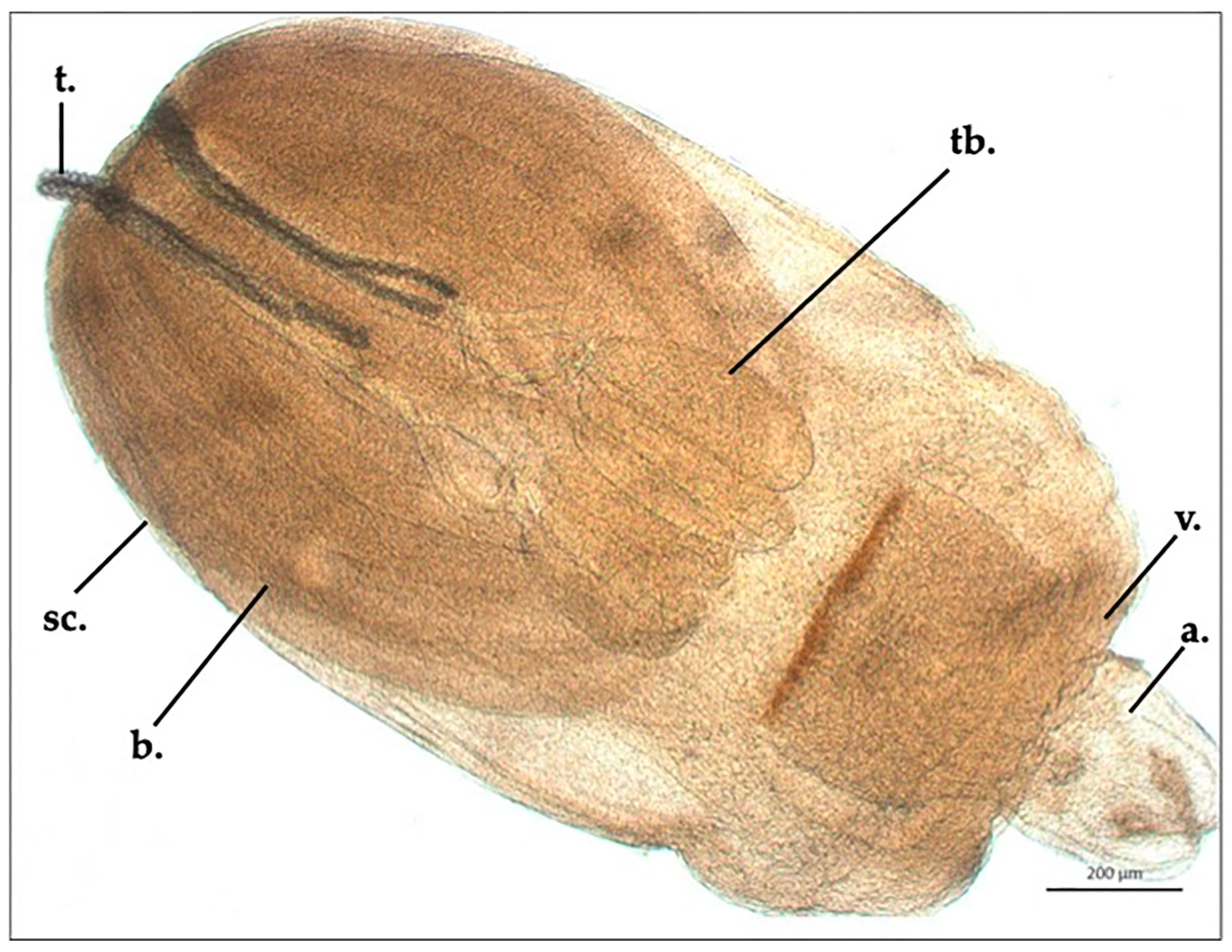
3.1. Nybelinia sp., Larvae (Trypanorhyncha: Tentaculariidae)

Nybelinia sp. plerocercoid appeared pyriform in shape; its ML and MW were 2324.6 ± 23.4 µm and 1227.6 ± 17.3 µm, respectively. The scolex’s ML was 1354.5 ± 6.4 µm, and two bothria were present (ML: 1298 ± 14.7 µm, MW: 567 ± 5.8 µm). The tentacular bulb’s length was 423 ± 8.2 µm. The appendix’s ML was 8118 ± 2.8 µm, and the velum’s ML was 488.5 ± 4.9 µm. Four cylindrical tentacles, in only one specimen partially everted, were present, of which everted parts were 544 to 586 µm long, and the tentacular widths were 28.6 µm (basal area), 30.9 µm (metabasal area) and 28.4 µm (apical area). Tentacles were covered with uncinate hooks disposed in oblique ringlet lines, which were 11.3 µm long and 8.4 µm wide (basal area), 14.8 µm long and 12.5 µm wide (metabasal area), and 13.5 µm long and 10.2 µm wide (apical area) (Figure 1).

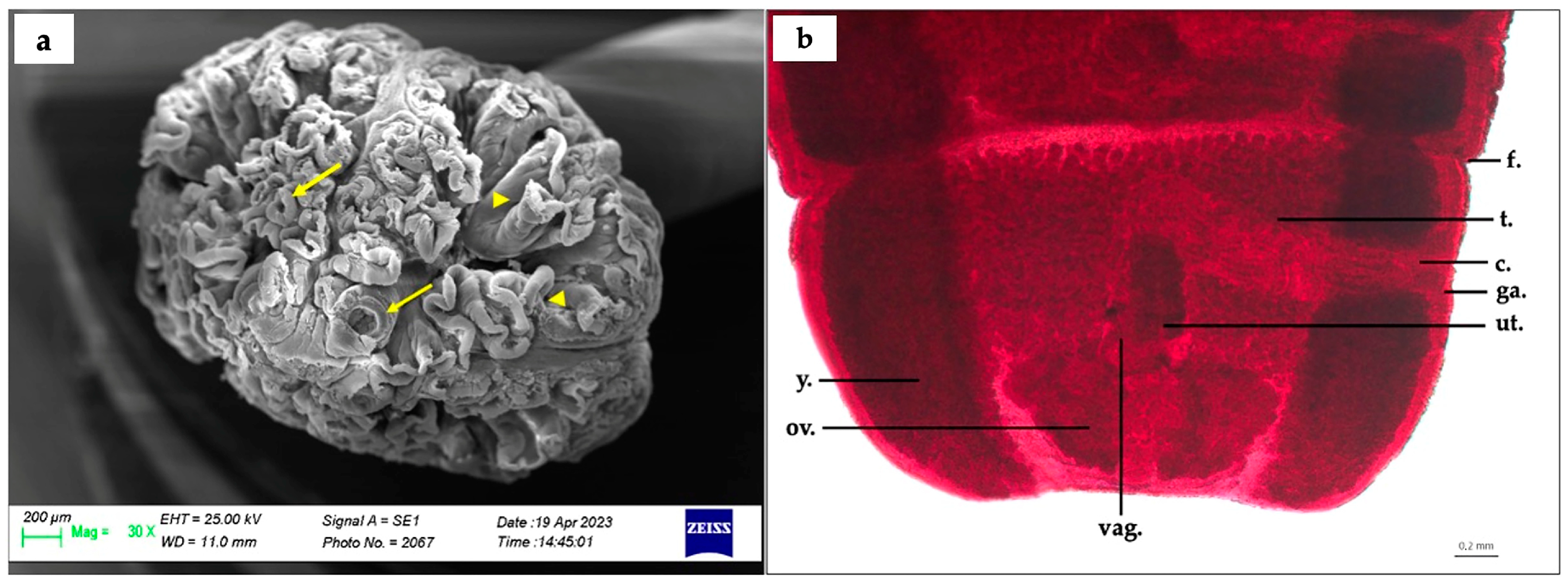
3.2. Phyllobothrium sinuosiceps (Phyllobothriidea: Phyllobothriidae) Adult Specimen

The scolex appeared spherical in shape (ML: 3130.7 ± 27.5 µm, MW: 2386.5 ± 20.5 µm), and presented with a series of folds, arranged in random and different positions. It was completely or partially surrounded by two to four folds. In general, these appeared as major folds, with two or three divisions; all the margins of major and minor folds appeared curled. Caudal folds of the scolex overlapped the anterior part of the strobila. Four small suckers, located at the top of the upper portion of the scolex and of variable size (MW: 304.5 ± 0.7 µm), were present. The strobila’s MW was 2046 ± 11.3 µm, and the cuticle appeared scaly for the entire strobila length; at the posterior end, the examined mature segments varied in shape, being generally wider than long (ML: 1639.8 ± 31.4 µm, MW: 3024 ± 7.1 µm). The excretory system of the scolex appeared simple in structure; the cirrus sac’s ML and MW were 1401.5 ± 0.7 µm and 397 ± 1.4 µm, respectively. Regarding the reproductive system, the testes were not counted, but appeared spherical in shape (MW: 49.7 ± 2.5 µm) and disposed in the middle of the mature segment, between the yolk glands, and associated with the vagina and cirrus sac. The vagina was characterized by thin walls and located in the middle of the uterus, directly connected to the cirrus sac. The uterus was localized above the ovary, in the middle region of the mature segment (ML: 791 ± 7.1 µm, MW: 489 ± 1.4 µm); the ovary was leaf-like in shape (ML: 1272.3 ± 4.3 µm, MW: 474.3 ± 3.5 µm) and set in the middle posterior part of the segment, between the yolk glands. The posterior flap of the evaluated segments did not appear clearly marked (Figure 2a,b).

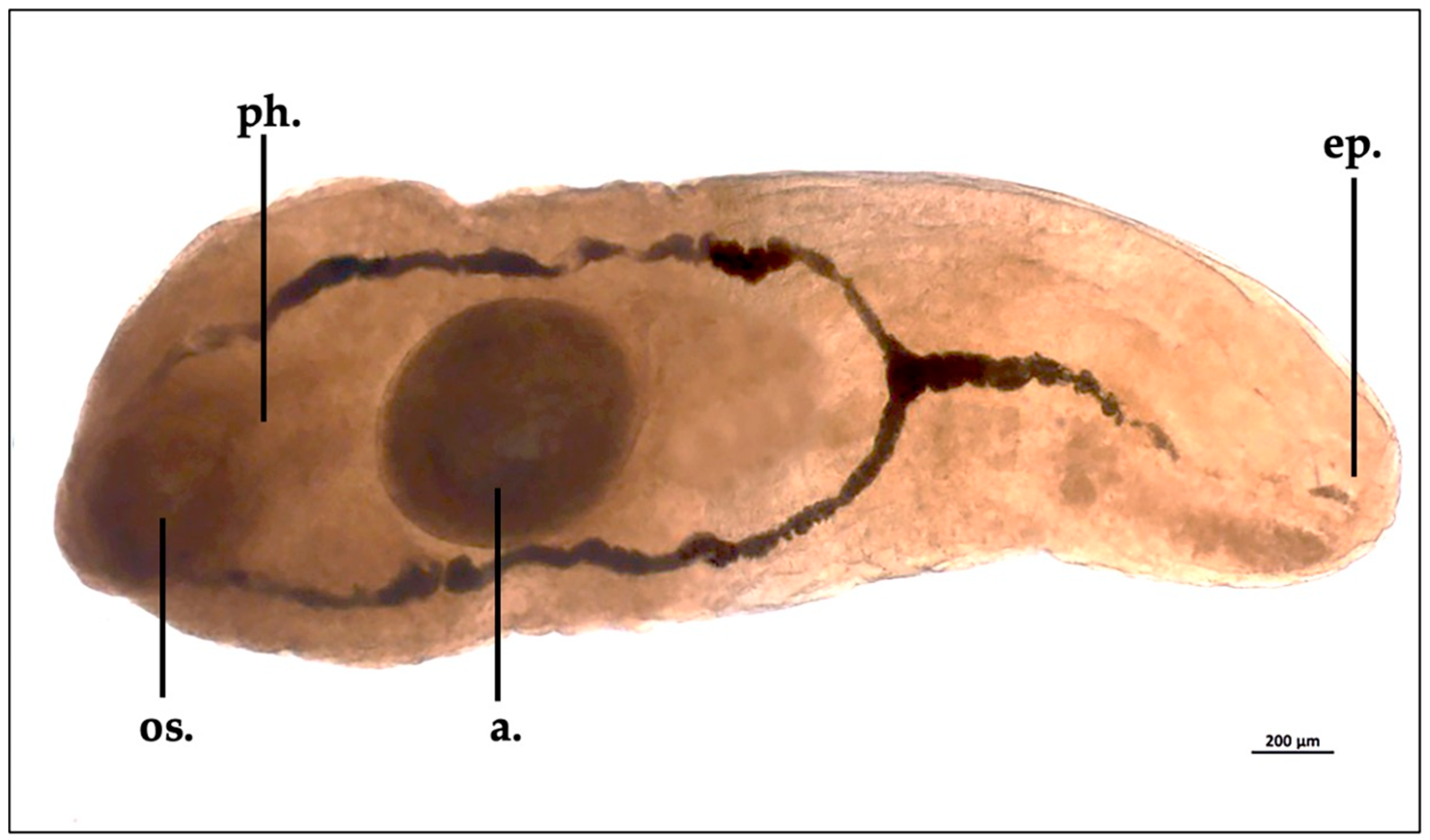
3.3. The trematode Otodistomum veliporum Creplin, 1837 Immature Specimen

Morphological features of the only specimen collected were a length of 3840 µm and a width of 1360 µm; the oral sucker width was 480 µm, and the acetabulum measured 748 µm in length. The yellow-whitish external cuticle appeared non-spinous. The Otodistomum veliporum specimen showed the pharynx closer to the oral sucker than the acetabulum (Figure 3).

4. Discussion

In this study, parasitological evaluation of H. griseus (caught off the Sicilian coasts) helminth fauna was carried out, describing three parasites in this area. Considering the H. griseus development stages described by Capapé et al. [5,6], biometric indices and gonadal development, the specimen examined in this study was considered juvenile. The parasite fauna found were partially in agreement with some previous findings.
Among them, Nybelinia sp. larvae have been reported in several elasmobranch species from the Mediterranean Sea [39]. Regarding the epidemiological indices of this species, low prevalence was reported by Pascual et al. [40,41] in the lumen and attached to the digestive tract or in the mantle cavity of the cephalopods Illex coindetii (Vérany, 1839) and Todaropsis eblanae (Ball, 1841), which are considered intermediate hosts in the Mediterranean Sea and Atlantic Ocean; these data were confirmed by Gestal [42]. In our study, we partially confirm this data, due to the empty stomach and spiral valve of the examined H. griseus. The Tentacularidae life cycle generally includes elasmobranchs as a definitive host, as reported by Mulas and colleagues [43], who carried out a survey of Hexanchus griseus and Scyliorhinus canicula sampled from the Central–Western Mediterranean Sea; these data were not confirmed by our results, due to the Nybelinia sp. plerocercoid findings. Nybelinia sp. larvae have been reported by Reinero et al. [39] in Lesser Spotted Dogfish (Scyliorhinus canicula) from the Central Tyrrhenian Sea with a prevalence of 24%; in that study, parasites were localized mainly in the spiral valve and stomach. However, in our study, Nybelinia sp. larvae were found only in the H. griseus stomach, confirming this tract as the elective localization of these parasites. According to Palm and Walter [44], who found three adult cestodes identified as belonging to the genus Nybelinia, H. griseus can be considered as a definitive host of these parasites. Instead, according to our results, we can speculate that this elasmobranch may be considered as an intermediate host of Nybelinia sp. Moreover, according to Hart’s [45] Pacific Ocean study, Tetrarhynchidea metacestodes can be found in H. griseus specimens, attached to the spiral valve wall, just below the pyloric region. Our results suggest that this finding can be considered as an erratic localization.
Phyllobothrium sinuosiceps, a tapeworm characterized by a complex life cycle, is considered a parasite that usually inhabits the spiral valve of various elasmobranchs. Thanks to its highly adaptive specialization to the unique environment of the shark digestive tract, it should also be considered from an ecological point of view [46]. Phyllobothrium sinuosiceps (as well as other cestodes) do not typically cause direct injuries to their hosts; however, a heavy parasite load may have indirect effects on host health. Indeed, possible lesions in the shark spiral valve due to a parasite may cause acute inflammation, with a consequent reduction in nutrient absorption, which is potentially dangerous for correct shark development [47]. In our case, the low parasite load collected, according to the general growth condition of the host, can exclude parasite infection as causative of death. Moreover, according to Caira et al. [46], the presence of P. sinuosiceps in sharks highlights the close ecological connections within marine ecosystems. As sharks have been considered predators in their habitat, feeding habits and interactions between these animals and their prey play a key role in the transmission and adaptation of their parasites [48]. To better understand the dynamics concerning these organisms, it is necessary to evaluate the food chain and the status of the ecosystem; unfortunately, in our case, the absence of food debris in the gastrointestinal tract meant this was not feasible. Adult Phyllobothrium sp. specimens have been found in Carcharhinus limbatus (Müller and Henle, 1839) and Carcharhinus obscurus (Lesueur, 1818) by Pramanik and colleagues [49]; among other shark species, Sphyrna zygaena (Linnaeus, 1758) [50], Galeocerdo cuvier (Péron and Lesueur, 1822) [51] and Mustelus mustelus (Linnaeus, 1758) [52] sampled from different areas, including the Mediterranean Sea, were positive for Phyllobothrium sp. found in the stomach and spiral valve. Our results confirm that this tapeworm is strictly related to many elasmobranch species from the Mediterranean Sea, as well as other basins worldwide. In our study, six specimens of P. sinuosiceps were collected. Morphological identification was performed following the morphological description reported by Williams [37] in H. griseus. This author described P. sinuosiceps for the first time in H. griseus caught in the Celtic Sea. According to our results, we can confirm the same morphological features in H. griseus from different regions. Henderson [31] reported P. sinuosiceps in H. griseus from the west of Ireland, localized in the stomach and spiral valve, but also encysted in the stomach wall; as previously discussed regarding other parasites, such as Nybelinia sp., and considering the habit of P. sinuosiceps to infect the spiral valve, we can consider the stomach as an erratic localization for this parasite. Phyllobothrium sinuosiceps infection intensity can be variable in H. griseus. Studies have shown that the intensity of this tapeworm can be relatively high in some populations of H. griseus [53]. In our case, the number of adult cestodes found may have been underestimated due to the possible emission of parasites before the host was found.
Based on morphometric features and the oral sucker and acetabulum size, the Digenean trematode reported was identified as belonging to the genus Otodistomum (Stafford, 1904, Family Azygiidae), confirming H. griseus as a definitive host, as previously reported by Kay [32]. Otodistomum veliporum has been described from sub-tropical, temperate and polar regions [54,55], including the Atlantic Ocean and the Central Mediterranean Sea, where Gibson and Bray [33] and Gibson [56] reported O. veliporum as a parasite of many bony fish and elasmobranchs, such as Squalus acanthias (Linnaeus, 1758), Raja radiata (Donovan, 1808), Centroscyllium fabricii (Reinhardt, 1825) and Pristiophorus cirratus (Latham, 1794). Our results allowed inclusion of H. griseus among the possible definitive hosts of O. veliporum inhabiting the Mediterranean Sea. Among other species belonging to the genus Otodistomum, up to one hundred specimens have been found in H. griseus (caught on the Pacific Ocean) stomachs’ pyloric region [32]. According to our results, the only O. veliporum specimen found suggests a low density of populations of intermediate hosts in the diet of H. griseus present in the Strait of Messina. Recently, Sperone and Milazzo [57] described O. veliporum in kitefin shark (Dalatias licha, Bonnaterre, 1788) from the Southern Tyrrhenian Sea (Central Mediterranean Sea). In this case, the authors reported one O. veliporum specimen, attached to the mucosa of a D. licha spiracle. This unusual localization, compared to our results, could be attributable to a postmortem accidental parasite movement from the gastrointestinal tract to the spiracle. Another Otodistomum sp. single finding was reported by Espínola and Novelo [58] in southern lanternshark (Etmopterus granulosus, Günther, 1880) collected from the southeastern Pacific Ocean. Similarly, in our study, as previously discussed, the single specimen found may have be related to the low availability of intermediate hosts.

5. Conclusions

This study reports a new parasitological evaluation in H. griseus from the Central Mediterranean Sea. The difference between the Strait of Messina and other study areas, where these parasites have already been reported, lies mainly in the conformation of teleost and cephalopods varieties as intermediate hosts. Moreover, the movement of these large elasmobranchs between the Ionian and Tyrrhenian Seas, including their daily vertical migration, adds many variables to this type of survey, making it difficult to evaluate possible host–parasite interactions.
Therefore, the results reported in this study confirm the bluntnose sixgill shark as a definitive host of P. sinuosiceps and O. veliporum, and provide new data about this species as an intermediate host of Nybelinia sp. Finally, we highlight the need to carry out further studies of the epidemiology of these parasite species in the central Mediterranean Sea.

Author Contributions

Conceptualization, G.G. and F.M.; methodology, G.D.B. and F.C.; validation, G.G. and F.M.; formal analysis, F.C. and C.I.; investigation, G.D.B., F.C. and C.I.; data curation, G.D.B. and C.I.; writing—original draft preparation, G.D.B. and F.C.; writing—review and editing, G.D.B., F.C. and C.I.; supervision, G.G. and F.M. All authors have read and agreed to the published version of the manuscript.

Funding

This research received no external funding.

Institutional Review Board Statement

No procedures on live animals were carried out; thus, the requirement for ethical approval was not applicable.

Informed Consent Statement

Not applicable.

Data Availability Statement

Not applicable.

Conflicts of Interest

The authors declare no conflict of interest.

References

  1. Coll, M.; Piroddi, C.; Steenbeek, J.; Kaschner, K.; Ben Rais Lasram, F.; Aguzzi, J.; Ballesteros, E.; Bianchi, C.N.; Corbera, J.; Dailianis, T.; et al. The biodiversity of the Mediterranean Sea: Estimates, patterns, and threats. PLoS ONE 2010, 5, e11842. [Google Scholar] [CrossRef] [PubMed] [Green Version]
  2. Nieto, A.; Ralph, G.M.; Comeros-Raynal, M.T.; Heessen, H.J.L.; Rijnsdorp, A.D. European Red List of Marine Fishes; Publications Office of the European Union: Luxembourg, 2015. [Google Scholar]
  3. Fricke, R.; Eschmeyer, W.N.; Van der Laan, R. Eschmeyer’s Catalog of Fishes: Genera, Species, References; California Academy of Sciences: San Francisco, CA, USA, 2022. [Google Scholar]
  4. Barrull, J.; Mate, I.; Bueno, M. Observaciones de tiburones (Chondrichthyes, Euselachii) en aguas de Cataluña, con algunos aspectos generales de su ecología. Sci. Gerund. 1999, 24, 127–151. [Google Scholar]
  5. Capapé, C.; Hemida, F.; Guélorget, O.; Barrull, J.; Mate, I.; Ben Souissi, J.; Bradaï, M.N. Reproductive biology of the bluntnose sixgill shark Hexanchus griseus (Bonnaterre, 1788) (Chondrichthyes: Hexanchidae) from the Mediterranean Sea: A review. Acta Adriat. 2004, 45, 95–106. [Google Scholar]
  6. FAO. Species Photographic Plates. Mediterranean Sharks, by Monica Barone, Fabrizio Serena and Mark Dimech. Rome, Italy. 2018. Available online: https://www.fao.org/publications/card/en/c/I8615EN/ (accessed on 26 May 2023).
  7. Vella, N.; Vella, A. Population genetics of the deep-sea bluntnose sixgill shark, Hexanchus griseus, revealing spatial genetic heterogeneity. Mar. Genom. 2017, 36, 25–32. [Google Scholar] [CrossRef]
  8. Farrugio, H.; Soldo, A. Sicily Channel/Tunisian Plateau: Status and Conservation of Fisheries. UNEP-MAP-RAC/SPA; Cebrian, D., Requena, S., Eds.; RAC/SPA: Tunis, Tunisia, 2015; p. 76. [Google Scholar]
  9. Mundy, B.C. Checklist of the fishes of the Hawaiian Archipelago. Bish. Mus. Bull. Zool. 2005, 6, 1–704. [Google Scholar]
  10. Last, P.R.; Stevens, J.D. Sharks and Rays of Australia. J. Mar. Biol. Assoc. UK 1994, 74, 744. [Google Scholar] [CrossRef]
  11. Ebert, D.A. Biological Aspects of the Sixgill Shark, Hexanchus griseus. Copeia 1986, 1, 131–135. [Google Scholar] [CrossRef]
  12. Barrull, J.; Mate, I. Biología de la cañabota Hexanchus griseus (Bonnaterre, 1788) en el Mar Mediterráneo. Bol. Esp. Elasmobranquios 2000, 3, 13–20. [Google Scholar]
  13. Soldo, A.; Bariche, M.; Buscher, E.; Cook, S.F.; Compagno, L.J.V. Hexanchus griseus. In The IUCN Red List of Threatened Species; IUCN: Geneva, Switzerland, 2016. [Google Scholar]
  14. Nuez, I.; Giovos, I.; Tiralongo, F.; Penadés-Suay, J.; Ćetković, I.; Di Lorenzo, M.; Kleitou, P.; Bakiu, R.; Bradai, M.; Al Mabruk, S.; et al. Assessing the current status of Hexanchus griseus in the Mediterranean Sea using local ecological knowledge. Mar. Policy 2023, 147, 105378. [Google Scholar] [CrossRef]
  15. Tserpes, G.; Maravelias, C.D.; Pantazi, M.; Peristeraki, P. Distribution of relatively rare demersal elasmobranchs in the eastern Mediterranean. Estuar. Coast. Shelf Sci. 2013, 117, 48–53. [Google Scholar] [CrossRef]
  16. Wood, C.L.; Byers, J.E.; Cottingham, K.L.; Altman, I.; Donahue, M.J.; Blakeslee, A.M.H. Parasites alter community structure. Proc. Natl. Acad. Sci. USA 2007, 104, 9335–9339. [Google Scholar] [CrossRef] [PubMed]
  17. Amundsen, P.A.; Lafferty, K.D.; Knudsen, R.; Primicerio, R.; Klemetsen, A.; Kuris, A.M. Food web topology and parasites in a subarctic lake. J. Anim. Ecol. 2009, 78, 563–572. [Google Scholar] [CrossRef] [PubMed]
  18. Sato, T.; Egusa, T.; Fukushima, K.; Oda, T.; Ohte, N.; Tokuchi, N.; Watanabe, K.; Kanaiwa, M.; Murakami, I.; Lafferty, K.D. Nematomorph parasites indirectly alter the food web and ecosystem function of streams through behavioural manipulation of their cricket hosts. Ecol. Lett. 2012, 15, 786–793. [Google Scholar] [CrossRef] [PubMed]
  19. Parker, G.A.; Ball, M.A.; Chubb, J.C. To grow or not to grow? Intermediate and paratenic hosts as helminth life cycle strategies. J. Theor. Biol. 2009, 258, 135–147. [Google Scholar] [CrossRef] [PubMed] [Green Version]
  20. Benesh, D.; Chubb, J.; Parker, G. The trophic transmission vacuum and the evolution of complex helminth life cycles. Proc. Biol. Sci. R. Soc. 2014, 281, 20141462. [Google Scholar] [CrossRef]
  21. Waeschenbach, A.; Webster, B.L.; Littlewood, D.T. Adding resolution to ordinal level relationships of tapeworms (Platyhelminthes: Cestoda) with large fragments of mtD-NA. Mol. Phyl. Evol. 2012, 63, 834–847. [Google Scholar] [CrossRef]
  22. Littlewood, D.T.J.; Bray, R.A.; Waeschenbach, A. Phylogenetic patterns of diversity in the cestodes and trematodes. In Parasite Diversity and Diversification: Evolutionary Ecology Meets Phylogenetics, 1st ed.; Morand, S., Krasnov, B.R., Littlewood, D.T.J., Eds.; Cambridge University Press: Cambridge, UK, 2015; pp. 304–319. [Google Scholar] [CrossRef]
  23. Caira, J.N.; Jensen, K.; Waeschenbach, A.; Olson, P.D.; Littlewood, D.T.J. Orders out of chaos—Molecular phylogenetics reveals the complexity of shark and stingray tapeworm relationships. Int. J. Parasitol. 2014, 44, 55–73. [Google Scholar] [CrossRef] [Green Version]
  24. Caira, J.N.; Jensen, K. A digest of elasmobranch tapeworms. J. Parasitol. 2014, 100, 373–391. [Google Scholar] [CrossRef]
  25. Carvajal, J.G. Grillotia dollfusi sp. n. (Cestoda: Trypanorhyncha) from the skate, Raja chilensis, from Chile, and a note on G. heptanchi. J. Parasitol. 1971, 57, 1269–1271. [Google Scholar] [CrossRef]
  26. Carvajal, J.G. Records of cestodes from Chilean sharks. J. Parasitol. 1974, 60, 29–34. [Google Scholar] [CrossRef]
  27. Beveridge, I.; Campbell, R.A. A new species of Grillotia Guiart, 1927 (Cestoda: Trypanorhyncha) with redescriptions of congeners and new synonyms. Syst. Parasitol. 2013, 85, 99–116. [Google Scholar] [CrossRef] [PubMed]
  28. Holzer, A.S.; Bartošová-Sojková, P.; Born-Torrijos, A.; Lövy, A.; Hartigan, A.; Fiala, I. The joint evolution of the Myxozoa and their alternate hosts: A cnidarian recipe for success and vast biodiversity. Mol. Ecol. 2018, 27, 1651–1666. [Google Scholar] [CrossRef] [PubMed]
  29. Lisnerová, M.; Fiala, I.; Cantatore, D.; Irigoitia, M.; Timi, J.; Pecková, H.; Bartošová-Sojková, P.; Sandoval, C.M.; Luer, C.; Morris, J.; et al. Mechanisms and Drivers for the Establishment of Life Cycle Complexity in Myxozoan Parasites. Biology 2020, 1, 10. [Google Scholar] [CrossRef] [Green Version]
  30. Lisnerová, M.; Martinek, I.N.; Alama-Bermejo, G.; Bouberlová, K.; Schaeffner, B.C.; Nkabi, N.; Holzer, A.S.; Bartošová-Sojková, P. An ancient alliance: Matching evolutionary patterns of cartilaginous fishes (Elasmobranchii) and chloromyxid parasites (Myxozoa). Infect. Genet. Evol. 2022, 103, 105346. [Google Scholar] [CrossRef]
  31. Henderson, A.C.; Flannery, K.; Dunne, J. Biological Observations on Shark Species Taken in Commercial Fisheries to the West of Ireland. Biol. Environ. 2003, 103B, 1–7. [Google Scholar] [CrossRef]
  32. Kay, M.W. Otodistomum plicatum n. sp. (Trematoda, Digenea) from Hexanchus griseus (Bonnaterre). Ohio J. Sci. 1947, 47, 79–83. [Google Scholar]
  33. Gibson, D.I.; Bray, R.A. The Azygiidae, Hirudineliidae, Ptychogonimidae, Sclerodistomidae and Syncoeliidae of fishes from the North-East Atlantic. Bull. Br. Mus. 1977, 32, 167–245. [Google Scholar]
  34. Knoff, M.; de São Clemente, S.C.; Pinto, R.M.; Gomes, D.C. Digenea and acanthocephala of elasmobranch fishes from the southern coast of Brazil. Mem. Inst. Oswaldo Cruz. 2001, 96, 1095–1101. [Google Scholar] [CrossRef] [Green Version]
  35. Oldewage, W.H.; Smale, M.J. Occurrence of piscine parasitic copepods (Crustacea) on sharks taken mainly off Cape Recife, South Africa. S. Afr. J. Mar. Sci. 1993, 13, 309–312. [Google Scholar] [CrossRef] [Green Version]
  36. Cable, R.M. An Illustrated Laboratory Manual of Parasitology; Burgess: Minniapolis, MN, USA, 1961. [Google Scholar]
  37. Williams, H.H. The anatomy of Phyllobothrium sinuosiceps sp.nov. (Cestoda: Tetraphyllidea) from Hexanchus griseus (Gmelin) the six gilled shark. Parasitology 1959, 49, 54–69. [Google Scholar] [CrossRef]
  38. Beveridge, I.; Bray, R.A.; Cribb, T.H.; Justine, J.L. Diversity of trypanorhynch metacestodes in teleost fishes from coral reefs off eastern Australia and New Caledonia. Parasite 2014, 21, 60. [Google Scholar] [CrossRef] [PubMed] [Green Version]
  39. Reinero, F.R.; Milazzo, C.; Minervino, M.; Marchio, C.; Filice, M.; Bevacqua, L.; Giglio, G.; Leonetti, F.L.; Micarelli, P.; Tripepi, S.; et al. Parasitic Load, Hematological Parameters, and Trace Elements Accumulation in the Lesser Spotted Dogfish Scyliorhinus canicula from the Central Tyrrhenian Sea. Biology 2022, 11, 663. [Google Scholar] [CrossRef] [PubMed]
  40. Pascual, S.; González, A.F.; Arias, C.; Guerra, A. Biotic relationships of Illex coindetii and Todaropsis eblanae (Cephalopoda, Ommastrephidae) in the Northeastern Atlantic: Evidence from parasites. Sarsia 1996, 81, 265–274. [Google Scholar] [CrossRef]
  41. Pascual, S.; Abollo, E.; Mladineo, I.; Gestal, C. Metazoa and Related Diseases. In Handbook of Pathogens and Diseases in Cephalopods; Gestal, C., Pascual, S., Guerra, Á., Fiorito, G., Vieites, J.M., Eds.; Springer International Publishing: Cham, Switzerland, 2019; Volume 1, pp. 169–179. [Google Scholar]
  42. Gestal, C.; Abollo, E.; Arias, C.; Pascual, S.S.E.M. study of the plerocercoid larval Phyllobothrium sp. (Tetraphyllidea, Phyllobothriidae) and Nybelinia lingualis (Trypanothyncha, Tentaculariidae), cestode parasite in Octopus vulgaris (Mollusca, Cephalopoda) off Vigo estuary. Iberus 1998, 16, 125–132. [Google Scholar]
  43. Mulas, A.; Bellodi, A.; Cau, A.; Gastoni, A.; Locci, I.; Follesa, M.C. Trophic interactions among Chondrichthyans in the central-western Mediterranean Sea. Biol. Mar. Mediterr. 2011, 18, 81–84. [Google Scholar]
  44. Palm, H.W.; Walter, T. Tentaculariid cestodes (Trypanorhyncha) from the Muséum national d’Histoire naturelle, Paris. Zoosystema 2000, 22, 641–666. [Google Scholar]
  45. Hart, J.F. Cestoda from Fishes of Puget Sound. II. Tetrarhynchoidea. Trans. Am. Microsc. Soc. 1936, 55, 369. [Google Scholar] [CrossRef]
  46. Caira, J.N.; Healy, C.J. Elasmobranchs as hosts of metazoan parasites. In Biology of Sharks and Their Relatives, 2nd ed; Carrier, J.C., Musick, J.A., Heithaus, M.R., Eds.; CRC Press: Boca Raton, FL, USA, 2004; Volume 1, pp. 523–551. [Google Scholar]
  47. Dick, T.A.; Chambers, C.; Isinguzo, I. Cestoidea (Phylum Platyhelminthes). In Fish Diseases and Disorders: Protozoan and Metazoan Infections, 2nd ed; Woo, P.T.K., Ed.; CAB international: London, UK, 2005; Volume 1, pp. 391–416. [Google Scholar]
  48. Rasmussen, T.; Randhawa, H. Host Diet Influences Parasite Diversity: A Case Study Looking at Tapeworm Diversity among Sharks. Mar. Ecol. Prog. Ser. 2018, 605, 1–16. [Google Scholar] [CrossRef] [Green Version]
  49. Pramanik, P.B.; Manna, B. Two new species of the genus Phyllobothrium van Beneden, 1850 from the cartilaginous fish Carcharhinus bleekeri Muller & Henle, 1841 at Digha coastal waters, Bay of Bengal, India. J. Nat. Hist. 2009, 5, 64–72. [Google Scholar]
  50. Sene, A.; Cheikh, B.; Bernard, M. Ultrastructure of spermiogenesis of Phyllobothrium lactuca (Cestoda, Tetraphyllidea, Phyllobothriidae). Folia Parasitol. 1999, 46, 191–198. [Google Scholar]
  51. Dicken, M.L.; Hussey, N.E.; Christiansen, H.M.; Smale, M.J.; Nkabi, N.; Cliff, G.; Wintner, S.P. Diet and Trophic Ecology of the Tiger Shark (Galeocerdo Cuvier) from South African Waters. PLoS ONE 2017, 12, e0177897. [Google Scholar] [CrossRef] [PubMed]
  52. Tedesco, P.; Gustinelli, A.; Caffara, M.; Marsella, A.; Toffan, A.; Fioravanti, M.L. Morphological and Molecular Characterization of Two Gastrointestinal Parasites in Mustelus Mustelus (Linnaeus, 1758) from Adriatic Sea. Eur. Zool. J. 2020, 87, 616–623. [Google Scholar] [CrossRef]
  53. Williams, H.H. The Taxonomy, Ecology and Host-Specificity of Some Phyllobothriidae (Cestoda: Tetraphyllidea), a Critical Revision of Phyllobothrium Beneden, 1849 and Comments on Some Allied Genera. Phil. Trans. R. Soc. Lond. B 1968, 253, 231–307. [Google Scholar] [CrossRef]
  54. Threlfall, W.; Carvajal, G.L. Otodistomum cestoides (van Beneden, 1871) from two species of skates taken in Chilean waters. Mem. Inst. Oswaldo Cruz. 1986, 81, 341–342. [Google Scholar] [CrossRef] [Green Version]
  55. Zdzitowiecki, K.; Pisano, E. New records of Digenea infection elasmobranch and teleost fish off Heard Island (Kerguelen, sub-region, sub-Antarctic). Arch. Fish. Mar. Res. 1996, 43, 265–272. [Google Scholar]
  56. Gibson, D.I.; Margolis, L. Trematoda. In Guide to the Parasites of Fishes of Canada; Margolis, L., Ed.; Department of Fisheries and Oceans: Ottawa, ON, Canada, 1996; ISBN 9780660164038. [Google Scholar]
  57. Sperone, E.; Milazzo, C. On the Occurrence of the Digenean Otodistomum Veliporum in the Spiracle of the Kitefin Shark Dalatias licha. Acta Adriat. 2018, 59, 137–140. [Google Scholar] [CrossRef] [Green Version]
  58. Espínola-Novelo, J.F.; Escribano, R.; Oliva, M.E. Metazoan Parasite Communities of Two Deep-Sea Elasmobranchs: The Southern Lanternshark, Etmopterus Granulosus, and the Largenose Catshark, Apristurus Nasutus, in the Southeastern Pacific Ocean. Parasite 2018, 25, 53. [Google Scholar] [CrossRef] [Green Version]
Figure 1. Nybelinia sp. (Poche, 1926) larvae collected from Hexanchus griseus stomach after glycerine diaphanization (t., tentacle; sc., scolex; b., bothria; tb., tentacular bulbs; v., velum; a., appendix).
Animals 13 02405 g001
Figure 2. Phyllobothrium sinuosiceps (Williams, 1959) collected from H. griseus spiral valve: (a) P. sinuosiceps scolex, scanning electric microscopy observation, apical sucker (arrow); curled margin of the scolex folds (arrowhead) and (b) P. sinuosiceps terminal segment after Semichon’s carmine staining (y., yolk sack; ov., ovary; vag., vagina; ut., uterus; f., posterior flap of segment; ga., genital atrium; c., cirrus; t., testis).
Animals 13 02405 g002
Figure 3. Otodistomum veliporum collected from H. griseus spiral valve (os., oral sucker; ph., pharynx; a., acetabulum; ep., excretory pore).
Animals 13 02405 g003
Disclaimer/Publisher’s Note: The statements, opinions and data contained in all publications are solely those of the individual author(s) and contributor(s) and not of MDPI and/or the editor(s). MDPI and/or the editor(s) disclaim responsibility for any injury to people or property resulting from any ideas, methods, instructions or products referred to in the content.

