
Serological Examinations of Significant Viral Infections in Domestic Donkeys at the Special Nature Reserve “Zasavica”, Serbia
 , , , and
Abstract
:Simple Summary
Abstract
1. Introduction
1.1. Equine Infectious Anemia (EIA)
1.2. African Horse Sickness (AHS)
1.3. Equine Herpesvirus Infection (Equine Rhinopneumonitis)
1.4. Equine Influenza (EI)
1.5. Equine Viral Arteritis (EVA)
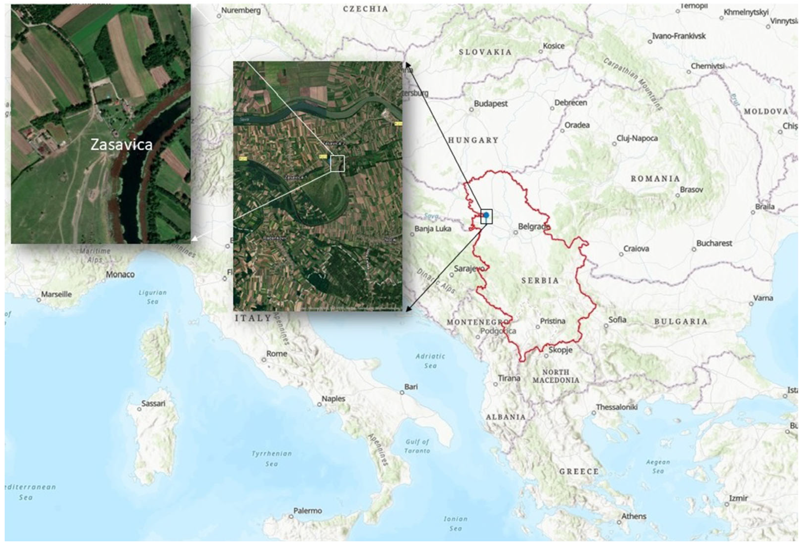
2. Materials and Methods
3. Results
4. Discussion
5. Conclusions
Author Contributions
Funding
Institutional Review Board Statement
Informed Consent Statement
Data Availability Statement
Acknowledgments
Conflicts of Interest
References
- Rossel, S.; Marshall, F.; Peters, J.; Pilgram, T.; Adams, M.D.; O’Connor, D. Domestication of the donkey: Timing, processes, and indicators. Proc. Natl. Acad. Sci. USA 2008, 105, 3715–3720. [Google Scholar] [CrossRef] [PubMed] [Green Version]
- Burden, F.; Thiemann, A. Donkeys Are Different. J. Equine Vet. Sci. 2015, 35, 376–382. [Google Scholar] [CrossRef]
- Maggs, H.C.; Ainslie, A.; Bennett, R.M. Donkey Ownership Provides a Range of Income Benefits to the Livelihoods of Rural Households in Northern Ghana. Animals 2021, 11, 3154. [Google Scholar] [CrossRef] [PubMed]
- Živkov Baloš, M.; Pelić, D.L.; Jakšić, S.; Lazić, S. Donkey Milk: An Overview of its Chemical Composition and Main Nutritional Properties or Human Health Benefit Properties. J. Equine Vet. Sci. 2023, 121, 104225. [Google Scholar] [CrossRef] [PubMed]
- Norris, S.L.; Little, H.A.; Ryding, J.; Raw, Z. Global donkey and mule populations: Figures and trends. PLoS ONE 2021, 16, e0247830. [Google Scholar] [CrossRef]
- FAOSTAT. Food and Agriculture Organization of the United Nations. Available online: https://www.fao.org/faostat/en/#data/QCL (accessed on 9 February 2023).
- Statistical Office of the Republic of Serbia. Statistical Yearbook of the Republic of Serbia 2022. Available online: https://publikacije.stat.gov.rs/G2022/PdfE/G20222055.pdf (accessed on 15 June 2023).
- Urosevic, M.; Drobnjak, D.; Ivanov, S. Autohtone Rase Magaraca u Srbiji [Indigenous Breeds of Donkeys in Serbia]. In Proceedings of the Protection of Agrobiodiversity and Preservation of Indigenous Breeds of Domestic Animals, Dimitrovgrad, Serbia, 28–30 June 2019. [Google Scholar]
- World Organization for Animal Health (WOAH). Available online: https://www.woah.org/en/what-we-do/animal-health-and-welfare/animal-diseases/?_tax_animal=terrestrials%2Cequine (accessed on 21 January 2023).
- Trailović, D.; Trailović, R.; Urošević, M.; Trailović, I. Uzgoj, Nega I Bolesti Magaraca [Breeding, Care and Diseases of Donkeys]; Centar za Očuvanje Autohtonih Rasa—COAR, Zemun: Beograd, Serbia, 2021; ISBN 978-86-920597-4-2. [Google Scholar]
- Barrandeguy, M.E.; Carossino, M. Infectious Diseases in Donkeys and Mules: An Overview and Update. J. Equine Vet. Sci. 2018, 65, 98–105. [Google Scholar] [CrossRef]
- Sellon, D.C. Equine Infectious Anemia. Vet. Clin. N. Am. Equine Pract. 1993, 9, 321–336. [Google Scholar] [CrossRef]
- Foil, L.D.; Issel, C.J. Transmission of Retroviruses by Arthropods. Annu. Rev. Entomol. 1991, 36, 355–381. [Google Scholar] [CrossRef]
- Mealey, R. Equine Infectious Anemia. In Equine Infectious Diseases; Long, M., Sellon, D., Eds.; Saunders: St. Louis, MO, USA, 2014; p. 232.e9. [Google Scholar]
- Kemen, M.J.; Coggins, L. Equine infectious anemia: Transmission from infected mares to foals. J. Am. Vet. Med. Assoc. 1972, 161, 496–499. [Google Scholar]
- World Organization for Animal Health (WOAH). Manual of Diagnostic Tests and Vaccines for Terrestrial Animals, Chapter: 3.6.1 African Horse Sickness (Infection with African Horse Sickness Virus). Available online: https://www.woah.org/fileadmin/Home/eng/Health_standards/tahm/3.06.01_AHS.pdf (accessed on 6 March 2023).
- Henning, M. African Horse Sickness, Perdesiekte, Pestis Equorum. In Animal Diseases in South Africa; Central News Agency: Sandton, South Africa, 1956; pp. 785–808. [Google Scholar]
- Hamblin, C.; Salt, J.S.; Mellor, P.S.; Graham, S.D.; Smith, P.R.; Wohlsein, P. Donkeys as reservoirs of African horse sickness virus. In African Horse Sickness; Springer: Vienna, Austria, 1998; pp. 37–47. [Google Scholar] [CrossRef]
- Coetzer, J.; Guthrie, A. African Horsesickness. In Infectious Diseases of Livestock; Coetzer, J., Tustin, R., Eds.; Oxford University Press: Cape Town, South Africa, 2005; pp. 1231–1246. [Google Scholar]
- Molini, U.; Zaccaria, G.; Kandiwa, E.; Mushonga, B.; Khaiseb, S.; Ntahonshikira, C.; Chiwome, B.; Baines, I.; Madzingira, O.; Savini, G.; et al. Seroprevalence of African horse sickness in selected donkey populations in Namibia. Vet. World 2020, 13, 1005–1009. [Google Scholar] [CrossRef]
- Lubroth, J. African Horse Sickness and the Epizootic in Spain 1987. Equine Pract. 1988, 10, 26–33. [Google Scholar]
- Castillo-Olivares, J. African horse sickness in Thailand: Challenges of controlling an outbreak by vaccination. Equine Vet. J. 2021, 53, 9–14. [Google Scholar] [CrossRef] [PubMed]
- Gordon, S.J.G.; Bolwell, C.; Rogers, C.W.; Musuka, G.; Kelly, P.; Guthrie, A.; Mellor, P.S.; Hamblin, C. The sero-prevalence and sero-incidence of African horse sickness and equine encephalosis in selected horse and donkey populations in Zimbabwe. Onderstepoort J. Vet. Res. 2017, 84, a1445. [Google Scholar] [CrossRef] [PubMed]
- Ndebé, M.M.F.; Mouiche, M.M.M.; Moffo, F.; Poueme, R.N.S.; Awah-Ndukum, J. Seroprevalence and Risk Factors of African Horse Sickness in Three Agroecological Zones of Cameroon. Vet. Med. Int. 2022, 2022, 2457772. [Google Scholar] [CrossRef]
- Câmara, R.J.F.; Bueno, B.L.; Resende, C.F.; Balasuriya, U.B.R.; Sakamoto, S.M.; dos Reis, J.K.P. Viral Diseases that Affect Donkeys and Mules. Animals 2020, 10, 2203. [Google Scholar] [CrossRef]
- International Committee on Taxonomy of Viruses. Available online: https://ictv.global/taxonomy (accessed on 20 August 2022).
- Crabb, B.S.; Studdert, M.J. Equine Herpesviruses 4 (Equine Rhinopneumonitis Virus) and 1 (Equine Abortion Virus). Adv. Virus Res. 1995, 45, 153–190. [Google Scholar] [CrossRef]
- Allen, G. Respiratory Infections by Equine Herpesvirus Types 1 and 4. In Equine Respiratory Diseases; Lekeux, P., Ed.; International Veterinary Information Service: Ithaca, NY, USA, 2002. [Google Scholar]
- Laval, K.; Poelaert, K.C.K.; Van Cleemput, J.; Zhao, J.; Vandekerckhove, A.P.; Gryspeerdt, A.C.; Garré, B.; van der Meulen, K.; Baghi, H.B.; Dubale, H.N.; et al. The Pathogenesis and Immune Evasive Mechanisms of Equine Herpesvirus Type. Front. Microbiol. 2021, 12, 662686. [Google Scholar] [CrossRef]
- World Organization for Animal Health (WOAH). Manual of Diagnostic Tests and Vaccines for Terrestrial Animals, Chapter: 3.6.9 Equine Rhinopneumonitis (Infection with Equid Herpesvirus-1 and -4). Available online: https://www.woah.org/fileadmin/Home/eng/Health_standards/tahm/3.06.09_EQUINE_RHINO.pdf (accessed on 6 March 2023).
- Laval, K.; Favoreel, H.W.; Nauwynck, H.J. Equine herpesvirus type 1 replication is delayed in CD172a+ monocytic cells and controlled by histone deacetylases. J. Gen. Virol. 2015, 96, 118–130. [Google Scholar] [CrossRef]
- Oladunni, F.S.; Horohov, D.W.; Chambers, T.M. EHV-1: A Constant Threat to the Horse Industry. Front. Microbiol. 2019, 10, 2668. [Google Scholar] [CrossRef] [Green Version]
- Ataseven, V.S.; Arslan, H.H. Equine infectious anemia in mules, donkeys, and horses: Epidemiologic studies in the different geographic regions of Turkey. J. Equine Vet. Sci. 2005, 25, 439–441. [Google Scholar] [CrossRef]
- Yildirim, Y.; Yilmaz, V.; Kirmizigul, A.H. Equine herpes virus type 1 (EHV-1) and 4 (EHV-4) infections in horses and donkeys in northeastern Turkey. Iran. J. Vet. Res. 2015, 16, 341–344. [Google Scholar] [CrossRef] [PubMed]
- Getachew, M.; Alemayehu, F.; Chala, C.; Amare, B.; Kassa, D.; Burden, F.; Wernery, R.; Wernery, U. A Cross-Sectional Sero-Survey of Some Infectious Diseases of Working Equids in Central Ethiopia. J. Vet. Med. Anim. Health 2014, 6, 231–238. [Google Scholar]
- Mekonnen, A.; Eshetu, A.; Gizaw, D. Equine herpesvirus 1 and/or 4 in working equids: Seroprevalence and risk factors in North Shewa Zone, Ethiopia. Ethiop. Vet. J. 2017, 21, 28–39. [Google Scholar] [CrossRef] [Green Version]
- Moghazy, M.; Mohamed, G.A.; Faiysal, I.H.; Elsayed, M.I.; Safaa, M.A.W. Seroprevalence of Equine Herpes Virus Type-1 in Horses and Donkeys in Qalubiah Governorate in Egypt. IJRDO-J. Biol. Sci. 2017, 3, 1–18. [Google Scholar]
- Azab, W.; Bedair, S.; AbdelGawad, A.; Eschke, K.; Farag, G.K.; Abdel-Raheim, A.; Greenwood, A.D.; Osterrieder, N.; Ali, A.A.H. Detection of equid herpesviruses among different Arabian horse populations in Egypt. Vet. Med. Sci. 2019, 5, 361–371. [Google Scholar] [CrossRef]
- Wegdan, H.A.; Intisar, K.S.; Shaza, M.M.; Algezoli, O.A.; Ballal, A.; Ihsan, H.A.; Sahar, M.E.; Baraa, A.M.; Manal, H.S.; Muna, E.A.; et al. Serological Detection of Equine Herpes Virus (EHV) Type 1 and 4 in Sudan. Microbiol. Res. J. Int. 2016, 14, 1–6. [Google Scholar] [CrossRef]
- Lara, M.d.C.C.d.S.H.; Villalobos, E.M.C.; Cunha, E.M.S.; De Oliveira, J.V.; Nassar, A.F.D.C.; Silva, L.M.P.; Okuda, L.H.; Romaldini, A.H.D.C.N.; Marques, E.C.; Mori, E. Occurrence of viral diseases in donkeys (Equus asinus) in São Paulo State, Brazil. Braz. J. Vet. Res. Anim. Sci. 2017, 54, 154–158. [Google Scholar] [CrossRef] [Green Version]
- Goodrich, E.L.; McLean, A.; Guarino, C. A Pilot Serosurvey for Selected Pathogens in Feral Donkeys (Equus asinus). Animals 2020, 10, 1796. [Google Scholar] [CrossRef]
- Seeber, P.A.; Quintard, B.; Sicks, F.; Dehnhard, M.; Greenwood, A.D.; Franz, M. Environmental stressors may cause equine herpesvirus reactivation in captive Grévy’s zebras (Equus grevyi). PeerJ 2018, 6, e5422. [Google Scholar] [CrossRef] [Green Version]
- Guevara, L.; Abdelgawad, A.; Onzere, C.; Greenwood, A.D.; Davidson, Z.; Bishop, R.; Mutinda, M. Seroprevalence of Equine Herpesviruses 1 and 9 (EHV-1 and EHV-9) in Wild Grévy’s Zebra (Equus grevyi) in Kenya. J. Wildl. Dis. 2018, 54, 848–851. [Google Scholar] [CrossRef]
- AbdelGawad, A.; Hermes, R.; Damiani, A.; Lamglait, B.; Czirják, G.; East, M.; Aschenborn, O.; Wenker, C.; Kasem, S.; Osterrieder, N.; et al. Comprehensive Serology Based on a Peptide ELISA to Assess the Prevalence of Closely Related Equine Herpesviruses in Zoo and Wild Animals. PLoS ONE 2015, 10, e0138370. [Google Scholar] [CrossRef] [PubMed]
- Centers for Disease Control and Prevention (CDC): Influenza (Flu). Available online: https://www.cdc.gov/flu/about/viruses/change.htm (accessed on 31 May 2023).
- Sovinova, O.; Tumova, B.; Pouska, F.; Nemec, J. Isolation of a virus causing respiratory disease in horses. Acta Virol. 1958, 2, 52–61. [Google Scholar] [PubMed]
- Waddell, G.H.; Teigland, M.B.; Sigel, M.M. A new influenza virus associated with equine respiratory disease. J. Am. Vet. Med. Assoc. 1963, 143, 587–590. [Google Scholar]
- Badji, A.; Faye, A.; Thior, Y.E.H.; Sarr, S.; Mbengue, B.; Sene, A. Risk factors for infection with equine influenza virus in donkeys (Equus asinus) in Senegal. Int. J. Biol. Chem. Sci. 2022, 15, 1783–1790. [Google Scholar] [CrossRef]
- Boone, S.A.; Gerba, C.P. The occurrence of influenza A virus on household and day care center fomites. J. Infect. 2005, 51, 103–109. [Google Scholar] [CrossRef] [PubMed]
- Waghmare, S.; Mode, S.; Kolte, A.; Babhulkar, N.; Vyavahare, S.; Patel, A. Equine Influenza: An Overview. Vet. World 2010, 3, 194–197. [Google Scholar]
- Crawford, P.C.; Dubovi, E.J.; Castleman, W.L.; Stephenson, I.; Gibbs, E.P.J.; Chen, L.; Smith, C.; Hill, R.C.; Ferro, P.; Pompey, J.; et al. Transmission of Equine Influenza Virus to Dogs. Science 2005, 310, 482–485. [Google Scholar] [CrossRef] [Green Version]
- Guo, Y.; Wang, M.; Kawaoka, Y.; Gorman, O.; Ito, T.; Saito, T.; Webster, R.G. Characterization of a new avian-like influenza A virus from horses in China. Virology 1992, 188, 245–255. [Google Scholar] [CrossRef]
- Xie, T.; Anderson, B.D.; Daramragchaa, U.; Chuluunbaatar, M.; Gray, G.C. A Review of Evidence that Equine Influenza Viruses Are Zoonotic. Pathogens 2016, 5, 50. [Google Scholar] [CrossRef] [Green Version]
- Moreira, R.; García, A.; Ahumada, C.; Badía, C.; Suárez, P.; Yangari, B.; Aguayo, C.; Herrera, J.; Espejo, G.; Pinto, E. Report of 2018 equine influenza outbreak in Chile. Austral J. Vet. Sci. 2019, 51, 27–31. [Google Scholar] [CrossRef] [Green Version]
- Yongfeng, Y.; Xiaobo, S.; Nan, X.; Jingwen, Z.; Wenqiang, L. Detection of the epidemic of the H3N8 subtype of the equine influenza virus in large-scale donkey farms. Int. J. Vet. Sci. Med. 2020, 8, 26–30. [Google Scholar] [CrossRef] [Green Version]
- Bryans, J.T.; Crowe, M.E.; Doll, E.R.; Mccollum, W.H. Isolation of a filterable agent causing arteritis of horses and abortion by mares; its differentiation from the equine abortion (influenza) virus. Cornell Vet. 1957, 47, 3–41. [Google Scholar] [PubMed]
- Balasuriya, U.B.; Go, Y.Y.; MacLachlan, N.J. Equine arteritis virus. Vet. Microbiol. 2013, 167, 93–122. [Google Scholar] [CrossRef] [PubMed]
- Stadejek, T.; Mittelholzer, C.; Oleksiewicz, M.B.; Paweska, J.; Belák, S. Highly diverse type of equine arteritis virus (EAV) from the semen of a South African donkey: Short communication. Acta Vet. Hung. 2006, 54, 263–270. [Google Scholar] [CrossRef]
- Yildirim, Y.; Kirmizigul, H.A.; Tan, T.; Gokce, E.; Irmak, K. Seroprevalence of Equine Viral Arteritis in Donkeys in Kars District, Turkey. J. Anim. Vet. Adv. 2008, 7, 1110–1112. [Google Scholar]
- Gür, S.; Irehan, B.; Gürçay, M.; Turan, T. Serological investigation of equine viral arteritis in donkeys in eastern and south-eastern Anatolia regions of Turkey. Acta Vet. Brno 2019, 88, 385–391. [Google Scholar] [CrossRef] [Green Version]
- Chenchev, I.; Rusenova, N.; Sandev, N. Sero-Epidemiological Studies of Donkeys’ Blood for Detection of Some Virus Infections on Ungulates. Trakia J. Sci. 2011, 9, 82–86. [Google Scholar]
- Ministry of Agriculture, Forestry and Water Management of Republic of Serbia, Veterinary Directorate. Rulebook on Establishing the Program of Animal Health Protection Measures of the Republic of Serbia for 2022. Available online: https://www.vet.minpolj.gov.rs/dokumenta/program-mera-za-2022-godinu (accessed on 14 May 2023).
- World Organization for Animal Health (WOAH). Manual of Diagnostic Tests and Vaccines for Terrestrial Animals, Chapter: 3.6.6 Equine Infectious Anaemia. Available online: https://www.woah.org/fileadmin/Home/eng/Health_standards/tahm/3.06.06_EIA.pdf (accessed on 6 March 2023).
- World Organization for Animal Health (WOAH). Manual of Diagnostic Tests and Vaccines for Terrestrial Animals, Chapter: 3.6.7, Equine Influenza (Infection with Equine Influenza Virus). Available online: https://www.woah.org/fileadmin/Home/eng/Health_standards/tahm/3.06.07_EQ_INF.pdf (accessed on 14 February 2023).
- Singh, R.K.; Dhama, K.; Karthik, K.; Khandia, R.; Munjal, A.; Khurana, S.K.; Chakraborty, S.; Malik, Y.S.; Virmani, N.; Singh, R.; et al. A Comprehensive Review on Equine Influenza Virus: Etiology, Epidemiology, Pathobiology, Advances in Developing Diagnostics, Vaccines, and Control Strategies. Front. Microbiol. 2018, 9, 1941. [Google Scholar] [CrossRef]
- World Organization for Animal Health (WOAH). Manual of Diagnostic Tests and Vaccines for Terrestrial Animals, Chapter: 3.6.10 Equine Viral Arteritis (Infection with Equine Arteritis Virus). Available online: https://www.woah.org/fileadmin/Home/eng/Health_standards/tahm/3.06.10_EVA.pdf (accessed on 14 February 2023).
- Cook, S.J.; Cook, R.F.; Montelaro, R.C.; Issel, C.J. Differential responses of Equus caballus and Equus asinus to infection with two pathogenic strains of equine infectious anemia virus. Vet. Microbiol. 2001, 79, 93–109. [Google Scholar] [CrossRef]
- Vidić, B.; Savić, S.; Grgić, Ž.; Bugarski, D.; Lupulović, D.; Prica, N.; Marčić, D. Serosurveillance of equine infectious anaemia in a region of vojvodina. Arch. Vet. Med. 2015, 7, 3–12. [Google Scholar] [CrossRef]
- Gaudaire, D.; Savic, S.; Lupulovic, D.; Amelot, G.; Hans, A. Evidence of Equine Infectious Anemia Virus (EIAV) in horses from Serbia. J. Equine Vet. Sci. 2016, 39, S41–S42. [Google Scholar] [CrossRef] [Green Version]
- Savić, S.; Gaudaire, D.; Lupulović, D.; Amelot, G.; Hans, A. Evidence of Equine Infectious Anemia Virus (EIAV) in Horses from Serbia. In Proceedings of the XVIIth international symposium of the world association of veterinary laboratory diagnosticians (WAVLD), Saskatoon, SK, Canada, 15–18 June 2015. [Google Scholar]
- Thompson, G.M.; Jess, S.; Murchie, A.K. A review of African horse sickness and its implications for Ireland. Ir. Vet. J. 2012, 65, 9. [Google Scholar] [CrossRef] [Green Version]
- Robin, M.; Archer, D.; Garros, C.; Gardès, L.; Baylis, M. The threat of midge-borne equine disease: Investigation of Culicoides species on UK equine premises. Vet. Rec. 2014, 174, 301. [Google Scholar] [CrossRef]
- Trailović, D.; Krstić, V.; Đuričić, B.; Lazić, S.; Jermolenko, G. Equine Viral Respiratory Diseases and Their Significance in the Actual Horse Pathology in Yugoslavia. In Proceedings of the Third Symposium in Animal Clinical Pathology and Therapy “Clinica Veterinaria”, Budva, Montenegro, 11–15 June 2001. [Google Scholar]
- Lazic, S.; Vidic, B.; Lalic, M.; Pavlovic, R.; Djurisic, S. Finding of Neutralizing Antibodies for the Herpes Virus in the Blood Serum of Race Horses in Vojvodina [Yugoslavia]. Vet. Glas. (Yugoslavia) 1993, 47, 387–390. [Google Scholar]
- Trailović, D.; Lazić, S. Book of Papers and Abstracts: XII Conference of Veterinarians of the Republic of Serbia. In Proceedings of the Equine Herpesvirus Infection How to React; Srpsko Veterinarsko Drustvo: Belgrade, Serbia, 2000; pp. 6–12. [Google Scholar]


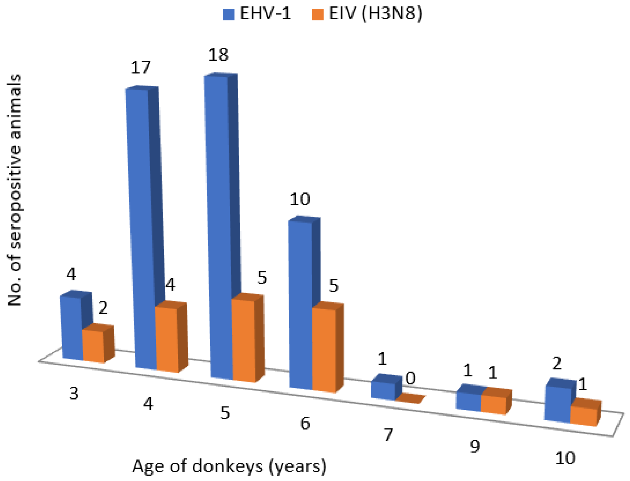
| Donkey Age | Number of Animals with Antibody Titer against EHV-1 | Total Number of Animals (%) | |||||||
|---|---|---|---|---|---|---|---|---|---|
| <1:2 | 1:2 | 1:4 | 1:8 | 1:16 | 1:32 | 1:64 | 1:128 | ||
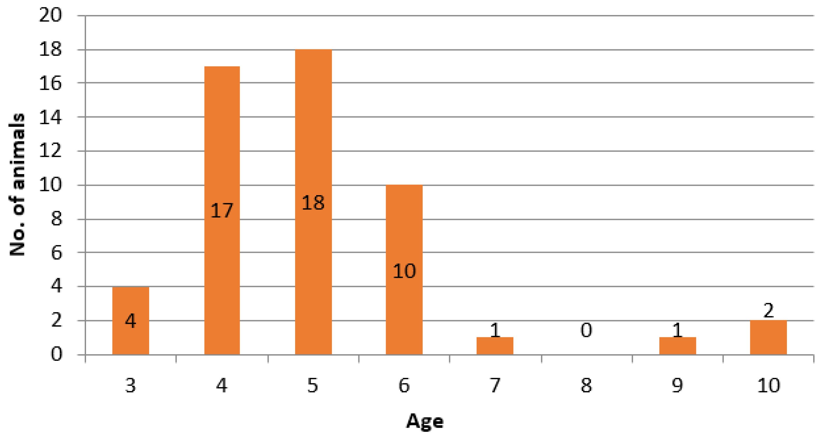
| 3 | 0 | 0 | 0 | 0 | 3 | 1 | 0 | 0 | 4 (7.55) |
| 4 | 0 | 0 | 1 | 1 | 8 | 5 | 2 | 0 | 17 (32.07) |
| 5 | 0 | 0 | 3 | 3 | 4 | 2 | 5 | 1 | 18 (33.96) |
| 6 | 0 | 0 | 0 | 0 | 1 | 5 | 4 | 0 | 10 (18.87) |
| 7 | 0 | 0 | 0 | 1 | 0 | 0 | 0 | 0 | 1 (1.89) |
| 9 | 0 | 0 | 0 | 1 | 0 | 0 | 0 | 0 | 1 (1.89) |
| 10 | 0 | 1 | 0 | 0 | 0 | 0 | 1 | 0 | 2 (3.77) |
| Total seropositive (%) | 0 (0%) | 1 (1.89%) | 4 (7.55%) | 6 (11.32%) | 16 (30.18%) | 13 (24.53%) | 12 (22.64%) | 1 (1.89%) | 53 (100.00%) |
| Donkey Age | Number of Animals with Antibody Titer against EIV H3N8 | Total Number of Animals (%) | ||||
|---|---|---|---|---|---|---|
| <1:2 | 1:2 | 1:4 | 1:8 | 1:16 | ||
| 3 | 0 | 1 | 1 | 0 | 2 | 4 (7.55) |
| 4 | 2 | 2 | 0 | 9 | 4 | 17 (32.07) |
| 5 | 7 | 3 | 1 | 2 | 5 | 18 (33.96) |
| 6 | 4 | 1 | 0 | 0 | 5 | 10 (18.87) |
| 7 | 0 | 0 | 0 | 1 | 0 | 1 (1.89) |
| 9 | 0 | 0 | 0 | 0 | 1 | 1 (1.89) |
| 10 | 0 | 0 | 0 | 1 | 1 | 2 (3.77) |
| Total seropositive (%) | 13 (24.53%) | 7 (13.21%) | 2 (3.77%) | 13 (24.53%) | 18 (33.96%) | 53 (100.00%) |
Disclaimer/Publisher’s Note: The statements, opinions and data contained in all publications are solely those of the individual author(s) and contributor(s) and not of MDPI and/or the editor(s). MDPI and/or the editor(s) disclaim responsibility for any injury to people or property resulting from any ideas, methods, instructions or products referred to in the content. |
© 2023 by the authors. Licensee MDPI, Basel, Switzerland. This article is an open access article distributed under the terms and conditions of the Creative Commons Attribution (CC BY) license (https://creativecommons.org/licenses/by/4.0/).
Share and Cite
Lazić, S.; Savić, S.; Petrović, T.; Lazić, G.; Žekić, M.; Drobnjak, D.; Lupulović, D. Serological Examinations of Significant Viral Infections in Domestic Donkeys at the Special Nature Reserve “Zasavica”, Serbia. Animals 2023, 13, 2056. https://doi.org/10.3390/ani13132056
Lazić S, Savić S, Petrović T, Lazić G, Žekić M, Drobnjak D, Lupulović D. Serological Examinations of Significant Viral Infections in Domestic Donkeys at the Special Nature Reserve “Zasavica”, Serbia. Animals. 2023; 13(13):2056. https://doi.org/10.3390/ani13132056
Chicago/Turabian StyleLazić, Sava, Sara Savić, Tamaš Petrović, Gospava Lazić, Marina Žekić, Darko Drobnjak, and Diana Lupulović. 2023. "Serological Examinations of Significant Viral Infections in Domestic Donkeys at the Special Nature Reserve “Zasavica”, Serbia" Animals 13, no. 13: 2056. https://doi.org/10.3390/ani13132056