Article

Biocontrol Potential of Endophytic Fungi for the Eco-Friendly Management of Root Rot of Cuminum cyminum Caused by Fusarium solani

by
Kamal A. M. Abo-Elyousr
1,*,
Omer H. M. Ibrahim
1,
Adel D. Al-Qurashi
1,
Magdi A. A. Mousa
1 and
Maged M. Saad
2
1
Department of Arid Land Agriculture, Faculty of Meteorology Environment and Arid Land Agriculture, King Abdulaziz University, Jeddah 80208, Saudi Arabia
2
Center for Desert Agriculture (CDA), King Abdullah University of Science and Technology, Thuwal 23955, Saudi Arabia
*
Author to whom correspondence should be addressed.
Agronomy 2022, 12(11), 2612; https://doi.org/10.3390/agronomy12112612
Submission received: 19 September 2022 / Revised: 18 October 2022 / Accepted: 21 October 2022 / Published: 24 October 2022
(This article belongs to the Special Issue Epidemiology and Control of Fungal Diseases of Crop Plants)

Abstract

:
Root rot disease of Cuminum cyminum caused by Fusarium solani is one of the most destructive diseases threatening cumin production. The present study investigates the biocontrol potential of some endophytes against F. solani and their effect on the induction of defense-related enzymes in a greenhouse. The results herein presented illustrate the strong biocontrol potential of three (out of twelve) endophytes. During the in vitro assay, three isolates demonstrated strong mycelial growth inhibition of F. solani: isolates 3, 4, and 9, with 87%, 65%, and 80% reductions, respectively, with respect to the control (100%). These isolates were identified as Trichoderma harzianum, T. longibrachiatum, and Chaetomium globosum, which produce siderophore and indole-3-acetic acid (IAA). Cumin seed priming with the culture filtrates of T. harzianum, C. globosum, and T. longibrachiatum positively affected the seed germination, as a higher germination (%) of culture filtrate-treated seeds was observed followed by infected and healthy control/untreated seeds. In the greenhouse, the application of T. harzianum, T. longibrachiatum, and C. globosum caused a reduction in disease severity (67.7%, 58.1%, and 59.3%, respectively) on cumin plants, with a lower disease severity (20%, 26%, and 25%, respectively) recorded in treated plants compared to the infected control (62%). Furthermore, a significant increase in defense-related enzymes in culture filtrate-treated cumin plants was recorded. Higher peroxidase (PO), polyphenoloxidase (PPO), and phenylalanine ammonia-lyase (PAL) activity, and a higher content of phenolic compounds, were found in culture filtrate-treated plants. These results indicate that the culture filtrates of these bioagents not only increased seed germination, but also protected the plants from F. solani infection by acting as important elements of the cellular antioxidant system in plants upon infection, conferring the biocontrol potential of C. globosum and Trichoderma species toward mitigating the root rot disease of cumin plants in a greenhouse.

1. Introduction

In the Apiaceae family, cumin (Cuminum cyminum L.) is an important plant which is best known for its valuable contribution in Asian food as a spice as well as for its medicinal uses. Moreover, cumin is widely consumed due to the abundant presence of metabolites [1]. This plant exists natively in the East Mediterranean but has been widely cultivated in Central and South Asia as well as in North Africa [2]. Various fungal diseases such as Fusarium wilt, powdery mildew, blight, and damping-off [3] diminish its production and cause significant yield losses.
Fusarium is a common soil-borne pathogen that invades a variety of plants and vegetables, including eggplants, tomato, potato, and peppers [4], causing Fusarium wilt on egg plants [5], root rot disease in cassava plant [6], and Sanqi ginseng Fusarium root rot [7]. Among the fungal plant diseases caused by various Fusarium species, namely, F. oxysporum, F. fujikuroi, and F. graminearum, the root rot caused by Fusarium solani is the most destructive disease, causing significant yield losses of various important crops [8], including cumin [9].
The application of fungicides to control fungal plant diseases is considered one of the most rapid methods; however, in severe disease conditions, multiple applications of fungicides lead to resistance development in fungal pathogens [10,11]. Importantly, the excessive use of chemical fungicides annihilates other beneficial soil microbes, contaminates the environment, and increases the toxicity of the soil by leaching into the rhizosphere. Various site-specific fungicides with different modes of action may provide effective control of F. solani, but the use of excessive fungicides, in addition to resistance problems, significantly pollutes the environment, poses a hazard to humans, and obliterates the soil microbiome [12]. Considering these serious problems of fungicides, various researchers are currently focusing on eco-friendly approaches for the control of plant diseases by using fungal and bacterial species with antimicrobial potential. Various fungal and bacterial endophytes with biocontrol potential have been widely used against various phytopathogens [13], and their direct application or seed priming can assist in reducing disease [14]. In fungal endophytes, hundreds of Trichoderma species used as biocontrol agents, including T. harzianum, T. atroviride, T. longibrachiatum [15], T. viridae, and T. polysporum, have been widely used against various plant diseases. When used as biocontrol agents, these Trichoderma species not only inhibit the in vitro mycelial growth of fungal pathogens, but also demonstrate astonishing results in greenhouse and field conditions to mitigate diseases. Furthermore, as plant growth regulators, these bioagents also stimulate plant growth, biomass, and fruit yield [13]. Root colonization by Trichoderma species causes root development and triggers the defense activity, with significant changes occurring in metabolic pathways. The genes in Trichoderma species that encode the oligopeptide transporter and extracellular protease are expressed during host–pathogen interaction [16]. Furthermore, these genes activate the defense system of the host plant through ethylene and jasmonic acid signaling [17].
In addition to Trichoderma species in the Ascomycota family, Chaetomiaceae is the most diverse saprophytic ascomycete family, containing more than 350 species [18]. Some species of Chaetomium have been used as antagonists against various phytopathogens due to their antimicrobial activity [19]. Among these species, Chaetomium globosum (Kunze ex Fr.) has been widely isolated from various endophytes [20] and has been identified as a promising biocontrol agent [21]. The in vitro and in vivo effects of C. cupreum, C. lucknowense, C. globosum, and their culture filtrates as biocontrol agents against Phytophthora nicotianae (causing citrus root rot disease) revealed their strong antifungal potential [22]. Furthermore, Chaetomium species have also been widely used against various plant diseases such as wheat common root rot [23] and potato black scurf [24]. In addition to their antimicrobial characteristics, these species demonstrated plant growth-promoting potential in cucumber [25]. Different metabolites of C. globosum were reported with strong antifungal activity against verticillium wilt of cotton [26].
The inhibition of fungal pathogens by bioagents involves various mechanisms such as mycoparasitism, antibiosis, and competition, whereas the stimulation of every process involves the biosynthesis of metabolites such as plant growth regulators, siderophores, antioxidant enzymes, and antibiotics [27]. The strong mycoparasitism and antagonistic potential of Trichoderma species enable them to diminish the plant disease incidence due to various phytopathogens, with hyperparasitism being one of the key mechanisms in Trichoderma species [28]. Additionally, the suppression of fungal plant pathogens by Trichoderma species mainly occurs directly (hyperparasitism, antibiosis, competition for food, space, and nutrient) [29] or indirectly by enhancing stress tolerance, bioremediating the contaminated rhizosphere, and improving plant growth and vigor, active nutrient uptake, and production of plant enzymes, secondary metabolites, and pathogenesis-related (PR) proteins [30]. The induction of resistance can provide an alternative approach for control plant pathogens, particularly those which cannot be controlled by site-specific fungicides with different modes of action [31]. The activation of defense-related enzymes, such as peroxidase (PO), polyphenol oxidase (PPO), catalase (CAT), phenylalanine ammonia lyase (PAL), and superoxide dismutase (SOD), and the accumulation of phenolic compounds, are most important to protect the plant from pathogen infections [32] by inducing resistance. Peroxidase (PO) is an important constituent of plants that responds to early infection of a pathogen and induces biosynthesis of lignin, ultimately restricting pathogen spread [33]. An increase in PO level quickly synthesizes reactive oxygen (RO) derivatives via oxidative burst, which leads to cell death and causes inhibition of the pathogen [34]. Polyphenol oxidase (PPO) assists in the oxidation process of phenols into quinine compounds, which are quinone toxic compounds that spread to affected plant tissues to restrict the pathogen [35]. Peroxidase (PO), together with PPO, catalyzes the formation of oxidative phenols and lignin, which assist in the formation of a defense barrier by altering the defense system of the cell structure [36]. Furthermore, phenylalanine ammonia lyase (PAL) activity and phenolic content can be induced via different mechanisms during plant–pathogen interaction including via elicitor compounds that maintain primary metabolites and regulate plant hormones [37].
Considering the importance of endophytes as bioagents and to minimize the fungicide resistance risks in pathogens, the main objectives of the present study were to isolate and identify the root rot pathogen of cumin, to isolate and identify endophytes as biocontrol agents against root rot pathogen, and to apply culture filtrates of endophytes in vivo with the aim of studying their effect on disease severity and defense-related enzymes in cumin plants.

2. Materials and Methods

2.1. Isolation and Morphological Characterization of Pathogen

To isolate the pathogen causing root rot of cumin, about 25 samples of infected root samples from cumin plants were collected from Assiut Governorate, Egypt (27°1801344″ N, 31°189283″ E). The root samples were placed in sterilized plastic polythene bags and stored at 4 °C.
For isolation of the pathogen, infected roots were collected and rinsed thrice with sterilized distilled water to eliminate the soil debris and other impurities. Then, the roots were immersed in sodium hypochlorite (2% NaOCl) solution for 3–5 min, and root samples were dried by transferring to sterile filter paper. The disinfected root samples (4–6 root samples/plate) were placed on Potato Dextrose Agar (PDA) medium plates supplemented with ampicillin (200 mg·L−1) and rifampicin (10 mg·L−1) [38] as antibiotics. Three plates were used for each sample. The plates were incubated at 27 °C for 5–7 days, and the germinated fungal colonies were observed. Colonies were subcultured on new PDA plates and then incubated at 27 °C for another 5 days. The colony cultures of the causal pathogen were purified, maintained, and preserved at 4 °C. Morphological and microscopic (OMAX, 40×–2500× LED Digital Trinocular Lab Compound Microscope with 5 MP Camera and Mechanical Stage, M83EZ-C50S) characteristics of the fungal colonies were observed. The identification of the species was based on the morphological characteristics of the isolates as described by Booth [39], Nelson et al. [40], and Burgess et al. [41].

2.2. Pathogenicity Screening and Selection of Virulent Isolate

Fungal isolates (n = 7) were assessed for their virulence level under greenhouse conditions in pots. Briefly, to evaluate the pathogenic effect of isolates on seed germination, pot experiments were conducted in the greenhouse of the Department of Arid Land Agriculture, King AbdulAziz University, Jeddah, Saudi Arabia, during the years 2019–2020. The pathogen inoculum was prepared by growing the fungal cultures on PDA plates for 5–7 days at 27 °C. The mycelium of all isolates was scraped and ground upon adding the appropriate volume of sterile distilled water. Then, cumin seeds of the variety “Baladi” were sown (10 seeds/pot) in 18 cm plastic pots, previously filled with peat soil/moss (1:3). Then, the pots were inoculated with the pathogen suspension (10 mL/pot) to infect the soil. For each isolate, four pots (each pot containing 10 seeds) were investigated as four replicates. Pots inoculated with sterilized distilled water (10 mL/pot) were used as a healthy control. Inoculated pots were covered with a sterile polythene sheet to retain the moisture content. The percentage of pre- and postemergence damping-off was recorded 20 and 40 days after the sowing date as follows [42]: germination percentage = S/T × 100, where S is the number of germinated seeds and T is the total number of seeds in a pot.
To validate Koch’s postulates, the pathogen from infected roots was reisolated and compared with the originally obtained isolate. The most virulent isolate was selected for further experiments.

2.3. Isolation of Fungal Endophytes

Naturally existing endophytes were isolated from healthy cucumber roots (10 samples) collected from the field station of Assiut University, Egypt (27° 1875″ N, 31° 1703″ E). After surface sterilization, the endophytes were isolated using the method described in [5] with slight modification. Briefly, the roots were thoroughly rinsed with sterilized distilled water, followed by surface sterilization in ethanol (70%) for 30 s and in 2% sodium hypochlorite (NaOCl) for 3–5 min, before thoroughly rerinsing in sterile distilled water. The root segments (5 mm size, 4–6 roots samples/plate, and three plates for each sample) were placed on the surface of Petri plates containing PDA [5], maintaining constant moisture under aseptic conditions. Plates were incubated at 27 °C to allow fungal germination from the roots. Colonies were observed and purified, and the culture was maintained in glass slants containing PDA at 4 °C for further use. A pure culture of isolate was deposited in the culture collection of the Plant Pathology Department, Faculty of Agriculture, Assiut University. The isolated fungi were identified on the basis of their cultural, morphological, and microscopical characteristics [43,44,45].

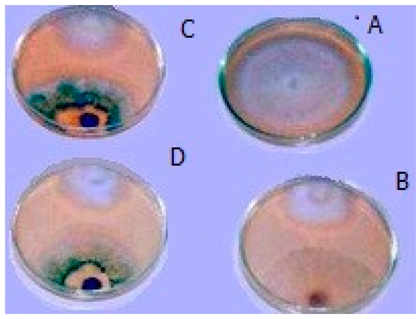
2.4. Screening of Endophytes for Biocontrol Potential

In order to assess the in vitro biocontrol potential of fungal endophytes against the highly virulent pathogenic isolate (Fusarium solani isolate 7), the fungal endophytes were grown on PDA plates. For in vitro antagonism, a dual-culture assay was performed; a 5 mm mycelial disc (3 days old, priorly grown on PDA) from the highly virulent isolate was placed face-down on a PDA plate at an equal distance from the edge, whereas, in the parallel direction, a mycelial disc (identical diameter) excised from fungal endophytes (3 day old culture) was placed. A pathogenic mycelial disc in the same position on a PDA plate but lacking an endophyte mycelial disc was used as the control. Four replicates were used for each fungal endophyte. Plates were incubated at 27 °C for 5–7 days. Colony diameter was measured and compared to control plates, and the growth reduction (%) of the pathogen due to endophytes was recorded [46]. The experiment was repeated twice. On the basis of the results, endophytes with strong antagonistic potential were selected and identified.

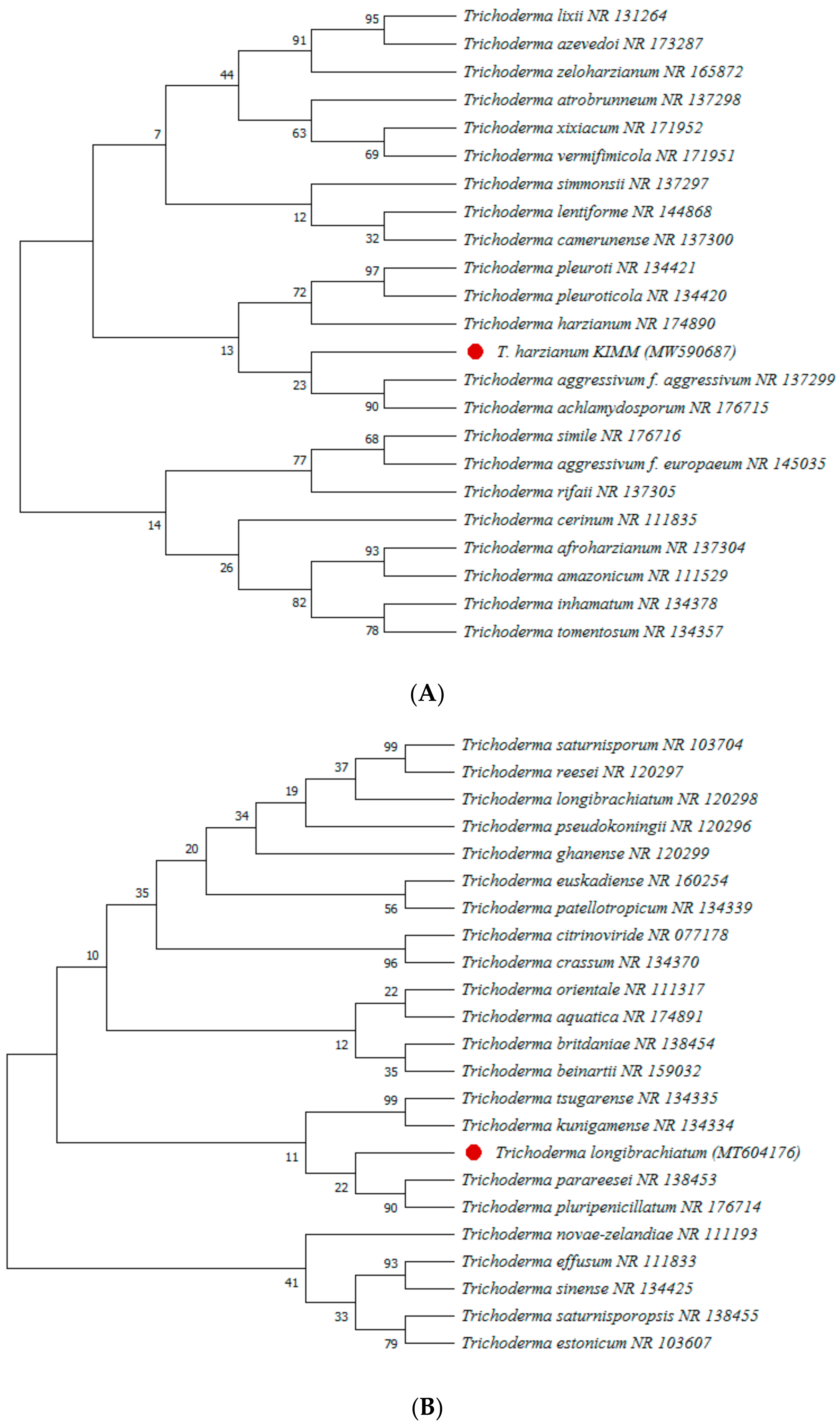
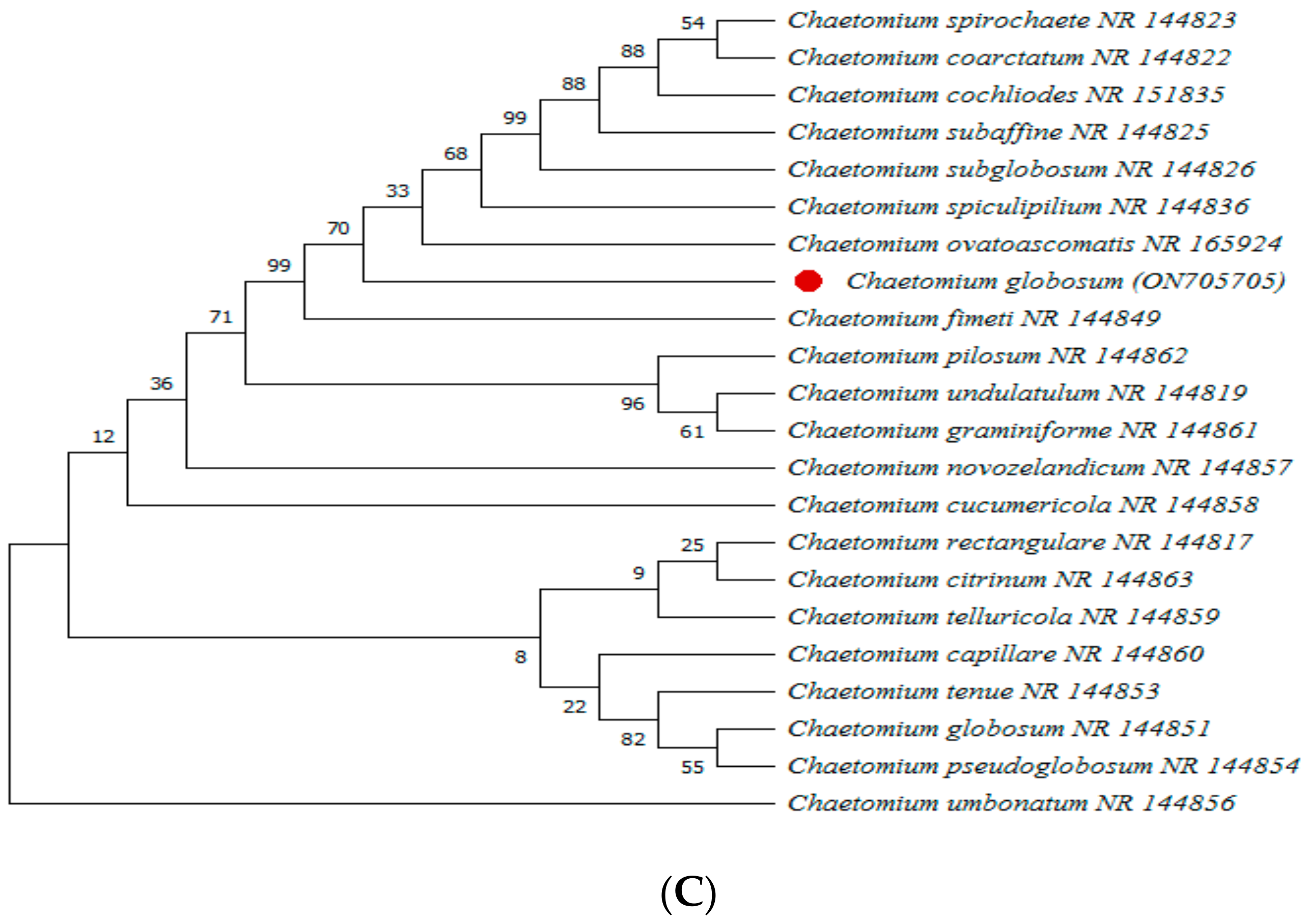
2.5. Molecular Identification of Selected Pathogen and Endophyte

The highly virulent isolate and the endophytes demonstrating strong antagonistic potential were selected for molecular identification by PCR analysis using DNA amplification of the ITS region. PDA plates with a cellophane layer containing a 5 mm mycelial disc from the 5-day-old fungal culture (virulent pathogen and selected endophytes) were incubated at 27 °C for 5–7 days. The fungal mycelium scraped using a sterile scraper was collected in 2 mL vials and ground in liquid nitrogen to obtain a fine powder for DNA extraction using the cetyltrimethylammonium bromide (CTAB) method [42]. The universal pair of ITS1 and ITS4 primers (ITS1: 5’–TCCGTAGGTGAACCTGCGG, ITS4: 5’–TCCTCCGCTTATTGATATGC [4]) was used for the identification of the pathogen, whereas the pair of ITS5 and ITS4 primers [47] with rpb2 fragments (fRPB27cR-RPB25F2 [48] and tef1 fragments (EF2-EF1728M) [49] was amplified from the selected endophytes. A standard PCR reaction with a final volume of 50 µL was prepared containing 5 μL of 10× Standard Taq Reaction Buffer, 4 μL of 10 mM dNTPs, 1 μL of 10 μM forward primer, 1 μL of 10 μM reverse primer, 0.5 μL of Taq DNA Polymerase enzyme (MBI; Fermentas, Leon-Rot, Germany), 1 μL of template DNA, and 37.5 μL of nuclease-free water. The PCR reaction was performed in a thermal cycler (Thermo Scientific® cyclone 25 (PeqLab, Erlangen, Germany) with the following conditions for the gene amplification of pathogen DNA: initial denaturation at 95 °C for 3 min, followed by 35 cycles of denaturation at 95 °C for 35 s, annealing at 55 °C for 1 min, extension at 72 °C for 1 min, and final extension for 10 min at 72 °C. PCR products were visualized on 1.5% agarose gel along a 1 kb DNA ladder (Zymo Research, Irvine, CA, USA). For the identification of endophytes, PCR reaction conditions for ITS were as follows: initial denaturation at 94 °C for 3 min, followed by 30 cycles at 94 °C for 1 min, 56 °C for 30 s, 72 °C for 30 s, and 72 °C for 10 min. PCR conditions for tef1 were as follows: 94 °C for 2 min, followed by nine cycles at 94 °C for 35 s and 66 °C for 55 s, and 35 cycles at 94 °C for 35 s, 56 °C for 55 s, and 72 °C for 1 min 30 s. The PCR conditions for rpb2 were as follows: 95 °C for 5 min., followed by 38 cycles at 95 °C for 1 min, 58 °C for 2 min, 72 °C for 2 min, and 72 °C for 10 min. The products were visualized on 2% agarose gel followed by staining with 0.5 µg of ethidium bromide solution for 10 min using an AlphaImagerTM gel imager system. The products were sequenced by Macrogen, Inc., Seoul, South Korea (http://www.macrogen.com, 13 –June 2022). The obtained sequences of products were compared with the sequence available in the public domain of the National Center for Biotechnology Information (NCBI) library using the Basic Local Alignment Search Tool (BLAST). Products were identified on the basis of their higher similarity, and the sequences were submitted to NCBI with a specific accession number. A phylogenetic tree was constructed with the sequenced products using the neighbor joining algorithm in the MEGA 6X package [50].

2.6. In Vitro Quantification of Siderophore and Indole Acetic Acid Production by Bioagents

2.6.1. Siderophore Detection

The production of siderophore by selected endophytes was assayed using the Chrome Azurol S (CAS) agar assay [51] with MM9 medium. Briefly, 5 mm mycelial discs from each endophyte (previously grown on iron-deficient MM9 medium supplemented with 1% w/v potato dextrose broth) was placed on CAS agar Petri plates and incubated at 27 °C for 24 h; the clear halo zone around the growing colonies was visualized, and the change in color (dark blue to yellow) was observed. The diameter of the halo zone around the colonies was measured. The experiment was performed twice in quadruplicate.

2.6.2. Indole-3-Acetic Acid Quantification

The production of IAA by selected endophyte isolates was determined using the method described by Bano and Musarrat [52]. Briefly, 5 mm mycelial discs (3-day-old mycelial cultures previously grown on PDA at 27 °C) from the selected fungal endophytes were transferred in a floating position (7–10 mycelial discs) to 25 mL of tryptone yeast extract broth (YB) containing 2 mg/mL l-tryptophan followed by 7 days of incubation at 27 °C in a shaker at 125 rpm. Then, the supernatant was centrifuged at 11,000 rpm for 15 min at 4 °C. To enumerate IAA, 1 mL of the supernatant was combined with 2 mL of Salkowski reagent [20], and the appearance of a pink color was observed to indicate the positive production of IAA. The optical density at 535 nm was recorded, and the level of IAA was expressed as µg/mL.

2.7. In Vitro Culture Filtrate Assay

2.7.1. Preparation of Culture Filtrates

Culture filtrates of the selected endophytes were initially prepared by transferring 5 mm mycelial discs (3 days old, previously grown on PDA) to 50 mL conical flasks containing 25 mL of Potato Dextrose Broth medium (PDA lacking agar). Mycelial discs (8–10 discs/flask) were carefully placed (in a floating position) in PDB medium flasks. The flasks were incubated at 27 °C in a shaker (125 rpm) for 7–10 days. The suspension was centrifuged at 10,000 rpm for 10 min at 4 °C and filtered through Whatman No. 1 filter paper (Sigma-Aldrich, Missouri, USA) followed by filtration through a microbiological filtration unit (Sigma-Aldrich, USA) for purification of the culture filtrates. The obtained supernatant (100% purity) was stored at 4 °C for further use.

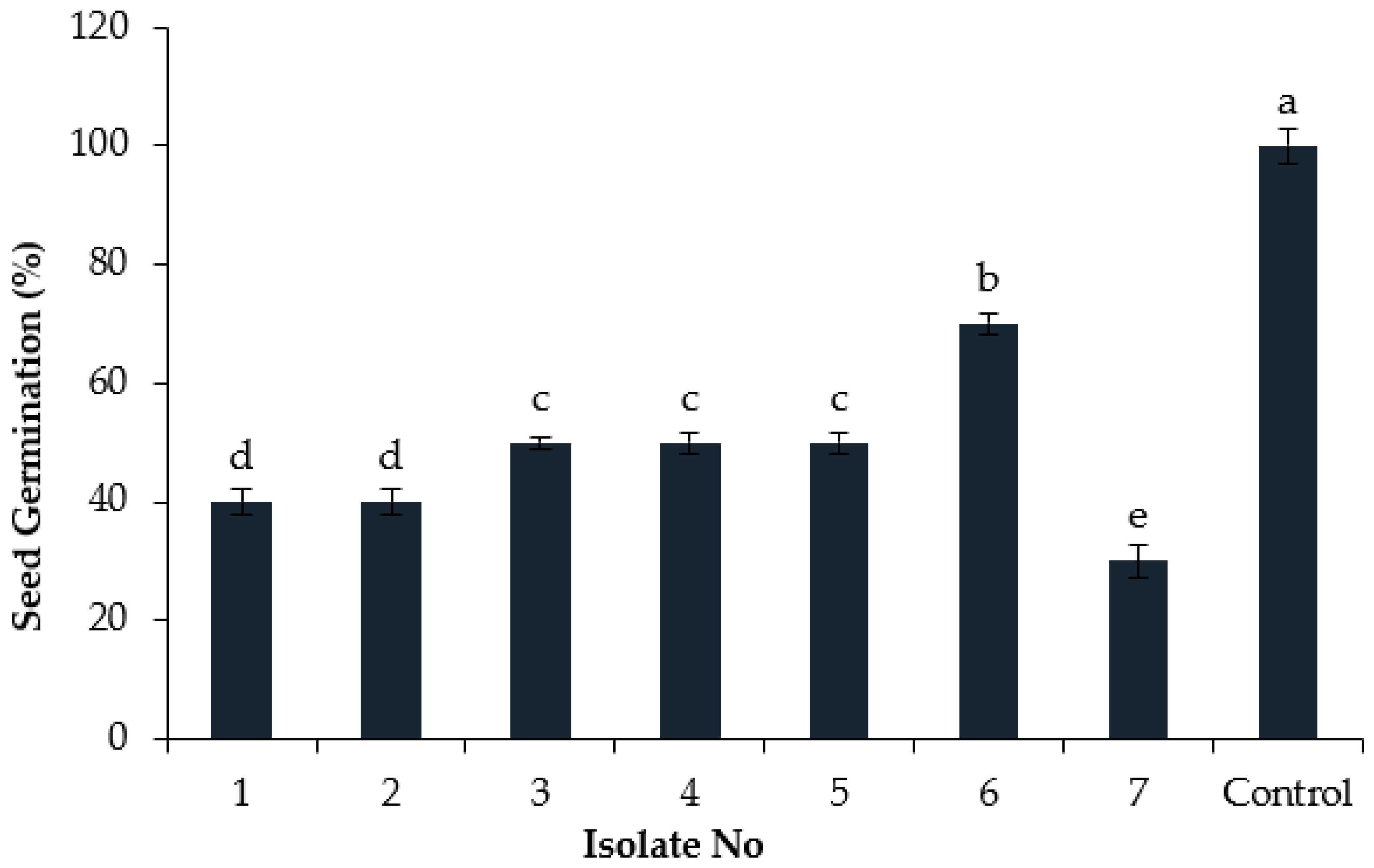
2.7.2. Effect of Endophyte Culture Filtrates on Cumin Seed Germination

Seeds of cumin variety “Baladi” obtained from the Laboratory of Agronomy, King Abdulaziz University were surface-sterilized with 2% sodium hypochlorite (NaOCl) solution for 15 min, followed by rinsing with sterile distilled water. Seeds primed (20 min) with the suspension of culture filtrates were placed in 90 mm Petri plates (20 seeds/plate) containing three layers of sterile filter paper with sufficient moisture and relative humidity (about 95%). Seeds treated with sterile distilled water were used as healthy controls, while seeds treated with a suspension of pathogen (1 × 10⁵ conidia/mL) only were used as infected controls. Plates were incubated at 25 °C for 7–10 days, and seed germination (%) was calculated as follows [42]: germination percentage = S/T × 100, where S is the number of germinated seeds, and T is the total number of seeds.

2.7.3. In Vivo Effects of Culture Filtrates

In order to study the effects of culture filtrates on disease reduction, greenhouse experiments were carried out using the previously mentioned cumin variety. Briefly, the cumin seeds (10 seeds/pot) were sown in pots containing soil/peat moss (1:3), placed in a greenhouse, and irrigated as required. Then, a conidial suspension of the selected highly virulent pathogenic isolate (Fusarium solani isolate 7) was prepared as described in Section 2.2. The number of conidia in the suspension was adjusted (104 conidia/mL) using a hemacytometer (Sigma Aldrich, Missouri, USA). Then, 14-day-old cumin seedlings were inoculated with the suspension of culture filtrate (100%) of the selected endophyte (20 mL/pot) and, 24 h later, inoculated with the cell suspension (20 mL/pot) of pathogen. Cumin seedlings inoculated with the pathogen suspension only were used as infected controls, whereas seedlings inoculated with sterile distilled water were used as healthy controls. The temperature and humidity inside the greenhouse were maintained. The disease severity was recorded using a rating scale (0 = healthy plants, 1 = 0–25% wilting, 2 = 26–50% wilting, 3 = 51–75% wilting, and 4 = severe/complete wilting). Disease severity (DS%) = (Σ (n x v)/4N) × 100, whereas n = Number of infected leaves in each category. v = Numerical values of each category. N = Total number of the infected. Disease symptoms such as stunted growth, wilting of leaves, stem discoloration, and plant fatality were observed [53].

2.8. Effect of Culture Filtrates on Defense Enzymes

The effect of culture filtrates of the selected endophytes on the induction of defense-related enzyme production, i.e., peroxidase (PO), polyphenol oxidase (PPO), and phenylalanine ammonia-lyase (PAL), as well as phenolic content, was determined in culture filtrate-treated cumin leaves. The leaf samples of cumin leaves were collected at different times (0, 2, 4, 6, and 8 days after full germination, i.e., 7 days after sowing date). The leaf samples were stored at −80 °C, and defense-related enzymes were assayed in leaf samples.
  • Protein Assay
The total protein content in cumin leaf samples was determined using the Bradford assay [54] according to a bovine serum albumin (BSA) (0–5 mg/mL) standard curve. The reaction was incubated at room temperature for 30 min, and the protein content was measured according to the absorbance at 595 nm for 4 min with an interval of 20 s to develop the standard curve.
  • Estimation of Defense Enzymes
Defense-related enzymes were assayed in collected cumin leaf samples, and the leaves were immersed in liquid nitrogen. To prepare the crude enzyme, 1 g of powder was homogenized in 10 mL of 0.1 M Na-acetate buffer (pH = 5.2), and the homogenate was centrifuged at 10,000 rpm for 30 min at 4 °C. The pellet was discarded, while the supernatant was transferred to a new centrifuge tube and stored at −20 °C to determine PO and PAL activity.

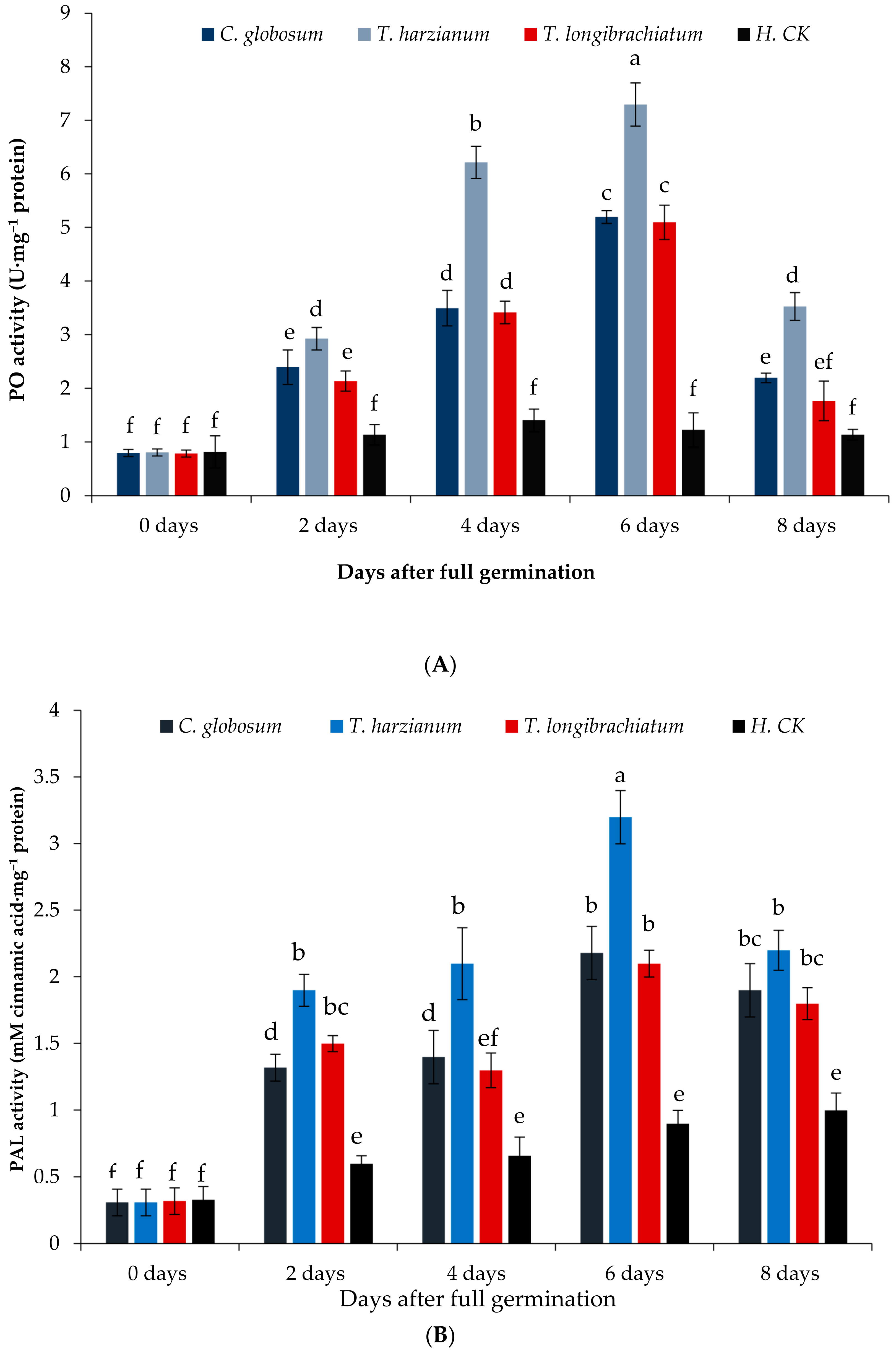
2.8.1. Peroxidase (PO) Assay

Peroxidase (PO) activity was determined using the method described by Pütter [55], with guaiacol as a substrate. The reaction mixture was prepared by dissolving 0.2 mL of supernatant (sample) in 1 mL of 0.1 M Na-acetate buffer (pH = 5.2) followed by the addition of 0.2 mL of 1% guaiacol and 0.2 mL of 1% H2O2. The reaction was incubated at 25 °C for 1 min, and the absorbance was measured at 436 nm using a spectrophotometer (UV-1800, Shimadzu USA Manufacturing Inc., Canby, UR, USA). A reaction mixture lacking sample was used as the blank, and the PO activity was expressed as U·mg−1 protein.

2.8.2. Phenylalanine Ammonia-Lyase (PAL) Assay

Phenylalanine ammonia-lyase (PAL) activity was determined using the method described by Zucker [56]. A reaction mixture containing 0.5 mL of supernatant (sample), 2 mL of sodium borate buffer (50 mM), and 1 mL of 60 mM phenylalanine was incubated at 37 °C for 2 h in a water bath. Cinnamic acid (0–5 mg) was used to establish a standard curve. After 2 h, the absorbance was measured at 290 nm using a spectrophotometer, and PAL activity was expressed as mM cinnamic acid/mg protein.

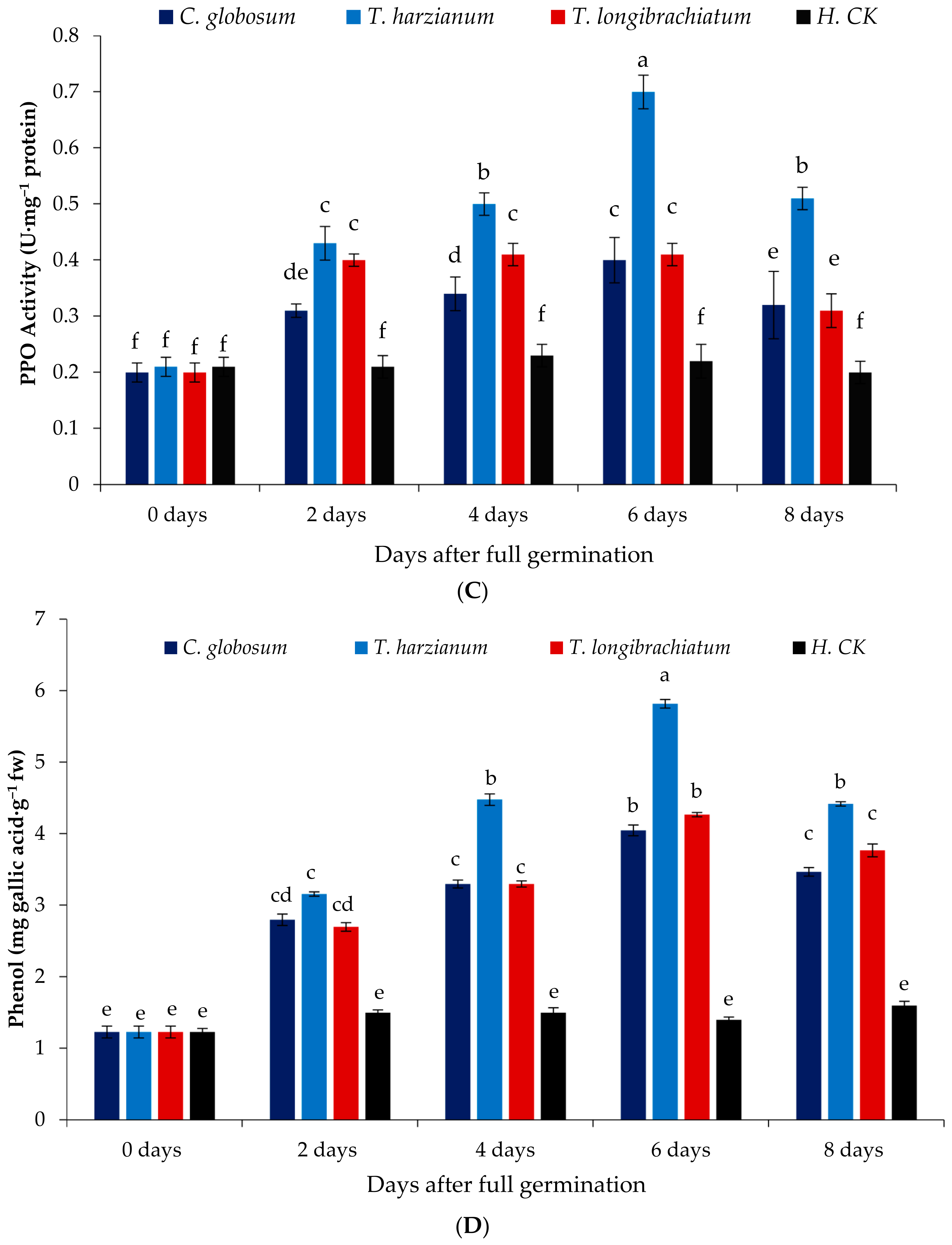
2.8.3. Polyphenol Oxidase (PPO) Assay

Polyphenol oxidase (PPO) activity in treated cumin leaf samples was determined using the method described by Jockusch [57]. A reaction mixture containing 2.5 g of powdered sample suspended in 5 mL of sodium phosphate buffer (0.05 M; pH 6.0) with 5% polyvinylpolypyrrolidone (w/v) was centrifuged at 13,000 rpm for 5 min at 4 °C followed by filtration through Whatman No. 1 filter paper. The supernatant was transferred to new tubes stored at −20 °C. In order to determine PPO activity, a reaction mixture containing 1 mL of the supernatant and 2.9 mL of sodium phosphate buffer (0.05 M), along with 1 mL of catechol (0.1 M), was incubated at room temperature for 3–5 min. Sodium phosphate buffer (identical volume of sample) was used as a blank. The absorbance was measured at 546 nm using a spectrophotometer, and PPO activity was expressed as U·mg−1 protein.

2.8.4. Total Phenolic Assay

The content of total phenols in treated cumin leaf samples was determined according to the method described by Şahin et al. (2004) [58] using gallic acid (0–5 mg) as a standard. A reaction mixture containing 0.02 mL of sample, 0.5 mL of Folin–Ciocâlteu reagent, 0.75 mL of 20% sodium carbonate (Na2CO3), and 8 mL of sterile distilled water was incubated at 37 °C for 60 min. A reaction mixture lacking sample was used as the control. The total phenolic content was assayed at 767 nm absorbance using a spectrophotometer and expressed as mg gallic acid·g−1 plant material.

2.9. Statistical Analysis

All in vitro experiments were conducted in quadruplicate. Greenhouse experiments were performed in a complete randomized design, and all collected data were analyzed using Statistix 8.1 analytical software (Statistix; Tallahassee, FL, USA, 1985–2003). The data collected from disease severity were transformed into arcsine values, and a one-way analysis of variance (ANOVA) was performed. Means of replicates in all treatments were compared using Fisher’s least significant difference (LSD) test at p = 0.05 [59].

3. Results

3.1. Isolation and Morphological Characterization of Pathogen

According to the reported cultural and morphological characteristics [39,40,41], all seven isolates were identical to Fusarium spp.; therefore, all isolates were further used to determine the level of virulence.

3.2. In Vivo Screening, Selection, and Identification of Pathogen

The in vivo screening of all isolates performed in a greenhouse on cumin seeds showed a significant (p = 0.05) difference in pathogenicity. A significant reduction in seed germination after the inoculation of isolate 7 (30%) was recorded (Figure 1). Comparatively, the seed germination by isolates 3, 4, and 5 was identical (50%) but lower than the control (100%), whereas the inoculation of isolates 1, 2, and 6 demonstrated 40% and 70% seed germination (%). The results presented herein show that the inoculation of isolate 7 significantly affected the cumin seed germination in pot trials under greenhouse conditions. Reisolation of the pathogen from the inoculated seeds was performed for comparison with the inoculated isolates. The reisolated pathogen isolates were identical to the original fungi inoculated in the soil in terms of their morphological and pathogenic characteristics, thus satisfying Koch’s postulates. Accordingly, isolate 7 was selected for identification and further experiments.
The molecular identification of highly virulent isolate 7 using PCR analysis showed that the length of the obtained sequenced products was 540 bp. Furthermore, BLAST analysis of the obtained sequence showed the highest similarity (100%) with Fusarium solani (GenBank Accession number AF178418). Therefore, isolate 7 was identified as F. solani. The sequences were submitted to NCBI under accession number MT730027. The phylogenetic tree (Figure 2) highlights the relationship between the sequence of the isolate identified in this study (identified as F. solani) and identical sequences obtained from the NCBI, constructed using the bootstrap method.

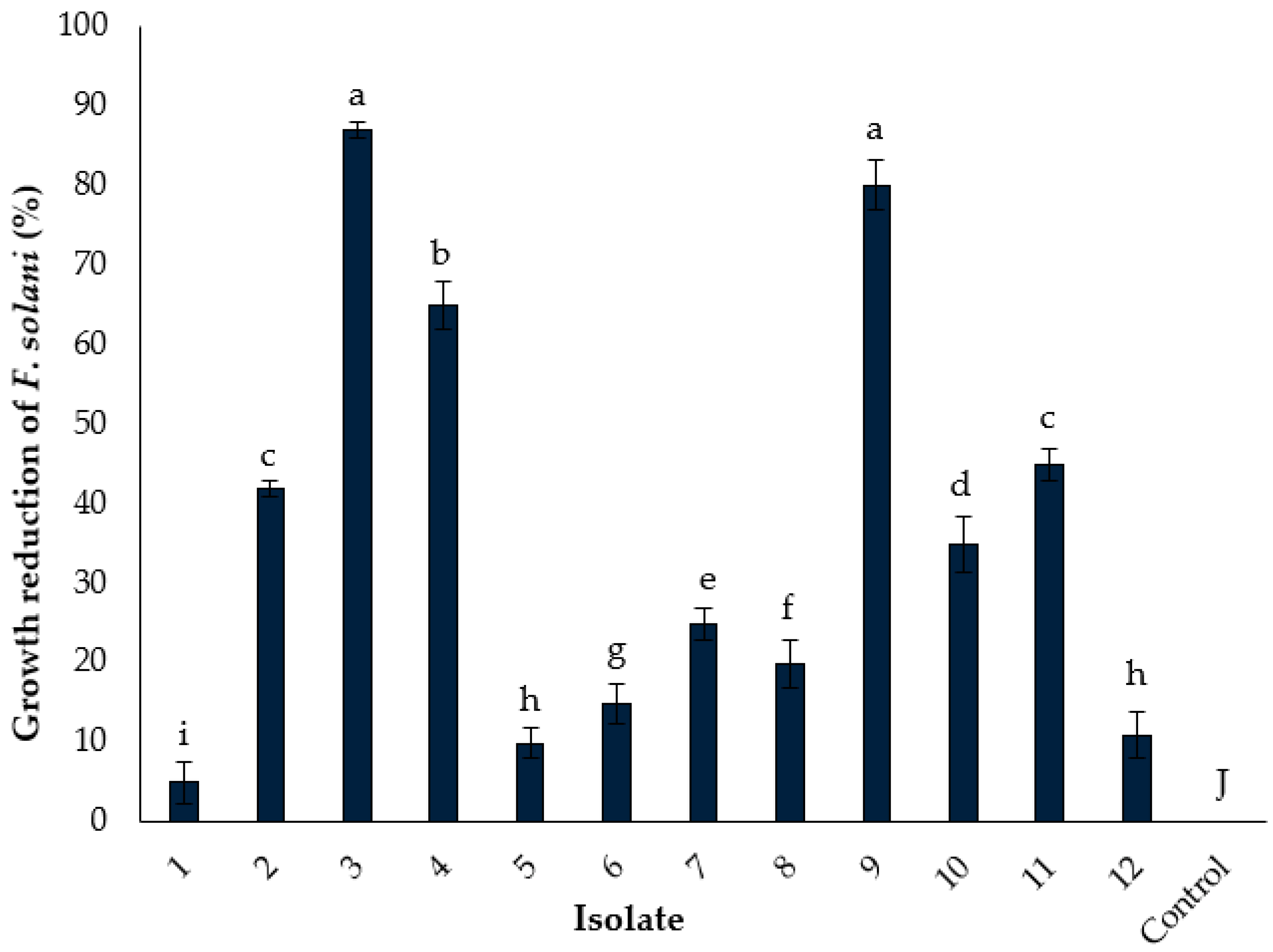
3.3. Isolation and In Vitro Screening of Fungal Endophytes for Biocontrol Potential

A total of 12 fungal endophytes were obtained from the roots of healthy cucumber plants. In the in vitro screening, all isolates showed antagonistic potential against F. solani. However, significant (p < 0.05) inhibition of the mycelial growth of F. solani was recorded. In the dual-culture assay, isolates 3, 4, and 9 (87%, 65%, and 80%, respectively) demonstrated higher mycelial growth reduction, followed by other endophyte isolates (Figure 3 and Figure 4). Isolates 2, 10, and 11 also demonstrated a significant reduction in the mycelial growth (42%, 35%, and 45% respectively) of F. solani, but the inhibition by these isolates was relatively lower, followed by isolates 3, 4, and 9. Comparatively, all isolates demonstrated biocontrol potential against F. solani; however, isolates 3, 4, and 9 caused a strong and significant reduction (Figure 3). Therefore, these isolates were selected for further evaluation.
The obtained sequences of PCR products of isolates 3, 4, and 9 were 611, 585, and 562 bp in length, respectively, and the BLAST analysis of these isolates performed in the NCBI database showed the highest similarity (100%) with Trichoderma harzianum (GenBank reference no. KR868283), Trichoderma longibrachiatum (GenBank reference no. MK871186), and Chaetomium globosum (GenBank reference no. MF461354), respectively. The identified sequences were submitted to the public domain of NCBI GenBank under accession numbers MW590687, MT604176.1, and ON705705, respectively. The phylogenetic tree of the identified isolates (Figure 5A–C) constructed using the bootstrap method signifies the relationship of the identified isolates with the sequences available in the public domain of NCBI (Figure 5).

3.4. In Vitro Quantification of Siderophore and Indole-3-Acetic Acid Production by Endophyte Antagonists

The production of siderophore and indole-3-acetic acid (IAA) by the fungi is mainly associated with their antagonistic potential; in this study, significant siderophore and IAA production was recorded (Table 1) by these bioagents. The siderophore production by T. harzianum was higher than that by C. globosum and T. longibrachiatum, as the diameter of hollow zones around T. harzianum was greater (4.3 mm) compared to C. globosum (3.6 mm) and T. longibrachiatum (3.5 mm). Furthermore, the production of IAA by T. harzianum was higher (3.6 µg/mL) compared to C. globosum (2.5 µg/mL) and T. longibrachiatum (2.4 µg/mL). The strong production of siderophores and IAA highlights the potential of these fungal endophytes as bioagents. A higher siderophore and IAA production was recorded by T. harzianum compared to C. globosum and T. longibrachiatum.

3.5. Effect of Culture Filtrates on Cumin Seed Germination

The priming of cumin seeds with the culture filtrates of selected endophyte antagonists demonstrated a positive effect on the germination rate (%) of seeds. Seed priming with the culture filtrates of T. harzianum demonstrated higher seed germination (90%) as compared to the C. globosum (80%) and T. longibrachiatum (80%) culture filtrate treatments. These results illustrate that the culture filtrates of these selected endophytes showed a positive and significant effect on the cumin seed germination (%) compared to the infected control, where a lower seed germination (40%) was recorded (Table 2). Thus, these results highlight the assertive effect of culture filtrates on cumin seeds.

3.6. In Vivo Effect of Culture Filtrates

The application of culture filtrates of the selected fungal endophytes under greenhouse conditions on cumin plants led to a significant (p < 0.05) reduction in disease severity. The application of T. harzianum and T. longibrachiatum culture filtrates significantly mitigated the disease severity (67.7% and 58.1%, respectively) on cumin plants, with lower disease severity recorded in these treatments (20% and 26%, respectively), compared to the infected control (62%). Furthermore, the application of C. globosum provided a significant reduction (59.3%) in the disease severity of cumin plants compared to the infected control. The results demonstrate that the application of culture filtrates of these fungal endophytes as bioagents could suppress the infection of F. solani on cumin plants. However, the inoculation of cumin seedlings with the culture filtrates of T. harzianum conferred significant protection to cumin plants by restraining F. solani infection compared to other treatments. Nevertheless, all treatments considerably reduced the disease severity (%) with respect to the infected control (Table 3).

3.7. Estimation of Defense-Related Enzymes

3.7.1. Peroxidase (PO)

Initially, the PO activity in the culture filtrate-treated cumin leaves was lower, followed by a slight increase after 2 days of inoculation. A significant increase in PO activity after the inoculation of T. harzianum culture filtrates was recorded, followed by an increase in other treatments after 4 and 6 days of inoculation (Figure 6A). In these results, T. harzianum-treated cumin plants demonstrated the maximum PO activity (7.3 U·mg−1 protein) after 6 days of application, whereas the other treatments (T. longibrachiatum and C. globosum) demonstrated lower PO activity. Although no significant difference between T. longibrachiatum and C. globosum after 4 and 6 days was observed, higher PO activity was recorded in both treatments after 6 days. Comparatively, in greenhouse conditions, the application of fungal endophytic bioagents increased the PO activity in cumin plants, potentially by activating the defense system of plants to prevent the infection of pathogen.

3.7.2. Phenylalanine Ammonia-Lyase (PAL)

Lower PAL activity in all treatments was recorded, which gradually increased after 2, 4, and 6 days, with a slight decrease after 8 days (Figure 6B). T. harzianum-treated plants demonstrated significantly higher (3.2 mM cinnamic acid·mg−1 protein) PAL activity after 6 days, followed by after 2, 4, and 8 days (1.9–2.2 mM cinnamic acid·mg−1 protein), where stable PAL activity was observed. Consequently, the treatment with T. longibrachiatum and C. globosum demonstrated a significant increase in PAL activity after 6 days (2.1 and 2.18, respectively). The PAL activity in T. harzianum-treated cumin plants was relatively higher than other treatments compared to the control (Figure 6B), highlighting the defensive potential of these endophytes against F. solani infection.

3.7.3. Polyphenol Oxidase (PPO)

A gradual increase in PPO activity after 2 and 4 days was recorded, which was initially stable in all treatments. Subsequently, a gradual increase in C. globosum treatments was recorded after 2, 4, and 6 days (0.31–0.40 U·mg−1 protein), whereas T. longibrachiatum treatments remained stable (0.41 U·mg−1 protein). T. harzianum-treated plants (0.21–0.70 U·mg−1 protein) demonstrated a gradual increase in PPO activity that was significantly (p < 0.05) higher after 6 days (0.70 U·mg−1 protein). However, in all treatments, PPO activity slightly decreased after 8 days, with no significant reduction recorded in the PPO activity of T. harzianum-treated plants. The PPO activity in control plants was relatively lower than that in the plants treated with the culture filtrates of selected endophytes. Overall, T. harzianum-treated plants showed higher PPO activity, highlighting the strong and significant antagonistic potential of T. harzianum (Figure 6C).

3.7.4. Phenolic Content

The results of the present study demonstrate the high level of phenolic content in all treatments. The levels were lower in all treatments after 2 days, before gradually increasing 4 days later (Figure 6D). The plants treated with the culture filtrates of T. harzianum showed higher phenolic compound production between 2 and 8 days. The phenolic content in T. harzianum-treated plants recorded after 6 days (5.82 mg gallic acid·g−1 fw) of inoculation slightly decreased after 8 days (4.42 mg gallic acid·g−1 fw) to a level similar to that after 4 days (4.48 mg gallic acid·g−1 fw). The phenolic content in T. longibrachiatum and C. globosum remained lower compared to T. harzianum after 2 and 4 days (Figure 6D) but increased after 6 days of inoculation (4.05 and 4.27 mg gallic acid·g−1 fw, respectively). Generally, the phenolic content in T. harzianum-treated plants was higher compared to T. longibrachiatum and C. globosum with respect to the control. The results of the present study highlight the high phenolic content production by these selected bioagents, which may assist in protecting cumin plants from F. solani infection in greenhouse conditions.

4. Discussion

Cumin (Cuminum cyminum L.) as a spice and herbal medicinal crop has attracted great attention worldwide; it is grown in sandy loam and clay soil throughout the postrainy seasons in arid to semiarid regions [3]. Fungal plant pathogens cause detrimental infections of various economically important crops including cumin, which results in substantial production losses worldwide [60]. Fusarium species have been extensively reported for their virulence, causing serious threats to agricultural production due to their persistent survival in soil. Many destructive diseases of cumin plants have been widely reported, with the root rot disease of cumin caused by Fusarium solani being among the most destructive diseases causing severe yield losses [9]. The resistance problem in fungal pathogens due to the extensive use of fungicides has become a challenge in agriculture, and alternative approaches such as biological control using indigenous fungal species, i.e., Trichoderma, are receiving more attention due to their significant and strong biocontrol potential, which has been extensively used against various fungal plants’ pathogens [61]. In the present study, Trichoderma species and Chaetomium globosum were used as biocontrol agents against a pathogenic fungus called F. solani, which was causing root rot disease of cumin, as various studies have reported the biocontrol potential of Trichoderma and C. globosum [20]. Furthermore, various Trichoderma species such as T. harzianum and T. longibrachiatum have been extensively used to control fungal plant pathogens through their culture filtrates [62]. In our results, the cultural and morphological characteristics of F. solani such as colony color, growth colony shape, shape and size of conidia, and conidiophores were identical to previously reported characteristics [63]. The pathogenicity tests performed on cumin seeds showed variation in the pathogenicity of isolates, as cumin seed germination was significantly affected by the F. solani pathogen. Different factors such as temperature, humidity, and moisture also influence the seed germination [14,42], whereas, in this study, ideal conditions (for cumin seed germination) were maintained, but the seeds demonstrated lower germination (%), potentially due to the pathogenic stress induced by F. solani.
The in vitro antagonistic mechanism of biocontrol agents involves various phenomena, such as competition for food, space, and nutrients, and the production of hormones such as IAA and siderophore molecules. Siderophore molecules are the most important metabolites with a low molecular weight fabricated for scavenging iron from the environment, as they have a high affinity for iron(III) [64]. In the present study, significant indoel-3-acetic acid (IAA) and siderophore production by the selected fungal bioagents was recorded, with the highest levels recorded in T. harzianum, followed by T. longibrachiatum and C. globosum, which is in line with previous findings [65]. The ability of various Trichoderma species to antagonize Fusarium was previously reported [57], supporting the results of this study.
Seed priming with different treatments, e.g., culture filtrates of fungal and bacterial bioagents, kinetin, gibberellic acid, soaking, and hormonal priming, prior to growing can significantly enhance the seed germination rate, growth, and adaptation of plants to various biotic and abiotic stresses [65]. In the present study, dressing seeds with the culture filtrates of selected endophytes led to a significant increase in cumin seed germination (%) compared to those infected and healthy controls. Furthermore, the application of culture filtrates in greenhouse conditions considerably reduced the root rot infection and mitigated the disease severity on cumin plants. The culture filtrate application of C. globosum, T. harzianum, and T. longibrachiatum bioagent endophytes demonstrated a reduction in disease severity, which is in agreement with the results in [20,21]. The antifungal potential of fungal endophytic bioagents mainly involves the production of secondary metabolites such as harzianic acid produced by T. harzianum, which has strong antifungal and plant growth-promoting activity [66] against various pathogens. Furthermore, the plants possess various inductive and constitutive mechanisms as protective barriers against the attack of various plant pathogens, presenting various protective mechanisms in response to pathogen invasion, such as constitution of the cell wall, activation and synthesis of secondary metabolites (i.e., phytoalexin), accumulation of defense proteins and peptides, and production of reactive oxygen species [67]. Secondary metabolites such as phenolic acid and lignin provide mechanical strength and rigidity to the cell wall, inhibiting the penetration of pathogens. In addition to the production of secondary metabolites, different defense enzymes such as polyphenol oxidase (PPO), peroxidase (PO), phenylalanine ammonia-lyase (PAL), and superoxide dismutase (SOD) are produced in plants, which may also play a significant role in the induction of disease resistance [68]. According to our results, the peroxidase (PO) and polyphenol oxidase (PPO) activity in the cumin plants treated with fungal endophytic bioagents significantly increased with respect to the control plants, thereby providing protection from F. solani. PO plays a key role in the biosynthesis of lignin, which is involved in the lignification that contributes to resistance [69], whereas PPO catalyzes the oxidation of o-diphenolic and monophenolic compounds. Similar results have also been reported by various researchers with regard to the induction of PPO and PO against various pathogens [69].
Phenylalanine ammonia-lyase (PAL) allows the entry of enzymes into the phenylpropanoid biosynthesis pathway, which synthesizes a variety of phenolic compounds including isoflavonoids, plant hormones, flavonoids, anthocyanins, lignins, and phytoalexins [70]. Furthermore, l-phenylalanine is converted to trans-Cinnamic acid, which acts as a precursor for the synthesis of most phenolic compounds [70]. PAL is also considered a marker of environmental stress in various plants, as it stimulates the defense enzyme activity in response to various stresses. In our study, a significant increase in PAL activity was recorded in response to the treatment of cumin seeds with culture filtrates of endophytic bioagents. The PAL activity in T. harzianum-treated plants was higher compared to T. longibrachiatum and C. globosum, as well as the control, indicating the significant and positive effect of culture filtrates of these bioagents on cumin plants. Generally, the culture filtrate of these bioagents corresponded well with a reduction in pathogen infection and an increase in PAL activity. These results are in agreement with findings reported by various researchers, where a significant increase in PAL activity was observed in response to various treatments [67]. The increase in PAL activity might be due to the increase in allelochemicals associated with the ferulic acid or p-coumaric acid group of phenolic compounds which increase PAL activity in plants [70]. Furthermore, the overproduction of reactive oxygen species (ROS) scavengers increases the PAL activity in response to various stresses, which evidently supports our findings. Secondary metabolites, specifically phenolic compounds, play an important role in medicating the defense activity [62]. In our study, a slight increase in total phenolic content was observed after 2 days, while a significant increase was recorded in cumin plants after 6 days of inoculation with endophytic culture filtrates. Thus, the accumulation of phenolic compounds boosted the activity of lignification enzymes in cumin plants, which may reflect the component of many defense signals activated by bioagents and hormonal inducers leading to the activation of the defense system in cumin plants against the attack of F. solani. Furthermore, the increase in PPO and PO restricted pathogen growth via phenolic compound oxidation to quinone, which increased the antimicrobial activity and ultimately increased the defense mechanisms. Various researchers reported identical findings, where an increase in phenolic content was recorded in response to various treatments [71,72,73] highlighting that increases in phenolic compounds activate the defense systems of various plants against fungal pathogen infections.
From the results of this study, it is clear that endophytic bioagents T. harzianum, T. longibrachiatum, and C. globosum had strong antagonistic potential, not only inhibiting the mycelial growth of F. solani causing root rot of cumin, but also significantly inhibiting the disease severity in greenhouse conditions. Furthermore, the culture filtrates of these bioagents had a crucial effect on the germination (%) of cumin seeds. The culture filtrates of these bioagents not only protected the seed embryo from the infection of F. solani, but also positively affected seed germination. Additionally, the application of culture filtrates on cumin plants demonstrated a significant production of phenolic compounds, as well as an increase in PPO, PO, and PAL activity, ultimately activating the defense system by inducing systemic resistance, which protected the plants from F. solani infection in greenhouse conditions. It is suggested that phytotoxic chemical compounds cause the overproduction of ROS in plants, which eventually elevate the level of phenolic compounds, acting as sufficient antioxidants preventing the formation of cellular deterioration due to the oxidative stress. The higher activity of PPO, PO, and PAL increased the oxidation of phenolic compounds, which led to distortion of the cell wall structure and restricted pathogen growth.

5. Conclusions

The results of the present study indicate that the culture filtrates of T. harzianum, T. longibrachiatum, and C. globosum endophytes promptly and strongly activated the defense response of cumin plants after F. solani infection. Furthermore, the culture filtrates of these bioagents not only increased germination (%), but also significantly increased the activity of defense-related enzymes and phenolic content, highlighting the defense response triggered by the root rot pathogen. Future studies should focus on the transcriptome and proteomic analysis of cumin plants inoculated with the culture filtrates of bioagents, which can assist researchers in better understanding the biochemical changes in cumin plants as well as the role of bioagents in increasing the defense mechanism toward the reduction in root rot infection of cumin plants.

Author Contributions

Conceptualization, M.A.A.M. and K.A.M.A.-E.; methodology, M.M.S., K.A.M.A.-E. and O.H.M.I.; software, K.A.M.A.-E.; validation, M.A.A.M., K.A.M.A.-E. and A.D.A.-Q.; formal analysis, M.A.A.M. and K.A.M.A.-E.; investigation, M.A.A.M., K.A.M.A.-E. and O.H.M.I.; resources, M.A.A.M. and O.H.M.I.; data curation, M.A.A.M. and A.D.A.-Q., writing—original draft preparation, O.H.M.I.; writing—review and editing, M.A.A.M. and K.A.M.A.-E.; visualization, O.H.M.I.; supervision, M.A.A.M. and K.A.M.A.-E.; project administration, M.A.A.M.; funding acquisition, M.A.A.M. All authors have read and agreed to the published version of the manuscript.

Funding

This research was funded by the Deputyship for Research and Innovation, Ministry of Education, Saudi Arabia, for funding this research work through the project number “IFPRC-156-155-2020” and King Abdulaziz University, DSR, Jeddah, Saudi Arabia.

Institutional Review Board Statement

Not applicable.

Informed Consent Statement

Not applicable.

Data Availability Statement

The data from this study are available at http://ncbi.nlm.nih.gov/, 13 June 2022.

Acknowledgments

The authors extend their appreciation to the Deputyship for Research and Innovation, Ministry of Education, Saudi Arabia, for funding this research work through the project number “IFPRC: 156178-155-2020”, and King Abdulaziz University, DSR, Jeddah, Saudi Arabia.

Conflicts of Interest

The authors declare no conflict of interest.

References

  1. Rebey, I.B.; Kefi, S.; Bourgou, S.; Ouerghemmi, I.; Ksouri, R.; Tounsi, M.S.; Marzouk, B. Ripening stage and extraction method effects on physical properties, polyphenol composition and antioxidant activities of cumin (Cuminumcyminum L.) seeds. Plant Foods Hum. Nutrit. 2014, 69, 358–364. [Google Scholar] [CrossRef] [PubMed]
  2. Mnif, S.; Aifa, S. Cumin (Cuminum cyminum L.) from traditional uses to potential biomedical applications. Chem. Biodivers 2015, 12, 733–742. [Google Scholar] [CrossRef] [PubMed]
  3. Rakesh, P.; Misra, A.K.; Singh, H.B.; Alok, K.; Dinesh, S. Diseases of Medicinal and Aromatic Plants and Their Management; Today and Tomorrow Printers and Publisher: New Delhi, India, 2019; pp. 339–352. [Google Scholar]
  4. Mehl, H.L.; Epstein, L. Identification of Fusarium solani f. sp. cucurbitae race 1 and race 2 with PCR and production of disease free pumpkin seeds. Plant Dis. 2007, 91, 1288–1292. [Google Scholar] [PubMed] [Green Version]
  5. Amer, M.A.; Salem, S.S.; Ahmed, M.A.K.; Deiaa, A.E.; Hossam, M.F.; Amr, H.H. Potential of biosynthesized zinc oxide nanoparticles to control Fusarium wilt disease in eggplant (Solanum melongena) and promote plant growth. Biometals 2022, 35, 601–616. [Google Scholar]
  6. Chanon, S.; Rungthip, S.; Toan, L.T.; Piyaporn, P.; Natthiya, B. Induced resistance against Fusarium solani root rot disease in cassava plant (Manihot esculenta Crantz) promoted by salicylic acid and Bacillus subtilis. Acta Agr. Sca. Section B-Soil Plant Sci. 2022, 72, 516–526. [Google Scholar]
  7. Yaojun, D.; Binbin, T.; Mimi, H.; Lili, W.; Kai, W.; Sixia, Y.; Jianfeng, L.; Hong, Y.; Changmei, W.; Fang, Y.; et al. High concentrations of antagonistic bacterial strains from diseased sanqi ginseng rhizosphere suppressed Fusarium root rot. Eur. J. Plant Pathol. 2022, 163, 143–153. [Google Scholar]
  8. Romberg, M.K.; Davis, R.M. Host Range and Phylogeny of Fusarium solani f. sp. eumartii from Potato and Tomato in California. Plant Dis. 2007, 91, 585–592. [Google Scholar] [CrossRef] [Green Version]
  9. Tuncturk, R.; Tuncturk, M. Effects of different phosphorus levels on the yield and quality components of cumin (Cuminum cyminum L.). Res. J. Agr. Biol. Sci. 2006, 2, 336–340. [Google Scholar]
  10. Imran, M.; Esmat, F.A.; Abo-Elyousr, K.A.M.; Sallam, N.M.A.; Khan, M.M.M.; Younas, M.W. Characterization and sensitivity of Botrytis cinerea to Benzimidazole and SDHI fungicides and illustration of resistance profile. Aus. Plant Pathol. 2021, 50, 589–601. [Google Scholar] [CrossRef]
  11. Can, Z.; Imran, M.; Xiao, L.; Hu, Z.; Li, G.; Zhang, F.; Liu, X. Difenoconazole Resistance Shift in Botrytis cinereal From Tomato in China Associated with Inducible Expression of CYP51. Plant Dis. 2021, 105, 400–407. [Google Scholar]
  12. Zhang, C.; Diao, Y.; Wang, W.; Hao, J.; Imran, M.; Duan, H.; Liu, X. Assessing the Risk for Resistance and Elucidating the Genetics of Colletotrichum truncatum That Is Only Sensitive to Some DMI Fungicides. Front. Microbiol. 2017, 8, 1779. [Google Scholar] [CrossRef] [PubMed] [Green Version]
  13. Imran, M.; Abo-Elyousr, K.A.M.; El-Sharnouby, M.E.; Ali, E.F.; Sallam, N.M.A.; Bagy, H.M.M.K.; Abdel-Rahim, I.R. Biocontrol Potential of Trichoderma harzianum and Zinc Nanoparticles to Mitigate Gray Mold Disease of Tomato. Gesunde Pflanz. 2022. [CrossRef]
  14. Zhang, W.-l.; Li, L.-I.; Zu, Y.-G.; Sonia, P. Effect of priming on the germination of Peltophorumdubium seeds under water stress. J. For. Res. 2004, 15, 287–290. [Google Scholar]
  15. Imran, M.; Abo-Elyousr, K.A.M.; Mousa, M.A.; Saad, M.M. A study on the synergetic effect of Bacillus amyloliquefaciens and dipotassium phosphate on Alternaria solani causing early blight disease of tomato. Eur. J. Plant Pathol. 2022, 162, 63–77. [Google Scholar] [CrossRef]
  16. Stappler, E.; Dattenböck, C.; Tisch, D.; Schmoll, M. Analysis of light-and carbon-specific transcriptomes implicates a class of G-protein-coupled receptors in cellulose sensing. Msphere 2017, 2, e00089-17. [Google Scholar] [CrossRef] [PubMed] [Green Version]
  17. Martínez-Medina, A.; VanWees, S.C.M.; Pieterse, C.M.J. Airborne signals from Trichoderma fungi stimulate iron uptake responses in roots resulting in priming of jasmonic acid-dependent defences in shoots of Arabidopsis thaliana and Solanum lycopersicum. Plant Cell Environ. 2017, 40, 2691–2705. [Google Scholar] [CrossRef] [PubMed]
  18. Zhang, Q.; Li, H.Q.; Zong, S.C.; Gao, J.M.; Zhang, A.L. Chemical and bioactive diversities of the genus Chaetomium secondary metabolites. Mini-Rev. Med. Chem. 2012, 12, 127–148. [Google Scholar] [CrossRef]
  19. Sibounnavong, P.; Sibounnavong, P.S.; Kanokmedhakul, S.; Soytong, K. Antifungal activities of Chaetomium brasilense CB01 and Chaetomium cupreum CC03 against Fusarium oxysporum f.sp. lycopersici race 2. J. Agric. Technol. 2012, 8, 1029–1038. [Google Scholar]
  20. Li, H.; Xiao, J.; Gao, Y.; Tang, J.; Zhang, A.; Gao, J. Chaetoglobosins from Chaetomium globosum, an endophytic fungus in Ginkgo biloba, and their phytotoxic and cytotoxic activities. J. Agric. Food Chem. 2014, 62, 3734–3741. [Google Scholar] [CrossRef]
  21. Zhao, S.; Zhang, Y.; Yan, W.; Cao, L.; Xiao, Y.; Ye, Y. Chaetomium globosum CDW7, a potential biological control strain and its antifungal metabolites. FEMS Microbiol. Lett. 2017, 364, fnw287. [Google Scholar] [CrossRef] [Green Version]
  22. Hung, P.M.; Pongnak, W.; Soytong, K.; Supatta, P. Efficacy of Chaetomium species as biological control agents against Phytophthora nicotianae root rot in citrus. Mycobiology 2015, 43, 288–296. [Google Scholar] [CrossRef] [PubMed] [Green Version]
  23. Yue, H.-M.; Wang, M.; Gong, W.-F.; Zhang, L.-Q. The screening and identification of the biological control fungi Chaetomium spp. against wheat common root rot. FEMS Microbiol. Lett. 2018, 365, fny242. [Google Scholar] [CrossRef] [PubMed]
  24. Arunkumar, K.; Parthasarathy, S.; Harish, S.; Lingan, R.; Raguchander, T. Evaluating an isotonic aqueous formulation of Chaetomium globosum Kunze for the management of potato black scurf disease caused by Rhizoctonia solani Kuhn in India. J. Plant Pathol. 2022, 104, 191–202. [Google Scholar]
  25. Tian, Y.; Fu, X.; Zhang, G.; Zhang, R.; Kang, Z.; Gao, K.; Mendgen, K. Mechanisms in Growth-Promoting of Cucumber by the Endophytic Fungus Chaetomium globosum Strain ND35. J. Fungi 2022, 8, 180. [Google Scholar] [CrossRef]
  26. Yun, Z.; Heqin, Z.; Yonghao, Y.; Canming, T. Antifungal Activity of Chaetoviridin A from Chaetomium globosum CEF-082 Metabolites Against Verticillium dahliae in Cotton. Mol. Plant-Microbe Inter. 2021, 34, 758–769. [Google Scholar]
  27. Monika, S.; Dhriti, K.; Vipul, K.; Sheteiwy, M.S.; Ramakrishnan, M.; Landi, M.; Araniti, F.; Anket, S. Trichoderma: The “Secrets” of a Multitalented Biocontrol Agent. Plants 2020, 9, 762. [Google Scholar]
  28. Kredics, L.; Chen, L.; Kedves, O.; Büchner, R.; Hatvani, L.; Allaga, H.; Nagy, V.D.; Khaled, J.M.; Alharbi, N.S.; Vágvölgyi, C. Molecular tools for monitoring Trichoderma in agricultural environments. Front. Microbiol. 2018, 9, 1599. [Google Scholar] [CrossRef]
  29. Zhang, J.; Chen, G.Y.; Li, X.Z.; Hu, M.; Wang, B.Y.; Ruan, B.H.; Zhou, H.; Zhao, L.X.; Zhou, J.; Ding, Z.T.; et al. Phytotoxic, antibacterial, and antioxidant activities of mycotoxins and other metabolites from Trichoderma sp. Nat. Prod. Res. 2017, 31, 2745–2752. [Google Scholar] [CrossRef]
  30. Kumar, S. Trichoderma: A biological weapon for managing plant diseases and promoting sustainability. Int. J. Agric. Sci. Med. Vet. 2013, 1, 106–121. [Google Scholar]
  31. Nandini, M.L.N.; Srinivasulu, B.; Gopal, K.; Ruth, C.; Devi, P.R.; Babu, M.R.; Padmaja, V.V. Induction of induced systemic resistance in capsicum chilli against Colletotrichum truncatum through fungal biocontrol agents. J. Agr. Ecol. 2022, 13, 99–111. [Google Scholar] [CrossRef]
  32. Candra, R.T.; Prasasty, V.D.; Karmawan, L.U. Biochemical Analysis of Banana Plants in Interaction between Endophytic Bacteria Kocuriarhi zophila and the Fungal Pathogen Fusarium oxysporum f. sp. cubense Tropical Race (Foc TR4). Biol. Life Sci. Forum. 2022, 11, 84. [Google Scholar]
  33. Vidhyasekaran, P. Fungal Pathogenesis in Plants and Crops; CRC Press: Boca Raton, FL, USA, 2007. [Google Scholar]
  34. Prasannath, K.; Dharmadasa, K.N.P.; De Costa, D.M.; Hemachandra, K.S. Variations of Incidence, Types of Virus Diseases and Insect Vector Populations of Tomato (Solanum lycopersicum L.), Grown in Different Agroecological Regions of Sri Lanka under Two Crop Management Systems. Trop. Agr. Res. 2015, 25, 376–395. [Google Scholar] [CrossRef]
  35. Vinale, F.; Sivasithamparam, K.; Ghisalberti, E.L.; Marra, R.; Woo, S.L.; Lorito, M. Trichoderma-plant-pathogen interactions. Soil Biol. Biochem. 2008, 40, 1–10. [Google Scholar] [CrossRef]
  36. Li, L.; Steffens, J.C. Over expression of polyphenol oxidase in transgenic tomato plants results in enhanced bacterial disease resistance. Planta 2002, 215, 239–247. [Google Scholar] [CrossRef] [PubMed]
  37. Hussain, A.; Kamran, M.A.; Javed, M.T.; Hayat, K.; Farooq, M.A.; Ali, N.; Ali, M.; Manghwar, H.; Jan, F.; Chaudhary, H.J. Individual and combinatorial application of Kocuriarhi zophila and citric acid on phytoextraction of multi-metal contaminated soils by Glycine max L. Environ. Exp. Bot. 2019, 159, 23–33. [Google Scholar] [CrossRef]
  38. Al-Jaradi, A.; Al-Mahmooli, I.; Janke, R.; Maharachchikumbura, S.; Al-Saady, N.; Al-Sadi, A.M. Isolation and identification of pathogenic fungi and oomycetes associated with beans and cowpea root diseases in Oman. PeerJ 2018, 6, e6064. [Google Scholar] [CrossRef] [Green Version]
  39. Booth, C. The Genus Fusarium Commonwealth Mycological Institute; CAB International: Wallingford, UK, 1971. [Google Scholar]
  40. Nelson, P.E.; Toussoun, T.A.; Marasas, W.F.O. Fusarium Species: An Illustrated Manual for Identification; Pennsylvania State University Press: University Park, PA, USA, 1983. [Google Scholar]
  41. Burgess, L.W.; Summerell, B.A.; Bullock, S.; Gott, K.P.; Backhouse, D. Laboratory Manual for Fusarium Research, 3rd ed.; University of Sydney and Botanic Garden: Sydney, Australia, 1994. [Google Scholar]
  42. Heidari, M.; Sadeghi, H. Germination and emergence of primed cumin (Cuminum cyminum L.) seeds with GA3 under different temperature regimes. Int. J. Biosci. 2014, 5, 266–272. [Google Scholar]
  43. Gams, W.; Bissett, J. Morphology and identification of Trichoderma. Basic biology, taxonomy and genetics. In Trichoderma and Glicladium; Kubicek, C.P., Harman, G.E., Eds.; Taylor & Francis Ltd.: London, UK, 1998; Volume 1, pp. 1–34. [Google Scholar]
  44. Rifai, E. A revision of the genus Trichoderma. Mycologia 1969, 116, 1–56. [Google Scholar]
  45. Onions, A.H.S.; Barron, G.L. Monophialidic Species of Paecilomyces; Commonwealth Mycological Institute: Richmond, UK, 1967. [Google Scholar]
  46. Bekker, T.F.; Kaiser, C.; Merwe, R.V.D.; Labuschagne, N. In-vitro inhibition of mycelial growth of several phytopathogenic fungi by soluble potassium silicate. S. Afr. J. Plant Soil 2006, 23, 169–172. [Google Scholar] [CrossRef] [Green Version]
  47. Jiang, Y.; Wang, J.L.; Chen, J.; Mao, L.J.; Feng, X.X.; Zhang, C.L. Trichoderma Biodiversity of Agricultural Fields in East China Reveals a Gradient Distribution of Species. PLoS ONE 2016, 11, e0160613. [Google Scholar] [CrossRef] [Green Version]
  48. Liu, Y.J.; Whelen, S.; Hall, B.D. Phylogenetic relationships among Ascomycetes: Evidence from an RNA Polymerse II Subunit. Mol. Biol. Evol. 1999, 16, 1799–1808. [Google Scholar] [CrossRef] [PubMed] [Green Version]
  49. White, T.J.; Bruns, T.; Lee, S.; Taylor, J. Amplification and direct sequencing of fungal ribosomal RNA genes for phylogenetics. Mycologia 2013, 64, 315–322. [Google Scholar]
  50. Tamura, K.; Stecher, G.; Peterson, D.; Filipski, A.; Kumar, S. MEGA6: Molecular Evolutionary Genetics Analysis version 6.0. Mol. Biol. Evolution 2013, 30, 2725–2729. [Google Scholar] [CrossRef] [PubMed] [Green Version]
  51. Schwyn, B.; Neilands, J.B. Universal chemical assay for the detection and determination of siderophores. Anal. Biochem. 1987, 160, 47–56. [Google Scholar] [CrossRef]
  52. Bano, N.; Musarrat, J. Characterization of a new Pseudomonas aeruginosa strain NJ-1 5 as a potential biocontrol agent. Curr. Microbiol. 2003, 46, 324–328. [Google Scholar] [CrossRef] [PubMed]
  53. Gour, H.N.; Agrawal, S. A wilt toxin from Fusarium oxysporum f.sp. cumini Patel and Prasad. Curr. Sci. 1988, 57, 849–851. [Google Scholar]
  54. Bradford, M. A rapid and sensitive method for the quantitation of microgram quantities of protein utilizing the principle of protein dye binding. Anal. Biochem. 1976, 72, 248–250. [Google Scholar] [CrossRef]
  55. Pütter, J. Peroxidases. In Methods of Enzymatic Analysis; Academic Press: Cambridge, MA, USA, 1974; pp. 685–690. [Google Scholar]
  56. Zucker, M. Sequential induction of phenylalanine ammonia-lyase and a lyase-inactivating system in potato tuber disks. Plant Physiol. 1968, 43, 365–374. [Google Scholar] [CrossRef]
  57. Jockusch, H. Induction of defence responses against Erwinia soft rot by an endogenous pectatelyase in potatoes. Physiol. Mol. Plant Pathol. 2002, 60, 91–100. [Google Scholar]
  58. Şahin, F.; Güllüce, M.; Daferera, D.; Sökmen, A.; Sökmen, M.; Polissiou, M.; Agar, G.; Özer, H. Biological activities of the essential oils and methanol extract of Origanum vulgare ssp. vulgare in the Eastern Anatolia region of Turkey. Food Control 2004, 15, 549–557. [Google Scholar] [CrossRef]
  59. Steel, R.G.D.; Torrie, J.H.; Dickey, D.A. Principles and Procedures of Statistics, A Biometrical Approach, 3rd ed.; McGraw Hill Book Co. Inc.: New York, NY, USA, 1996. [Google Scholar]
  60. Garima, B.; Indu, R.; Sanjay, K.; Vinay, S. Induction of β-1,3-glucanase and chitinase activity, cloning and their characterization in the defense response of Cuminum cyminum plant against the fungal pathogen Fusarium oxysporum. Arch. Phytopathol. Plant Prot. 2021, 54, 1617–1632. [Google Scholar]
  61. Mohammadi, A.; Shams-Ghahfarokhi, M.; Nazarian-Firouzabadi, F.; Kachuei, R.; Gholami-Shabani, M.; Razzaghi-Abyaneh, M. Giberellafujikuroi species complex isolated from maize and wheat in Iran: Distribution, molecular identification and fumonisin B1 in vitro biosynthesis. J. Sci. Food Agric. 2016, 96, 1333–1340. [Google Scholar] [CrossRef] [PubMed]
  62. Nurbailis, A.D.; Haliatur, R.; Yenny, L. Potential of culture filtrate from Trichoderma spp. as biofungicide to Colletotrichum gloeosporioides causing anthracnose disease in chili. Biodiversitas 2019, 20, 2915–2920. [Google Scholar] [CrossRef]
  63. Leslie, J.F.; Summerell, B.A. Fusarium laboratory workshops—A recent history. Mycotoxin Res. 2006, 22, 73. [Google Scholar] [CrossRef]
  64. Hider, R.C.; Kong, X. Chemistry and biology of siderophores. Nat. Prod. Rep. 2010, 27, 637–657. [Google Scholar] [CrossRef] [PubMed]
  65. Debbi, A.; Boureghda, H.; Monte, E.; Hermosa, R. Distribution and genetic variability of Fusariumoxysporum associated with tomato diseases in Algeria and biocontrol strategy with indigenous Trichoderma spp. Front. Microbiol. 2018, 9, 282. [Google Scholar] [CrossRef] [Green Version]
  66. Nguyen, T.N.; Son, S.H.; Jordan, M.C.; Levin, D.B.; Ayele, B.T. Lignin biosynthesis in wheat (Triticumaestivum L.): Its response to water logging and association with hormonal levels. BMC Plant Biol. 2016, 16, 28. [Google Scholar] [CrossRef] [Green Version]
  67. Lehmann, S.; Serrano, M.; Haridon, F.L.; Tjamos, S.E.; Metraux, J.P. Reactive oxygen species and plant resistance to fungal pathogens. Phytochem 2015, 112, 54–62. [Google Scholar] [CrossRef]
  68. Okey, E.N.; Duncan, E.J.; Sirju-charran, G.; Sreenivasan, T.N. Phytopthora canker resistance in cacao: Role of peroxidase, polyphenol oxidase and phenylalanine ammonia lyase. J. Phytopathol. 1997, 145, 295–299. [Google Scholar] [CrossRef]
  69. Fotoohiyan, Z.; Rezaee, S.; Bonjar, G.H.S.; Mohammadi, A.H.; Moradi, M. Induction of systemic resistance by Trichoderma harzianum isolates in pistachio plants infected with Verticillium dahliae. J. Nuts 2015, 6, 95–111. [Google Scholar]
  70. La Camera, S.; Gouzerh, G.; Dhondt, S.; Hoffmann, L.; Fritig, B.; Legrand, M.; Heitz, T. Metabolic reprogramming in plant innate immunity: The contributions of phenylpropanoid and oxylipin pathways. Immunol. Rev. 2004, 198, 267–284. [Google Scholar] [CrossRef] [PubMed]
  71. Pawel, S.; Urszula, K.; Bederska-Błaszczyk, M.; Agnieszka, G. Canavanine Increases the Content of Phenolic Compounds in Tomato (Solanum lycopersicum L.) Roots. Plants 2020, 9, 1595. [Google Scholar]
  72. Abo-Elyousr, K.A.M.; Ibrahim, Y.E.; Balabel, N.M. Induction of disease defensive enzymes in response to treatment with acibenzolar-S-methyl (ASM) and Pseudomonas fluorescens Pf2 and inoculated with Ralstonia solanacearum race 3, biovar2 (phylotype II). J. Phytopathol. 2012, 160, 382–389. [Google Scholar] [CrossRef]
  73. Youssef, K.; Roberto, S.R.; Tiepo, A.N.; Constantino, L.V.; de Resende, J.T.V.; Abo-Elyousr, K.A.M. Salt Solution Treatments Trigger Antioxidant Defense Response against Gray Mold Disease in Table Grapes. J. Fungi 2020, 6, 179. [Google Scholar] [CrossRef] [PubMed]
Figure 1. Pathogenicity screening of isolates with respect to seed germination of cumin under greenhouse conditions. Values followed by different letters indicate that means are significantly different, while identical letters indicate that means are not significantly different according to Fisher’s least significant difference at p = 0.05.
Agronomy 12 02612 g001
Figure 2. Semistrict concurrence of most parsimonious identified isolate No. 7 (Fusarium solani) based on sequenced data. This tree was overall compatible with highly supported lineages according to the Bayesian 50% majority-rule consensus tree. Jackknife frequencies (10,000 replicates) are shown above each node. The red dot in the figure represents isolate 7, identified as Fusarium solani.
Agronomy 12 02612 g002
Figure 3. In vitro screening of fungal endophytes for their biocontrol potential against the highly virulent Fusarium solani isolate. Identical letters within each treatment are not significantly different at the p < 0.05 level of confidence according to the least significant difference (LSD) multiple range test.
Agronomy 12 02612 g003
Figure 4. In vitro screening of the endophytic fungi for their biocontrol potential against the highly virulent Fusarium solani isolate. (A), Control; (B), Chaetomium globosum; (C), Trichoderma harzianum; (D), T. longibrachiatum.
Agronomy 12 02612 g004
Figure 5. Phylogenetic trees of identified fungal bioagent isolates. Semistrict concurrence of most parsimonious trees based on ITS sequence data. This tree is overall compatible with highly supported lineages according to the Bayesian 50% majority-rule consensus tree. Jackknife frequencies (10,000 replicates) are shown above each node. The dots in each figure represent the isolates identified in this study. A, Trichoderma harzianum, B, Trichoderma longibrachiatum and C, Chaetomium globosum.
Agronomy 12 02612 g005aAgronomy 12 02612 g005b
Figure 6. Effect of culture filtrate of fungal endophytic bioagents on the induction of (A) peroxidase (PO), (B) phenylalanine ammonia-lyase (PAL), (C) polyphenol oxidase (PPO), and (D) phenol content in cumin plants under greenhouse conditions. Identical letters within each treatment are not significantly different at the p < 0.05 level of confidence according to the least significant difference (LSD) multiple range test. C. globosum, Chaetomium globosum; T. harzianum, Trichoderma harzianum; T. longibrachiatum, Trichoderma longibrachiatum; H. CK, healthy control.
Agronomy 12 02612 g006aAgronomy 12 02612 g006b
Table 1. Production of siderophore and indole-3-acetic acid (IAA) by selected endophytic fungal bioagents.
IsolatesHalo Zone (mm)IAA (μg/mL)
Chaetomium globosum3.6 ± 0.67 b2.5 ± 1.02 b
Trichoderma harzianum4.3 ± 1.03 a3.6 ± 0.48 a
Trichoderma longibrachiatum3.5 ± 0.96 b2.4 ± 0.87 b
Means followed by the same letter are not significantly different according to Duncan’s multiple range tests at p < 0.05. Results are shown as the mean ± SE of four replicates for each treatment.
Table 2. Effect of culture filtrate of different bioagents on seed germination of cumin Baladi in vitro.
Treatment of Culture FiltratesSeed Germination (%)
Chaetomium globosum80 ± 0.46 b
Trichoderma harzianum90 ± 1.93 a
Trichoderma longibrachiatum80 ± 0.63 b
Infected control40 ± 0.39 d
Identical letters within each treatment are not significantly different at the p < 0.05 level of confidence according to the LSD multiple range test. Results are shown as the mean ± SE of four replicates for each treatment.
Table 3. Foliar effect of cultural filtrates of bioagents on disease severity (%) after the artificial inoculation of F. solani pathogen under greenhouse conditions.
IsolatesDS (%)DR (%)
Chaetomium globosum25 ± 0.56 b59.3
Trichoderma harzianum20 ± 0.21 c67.7
Trichoderma longibrachiatum26 ± 0.87 b58.1
Infected control62 ± 0.47 a0
Healthy control00 ± 0.00 d0
DS, disease severity; DR, disease reduction. Values in the same column followed by the same letter are not significantly different according to the LSD test (p = 0.05). Results are presented as the mean ± SE of four replicates for each treatment. Disease reduction (DR%) was calculated as = (DS control plants—DS treated plants)/DS control plant) × 100.
Publisher’s Note: MDPI stays neutral with regard to jurisdictional claims in published maps and institutional affiliations.

