Article

Pathogenicity of Cordyceps javanica (Hypocreales: Cordycipitaceae) to Diaphorina citri (Hemiptera: Liviidae) Adults, with Ultrastructural Observations on the Fungal Infection Process

1
Guangdong Key Laboratory of Animal Conservation and Resource Utilization, Guangdong Public Laboratory of Wild Animal Conservation and Utilization, Guangdong Engineering Research Center for Mineral Oil Pesticides, Institute of Zoology, Guangdong Academy of Science, Guangzhou 510260, China
2
Department of Entomology and Nematology, Indian River Research and Education Center, Institute of Food and Agricultural Sciences, University of Florida, 2199 South Rock Road, Fort Pierce, FL 34945, USA
3
Department of Plant Pathology, Indian River Research and Education Center, Institute of Food and Agricultural Sciences, University of Florida, 2199 South Rock Road, Fort Pierce, FL 34945, USA
4
BioBee USA, 5126 S. Royal Atlanta Dr, Tucker, GA 30084, USA
5
College of Horticulture, Fujian Agriculture and Forestry University, Fuzhou 350002, China
6
College of Plant Protection, Fujian Agriculture and Forestry University, Fuzhou 350002, China
*
Authors to whom correspondence should be addressed.
Agronomy 2021, 11(12), 2476; https://doi.org/10.3390/agronomy11122476
Submission received: 10 November 2021 / Revised: 25 November 2021 / Accepted: 2 December 2021 / Published: 6 December 2021
(This article belongs to the Special Issue Citrus Production and Protection from Pests and Diseases)

Abstract

:
Entomopathogenic fungi are proposed biological control agents against the Asian citrus psyllid (Diaphorina citri). We quantified the pathogenicity of Cordyceps javanica strain Apopka 97 strain (Cja Apopka 97) (blastospores and conidia) against D. citri using the spray technique. We also used light and scanning electron microscopy to observe the Cja Apopka 97, infection process against D. citri adults at different stages pre- and post-mortem. Our findings demonstrated that psyllid mortality in the blastospore treatment ranged from 8 to 25% within 24–48 h of exposure, compared to 0% in the conidial and control treatments. However, psyllid mortality rate had reached 100% by 7 days after exposure at a concentration of 107 spores/mL, under both fungal treatments compared to the controls (0%). SEM and light microscopy revealed several stages in in the Cja Apopka 97 infection process of D. citri, including spore adherence and germ tube formation within 24 h post-inoculation, penetration pegs and mycelia growth on wings after 72 h, rupturing of cuticle after 96 h and mycelial mass colonizing host body after 144 h. Our study findings provide basic information on the interaction of entomopathogenic fungi with D. citri which will assist in the understanding of the infection process and the potential roles of entomopathogenic fungi in its management.

1. Introduction

Diaphorina citri (Hemiptera: Liviidae) is an economically important pest of citrus worldwide and the vector for Candidatus Liberibacter asiaticus, a Gram-negative bacterium which is associated with citrus Huanglongbing [1,2]. Currently, D. citri management is mostly based on intensive use of synthetic foliar insecticides, sometimes in combination with soil-applied systemic insecticides, such as aldicarb or imidacloprid [3,4]. However, these treatments are not sustainable because D. citri has a rapid generation time and can build up resistance to these chemical insecticides and can multiply very rapidly. Furthermore, broad spectrum synthetic chemical pesticides are becoming less acceptable because they have low selectivity and increasing safety issues [5]. Therefore, there is a clear need to develop alternative control and management strategies based on the use of biocontrol agents which are effective for D. citri management as well as ecologically and user friendly [6].
Currently under research are entomopathogenic fungi belonging to the Hypocreales: Cordycipitaceae [7]. Cordyceps species have a worldwide distribution with wide-ranging host associations among several insect orders. These fungi are being used or developed for the management of several economically important pests, such as lepidopterans, psyllids, thrips and whiteflies [8,9,10].
Previous authors investigated the virulence and host–pathogen associations with Cordyceps and D citri [2,11,12,13,14]. However, few quantified how the pathogenic mode of action relates to the ultrastructural investigations of fungal infection processes [15,16,17]. In this study, we describe the ultrastructural observations on fungal infection process of Cordyceps javanica (Frieder. & Bally) Kepler, B. Shrestha & Spatafora (Hypocreales: Cordycipitaceae) strain Apopka 97 (henceforth Cja Apopka 97) colonizing D. citri through SEM (scanning transmission electron microscopy) and light microscopy techniques. We also compared the relative virulence of Cja Apopka 97 blastospore and conidia, two infective propagule types that can be produced as inoculum for microbial insecticides.

2. Materials and Methods

2.1. Insects

A population of healthy D. citri adults were collected from a citrus orchard located in Guangzhou city, Guangdong province, China, and maintained on orange jasmine, Murraya paniculata (L.) Jack (Rutaceae). The insects were reared in the Institute of Zoology, Guangdong Academy of Sciences, Guangzhou, China. Psyllids were reared at 25 °C, with 60% relative under a 14-h photophase. Four successive generations were reared prior to the start of the study.

2.2. Plants and Fungus

Orange jasmine, Murraya paniculata (L.) leaves naturally infested with Cja Apopka 97 were sampled in the laboratory and subsequently maintained on Czapek Dox (CD) media (Thermo Fisher Scientific (China) Co., Ltd., Shanghai, China) for one month. For long-term storage (two months), conidial suspensions were prepared in 20–30% glycerol solution, and then stored at −80 °C in chamber (Jinan Qianshi Biotechnology Co., Ltd., Jinan, China) containing liquid nitrogen as stock cultures and sub-cultured in our laboratory until needed. Passaging was conducted on the D. citri (host insect) after every two to three months and the subculture was re-isolated and maintained on CD and preserved by lyophilization. The subculture stock of Cja Apopka 97 was used for all the experiments.

2.3. Molecular Characterization of Entomopathogenic Fungi

Fungal identification was carried out using molecular analysis. The Cja Apopka 97 was grown in CD culture media and incubated for 10 days at 25 °C. DNA extraction was performed from the mycelium using the FastDNA® spin kit (MP Biomedicals, Illkirch, France). The precipitated DNA pellet was dissolved in sterile water (30 μL) and stored at −20 °C until needed. Additionally, a spectrophotometer was used at 260/280 nm to quantify the DNA concentration.
Detection and identification of Cja Apopka 97 was performed through polymerase chain reaction using a universal fungal primer designed for amplification of 18S rRNA gene using an Applied Biosystems 2720 thermal cycler. 18S (sequence: TCCGTCGGTGAACCTGCGG) was used as the forward primer and 18S (sequence: TCCTCCGCTTATTGATATGC) as the reverse primer [18,19]. The reaction was set up as follows: an initial denaturation step at 95 °C for 5 min, followed by 30 cycles at 95 °C for 30 s, 57 °C for 1 min, and 72 °C for 1 min, followed by a final extension step at 72 °C for 6 min. Gel Extraction Kit (E.Z.N.A®, Shanghai, China) was used to purify amplified DNA products separated on a 1% agarose gel stained with ethidium bromide solution.

2.4. Preparation of Conidia

Cja Apopka 97 was sub-cultured on CD culture media and incubated for 10 days at 27 °C to sporulate. For conidial production, a suspension of mycelia fragments with conidia was prepared by scraping the fungal growth with a sterile spatula and suspending in 10 mL of Tween 80 (v/v; 0.01%). The suspension was vortexed and filtered using sterile cotton for the elimination of mycelial fragments. Conidial concentration was finally adjusted with sterile 0.01% Tween 80 (v/v) to 1 × 10⁷ conidia/mL using hemocytometer. Conidial viability was determined by plating 0.1 mL aliquots on CD plates and incubated at 27 °C with 65–70% RH under a light (14 h): dark (10 h) photoperiod for 24 h. The conidia were 99 viable after 24 h incubation period).

2.5. Inoculation of D. citri with Entomopathogenic Fungi

Murraya paniculata plants (2–3 months old) were selected at random for experimental use. Mated males and females (100) of D. citri were released into the center of each mesh cage. Females were allowed to oviposit for 24 h before adults were removed. All leaflets were subsequently observed under a stereomicroscope (SZ680, CNOPTEC) and the numbers of eggs on the abaxial surface were counted. The detached leaf petioles were covered with a piece of sterile cotton, moistened with 2 mL of sterile distilled water and placed in sterile 90 mm plastic Petri dishes containing three sterile Whatman filter papers, also moistened with sterile distilled water (2 mL) (Figure 1), based on earlier method [20]. Control leaflets were dipped into Tween 80 (0.01%; v/v). Both dishes (treated and control) were sealed and incubated in a growth chamber set at constant temperature (25 °C) and R.H. (70 ± 2%) with a photoperiod of 14 h (L):10 h (D) for 7 days.
Mated gravid female D. citri (100) were released into the center of new mesh cages. Females were permitted to oviposit (for 24 h) before adults were removed, as described above. The eggs were allowed to develop into third or fourth instar nymphs. Twenty nymphs were separately kept in each Petri dish, and were treated topically, with a concentration of conidial suspension (107 conidia/mL) of Cja Apopka 97 strain or 0.01% v/v Tween 80 solution (control) for 30 sec. Following inoculation, leaf samples with nymphs were air dried at room temperature (27 ± 1 °C) for 10 min. Leaf petioles were wrapped in moistened cotton and then transferred into Petri dishes (Figure 1). Each fungal inoculation and the control were replicated 5 times. The Petri dishes were incubated in a growth chamber at 25 ± 1 °C and 70 ± 2.0% R.H. under a 14 h:10 h L:D photoperiod.
To rear D. citri adults, mated males together with females (100) were released into other insect cages for oviposition. After 24 h all the females were removed. The leaves were incubated for 16–17 days to generate fresh adult D. citri. Three to four day-old adults were used for pathogenicity test following the same procedure as described above for the eggs.

2.6. Psyllid Mortality Bioassays

The mortality of the adult D. citri in the leaf disks bioassays was determined using the protocol described by Avery, et al. [21]. Treatments consisted of the following (1) blastospores or (2) conidia of Cja Apopka strain and (3) tween for the control. Adult D. citri mortality was assessed daily for 7 days.

2.7. Light Microscopy (LM) and Electron Microscopy (EM)

The morphological development of Cja Apopka-97 growth was observed on the pre-mortem and dead D. citri over time as well as culture medium, using scanning electron microscopy (SEM), and light microscopy (LM).
(a) For light microscopic observations, a piece of PDA agar with grown fungus was collected using a sterile scalpel and mounted on a glass slide. A drop of lactophenol blue solution was used to enable observation under a bright field light microscope (OLYMPUS BX41, Shinjuku, Tokyo, Japan).
(b) For conventional SEM, at 24, 48, 72, 96, 120, 144, and 168 h, the infected D. citri were collected (post-inoculation) and observed using a Hitachi SU-3500 SEM (Hitachi, Tokyo, Japan) operating at 15 KV. Adult D. citri were prepared by first being immersed in 4% glutaraldehyde for 24 h, rinsed thrice for 10 min (each time) in phosphate buffer (0.2 M) and then dehydrated in an ethanol series (75, 80, 85, 90, 95, and 100% ethanol [v/v]). Samples were then immersed in chloroform (for 1 h) to dissolve the surface wax [13,16], followed by a final rinse in ethanol (100%). The ethanol was then exchanged for liquid (CO2) and D. citri samples were prepared using a drying apparatus (K850). D. citri samples were mounted on a graphite pad and sputter coated with gold to a standard thickness (20 nm). Micrographs were taken with a digital camera (Canon EOS 350D).
(c) For SEM, fungal-treated psyllids were fixed for 2 h in a solution of 2% glutaraldehyde, 3% paraformaldehyde, in sodium cacodylate buffer 0.2 M, pH 7.4 (#11650 Electron Microscopy Sciences, Hatfield, PA, USA), followed by dehydration in an ethanol series of 35, 50, 70, 95, and three changes in 100%, each for 1 h. Dehydrated samples were lyophilized, and sputter coated with gold (at thickness of 20 mm). A Hitachi 4800, SEM microscope was used to examine the specimens (Tokyo, Japan).

2.8. Statistical Analysis

For mortality bioassays, within each treatment group (20 disks/treatment), each disk (Petri dish) was counted as an independent replication, and each of the three treatments was evaluated on three successive dates (n = 60/treatment) until the insects started dying (n = 60/treatment—# dying). Cumulative mean percent mortality was determined using Abbott’s formula. An ANOVA was used to compare the means of mortality (%), followed by a Tukey’s HSD test (p < 0.05). All statistical analyses were carried out on a WIN PRO platform using SAS Proc GLM routines (SAS 1999–2001).

3. Results

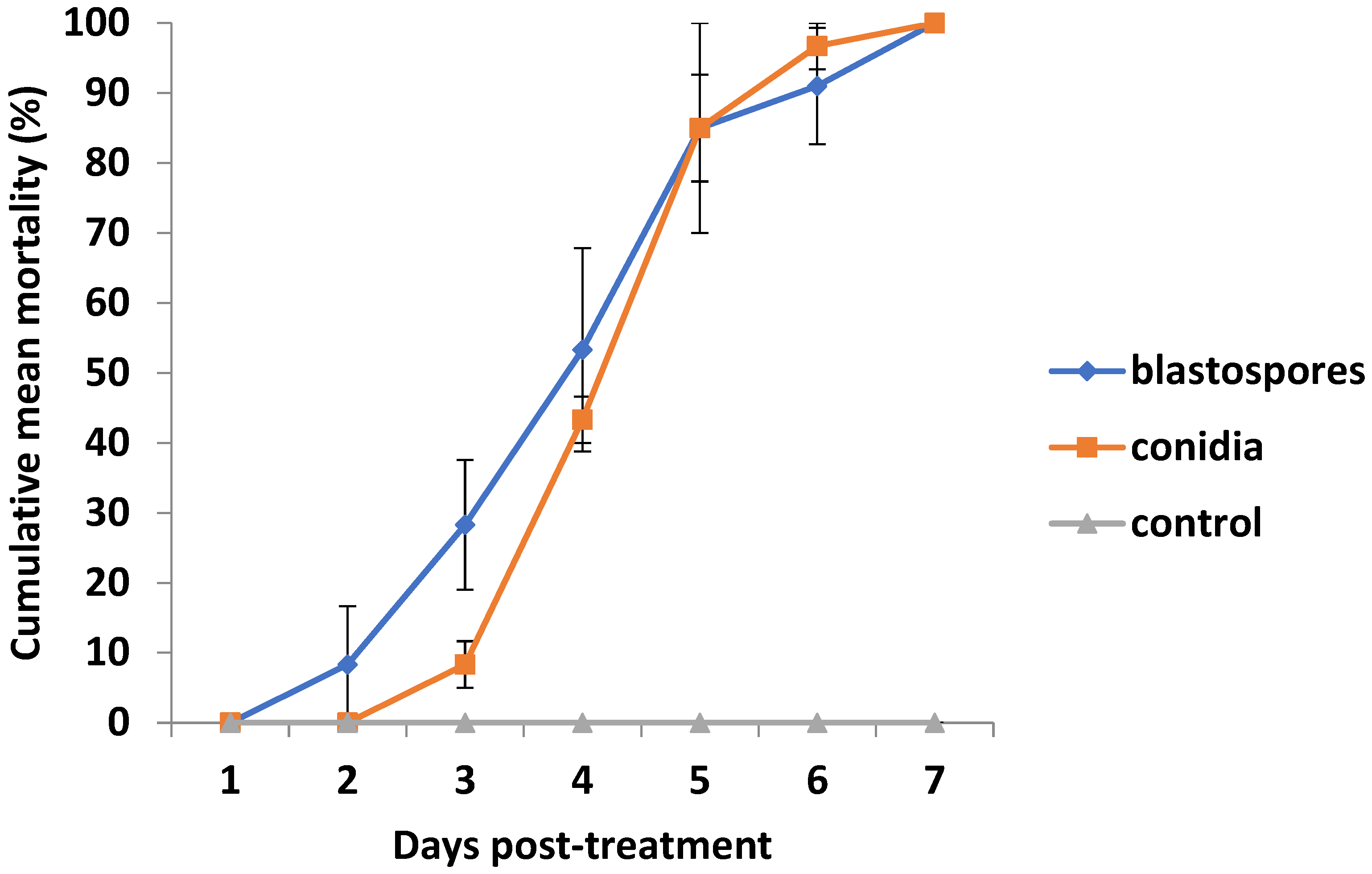
3.1. Percent Psyllid Mortality

Psyllids started dying earlier in the blastospore treatment, with initial mortality observed on day 2 post-inoculation (Figure 2). However, both blastospore and conidia treatments significantly affected cumulative percent mortality of adult psyllids that died after 4 days post-exposure (F = 7.82; df = 2, 8; p = 0.04). Both blastospore and conidial treatments had 91–97% mortality at 6 days. Seven days after exposure to Cja Apopka 97 blastospores or conidia, all psyllid adults died. No psyllids died in the control over the 7 days.

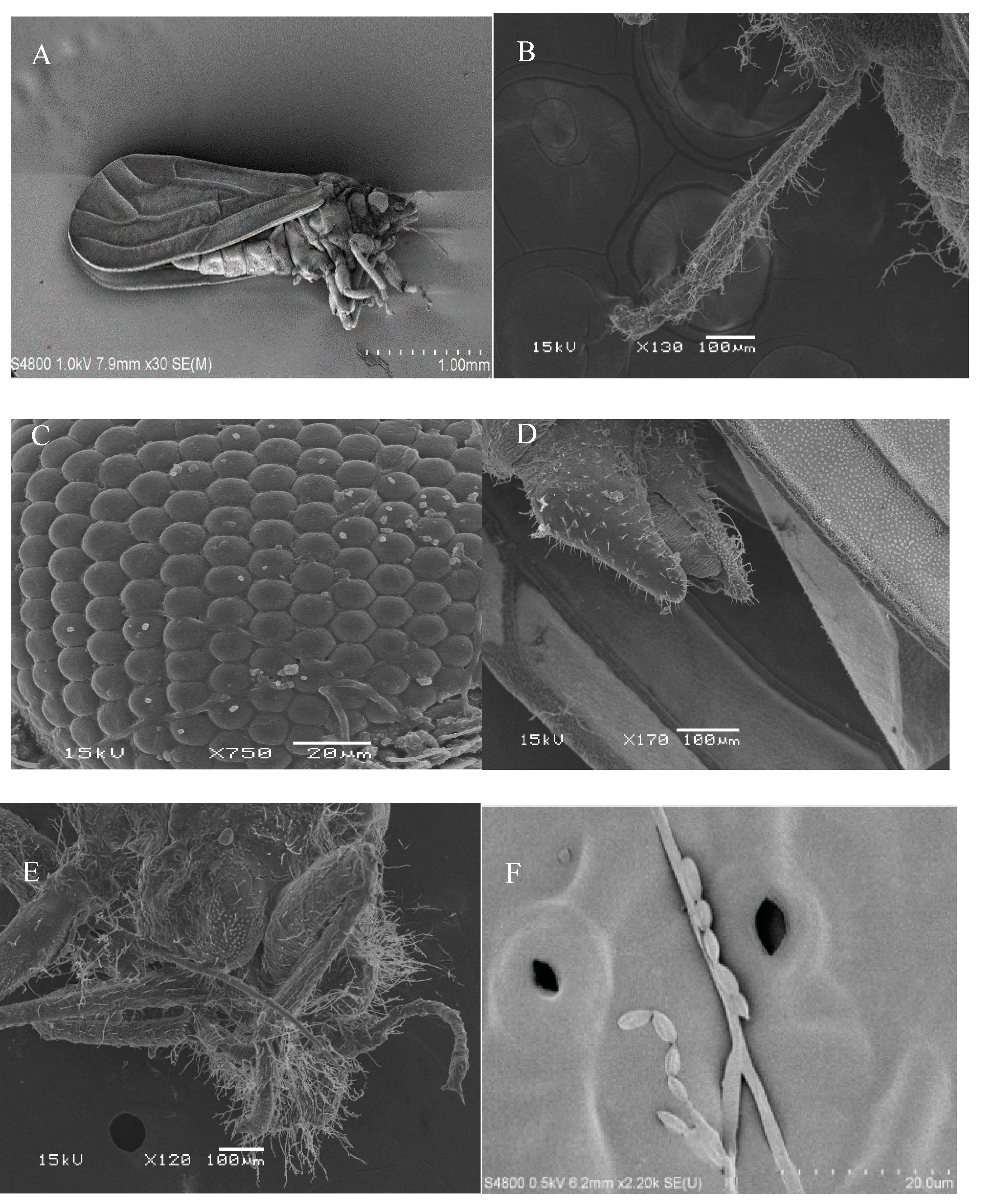
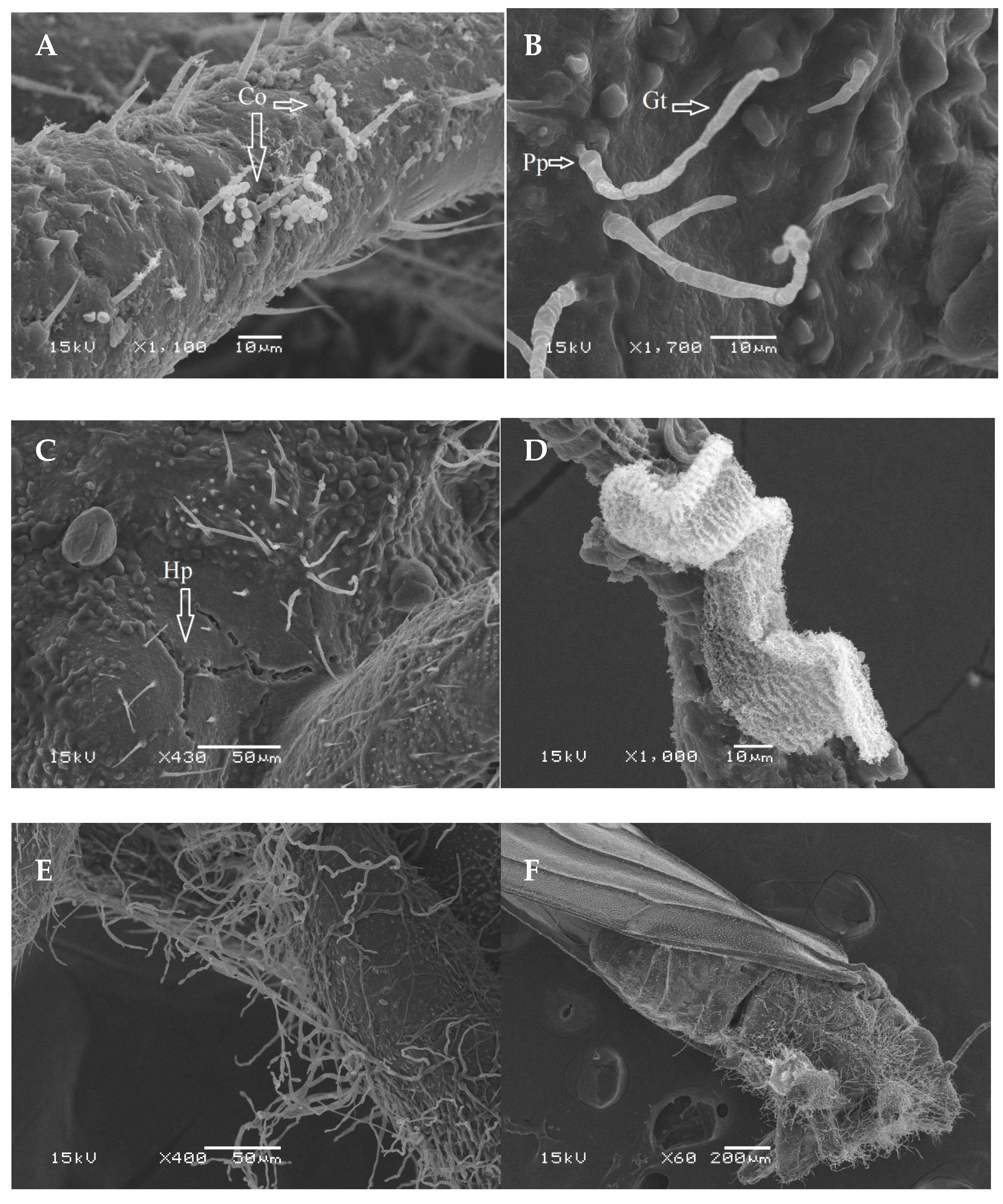
3.2. Scanning Electron Microscopy

SEM observations revealed that the fungal spores were evenly distributed on the infected body of the adult D. citri, including leg and head area (Figure 3). In addition, the hyphae of Cja Apopka 97 strain grew and formed the conidia on the leaf surface (Figure 3F) which were similar to the conidia and hyphae observed on the wings and abdomen of infected psyllids). Blastospores germinated 6–8 h after the D. citri was inoculated, whereas only the conidia adhering to the legs (Figure 4A) successfully germinated and formed a germ tube after 24 h post-inoculation (Figure 4B). The penetration of the germ tube caused cracks in the D. citri cuticle (Figure 4C), while some hyphae developed into mycelial mass on antennae by 120 h post-inoculation (Figure 4D). After that time, it was observed that the germ tube elongated toward the body wall, and then penetrated the cuticle of the psyllids forming hyphae. Some hyphae grew upward while others grew parallel along the body surface (Figure 4E). Some hyphae colonized the wings after 72 h post-inoculation (Figure 4F). After that, the hyphae grew rapidly on the surface of the insect cuticle, intertwined to form mycelium (Figure 4E). Finally, mycelia colonized D. citri body after 144 h post-inoculation, and the insect body became desiccated.

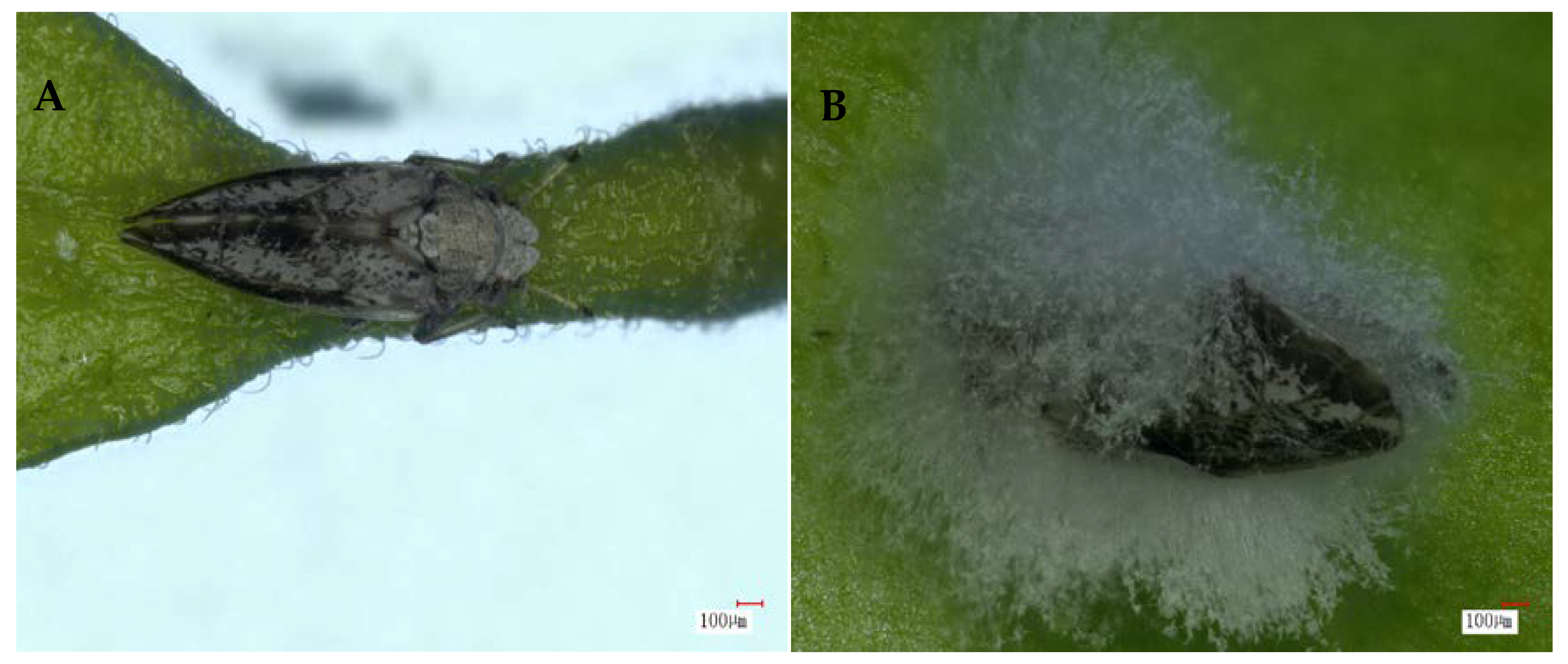
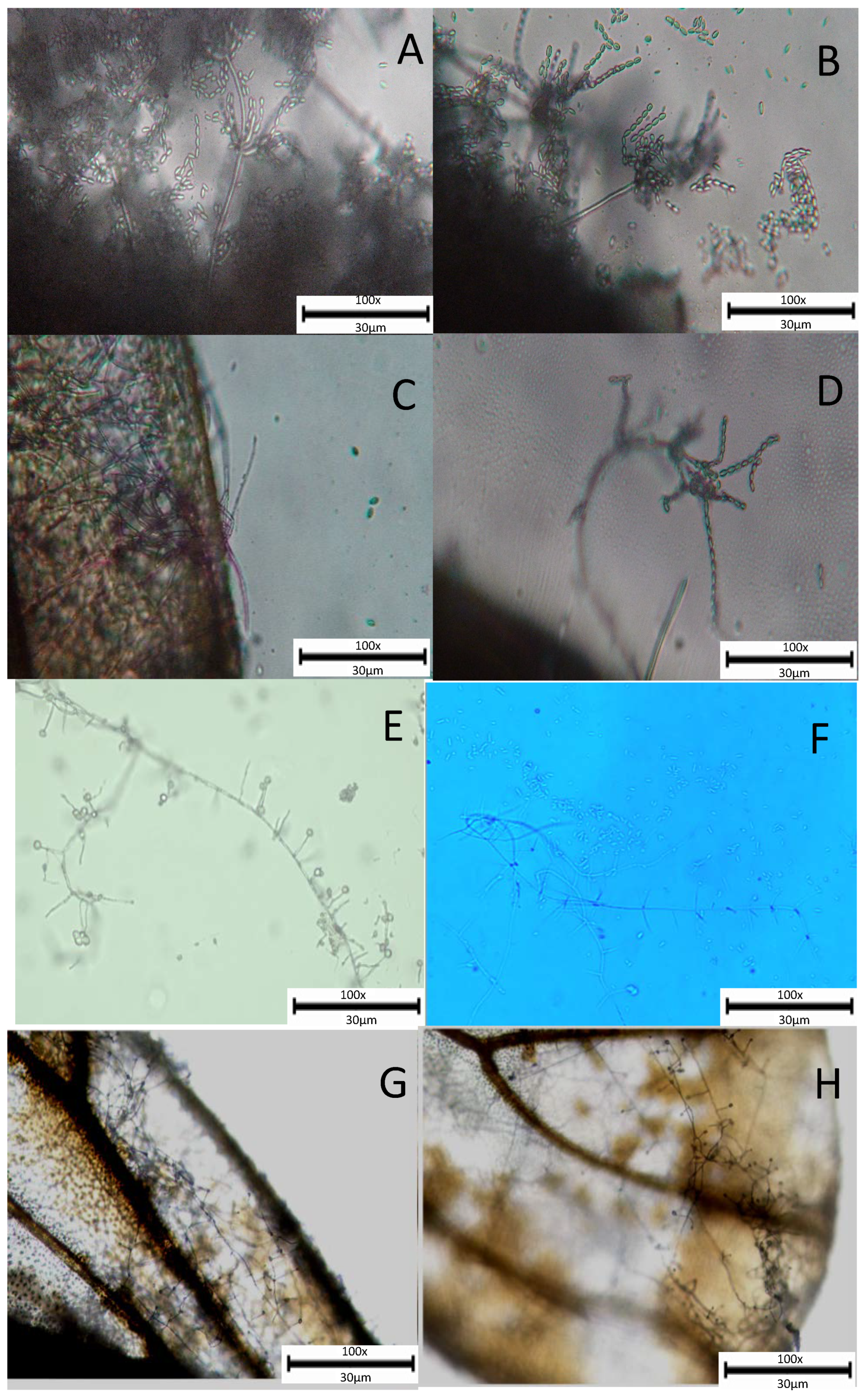
3.3. Light Microscopy

We observed that after infection with Cja the psyllid remained at the same angle while the fungus penetrates through the tarsi and cements it to the leaf surface (Figure 5). Additionally, we observed Cja hyphae and conidia growing on phialides and dispersing from the infected host. Branched hyphae with conidia were observed (Figure 6).

4. Discussion

In the mortality bioassay, we quantified the pathogenicity of Cja Apopka 97 spores against D. citri. The corrected mortality for D. citri treated with 107 spores/mL were higher than control, while we also observed faster initial mortality from D. citri exposed to the blastospore formulation, suggesting these germinate more quickly compared with the conidia (Figure 3). Our results revealed the ability of the fungus to infect different regions of the host, leading to an increase in host mortality and conidial production via colonization of the cadaver. In addition, our results indicate that infection of D. citri was dependent on the spore residual concentration on the leaf disk surface under high R.H. However, when D. citri adults were exposed to the spore suspension, rapid fungal growth on the D. citri body parts began after 24 h to 72 h post-inoculation, which contributed to higher mortality (Figure 5). After the adult psyllid becomes infected, the antennae and legs begin to twitch, and the body becomes unstable (see Supplementary Video S1).
Our ultrastructural studies illustrate the successful invasion of D. citri by the fungal entomopathogen Cja Apopka 97 (Figure 3, Figure 4, Figure 5 and Figure 6). Our results expand on observations with the entomopathogenic fungal infection process in whiteflies [10] and other insects [21,22,23,24]. We observed that Cja Apopka 97 infect D. citri (Figure 3 and Figure 4), with the infection process, similar to Cja Apopka 97 infecting other insects, such as whitefly, scale insects, and mites [25,26,27]. Our studies show how Cja Apopka 97 infected D. citri integument. It was observed that the infection process involved adherence of spores to the host body after 24 h post-inoculation (Figure 4A), germ tube (Gt) formation after 24 h post-inoculation and penetration pegs (Pp) along with mycelia growth out of host body after 72 h post-inoculation (Figure 4B); rupturing of cuticle due to hyphae growth (Hp) after 96 h post-inoculation (Figure 4C), surrounding of fungal mycelial mass on host antennae after 120 h post-inoculation (Figure 4D), growing of fungal hyphae on the wings after 72 h post-inoculation (Figure 4E), and fungal mycelia colonizing host body after 144 h post-inoculation (Figure 4F).
Our results showed that under our experimental conditions, Cja Apopka 97 can also colonize the leaf surface (Figure 3F). This observation suggests that under certain conditions, Cja Apopka 97 may persist in the environment even without an insect host, potentially contributing to later epizootics. This aspect of fungal colonization on plants by Cordyceps species expands on previous observations [21,28] but also requires further study. Pick et al. 2021 [29] also noted that Cja Apopka 97 may also colonize the phylloplane of citrus leaves under field conditions following rainfall events. On psyllids, Cja Apopka 97 fungal adherence and growth also seems to grow in the wax layer [30]. A thorough understanding of the interactions between entomopathogenic fungi and their potential hosts is required before they can be developed as biocontrol agents [24]. Appropriate targets on the host insects must be identified in such studies; as a result, the efficacy of formulation and application methods will be improved. For understanding how different fungi infect and colonize insect pests, light and scanning electron microscopy are valuable tools for studying the entomopathogenic fungus mode of action [30,31,32].
Previous studies have investigated fungal adherence and penetration structures on other insects using scanning electron microscopy [33,34]. These fungi could be effective biological control agents for insect pest populations on various host plants. Therefore, microscopic investigations have been helpful in determining how entomopathogenic fungi, such as Cja Apopka 97, interact with the D. citri. This research has led to a better understanding of the fungal infection process on adult psyllids with Cja Apopka 97 and the potential for using entomopathogenic fungi to control it.

5. Conclusions

In conclusion, our study provides basic and applied data on the interaction of Cja Apopka 97 with D. citri. We noted several new observations on the differences between infective propagules and the progression of the disease in D. citri. Therefore, the findings of the present study will help to increase the available data concerning the efficacy of using entomopathogenic fungi as potential biological agents against the D. citri. In the long run, the use of entomopathogenic fungi can serve as a sustainable alternative to the broad spectrum synthetic chemical insecticides commonly used to regulate and manage the D. citri populations in the field.

Supplementary Materials

The following are available online at https://www.mdpi.com/article/10.3390/agronomy11122476/s1, Video S1: Effect of blastospore and conidial Cordyceps javanica formulation in inoculated Diaphorina citri displayed disease symptoms including twitching of legs, antennae, and wing. Prior death inoculated psyllids present fungal hyphae emerging from the body. Note: The progression of the disease on adult D. citri included moribund behaviors, one week after inoculation infected insects form a cemented sporulating cadaver on the surface of the leaf with the hyphae colonizing on the entire exoskeleton.

Author Contributions

Conceptualization, M.H., P.B.A., L.W., D.Q. and R.M.; Formal analysis, M.H., P.B.A., W.Z. and R.M.; Funding acquisition, M.H., P.B.A., S.P.A. and R.M.; Investigation, M.H., P.B.A., W.Z., M.P., D.Q. and R.M.; Methodology, M.H., P.B.A., W.Z., M.P., L.W., D.Q. and R.M.; Project administration, M.H., S.P.A., D.Q. and R.M.; Software, M.H. and M.P.; Supervision, R.M., D.Q. and L.W.; Validation, M.H., P.B.A. and R.M.; Visualization, M.H., P.B.A., M.P. and S.P.A.; Writing—original draft, M.H., P.B.A., W.Z., L.W., D.Q. and R.M.; Writing—review & editing, M.H., P.B.A., W.Z., M.P., S.P.A., L.W., D.Q. and R.M. All authors have read and agreed to the published version of the manuscript.

Funding

This study was supported by the GDAS Special Project of Science and Technology Development (2020GDASYL-20200103096, 2021GDASYL-20210103051, and 2019GDASYL-0501006); The Research Programs of Guangzhou (202103000065), and IR-4 Project (USDA-CSREES) UF (09100905).

Institutional Review Board Statement

Not applicable.

Informed Consent Statement

Not applicable.

Data Availability Statement

The data presented in this study are available on request from the corresponding author.

Acknowledgments

We are grateful to the Michael S. Grace and Gayle Duncombe (Florida Institute of Technology, Department of Biological Sciences, 150 W. University Blvd., Melbourne, Florida), for their contribution. We thankfully acknowledge the expertise of Maria T. Gonzalez, Biological Science Technician, for sample preparation and visualization using the STEM 4800 electron microscope and Wayne Hunter, USDA, ARS, Fort Pierce, Florida, U.S.A.

Conflicts of Interest

The authors declare no conflict of interest. The funders had no role in the design of the study; in the collection, analyses, or interpretation of data; in the writing of the manuscript, or in the decision to publish the results. S.P.A. works for a commercial company that has no commercial relationship to any products or services mentioned in this manuscript.

References

  1. Hussain, M.; Akutse, K.S.; Ravindran, K.; Lin, Y.; Bamisile, B.S.; Qasim, M.; Dash, C.K.; Wang, L. Effects of different temperature regimes on survival of Diaphorina citri and its endosymbiotic bacterial communities. Environ. Microbiol. 2017, 19, 3439–3449. [Google Scholar] [CrossRef] [PubMed]
  2. Ibarra-Cortés, K.; Guzmán-Franco, A.; González-Hernández, H.; Ortega-Arenas, L.; Villanueva-Jiménez, J.; Robles-Bermúdez, A. Susceptibility of Diaphorina citri (Hemiptera: Liviidae) and its parasitoid Tamarixia radiata (Hymenoptera: Eulophidae) to entomopathogenic fungi under laboratory conditions. Neotrop. Entomol. 2018, 47, 131–138. [Google Scholar] [CrossRef]
  3. Boina, D.R.; Rogers, M.E.; Wang, N.; Stelinski, L.L. Effect of pyriproxyfen, a juvenile hormone mimic, on egg hatch, nymph development, adult emergence and reproduction of the Asian citrus psyllid, Diaphorina citri Kuwayama. Pest Manag. Sci. Former. Pestic. Sci. 2010, 66, 349–357. [Google Scholar] [CrossRef] [PubMed]
  4. Qureshi, J.A.; Stansly, P.A. Rate, placement and timing of aldicarb applications to control Asian citrus psyllid, Diaphorina citri Kuwayama (Hemiptera: Psyllidae), in oranges. Pest Manag. Sci. Former. Pestic. Sci. 2008, 64, 1159–1169. [Google Scholar] [CrossRef] [PubMed]
  5. Ghaffari, S.; Karimi, J.; Kamali, S.; Moghadam, E.M. Biocontrol of Planococcus citri (Hemiptera: Pseudococcidae) by Lecanicillium longisporum and Lecanicillium lecanii under laboratory and greenhouse conditions. J. Asia-Paci. Entomol. 2017, 20, 605–612. [Google Scholar] [CrossRef]
  6. Beloti, V.; Santos, F.; Alves, G.; Bento, J.; Yamamoto, P. Curry leaf smells better than citrus to females of Diaphorina citri (Hemiptera: Liviidae). Arth. Plant Interac. 2017, 11, 709–716. [Google Scholar] [CrossRef]
  7. Gallou, A.; Serna-Domínguez, M.G.; Berlanga-Padilla, A.M.; Ayala-Zermeño, M.A.; Mellín-Rosas, M.A.; Montesinos-Matías, R.; Arredondo-Bernal, H.C. Species clarification of Isaria isolates used as biocontrol agents against Diaphorina citri (Hemiptera: Liviidae) in Mexico. Fungal Bioloy 2016, 120, 414–423. [Google Scholar] [CrossRef] [PubMed]
  8. Boopathi, T.; Karuppuchamy, P.; Singh, S.B.; Kalyanasundaram, M.; Mohankumar, S.; Ravi, M. Microbial control of the invasive spiraling whitefly on cassava with entomopathogenic fungi. Braz. J. Microbiol. 2015, 46, 1077–1085. [Google Scholar] [CrossRef] [PubMed] [Green Version]
  9. Saito, T.; Takatsuka, J.; Shimazu, M. Characterization of Paecilomyces cinnamomeus from the camellia whitefly, Aleurocanthus camelliae (Hemiptera: Aleyrodidae), infesting tea in Japan. J. Invert. Pathol. 2012, 110, 14–23. [Google Scholar] [CrossRef] [Green Version]
  10. Tian, J.; Diao, H.; Liang, L.; Hao, C.; Arthurs, S.; Ma, R. Pathogenicity of Isaria fumosorosea to Bemisia tabaci, with some observations on the fungal infection process and host immune response. J. Invert. Pathol. 2015, 130, 147–153. [Google Scholar] [CrossRef] [PubMed]
  11. Chow, A.; Dunlap, C.A.; Jackson, M.A.; Flores, D.; Patt, J.M.; Sétamou, M. Oviposition behavior and survival of Tamarixia radiata (Hymenoptera: Eulophidae), an ectoparasitoid of the Asian Citrus Psyllid, Diaphorina citri (Hemiptera: Liviidae), on hosts exposed to an entomopathogenic fungus, Isaria fumosorosea (Hypocreales: Cordycipitaceae), under laboratory conditions. J. Econ. Entomol. 2016, 109, 1995–2005. [Google Scholar]
  12. Kumar, V.; Avery, P.B.; Ahmed, J.; Cave, R.D.; McKenzie, C.L.; Osborne, L.S. Compatibility and efficacy of Isaria fumosorosea with horticultural oils for mitigation of the Asian citrus psyllid, Diaphorina citri (Hemiptera: Liviidae). Insects 2017, 8, 119. [Google Scholar] [CrossRef] [Green Version]
  13. Lu, L.; Cheng, B.; Du, D.; Hu, X.; Peng, A.; Pu, Z.; Zhang, X.; Huang, Z.; Chen, G. Morphological, molecular and virulence characterization of three Lencanicillium species infecting Asian citrus psyllids in Huangyan citrus groves. J. Invert. Pathol. 2015, 125, 45–55. [Google Scholar] [CrossRef]
  14. Milosavljević, I.; Amrich, R.; Strode, V.; Hoddle, M.S. Modeling the phenology of Asian citrus psyllid (Hemiptera: Liviidae) in urban southern california: Effects of environment, habitat, and natural enemies. Environ. Entomol. 2018, 47, 233–243. [Google Scholar] [CrossRef]
  15. Lopez-Llorca, L.; Carbonell, T.; Salinas, J. Colonization of plant waste substrates by entomopathogenic and mycoparasitic fungi—a SEM study. Micron 1999, 30, 325–333. [Google Scholar] [CrossRef]
  16. Lopez-Llorca, L.V.; Carbonell, T.; Gomez-Vidal, S. Degradation of insect cuticle by Paecilomyces farinosus proteases. Mycol. Progress 2002, 1, 249–256. [Google Scholar] [CrossRef]
  17. Meyer, J.M.; Hoy, M.A.; Boucias, D.G. Isolation and characterization of an Isaria fumosorosea isolate infecting the Asian citrus psyllid in Florida. J. Invert. Pathol. 2008, 99, 96–102. [Google Scholar] [CrossRef]
  18. Kwiatkowski, N.P.; Babiker, W.M.; Merz, W.G.; Carroll, K.C.; Zhang, S.X. Evaluation of nucleic acid sequencing of the D1/D2 region of the large subunit of the 28S rDNA and the internal transcribed spacer region using SmartGene IDNS software for identification of filamentous fungi in a clinical laboratory. J. Mol. Diag. 2012, 14, 393–401. [Google Scholar] [CrossRef] [PubMed]
  19. Petti, C.; Bosshard, P.; Brandt, M.; Clarridge, J.; Feldblyum, T.; Foxall, P.; Furtado, M.; Pace, N.; Procop, G. Interpretive Criteria for Identification of Bacteria and Fungi by DNA Target Sequencing; Approved Guideline; Clinical and Laboratory Standards Institute (CLSI): Wayne, PA, USA, 2008; Volume 28. [Google Scholar]
  20. Gurulingappa, P.; Mc Gee, P.; Sword, G.A. In vitro and in planta compatibility of insecticides and the endophytic entomopathogen, Lecanicillium lecanii. Mycopathologia 2011, 172, 161–168. [Google Scholar] [CrossRef]
  21. Avery, P.B.; Wekesa, V.W.; Hunter, W.B.; Hall, D.G.; McKenzie, C.L.; Osborne, L.S.; Powell, C.A.; Rogers, M.E. Effects of the fungus Isaria fumosorosea (Hypocreales: Cordycipitaceae) on reduced feeding and mortality of the Asian citrus psyllid, Diaphorina citri (Hemiptera: Psyllidae). Bio. Sci. Tech. 2011, 21, 1065–1078. [Google Scholar] [CrossRef]
  22. Lu, L.; Cheng, B.; Hu, X.; Pu, Z.; Chen, G. Genetic diversity of Lecanicillium lecanii and its pathogenicity against Diaphorina citri. J. Zhejiang Univ. (Agric. Life Sci.) 2015, 41, 34–43. [Google Scholar]
  23. Anderson, C.M.; McGee, P.A.; Nehl, D.B.; Mensah, R.K. The fungus Lecanicillium lecanii colonises the plant Gossypium hirsutum and the aphid Aphis gossypii. Aust. Mycol. 2007, 26, 65–70. [Google Scholar]
  24. Hoy, M.A.; Singh, R.; Rogers, M.E. Evaluations of a novel isolate of Isaria fumosorosea for control of the Asian citrus psyllid, Diaphorina citri (Hemiptera: Psyllidae). Florida Entomol. 2010, 93, 24–32. [Google Scholar] [CrossRef]
  25. Sewify, G.; Mabrouk, A. The susceptibility of different stages of citrus brown mite Eutetranychus orientalis Oudemans (family: Tetranychidae) to entomopathogenic fungus Verticillium lecanii. In Proceedings of the Vth International Colloquium on Invertebrate Pathology and Microbial Control, Adelaide, Australia, 20–24 August 1990. [Google Scholar]
  26. Liu, W.; Xie, Y.; Xue, J.; Zhang, Y.; Zhang, X. Ultrastructural and cytochemical characterization of brown soft scale Coccus hesperidum (Hemiptera: Coccidae) infected by the Lecanicillium lecanii (Ascomycota: Hypocreales). Micron 2011, 42, 71–79. [Google Scholar] [CrossRef]
  27. Cuthbertson, A.G.; Walters, K.F. Pathogenicity of the entomopathogenic fungus, Lecanicillium muscarium, against the sweetpotato whitefly Bemisia tabaci under laboratory and glasshouse conditions. Mycopathologia 2005, 160, 315–319. [Google Scholar] [CrossRef]
  28. Avery, P.B.; Pick, D.A.; Aristizábal, L.F.; Kerrigan, J.; Powell, C.A.; Rogers, M.E.; Arthurs, S.P. Compatibility of Isaria fumosorosea (Hypocreales: Cordycipitaceae) blastospores with agricultural chemicals used for management of the Asian citrus psyllid, Diaphorina citri (Hemiptera: Liviidae). Insects 2013, 4, 694–711. [Google Scholar] [CrossRef]
  29. Pick, D.A.; Avery, P.B.; Qureshi, J.A.; Arthurs, S.P.; Powell, C.A. Field persistence and pathogenicity of Cordyceps fumosorosea for management of Diaphorina citri. Biocontrol Sci. Technol. 2021, 1–12. [Google Scholar] [CrossRef]
  30. Ammar, E.-D.; Alessandro, R.; Shatters, R.G., Jr.; Hall, D.G. Behavioral, ultrastructural and chemical studies on the honeydew and waxy secretions by nymphs and adults of the Asian citrus psyllid Diaphorina citri (Hemiptera: Psyllidae). PLoS ONE 2013, 8, e64938. [Google Scholar] [CrossRef] [Green Version]
  31. Meyer, J.M.; Hoy, M.A.; Boucias, D.G. Morphological and molecular characterization of a Hirsutella species infecting the Asian citrus psyllid, Diaphorina citri Kuwayama (Hemiptera: Psyllidae), in Florida. J. Invert. Pathol. 2007, 95, 101–109. [Google Scholar] [CrossRef]
  32. Hussain, M.; Akutse, K.S.; Lin, Y.; Chen, S.; Huang, W.; Zhang, J.; Idrees, A.; Qiu, D.; Wang, L. Susceptibilities of Candidatus Liberibacter asiaticus—Infected and noninfected Diaphorina citri to entomopathogenic fungi and their detoxification enzyme activities under different temperatures. MicrobiologyOpen 2018, 7, e00607. [Google Scholar] [CrossRef] [PubMed]
  33. Liu, W.; Xie, Y.; Xue, J.; Gao, Y.; Zhang, Y.; Zhang, X.; Tan, J. Histopathological changes of Ceroplastes japonicus infected by Lecanicillium lecanii. J. Invert. Pathol. 2009, 101, 96–105. [Google Scholar] [CrossRef] [PubMed]
  34. De Oliveira, M.A.; de Souza, W. An electron microscopic study of penetration by Trypanosoma rangeli into midgut cells of Rhodnius prolixus. J. Invert. Pathol. 2001, 77, 22–26. [Google Scholar] [CrossRef] [PubMed]
Figure 1. Murraya paniculata leaf petioles covered with a piece of sterile cotton in a sterile plastic Petri dish used for inoculation of D. citri with Cja Apopka 97 strain.
Agronomy 11 02476 g001
Figure 2. Mortality of Diaphorina citri adults after feeding on a leaf disk sprayed with blastospores, conidia of Cja Apopka or Tween 80 (control) over time.
Agronomy 11 02476 g002
Figure 3. (A) SEM photomicrographs of Diaphorina citri uninfected adult; (B) Cja Apopka 97 hyphae growing on leg; (C) Cja hyphae growing on compound eyes; (D) Cja hyphal growth on genitalia; (E) head area covered with hyphae; (F) hyphae growing and forming conidia on the leaf surface.
Agronomy 11 02476 g003
Figure 4. Ultrastructural observations of Diaphorina citri infected with Cja Apopka 97; (A) conidia (Co) adhering to the legs after 24 h post-inoculation; (B) germ tube formation (Gt) after 24 h post-inoculation formation of penetration pegs (Pp), and mycelia growth after 72 h post-inoculation; (C) rupturing of cuticle due to hyphal penetration (Hp) after 96 h post-inoculation; (D) mycelial mass on antennae surrounded after 120 h post-inoculation; (E) hyphae growing on the wings after 72 h post-inoculation; (F) mycelia colonizing D. citri body after 144 h post-inoculation.
Agronomy 11 02476 g004
Figure 5. (A) Healthy Diaphorina citri adult (B) D. citri adult colonized with Cordyceps javanica.
Agronomy 11 02476 g005
Figure 6. Light microscopic picture of Cja Apopka 97 hyphae and conidia, outgrowth from infected adult Diaphorina citri (A) vertically branched conidiophore with conidia dispersing from D. citri adult cadaver; (B) dense conidia and hyphae extending from the edge of the psyllid body (aerial conidial dispersal); (C) hyphae growing on the wing surface of infected psyllid; hyphae growing through the veins and forming mycelium; (D) hyphae and conidia growing on phialides extending from infected insect; (E, F) hyphae and conidia growing on phialides and dispersing from infected insect (aerial conidial dispersal); (G) hyphae growing on the infected psyllid wing surface; (H) hyphae with conidia growing on phialides on wing surface.
Agronomy 11 02476 g006
Publisher’s Note: MDPI stays neutral with regard to jurisdictional claims in published maps and institutional affiliations.

