Article

Phenotypic Analysis and Gene Cloning of a New Allelic Mutant of SPL5 in Rice

1
School of Life Science and Engineering, Southwest University of Science and Technology, Mianyang 621010, China
2
Rice Research Institute, Southwest University of Science and Technology, Mianyang 621010, China
3
Rice Research Institute, Sichuan Agricultural University, Chengdu 611130, China
*
Authors to whom correspondence should be addressed.
These authors contributed equally to this work.
Agriculture 2023, 13(10), 1875; https://doi.org/10.3390/agriculture13101875
Submission received: 9 August 2023 / Revised: 11 September 2023 / Accepted: 20 September 2023 / Published: 25 September 2023
(This article belongs to the Section Genotype Evaluation and Breeding)

Abstract

:
This study was conducted on the lesion-mimic mutant lm5, which was produced by mutagenesis of WYJ21 (WT) using ethyl methane sulfonate (EMS). The mutant lm5 was short in the seedling stage and displayed yellowish-brown disease-like spots on leaves that were yellowish-brown when the plant was at the tillering stage. The disease-like spots gradually grew larger as the plant grew until it reached maturity. Compared to WT, lm5 had considerably reduced the plant height, ear panicle length, tiller number, and 1000-grain weight. A single recessive gene was found to be in control of lm5, according to a genetic study. It was physically located 245 kb apart between the RM21160 and RM180 markers on chromosome 7. Using RiceData and other websites, analyze and sequence potential gene candidates. Exon 7 of LOC_Os07g10390 (OsLM5) was identified to have a mutation that changed the 1560 base from G to A, changing the 788 amino acids from Arg to Lys. The OsLM5 gene was found to be a new allele of the SPL5 gene, encoding the protein shear factor SF3b3. Studies showed that OsLM5 was localized in the nucleus, and OsLM5 was significantly expressed in leaves. Reactive oxygen species (ROS) accumulation occurred in the leaves and roots of mutant lm5, and qPCR results showed abnormal expression of genes related to chloroplast development as well as significantly increased expression of genes related to aging and disease course. The OsLM5 gene may have a significant impact on the regulation of apoptosis in rice cells.

1. Introduction

Rice is one of the important food crops in China [1], and its planting area accounts for more than 30% of the national food crop area [2]. During the growing process of rice, it is easy to be affected by external stress factors, such as climate, pests, diseases, etc. These elements lead to the abnormal physiological activities of rice and ultimately affect the yield and quality of rice. Lesion Mimic Mutant (LMM) is a type of mutant that is characterized by the spontaneous formation of disease-like spots mediated by programmed cell death (PCD) in the absence of pathogen infection, environmental stress, or mechanical damage [3,4,5]. On the leaves, leaf sheaths, stalks, or seed shells of plants, disease-like spots frequently manifest [6,7]. Early lesion-mimic mutants have been extensively reported in Arabidopsis thaliana rice [8], maize [9], barley [10], wheat [11], tomato [12], and other crops, with ongoing research. Meanwhile, it was found that most of the lesion-mimic mutants were related to plant disease resistance, showing increased expression of defense-related genes and activation of defense-related pathways. It exhibits heightened resistance to specific pathogens [13], and the lesion-mimic mutants are regarded as important materials for studying the molecular mechanism of plant resistance, which is of great significance for clarifying the molecular mechanism of plant resistance to external stresses such as diseases and pests and also providing support for the breeding of high-resistance improved varieties [14,15].
It was discovered that there were great differences in the shape and color of disease-like spots in different mutants. Spl36 [16] and spl(Y181) [17] showed irregular shapes, while spl24 [18,19] had regular shapes. The disease-like spots of SPL3 [20] and spl16 [21] were black, and those of spl18 [22] and lil1 [23] were reddish brown. In addition, the stage of appearance of disease-like spots is also different, as spl2 [24], spl3 [20], and other lesion-mimic mutants will show disease-like spots at the seedling stage. The spl6 [25] mutant does not show the phenotype of disease-like spots until the tiller stage and the booting stage. With the growth and development of the plant, the phenotype of the lesion-mimic mutant will become more and more severe and even spread to the whole plant.
The occurrence of regulatory disease-like spots in rice is influenced by a variety of factors. With the mining, cloning, and functional research of more and more genes related to these phenotypes, the mechanism of their occurrence is gradually understood. Important signaling molecules called reactive oxygen species (ROS) are primarily formed in organelles, including plant mitochondria, plasma membranes, chloroplasts, peroxisomes, etc. Each plant cell has a redox pathway regulated by ROS, which regulates the physiological processes of cells, including gene expression, aging and death, and metabolism. The hydrogen peroxide (H2O2) produced by the respiratory chain and enzymatic reactions in mitochondria will be transferred to the peroxisome and finally transported to the nucleus and cytoplasm. The H2O2 formed in chloroplasts will eventually enter the nucleus and cytoplasm [26]. PCD, which is closely related to hypertensive response (HR) [27], can be caused by ROS. Studies have found that the Arabidopsis gene Lsd1 is a member of the LSD1-like gene family, and its mutant lsd1 shows disease-like spots. The mutation of this gene will cause the PCD and associated defense mechanisms to be activated [28]. Zeng et al. [29] studied that ubiquitin ligase E3, a protein encoded by rice Spl11, negatively regulates PCD and immune function in plants. Thus, it was found that the formation of disease-like spots in plants was accompanied by PCD [30]. Excessive ROS accumulation will cause damage to plant cells and eventually lead to cell death [31].
ROS can influence the development of disease-like spots in two separate ways. First, they can react with a large number of biomolecules, causing irreversible cell damage and ultimately cell death in plants [32,33]. In addition, ROS can also disrupt signal transduction pathways and alter gene expression. Studies have found that ROS plays a crucial role in pathogen defense, gene expression, and cell signal transduction in response to a variety of pathogens [34]. Excessive accumulation of superoxide free radicals (O2) and H2O2 in plants can disrupt the redox balance in cells and lead to severe oxidative damage to nucleic acids, proteins, and lipid membranes [35,36]. In addition, the accumulation of ROS is closely related to the occurrence of PCD. For instance, the formation of Arabidopsis [37], wheat [38], rice [39,40], and other disease-like spots is accompanied by the accumulation of ROS and the occurrence of PCD. SPL33, encoding the eEF1A protein, contains one non-functional zinc finger domain and three functional EF-Tu domains. Wang et al. [41] found that the loss of SPL33 function leads to the accumulation of H2O2, accelerated leaf aging, and PCD, and finally produces reddish-brown lesions. ROS not only affects the formation of disease-like spots but also plays an important role in root growth and lateral root formation [42,43,44]. ROS production and related signaling pathways are involved in root formation [45,46]. Tsukagoshi [42] and Silva-Navas [43] found that primary root growth was strictly regulated by the differential accumulation of ROS at the apex. UPBEAT1 (UPB1) transcription factor is independent of the auxin pathway by inhibiting peroxidase gene expression in roots and regulating the distribution of H2O2 and O2− in the root elongation region and meristem [42]. Therefore, the study of the characteristics and regulatory mechanisms of rice lesion-mimic mutants is helpful in revealing the molecular mechanism of PCD in plants.
In this study, Wuyunjing21 was mutated by EMS to identify a mutant named lm5. The mutant was short in plant height and had brown disease-like spots on leaves at the tillering stage. This gene encodes the protein shear factor SF3b3. We identified the mutant gene and discovered that the lesion-mimic phenotype of the lm5 mutant was caused by the single base mutation of the OsLM5 gene, which is an allele of the OsLM5 gene and the SPL5 gene [47]. Mutations in the OsLM5 gene disrupt the ROS balance in cells, disrupt the structure of chloroplasts, and lead to local cell death at the lesion mimic. The results suggest that OsLM5 plays an important role in regulating PCD and ROS balance in rice.

2. Materials and Methods

2.1. Plant Materials and Growing Conditions

Lm5 mutants were obtained from the Wuyunjing21 (WYJ21, WT) population by EMS mutagenesis. All the plants were grown in April 2022 in the paddy field at Southwest University of Science and Technology and in October 2022 in Lingshui, Hainan, under the natural conditions of 28~37 °C. Field management follows standard agricultural practices. Tests related to roots were completed in March 2023, measuring rice roots after 15 days of growth. The cultivation condition of rice was 14 h light/10 h dark, and the light intensity was 1200 µmol photons m−2·;s−1.

2.2. Investigation of Chlorophyll Content

The leaves of WTand mutant lm5 at the tillering stage were collected, and chlorophyll (Chl) content was determined. The concentrations of carotenoids (Car), chlorophyll a (Chla), and chlorophyll b (Chlb) were determined by spectrophotometry using the method described by Wellburn [48]. Simply put, the leaf samples were cut into pieces of about 0.5 cm, soaked in 80% acetone, and treated in dark conditions for more than 24 h. The optical density (OD) of the extracts was measured by spectrophotometry at 663, 646, and 470 nm, with three biological replicates measured per sample.

2.3. Determination of Various Antioxidant Indexes

Superoxide dismutase (SOD), peroxidase (POD), malondialdehyde (MDA), and catalase (CAT) from a catalase assay kit (visible light) were purchased from the Nanjing Jianjiancheng Bioengineering Institute. Enzyme activity is measured according to the methods in the manufacturer’s instructions. All tests were carried out on leaves at the tillering stage and roots at the seedling stage.

2.4. Real-Time PCR Analysis

Total RNA was extracted from rice leaves using the TIANGEN RNAprep Pure Plant Kit and reverse-transcribed into cDNA using the Ecorry EvoM-MLV reverse transcription kit. The ACTIN gene was used as the internal reference gene, and the gene expression was detected by the SYBR Green Pro Taq HS premixed qPCR kit. There were three replicates per sample. The housekeeping gene we use is Actin, and the primers used in qRT-PCR analysis are shown in Supplementary Table S2.

2.5. Histochemical Label Staining

Nitrotetrazolium blue chloride (NBT) staining for superoxide anion accumulation and DAB staining for H2O2 accumulation were tested [49]. In simple terms, the sample is placed in diaminobenzidine (DAB) or NBT stain solution at 28 °C for staining, then all materials are decolorized in 95% ethanol at 70 °C until there is no chlorophyll, and then transferred to 70% glycerin for photographing. Trypan blue staining was used to detect cell death [18]. The sample was soaked in a basin blue stain solution and kept in the dark for more than 48 h, then all materials were decolorized in 95% ethanol at 70 °C and then transferred to 70% glycerin for photography.
The DCFH-DA, 2′,7′-Dichlorodihydrofluorescein diacetate (H2DCFDA) experimental scheme was modified according to Leshem’s method [50]. The leaves of WT and mutant lm5 growing in the same growth period were selected, and the leaves were cut into 1 cm for incubation. Chlorophyll autofluorescence and oxidation of H2DCFDA were observed by Nikon AXR laser confocal microscopy.
The roots of WT and mutant lm5 at the seedling stage were fixed with FAA. After dehydration, transparency, and penetration, the samples were cut into thin slices with a Lycra slicer. After paraffin eluting with xylene, the slices were stained with aniline blue and then observed with a Lycra optical microscope.

2.6. Gene Mapping

In the genetic analysis, we hybridized with R498 using lm5 as the maternal parent to obtain F1 offspring to self-breed and produce an F2 population. The phenotype of the F2 generation population was identified, genetic analysis was conducted, and 40 mutant phenotypic single strains were selected for mixing to obtain a mixing pool. Gene mapping was performed by mapping cloning analysis. We used the software Primer5 for primer design and the software SnapGene for sequence alignment and peak mapping. The RiceData (https://www.ricedata.cn/gene/ accessed on 17 March 2023) was used to search the genetic information. The primers used for gene mapping are shown in Supplementary Table S3.

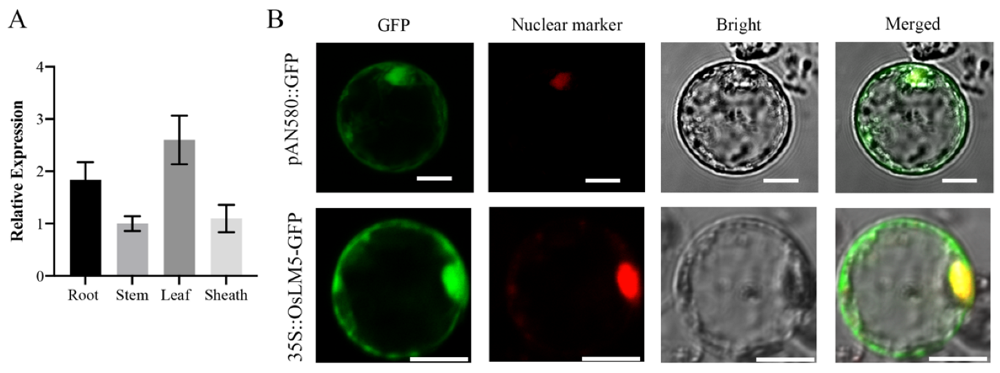
2.7. Subcellular Localization

The full-length OsLM5 coding sequence 4068 bp was inserted into PAN580-GFP to construct the 35S::OsLM5::GFP vector, and the nuclear marker and the constructed vector were transformed into rice protoplasts for transient expression. Fluorescence signals were observed using a Zeiss LSM700 laser-scanning confocal microscope. Primers are shown in Supplementary Table S4.

3. Results

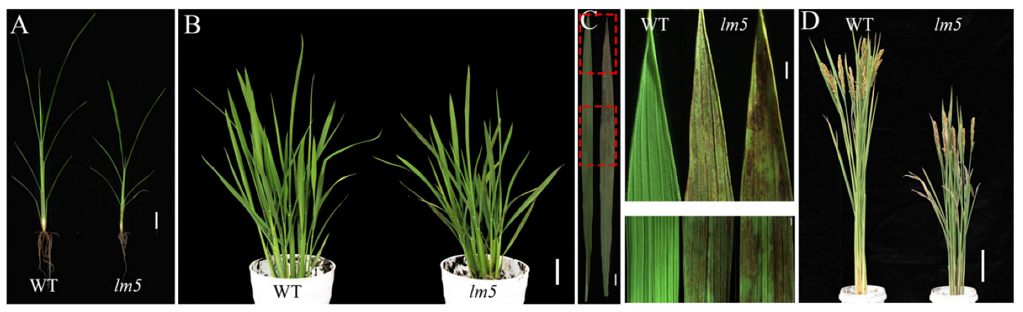
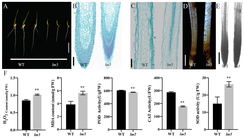
3.1. Phenotypic Characteristics of Mutant lm5

The rice variety Wuyunjing 21 (wild-type, WT) was mutated by EMS (ethyl methylsulfonate), and a leaf lesion mutant named lm5 was identified. The mutant was short in plant size (Figure 1A). During the tillering stage, mutant lm5 showed yellowish-brown lesions from the middle of the leaf to the tip of the leaf (Figure 1B,C). With the progress of growth and development, the leaf lesions of the mutant became more and more obvious. In addition, compared with WT, mutant lm5 had lower plant height at maturity and shorter internode and panicle length than WT (Figure S1A,C), and the number of tillers, number of branches, stems, seed setting rate, number of grains per panicle, and 1000-grain weight were all decreased (Figure S1D–K). The grain width and length of lm5 mutants were significantly smaller than those of WT (Figure S1B), suggesting that lm5 mutations may indirectly affect grain size. These results indicated that the formation of disease-like spots seriously affected the growth and development of lm5 mutants.

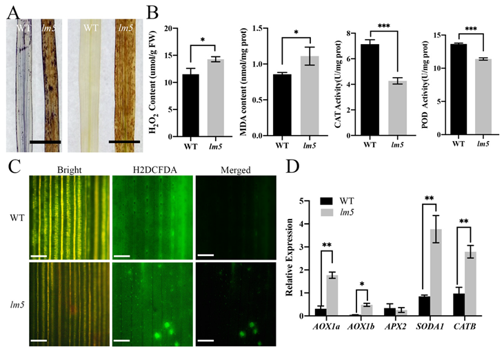
3.2. ROS Accumulation Occurred in lm5 Mutants

To further investigate the reason underlying the development of disease-like spots, we measured ROS levels in lm5 mutants. Trypan blue (left in Figure 2A) and diaminobenzidine (right in Figure 2A) were used to stain the lesion sites of WT and lm5 leaves. Leaves of mutant lm5 were found to have an accumulation of dark blue and tan precipitates, which were not detected in WT leaves (Figure 2A). These results indicate that the occurrence of disease-like spots in lm5 is accompanied by PCD and peroxide accumulation. To explore the accumulation of ROS in the leaves of mutant lm5, we measured the contents of H2O2 and MDA at the tillering stage (Figure 2B) and found that, compared with the WT, the concentrations of H2O2 and MDA in mutant lm5 were significantly increased, and the accumulation of MDA would aggravate membrane damage [51]. Therefore, MDA content indirectly reflects the damage degree of mutant cells. We also measured the activities of CAT and POD (Figure 2B). The POD activity in the leaves of mutant lm5 was lower than that of the WT, and the activity of ROS scavenging enzymes and CAT enzymes decreased. Abnormal ROS in the mutant and excessive accumulation of ROS and H2O2 may lead to the occurrence of disease-like spots in the leaves.
We incubated the leaves of WT and mutant lm5 with an H2DCFDA fluorescent probe to detect ROS accumulation and distribution in tissues (Figure 2C). Observations under laser confocal microscopy showed that the probe’s green oxidation state fluorescence signal was observed in lm5 mutants but not in the WT.
Since ROS levels are strictly regulated by the antioxidant system, we detected the expression of genes related to ROS clearance and found that the expression levels of AOX1a, AOX1b, APX2, SODA1, and CATB genes were increased (Figure 2D). The high expression of ROS detoxification genes in mutant lm5 may be caused by the elevated level of ROS in cells. Compared with WT, the expression levels of aging genes such as OsWRKY2, OsWRKY7, OsNAC2, and SGR in lm5 mutants increased (Figure S2A), and the expression of disease-like spots course-related genes PR10 and JIOsPR10 increased significantly (Figure S2B). The above results indicate that the activity of ROS scavenging enzymes in lm5 is reduced, the balance of the scavenging system is destroyed, resulting in a large accumulation of ROS, lipid peroxidation, increased MDA content, and finally cell damage resulting in the occurrence of disease-like spots.

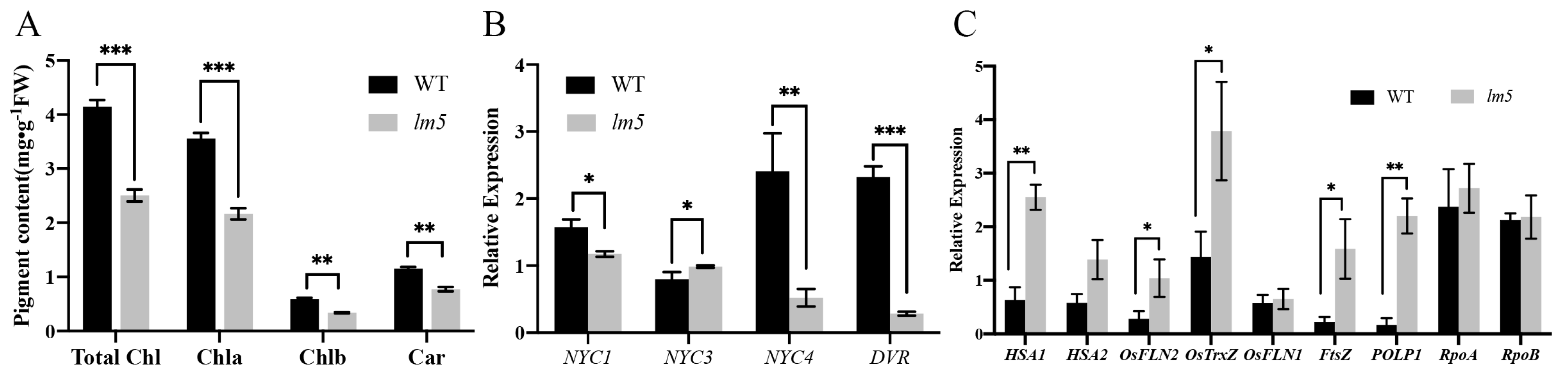
3.3. The Chlorophyll Content of the lm5 Mutant Decreased

The formation of disease-like spots caused the difference in leaf color, so the chlorophyll content of leaves was detected to determine whether it would affect the change in pigment. It was found that the contents of chlorophyll a, chlorophyll b, carotenoid, and total chlorophyll in the leaves of lm5 at the tillering stage were significantly lower than those of WT. Among them, the chlorophyll content of mutant lm5 was 39.2% lower than that of WT, the chlorophyll b content was 25.5% lower than that of WT, the total chlorophyll content was 39.7% lower than that of WT, and the carotenoid content of mutant lm5 was 33% lower than that of WT. The results showed that the formation of disease-like spots in mutant lm5 affected chlorophyll synthesis.
The leaf of mutant lm5 has disease-like spots, and the chlorophyll content in the leaves is significantly reduced. The formation of disease-like spots in mutant lm5 may lead to the destruction of chloroplast structure. We further quantitatively analyzed chloroplast-related genes and chlorophyll-related genes in the leaves. The expression of chloroplast genes depends on the activity of two RNA polymerases, plastid-encoded RNA polymerase (PEP) and nucleus-encoded RNA polymerase (NEP) [52,53]. HSA1, OsFLN1, OsFLN2, OsTrxZ, POLP1, RpoA, and RpoB belong to plastid-dependent RNA polymerase genes [54,55]. Through the analysis of chlorophyll gene expression, it was found that, compared with WT, the expression of NYC4 and DVR genes in the leaves of mutant lm5 decreased while the expression of NYC3 increased (Figure 3B), indicating that the change in chlorophyll content of the mutant may be caused by the destruction of the chloroplast structure of the mutant. Quantitative analysis showed that the expression levels of chloroplast development genes HSA1, OsFLN2, OsTrxZ, POLP1, and FtsZ increased (Figure 3C), indicating that the disturbance of chloroplast development gene expression interfered with PEP activity, thus affecting chloroplast development.

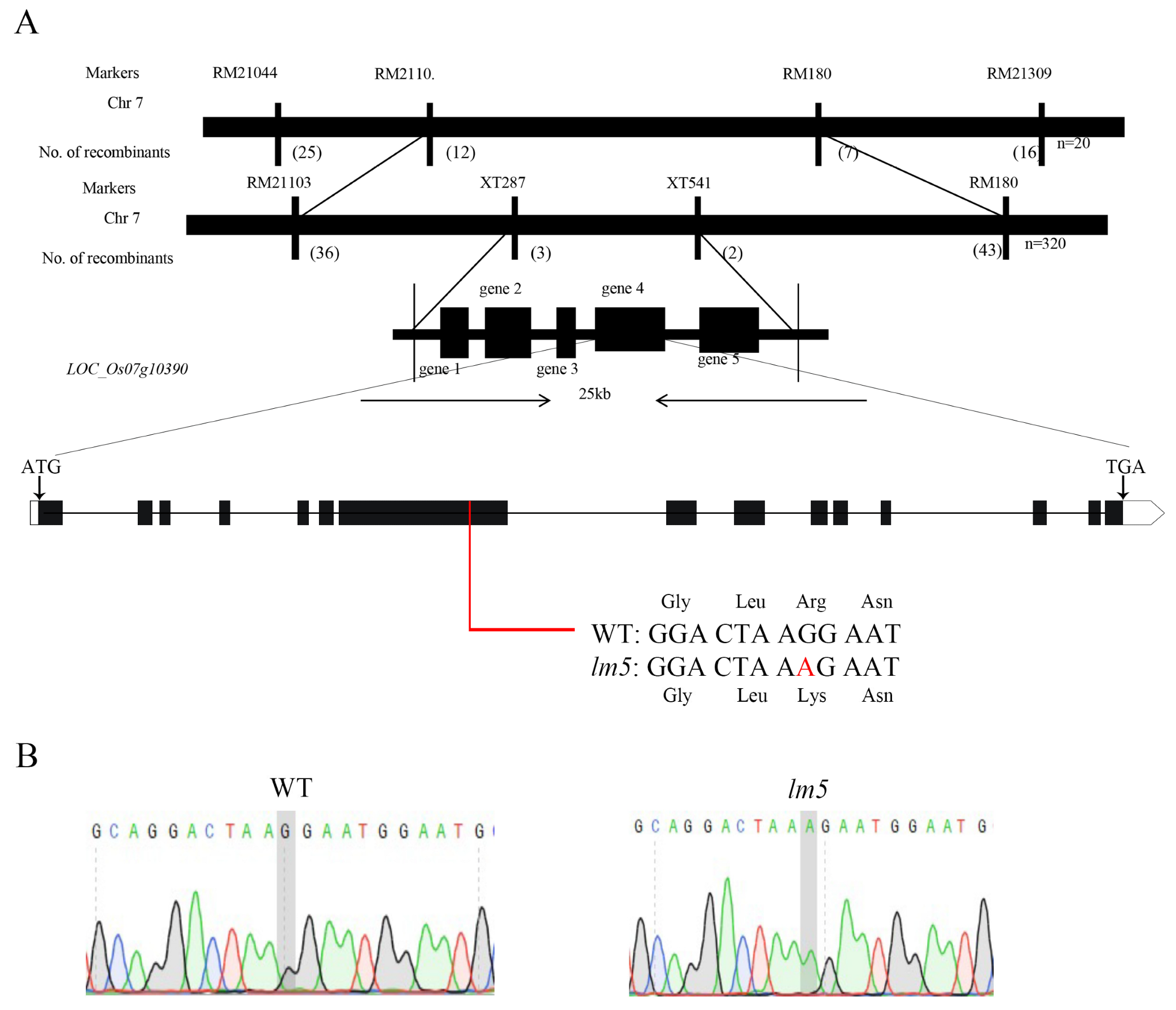
3.4. Genetic Analysis and Candidate Gene Mapping of Macular Mutant lm5

To reveal the molecular mechanism behind the disease-like spots phenotype of the mutant lm5, we hybridized with R498 using lm5 as the maternal parent to obtain F1 progeny for the self-breed F2 population. The phenotype of the F2 progeny population was identified, and genetic analysis was conducted. It was found that there were 978 normal strains and 342 mutant strains of lm5. The Chi-square test results show that the separation ratio is 3:1 ( χ 2 = 0.58 < χ 0.05 2 = 3.84 ), indicating that the phenotype of mutant lm5 was controlled by a pair of recessive nuclear genes.
Linkage analysis of two parents of R498 and lm5 was performed by designing polymorphic markers, and lm5 was initially located between XT-1 and RM21309. A total of 320 recessive plaque single strains from the F2 population were used in this interval for fine localization by screening new polymorphism primers. It was found that there were three exchange single strains at RM21160 and two exchange strains at RM180. Finally, lm5 was located between the markers RM21160 and RM180, with a physical distance of 245 kb (Figure 4A). By gramene, RiceData, and other websites analyzed the prediction analysis of genes in the 245 kb location interval and found that there were 33 annotated genes in the interval (Table S1). Of these, 16 encode expression proteins, and 14 genes have been annotated for possible functions; By analyzing the phenotypes of the genes encoding predicted splicing factor 3b subunit 3, encoding zeta-carotene dehydrogenase, and encoding cysteine-rich alcohol-soluble gluten of the reported genes, it was found that the phenotypes of the genes encoding predicted splicing factor 3b subunit 3 were similar to this gene. Therefore, WT and the mutant lm5 were sequenced (Figure 4B). A difference was found between WT and the mutant lm5, in which the 1560 base of exon 7 was mutated from G to A, resulting in a change in the 788 amino acids (Arg to Lys). At the same time, the F2 mutant material was also sequenced, which further confirmed that the gene had a single base mutation (Figure S3). Therefore, LOC_Os07g10390 was taken as a candidate gene and named OsLM5 (Lesion Mimic 5). The OsLM5 gene was found to be an allele of the SPL5 gene through analysis [47].

3.5. OsLM5 Expression Pattern Analysis

To understand the expression of the OsLM5 gene in different tissues, we collected WYJ21 tissue materials and detected the expression level of the OsLM5 gene by qRT-PCR. The OsLM5 gene was found to have the highest expression level in leaf tissues (Figure 5A), followed by a higher expression level in roots, indicating that the OsLM5 gene plays a major role in leaves. To determine the subcellular localization of the OsLM5 protein, we constructed a 35S::LM5::GFP vector and performed a transient transformation assay with nuclear marker-transformed rice protoplasts for co-expression. Confocal laser scanning microscopy showed that the GFP signal of the 35S::LM5::GFP fusion protein was co-located with the red fluorescence of nuclear markers in protoplasts (Figure 5B), and the OsLM5 protein may also play a role in the cytoplasm.

3.6. OsLM5 Mutations Affect Root Development

Compared with WT, mutant lm5 had short growth and short root length (Figure 6A and Figure S4A,B). Expression profile analysis found that the OsLM5 gene was active in roots (Figure 5A). Paraffin sections were performed on the roots of WT and mutant lm5, and obvious changes were found in root cell morphology. Compared with WT, the root cells of mutant lm5 became smaller, the number of cells increased, and the size of single cells in the root crown, meristem zone, elongation zone, and mature zone decreased significantly (Figure S4D). Previous studies have found that ROS accumulation in roots can affect root growth. DAB and NBT staining were performed on taproots of WT and mutant lm5, and grayscale analysis of NBT staining was performed, and it was found that mutant lm5 roots were dyed brown more deeply by DAB (Figure 6D). NBT staining and grayscale analysis showed that O2− in mutant lm5 roots was higher than WT (Figure 6E and Figure S4E), indicating ROS accumulation in mutant lm5. The H2O2-related indexes of roots of WT and lm5 were determined, and it was found that the H2O2 content and MDA content of mutant lm5 were significantly higher than those of WT, the H2O2 scavenging ability of CAT and POD was significantly reduced (Figure 6F), and the catalytic capacity of SOD to generate H2O2 was significantly increased. OsLM5 mutations lead to H2O2 accumulation in roots, which affects root development.

4. Discussion

4.1. The Formation of Leaf Lesions Affected the Agronomic Traits of Mutant lm5

The appearance of disease-like spots on rice leaves will reduce the chlorophyll content and photosynthetic rate of rice leaves, affect the photosynthesis and dry matter accumulation of rice leaves, and then affect the agronomic traits of rice mutants. Qiao et al. [56] reported that the chlorophyll content and photosynthetic system II efficiency of the spot-like mutant spl28 decreased, and the yield decreased. Studies on the lesion-mimic mutant lmm8 found that its chlorophyll content and PSII efficiency decreased, and the final thousand-grain weight and seed-setting rate were significantly reduced [57]. The main agronomic characters of spl36, such as plant height, effective panicle number, panicle length, kernel number per panicle, seed setting rate, and 1000-grain weight, were significantly decreased [6]. Ma et al. [58] studied the mottle leaf mutant spl35 and found that due to the emergence of reddish-brown lesions, chloroplast development was abnormal, resulting in a decrease in the seed setting rate and 1000-grain weight of the mutant spl35. Consistent with the above studies, the mutant lm5 appeared with yellow-brown lesions from the early tillering stage and lasted until maturity (Figure 1). Some agronomic traits of mutant lm5, such as plant height, effective panicle number, 1000-grain weight, and seed setting rate, were significantly lower than those of WT due to the appearance of disease-like spots, which ultimately led to a decrease in its yield (Figure S1).

4.2. ROS Accumulation Led to the Formation of Disease-like Spots in Mutant lm5

The accumulation of PCD and ROS around leaf lesions can lead to oxidative damage to plants, which is a way for plants to resist external infection [20,59]. Many studies have shown that plants have their own ROS clearance mechanism, and the production of ROS in plant cells is clearly in dynamic equilibrium to balance their own ROS [3,34,40]. ROS accumulation has been detected in more than 40 kinds of rice lesion-mimic mutants that have been cloned [60]. A large amount of ROS accumulates around the leaf lesion of mutant spl28 [56]. ROS within the normal range are important signals in response to stress, regulation of plant growth and development, and PCD [61]. Mutant ell1 accumulated a large amount of ROS and a large amount of H2O2 accumulation, and the presence of dead cells was found in ell1. Gene expression related to oxygen binding and ROS clearance was upregulated. Abnormal PCD occurred in the ell1 mutant, and excessive ROS accumulation can mediate cell death [3]. Our study had similar results. Through the determination of Trypan blue and DAB staining on the leaves of mutant lm5 and related physiological indexes (Figure 2A), it was found that a large number of ROS accumulated and cell death existed in the leaves of the lesion-mimic mutant, and the activities of CAT- and POD-related H2O2 protective enzymes were significantly reduced, while the activity of SOD, which catalyzed H2O2 generation, was significantly increased, possibly due to the increase in H2O2 content (Figure 2B). The clear mechanism of ROS is disturbed, and the cell structure is destroyed. The increase in MDA content aggravated the damage to the membrane of mutant leaves. At the same time, it was also found that there was cell death around the leaf lesions of mutant lm5, indicating that excessive accumulation of ROS caused the death of leaf cells.
ROS is a key signal in the process of plant root elongation and differentiation. Studies in Arabidopsis have shown that a decrease in O2− concentration reduces root elongation, but the removal of H2O2 can promote root elongation [62]. RITF1 overexpression can induce the ROS signal downstream of RGF1 (enhanced O2− signal in the meristem region) and promote root meristem development [60]. Interestingly, we also found an accumulation of ROS and O2− in mutant lm5 roots, which inhibited the root elongation of mutant lm5. The electron transport chain of plant mitochondria has multiple pathways, mainly the cytochrome pathway (CP) and alternative respiratory pathway (AP). The alternate respiratory pathway is a branch of the main respiratory chain that contains an alternative oxidase (AOX), the terminal oxidase in the respiratory chain [63], which is consumed in the form of heat energy to reduce oxidative damage, maintain oxidative balance in mitochondria, and limit the production of ROS [64]. AOX has an important relationship with stress resistance [65], resistance to high temperature stress [66], and maintenance of metabolic balance [67]. Related studies have reported that AOX1a and AOX1b [68] participate in alternate oxidase through respiration, and APX2 [69] is related to the gene of ascorbate peroxidase, which mainly acts to clear ROS. OsCATA and OsCATB [70] are genes involved in the H2O2 metabolic pathway. In this study, aging, disease course, and ROS-related quantification were performed on the macular mutant lm5, and it was found that the expression of ROS genes AOX1a, AOX1b, SODA1, and CATB of the mutant were significantly up-regulated (Figure 2D), indicating that the accumulation of ROS in the mutant lm5 induced the expression of AOX1 and other genes to affect the occurrence of cyanogen-resistant respiration. At the same time, the expression levels of aging genes OsWRKY2, OsWRKY7, OsNAC2, and SGR were up-regulated, indicating that the accumulation of ROS could stimulate senescence-related mechanisms and accelerate the aging and death of plants. Due to the formation of disease-like spots, the plant’s defense mechanism is activated, and the expression of PR10, JIOsPR10, and other resistance genes in mutant lm5 is increased (Figure S2A,B). The mutation of OsLM5 affects a series of resistance gene expressions in rice. Therefore, OsLM5 is of great significance in theoretical research and disease-resistance breeding. The above studies indicated that the lesion-mimic mutant would be accompanied by ROS accumulation and cell death during the disease-like spot formation process, resulting in the phenotype of mutant lm5. OsLM5 can regulate ROS metabolism in leaves and roots, thereby affecting the formation of leaf lesions and root elongation.

4.3. Effects of Mutant lm5 on Photosynthetic Function in Leaves

It was found that the chlorophyll content of rice spot-like mutant spl41 gradually decreased with the increase of the spot-like phenotype, indicating that the formation of disease-like spots would affect chlorophyll synthesis and lead to premature plant aging [71]. Ma et al. [57] found that the leaf mutant spl35 with spotted leaves showed decreased pigment content and increased ROS accumulation. It was found that the phenotype of mutant llm1 was induced by light, the content of photosynthetic pigment was significantly decreased, the number of chloroplasts in the mutant mesophyll cells was reduced, and the chloroplast structure was destroyed [13]. In this study, the chlorophyll content of the leaf after the formation of the morphed mutant lm5 was detected, and it was found that the chlorophyll content of the mutant lm5 was significantly lower than that of WT. In mutant lm5, chloroplast development genes HSA1, OsFLN2, OsTrxZ, FtsZ, POLP1, and other genes were up-regulated, and the expression of chlorophyll-related genes NYC4 and DVR decreased while NYC3 expression increased (Figure 3), indicating that the formation of disease-like spots in mutant lm5 affected chloroplast development and chlorophyll synthesis. Chloroplast damage was caused by the leaf lesion of the mutant, which affected the normal photosynthesis of the plant.

4.4. OsLM5 Is a New Allele of SPL5

OsLM5 is a new allele of the SPL5 gene encoding SF3b3, belonging to the SF3b3 splicing family. SF3b3, a 130-kDa protein, is a component of a multi-subunit complex identified as a splicing factor that required the addition of U2 snRNP during pre-spliceosome formation [72]. As an important component of the U2 snRNP, SF3b3 is involved in the recognition of pre-messenger RNA branch sites in the splice, which is crucial for the accurate excision of pre-messenger RNA introns in yeast [73]. Menon et al. [74] found that SF3b3 is related to the cullin-RING E3 ubiquitin ligase, which plays a role in stabilizing the genome during the cell cycle. Yamasaki et al. [75] found that SF3b3 can bind to C-type lectin in macrophages to form receptors, induce the production of some inflammatory cells, such as neutrophils, and enter damaged tissues to induce cell death. In mice, the SF3b3 gene can induce significant downregulation of the early sac apoptosis gene of the embryonic stem cell sac [76]. Chen et al. [47] discovered that the Spotted mutant Spotted leaf 5 (spl5), which appeared for the first time in rice, was missing a G-base in exon 7, leading to a frameshift mutation and an advanced stop codon. Ge et al. [77] discovered a new spl5 allele, OsSL5, which has a single base mutation at site 3647, resulting in amino acid changes. The plant height, ear length, and stem number of the sl5 mutant were significantly lower than those of the WT. Sl5 mutants begin to develop disease-like spots at the tip of the leaf at the seedling stage, and these spots spread throughout the leaf as they grow [77]. In the later stages of growth, the size and number of spots increased further, and SPL5 may be involved in the apoptosis of rice cells. The lm5 mutant obtained in this study is a mottle leaf spot mutant, whose phenotype first appeared at the tillering stage and gradually expanded with the growth process until maturity. Our study found that the OsLM5 gene may be involved in apoptosis regulation by encoding the SF3b3 protein. It is a new allelic mutant of SPL5, which has a single base mutation on the 1560th base of the gene, resulting in an amino acid change (Figure 4). The mutation site is located in an important conserved domain of the gene, so the single base mutation causes the mutant lm5 to produce very severe disease-like spots. By studying roots, we found that this gene mutation can regulate the size of root cells through ROS (Figure 5 and Figure S4), indicating that OsLM5 not only affects the leaves’ development but also affects the elongation of roots through ROS.
There have been no reports on the involvement of SF3b3 in rice defense response, and the specific regulatory mechanism of splicing involved in this gene in rice remains unclear and needs to be further explored. Therefore, lm5 mutants can be used as an effective tool to study the regulatory mechanism of SF3b3 defense in plants and molecular breeding for crop disease resistance.

5. Conclusions

In this study, we discovered that mutations in the OsLM5 gene disrupt the ROS balance in cells, disrupt the structure of chloroplasts, and lead to local cell death at disease-like spots on leaves. OsLM5 plays an important role in regulating cell death and ROS balance in rice. The OsLM5 gene encodes the protein splicing factor SF3b3. Currently, there are no reports on the involvement of SF3b3 in defense responses in rice. The specific regulatory mechanism of splicing involved in this gene in rice remains unclear, and further exploration is needed. Therefore, lm5 mutants can be used as an effective tool to study the regulatory mechanism of SF3b3 defense in plants and molecular breeding for crop disease resistance.

Supplementary Materials

The following supporting information can be downloaded at: https://www.mdpi.com/article/10.3390/agriculture13101875/s1. Figure S1: Agronomic traits of WT and mutant lm5 at maturity; Figure S2: Expression of genes related to aging and disease progression; Figure S3: F2 generation sequencing analysis; Figure S4: Root differences between WT and mutant lm5; Table S1. Candidate genes of mutant lm5; Table S2. Primers used for qRT–PCR analysis in this study; Table S3. Primers for polymorphism screening; Table S4. Primers used for PCR amplification and plasmid constructions.

Author Contributions

Conceptualization, P.L. and N.X.; methodology, P.L. and M.T.; software, P.L., W.Y., J.Z. and Y.L.; validation, P.L., F.G., M.T., N.X., B.L. and Y.S.; formal analysis, P.L., M.T., P.Q., Q.H. and D.B.; investigation, P.L., N.X., M.T. and F.G.; resources, Y.P.; data curation, P.L. and N.X.; writing—review and editing, P.L.; visualization, P.L.; supervision, Y.P. and Y.H.; project administration, Y.P. and Y.H.; funding acquisition, Y.P. and Y.H. All authors have read and agreed to the published version of the manuscript.

Funding

This research was supported by the National Natural Science Foundation of China, Grant Number: 32001491; the Natural Science Foundation of Sichuan Province, Grant Number: 2022NSFSC0153; and the Key Research and Development Program of Sichuan, Grant Number: 2021YFYZ0016.

Institutional Review Board Statement

Not applicable.

Data Availability Statement

The data reported in this study are contained within the article.

Conflicts of Interest

The authors declare no conflict of interest.

References

  1. Gross, B.L.; Zhao, Z. Archaeological and genetic insights into the origins of domesticated rice. Proc. Natl. Acad. Sci. USA 2014, 111, 6190–6197. [Google Scholar] [CrossRef] [PubMed]
  2. Muthayya, S.; Sugimoto, J.D.; Montgomery, S.; Maberly, G.F. An overview of global rice production, supply, trade, and consumption. Ann. N. Y. Acad. Sci. 2014, 1324, 7–14. [Google Scholar] [CrossRef] [PubMed]
  3. Cui, Y.; Peng, Y.; Zhang, Q.; Xia, S.; Ruan, B.; Xu, Q.; Yu, X.; Zhou, T.; Liu, H.; Zeng, D.; et al. Disruption of EARLY LESION LEAF 1, encoding a cytochrome P450 monooxygenase, induces ROS accumulation and cell death in rice. Plant J. 2020, 105, 942–956. [Google Scholar] [CrossRef] [PubMed]
  4. Hu, G.; Richter, T.E.; Hulbert, S.H.; Pryor, T. Disease Lesion Mimicry Caused by Mutations in the Rust Resistance Gene rp1. Plant Cell 1996, 8, 1367–1376. [Google Scholar] [CrossRef]
  5. Wu, C.; Bordeos, A.; Madamba, M.R.S.; Baraoidan, M.; Ramos, M.; Wang, G.-L.; Leach, J.E.; Leung, H. Rice lesion mimic mutants with enhanced resistance to diseases. Mol. Genet. Genom. 2008, 279, 605–619. [Google Scholar] [CrossRef] [PubMed]
  6. Cai, L.; Yan, M.; Yun, H.; Tan, J.; Du, D.; Sun, H.; Guo, Y.; Sang, X.; Zhang, C. Identification and fine mapping of lesion mimic mutant spl36 in rice (Oryza sativa L.). Breed. Sci. 2021, 71, 510–519. [Google Scholar] [CrossRef]
  7. Kelly, D.; Vatsa, A.; Mayham, W.; Kazic, T. Extracting complex lesion phenotypes in Zea mays. Mach. Vis. Appl. 2015, 27, 145–156. [Google Scholar] [CrossRef]
  8. Shirsekar, G.S.; Vega-Sanchez, M.E.; Bordeos, A.; Baraoidan, M.; Swisshelm, A.; Fan, J.; Park, C.H.; Leung, H.; Wang, G.-L. Identification and characterization of suppressor mutants of spl11-mediated cell death in rice. Mol. Plant-Microbe Interact. 2014, 27, 528–536. [Google Scholar] [CrossRef]
  9. Hoisington, D.; Neuffer, M.; Walbot, V. Disease lesion mimics in maize: I. Effect of genetic background, temperature, developmental age, and wounding on necrotic spot formation with Les1. Dev. Biol. 1982, 93, 381–388. [Google Scholar] [CrossRef]
  10. Wolter, M.; Hollricher, K.; Salamini, F.; Schulze-Lefert, P. The mlo resistance alleles to powdery mildew infection in barley trigger a developmentally controlled defence mimic phenotype. Mol. Genet. Genom. 1993, 239, 122–128. [Google Scholar] [CrossRef]
  11. Yao, Q.; Zhou, R.; Fu, T.; Wu, W.; Zhu, Z.; Li, A.; Jia, J. Characterization and mapping of complementary lesion-mimic genes lm1 and lm2 in common wheat. Theor. Appl. Genet. 2009, 119, 1005–1012. [Google Scholar] [CrossRef] [PubMed]
  12. Spassieva, S.; Hille, J. A lesion mimic phenotype in tomato obtained by isolating and silencing an Lls1 homologue. Plant Sci. 2002, 162, 543–549. [Google Scholar] [CrossRef]
  13. Wang, S.-H.; Lim, J.-H.; Kim, S.-S.; Cho, S.-H.; Yoo, S.-C.; Koh, H.-J.; Sakuraba, Y.; Paek, N.-C. Mutation of SPOTTED LEAF3 (SPL3) impairs abscisic acid-responsive signalling and delays leaf senescence in rice. J. Exp. Bot. 2015, 66, 7045–7059. [Google Scholar] [CrossRef] [PubMed]
  14. Tsuda, K.; Katagiri, F. Comparing signaling mechanisms engaged in pattern-triggered and effector-triggered immunity. Curr. Opin. Plant Biol. 2010, 13, 459–465. [Google Scholar] [CrossRef]
  15. Nurnberger, T.; Brunner, F.; Kemmerling, B.; Piater, L. Innate immunity in plants and animals: Striking similarities and obvious differences. Immunol. Rev. 2004, 198, 249–266. [Google Scholar] [CrossRef]
  16. Yuchun, R.A.O.; Ran, J.I.A.O.; Sheng, W.A.N.G.; Xianmei, W.U.; Hanfei, Y.E.; Chenyang, P.A.N.; Sanfeng, L.I.; Dedong, X.; Weiyong, Z.H.O.U.; Gaoxing, D.A.I.; et al. SPL36 Encodes a Receptor-like Protein Kinase that Regulates Programmed Cell Death and Defense Responses in Rice. Rice 2021, 14, 34. [Google Scholar] [CrossRef]
  17. Sun, H.; Mao, J.; Lan, B.; Zhang, C.; Zhao, C.; Pan, G.; Pan, X. Characterization and mapping of a spotted-leaf genotype, spl (Y181) that confers blast susceptibility in rice. Eur. J. Plant Pathol. 2014, 140, 407–417. [Google Scholar] [CrossRef]
  18. Yin, Z.; Chen, J.; Zeng, L.; Goh, M.; Leung, H.; Khush, G.S.; Wang, G.-L. Characterizing rice lesion mimic mutants and identifying a mutant with broad-spectrum resistance to rice blast and bacterial blight. Mol. Plant-Microbe Interact. 2000, 13, 869–876. [Google Scholar] [CrossRef]
  19. Shang, H.; Li, P.; Zhang, X.; Xu, X.; Gong, J.; Yang, S.; He, Y.; Wu, J.-L. The Gain-of-Function Mutation, OsSpl26, Positively Regulates Plant Immunity in Rice. Int. J. Mol. Sci. 2022, 23, 14168. [Google Scholar] [CrossRef]
  20. Wang, L.; Han, S.; Zhong, S.; Wei, H.; Zhang, Y.; Zhao, Y.; Liu, B. Characterization and fine mapping of a necrotic leaf mutant in maize (Zea mays L.). J. Genet. Genom. 2013, 40, 307–314. [Google Scholar] [CrossRef]
  21. Wang, S.; Wu, K.; Yuan, Q.; Liu, X.; Liu, Z.; Lin, X.; Zeng, R.; Zhu, H.; Dong, G.; Qian, Q.; et al. Control of grain size, shape and quality by OsSPL16 in rice. Nat. Genet. 2012, 44, 950–954. [Google Scholar] [CrossRef] [PubMed]
  22. Mori, M.; Tomita, C.; Sugimoto, K.; Hasegawa, M.; Hayashi, N.; Dubouzet, J.G.; Ochiai, H.; Sekimoto, H.; Hirochika, H.; Kikuchi, S. Isolation and molecular characterization of a Spotted leaf 18 mutant by modified activation-tagging in rice. Plant Mol. Biol. 2007, 63, 847–860. [Google Scholar] [CrossRef] [PubMed]
  23. Zhou, Q.; Zhang, Z.; Liu, T.; Gao, B.; Xiong, X. Identification and Map-Based Cloning of the Light-Induced Lesion Mimic Mutant 1 (LIL1) Gene in Rice. Front. Plant Sci. 2017, 8, 2122. [Google Scholar] [CrossRef] [PubMed]
  24. Kojo, K.; Yaeno, T.; Kusumi, K.; Matsumura, H.; Fujisawa, S.; Terauchi, R.; Iba, K. Regulatory mechanisms of ROI generation are affected by rice spl mutations. Plant Cell Physiol. 2006, 47, 1035–1044. [Google Scholar] [CrossRef] [PubMed]
  25. Wang, Q.-L.; Sun, A.-Z.; Chen, S.-T.; Chen, L.-S.; Guo, F.-Q. SPL6 represses signalling outputs of ER stress in control of panicle cell death in rice. Nat. Plants 2018, 4, 280–288. [Google Scholar] [CrossRef]
  26. Huang, S.; Van Aken, O.; Schwarzländer, M.; Belt, K.; Millar, A.H. The Roles of Mitochondrial Reactive Oxygen Species in Cellular Signaling and Stress Response in Plants. Plant Physiol. 2016, 171, 1551–1559. [Google Scholar] [CrossRef]
  27. Dickman, M.B.; Fluhr, R. Centrality of host cell death in plant-microbe interactions. Annu. Rev. Phytopathol. 2013, 51, 543–570. [Google Scholar] [CrossRef]
  28. Wang, L.; Pei, Z.; Tian, Y.; He, C. OsLSD1, a rice zinc finger protein, regulates programmed cell death and callus differentiation. Mol. Plant-Microbe Interact. 2005, 18, 375–384. [Google Scholar] [CrossRef]
  29. Zeng, L.-R.; Qu, S.; Bordeos, A.; Yang, C.; Baraoidan, M.; Yan, H.; Xie, Q.; Nahm, B.H.; Leung, H.; Wang, G.-L. Spotted leaf11, a negative regulator of plant cell death and defense, encodes a U-box/armadillo repeat protein endowed with E3 ubiquitin ligase activity. Plant Cell 2004, 16, 2795–2808. [Google Scholar] [CrossRef]
  30. Hu, P.; Tan, Y.; Wen, Y.; Fang, Y.; Wang, Y.; Wu, H.; Wang, J.; Wu, K.; Chai, B.; Zhu, L.; et al. LMPA Regulates Lesion Mimic Leaf and Panicle Development Through ROS-Induced PCD in Rice. Front. Plant Sci. 2022, 13, 875038. [Google Scholar] [CrossRef]
  31. Pei, Z.-M.; Murata, Y.; Benning, G.; Thomine, S.; Klüsener, B.; Allen, G.J.; Grill, E.; Schroeder, J.I. Calcium channels activated by hydrogen peroxide mediate abscisic acid signalling in guard cells. Nature 2000, 406, 731–734. [Google Scholar] [CrossRef] [PubMed]
  32. Girotti, A.W. Photosensitized oxidation of membrane lipids: Reaction pathways, cytotoxic effects, and cytoprotective mechanisms. J. Photochem. Photobiol. B Biol. 2001, 63, 103–113. [Google Scholar] [CrossRef] [PubMed]
  33. Rebeiz, C.A.; Montazer-Zouhoor, A.; Mayasich, J.M.; Tripathy, B.C.; Wu, S.; Rebeiz, C.C.; Friedmann, H.C. Photodynamic herbicides. Recent developments and molecular basis of selectivity. Crit. Rev. Plant Sci. 1988, 6, 385–436. [Google Scholar] [CrossRef]
  34. Apel, K.; Hirt, H. Reactive oxygen species: Metabolism, oxidative stress, and signal transduction. Annu. Rev. Plant Biol. 2004, 55, 373–399. [Google Scholar] [CrossRef] [PubMed]
  35. Hu, S.; Yu, Y.; Chen, Q.; Mu, G.; Shen, Z.; Zheng, L. OsMYB45 plays an important role in rice resistance to cadmium stress. Plant Sci. 2017, 264, 1–8. [Google Scholar] [CrossRef]
  36. Foyer, C.H.; Noctor, G. Redox homeostasis and antioxidant signaling: A metabolic interface between stress perception and physiological responses. Plant Cell 2005, 17, 1866–1875. [Google Scholar] [CrossRef]
  37. Chai, T.; Zhou, J.; Liu, J.; Xing, D. LSD1 and HY5 antagonistically regulate red light induced-programmed cell death in Arabidopsis. Front. Plant Sci. 2015, 6, 292. [Google Scholar] [CrossRef]
  38. Abou-Attia, M.A.; Wang, X.; Al-Attala, M.N.; Xu, Q.; Zhan, G.; Kang, Z. TaMDAR6 acts as a negative regulator of plant cell death and participates indirectly in stomatal regulation during the wheat stripe rust-fungus interaction. Physiol. Plant. 2015, 156, 262–277. [Google Scholar] [CrossRef]
  39. Xiao, G.; Zhou, J.; Lu, X.; Huang, R.; Zhang, H. Excessive UDPG resulting from the mutation of UAP1 causes programmed cell death by triggering reactive oxygen species accumulation and caspase-like activity in rice. New Phytol. 2017, 217, 332–343. [Google Scholar] [CrossRef]
  40. Yang, C.; Li, W.; Cao, J.; Meng, F.; Yu, Y.; Huang, J.; Jiang, L.; Liu, M.; Zhang, Z.; Chen, X.; et al. Activation of ethylene signaling pathways enhances disease resistance by regulating ROS and phytoalexin production in rice. Plant J. 2017, 89, 338–353. [Google Scholar] [CrossRef]
  41. Wang, S.; Lei, C.; Wang, J.; Ma, J.; Tang, S.; Wang, C.; Zhao, K.; Tian, P.; Zhang, H.; Qi, C.; et al. SPL33, encoding an eEF1A-like protein, negatively regulates cell death and defense responses in rice. J. Exp. Bot. 2017, 68, 899–913. [Google Scholar] [CrossRef] [PubMed]
  42. Tsukagoshi, H.; Busch, W.; Benfey, P.N. Transcriptional regulation of ROS controls transition from proliferation to differentiation in the root. Cell 2010, 143, 606–616. [Google Scholar] [CrossRef]
  43. Silva-Navas, J.; Moreno-Risueno, M.A.; Manzano, C.; Téllez-Robledo, B.; Navarro-Neila, S.; Carrasco, V.; Pollmann, S.; Gallego, F.J.; del Pozo, J.C. Flavonols Mediate Root Phototropism and Growth through Regulation of Proliferation-to-Differentiation Transition. Plant Cell 2016, 28, 1372–1387. [Google Scholar] [CrossRef] [PubMed]
  44. Manzano, C.; Pallero-Baena, M.; Casimiro, I.; De Rybel, B.; Orman-Ligeza, B.; Van Isterdael, G.; Beeckman, T.; Draye, X.; Casero, P.; del Pozo, J.C. The Emerging Role of Reactive Oxygen Species Signaling during Lateral Root Development. Plant Physiol. 2014, 165, 1105–1119. [Google Scholar] [CrossRef] [PubMed]
  45. Clore, A.M.; Doore, S.M.; Tinnirello, S.M.N. Increased levels of reactive oxygen species and expression of a cytoplasmic aconitase/iron regulatory protein 1 homolog during the early response of maize pulvini to gravistimulation. Plant Cell Environ. 2007, 31, 144–158. [Google Scholar] [CrossRef] [PubMed]
  46. Joo, J.H.; Bae, Y.S.; Lee, J.S. Role of auxin-induced reactive oxygen species in root gravitropism. Plant Physiol. 2001, 126, 1055–1060. [Google Scholar] [CrossRef] [PubMed]
  47. Chen, X.; Hao, L.; Pan, J.; Zheng, X.; Jiang, G.; Jin, Y.; Gu, Z.; Qian, Q.; Zhai, W.; Ma, B. SPL5, a cell death and defense-related gene, encodes a putative splicing factor 3b subunit 3 (SF3b3) in rice. Mol. Breed. 2011, 30, 939–949. [Google Scholar] [CrossRef]
  48. Wellburn, A.R. The Spectral Determination of Chlorophylls a and b, as well as Total Carotenoids, Using Various Solvents with Spectrophotometers of Different Resolution. J. Plant Physiol. 1994, 144, 307–313. [Google Scholar] [CrossRef]
  49. Han, S.-H.; Sakuraba, Y.; Koh, H.-J.; Paek, N.-C. Leaf variegation in the rice zebra2 mutant is caused by photoperiodic accumulation of tetra-Cis-lycopene and singlet oxygen. Mol. Cells 2011, 33, 87–97. [Google Scholar] [CrossRef]
  50. Leshem, Y.; Melamed-Book, N.; Cagnac, O.; Ronen, G.; Nishri, Y.; Solomon, M.; Cohen, G.; Levine, A. Suppression of Arabidopsis vesicle-SNARE expression inhibited fusion of H2O2-containing vesicles with tonoplast and increased salt tolerance. Proc. Natl. Acad. Sci. USA 2006, 103, 18008–18013. [Google Scholar] [CrossRef]
  51. Li, Z.; Zhang, Y.; Liu, L.; Liu, Q.; Bi, Z.; Yu, N.; Cheng, S.; Cao, L. Fine mapping of the lesion mimic and early senescence 1 (lmes1) in rice (Oryza sativa). Plant Physiol. Biochem. 2014, 80, 300–307. [Google Scholar] [CrossRef] [PubMed]
  52. Hess, W.R.; Börner, T. Organellar RNA polymerases of higher plants. Int. Rev. Cytol. 1999, 190, 1–59. [Google Scholar] [CrossRef] [PubMed]
  53. Shiina, T.; Tsunoyama, Y.; Nakahira, Y.; Khan, M.S. Plastid RNA polymerases, promoters, and transcription regulators in higher plants. Int. Rev. Cytol. 2005, 244, 1–68. [Google Scholar] [CrossRef] [PubMed]
  54. He, L.; Zhang, S.; Qiu, Z.; Zhao, J.; Nie, W.; Lin, H.; Zhu, Z.; Zeng, D.; Qian, Q.; Zhu, L. FRUCTOKINASE-LIKE PROTEIN 1 interacts with TRXz to regulate chloroplast development in rice. J. Integr. Plant Biol. 2018, 60, 94–111. [Google Scholar] [CrossRef]
  55. Takeuchi, R.; Kimura, S.; Saotome, A.; Sakaguchi, K. Biochemical properties of a plastidial DNA polymerase of rice. Plant Mol. Biol. 2007, 64, 601–611. [Google Scholar] [CrossRef]
  56. Qiao, Y.; Jiang, W.; Lee, J.; Park, B.; Choi, M.; Piao, R.; Woo, M.; Roh, J.; Han, L.; Paek, N.; et al. SPL28 encodes a clathrin-associated adaptor protein complex 1, medium subunit μ1 (AP1M1) and is responsible for spotted leaf and early senescence in rice (Oryza sativa). New Phytol. 2009, 185, 258–274. [Google Scholar] [CrossRef]
  57. Zhao, M.; Guo, Y.; Sun, H.; Dai, J.; Peng, X.; Wu, X.; Yun, H.; Zhang, L.; Qian, Y.; Li, X.; et al. Lesion mimic mutant 8 balances disease resistance and growth in rice. Front. Plant Sci. 2023, 14, 1189926. [Google Scholar] [CrossRef]
  58. Ma, J.; Wang, Y.; Ma, X.; Meng, L.; Jing, R.; Wang, F.; Wang, S.; Cheng, Z.; Zhang, X.; Jiang, L.; et al. Disruption of gene SPL35, encoding a novel CUE domain-containing protein, leads to cell death and enhanced disease response in rice. Plant Biotechnol. J. 2019, 17, 1679–1693. [Google Scholar] [CrossRef]
  59. Yang, Y.; Qi, M.; Mei, C. Endogenous salicylic acid protects rice plants from oxidative damage caused by aging as well as biotic and abiotic stress. Plant J. 2004, 40, 909–919. [Google Scholar] [CrossRef]
  60. Kang, S.G.; Lee, K.E.; Singh, M.; Kumar, P.; Matin, M.N. Rice Lesion Mimic Mutants (LMM): The Current Understanding of Genetic Mutations in the Failure of ROS Scavenging during Lesion Formation. Plants 2021, 10, 1598. [Google Scholar] [CrossRef]
  61. Yamada, M.; Han, X.; Benfey, P.N. RGF1 controls root meristem size through ROS signalling. Nature 2020, 577, 85–88. [Google Scholar] [CrossRef] [PubMed]
  62. Dunand, C.; Crèvecoeur, M.; Penel, C. Distribution of superoxide and hydrogen peroxide in Arabidopsis root and their influence on root development: Possible interaction with peroxidases. New Phytol. 2007, 174, 332–341. [Google Scholar] [CrossRef] [PubMed]
  63. Berthold, D.A.; Voevodskaya, N.; Stenmark, P.; Gräslund, A.; Nordlund, P. EPR studies of the mitochondrial alternative oxidase: Evidence for a diiron carboxylate center. Perspect. Surg. 2002, 277, 43608–43614. [Google Scholar] [CrossRef]
  64. Affourtit, C.; Albury, M.S.; Crichton, P.G.; Moore, A.L. Exploring the molecular nature of alternative oxidase regulation and catalysis. FEBS Lett. 2001, 510, 121–126. [Google Scholar] [CrossRef]
  65. Li, H.; Zhang, H.; Yang, Y.; Fu, G.; Tao, L.; Xiong, J. Effects and oxygen-regulated mechanisms of water management on cadmium (Cd) accumulation in rice (Oryza sativa). Sci. Total Environ. 2022, 846, 157484. [Google Scholar] [CrossRef]
  66. Murakami, Y.; Toriyama, K. Enhanced high temperature tolerance in transgenic rice seedlings with elevated levels of alternative oxidase, OsAOX1a. Plant Biotechnol. 2008, 25, 361–364. [Google Scholar] [CrossRef]
  67. Vanlerberghe, G.C.; Cvetkovska, M.; Wang, J. Is the maintenance of homeostatic mitochondrial signaling during stress a physiological role for alternative oxidase? Physiol. Plant. 2009, 137, 392–406. [Google Scholar] [CrossRef]
  68. Saika, H.; Ohtsu, K.; Hamanaka, S.; Nakazono, M.; Tsutsumi, N.; Hirai, A. AOX1c, a novel rice gene for alternative oxidase; Comparison with rice AOX1a and AOX1b. Genes Genet. Syst. 2002, 77, 31–38. [Google Scholar] [CrossRef]
  69. Guan, Q.; Takano, T.; Liu, S. Genetic transformation and analysis of rice OsAPx2 gene in Medicago sativa. PLoS ONE 2012, 7, e41233. [Google Scholar] [CrossRef]
  70. Lin, A.; Wang, Y.; Tang, J.; Xue, P.; Li, C.; Liu, L.; Hu, B.; Yang, F.; Loake, G.J.; Chu, C. Nitric oxide and protein S-nitrosylation are integral to hydrogen peroxide-induced leaf cell death in rice. Plant Physiol. 2011, 158, 451–464. [Google Scholar] [CrossRef]
  71. Zheng, Y.; Xu, J.; Wang, F.; Tang, Y.; Wei, Z.; Ji, Z.; Wang, C.; Zhao, K. Mutation Types of CYP71P1 Cause Different Phenotypes of Mosaic Spot Lesion and Premature Leaf Senescence in Rice. Front. Plant Sci. 2021, 12, 641300. [Google Scholar] [CrossRef] [PubMed]
  72. Das, B.K.; Xia, L.; Palandjian, L.; Gozani, O.; Chyung, Y.; Reed, R. Characterization of a protein complex containing spliceosomal proteins SAPs 49, 130, 145, and 155. Mol. Cell. Biol. 1999, 19, 6796–6802. [Google Scholar] [CrossRef] [PubMed]
  73. Golas, M.M.; Sander, B.; Will, C.L.; Lührmann, R.; Stark, H. Molecular architecture of the multiprotein splicing factor SF3b. Science 2003, 300, 980–984. [Google Scholar] [CrossRef] [PubMed]
  74. Menon, S.; Tsuge, T.; Dohmae, N.; Takio, K.; Wei, N. Association of SAP130/SF3b-3 with Cullin-RING ubiquitin ligase complexes and its regulation by the COP9 signalosome. BMC Biochem. 2008, 9, 1. [Google Scholar] [CrossRef]
  75. Yamasaki, S.; Ishikawa, E.; Sakuma, M.; Hara, H.; Ogata, K.; Saito, T. Mincle is an ITAM-coupled activating receptor that senses damaged cells. Nat. Immunol. 2008, 9, 1179–1188. [Google Scholar] [CrossRef]
  76. Jincho, Y.; Sotomaru, Y.; Kawahara, M.; Ono, Y.; Ogawa, H.; Obata, Y.; Kono, T. Identification of genes aberrantly expressed in mouse embryonic stem cell-cloned blastocysts. Biol. Reprod. 2008, 78, 568–576. [Google Scholar] [CrossRef]
  77. Ge, C.-W.; E, Z.-G.; Pan, J.-J.; Jiang, H.; Zhang, X.-Q.; Zeng, D.-L.; Dong, G.-J.; Hu, J.; Xue, D.-W. Map-based cloning of a spotted-leaf mutant gene OsSL5 in Japonica rice. Plant Growth Regul. 2014, 75, 595–603. [Google Scholar] [CrossRef]
Figure 1. Phenotypic characteristics of lesion-mimic mutant lm5. (A,B) WT and lm5 phenotypes at seedling and tillering stages, bar = 2 cm. (C) Phenotypes of WT and the disease-like spots of the lm5 mutant in early tillering leaves, the red box is selected as the different phenotype at the tip and middle of the leaves of WT and the lm5 mutant; on the right is an enlarged image, bar = 2 cm. (D) The whole phenotype of WT and mutant lm5 at maturity, bar = 10 cm.
Agriculture 13 01875 g001
Figure 2. ROS accumulation in WT and lm5 mutants. (A) Trypan blue and DAB staining of leaves of WT and mutant lm5, bar = 2 cm. (B) Determination of H2O2, MDA, CAT, and POD contents in WT and mutant lm5. (C) Microscopic observation of H2DCF-DA in WT and mutant lm5, with green representing oxidized H2DCFDA and red representing chlorophyll, bar = 20 μm. (D) Differences in ROS-related gene expression levels between WT and lm5 at the tillering stage. *, p < 0.05; **, p < 0.01; ***, p < 0.001.
Agriculture 13 01875 g002
Figure 3. Analysis of the chlorophyll content difference between WT and lm5 at the tillering stage. (A) Determination of chlorophyll content of WT, lm5, at the tillering stage. (B) Expression of WT and lm5 and chlorophyll-related genes. (C) Expression of genes related to chloroplast development in WT and lm5. *, p < 0.05; **, p < 0.01; ***, p < 0.001.
Agriculture 13 01875 g003
Figure 4. Candidate gene localization. (A) In mapping cloning and gene mapping of candidate genes, n = 20 was the initial population number; n = 320 is the number of fine localization populations. (B) Sequencing and identification of WT and lm5 candidate genes. SnapGene for sequence alignment.
Agriculture 13 01875 g004
Figure 5. OsLM5 expression pattern analysis. (A) OsLM5 expression profile analysis. (B) Subcellular localization of OsLM5 protein, bar = 10 μm.
Agriculture 13 01875 g005
Figure 6. Root growth difference between WT and mutant lm5 at the seedling stage. (A) Root phenotype of WT and mutant lm5, bar = 2 cm. (B,C) The root of WT and mutant lm5 was observed in paraffin sections, bar = 100 μm. (D,E) The roots of WT and mutant lm5 were stained with DAB and NBT, bar = 100 μm. (F) Determination of H2O2, MDA, POD, CAT, and SOD contents in the roots of WT and mutant lm5 at the seedling stage. **, p < 0.01.
Agriculture 13 01875 g006
Disclaimer/Publisher’s Note: The statements, opinions and data contained in all publications are solely those of the individual author(s) and contributor(s) and not of MDPI and/or the editor(s). MDPI and/or the editor(s) disclaim responsibility for any injury to people or property resulting from any ideas, methods, instructions or products referred to in the content.

